

Submitted:
03 August 2026
Posted:
04 August 2026
You are already at the latest version
Abstract
Osteoarthritis (OA) is a debilitating degenerative joint disease urgently requiring disease-modifying therapies. Although multimodal RNA therapeutics (e.g., siRNA, miRNA, and mRNA) exhibit immense potential in reprogramming the complex OA pathological network, their clinical translation remains severely impeded by the hostile intra-articular microenvironment and rapid nucleolytic degradation. This review comprehensively evaluates the evolutionary trajectory of RNA delivery systems tailored for OA, advancing from conventional lipid nanoparticles (LNPs) and natural exosomes to sophisticated hierarchical platforms, including lipo-exosome hybrids, stimuli-responsive metal-organic frameworks (MOFs), and injectable hydrogels. These engineered vectors successfully breach cartilaginous barriers, enabling deep tissue penetration, prolonged joint retention, and microenvironment-responsive payload release. Finally, by synergizing advanced biomaterial engineering with precision genetic modulation, we outline the current translational bottlenecks and highlight future trajectories—such as the emerging role of artificial intelligence in rational nanocarrier design—to develop next-generation, dynamically responsive RNA therapeutics for authentic joint microenvironment remodeling.
Keywords:
osteoarthritis
; RNA delivery systems
; gene therapy
; artificial intelligence
1. Introduction
Osteoarthritis (OA) is a chronic degenerative disease centrally characterized by articular cartilage degeneration. With a globally escalating prevalence that affected an estimated 600 million individuals in 2020, OA has emerged as a leading cause of disability and diminished quality of life among middle-aged and elderly populations [1]. Driven by an aging demographic and rising obesity rates, the incidence of OA continues to surge, posing a profound socioeconomic burden[2]. Its pathological profile involves the complex interplay of multiple factors—including cartilage matrix degradation, synovial inflammation, aberrant subchondral bone remodeling, and oxidative stress imbalance—which ultimately culminate in structural joint destruction and functional impairment[3]. Advancing age, obesity, abnormal mechanical loading, and genetic susceptibility serve as the principal risk factors for OA, with the widespread prevalence of obesity and metabolic syndrome further exacerbating the overall disease burden [4].
Currently, the clinical management of OA remains predominantly palliative, with a notable absence of disease-modifying therapeutic modalities. Although nonsteroidal anti-inflammatory drugs (NSAIDs) serve as first-line pharmacological agents to alleviate pain and inflammation via the inhibition of cyclooxygenase (COX) activity, their long-term application is heavily constrained by adverse cardiovascular events (e.g., elevated risk of myocardial infarction) and gastrointestinal toxicity (e.g., ulceration and hemorrhage) [5]. While selective COX-2 inhibitors (such as celecoxib) mitigate gastrointestinal adverse effects, controversies regarding their cardiovascular safety persist[6]; moreover, they fail to reverse the progression of cartilage degradation. Opioids, often employed as second-line alternatives, offer short-term relief for moderate-to-severe pain [7]; however, their clinical utility is severely hampered by risks of addiction, tolerance, and adverse effects on the central nervous system. Intra-articular injections of glucocorticoids or hyaluronic acid provide localized symptomatic improvement, yet their therapeutic duration is transient, and repeated administrations may paradoxically accelerate cartilage degeneration [8,9]. Surgical interventions, such as total joint arthroplasty, can partially restore joint function but are associated with significant surgical trauma, prolonged rehabilitation periods, and the finite lifespan of prostheses [10]. Consequently, there remains a critical unmet need for genuine disease-modifying therapies.
In recent years, nucleic acid therapeutics, particularly RNA therapies, have achieved remarkable breakthroughs in various fields—such as oncology, inherited metabolic disorders, and viral vaccines—owing to their intrinsic advantages of highly programmable design, precise tunability, and targeted action [11,12]. Notably, the unprecedented clinical success of messenger RNA (mRNA) vaccines and small interfering RNA (siRNA) therapeutics has provided a strong impetus for translational research in the OA field. Within the context of OA, the rationale for RNA-based therapies follows a similar paradigm: interfering with pathogenic genes via siRNA or microRNA (miRNA), and supplementing or activating endogenous repair pathways through mRNA or gene editing, thereby achieving both structural repair and functional restoration. For instance, existing literature indicates that RNA-mediated gene therapy can precisely modulate crucial OA-associated signaling networks, including the NF-κB, Wnt/β-catenin, and TGF-β/SMAD pathways [13]. Nevertheless, the clinical translation of RNA therapeutics for OA treatment is still hindered by several critical challenges. First, the articular cavity represents a relatively isolated microenvironment fortified by robust extracellular barriers, such as the dense cartilage matrix, synovial fluid, and glycosaminoglycans (GAGs); consequently, naked RNA molecules exhibit poor stability and are highly susceptible to nuclease-mediated degradation. Second, delivery vehicles must be engineered to facilitate highly efficient cellular uptake by chondrocytes or synovial cells, while ensuring sufficient intra-articular retention and controlled release kinetics. Third, safety concerns (e.g., immunogenicity and off-target effects), alongside difficulties in large-scale manufacturing and stringent quality control under Good Manufacturing Practice (GMP) standards, remain significant translational bottlenecks [11]. In light of these obstacles, an increasing number of studies have focused on integrating RNA therapeutics with advanced delivery systems—such as lipid nanoparticles (LNPs), exosomes, and hydrogel platforms—to substantially enhance the efficiency of intra-articular administration and prolong therapeutic durability.
Over the past decade, nanomedicine has continuously evolved in therapeutic research across a broad spectrum of pathologies, including oncology, infectious diseases, and cardiovascular conditions [14]. Multiple therapeutic nanoplatforms, such as liposomes and LNPs, have been approved for oncological treatments, with an expanding array of modalities successfully commercialized or currently undergoing clinical trials [15]. Specifically regarding RNA-loaded nanocarriers, the United States Food and Drug Administration (FDA) has approved Onpattro—an siRNA delivery system specifically designed for treating polyneuropathy caused by hereditary transthyretin-mediated amyloidosis—alongside the landmark success of the COVID-19 vaccines developed by Pfizer-BioNTech and Moderna, which deliver mRNA encoding the SARS-CoV-2 spike protein [16]. Furthermore, numerous investigators are actively exploring a wide array of RNA-encapsulating formulations. The fundamental success of these RNA-based therapeutics is universally anchored in the utilization of nanoparticles to efficiently mediate the delivery of RNA agents into target cells. In the context of OA, nanoparticles can stably encapsulate therapeutic agents and execute controlled release profiles, thereby prolonging local retention times and mitigating site-specific toxicity. Simultaneously, rationally designed nanoparticles possess the capacity to diffuse and penetrate the extracellular matrix (ECM) and deeper articular tissues, thereby facilitating cartilage repair [17]. Nanoparticle-based delivery platforms can be engineered to actively or passively target specific anatomical or cellular sites, augmenting therapeutic efficacy while minimizing the off-target side effects of the payload. Consequently, encapsulating therapeutic RNA within nanoparticles presents a highly efficient gene-therapy strategy for OA, effectively circumventing the adverse effects associated with conventional pharmacological agents and their non-nucleic acid nanoparticle counterparts. Emerging studies further highlight the escalating demand for precision-targeted therapies in OA; hence, precision nanomedicine leveraging RNA therapeutics has emerged as an exceptionally promising strategy. Against this backdrop, this comprehensive review aims to systematically summarize the recent advances in "RNA delivery systems for OA therapy". Specifically, we will first delineate the distinct formats of RNA—including siRNA, miRNA, mRNA, long non-coding RNA (lncRNA), and circular RNA (circRNA)—and elucidate their therapeutic potential in OA. Subsequently, we will provide an in-depth discussion on the evolution and current challenges of RNA delivery platforms. Finally, we will prospect the clinical translational potential and delineate future research directions for these strategies. By synthesizing the latest literature, this review endeavors to offer novel insights into OA management and highlight pivotal future research focuses, ultimately aiming to accelerate the practical application of RNA-based gene therapies in osteoarticular diseases.
2. Osteoarthritis Pathological Characteristics and Therapeutic Targets
Pathomechanistically, OA is now widely recognized as a whole-joint disease. Its hallmarks extend beyond the progressive degeneration of articular cartilage to encompass multi-tissue and multi-level pathological alterations, including synovial inflammation, aberrant subchondral bone remodeling, oxidative stress imbalance, and cellular senescence [18,19]. While traditional perspectives predominantly attributed OA to mechanical "wear and tear," an accumulating body of evidence indicates that its fundamental nature is a highly complex disorder driven by localized microenvironmental dysregulation. These pathological processes intricately interact to orchestrate a vicious cycle, ultimately culminating in the profound destruction of joint architecture and the concomitant loss of mechanical function. Consequently, achieving a profound understanding of the pathological characteristics and identifying pivotal therapeutic targets of OA are of paramount importance for the development of authentic disease-modifying treatment strategies.
2.1. Extracellular Matrix Degradation and Cartilage Destruction
Articular cartilage is primarily composed of chondrocytes and their secreted ECM, wherein type II collagen (Collagen II) and aggrecan constitute the fundamental components responsible for maintaining the biomechanical properties and tissue homeostasis of the cartilage [20]. Under healthy conditions, the synthesis and degradation of the cartilage matrix are maintained in a dynamic equilibrium. However, during the progression of OA, this delicate balance is disrupted, manifesting as an ECM degradation rate that significantly outpaces its synthesis rate.
Extensive research has corroborated that matrix metalloproteinase-13 (MMP-13) and a disintegrin and metalloproteinase with thrombospondin motifs 5 (ADAMTS5) are the pivotal effector molecules driving cartilage matrix destruction [13,21]. MMP-13 is capable of highly efficient degradation of Collagen II, whereas ADAMTS5 primarily mediates the cleavage of aggrecan. As the expression of both enzymes persistently escalates, the cartilage matrix is progressively depleted, resulting in cartilage thinning, fissure formation, and the ultimate loss of structural integrity. Therefore, the inhibition of catabolic factors, such as MMP-13 and ADAMTS5, has emerged as one of the most critical interventional strategies in the therapeutic management of OA.
2.2. Chronic Inflammation and Synovial Microenvironment Dysregulation
Although OA has historically been characterized as a "non-inflammatory" condition, an accumulating body of evidence now emphasizes that chronic, low-grade inflammation permeates the entire onset and progression of the disease. Damage-associated molecular patterns (DAMPs) liberated from injured cartilage can activate inflammatory signaling cascades, such as Toll-like receptor 4 (TLR4) and NF-κB, in both synoviocytes and chondrocytes. This activation subsequently triggers the persistent secretion of pro-inflammatory cytokines, including interleukin-1 beta (IL-1β), tumor necrosis factor-alpha (TNF-α), and interleukin-6 (IL-6) [22,23]. These inflammatory mediators not only directly precipitate chondrocyte apoptosis but also synergistically upregulate the expression of MMP-13 and ADAMTS5, thereby accelerating ECM degradation. Furthermore, recent studies have identified the cGAS-STING pathway as a pivotal player in OA-related senescent inflammation. The persistent activation of this pathway drives the release of inflammatory factors and induces the formation of the senescence-associated secretory phenotype (SASP), consequently establishing a vicious positive feedback loop between local inflammation and cartilage degeneration [24]. Therefore, modulating the inflammatory microenvironment and antagonizing key inflammatory signaling pathways have emerged as critical directions for RNA-based therapeutics.
2.3. Oxidative Stress and Mitochondrial Dysfunction
Oxidative stress constitutes another crucial driving force in the pathobiology of OA. Under physiological conditions, the generation and scavenging of reactive oxygen species (ROS) remain in a strict dynamic equilibrium. However, within the hostile OA articular microenvironment, the persistent accumulation of ROS precipitates lipid peroxidation, DNA damage, and the oxidative modification of proteins [21]. Concurrently, excessive ROS severely impairs the mitochondrial function of chondrocytes, thereby diminishing the efficiency of adenosine triphosphate (ATP) production and triggering mitochondria-dependent apoptosisp [25]. Additionally, ROS can activate inflammatory signaling cascades, such as NF-κB, which in turn further potentiates the secretion of pro-inflammatory cytokines and the expression of matrix-degrading enzymes [26]. Consequently, restoring redox homeostasis and ameliorating mitochondrial dysfunction are recognized as vital therapeutic strategies to decelerate the progression of OA.
2.4. Chondrocyte Senescence and Cell Death
Chondrocytes are the sole cell type responsible for maintaining cartilage homeostasis, and their functional state directly dictates the structural integrity of the tissue. As OA progresses, a substantial number of chondrocytes enter a state of cellular senescence, characterized by the upregulation of senescence markers such as p16INK4a, p21, and p53, concomitant with the persistent release of SASP factors [27,28,29]. These senescent cells not only lose their normal capacity for matrix synthesis but also continually secrete pro-inflammatory cytokines and matrix-degrading enzymes, thereby exacerbating the deterioration of the local pathological microenvironment. Furthermore, inflammation and oxidative stress can induce chondrocyte apoptosis, impaired autophagy, and programmed necrosis, which collectively further diminish the intrinsic reparative capacity of the cartilage[30]. Therefore, suppressing cellular senescence and preserving chondrocyte viability have emerged as pivotal research directions in OA therapeutics in recent years.
2.5. Insufficient Cartilage Regenerative Capacity
Unlike most biological tissues, articular cartilage is avascular, aneural, and alymphatic, resulting in an extremely limited endogenous reparative capacity [31]. Once the cartilaginous architecture is compromised, achieving complete regeneration is often unattainable. Research indicates that factors such as SOX9, TGF-β, and FGF18 play critical roles in chondrogenesis and tissue repair [32,33,34]. Among these, FGF18 is capable of promoting chondrocyte proliferation, enhancing the synthesis of Collagen II and aggrecan, and maintaining subchondral bone homeostasis.
Consequently, rather than merely suppressing inflammatory or catabolic processes, stimulating cartilage regeneration and restoring joint tissue homeostasis have progressively become the overarching goals of disease-modifying OA therapies.
3. Multimodal RNA Therapeutics for Osteoarthritis: Molecular Mechanisms and Strategies Targeting Pathological Processes
3.1. Molecular Basis of Diverse RNAs in Mediating OA Microenvironment Regulation
In the landscape of RNA-mediated OA therapeutics, the predominant RNA species harnessed for gene therapy or genetic regulation encompass siRNA, miRNA, mRNA, lncRNA, and circRNA. However, given that distinct sequences within the same RNA class can yield diametrically opposing phenotypic outcomes—either exacerbating pathological progression or exerting therapeutic efficacy—it is imperative to first delineate the specific modalities of "RNA" within this context and elucidate their respective therapeutic potentials.
3.1.1. miRNA
MicroRNAs (miRNAs) are single-stranded, non-coding RNA molecules discovered within plant and animal genomes [35]. They can bind to target mRNAs to form miRNA-mRNA complexes, resulting in the degradation and translational repression of the target transcripts. In the context of OA, miRNAs are recognized as crucial regulators of the anabolic and catabolic processes in articular cartilage, as well as mediators of inflammatory responses and degenerative progression. Given that miRNAs can simultaneously influence ECM synthesis/degradation, inflammatory signaling, and cellular fate (including proliferation, apoptosis, autophagy, and senescence), the combinatory injection of miRNAs may yield more efficacious therapeutic outcomes for OA [36].
3.1.2. siRNA
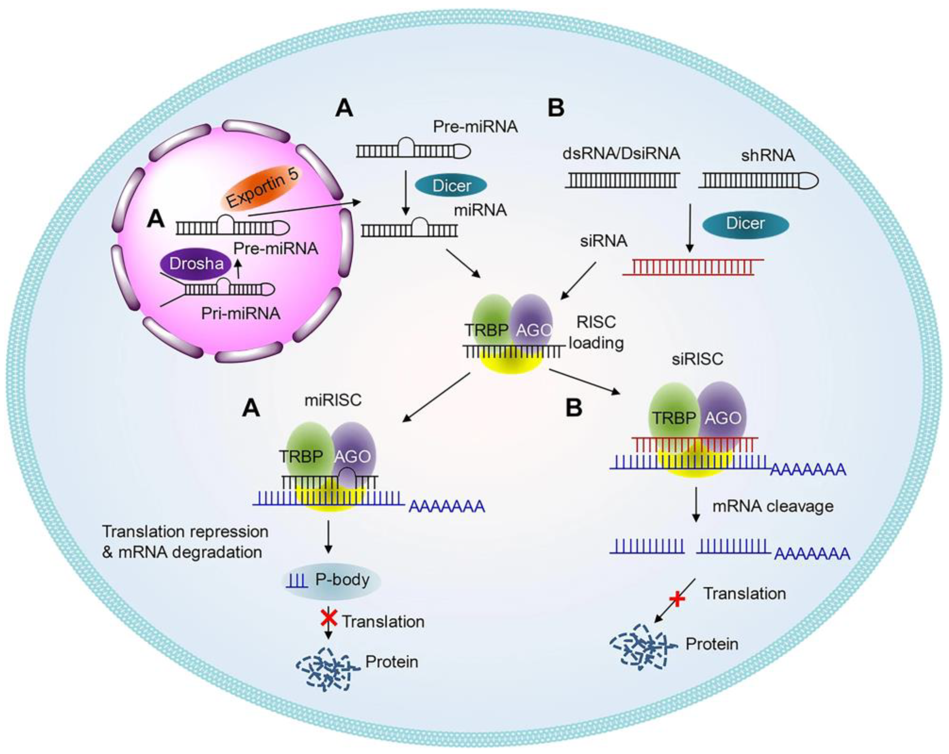
Small interfering RNA (siRNA) is currently one of the most widely studied RNAs in the field of gene therapy. RNA interference (RNAi) represents a highly specific gene-silencing mechanism whereby molecules such as siRNA and miRNA can knock down target gene expression in a sequence-specific manner [37]. This is accomplished by mediating either targeted mRNA degradation (applicable to both siRNA and miRNA) or translational repression (specific to miRNA). While a single miRNA possesses the capacity to simultaneously disrupt the expression of multiple distinct target genes, siRNA generally triggers gene silencing with superior efficiency and target specificity compared to miRNA [38]; consequently, it has been extensively employed in the targeted modulation of disease-associated genes. Mechanistically, siRNA primarily associates with the RNA-induced silencing complex (RISC) to recognize and bind complementary target mRNAs, subsequently inducing their degradation or translational inhibition to achieve the precise silencing of pathogenic genes. Within the realm of OA research, siRNA is predominantly harnessed to target genes implicated in cartilage matrix degradation and inflammatory cascades, thereby effectively retarding the progression of articular degeneration [40].
Figure 1.
The gene silencing mechanism of RNAi [37], Copyright 2025, with the permission from the authors, licensed under CC BY.
Figure 1.
The gene silencing mechanism of RNAi [37], Copyright 2025, with the permission from the authors, licensed under CC BY.
3.1.3. mRNA
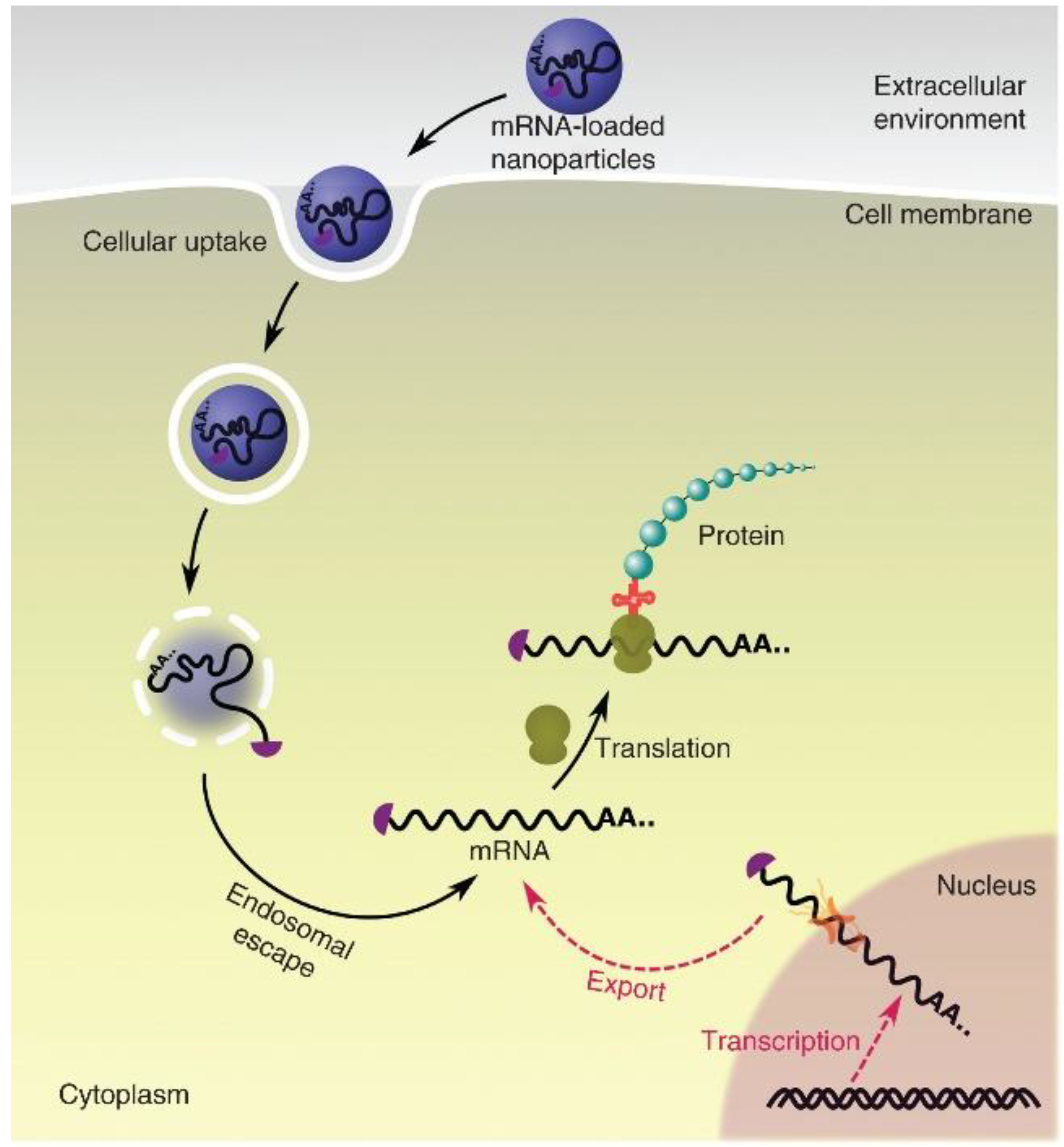
Messenger RNA (mRNA), which encodes specific proteins, can be exogenously delivered into cells to undergo translation into target proteins, thereby altering cellular phenotypes and restoring physiological functions to achieve therapeutic purposes through the supplementation or activation of endogenous repair pathways [41,42]. In recent years, mRNA therapeutics have emerged as a highly promising modality; upon successfully reaching the cytoplasm, it can be directly translated into functional proteins. In the context of OA treatment, the targeted delivery of in vitro-transcribed mRNA into articular chondrocytes robustly enhances anabolic activities and promotes cartilage regeneration [44,45]. Consequently, mRNA-based therapies have demonstrated a favorable safety profile for intra-articular applications.
Figure 2.
Schematic diagram of intracellular mRNA delivery. Endogenous mRNA is transcribed from DNA, processed, and exported to cytoplasm for translation (red pathway). Alternatively, in vitro transcribed mRNA can be introduced into cytoplasm by nanoscale platforms (black pathway). Using exogenous mRNAs, functional proteins can be produced in the cytoplasm [43], Copyright 2018, with the permission from Wiley Periodicals, Inc.
Figure 2.
Schematic diagram of intracellular mRNA delivery. Endogenous mRNA is transcribed from DNA, processed, and exported to cytoplasm for translation (red pathway). Alternatively, in vitro transcribed mRNA can be introduced into cytoplasm by nanoscale platforms (black pathway). Using exogenous mRNAs, functional proteins can be produced in the cytoplasm [43], Copyright 2018, with the permission from Wiley Periodicals, Inc.
3.1.4. lncRNA
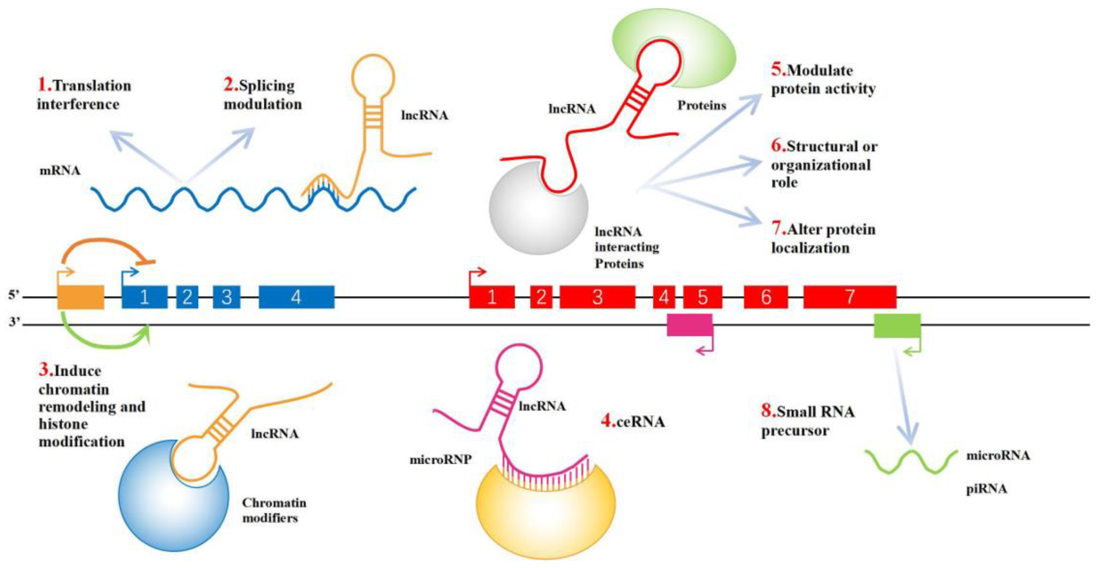
Long non-coding RNAs (lncRNAs) are RNA transcripts exceeding 200 nucleotides in length that lack distinct open reading frames and protein-coding capacity, yet they exert profound regulatory effects on gene expression[46]. Based on their relative chromosomal positions to coding genes, lncRNAs can be classified into five categories: sense, antisense, bidirectional, intronic, and intergenic. They modulate gene expression by folding into unique conformations and interacting with DNA, RNA, or proteins[47]. In the context of OA research, lncRNAs have been implicated in driving pathological progression; specifically, certain lncRNAs are upregulated in response to inflammatory stimuli, which subsequently activates the Wnt/β-catenin and NF-κB signaling pathways to upregulate the expression of matrix-degrading enzymes, such as MMP-13 and ADAMTS5, thereby precipitating cartilage matrix destruction[48]. Conversely, numerous mechanistic studies have elucidated the protective roles of other lncRNAs, which exert "anti-inflammatory and pro-repair" effects by inhibiting chondrocyte apoptosis, augmenting matrix synthesis, or suppressing pro-inflammatory signaling, highlighting their immense therapeutic potential[49]. Consequently, lncRNAs manifest a "dual role" in OA pathobiology, functioning simultaneously as pathogenic drivers and as viable therapeutic targets or reparative tools.
Figure 3.
Schematic diagram of lncRNA function. 1) LncRNA can be transcribed with the upstream promoter region of a protein-coding gene to interfere with the expression of downstream genes. 2) LncRNA can form complementary double strands with the transcript of a protein-coding gene, interfering with the splicing of messenger RNA (mRNA) and forming different forms of splicing. 3) LncRNA can mediate chromatin remodelling and histone modification, affecting the expression of downstream genes. 4) LncRNA has microRNA (miRNA) action sites, which can be competitively combined with miRNA. RNAs that act this way are known as miRNA sponges (competitive endogenous RNAs (ceRNAs)). 5) In combination with specific proteins, lncRNA transcripts can regulate the activity of corresponding proteins. 6) As a structural component, it forms a nucleic acid–protein complex with protein. 7) LncRNA can bind to a specific protein, changing its cellular location. 8) LncRNA can form the precursor molecule of small RNAs (such as miRNA, PIWI-interacting RNA (piRNA)). [47], Copyright 2021, with the permission from the authors, licensed under CC BY-NC-ND 4.0.
Figure 3.
Schematic diagram of lncRNA function. 1) LncRNA can be transcribed with the upstream promoter region of a protein-coding gene to interfere with the expression of downstream genes. 2) LncRNA can form complementary double strands with the transcript of a protein-coding gene, interfering with the splicing of messenger RNA (mRNA) and forming different forms of splicing. 3) LncRNA can mediate chromatin remodelling and histone modification, affecting the expression of downstream genes. 4) LncRNA has microRNA (miRNA) action sites, which can be competitively combined with miRNA. RNAs that act this way are known as miRNA sponges (competitive endogenous RNAs (ceRNAs)). 5) In combination with specific proteins, lncRNA transcripts can regulate the activity of corresponding proteins. 6) As a structural component, it forms a nucleic acid–protein complex with protein. 7) LncRNA can bind to a specific protein, changing its cellular location. 8) LncRNA can form the precursor molecule of small RNAs (such as miRNA, PIWI-interacting RNA (piRNA)). [47], Copyright 2021, with the permission from the authors, licensed under CC BY-NC-ND 4.0.
3.1.5. circRNA
Circular RNAs (circRNAs) represent a novel class of RNA molecules originating from exon skipping events during the alternative splicing of precursor mRNAs (pre-mRNAs)[50]. They are ubiquitously expressed across diverse organisms, ranging from prokaryotes and eukaryotes to mammals. Distinct from their linear counterparts, such as mRNAs and lncRNAs, circRNAs are characterized by a covalently closed-loop structure devoid of 5′ caps and 3′ poly-A tails[51,52]. This unique structural topology renders them highly resistant to exonuclease-mediated degradation, thereby ensuring their stable expression in vivo. In recent years, synthetic circRNAs have been extensively engineered with the aim of exploring their potential applications as a novel modality for mRNA therapeutics and vaccines[53]. Within the context of OA, circRNAs possess the capability to orchestrate chondrocyte proliferation, apoptosis, differentiation, and autophagy[54]. Furthermore, they critically modulate ECM degradation, as well as the oxidative stress and inflammatory cascades within chondrocytes. On the other hand, circRNAs regulate the broader intra-articular microenvironment—encompassing the synovium, meniscus, and subchondral bone—and hold significant promise as diagnostic biomarkers in liquid biopsies.
Figure 4.
Functions of circRNAs. (A) Acting as an miRNA sponge or ceRNA. (B) Directly targeting mRNA by partly base pairing. (C) Binding RNA binding protein (RBP) and AGO to regulate protein expression. (D) Used as the template of protein synthesis. IRES: Internal ribosome entry site. [50], Copyright 2017, with the permission from Elsevier B.V.
Figure 4.
Functions of circRNAs. (A) Acting as an miRNA sponge or ceRNA. (B) Directly targeting mRNA by partly base pairing. (C) Binding RNA binding protein (RBP) and AGO to regulate protein expression. (D) Used as the template of protein synthesis. IRES: Internal ribosome entry site. [50], Copyright 2017, with the permission from Elsevier B.V.
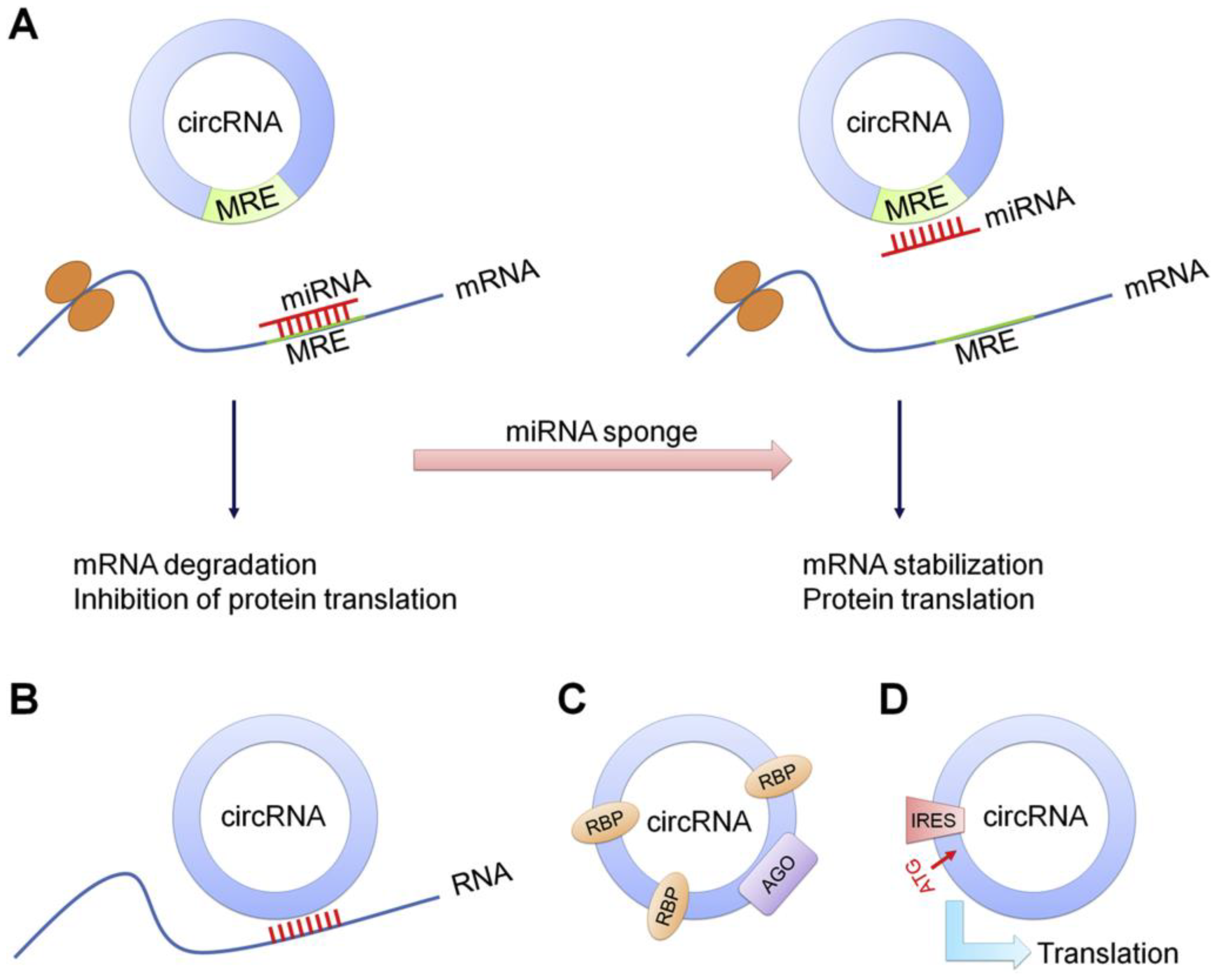
Beyond the aforementioned RNA classes, other RNA variants, such as single guide RNAs (sgRNAs), also exert specific therapeutic effects in the management of OA. The diverse spectrum of RNA molecules exhibits multifaceted and complementary regulatory potentials in OA therapeutics. By suppressing inflammatory responses, retarding matrix degradation, or activating cartilaginous repair pathways, these molecules enable the profound remodeling of the articular microenvironment at the molecular level. However, as delineated in the introduction, their clinical translation remains severely bottlenecked by poor in vivo stability, high susceptibility to nuclease degradation, and suboptimal targeting efficiency. Therefore, the development of highly efficient, safe, and spatiotemporally controllable RNA delivery systems—designed to enhance local accumulation and sustain therapeutic duration at the pathological site—has emerged as a paramount direction to propel the clinical translation of RNA-based gene therapies.
3.2. RNA-Based Therapeutic Strategies Targeting OA Pathological Processes
RNA therapeutics possess the capability to precisely modulate disease-implicated genes at both the transcriptional and post-transcriptional levels; consequently, they are universally recognized as a pivotal strategy for realizing disease-modifying OA therapies. In recent years, propelled by the continuous discovery of diverse functional RNA species, researchers have been empowered to rationally design tailored RNA interventional regimens targeting the distinct pathological processes inherent to OA.
3.2.1. RNA Therapeutics Targeting ECM Degradation
ECM degradation represents the core pathological event driving cartilage degeneration in OA; consequently, a multitude of RNA-based therapeutic strategies have primarily focused on the inhibition of matrix-degrading factors. In an in vivo OA model, Akagi et al. utilized MMP-13 siRNA to significantly downregulate MMP-13 expression levels, thereby retarding the degradation of Collagen II and overall cartilage degeneration[55]. Similarly, siRNA therapies targeting ADAMTS5 have demonstrated marked efficacy in mitigating aggrecan depletion and ameliorating cartilage tissue architecture[56]. Beyond the direct silencing of terminal catabolic enzymes, Pi et al. employed hypoxia-inducible factor-2 alpha (HIF-2α) siRNA to suppress the upstream regulators of MMP-13 and ADAMTS5, thereby exerting a broader, more comprehensive anti-catabolic effect[57]. In the realm of miRNAs, miR-140 remains one of the most extensively characterized chondroprotective miRNAs to date. It not only suppresses the expression of MMP-13 and ADAMTS5 but also actively stimulates Collagen II synthesis, thereby fulfilling a dual function of halting degradation and promoting structural repair[58,59]. Similarly, miR-17 is capable of concurrently targeting a panel of catabolic factors—including matrix metalloproteinase-3 (MMP-3), MMP-13, ADAMTS5, and nitric oxide synthase 2 (NOS2)—highlighting its multi-target regulatory superiority in preserving cartilage homeostasis[60].
Collectively, RNA therapeutic strategies targeting ECM degradation possess the capacity to modulate the cartilaginous catabolic network across multiple hierarchical levels, rendering this approach one of the most mature and well-established paradigms in contemporary OA gene therapy research.
3.2.2. RNA Therapeutics Targeting the Inflammatory Microenvironment
Chronic inflammation serves as a pivotal driver in the relentless progression of OA; consequently, the targeted blockade of inflammatory signaling cascades has emerged as a fundamental strategy in RNA therapeutics.
Ding et al. discovered that miR-93 effectively mitigates chondrocyte inflammatory responses and apoptosis by suppressing the TLR4/NF-κB signaling pathway[61]. Other chondroprotective miRNAs, such as miR-21 and miR-142-3p, have similarly been validated for their capacity to downregulate the expression of pro-inflammatory cytokines and ameliorate the articular microenvironment[62,63,64]. Within the landscape of lncRNAs, maternally expressed gene 3 (MEG3) represents one of the most extensively investigated protective lncRNAs in recent years. Mechanistic studies demonstrate that the overexpression of MEG3 restrains the secretion of inflammatory mediators and revitalizes chondrocyte viability via the miR-9-5p/Krüppel-like factor 4 (KLF4) axis[65]. Furthermore, miRNAs delivered via diverse engineered exosomes have been corroborated to attenuate inflammatory responses by modulating key signaling networks, including the NF-κB and phosphoinositide 3-kinase/protein kinase B (PI3K/AKT) pathways[66].
Consequently, harnessing RNA therapeutics to rewire inflammatory signaling networks not only profoundly alleviates localized inflammation but also indirectly abrogates the deleterious process of cartilage matrix degradation.
3.2.3. RNA Therapeutics Targeting Oxidative Stress
Oxidative stress serves as a crucial nexus connecting inflammation, cell death, and tissue degeneration. In recent years, researchers have actively begun to harness RNA to modulate ROS-related pathways in an effort to restore articular microenvironmental homeostasis.
Several protective lncRNAs have also been demonstrated to mitigate ROS accumulation and ameliorate mitochondrial function. Gu et al. discovered that the lncRNA zinc finger NFX1-type containing 1 antisense 1 (ZFAS1) promotes chondrocyte proliferation and suppresses oxidative stress by targeting the miR-1323/nuclear factor erythroid 2-related factor 2 (Nrf2) signaling axis, while concurrently activating the antioxidant Nrf2/heme oxygenase-1 (HO-1) pathway[67]. Notably, FGF18 mRNA therapy not only facilitates robust cartilage repair but may also indirectly abrogate oxidative stress-induced damage by improving cellular metabolic profiles and tissue homeostasis[34]. Furthermore, Shang et al. revealed that circHIPK3 is capable of modulating the miR-30a-3p/paraoxonase 2 (PON2) axis in chondrocytes, thereby downregulating intracellular ROS levels and inhibiting mitochondrial outer membrane permeabilization (MOMP) to successfully counteract apoptosis[68].
Consequently, RNA-based interventions specifically targeted at ROS imbalances are poised to emerge as a vital complementary direction for future OA therapeutics.
3.2.4. RNA Therapeutics Targeting Cellular Senescence
In recent years, emerging evidence has indicated that chondrocyte senescence may constitute a pivotal driving factor in the pathogenesis and progression of OA. Park et al. employed p16INK4a siRNA to successfully silence the expression of this hallmark senescence marker, thereby significantly attenuating cartilage structural damage and retarding OA progression[69]. Within the realm of lncRNA research, PTS-1 has been corroborated to modulate the miR-8085/E2F transcription factor 2 (E2F2) signaling axis[70]. This modulation upregulates critical regulators of cell cycle progression and DNA synthesis, thereby markedly alleviating IL-1β-induced cellular senescence and inflammatory damage. Recent investigations have further illuminated that circPLXNC1 functions as a potent senescence inhibitor, effectively treating OA by orchestrating the delicate balance between cellular senescence and autophagy[71]. Furthermore, the recently highlighted cGAS-STING signaling pathway is intrinsically linked to senescence-associated inflammation; consequently, siRNA-mediated interventions targeting this cascade have exhibited highly promising translational prospects.
3.2.5. RNA Therapeutics Promoting Cartilage Regeneration
Beyond the mere suppression of pathological processes, stimulating genuine cartilage regeneration remains the ultimate objective for realizing true disease-modifying OA therapies. At present, the most representative paradigm in this domain is FGF18 mRNA therapy[34]. As a paramount regulatory factor governing chondrogenesis and tissue repair, FGF18 effectively stimulates chondrocyte proliferation, significantly upregulates the expression of Collagen II and aggrecan, and actively orchestrates the regeneration of compromised cartilaginous tissue. Additionally, a diverse array of protective lncRNAs and circRNAs is intricately involved in the molecular regulation of cartilage repair. For instance, the lncRNA colorectal neoplasia differentially expressed (CRNDE) can robustly promote the chondrogenic differentiation of mesenchymal stem cells (MSCs) via the sirtuin 1 (SIRT1)/SOX9 signaling axis[72]. Moreover, the circRNA derived from the serpin family E member 2 gene (circSERPINE2) is capable of augmenting ECM synthesis while concurrently abrogating chondrocyte apoptosis by modulating the miR-1271 pathway[73]. Similarly, the circRNA originating from human phosphodiesterase 4D (circPDE4D) can function as a competitive endogenous molecular sponge for miR-103a-3p[74]. This sponging effect relieves the post-transcriptional repression of FGF18, thereby fostering cartilage matrix maintenance and repair while concomitantly suppressing the elevation of pro-inflammatory biomarkers. Consequently, in stark contrast to conventional anti-inflammatory modalities, RNA-based strategies aimed at driving intrinsic cartilage regeneration more closely align with the developmental trajectory of disease-modifying OA therapeutics, undoubtedly representing a paramount trend for future RNA translational research.
4. Delivery Vehicles Enhancing RNA Therapeutics for OA
4.1. LNPs
Over the past two decades, LNPs have continuously evolved as an efficacious, highly biocompatible, and biodegradable RNA delivery platform within the realms of nanomedicine, biotechnology, and drug delivery, establishing themselves as one of the most mature systems in contemporary RNA delivery research. By forming electrostatic complexes with RNA via cationic or ionizable lipids, LNPs leverage their superior biocompatibility and biodegradability not only to shield RNA from nucleolytic degradation but also to facilitate the cellular uptake and subsequent cytosolic delivery of RNA therapeutics into target cells[75]. The triumphant regulatory approval and commercialization of mRNA COVID-19 vaccines and Givosiran—an siRNA therapeutic targeting aminolevulinate synthase 1 (ALAS1)—have profoundly inspired numerous researchers to explore the translational applications of LNPs in OA therapy.
In the investigation of OA therapeutics, Kong et al. encapsulated FGF18 mRNA with an optimized 5' untranslated region (UTR) into LNPs, which enabled the stable expression of therapeutic doses of the FGF18 protein, efficiently delivered the mRNA into the deep zones of the cartilage, and sustained in vivo expression within the murine knee joint cavity for over six days[76]. This LNP-FGF18 mRNA formulation effectively alleviated chondrocyte degeneration and senescence induced by pro-inflammatory cytokines, while concurrently stimulating chondrocyte proliferation. Mechanistically, LNP-FGF18 mRNA also upregulated the expression of FOXO3a and robustly activated autophagy in chondrocytes. Furthermore, Sun et al. engineered a novel, intra-articularly retentive LNP system (WG-PL14)[77]. This platform effectively circumvents systemic leakage and hepatotoxicity, yielding an mRNA transduction efficiency nearly 30-fold higher than that of the commercial MC3 lipid formulation. Administered at an exceptionally low dose, it mediated the high-activity expression of endogenous recombinant human FGF18 (rhFGF18), which significantly remodeled subchondral bone homeostasis and facilitated cartilage regeneration. Simultaneously, it efficaciously ameliorated pain responses, upregulated ECM-associated genes—such as agrin (AGRN) and hyaluronan synthase 2 (HAS2)—and reestablished subchondral bone homeostasis. Zhao et al. loaded siRNA targeting fibroblast activation protein (FAP), a soluble factor secreted by the OA synovium, into LNPs[78]. By incorporating nuclear localization signal (NLS)-modified lipids (NLS-SM102) and PEG lipids conjugated with TAT peptides (a class of cell-penetrating peptides), they significantly augmented the efficiency of the LNPs in penetrating the cartilage matrix and entering chondrocytes. In vitro assays corroborated the exceptional physiological stability of these LNPs, while in vivo imaging demonstrated that a single intra-articular injection could achieve sustained drug release for up to one week. This highly efficient and low-toxicity delivery system successfully silenced FAP expression in chondrocytes, subsequently downregulating the SASP via the inhibition of the NF-κB pathway, thereby significantly retarding OA progression.
From a materials science perspective, the application of LNPs in RNA therapeutics for OA presents distinct advantages compared to delivery strategies targeting hepatic or systemic diseases. Firstly, given that OA is predominantly managed via localized intra-articular injection, LNPs are spared from systemic circulation and extensive clearance by the reticuloendothelial system. This enables the achievement of high effective local concentrations at relatively low administrable doses, thereby profoundly minimizing the risk of systemic toxicity. Secondly, the lipid composition, particle size, and surface charge of LNPs are highly tunable. By incorporating cationic or cartilage-affinity modifications, their interactions with the negatively charged cartilage matrix can be substantially enhanced, thereby improving tissue retention and cellular uptake efficiencies. Furthermore, LNPs possess well-established mechanisms for the encapsulation and endosomal escape of mRNAs and siRNAs; the material platform already has a robust foundation for clinical translation, characterized by clear pathways for process scale-up and quality control[79,80]. When synergistically combined with localized administration and injectable scaffold biomaterials, LNPs are highly amenable to constructing intra-articular RNA delivery platforms characterized by "high stability, prolonged retention, and controlled release". Consequently, they hold prominent material and translational advantages in the landscape of RNA-based OA therapeutic regimens.
4.2. Exosomes
Exosomes are nanoscale lipid membrane vesicles secreted by cells that facilitate intercellular communication by transferring active substances from parent cells to recipient cells, transporting a diverse array of bioactive molecules, including RNA, proteins, and lipids[81]. Owing to their natural origins, exosomes exhibit high biocompatibility and low immunogenicity, typically retaining intrinsic functions that reflect their cellular source[82,83]. Furthermore, in recent years, exosomes have emerged as a focal point in the therapeutic landscape of numerous diseases, similarly demonstrating immense potential in nucleic acid delivery for OA. Currently, exosome-based RNA delivery strategies are primarily categorized into two paradigms: the first leverages the endogenous therapeutic RNA carried by naturally derived exosomes, such as those from stem cells; the second involves engineering modifications to actively load exogenously designed therapeutic RNAs into exosomes for targeted delivery.
Figure 5.
Natural exosomes and engineered exosomes loaded with endogenous or exogenous cargos for therapeutic purposes[84], Copyright 2020, with the permission from the authors, licensed under CC BY.
Figure 5.
Natural exosomes and engineered exosomes loaded with endogenous or exogenous cargos for therapeutic purposes[84], Copyright 2020, with the permission from the authors, licensed under CC BY.
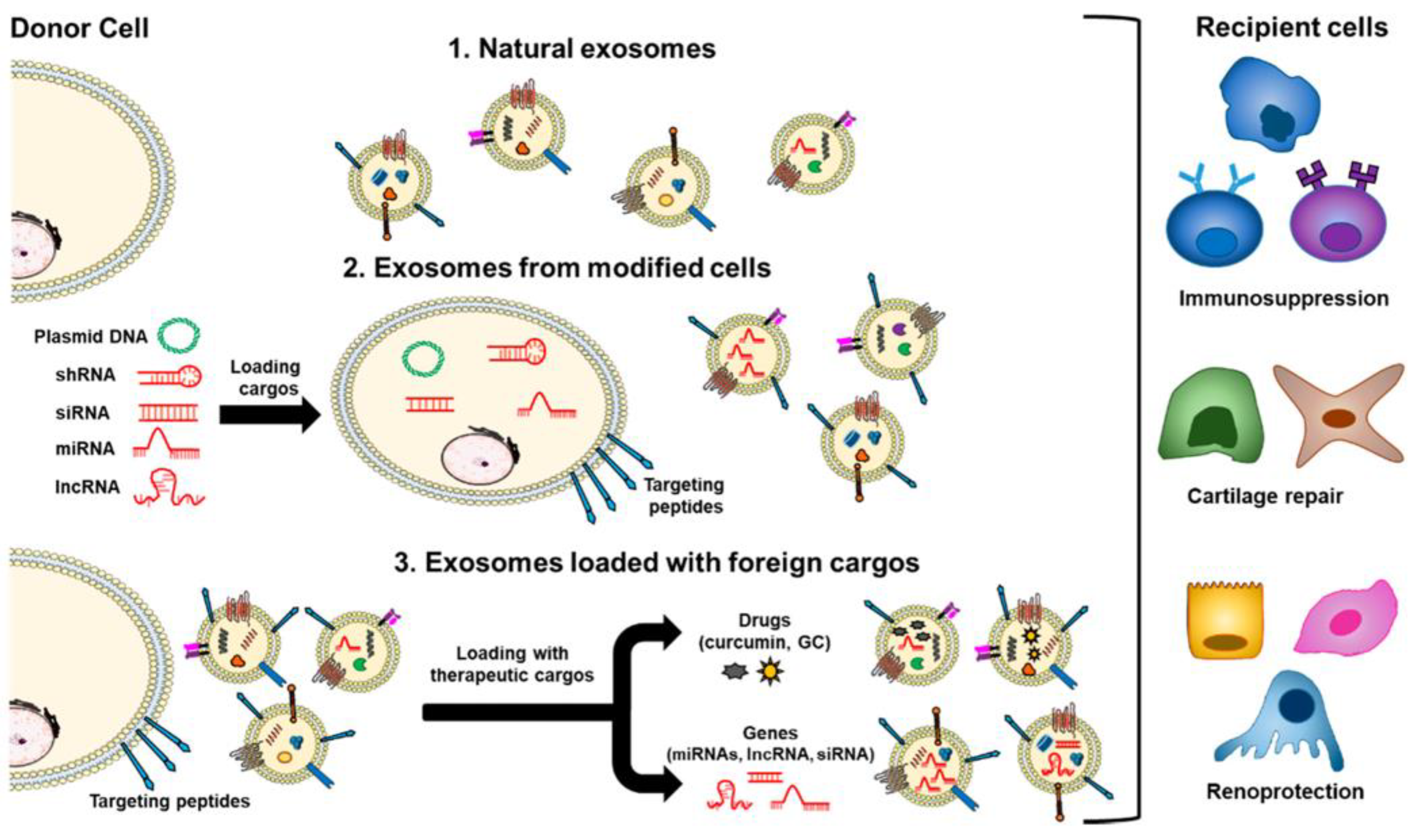
Regarding the utilization of endogenous RNA, exosomes derived from MSCs have garnered considerable attention due to their enrichment with chondro-regenerative miRNAs. Studies indicate that specific preconditioning of MSCs can significantly optimize the RNA payload within their secreted exosomes. For instance, in MSC exosomes preconditioned with fucoidan, the expression of the therapeutic miR-146b-5p is substantially enriched[85]. Acting as natural vehicles for cell-free therapy, these miRNAs exert chondroprotective effects by targeting TNF receptor-associated factor 6 (TRAF6) and suppressing the PI3K/AKT/ mTOR signaling pathway, thereby effectively mitigating inflammatory responses and ECM degradation while simultaneously promoting chondrocyte autophagy. Additionally, exosomes derived from umbilical cord mesenchymal stem cells (UCMSC-EXOs) are similarly enriched with a variety of therapeutic miRNAs[86]. These endogenous miRNAs can reverse the senescent state of OA chondrocytes by inhibiting the p53 signaling pathway and downregulating the expression of SASP factors. Moreover, intercellular RNA communication under pathological conditions offers reciprocal insights for therapeutic interventions. For example, osteoclast-derived exosomes can transfer their abundant endogenous miRNAs (such as miR-214-3p) to chondrocytes, accelerating cartilage degeneration via the suppression of TIMP-2/3 expression[87]. Therefore, harnessing protective exosomal RNAs or intercepting the transmission of such pathogenic exosomal RNAs both represent efficacious strategies for intervening in intercellular communication and delaying OA progression.
In the realm of engineering the active loading of exogenous RNA, exosomes have proven to be ideal vectors for the delivery of gene-based therapeutics. Addressing the avascular and dense architecture of cartilage tissue, endowing exosomes with targeting capabilities via surface display technology is crucial for achieving the deep penetration of nucleic acid drugs. To overcome the rapid clearance of exosomes within the articular cavity, numerous researchers have modified their surfaces with cartilage affinity peptides (CAPs), thereby constructing exosome delivery systems with potent cartilage-targeting capacity. Liang et al. generated specific CAP-Exos by fusing the CAP sequence with the exosomal membrane protein Lamp2b, successfully encapsulating the chondroprotective miR-140 within them[88]. This delivery system is capable of penetrating the dense matrix to access deep-zone chondrocytes, significantly downregulating the expression of matrix-degrading enzymes such as MMP-13 and ADAMTS5, thereby achieving targeted, cell-free OA therapy. Beyond delivering miRNAs, exosomes also demonstrate exceptional performance in mediating the targeted gene silencing of siRNAs. Utilizing lipid insertion technology, researchers anchored a CAP-containing conjugate (DSPE-PEG-MAL-CAP) onto the exosomal surface and highly efficiently loaded MMP-13-targeted siRNA (siMMP13) via electroporation[89]. This engineered exosome (CAP-Exo/siMMP13) achieved highly efficient and specific knockdown of MMP-13 in chondrocytes, which not only promoted Collagen II synthesis and cartilage regeneration but also fundamentally circumvented the off-target toxicities potentially induced by traditional small-molecule MMP inhibitors.
As natural nanovesicles, exosome-mediated RNA delivery systems ingeniously synergize the favorable pharmacokinetic properties of endogenous vesicles with the targeted regulatory prowess of nucleic acid therapeutics. Whether by directly uncovering and potentiating the activity of their endogenous therapeutic RNAs or by employing them as engineered vectors for the delivery of customized RNA therapeutics, this approach holds the promise to transcend the limitations of conventional OA treatments. Consequently, it provides a highly translatable novel pathway for realizing cell-free, molecularly targeted therapies directed against the pathological mechanisms of cartilage degeneration.
4.3. Lipo-Exosome Hybrids
As delineated above, while LNPs and natural exosomes serve as the two predominant RNA delivery vectors and both exhibit unique potential in OA therapeutics, they inherently manifest unavoidable limitations hindering further clinical translation. Traditional LNPs possess remarkable nucleic acid loading capacity and highly customizable surface properties; however, their cartilage-targeting efficacy within the joint cavity is often suboptimal, and their potential immunogenicity restricts long-term intra-articular applications. Conversely, natural exosomes, leveraging their excellent biocompatibility and inherent advantage of penetrating the dense cartilage matrix, are regarded as ideal endogenous vectors; nevertheless, their limited vesicular volume and membrane charge characteristics result in exceedingly low encapsulation efficiencies for large-molecule nucleic acids, such as long mRNAs or CRISPR/Cas9 plasmids. To overcome the technological bottlenecks of singular vectors, Lipo-Exosome Hybrids, constructed by fusing liposomes with exosomes via membrane fusion technologies, have emerged as a highly innovative solution[90]. For instance, by fusing vesicles overexpressing growth factors (e.g., TGF-β1) with anti-inflammatory liposomes to prepare hybrid nanoparticles, the simultaneous immunomodulation and tissue reconstruction of the cartilage microenvironment have been achieved[91]. This biomimetic hybrid system realizes a synergistic effect that perfectly complements the strengths of both modalities; it not only inherits the low immunogenicity of exosomes but also significantly enhances the loading efficacy for large-molecule mRNAs and sgRNAs through the incorporation of lipid components. Studies indicate that while traditional pure liposomes struggle to effectively transfect MSCs, hybrid nanoparticles can highly efficiently encapsulate the bulky CRISPR/Cas9 and sgRNA, successfully executing cytosolic translation and genomic editing within MSCs[92]. This delivery strategy circumvents the risk of random genomic integration potentially associated with traditional plasmid DNA delivery and obviates the need to cross the nuclear envelope barrier, thereby significantly elevating editing efficiency. Similarly, this hybrid architecture has been validated to efficiently load and protect the therapeutically functional N6-methyladenosine (m6A) demethylase alkB homolog 5 (ALKBH5) mRNA, successfully maintaining RNA stability and translational activity within complex microenvironments[93]. Martijn further empirically demonstrated that, compared to pure liposomes, the encapsulation of siRNA into hybrids profoundly altered their functional behaviors regarding cellular uptake, toxicity, and gene-silencing efficacy, which varied depending on the recipient cell type[94]. Collectively, these findings underscore the broad-spectrum applicability of hybrids in the co-delivery of multicomponent RNAs.
Addressing cartilage degeneration within the pathological environment of OA, hybrid systems endow RNA delivery platforms with exceptional capabilities for deep tissue penetration and in situ activation. Researchers have successfully constructed hybrid vectors with chondrocyte-specific targeting capacity by fusing CAP-modified liposomes with natural exosomes[95]. This platform successfully enhances plasmid stability and loading capacity while mitigating off-target organ diffusion, extending cartilage localization time, and deepening matrix penetration. Consequently, it enables the precise delivery of the CRISPR-Cas9/sgRNA system targeting degeneration-associated genes directly into the deep-zone cartilage tissue, achieving the specific remodeling of the genome in degenerated chondrocytes. Building upon this, Chen et al. further encapsulated the targeted hybrids loaded with this system into a methacrylated hyaluronic acid (HAMA) microgel[96]. The constructed composite system not only in situ activates FGF18 expression at the molecular level to promote cartilage regeneration, but also utilizes its microgel scaffold to provide a self-renewing hydration layer. This affords durable lubrication during tribological wear, thereby realizing a synergistic orchestration of biological gene regulation and physical biomechanical lubrication.
Figure 6.
Schematic illustration of chondrocyte-specific genome editing by hybrid exosomes [95], Copyright 2022, with the permission from the authors, licensed under CC BY.
Figure 6.
Schematic illustration of chondrocyte-specific genome editing by hybrid exosomes [95], Copyright 2022, with the permission from the authors, licensed under CC BY.
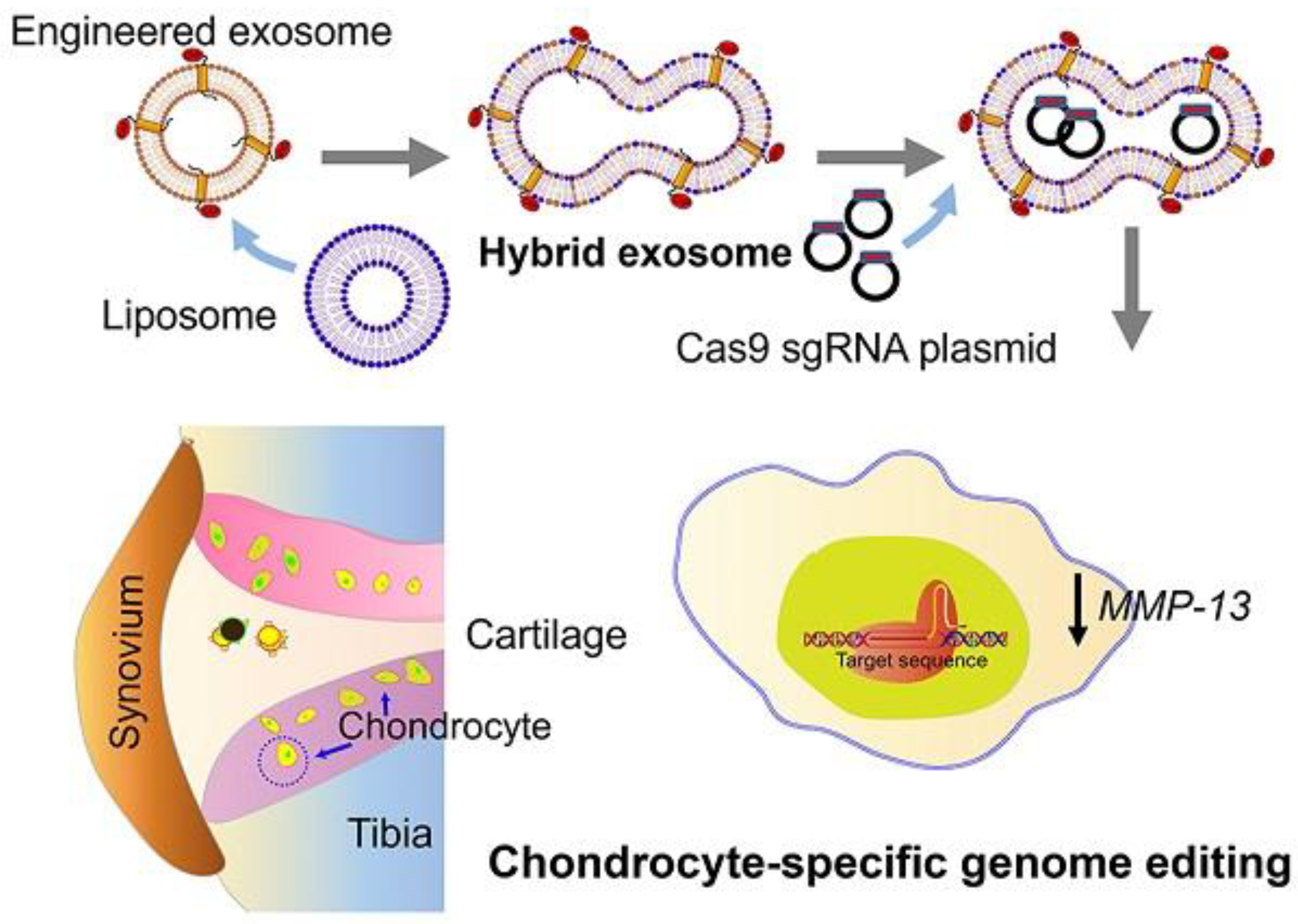
In summary, lipo-exosome hybrids ingeniously bridge the chasm between natural vesicles and artificial vectors, furnishing a versatile platform with profound clinical translational prospects for the targeted delivery and multimodal combinatorial therapy of complex RNA systems in OA management.
4.4. Metal Nanoparticles
Metal nanoparticles (MNPs) and metal-organic frameworks (MOFs), characterized by their highly uniform architectures, exceptionally large specific surface areas, and readily functionalizable surface properties, have occupied a prominent position in the field of targeted drug delivery[97,98]. In therapeutic applications for OA, metal-based carriers exhibit unique microenvironmental adaptability and multifaceted synergistic efficacy. On one hand, they can precisely respond to the low pH within the OA articular cavity or exogenous physical stimuli (such as light irradiation), effectively resolving the clinical bottlenecks of "burst release" and "suboptimal controlled release" inherent to conventional intra-articular vectors[99,100]. On the other hand, certain metallic carriers (such as Mg- or Zn-coordinated MOFs, alongside noble metal nanoparticles harboring intrinsic anti-inflammatory properties) release metal ions upon in vivo degradation or utilize their inherent structural motifs to exert endogenous biological activities[101]. These attributes enable them to directly orchestrate chondrocyte anabolism and synovial microenvironmental homeostasis.
Capitalizing on these superior physicochemical and biological properties, deploying metal nanomaterials as RNA delivery vectors is rapidly emerging as a vanguard strategy to breach the prevailing bottlenecks in OA gene therapy. Metallic vectors not only effectively shield fragile RNA molecules from nuclease-mediated degradation via physical coordination or mesoporous adsorption, but they also highly efficiently mediate endosomal escape, thereby markedly elevating the intracellular bioavailability of RNA. For instance, to specifically address the acidic microenvironment characterized by the intricate interplay of hypoxia and inflammation in OA cartilage, investigators have engineered aminated MIL-101 nano-frameworks for the targeted co-delivery of HIF-2α siRNA and the anti-inflammatory agent curcumin[102]. Upon exposure to the acidic inflammatory milieu, this pH-responsive MOF undergoes targeted structural dissociation. This mechanism not only facilitates the endosomal escape of the siHIF-2α—thereby realizing highly efficient intracellular release and precise gene silencing—but also engenders a dual synergistic chondroprotective effect in tandem with curcumin. In the domain of noble metal nanoparticles, Ag and Au nanocarriers have further expanded the frontiers of multimodal RNA delivery, leveraging their unique physicochemical properties. Studies demonstrate that the Lipo-AgPEI system—synthesized via the complexation of silver ions with polyethylenimine (PEI)—not only achieves the highly efficient encapsulation of miR-200c-3p employing microfluidic technologies but also harnesses the intrinsic antioxidant, anti-inflammatory, and anti-apoptotic attributes of silver ions[103]. This rational design empowers the miRNA to more efficiently target pivotal pathogenic factors, such as zinc finger E-box-binding homeobox 2 (ZEB2), thereby ameliorating structural cartilage damage across multiple dimensions. Furthermore, capitalizing on the distinct optical properties of gold nanomaterials allows for the construction of interventional systems endowed with superior spatiotemporal responsiveness. For example, Qiao et al. leveraged the exceptional near-infrared photothermal conversion efficacy of gold nanocages (AuNCs), integrating them with phase-change materials to engineer a photo-induced hybrid nanoplatform[104]. Upon external light irradiation, the localized and controlled hyperthermia not only directly palliates OA-associated joint pain via thermotherapy but also precisely triggers the on-demand release of the encapsulated siRNA targeting nerve growth factor (siNGF). This elegantly achieves a profound integration of physical physiotherapy and molecular gene silencing.
In summary, RNA-encapsulating metallic nanovectors not only successfully breach the permeation barriers imposed by the dense cartilage matrix against macromolecular nucleic acids but also integrate external-field responsiveness, environmental sensitivity, and the intrinsic biological effects of the carriers. This integration facilitates the construction of a "physical-chemical-genetic" unified synergistic therapeutic system, thereby furnishing an advanced platform for the realization of spatiotemporally controllable and precisely targeted OA therapies.
4.5. Hydrogels and Hydrogel Microspheres
Although nanocarriers such as LNPs and exosomes demonstrate exceptional performance in RNA delivery, the high-frequency mechanical friction and rapid synovial clearance mechanisms within the articular cavity often result in extremely short retention times for free nanocarriers, making it challenging to maintain effective therapeutic concentrations. To transcend these spatiotemporal limitations, injectable hydrogels have emerged as ideal long-acting intra-articular depot vectors, owing to their outstanding in situ gelation properties and structural support that mimics the natural cartilaginous ECM[105,106]. Consequently, encapsulating RNA-loaded nanocarriers within the three-dimensional porous networks of hydrogels or hydrogel microspheres not only prevents the rapid efflux of the primary vectors but also functions as a physical lubricant and biomechanical cushion within the joint cavity. At the microscopic level, the continuously and controllably released nanocarriers can deeply penetrate the dense cartilage tissue to execute precise molecular remodeling.
Figure 7.
Feasibility diagram of injectable hydrogel in OA treatment[107], Copyright 2025, with the permission from the authors, licensed under CC BY.
Figure 7.
Feasibility diagram of injectable hydrogel in OA treatment[107], Copyright 2025, with the permission from the authors, licensed under CC BY.
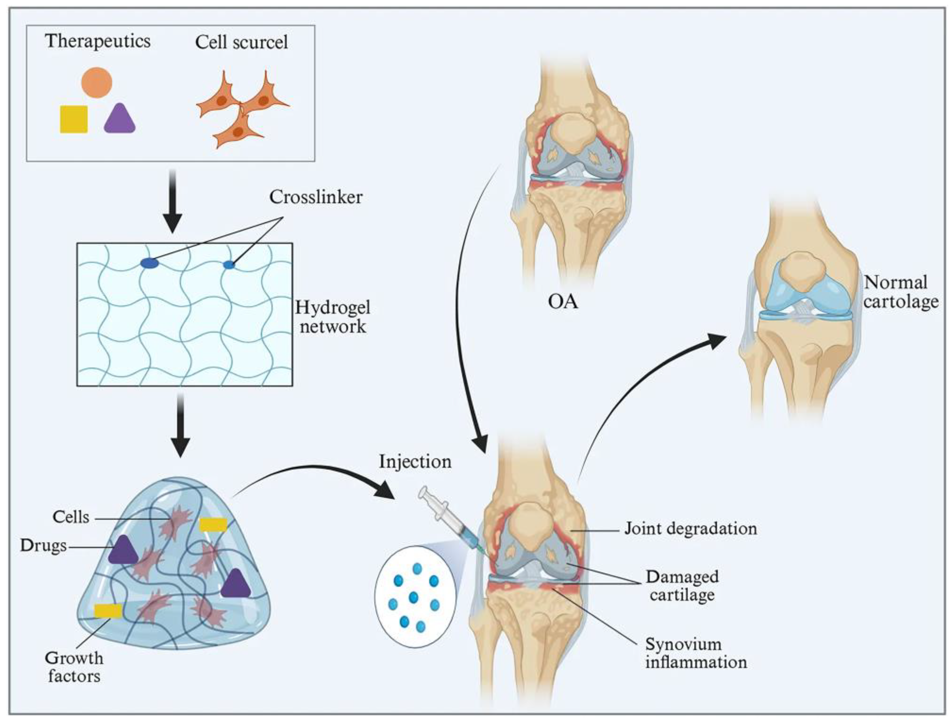
More crucially, through the intricate design of dynamic chemical bonds, this hydrogel network can achieve intelligent responsiveness to the pathological OA microenvironment and facilitate on-demand drug release. For instance, specifically targeting the overloaded ROS within the OA microenvironment, researchers developed a ROS-responsive hydrogel encapsulating siMMP-13-loaded iron nanoparticles (si-Fe NPs)[108]. In this system, the hydrogel backbone actively consumes and scavenges pathogenic ROS (exerting a self-therapeutic effect); concomitantly, the moderate dissociation of the backbone precisely triggers and sustains the prolonged controlled release of the internal siRNA nanoparticles, achieving a profound synergy between macroscopic local antioxidation and microscopic deep-tissue gene silencing. Furthermore, addressing the aberrant enzymatic microenvironment at OA cartilage defect sites, Gao et al. encapsulated miR-17 within LNPs and utilized an aggrecanase-responsive hydrogel[109]. While providing physical filling and structural support to the cartilage defect, this hydrogel undergoes pathological enzymatic degradation to enable the on-demand release of miR-17, effectively reprogramming the hostile localized inflammatory niche and significantly promoting endogenous cartilage regeneration following microfracture surgery. Feng et al. constructed a biomimetic hydration layer using zwitterionic hyperbranched polymers, which simultaneously reduces articular friction and mitigates mechanical wear while cultivating a more favorable localized microenvironment for RNA therapeutics[110]. Facilitated by the synergistic action of hyaluronic acid (HA) and a Collagen II-targeting dual-headed peptide, the hydrogel can stably adhere to the compromised cartilage surface, achieving long-term local drug accumulation. Moreover, CAP-modified exosomes have been shown to significantly enhance the delivery efficiency and cellular uptake of si-STING into chondrocytes, persistently suppressing the cGAS-STING inflammatory senescence pathway, thereby attenuating inflammatory responses and delaying cartilage degeneration. In another study, LNPs encapsulating a circRNA that encodes the p65 super-repressor IκBα (srIκBα) were surface-modified with Collagen II-targeting peptides and antibodies against fibroblast activation protein alpha (FAPα), enabling the simultaneous targeting of chondrocytes and synovial fibroblasts[111]. By further utilizing a silk fibroin/HA/chondroitin sulfate (CS) composite hydrogel, long-term intra-articular retention was achieved. Coupled with an MMP-13-sensitive peptide, this system enables inflammatory microenvironment-responsive drug release, effectively blocking the NF-κB pathway and thereby enhancing the local therapeutic efficacy and durability in OA management.
Therefore, the hierarchical combinatorial system of "hydrogel + RNA nanocarriers" ingeniously circumvents the dual hurdles of extreme susceptibility to degradation inherent to naked RNA and the rapid efflux characteristic of solitary nanocarriers. Beyond conventional hydrogels, researchers are increasingly focusing on stimuli-responsive hydrogels tailored to the OA microenvironment, enabling intelligent responsiveness and on-demand drug release precisely at the lesional sites. This strategy achieves the spatiotemporal coupling of "macroscopic physical lubrication, intelligent microenvironmental responsiveness, and microscopic long-acting gene regulation," unequivocally representing the vanguard paradigm for precision molecularly targeted interventions in OA.
5. Artificial Intelligence-Driven RNA Delivery Systems for OA Therapy
Artificial intelligence (AI) represents a prominent branch of computer science dedicated to executing complex tasks that traditionally necessitate "human intelligence" via computers or computer-controlled machineries[112]. Within this domain, drug delivery constitutes one of the foremost applications of AI-driven targeted nanoparticles. By forecasting nanocarrier behaviors within intricate biological environments and simulating the elaborate interactions between nanocarriers and biological systems, AI optimizes their physicochemical properties, predicts therapeutic efficacies, identifies precise targets, designs payloads, and ultimately realizes end-to-end digitalization[113].
With the deepening of scientific investigations, the contributions of AI models (e.g., ChatGPT, Gemini, and DeepSeek) to the medical field have progressively matured[114]. At present, the majority of research leveraging AI for targeted nanoparticles is concentrated within the field of oncology. For instance, utilizing data from nano-tumor databases, Lin et al. integrated AI with physiologically based pharmacokinetic (PBPK) models to substantiate the feasibility of expediting cancer nanomedicine research and development[115]. Furthermore, through AI-empowered analysis, Nuhn elucidated two critical penetration mechanisms: predominantly relying on passive extravasation in highly permeable tumors, whereas facilitating active transendothelial transport via pH-responsive peptide modifications in low-permeability tumors[116]. This AI-driven predictive model guided the rational design of a bifunctional nanocarrier endowed with both pH-responsiveness and an albumin-binding domain, which consequently amplified the delivery efficiency of doxorubicin in low-permeability tumors by threefold. Such research establishes an intelligent screening platform for the personalized design of nanomedicines, successfully circumventing the inherent limitations of the conventional enhanced permeability and retention (EPR) effect.
Undoubtedly, AI has also progressively garnered widespread attention in the realm of RNA delivery. Currently, AI is actively reshaping the research and development paradigm of RNA delivery systems through cutting-edge algorithms, realizing a quantum leap from vector engineering optimization to the precise design of nucleic acid sequences[117]. In terms of RNA sequence engineering, AI plays a quintessential role in optimizing RNA secondary structures and translational activities, as well as reinforcing the stability of nucleic acid molecules; through multimodal synergistic optimization, it significantly prolongs the in vivo half-life of therapeutic RNAs[118]. At the vector level, machine learning (ML) and deep learning (DL) models have been extensively deployed to predict the physicochemical properties, encapsulation efficiencies, and in vivo stabilities of carriers such as LNPs[119]. By means of high-throughput, data-driven simulated screening, these models effectively assist researchers in surmounting biological barriers to accomplish tissue-targeted delivery. Furthermore, Su et al. comprehensively reviewed the integration of AI into mRNA-LNP frameworks[120]. The AI-driven optimization of mRNA delivery can augment the precise localization of therapeutic cargos to specific cells or tissues, thereby potentiating therapeutic functionalities while minimizing off-target effects. Concurrently, predictive models empower researchers to more accurately forecast the in vivo pharmacokinetics and biodistribution profiles of nanoparticles. Additionally, AI has exhibited exquisite precision in predicting the activity of sgRNAs and assessing the risks of off-target effects for gene-editing tools (e.g., CRISPR/Cas9)[121].
Figure 8.
AI-based methods for RNA structure and function prediction.
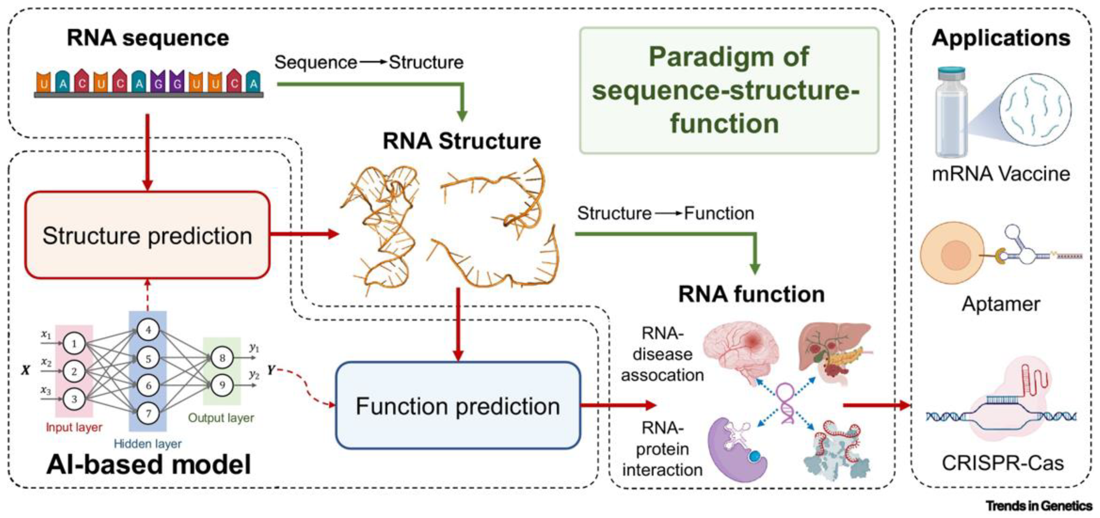
Artificial neural networks (ANNs) serve as the foundation of AI, and are designed to process enormous quantities of data for complicated analysis. ANNs comprise several layers of nodes, containing an input layer, one or more hidden layers, and an output layer. RNA sequences can be utilized as the input of the AI model for RNA structure prediction, with the output being anticipated tertiary structures. AI models may also be used to predict RNA functions, such as RNA–disease associations or RNA–protein interactions, using sequence and structural data as input. The reliability of AI-based RNA structure and function prediction may also be increased by integrating sequence, structural, and functional data. Following the sequence–structure–function paradigm, AI will provide unprecedented opportunities for the design of functional RNA molecules with potential biomedical applications, such as mRNA vaccines, aptamers, and CRISPR/Cas systems, among others.[118], Copyright 2023, with the permission from Elsevier B.V.
Although specific research on AI-driven RNA delivery tailored for OA remains in its nascent stages, its resounding success in the fields of tumor immunology and vaccinology provides profound insights for the precision intervention of OA. Confronting the dense, avascular physical barrier of OA cartilage tissue and the pathological microenvironment highly susceptible to rapid synovial clearance, AI can seamlessly assist in the rational design of customized nanocarriers endowed with high cartilage affinity and intelligent stimulus-responsive release profiles. Zhou et al. developed a system termed multiobjective LNP engineering with artificial intelligence (MOLEA)[122]. The K9 LNP, identified through this AI-guided screening, achieved a transfection efficiency exceeding 90% in murine articular chondrocytes. Furthermore, it demonstrated a 13.5-fold increase in knee-to-liver tissue selectivity relative to the clinical benchmark, SM-102. This exceptional selectivity empowers K9 to safely and efficaciously mediate in vivo CRISPR genome editing (specifically targeting the Mmp13 gene), thereby realizing sustained cartilage protection and abrogating joint degeneration. Simultaneously, leveraging AI-optimized, long-acting expressional RNA sequences holds the promise of achieving a sustained supply of therapeutic proteins (such as growth factors or anti-inflammatory cytokines) within the chronically inflamed microenvironment. This strategy intrinsically overcomes the critical limitation of transient therapeutic efficacy associated with conventional delivery systems during the OA disease course, thereby providing a more highly translatable, individualized, and intelligent paradigm for the profound reconstitution of intra-articular homeostasis.
6. Conclusion and Perspectives
The profoundly complex pathological microenvironment of OA imposes highly stringent spatiotemporal demands on RNA delivery vectors. In recent years, the design of delivery materials has undergone a paradigm shift, evolving from foundational lipid-based carriers (LNPs) and biomimetic natural vesicles (exosomes) toward advanced intelligent systems endowed with multidimensional environmental responsiveness, such as hybrid nanovesicles, MOFs, and hierarchical hydrogels. These advanced materials not only surmount the inherent deficiencies of naked RNA—including its fleeting half-life and poor cellular uptake—but also achieve the spatiotemporal synergy of biomechanical/tribological remodeling and localized gene silencing through their hierarchical architectures. This comprehensive interventional strategy, deeply rooted in materials engineering, is progressively propelling OA therapeutics from passive symptomatic palliation toward the proactive reconstitution of the microenvironmental niche.
Although novel nucleic acid delivery materials have demonstrated exceptional chondroprotective efficacy in preclinical models, their leap toward clinical translation remains obstructed by formidable materials science and biological barriers. Firstly, confronting the dense and avascular physiological barrier of the cartilage matrix, the deep-tissue penetration efficiency of existing vectors urgently requires enhancement; thus, designing surface and interfacial modification materials with superior matrix affinity and penetrability constitutes a pivotal future direction. Secondly, the long-term intra-articular pharmacokinetics, the toxicity of degradation byproducts, and the potential immunogenicity of multicomponent delivery systems (e.g., metal ion-incorporated MOFs or peptide-functionalized hybrid vectors) necessitate more rigorous and systemic toxicological evaluations in large animal models, such as equines or ovines. Furthermore, ensuring batch-to-batch consistency and establishing robust Chemistry, Manufacturing, and Controls (CMC) processes for the large-scale production of highly functionalized vectors—particularly engineered exosomes and complex hydrogel systems—remain the critical engineering bottlenecks hindering their clinical deployment.
Confronted with the aforementioned high-dimensional material design space and formidable optimization challenges, the integration of AI and high-throughput computational platforms undeniably represents the inevitable trajectory for the research and development of next-generation delivery systems. By leveraging ML to decipher the complex mapping relationships among the physicochemical properties of nanomaterials, the pathological microenvironment, and in vivo delivery efficiencies, researchers can realize a paradigm shift from traditional "trial-and-error" methodologies to "data-driven rational design". Concurrently, the application of AI in computational structural biology will tremendously accelerate the fundamental optimization of RNA sequences. Looking forward, deeply converging AI-assisted, highly active RNA molecules with multimodal, adaptive-responsive materials to construct a closed-loop interventional system—one that seamlessly integrates biomechanical support, microenvironmental homeostasis regulation, and targeted gene repair—holds immense promise for accelerating the realization of disease-modifying OA therapies, thereby providing a transformative platform technology for the clinical cure of degenerative joint diseases.
Author Contributions
Conceptualization, J.D.; investigation, L.Y., Z.S., M.L.; resources, Y.Z., Z.S.; visualization, J.H.; project administration, J.D.; writing—original draft preparation, Z.L.; writing—review and editing, Z.L., J.H.; funding acquisition, B.X., J.D. All authors have read and agreed to the published version of the manuscript.
Funding
This work was supported by the grants from the Hebei Provincial Natural Science Foundation (H2022206404); National Natural Science Foundation of China (81973251); Hebei Province Key Science and Technology Support Program (25242701D); Shijiazhuang Basic Research Project (241791397A).
Conflicts of Interest
The authors declare no conflicts of interest.
References
- GBD 2021 Osteoarthritis Collaborators. Global, regional, and national burden of osteoarthritis, 1990–2020 and projections to 2050: a systematic analysis for the Global Burden of Disease Study 2021. Lancet Rheumatol. 2023, 5, e508–e522. [Google Scholar] [CrossRef] [PubMed]
- da Costa, B.R.; Pereira, T.V.; Saadat, P.; Rudnicki, M.; Iskander, S. M.; Bodmer, N.S.; Bobos, P.; Gao, L.; Kiyomoto, H.D.; Montezuma, T.; Almeida, M.O.; Cheng, P.S.; Hincapié, C.A.; Hari, R.; Sutton, A.J.; Tugwell, P.; Hawker, G.A.; Jüni, P. Effectiveness and safety of non-steroidal anti-inflammatory drugs and opioid treatment for knee and hip osteoarthritis: network meta-analysis. BMJ. 2021, 375, n2321. [Google Scholar] [CrossRef] [PubMed]
- Mohammadinejad, R.; Ashrafizadeh, M.; Pardakhty, A.; Uzieliene, I.; Denkovskij, J.; Bernotiene, E.; Janssen, L.; Lorite, G. S.; Saarakkala, S.; Mobasheri, A. Nanotechnological strategies for osteoarthritis diagnosis, monitoring, clinical management, and regenerative medicine: Recent advances and future opportunities. Curr. Rheumatol. Rep. 2020, 22, 12. [Google Scholar] [CrossRef] [PubMed]
- Yang, W.; Sun, C.; He, S.Q.; Chen, J.Y.; Wang, Y.; Zhuo, Q. The efficacy and safety of disease-modifying osteoarthritis drugs for knee and hip osteoarthritis-a systematic review and network meta-analysis. J. Gen. Intern. Med. 2021, 36, 2085–2093. [Google Scholar] [CrossRef] [PubMed]
- Rane, M.A.; Foster, J.G.; Wood, S.K.; Hebert, P.R.; Hennekens, C.H. Benefits and risks of nonsteroidal anti-inflammatory drugs: Methodologic limitations lead to clinical uncertainties. Ther. Innov. Regul. Sci. 2019, 53, 502–505. [Google Scholar] [CrossRef] [PubMed]
- Veronese, N.; Cooper, C.; Bruyère, O.; Al-Daghri, N.M.; Branco, J.; Cavalier, E.; Cheleschi, S.; da Silva Rosa, M.S.; Conaghan, P.G.; Dennison, E.M.; de Wit, M.; Fioravanti, A.; Fuggle, N.R.; Haugen, I.K.; Herrero-Beaumont, G.; Honvo, G.; Laslop, A.; Matijevic, R.; Migliore, A.; Mobasheri, A.; Pelletier, J.P.; Prieto Yerro, M.C.; Radermecker, R.P.; Rannou, F.; Rizzoli, R.; Reginster, J.Y. Multimodal multidisciplinary management of patients with moderate to severe pain in knee osteoarthritis: A need to meet patient expectations. Drugs 2020, 82, 1347–1355. [Google Scholar]
- Yip, K.; Oettinger, J. Why are we still using opioids for osteoarthritis? Int. J. Clin. Pract. 2020, 74, e13416. [Google Scholar] [PubMed]
- Block, J.A. Are intraarticular glucocorticoids safe in osteoarthritis? Arthritis Rheumatol. 2022, 74, 181–183. [Google Scholar] [CrossRef] [PubMed]
- Bowman, S.; Awad, M.E.; Hamrick, M.W.; Hunter, M.; Fulzele, S. Recent advances in hyaluronic acid based therapy for osteoarthritis. Clin. Transl. Med. 2018, 7, 6. [Google Scholar] [CrossRef] [PubMed]
- Madry, H. Surgical therapy in osteoarthritis. Osteoarthr. Cartil. 2022, 30, 1019–1034. [Google Scholar] [CrossRef] [PubMed]
- DeJulius, C.R.; Walton, B.L.; Colazo, J.M.; d'Arcy, R.; Francini, N.; Brunger, J.M.; Duvall, C.L. Engineering approaches for RNA-based and cell-based osteoarthritis therapies. Nat. Rev. Rheumatol. 2024, 20, 81–100. [Google Scholar] [CrossRef] [PubMed]
- Liu, Y.; Wang, C.; Fu, X.; Ren, M. The progress and evolving trends in nucleic-acid-based therapies. Biomolecules 2025, 15, 376. [Google Scholar] [CrossRef] [PubMed]
- Radbakhsh, S.; Najar, M.; Merimi, M.; Benderdour, M.; Fernandes, J.C.; Martel-Pelletier, J.; Pelletier, J.P.; Fahmi, H. RNA modifications in osteoarthritis: Epitranscriptomic insights into pathogenesis and therapeutic targets. Int. J. Mol. Sci. 2025, 26, 4955. [Google Scholar] [CrossRef] [PubMed]
- Hao, J.; Li, Y.; Huang, L.; Qi, N.; Sun, Y.; He, C.; Yang, S.; Niu, Z.; Qi, X.; Xiang, B. Smart nanoarchitectures for precision RNA delivery: Harnessing endogenous and exogenous stimuli in cancer treatment. Theranostics 2025, 15, 7747–7778. [Google Scholar] [CrossRef] [PubMed]
- Su, L.J.; Ji, Z.H.; Xu, M.X.; Zhu, J.Q.; Chen, Y.H.; Qiao, J.F.; Wang, Y.; Lin, Y.X. RNA-based nanomedicines and their clinical applications. Nano Res. 2023, 16, 13182–13204. [Google Scholar] [CrossRef]
- Lim, S.A.; Cox, A.; Tung, M.; Chung, E.J. Clinical progress of nanomedicine-based RNA therapies. Bioact. Mater. 2022, 12, 203–213. [Google Scholar] [CrossRef] [PubMed]
- Zhou, D.; Zhou, F.; Sheng, S.; Wei, Y.; Chen, X.; Su, J. Intra-articular nanodrug delivery strategies for treating osteoarthritis. Drug Discov. Today. 2023, 28, 103482. [Google Scholar] [CrossRef] [PubMed]
- Tang, S.; Zhang, C.; Oo, W.M.; Fu, K.; Risberg, M.A.; Bierma-Zeinstra, S.M.; Neogi, T.; Atukorala, I.; Malfait, A.M.; Ding, C.; Hunter, D.J. Osteoarthritis. Nat. Rev. Dis. Prim. 2025, 11, 10. [Google Scholar] [CrossRef] [PubMed]
- Kloppenburg, M.; Namane, M.; Cicuttini, F. Osteoarthritis. Lancet 2025, 405, 71–85. [Google Scholar] [PubMed]
- Fujii, Y.; Liu, L.; Yagasaki, L.; Inotsume, M.; Chiba, T.; Asahara, H. Cartilage homeostasis and osteoarthritis. Int. J. Mol. Sci. 2022, 23, 6316. [Google Scholar] [CrossRef] [PubMed]
- Yi, X.; Leng, P.; Wang, S.; Liu, L.; Xie, B. Functional nanomaterials for the treatment of osteoarthritis. Int. J. Nanomedicine. 2024, 19, 6731–6756. [Google Scholar] [CrossRef] [PubMed]
- De Roover, A.; Escribano-Núñez, A.; Monteagudo, S.; Lories, R. Fundamentals of osteoarthritis: Inflammatory mediators in osteoarthritis. Osteoarthr. Cartil. 2023, 31, 1303–1311. [Google Scholar] [CrossRef] [PubMed]
- Mehana, E.E.; Khafaga, A.F.; El-Blehi, S.S. The role of matrix metalloproteinases in osteoarthritis pathogenesis: An updated review. Life Sci. 2019, 234, 116786. [Google Scholar] [CrossRef] [PubMed]
- Yang, X.; Zhao, L.; Pang, Y. cGAS-STING pathway in pathogenesis and treatment of osteoarthritis and rheumatoid arthritis. Front. Immunol. 2024, 15, 1384372. [Google Scholar] [CrossRef] [PubMed]
- Coryell, P.R.; Diekman, B.O.; Loeser, R.F. Mechanisms and therapeutic implications of cellular senescence in osteoarthritis. Nat. Rev. Rheumatol. 2021, 17, 47–57. [Google Scholar] [PubMed]
- Lepetsos, P.; Papavassiliou, K.A.; Papavassiliou, A.G. Redox and NF-κB signaling in osteoarthritis. Free Radic. Biol. Med. 2019, 132, 90–100. [Google Scholar] [CrossRef] [PubMed]
- Liu, Y.; Duan, J.; Dang, Y.; Hao, R.; Wang, H.; Tan, E.; Wang, R.; Li, Y.; Zhang, S.; Wang, Y.; Lv, J.; Qi, Y.; Zhang, X.; Cheng, Y. Remodeling of senescent macrophages in synovium alleviates trauma- and aging-induced osteoarthritis. Bioact. Mater. 2026, 55, 42–56. [Google Scholar] [CrossRef] [PubMed]
- Astrike-Davis, E.M.; Coryell, P.; Loeser, R.F. Targeting cellular senescence as a novel treatment for osteoarthritis. Curr. Opin. Pharmacol. 2022, 64, 102213. [Google Scholar] [CrossRef] [PubMed]
- Loeser, R.F.; Collins, J.A.; Diekman, B.O. Ageing and the pathogenesis of osteoarthritis. Nat. Rev. Rheumatol. 2016, 12, 412–420. [Google Scholar] [CrossRef] [PubMed]
- Sacitharan, P.K.; Vincent, T.L. Cellular ageing mechanisms in osteoarthritis. Mamm. Genome. 2016, 27, 421–429. [Google Scholar] [CrossRef] [PubMed]
- Zelinka, A.; Roelofs, A.J.; Kandel, R.A.; De Bari, C. Cellular therapy and tissue engineering for cartilage repair. Osteoarthr. Cartil. 2022, 30, 1547–1560. [Google Scholar] [CrossRef] [PubMed]
- Blaney Davidson, E.N.; van der Kraan, P.M.; van den Berg, W.B. TGF-beta and osteoarthritis. Osteoarthr. Cartil. 2007, 15, 597–604. [Google Scholar] [CrossRef] [PubMed]
- Govindaraj, K.; Kannan, S.; de Almeida, R.C.; Klomp, L.J.; Karperien, M.; Meulenbelt, I.; Post, J.N. Dissecting SOX9 dynamics reveals its differential regulation in osteoarthritis. J. Cell. Physiol. 2024, 239, e31443. [Google Scholar] [CrossRef] [PubMed]
- Yao, X.; Zhang, J.; Jing, X.; Ye, Y.; Guo, J.; Sun, K.; Guo, F. Fibroblast growth factor 18 exerts anti-osteoarthritic effects through PI3K-AKT signaling and mitochondrial fusion and fission. Pharmacol. Res. 2019, 139, 314–324. [Google Scholar] [CrossRef] [PubMed]
- Kim, H.; Lee, Y.Y.; Kim, V.N. The biogenesis and regulation of animal microRNAs. Nat. Rev. Mol. Cell Biol. 2025, 26, 276–296. [Google Scholar] [CrossRef] [PubMed]
- Fu, L.W.; Wu, J.; Shi, S.R.; Zhang, Z.C.; Zheng, Y.Z.; Li, P.X.; Yuan, X.; Ding, Z.G.; Ning, C.; Sui, X.; Liu, S.Y.; Guo, Q.Y.; Lin, Y.F. A tetrahedral framework nucleic acids-based gene therapeutic nanococktail alleviates cartilage damage and protects against osteoarthritis progression. Chem. Eng. J. 2024, 498, 155189. [Google Scholar] [CrossRef]
- Hu, B.; Zhong, L.; Weng, Y.; Peng, L.; Huang, Y.; Zhao, Y.; Liang, X.J. Therapeutic siRNA: State of the art. Signal Transduct. Target. Ther. 2020, 5, 101. [Google Scholar] [CrossRef] [PubMed]
- Jadhav, V.; Vaishnaw, A.; Fitzgerald, K.; Maier, M.A. RNA interference in the era of nucleic acid therapeutics. Nat. Biotechnol. 2024, 42, 394–405. [Google Scholar] [CrossRef] [PubMed]
- Song, S.; Liu, W.; Wang, S.; Guo, N.; Li, M.; Xie, J.; Luo, X.; Yan, X.; Xu, B.; Xu, Y. Progress in siRNA therapy and delivery platforms for osteoarthritis. iScience 2026, 29, 115163. [Google Scholar] [CrossRef] [PubMed]
- Li, Y.; Zhao, J.; Guo, S.; He, D. siRNA therapy in osteoarthritis: Targeting cellular pathways for advanced treatment approaches. Front. Immunol. 2024, 15, 1382689. [Google Scholar] [CrossRef] [PubMed]
- Faraway, R.; Zenklusen, D.; Plaschka, C. Mechanisms of messenger RNA packaging and export. Annu. Rev. Cell Dev. Biol. 2025, 41, 479–504. [Google Scholar] [CrossRef] [PubMed]
- He, Y.; Johnston, A.P.R.; Pouton, C.W. Therapeutic applications of cell engineering using mRNA technology. Trends Biotechnol. 2025, 43, 83–97. [Google Scholar] [CrossRef] [PubMed]
- Li, B.; Zhang, X.; Dong, Y. Nanoscale platforms for messenger RNA delivery. Wiley Interdiscip. Rev. Nanomed. Nanobiotechnol. 2019, 11, e1530. [Google Scholar] [PubMed]
- Terai, Y.; Yada, E.; Nakanishi, H.; Itaka, K. mRNA-based combination therapy for inflammation-driven osteoarthritis induced by monosodium iodoacetate. Pharmaceutics 2025, 17, 1254. [Google Scholar] [CrossRef] [PubMed]
- Pezzotti, G.; Zhu, W.; Terai, Y.; Marin, E.; Boschetto, F.; Kawamoto, K.; Itaka, K. Raman spectroscopic insight into osteoarthritic cartilage regeneration by mRNA therapeutics encoding cartilage-anabolic transcription factor Runx1. Mater. Today Bio. 2022, 13, 100210. [Google Scholar] [CrossRef] [PubMed]
- Mercer, T.R.; Dinger, M.E.; Mattick, J.S. Long non-coding RNAs: Insights into functions. Nat. Rev. Genet. 2009, 10, 155–159. [Google Scholar] [CrossRef] [PubMed]
- He, C.P.; Jiang, X.C.; Chen, C.; Zhang, H.B.; Cao, W.D.; Wu, Q.; Ma, C. The function of lncRNAs in the pathogenesis of osteoarthritis. Bone Jt. Res. 2021, 10, 122–133. [Google Scholar] [CrossRef] [PubMed]
- Hu, K.; Wen, H.; Song, T.; Che, Z.; Song, Y.; Song, M. Deciphering the role of LncRNAs in osteoarthritis: Inflammatory pathways unveiled. J. Inflamm. Res. 2024, 17, 6563–6581. [Google Scholar] [CrossRef] [PubMed]
- Shi, D.; Mei, Y.; Hao, W.; Li, J.; Liu, S.; Lin, X. Biological functions and applications of LncRNAs in the regulation of the extracellular matrix in osteoarthritis. Front. Cell Dev. Biol. 2024, 11, 1330624. [Google Scholar] [CrossRef] [PubMed]
- He, J.; Xie, Q.; Xu, H.; Li, J.; Li, Y. Circular RNAs and cancer. Cancer Lett. 2017, 396, 138–144. [Google Scholar] [CrossRef] [PubMed]
- Margvelani, G.; Maquera, K.A.A.; Welden, J.R.; Rodgers, D.W.; Stamm, S. Translation of circular RNAs. Nucleic Acids Res. 2025, 53, gkae1167. [Google Scholar] [PubMed]
- Hwang, H.J.; Kim, Y.K. Molecular mechanisms of circular RNA translation. Exp. Mol. Med. 2024, 56, 1272–1280. [Google Scholar] [CrossRef] [PubMed]
- Liu, X.; Zhang, Y.; Zhou, S.; Dain, L.; Mei, L.; Zhu, G. Circular RNA: An emerging frontier in RNA therapeutic targets, RNA therapeutics, and mRNA vaccines. J. Control. Release. 2022, 348, 84–94. [Google Scholar] [CrossRef] [PubMed]
- Mao, X.; Cao, Y.; Guo, Z.; Wang, L.; Xiang, C. Biological roles and therapeutic potential of circular RNAs in osteoarthritis. Mol. Ther. Nucleic Acids. 2021, 24, 856–867. [Google Scholar] [CrossRef] [PubMed]
- Akagi, R.; Sasho, T.; Saito, M.; Endo, J.; Yamaguchi, S.; Muramatsu, Y.; Mukoyama, S.; Akatsu, Y.; Katsuragi, J.; Fukawa, T.; Takahashi, K. Effective knock down of matrix metalloproteinase-13 by an intra-articular injection of small interfering RNA (siRNA) in a murine surgically-induced osteoarthritis model. J. Orthop. Res. 2014, 32, 1175–1180. [Google Scholar] [CrossRef] [PubMed]
- Zhao, S.S.; Karhunen, V.; Morris, A.P.; Gill, D. ADAMTS5 as a therapeutic target for osteoarthritis: Mendelian randomisation study. Ann. Rheum. Dis. 2022, 81, 903–904. [Google Scholar] [CrossRef] [PubMed]
- Pi, Y.; Zhang, X.; Shao, Z.; Zhao, F.; Hu, X.; Ao, Y. Intra-articular delivery of anti-Hif-2α siRNA by chondrocyte-homing nanoparticles to prevent cartilage degeneration in arthritic mice. Gene Ther. 2015, 22, 439–448. [Google Scholar] [CrossRef] [PubMed]
- Yin, C.M.; Suen, W.C.; Lin, S.; Wu, X.M.; Li, G.; Pan, X.M. Dysregulation of both miR-140-3p and miR-140-5p in synovial fluid correlate with osteoarthritis severity. Bone Jt. Res. 2017, 6, 612–618. [Google Scholar] [CrossRef] [PubMed]
- Duan, L.; Liang, Y.; Xu, X.; Xiao, Y.; Wang, D. Recent progress on the role of miR-140 in cartilage matrix remodelling and its implications for osteoarthritis treatment. Arthritis Res. Ther. 2020, 22, 194. [Google Scholar] [CrossRef] [PubMed]
- Zhang, Y.; Li, S.; Jin, P.; Shang, T.; Sun, R.; Lu, L.; Guo, K.; Liu, J.; Tong, Y.; Wang, J.; Liu, S.; Wang, C.; Kang, Y.; Zhu, W.; Wang, Q.; Zhang, X.; Yin, F.; Sun, Y. E.; Cui, L. Dual functions of microRNA-17 in maintaining cartilage homeostasis and protection against osteoarthritis. Nat. Commun. 2022, 13, 2447. [Google Scholar] [CrossRef] [PubMed]
- Ding, Y.; Wang, L.; Zhao, Q.; Wu, Z.; Kong, L. MicroRNA 93 inhibits chondrocyte apoptosis and inflammation in osteoarthritis by targeting the TLR4/NF κB signaling pathway. Int. J. Mol. Med. 2019, 43, 779–790. [Google Scholar] [PubMed]
- Zhang, Y.; Jia, J.; Yang, S.; Liu, X.; Ye, S.; Tian, H. MicroRNA-21 controls the development of osteoarthritis by targeting GDF-5 in chondrocytes. Exp. Mol. Med. 2014, 46, e79. [Google Scholar] [CrossRef] [PubMed]
- Zhang, L.; Peng, H.; Zhang, W.; Li, Y.; Liu, L.; Leng, T. Yeast cell wall particle mediated nanotube-RNA delivery system loaded with miR365 antagomir for post-traumatic osteoarthritis therapy via oral route. Theranostics 2020, 10, 8479–8493. [Google Scholar] [CrossRef] [PubMed]
- Wang, X.; Guo, Y.; Wang, C.; Yu, H.; Yu, X.; Yu, H. MicroRNA-142-3p inhibits chondrocyte apoptosis and inflammation in osteoarthritis by targeting HMGB1. Inflammation 2016, 39, 1718–1728. [Google Scholar] [CrossRef] [PubMed]
- Huang, Y.; Chen, D.; Yan, Z.; Zhan, J.; Xue, X.; Pan, X.; Yu, H. LncRNA MEG3 protects chondrocytes from IL-1β-induced inflammation via regulating miR-9-5p/KLF4 axis. Front. Physiol. 2021, 12, 617654. [Google Scholar] [CrossRef] [PubMed]
- Lu, J.; Wen, J.; Ji, J. Roles of exosomal non-coding RNAs in osteoarthritis. Front. Immunol. 2026, 17, 1829319. [Google Scholar] [CrossRef] [PubMed]
- Gu, Y.; Wang, G.; Xu, H. Long non-coding RNA ZNFX1 antisense 1 (ZFAS1) suppresses anti-oxidative stress in chondrocytes during osteoarthritis by sponging microRNA-1323. Bioengineered 2022, 13, 13188–13200. [Google Scholar] [CrossRef] [PubMed]
- Shang, J.; Li, H.; Wu, B.; Jiang, N.; Wang, B.; Wang, D.; Zhong, J.; Chen, Y.; Xu, X.; Lu, H. CircHIPK3 prevents chondrocyte apoptosis and cartilage degradation by sponging miR-30a-3p and promoting PON2. Cell Prolif. 2022, 55, e13285. [Google Scholar] [CrossRef] [PubMed]
- Park, H.; Lee, H.R.; Shin, H.J.; Park, J.A.; Joo, Y.; Kim, S.M.; Beom, J.; Kang, S.W.; Kim, D.W.; Kim, J. p16INK4a-siRNA nanoparticles attenuate cartilage degeneration in osteoarthritis by inhibiting inflammation in fibroblast-like synoviocytes. Biomater. Sci. 2022, 10, 3223–3235. [Google Scholar] [CrossRef] [PubMed]
- Ma, C.; Chen, Q.; Wei, Y.F.; Chen, S.W.; Liu, H.; Xin, F.; Ren, Y.X. LncRNA PTS-1 protects against osteoarthritis through the miR-8085/E2F2 axis. J. Inflamm. Res. 2025, 18, 347–366. [Google Scholar] [CrossRef] [PubMed]
- Zhou, H.; Ji, Z.; Sun, H.; Zhang, W.; Huang, Y.; Jin, G.; Cai, H.; Huang, X.; Dai, J.; Zhao, J.; Chen, H.; Fan, S.; Wu, Y.; Shen, J. Circular RNA circPLXNC1 functions as an inhibitor of senescence by regulating autophagy in osteoarthritis. Int. J. Biol. Sci. 2026, 22, 346–364. [Google Scholar] [CrossRef] [PubMed]
- Shi, C.; Zheng, W.; Wang, J. lncRNA-CRNDE regulates BMSC chondrogenic differentiation and promotes cartilage repair in osteoarthritis through SIRT1/SOX9. Mol. Cell Biochem. 2021, 476, 1881–1890. [Google Scholar] [CrossRef] [PubMed]
- Shen, S.; Wu, Y.; Chen, J.; Xie, Z.; Huang, K.; Wang, G.; Yang, Y.; Ni, W.; Chen, Z.; Shi, P.; Ma, Y.; Fan, S. CircSERPINE2 protects against osteoarthritis by targeting miR-1271 and ETS-related gene. Ann. Rheum. Dis. 2019, 78, 826–836. [Google Scholar] [CrossRef] [PubMed]
- Wu, Y.; Hong, Z.; Xu, W.; Chen, J.; Wang, Q.; Chen, J.; Ni, W.; Mei, Z.; Xie, Z.; Ma, Y.; Wang, J.; Lu, J.; Chen, C.; Fan, S.; Shen, S. Circular RNA circPDE4D protects against osteoarthritis by binding to miR-103a-3p and regulating FGF18. Mol. Ther. 2021, 29, 308–323. [Google Scholar] [CrossRef] [PubMed]
- Ma, Y.; Li, S.; Lin, X.; Chen, Y. A perspective of lipid nanoparticles for RNA delivery. Exploration 2024, 4, 20230147. [Google Scholar] [CrossRef] [PubMed]
- Kong, K.; Li, B.; Chang, Y.; Zhao, C.; Qiao, H.; Jin, M.; Wu, X.; Fan, W.; Wang, L.; Qi, Y.; Xu, Y.; Zhai, Z.; Ma, P.; Li, H. Delivery of FGF18 using mRNA-LNP protects the cartilage against degeneration via alleviating chondrocyte senescence. J. Nanobiotechnology. 2025, 23, 34. [Google Scholar] [CrossRef] [PubMed]
- Sun, M.; Ma, B.; Pan, Z.; Zhao, Y.; Tian, L.; Fan, Y.; Kong, W.; Wang, J.; Xu, B.; Ao, Y.; Guo, Q.; Wang, X.; Peng, X.; Li, X.; Cheng, J.; Miao, L.; Wang, K.; Hu, X. Targeted therapy of osteoarthritis via intra-articular delivery of lipid-nanoparticle-encapsulated recombinant human FGF18 mRNA. Adv. Healthc. Mater. 2024, 13, e2400804. [Google Scholar] [CrossRef] [PubMed]
- Zhao, X.; Lin, J.; Liu, M.; Jiang, D.; Zhang, Y.; Li, X.; Shi, B.; Jiang, J.; Ma, C.; Shao, H.; Xu, Q.; Ping, H.; Li, J.; Gao, Y. Targeting FAP-positive chondrocytes in osteoarthritis: A novel lipid nanoparticle siRNA approach to mitigate cartilage degeneration. J. Nanobiotechnology. 2024, 22, 659. [Google Scholar] [CrossRef] [PubMed]
- Pei, D. Endosomal escape of lipid nanoparticles: A perspective on the literature data. ACS Nano 2025, 19, 40293–40303. [Google Scholar] [CrossRef] [PubMed]
- Kustrzyńska, K.; Soińska, G.; Skupin-Mrugalska, P. Diverse structures and shapes of lipid-based nanoparticles: Toward clinical translation. Biomed. Pharmacother. 2026, 199, 119398. [Google Scholar] [CrossRef] [PubMed]
- Kalluri, R.; LeBleu, V.S. The biology, function, and biomedical applications of exosomes. Science 2020, 367, eaau6977. [Google Scholar] [CrossRef] [PubMed]
- Wen, S.; Huang, X.; Ma, J.; Zhao, G.; Ma, T.; Chen, K.; Huang, G.; Chen, J.; Shi, J.; Wang, S. Exosomes derived from MSC as drug system in osteoarthritis therapy. Front. Bioeng. Biotechnol. 2024, 12, 1331218. [Google Scholar] [CrossRef] [PubMed]
- Ni, Z.; Zhou, S.; Li, S.; Kuang, L.; Chen, H.; Luo, X.; Ouyang, J.; He, M.; Du, X.; Chen, L. Exosomes: Roles and therapeutic potential in osteoarthritis. Bone Res. 2020, 8, 25. [Google Scholar] [CrossRef] [PubMed]
- Ortega, A.; Martinez-Arroyo, O.; Forner, M.J.; Cortes, R. Exosomes as drug delivery systems: Endogenous nanovehicles for treatment of systemic lupus erythematosus. Pharmaceutics 2020, 13, 3. [Google Scholar] [CrossRef] [PubMed]
- Lou, C.; Jiang, H.; Lin, Z.; Xia, T.; Wang, W.; Lin, C.; Zhang, Z.; Fu, H.; Iqbal, S.; Liu, H.; Lin, J.; Wang, J.; Pan, X.; Xue, X. MiR-146b-5p enriched bioinspired exosomes derived from fucoidan-directed induction mesenchymal stem cells protect chondrocytes in osteoarthritis by targeting TRAF6. J. Nanobiotechnology. 2023, 21, 486. [Google Scholar] [CrossRef] [PubMed]
- Cao, H.; Chen, M.; Cui, X.; Liu, Y.; Liu, Y.; Deng, S.; Yuan, T.; Fan, Y.; Wang, Q.; Zhang, X. Cell-free osteoarthritis treatment with sustained-release of chondrocyte-targeting exosomes from umbilical cord-derived mesenchymal stem cells to rejuvenate aging chondrocytes. ACS Nano 2023, 17, 13358–13376. [Google Scholar] [CrossRef] [PubMed]
- Liu, J.; Wu, X.; Lu, J.; Huang, G.; Dang, L.; Zhang, H.; Zhong, C.; Zhang, Z.; Li, D.; Li, F.; Liang, C.; Yu, Y.; Zhang, B. T.; Chen, L.; Lu, A.; Zhang, G. Exosomal transfer of osteoclast-derived miRNAs to chondrocytes contributes to osteoarthritis progression. Nat. Aging. 2021, 1, 368–384. [Google Scholar] [CrossRef] [PubMed]
- Liang, Y.; Xu, X.; Li, X.; Xiong, J.; Li, B.; Duan, L.; Wang, D.; Xia, J. Chondrocyte-targeted microRNA delivery by engineered exosomes toward a cell-free osteoarthritis therapy. ACS Appl. Mater. Interfaces. 2020, 12, 36938–36947. [Google Scholar] [CrossRef] [PubMed]
- Zhang, H.; Yan, W.; Wang, J.; Xie, S.; Tao, W. A.; Lee, C. W.; Zhang, X.; Zhang, G.; Liu, Y.; Wei, D.; Hu, J.; Liu, H.; Liu, F.; Nie, Y.; Chen, X.; Xu, H.; Xia, J.; Wang, S. Surface functionalization of exosomes for chondrocyte-targeted siRNA delivery and cartilage regeneration. J. Control. Release. 2024, 369, 493–505. [Google Scholar] [CrossRef] [PubMed]
- Mondal, J.; Pillarisetti, S.; Junnuthula, V.; Saha, M.; Hwang, S.R.; Park, I.K.; Lee, Y.K. Hybrid exosomes, exosome-like nanovesicles and engineered exosomes for therapeutic applications. J. Control. Release. 2023, 353, 1127–1149. [Google Scholar] [CrossRef] [PubMed]
- Kim, J.Y.; Rhim, W.K.; Lee, S.Y.; Park, J.M.; Song, D.H.; Cha, S.G.; Lee, S.H.; Hwang, D.Y.; Kim, B.J.; Rho, S.; Ahn, T.K.; Park, C.G.; Han, D.K. Hybrid nanoparticle engineered with transforming growth factor-β1-overexpressed extracellular vesicle and cartilage-targeted anti-inflammatory liposome for osteoarthritis. ACS Nano 2024, 18, 33937–33952. [Google Scholar] [CrossRef] [PubMed]
- Lin, Y.; Wu, J.; Gu, W.; Huang, Y.; Tong, Z.; Huang, L.; Tan, J. Exosome-liposome hybrid nanoparticles deliver CRISPR/Cas9 system in MSCs. Adv. Sci. (Weinh) 2018, 5, 1700611. [Google Scholar] [CrossRef] [PubMed]
- Wu, S.; Yun, J.; Tang, W.; Familiari, G.; Relucenti, M.; Wu, J.; Li, X.; Chen, H.; Chen, R. Therapeutic m6A eraser ALKBH5 mRNA-loaded exosome-liposome hybrid nanoparticles inhibit progression of colorectal cancer in preclinical tumor models. ACS Nano 2023, 17, 11838–11854. [Google Scholar] [CrossRef] [PubMed]
- Evers, M.J. W.; van de Wakker, S.I.; de Groot, E.M.; de Jong, O.G.; Gitz-François, J.J.J.; Seinen, C.S.; Sluijter, J.P.G.; Schiffelers, R.M.; Vader, P. Functional siRNA delivery by extracellular vesicle-liposome hybrid nanoparticles. Adv. Healthc. Mater. 2022, 11, e2101202. [Google Scholar] [CrossRef] [PubMed]
- Liang, Y.; Xu, X.; Xu, L.; Iqbal, Z.; Ouyang, K.; Zhang, H.; Wen, C.; Duan, L.; Xia, J. Chondrocyte-specific genomic editing enabled by hybrid exosomes for osteoarthritis treatment. Theranostics 2022, 12, 4866–4878. [Google Scholar] [CrossRef] [PubMed]
- Chen, M.; Lu, Y.; Liu, Y.; Liu, Q.; Deng, S.; Liu, Y.; Cui, X.; Liang, J.; Zhang, X.; Fan, Y.; Wang, Q. Injectable microgels with hybrid exosomes of chondrocyte-targeted FGF18 gene-editing and self-renewable lubrication for osteoarthritis therapy. Adv. Mater. 2024, 36, e2312559. [Google Scholar] [CrossRef] [PubMed]
- Ullah, M.; Shah, M.I.; Hasan, M.W.; Jamshed, M.; Mustafa, U.; Inam, M. Engineered metal nanoparticles for precision drug delivery: Pioneering the future of medicine: Mini review. J. Chin. Chem. Soc. 2024, 71, 1358–1367. [Google Scholar] [CrossRef]
- Mehmood, S.; Iraqui, S.; Ojha, R.K.; Sharma, N.; Marlinda, A.R. Therapeutic potential and toxicological challenges of metal nanoparticles in drug delivery: A comprehensive review. Nanomedicine 2025, 70, 102862. [Google Scholar] [CrossRef] [PubMed]
- Chen, H.; Qin, Z.; Zhao, J.; He, Y.; Ren, E.; Zhu, Y.; Liu, G.; Mao, C.; Zheng, L. Cartilage-targeting and dual MMP-13/pH responsive theranostic nanoprobes for osteoarthritis imaging and precision therapy. Biomaterials 2019, 225, 119520. [Google Scholar] [CrossRef] [PubMed]
- Shi, G.; Jiang, H.; Yang, F.; Lin, Z.; Li, M.; Guo, J.; Liao, X.; Lin, Y.; Cai, X.; Li, D. NIR-responsive molybdenum (Mo)-based nanoclusters enhance ROS scavenging for osteoarthritis therapy. Pharmacol. Res. 2023, 192, 106768. [Google Scholar] [CrossRef] [PubMed]
- Shao, H.; Xu, S.; Zeng, F.; Jin, Z.; Ji, X.; Shan, Z.; Kang, J.; Tian, Q.; Zhang, S.; Mao, Z.; Zhang, Y.; Xu, J.; Tong, W.; Qin, L. Metal-organic framework-integrated nanoplatform orchestrates osteochondral-synovial homeostasis for osteoarthritis therapy. ACS Nano 2025, 19, 32744–32766. [Google Scholar] [CrossRef] [PubMed]
- Zhang, Z.J.; Hou, Y.K.; Chen, M.W.; Yu, X.Z.; Chen, S.Y.; Yue, Y.R.; Guo, X.T.; Chen, J.X.; Zhou, Q. A pH-responsive metal-organic framework for the co-delivery of HIF-2α siRNA and curcumin for enhanced therapy of osteoarthritis. J. Nanobiotechnology. 2023, 21, 18. [Google Scholar] [CrossRef] [PubMed]
- Zheng, D.; Chen, T.; Yang, K.; Yin, G.; Chen, Y.; Gui, J.; Xu, C.; Lv, S. Microfluidic synthesis of miR-200c-3p lipid nanoparticles: Targeting ZEB2 to alleviate chondrocyte damage in osteoarthritis. Int. J. Nanomedicine. 2025, 20, 505–521. [Google Scholar] [CrossRef] [PubMed]
- Qiao, L.; Li, Z.; Li, B.; Zhang, F.; Yao, Z.; Wu, C.; Tang, H.; Pan, Q.; Shi, P.; Ping, Y. Combination of anti-inflammatory therapy and RNA interference by light-inducible hybrid nanomedicine for osteoarthritis treatment. Acta Pharm. Sin. B. 2024, 14, 5008–5025. [Google Scholar] [CrossRef] [PubMed]
- Yang, Y.; Zhang, H. Intra-articular injection of nanomaterials for the treatment of osteoarthritis: From lubrication function restoration to cell and gene therapy. Adv. Funct. Mater. 2024, 34, 2401547. [Google Scholar] [CrossRef]
- Zhou, R.; Guo, J.; Jin, Z. Advancing osteoarthritis therapy with GMOCS hydrogel-loaded BMSCs-exos. J. Nanobiotechnology. 2024, 22, 493. [Google Scholar] [CrossRef] [PubMed]
- Zhang, B.G.; Liu, Q.; Ma, T.; Liu, J.J.; Zhang, Y.; Liu, F.; Wen, X.M.; Wang, D.X.; Jiang, W.; An, W.B. Mechanism and application of injectable hydrogel as carrier system in the treatment of osteoarthritis. Front. Bioeng. Biotechnol. 2025, 13, 1636518. [Google Scholar] [CrossRef] [PubMed]
- Wang, Q.; Feng, K.; Wan, G.; Liao, W.; Jin, J.; Wang, P.; Sun, X.; Wang, W.; Jiang, Q. A ROS-responsive hydrogel encapsulated with matrix metalloproteinase-13 siRNA nanocarriers to attenuate osteoarthritis progression. J. Nanobiotechnology. 2025, 23, 18. [Google Scholar] [CrossRef] [PubMed]
- Gao, T.; Li, H.; Yang, Y.; Zhao, T.; Chen, W.; Li, R.; Zhang, R.; Deng, H.; Li, J.; Ren, Y.; Yuan, Z.; Guo, Q.; Liu, S. An aggrecanase-responsive delivery hydrogel system of miRNA-17 facilitates microfracture-mediated articular cartilage regeneration by reprogramming hostile inflammatory niche. ACS Nano 2025, 19, 32063–32081. [Google Scholar] [CrossRef] [PubMed]
- Feng, G.; Wu, Y.; He, X.; Ye, T.; Chi, S.; Ji, X.; Kang, J.; Xu, K.; Zhou, J. F.; Xiang, Z.; Wang, W.; Li, Y.; Qi, Y. Lubricated hydrogel with STING-inhibiting EXOs protect the osteoarthritis by suppressing the senescent microenvironment. Nano Today 2025, 62, 102688. [Google Scholar] [CrossRef]
- Li, M.; Jia, X.; Lai, P.; Ma, Y.; Li, F.; Yang, L.; Zhang, S.; Yang, C.; Luo, Y.; Wang, Q. Injectable microenvironment-responsive hydrogels encapsulating engineered NF-κB-targeting circular RNA for osteoarthritis therapy. J. Control. Release. 2025, 385, 114039. [Google Scholar] [CrossRef] [PubMed]
- Sun, L.; Liu, H.; Ye, Y.; Lei, Y.; Islam, R.; Tan, S.; Tong, R.; Miao, Y. B.; Cai, L. Smart nanoparticles for cancer therapy. Signal Transduct. Target. Ther. 2023, 8, 418. [Google Scholar] [CrossRef] [PubMed]
- Chen, G.; Roy, I.; Yang, C.; Prasad, P.N. Nanochemistry and nanomedicine for nanoparticle-based diagnostics and therapy. Chem. Rev. 2016, 116, 2826–2885. [Google Scholar] [CrossRef] [PubMed]
- Sezer, B.; Aydoğdu, T. Performance of advanced artificial intelligence models in pulp therapy for immature permanent teeth: A comparison of ChatGPT-4 Omni, DeepSeek, and Gemini Advanced in accuracy, completeness, response time, and readability. J. Endod. 2025, 51, 1675–1684. [Google Scholar] [CrossRef] [PubMed]
- Lin, Z.; Chou, W.C.; Cheng, Y.H.; He, C.; Monteiro-Riviere, N.A.; Riviere, J.E. Predicting nanoparticle delivery to tumors using machine learning and artificial intelligence approaches. Int. J. Nanomedicine. 2022, 17, 1365–1379. [Google Scholar] [CrossRef] [PubMed]
- Nuhn, L. Artificial intelligence assists nanoparticles to enter solid tumours. Nat. Nanotechnol. 2023, 18, 550–551. [Google Scholar] [CrossRef] [PubMed]
- Zhao, Y.; Wang, H. Artificial intelligence-driven circRNA vaccine development: Multimodal collaborative optimization and a new paradigm for biomedical applications. Brief. Bioinform. 2025, 26, bbaf263. [Google Scholar] [CrossRef] [PubMed]
- Zhang, J.; Lang, M.; Zhou, Y.; Zhang, Y. Predicting RNA structures and functions by artificial intelligence. Trends Genet. 2023, 40, 94–107. [Google Scholar] [CrossRef] [PubMed]
- Bhujel, R.; Enkmann, V.; Burgstaller, H.; Maharjan, R. Artificial intelligence-driven strategies for targeted delivery and enhanced stability of RNA-based lipid nanoparticle cancer vaccines. Pharmaceutics 2025, 17, 992. [Google Scholar] [CrossRef] [PubMed]
- Su, K.; Qiu, J.; Xu, T.; Liu, S. Artificial intelligence-guided design of lipid nanoparticles for mRNA delivery. Acta Pharm. Sin. B. 2026, 16, 709–727. [Google Scholar] [CrossRef] [PubMed]
- Dixit, S.; Kumar, A.; Srinivasan, K.; Vincent, P.M.D.R.; Krishnan, N.R. Advancing genome editing with artificial intelligence: Opportunities, challenges, and future directions. Front. Bioeng. Biotechnol. 2024, 11, 1335901. [Google Scholar] [CrossRef] [PubMed]
- Zhou, M.; Xu, Y.; Li, G.; Chen, J.; Savguira, M.; Seto, B.; Gong, F.; Thomson, T.; Chen, J.; Lu, R.X.Z.; Dong, S.; Chen, D.; Eileen, C.; Wu, S.; Zheng, G.; Li, B. A multiobjective AI model for LNP engineering enhances tissue-selective mRNA delivery. Nat. Biotechnol. 2026, 1–13. [Google Scholar] [CrossRef] [PubMed]
Disclaimer/Publisher’s Note: The statements, opinions and data contained in all publications are solely those of the individual author(s) and contributor(s) and not of MDPI and/or the editor(s). MDPI and/or the editor(s) disclaim responsibility for any injury to people or property resulting from any ideas, methods, instructions or products referred to in the content. |
© 2026 by the authors. Licensee MDPI, Basel, Switzerland. This article is an open access article distributed under the terms and conditions of the Creative Commons Attribution (CC BY) license (http://creativecommons.org/licenses/by/4.0/).
Copyright: This open access article is published under a Creative Commons CC BY 4.0 license, which permit the free download, distribution, and reuse, provided that the author and preprint are cited in any reuse.