

Submitted:
31 July 2026
Posted:
31 July 2026
You are already at the latest version
Abstract
Background: Infrared thermography (IRT) has become a valuable non-invasive tool for monitoring exercise-induced physiological responses in soccer players. However, evidence regarding the influence of sex on skin temperature responses following soccer-specific exercise remains limited. This study compared changes in skin temperature (ΔTsk) and thermal asymmetry between women and men soccer players following a standardised small-sided game (SSG). Methods: Forty-six university soccer players (22 women and 24 men) completed a standardised 4 vs. 4 SSG. Thermographic images were acquired before (Pre), immediately after (Post), and 10 min after exercise (Post_10). Skin temperature changes (ΔPost–Pre, ΔPost_10–Post, and ΔPost_10–Pre) and bilateral thermal asymmetries were calculated for the anterior and posterior thigh and lower leg. Between-sex comparisons were performed using independent-samples Student's t-tests, and effect sizes were estimated using Hedges' g. Results: Women players exhibited significantly greater increases in ΔTsk than men players across most anatomical regions. The largest differences were observed during recovery (ΔPost_10–Pre), when all regions demonstrated greater thermal responses in women (p < 0.001), with large to very large effect sizes (g = 1.42–1.89). In contrast, thermal asymmetry did not differ significantly between sexes at any anatomical region or assessment time point (p > 0.05). Conclusions: Women soccer players demonstrated a greater acute thermoregulatory response than men players following an SSG, particularly during the early recovery period, whereas thermal asymmetry remained stable regardless of sex. These findings indicate that sex should be considered when interpreting exercise-induced skin temperature responses and suggest that assessment performed 10 min after exercise represents the most sensitive time point for detecting physiological differences using infrared thermography.
Keywords:
infrared thermography
; skin temperature
; thermal asymmetry
; soccer
; sex differences
; small-sided games
1. Introduction
Soccer is an intermittent sport characterised by repeated high-intensity actions, including accelerations, decelerations, changes of direction, jumps, and sprints, which impose substantial physiological and neuromuscular demands during both training and competition [1,2]. During these activities, approximately 70–80% of the metabolic energy produced is released as heat, activating thermoregulatory mechanisms that maintain thermal homeostasis through adjustments in cutaneous blood flow and heat dissipation [3,4]. Consequently, skin temperature (Tsk) represents an indirect physiological biomarker of the metabolic and vascular processes associated with the acute response to exercise and subsequent recovery.
In recent years, infrared thermography (IRT) has become established as a valid, non-invasive, and rapid technique for assessing the spatial distribution of skin temperature in athletes [5]. Its growing application in sport science is attributed to its ability to detect temperature changes associated with muscle activation, fatigue, inflammation, and post-exercise recovery [6,7]. More recently, a systematic review concluded that IRT is a valid and promising tool for assessing skin temperature, monitoring structural and metabolic fatigue, evaluating thermal asymmetry, and supporting the development of individualised recovery strategies in soccer players [8].
Within soccer, small-sided games (SSG) are among the most frequently used training methods because they simultaneously reproduce the physical, physiological, technical, and tactical demands of match play [9,10]. These training tasks induce substantial increases in metabolic activity and muscle blood flow, resulting in acute changes in skin temperature that may persist during the early stages of recovery. In this context, IRT has proven useful for monitoring fatigue, recovery, and thermal asymmetry in soccer players, providing complementary information for training-load management and injury prevention [11]. Nevertheless, the systematic review by [8] highlighted the limited evidence regarding the influence of biological variables, such as sex, on thermal responses in soccer players, identifying this issue as a priority for future research.
Exercise-induced thermal responses are influenced by several physiological factors, including sex. Differences in body composition, muscle mass, adipose tissue percentage, body surface area-to-mass ratio, and cutaneous blood flow regulation may affect both heat production and heat dissipation during exercise [4,12]. Although previous studies have reported sex-related differences in resting skin temperature and post-exercise thermal responses [13,14], evidence remains scarce within the context of soccer-specific training. Most previous investigations have included only one sex or have been conducted under laboratory conditions. Consequently, it remains unclear whether women and men soccer players exhibit different thermal responses following small-sided games.
Understanding the influence of sex on exercise-induced thermal responses during SSG has important implications for the physiological interpretation of thermographic images and for the development of individualised strategies for training-load monitoring, recovery, and injury prevention in soccer players. Therefore, the aim of the present study was to compare changes in skin temperature and thermal asymmetry between women and men university soccer players before, immediately after, and 10 min after a small-sided game using infrared thermography. We hypothesised that women players would exhibit greater increases in skin temperature during both the acute response and the early recovery period following the SSG, whereas thermal asymmetry would remain similar between sexes.
2. Materials and Methods
2.1. Study Design
An experimental study with repeated measures was conducted to examine sex-related differences in skin temperature responses induced by a SSG in university soccer players. The between-subject factor was sex (women and men), whereas the within-subject factor was the time of skin temperature assessment: before the SSG (Pre), immediately after the SSG (Post), and 10 min after the completion of the SSG (Post_10). The dependent variables were changes in skin temperature (ΔTsk) and bilateral thermal asymmetry calculated for the different regions of interest (ROIs) of the lower limbs.
2.2. Participants
Forty-six university soccer players (22 women and 24 men) from university representative teams volunteered to participate in the study, Body fat percentage was assessed using bioelectrical impedance analysis (BIA) with a Tanita BC-418 MA body composition analyzer (Tanita Corp., Tokyo, Japan). (Table 1). All players had at least five years of competitive soccer experience, trained at least three times per week, and regularly competed in official matches.
The inclusion criteria were: (i) regular participation in training sessions and competitive matches; (ii) absence of musculoskeletal injuries during the three months preceding data collection; and (iii) completion of all experimental assessments. Participants were excluded if they presented with an acute illness, sustained an injury during the experimental protocol, or failed to comply with the pre-assessment thermographic recommendations.
Before data collection, all participants received detailed information regarding the study procedures and provided written informed consent. The study protocol was conducted in accordance with the Declaration of Helsinki and was approved by the Ethics Committee of the Politecnico Colombiano Jaime Isaza Cadavid (under Min No. 20, dated 6 December 2024).
2.3. Procedures
All participants attended a single experimental session conducted on the institution’s soccer field between 10:00 and 12:00 h to minimise the influence of circadian variation on skin temperature.
Before testing, participants were instructed to refrain from vigorous physical exercise for 24 h, avoid caffeine, alcohol, and other stimulants for 12 h, and refrain from applying creams, lotions, or cosmetic products to the skin on the day of testing. They were also instructed to maintain their usual dietary and hydration habits.
Upon arrival, participants remained in a controlled environment for 15 min to allow thermal stabilisation before thermographic image acquisition. During this period, clothing covering the anatomical regions of interest was removed, and any physical contact capable of altering skin temperature was avoided.
Following thermal stabilisation, baseline thermographic images (Pre) were acquired using a standardised body positioning and image acquisition protocol. Participants then completed a standardised warm-up before performing the SSG. Immediately after the intervention, a second thermographic assessment (Post) was conducted. Following 10 min of passive recovery, a third thermographic assessment (Post_10) was obtained.
All thermographic images acquired at the three assessment time points were analysed using the same processing procedure to determine the mean skin temperature of each ROI. These values were subsequently used to calculate changes in skin temperature between time points (ΔPost–Pre, ΔPost_10–Post, and ΔPost_10–Pre), as well as bilateral thermal asymmetry between the right and left sides of the body.
The entire experimental protocol was conducted under identical environmental conditions and supervised by the same investigators to ensure consistency and standardisation throughout all measurements (Figure 1).
2.4. Assessment of Skin Temperature
Before the assessments, participants remained seated at rest for 10 min in a controlled environment to promote skin thermal stabilisation. Subsequently, thermographic images were acquired using an infrared camera (HIKMICRO M31T, 384 × 288 pixels; thermal sensitivity [NETD] < 0.05 °C; accuracy ± 0.5 °C; HIKMICRO, Hangzhou, China) and analysed using HIKMICRO Analyzer software (HIKMICRO, Hangzhou, China). The camera was switched on 10 min before image acquisition to ensure sensor stabilisation and positioned 1 m from the participant, perpendicular to the ROI.
Thermographic images were obtained with participants barefoot, standing in the anatomical position, and wearing only underwear to ensure full exposure of the anatomical regions under investigation [15]. All assessments were performed under controlled environmental conditions (26 °C and 86% relative humidity). Furthermore, participants followed the pre-assessment recommendations for thermographic imaging, including abstaining from alcohol, caffeine, and tobacco consumption, vigorous physical exercise, prolonged sun exposure, large meals, and the application of creams, ointments, or cosmetic products before testing to minimise factors that could influence skin temperature [16]. Compliance with these recommendations was verified using the Thermographic Imaging in Sports and Exercise Medicine (TISEM) checklist [16].
Four ROIs of the lower limbs were analysed: anterior thigh (AT), posterior thigh (PT), anterior leg (AL), and posterior leg (PL) (Figure 2). Mean skin temperature (°C) was recorded for each ROI at the three experimental time points (Pre, Post, and Post_10). These measurements were subsequently used to calculate skin temperature changes and bilateral thermal asymmetries.
2.5. Calculation of Skin Temperature Changes and Thermal Asymmetry
Skin temperature changes were calculated from the mean skin temperature recorded in each ROI at the different assessment time points, as follows:
- ΔTsk (Post–Pre) = Post skin temperature − Pre skin temperature.
- ΔTsk (Post_10–Post) = Post_10 skin temperature − Post skin temperature.
- ΔTsk (Post_10–Pre) = Post_10 skin temperature − Pre skin temperature.
Bilateral thermal asymmetry was determined as the absolute difference in skin temperature between the right and left homologous anatomical regions using the following equation:
Thermal asymmetry = {TRight − TLeft}
where T Right and T Left: represent the mean skin temperature of the right and left homologous ROIs, respectively.
2.6. Small-Sided Game
The intervention consisted of a 4 vs. 4 SSG performed on a 30 × 15 m pitch, providing an individual playing area of 56 m2 per player, with small goals. The offside rule was not applied, and ball replacement was performed manually whenever the ball left the playing area [17]. To minimise interruptions and maintain continuous play, more than four balls were strategically positioned around the pitch to allow rapid ball replacement. The protocol consisted of five 3-min bouts, interspersed with 1-min passive recovery periods.
No verbal encouragement was provided by the investigators during the SSG. Teams were selected by the head coach to ensure a balanced distribution of technical ability, tactical competence, and playing positions. Each team included at least one defender, one midfielder, and one forward. Team compositions were predetermined and remained unchanged throughout all experimental sessions to optimise team dynamics and ensure consistency between assessments.
Exercise intensity during the intervention was monitored continuously using Sigma Sport iD.RUN HR heart rate monitors (Sigma Sport, Germany). Participants were required to achieve at least 95% of their age-predicted maximal heart rate (HRmax), which was estimated using the equation proposed by [18]:
HRmax = 208 − (0.7 × age)
Immediately after completing the SSG, participants reported their session rating of perceived exertion (sRPE) using the Category Ratio-10 (CR-10) Borg scale by answering the question, “How intense was the session?” [18]. The CR-10 scale was administered individually to each participant to minimise the influence of peer responses.
2.7. Statistical Analysis
Statistical analyses were performed using JASP (version 0.97.1; JASP Team, Amsterdam, The Netherlands). Data normality was assessed using the Shapiro–Wilk test, while homogeneity of variances was evaluated using Levene’s test. Continuous variables are presented as mean ± standard deviation (SD). Statistical significance was set at p < 0.05.
Anthropometric characteristics were compared between women and men players using independent-samples Student’s t-tests. Between-sex comparisons of ΔTsk and thermal asymmetry were conducted separately for each assessment interval (Post–Pre, Post_10–Post, and Post_10–Pre) using independent-samples Student’s t-tests. When the assumption of homogeneity of variances was violated, Welch’s t-test was applied.
The magnitude of between-group differences was quantified using Hedges’ g and interpreted according to the thresholds proposed by [19]: 0.20–0.49, small effect; 0.50–0.79, moderate effect; and ≥ 0.80, large effect.
3. Results
3.1. Changes in Skin Temperature in the Thigh
Changes in ΔTsk in the thigh regions are presented in Table 2. Compared with men players, women players exhibited significantly greater increases in ΔTsk across most thigh ROIs. The largest between-sex differences were observed immediately after the SSG (Post–Pre), particularly in the right anterior thigh (RAT), left anterior thigh (LAT), right posterior thigh (RPT), and left posterior thigh (LPT), with moderate-to-large effect sizes (g = 0.78–1.15).
During the recovery period Post_10–Post, significant between-sex differences were identified only for the RAT, with women exhibiting greater ΔTsk than men (p = 0.005, g = 0.85), whereas no significant differences were observed in the remaining thigh regions (p > 0.05).
The most consistent differences were found for Post_10–Pre, where women players demonstrated significantly greater ΔTsk than men players in all thigh ROIs (p < 0.001). Effect sizes were large to very large (g = 1.43–1.89), with the greatest differences observed in the RAT (g = 1.89) and LPT (g = 1.63).
3.2. Changes in Skin Temperature in the Lower Leg
Changes in skin temperature in the lower-leg regions are presented in Table 3. Women players exhibited significantly greater ΔTsk than men players in both the anterior and posterior lower-leg regions.
Immediately after the SSG Post–Pre, significant between-sex differences were observed in all ROIs, with large effect sizes (g = 0.80–1.45). No significant differences were detected during the recovery interval Post_10–Post (p > 0.05).
The greatest between-sex differences occurred for Post_10–Pre, when all lower-leg ROIs demonstrated significantly greater ΔTsk in women than in men (p < 0.001). Effect sizes ranged from large to very large (g = 1.42–1.62), with the largest values observed for the left posterior leg (LPL) (g = 1.62) and the left anterior leg (LAL) (g = 1.61).
3.3. Thermal Asymmetry
Overall, women players exhibited greater exercise-induced increases in skin temperature than men players across multiple lower-limb regions immediately after the SSG and throughout the recovery period. The most pronounced between-sex differences were observed for the Post_10–Pre comparison, in which all anatomical regions demonstrated large to very large effect sizes. In contrast, bilateral thermal asymmetry remained comparable between sexes across all ROIs and assessment intervals (Figure 3).
4. Discussion
The present study compared changes in ΔTsk and thermal asymmetry between women and men soccer players following a standardised SSG using an IRT protocol. The principal findings were as follows: (i) women players exhibited greater increases in ΔTsk across the anterior and posterior regions of both the thigh and lower leg; (ii) these between-sex differences became more pronounced during the recovery period, particularly for the Post_10–Pre comparison; and (iii) thermal asymmetry remained comparable between sexes across all assessment intervals. Collectively, these findings indicate that sex influences the magnitude of the acute thermoregulatory response induced by an SSG, whereas bilateral thermal symmetry remains preserved.
These findings extend the current evidence regarding the application of IRT in soccer. Although IRT has proven useful for monitoring exercise-induced responses, post-exercise recovery, and indicators associated with training load, most previous studies have been conducted in men soccer players or have not considered sex as an analytical variable [8,20]. Consequently, evidence regarding sex-specific thermal responses remains limited, particularly in women players performing intermittent high-intensity tasks such as SSGs.
The greater ΔTsk observed in women players suggests a more pronounced peripheral thermoregulatory response during post-exercise recovery. It is important to note that skin temperature does not directly reflect muscle heat production, but rather represents the net result of the dynamic interplay between metabolic heat production, cutaneous blood perfusion, and heat dissipation mechanisms. Therefore, the observed between-sex differences are more likely to reflect alterations in cutaneous blood flow redistribution than differences in physiological load or exercise-induced muscle damage. This interpretation is consistent with recent reviews describing skin temperature as an indirect marker of the physiological response to exercise, the magnitude of which is influenced by multiple biological and environmental factors [8,20].
Our findings partially agree with those reported by [21], who observed that although men players exhibited higher absolute skin temperatures following the FIFA 11+ protocol, women players showed greater relative thermal changes. Similarly, in the present study, the between-sex differences were more evident in the magnitude of ΔTsk than in absolute skin temperature during the recovery period. Together, these findings suggest that women players may exhibit a greater thermal responsiveness to intermittent exercise, although the physiological mechanisms underlying this response remain incompletely understood.
Several physiological factors may contribute to this differential response. Sex-related differences in body composition, body surface area-to-mass ratio, cutaneous blood flow regulation, and sudomotor function may influence the rate at which exercise-generated heat is transferred to the skin surface [22]. However, these variables were not assessed in the present study, and skin temperature responses are also influenced by factors such as the relative exercise intensity, training status, and environmental conditions. Therefore, sex should be interpreted as a modulating factor of the thermal response rather than as an independent causal mechanism.
A particularly relevant finding was that the between-sex differences were more pronounced for the Post_10–Pre comparison than immediately after the SSG. This result indicates that skin temperature continues to evolve during the early recovery period, when cutaneous vasodilation, blood flow redistribution, and heat transfer from deeper tissues to the skin remain active. Recent studies have demonstrated that post-exercise thermal recovery kinetics may provide more sensitive physiological information than measurements obtained immediately after exercise [20,23]. Accordingly, assessment performed 10 min after exercise may represent a more appropriate time point for detecting physiological differences between women and men soccer players.
Furthermore, the observed differences across both the anterior and posterior regions of the thigh and lower leg indicate that the thermal response was not confined to a single muscle group but reflected the coordinated involvement of multiple anatomical segments during the mechanical demands imposed by SSGs. This observation is consistent with previous studies showing that regional skin temperature responses depend on both the type of exercise performed and the distribution of mechanical load during activity [21,24]. Consequently, analysing specific anatomical regions provides more physiologically meaningful information than using a single mean temperature for the entire lower limb.
In contrast to the differences observed in ΔTsk, thermal asymmetry remained comparable between women and men players. This finding indicates that sex influences the absolute magnitude of the thermal response without altering the bilateral organisation of superficial heat distribution. Healthy athletes have consistently been shown to exhibit a high degree of bilateral thermal symmetry, with differences between homologous limbs generally remaining below 0.2 °C, supporting the stability of this parameter in the absence of pathological conditions [15,25].
Moreover, the absence of between-sex differences reinforces the concept that thermal asymmetry represents a physiological phenomenon distinct from the magnitude of ΔTsk. Previous studies have reported inconsistent findings regarding its association with neuromuscular imbalances and physical performance. Whereas some authors found no relationship between thermal asymmetry and isokinetic imbalances, others reported associations with explosive performance variables, such as the rate of force development [26,27]. Collectively, these findings suggest that thermal asymmetry should be interpreted as a complementary indicator of physiological status rather than as an isolated marker of fatigue or injury risk.
From a practical perspective, the present findings suggest that the interpretation of post-exercise ΔTsk following SSGs should consider the athlete’s sex, particularly when assessments are performed during the recovery period. Conversely, the absence of between-sex differences in thermal asymmetry indicates that this parameter may be interpreted using similar criteria for women and men players. Furthermore, the greater sensitivity observed at the Post_10 assessment supports the inclusion of this time point in infrared thermography monitoring protocols.
The present study has several strengths, including the direct comparison of women and men soccer players using a standardised IRT acquisition protocol and a soccer-specific exercise task. Nevertheless, several limitations should be acknowledged. Internal and external training load variables were not recorded; therefore, it was not possible to determine the relationship between thermal responses and the physiological demands imposed by the SSG. Likewise, potentially influential factors such as body composition, cutaneous blood flow, and menstrual cycle phase were not assessed and may have contributed to the observed between-sex differences. Finally, thermal assessments were limited to three time points (Pre, Post, and Post_10). Future studies should incorporate a longer recovery follow-up and combine infrared thermography with mechanical, physiological, and biochemical markers to provide a more comprehensive characterisation of recovery following soccer-specific exercise.
5. Conclusions
These findings provide new evidence that sex influences the magnitude of post-exercise skin temperature responses following soccer-specific exercise, whereas bilateral thermal symmetry remains preserved. Furthermore, the present results highlight that thermographic assessment performed 10 min after exercise provides greater physiological sensitivity than immediate post-exercise measurements, supporting the incorporation of sex-specific reference values and recovery-based assessment protocols when infrared thermography is used to monitor athletes.
Supplementary Materials
The following supporting information can be downloaded at the website of this paper posted on Preprints.org.
Author Contributions
Conceptualization, M.M.O; A.M.A and W.G-S; methodology, M.M.O., E.E.O and W.G-S; software, M.M.O; formal analysis, M.M.O and W.G-S; investigation, M.M.O; J.D.F.V; J.G.T; DGS; E.E.O, resources, M.M.O; J.D.F.V; J.G.T; DGS and WG-S; data curation, M.M.O. and W.G.-S; writing—original draft preparation, M.M.O, writing—review and editing, D.G.S; A.M-A and E.E O; visualization, M.M.O; supervision, E.E.O. and M.M..O. All authors have read and agreed to the published version of the manuscript.
Funding
This research received no external funding.
Institutional Review Board Statement
The study was conducted in accordance with the Declaration of Helsinki and approved by the Institutional Ethics Committee of the Politecnico Colombiano Jaime Isaza Cadavid (Min No. 20, December 06, 2024). Consent to participate.
Informed Consent Statement
Informed consent was obtained from all subjects involved in the study.
Data Availability Statement
The data presented in this study are available from the corresponding author upon reasonable request. The authors will make the raw data supporting the conclusions of this article available upon reasonable request. The datasets are not publicly available because they contain data from human participants and are subject to ethical and confidentiality restrictions established in the informed consent signed by the participants and approved by the Institutional Ethics Committee.
Acknowledgments
The authors express their gratitude to all the players involved in this study.
Conflicts of Interest
The authors declare no conflicts of interest.
References
- Stølen T, Chamari K, Castagna C, Wisløff U. Physiology of soccer: an update. Sports Med. 2005;35(6):501-36. [CrossRef]
- Paul DJ, Nassis GP. Physical fitness testing in youth soccer: issues and considerations regarding reliability, validity and sensitivity. Pediatr Exerc Sci. 2015;27(3):301-13. [CrossRef]
- Formenti D, Ludwig N, Gargano M, Gondola M, Dellerma N, Caumo A, et al. Thermal imaging of exercise-associated skin temperature changes in trained and untrained female subjects. Ann Biomed Eng. 2013;41(4):863-71. [CrossRef]
- Notley SR, Park J, Tagami K, Ohnishi N, Taylor NAS. Variations in body morphology explain sex differences in thermoeffector function during compensable heat stress. Exp Physiol. 2017;102(5):545-62. [CrossRef]
- Fernández-Cuevas I, Bouzas Marins JC, Arnáiz-Lastras J, Gómez-Carmona PM, Piñonosa-Cano S, García-Concepción MA, et al. Classification of factors influencing the use of infrared thermography in humans: a review. Infrared Phys Technol. 2015;71:28-55. [CrossRef]
- Priego-Quesada JI, Oficial-Casado F, Gandia-Soriano A, Carpes FP. A preliminary investigation about the observation of regional skin temperatures following cumulative training loads in triathletes during a training camp. J Therm Biol. 2019;84:431-8. [CrossRef]
- Machado ÁS, Priego-Quesada JI, Jiménez-Pérez I, Gil-Calvo M, Carpes FP, Pérez-Soriano P. Influence of infrared camera model and evaluator reproducibility in the assessment of skin temperature responses to physical exercise. J Therm Biol. 2021;98:102913. [CrossRef]
- Barajas Ramón Y, Calleja-González J, Luaces-Carreño J, Velarde-Sotres Á. Infrared thermography to assess fatigue, injury risk factors and recovery in soccer: a systematic review of original studies. Front Physiol. 2026;17:1835464. [CrossRef]
- Hill-Haas SV, Dawson B, Impellizzeri FM, Coutts AJ. Physiology of small-sided games training in football: a systematic review. Sports Med. 2011;41(3):199-220. [CrossRef]
- Clemente FM, Praça G, Aquino R, Castillo D, Raya-González J, Rico-González M, et al. Effects of pitch size on soccer players’ physiological, physical, technical, and tactical responses during small-sided games: a meta-analytical comparison. Biol Sport. 2023;40(1):111-47. [CrossRef]
- Moya Ortega M, Gómez Sánchez W, Estrada Ocampo E, Fernández Villada JD, García Tamayo. Skin thermal response assessed by infrared thermography during small-sided games in soccer players: a pilot study. Retos. 2026; 74:578-88. [CrossRef]
- Marins JCB, Formenti D, Costa CMA, Fernandes AA, Sillero-Quintana M. Circadian and gender differences in skin temperature in militaries by thermography. Infrared Phys Technol. 2015;71:322-8. [CrossRef]
- Pinheiro AM, Tuono AT, Vieira NA, Rezende TM, Borin SH, Hartz CS, et al. Acute effect of a speed test on the skin temperature of elite female soccer players. Rev Bras Med Esporte. 2021;27(1):55-9. [CrossRef]
- Sánchez-Jiménez JL, Sánchez-Ribes E, Priego-Quesada JI, Encarnación-Martínez A, Sanchis-Sanchis R, Pérez-Soriano P. Sex differences in young adults on comfort and foot skin temperature using different running shoes lacing. Appl Ergon. 2026;130:104660. [CrossRef]
- Marins JCB, Moreira DG, Cano SP, Sillero-Quintana M, Soares DD, Fernandes AA, et al. Time required to stabilize thermographic images at rest. Infrared Phys Technol. 2014;65:30-5. [CrossRef]
- Moreira DG, Costello JT, Brito CJ, Adamczyk JG, Ammer K, Bach AJE, et al. Thermographic imaging in sports and exercise medicine: a Delphi study and consensus statement on the measurement of human skin temperature. J Therm Biol. 2017;69:155-62. [CrossRef]
- Wen X, Song F, Yang L, Xu Q. Small-sided soccer games promote greater adaptations on vertical jump and change-of-direction deficit and similar adaptations in aerobic capacity than high-intensity interval training in females. J Sports SciMed. 2024;23(2):445-54. . [CrossRef]
- Tanaka H, Monahan KD, Seals DR. Age-predicted maximal heart rate revisited. J Am Coll Cardiol. 2001;37(1):153-6. [CrossRef]
- Ellis PD. The essential guide to effect sizes: statistical power, meta-analysis, and the interpretation of research results. Cambridge: Cambridge University Press; 2010.
- Rojas-Valverde D, Tomás-Carús P, Timón R, et al. Short-Term Skin Temperature Responses to Endurance Exercise: A Systematic Review of Methods and Future Challenges in the Use of Infrared Thermography. Life (Basel). 2021;11(12):1286. [CrossRef]
- Caudet P, Baiget E, Batalla A, Colomar J, Corbi F. Acute effects of FIFA 11+ warm-up on skin temperature in male and female amateur soccer players. Physiol Meas. 2026;47(5). [CrossRef]
- Yanovich R, Ketko I, Charkoudian N. Sex Differences in Human Thermoregulation: Relevance for 2020 and Beyond. Physiology (Bethesda). 2020;35(3):177-184. [CrossRef]
- Vasquez-Bonilla AA, Boschetti F, Yañez-Sepulveda R, Sillero-Quintana M. Effects of exercise intensity on skin temperature and muscle oxygen saturation in the thigh during and after strength resistance training. J Therm Biol. 2025; 130:104132. [CrossRef]
- Escamilla-Galindo VL, Vega-Ramos A, Felipe JL, Alonso-Callejo A, Fernandez-Cuevas I. Skin temperature as a marker of physical fitness profile: the impact of high-speed running in professional soccer players. Sports (Basel). 2025;13(12):443. [CrossRef]
- Dias FAM, Silva AG da, Marins JCB. Thermal profile of soccer players’ lower limbs: a comparison between young athletes and professionals. Rev Bras Educ Fís Esporte [Internet]. 2023;37:e37189306. https://revistas.usp.br/rbefe/article/view/189306.
- Gómez-Carmona P, Fernández-Cuevas I, Sillero-Quintana M, Arnaiz-Lastras J, Navandar A. Infrared thermography protocol on reducing the incidence of soccer injuries. J Sport Rehabil. 2020;29(8):1222-7. [CrossRef]
- Rodrigues Júnior JL, Duarte W, Falqueto H, Andrade AGP, Morandi RF, Albuquerque MR, et al. Correlation between strength and skin temperature asymmetries in the lower limbs of Brazilian elite soccer players before and after a competitive season. J Therm Biol. 2021;99:102919. [CrossRef]
Figure 1.
Schematic representation of the experimental protocol.
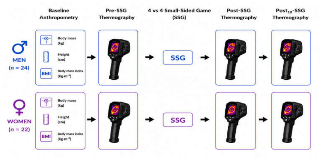
Figure 2.
Representative infrared thermographic images of the regions of interest. Note. Representative infrared thermographic images showing the anterior (A) and posterior (B) regions of interest (ROIs) used for skin temperature analysis. The ROIs included the right anterior thigh (RAT), left anterior thigh (LAT), right anterior leg (RAL), left anterior leg (LAL), right posterior thigh (RPT), left posterior thigh (LPT), right posterior leg (RPL), and left posterior leg (LPL). The central color bar represents skin temperature (°C).
Figure 2.
Representative infrared thermographic images of the regions of interest. Note. Representative infrared thermographic images showing the anterior (A) and posterior (B) regions of interest (ROIs) used for skin temperature analysis. The ROIs included the right anterior thigh (RAT), left anterior thigh (LAT), right anterior leg (RAL), left anterior leg (LAL), right posterior thigh (RPT), left posterior thigh (LPT), right posterior leg (RPL), and left posterior leg (LPL). The central color bar represents skin temperature (°C).
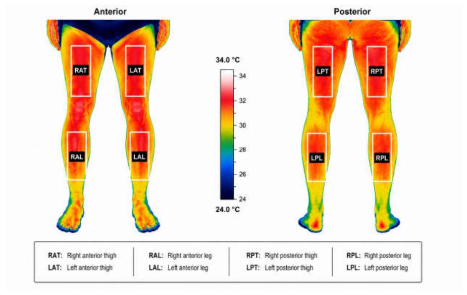
Figure 3.
Between-sex comparison of thermal asymmetry following the small-sided game (SSG). Note. Anterior thigh (AT), posterior thigh (PT), anterior leg (AL), and posterior leg (PL) for the Post–Pre (A), Post_10–Post (B), and Post_10–Pre (C) comparisons. Points represent mean values, and horizontal bars indicate 95% confidence intervals (95% CI). No significant sex-related differences were observed for any thermal asymmetry variable (p > 0.05).
Figure 3.
Between-sex comparison of thermal asymmetry following the small-sided game (SSG). Note. Anterior thigh (AT), posterior thigh (PT), anterior leg (AL), and posterior leg (PL) for the Post–Pre (A), Post_10–Post (B), and Post_10–Pre (C) comparisons. Points represent mean values, and horizontal bars indicate 95% confidence intervals (95% CI). No significant sex-related differences were observed for any thermal asymmetry variable (p > 0.05).
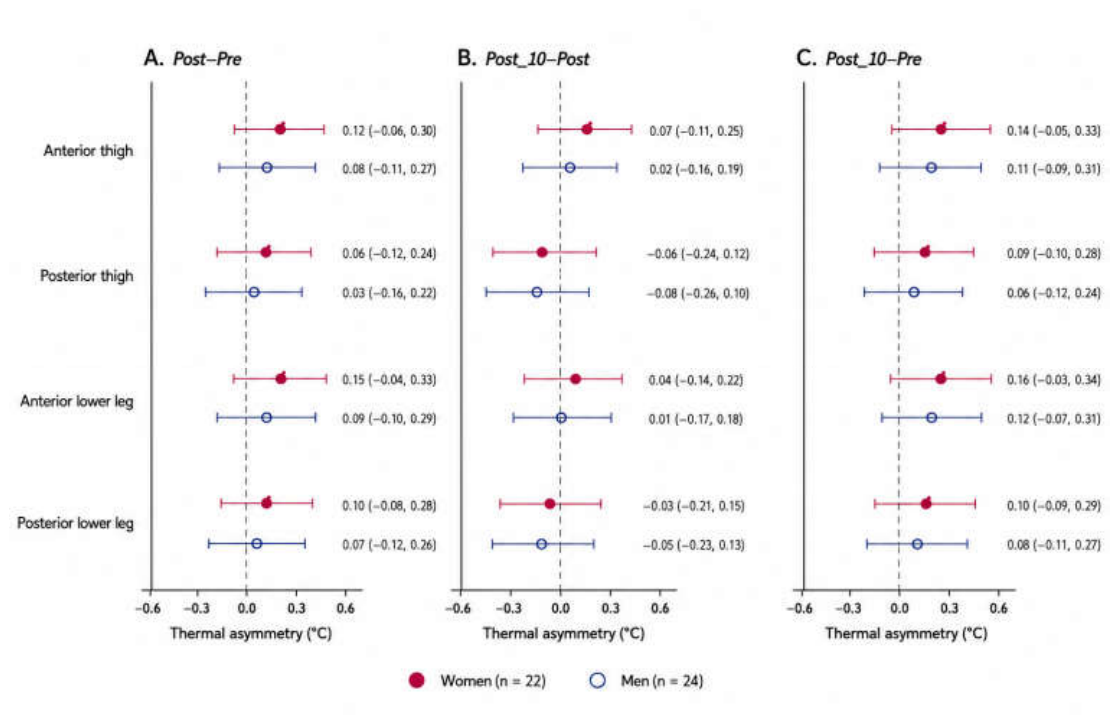

Table 1.
Participant characteristics (mean ± standard deviation).
| Characteristic | Women (n = 22) | Men (n = 24) | p value |
|---|---|---|---|
| Age (years) | 23 ± 4 | 24 ± 8 | 0.708 |
| Body mass (kg) | 60.6 ± 8.5 | 79.0 ± 6.7 | <0.001 |
| Height (cm) | 158 ± 5 | 178 ± 6 | <0.001 |
| Body mass index (kg·m−2) | 24.0 ± 8.3 | 24.9 ± 2.1 | 0.685 |
| Body fat (%) | 24.5 ± 5.5 | 18.2 ± 2.5 | <0.001 |
Note. Data are presented as mean ± standard deviation (SD). p values refer to comparisons between women and men participants.
Table 2.
Comparison of changes in skin temperature (ΔTsk) between women and men soccer players in the thigh regions.
Table 2.
Comparison of changes in skin temperature (ΔTsk) between women and men soccer players in the thigh regions.
| ROI | Time | Women Δ Tsk (°C) Mean ± SD | Men Δ Tsk (°C) Mean ± SD |
P | Hedges’ g |
|---|---|---|---|---|---|
| RAT | Post–Pre | 1.69 ± 0.92 | 0.43 ± 1.21 | <0.001 | 1.15 |
| Post_10–Post | 1.21 ± 1.06 | 0.17 ± 1.34 | 0.005 | 0.85 | |
| Post_10–Pre | 1.81 ± 1.04 | 0.10 ± 0.71 | <0.001 | 1.89 | |
| LAT | Post–Pre | 1.61 ± 0.95 | 0.45 ± 1.07 | <0.001 | 1.13 |
| Post_10–Post | 0.11 ± 0.94 | −0.27 ± 1.06 | 0.199 | 0.38 | |
| Post_10–Pre | 1.72 ± 1.19 | 0.18 ± 0.73 | <0.001 | 1.55 | |
| RPT | Post–Pre | 1.32 ± 1.10 | 0.46 ± 1.06 | 0.010 | 0.78 |
| Post_10–Post | −0.01 ± 0.88 | −0.31 ± 1.03 | 0.628 | 0.31 | |
| Post_10–Pre | 1.31 ± 0.98 | 0.15 ± 0.58 | <0.001 | 1.43 | |
| LPT | Post–Pre | 1.44 ± 0.96 | 0.36 ± 1.04 | 0.001 | 1.06 |
| Post_10–Post | −0.12 ± 0.91 | −0.31 ± 0.95 | 0.800 | 0.20 | |
| Post_10–Pre | 1.32 ± 0.98 | 0.05 ± 0.50 | <0.001 | 1.63 |
Note. ROI = region of interest; SD = standard deviation; RAT = right anterior thigh; LAT = left anterior thigh; RPT = right posterior thigh; LPT = left posterior thigh. Data are presented as mean ± standard deviation. Significant differences were considered at p < 0.05. g = Hedges’ g.
Table 3.
Comparison of changes in skin temperature (ΔTsk) between women and men soccer players in the lower-leg regions.
Table 3.
Comparison of changes in skin temperature (ΔTsk) between women and men soccer players in the lower-leg regions.
| ROI | Time | Women Δ Tsk (°C) Mean ± SD | Men Δ Tsk (°C) Mean ± SD |
P | Hedges’ g |
|---|---|---|---|---|---|
| RAL | Post–Pre | 1.74 ± 0.87 | 0.35 ± 1.22 | <0.001 | 1.29 |
| Post_10–Post | −0.02 ± 1.05 | −0.15 ± 1.14 | 0.685 | 0.12 | |
| Post_10–Pre | 1.72 ± 1.31 | 0.19 ± 0.47 | <0.001 | 1.56 | |
| LAL | Post–Pre | 1.89 ± 0.86 | 0.34 ± 1.21 | <0.001 | 1.45 |
| Post_10–Post | −0.08 ± 1.10 | −0.23 ± 1.24 | 0.671 | 0.12 | |
| Post_10–Pre | 1.81 ± 1.43 | 0.11 ± 0.46 | <0.001 | 1.61 | |
| RPL | Post–Pre | 1.61 ± 1.17 | 0.70 ± 0.98 | 0.006 | 0.84 |
| Post_10–Post | −0.11 ± 1.07 | −0.47 ± 0.92 | 0.233 | 0.35 | |
| Post_10–Pre | 1.50 ± 1.12 | 0.23 ± 0.59 | <0.001 | 1.42 | |
| LPL | Post–Pre | 1.60 ± 1.10 | 0.74 ± 1.02 | 0.009 | 0.80 |
| Post_10–Post | −0.04 ± 0.96 | −0.53 ± 0.94 | 0.093 | 0.50 | |
| Post_10–Pre | 1.56 ± 1.09 | 0.21 ± 0.43 | <0.001 | 1.62 |
Note. ROI = region of interest; SD = standard deviation; RAL = right anterior leg; LAL = left anterior leg; RPL = right posterior leg; LPL = left posterior leg. Data are presented as mean ± standard deviation. Significant differences were considered at p < 0.05. g = Hedges’ g.
Disclaimer/Publisher’s Note: The statements, opinions and data contained in all publications are solely those of the individual author(s) and contributor(s) and not of MDPI and/or the editor(s). MDPI and/or the editor(s) disclaim responsibility for any injury to people or property resulting from any ideas, methods, instructions or products referred to in the content. |
© 2026 by the authors. Licensee MDPI, Basel, Switzerland. This article is an open access article distributed under the terms and conditions of the Creative Commons Attribution (CC BY) license (http://creativecommons.org/licenses/by/4.0/).
Copyright: This open access article is published under a Creative Commons CC BY 4.0 license, which permit the free download, distribution, and reuse, provided that the author and preprint are cited in any reuse.