

Submitted:
30 July 2026
Posted:
31 July 2026
You are already at the latest version
Abstract
HIV-2 infection results in progressive CD4 lymphopenia and death if untreated. The course of HIV-2 infection is more indolent than HIV-1, and the viral and immune mechanisms responsible for this slower pathogenesis are incompletely understood. Persistent inflammation with elevations in cellular and soluble markers of immune activation is a characteristic feature of HIV-2 infection and has been reported to be related to levels of HIV-infected cells in peripheral blood. Here we report an individual with HIV-2 who had persistently elevated levels of activated CD8 lymphocyte subsets that were significantly higher than those quantified in a set of controls without HIV matched for age, sex, and race. Despite elevated levels of immune activation, no detectable HIV-2 RNA or proviral DNA was present in blood. Without undergoing antiretroviral therapy, longitudinal analysis revealed this sustained immune activation was not associated with a progressive CD4 lymphopenia or evidence of disease complications. The presence of strong immune responses without detectable levels of HIV-2 nucleic acid complicates diagnostic testing for HIV-2, and highlights gaps in our understanding of HIV-2 pathogenesis.
Keywords:
HIV-2
; immune activation
; HIV proviral DNA
Introduction
Human Immunodeficiency Virus Type 2 (HIV-2) is a lentivirus with an overall structure and replication cycle similar to HIV-1[1]. Both viruses cause an incurable immunodeficiency leading to death if untreated. These viruses differ, however, in origin, genetic composition, spread, epidemiology, pathogenesis, clinical complications, antiretroviral therapeutics, and public health impact [2,3,4,5,6,7]. Disease progression is slower with HIV-2, including a slower rate of CD4 lymphopenia and prolonged duration to development of AIDS or death [8].Viral characteristics, host responses, and their interactions are central to HIV pathogenesis and responsible for the differences between HIV-2 and HIV-1.
Viral characteristics of HIV-2 and HIV-1 infection are significantly different in vivo. In contrast to persons with HIV-1 (PWH1), in whom viral RNA levels are substantial (median HIV-1 is c. 50,000 copies HIV-1 RNA/ml plasma) [9]), persons with HIV-2 (PWH2) have levels of HIV-2 RNA that are frequently low or absent. This may result in slower disease progression [10] but also frequently complicates diagnostic efforts. Despite the absence of viremia, transcriptionally active HIV-infected cells are present in peripheral blood lymphocytes in PWH2 [11]; in fact, levels of proviruses have been reportedly comparable to those measured in PWH1 [12]. As such, the contribution of viremia and the abundance of infected cells to disease pathogenesis is uncertain.
HIV-2 infection elicits strong specific and generalized host immune responses. Serologic responses to HIV-2 components include prominent Env reactivity and presence of neutralizing antibodies at higher frequency than that detected in HIV-1, which may contribute to slower disease progression [13]. Nevertheless, characteristics of antibody specificity are not well understood with practical consequences for HIV diagnostics. Several FDA-approved commercial assays, including GeeniusTM and VioOneTM, can differentiate HIV-1 and HIV-2 infection [14]. Despite specific antibody responses to HIV-2, significant cross-reactivity between the two viruses persists, which can lead to uncertainties regarding HIV-2 diagnosis. The frequent absence of viremia precludes routine viral RNA testing as a reliable aid to diagnosis. Consequences of diagnostic ambiguity are severe in high prevalence areas, such as West Africa, but are also significant in low-prevalence areas like the United States, where misclassification of HIV-2 as HIV-1 is reported [15]. In fact, several studies demonstrated more HIV-2 false-positives and HIV-2 indeterminate results among PWH1 than all confirmed and probable HIV-2 cases [16,17]. As therapeutic options for HIV-2 are more restrictive than for HIV-1 (HIV-2 is not sensitive to any approved non-nucleoside reverse transcriptase inhibitors or capsid inhibitors), misdiagnosis has critical clinical consequences.
Cellular immune responses against viral components are readily detectable in persons with HIV (PWH), including CD8 cytotoxic and natural killer (NK) cell activity; although responses in HIV-2 infection differ in magnitude and composition [18], with skewing of responses to increases in T cell exhaustion markers [19]. Cellular immune responses have demonstrated impact on virus replication with effects on intra-host HIV-2 molecular evolution [20]. Differences in cellular immune responses may contribute to distinct rates of disease progression in HIV-1 and HIV-2 infection [18,21,22].
In addition to specific humoral and cellular immune responses, HIV-2 infection results in elevations in general immune activation. This includes increases in soluble markers of immune activation [23,24] and elevated levels of activated CD8 and CD4 T cell subsets such as CD8+CD38+DR+ and subsets with exhaustion markers [25,26]. Similar generalized immune abnormalities are present in HIV-1 infection, some of which have been linked to progressive morbidity and mortality, even after introduction of antiretroviral therapy (ART) [27]. The role of immune activation in disease progression is uncertain, although it has been proposed that such high inflammatory responses are the drivers of pathogenesis [26]. Key gaps in understanding virus-host interactions remain, and new studies are essential to inform diagnostic, therapeutic, and eradication efforts.
Here we report a detailed longitudinal analysis of a PWH2 with no evidence of disease progression despite high levels of immune activation. Levels of cellular markers of immune activation exceeded those we measured in a comparison group of otherwise healthy individuals without HIV matched for age, sex, and race. This individual had evidence of strong persistent immune responses but no evidence of HIV-2 RNA in plasma or HIV-2 RNA or DNA in peripheral blood lymphocytes. These studies shed new light on HIV-2 disease pathogenesis and clarify the diagnostic approach in such circumstances.
Materials and Methods
The study participant was evaluated under the protocols, Diagnosis and Management of Inflammatory and Infectious Diseases (NCT00557726; protocol 78-I-0023) and Studies of the Pathogenesis of HIV Infection in Human Peripheral Blood Cells and/or Body Fluids in People Living With and Without HIV (NCT00001281; protocol 91-I-0140). Healthy and diabetic controls were seen under Safety and Immunogenicity of Recombinant Varicella Zoster Virus Vaccine in People With HIV Who Have a CD4 Count Less Than 300 or Greater Than or Equal to 300 and a Healthy Control Population (NCT05580458; protocol 000866-I), An Evaluation of HIV-associated Neurocognitive Disorders (HAND) in Virologically Controlled Patients (NCT01875588; protocol 13-N-0149), A Study of Viral Burden in Peripheral Blood Versus Lymphoid and Bone Marrow Tissue in People Living With HIV (NCT00001316; protocol 92-I-0125), and Studies of the Pathogenesis of HIV Infection in Human Peripheral Blood and/or Body Fluids in People Living With and Without HIV (NCT00001281; protocol 91-I-0140). PWH2 controls were evaluated under Leukapheresis Procedures to Obtain Plasma and Lymphocytes for Research Studies on Antiretroviral Naïve HIV-1 Research Participants (NCT00039689; protocol 02-I-0202, 12 Jul 2002), and Blood Markers of Inflammation, Blood Clotting and Blood Vessel Function in HIV-infected Adults (NCT00776412; protocol 09-I-0013, 20 Nov 2008). Studies were conducted in accordance with the Declaration of Helsinki. All protocols were approved by the National Institute of Allergy and Infectious Disease IRB. Informed consent was obtained under the respective protocol for all subjects involved in the study. Clinical, laboratory, and ancillary study data were collected during clinic visits and via chart review.
Quantitative assays of cell-associated HIV DNA and HIV RNA were performed. Peripheral blood mononuclear cells were isolated [28] and multiplexed ddPCR conducted as previously described [29]. Plasma HIV RNA was quantified using plasma from phlebotomy samples by clinical PCR (Amplicor, Roche Diagnostic Systems, Branchburg, NJ, USA) with a lower limit of detection depending on the commercial assay used. Plasma HIV-2 RNA was quantified at the Wadsworth Center through clinically validated real-time RT-PCR HIV-2 testing [30]. Plasma antibodies to HIV-2 and HIV-1 proteins were qualitatively detected according to manufacturer’s instructions at the following locations: 1) NIH Clinical Center (DetermineTM HIV 1/2 antibody/antigen combo, GeeniusTM HIV1/2 Supplemental AssayTM), 2)The Virus Isolation and Serology Laboratory, Frederick National Laboratory for Cancer Research (DetermineTM HIV 1/2 antibody/antigen combo, GeeniusTM HIV1/2 Supplemental Assay, VioOneTM HIV profile Supplemental Assay) and 3) The Centers for Disease Control’s Division of HIV Prevention Laboratory Branch (CDC DHP, HIV-2 Western Blot; [MP Diagnostics™ HIV-2 Blot 1.2 Western Blot, MP Biomedicals Asia Pacific Pte Ltd, Singapore] and Bio-Rad HIV-1 Western Blot testing [Bio-Rad Laboratories, Clinical Diagnostics, Hercules, CA, USA]). Highly sensitive single-copy RNA determinations by real-time RT-PCR were also performed using SIV primers [31].
Flow cytometry was used for immunophenotyping a panel of CD4 and CD8 subsets [32] that correspond to immune activation and lymphocyte differentiation. Specimens for flow cytometry were collected at time points that were as close as possible to the collection dates of specimens used for HIV DNA testing.
The following tests were performed at the NIH Clinical Center Department of Laboratory Medicine using Clinical Laboratory Improvement Amendments (CLIA)-certified assays:
Complete blood count with differential, immunoglobulins (quantitative), Rheumatoid factor, and HLA determination. Additional CLIA-certified tests performed at Mayo Clinic Laboratories included antibodies to Schistosoma, Strongyloides, Human T-lymphotropic virus I/II, C3 and C4 complement, Anti-nuclear antibody, Anti-ds DNA antibody, Anti-CCP antibodies, and Anti-JO-1 antibody. Bictegravir levels were tested by the University of Florida Infectious Disease Pharmacokinetics Laboratory.
Results
An asymptomatic 61-year-old woman with well-controlled type 2 diabetes, mild hypertension, and no prior HIV testing was reported to be HIV-1 indeterminate/HIV-2 positive by serologic testing on routine examination. She had immigrated from Ghana 21 years earlier and denied sexual activity for the previous 10 years. When last sexually active, she had condomless sex with one long-term partner who has since tested negative for HIV. She denied past blood transfusions, and use of alcohol, tobacco, and illicit drugs. HIV-1/2 RNA (HIV-1/2 qualitative RNA testing) was not detected; CD4 was 815 cells/µl (45.3%). Her home provider prescribed combination ART with tenofovir alafenamide/emtricitabine/bictegravir (Biktarvy®).
She discontinued ART after one dose, requested additional evaluation, and was enrolled in HIV studies at the National Institutes of Health (NIH) Clinical Center.
Upon evaluation, the study participant’s blood sample was repeatedly reactive (DetermineTM HIV 1/2 antibody/antigen combo). Supplemental testing with GeeniusTM and VioOneTM (HIV-1/2 antibody differentiation assays) yielded conflicting test results (Figure 1): GeeniusTM reported HIV-1 indeterminate and HIV-2 negative; VioOneTM noted HIV-1 indeterminate and HIV-2 positive. HIV-1/2 RNA and DNA were not detected by qualitative RT-PCR, and HIV-2 RNA was not detected by sensitive HIV-2 RNA testing [30]. In single-copy research assays, cell-associated (ca-) HIV-2 DNA and ca-RNA were not detected (<0.02 copies/million cells and <0.24 copies/million cells, respectively) using primers and probes for sequences common to HIV-2 and simian immunodeficiency virus [31]. No bictegravir was detected in plasma, and Human T-lymphotropic virus I/II testing was non-reactive. HLA typing revealed HLA-A*: 02,36; HLA-B*: 18, 53, HLA-C*: 02,16; DRB1*: 09, 15, DQB1*: 02,06, DRB*: 4*01, 5*01.
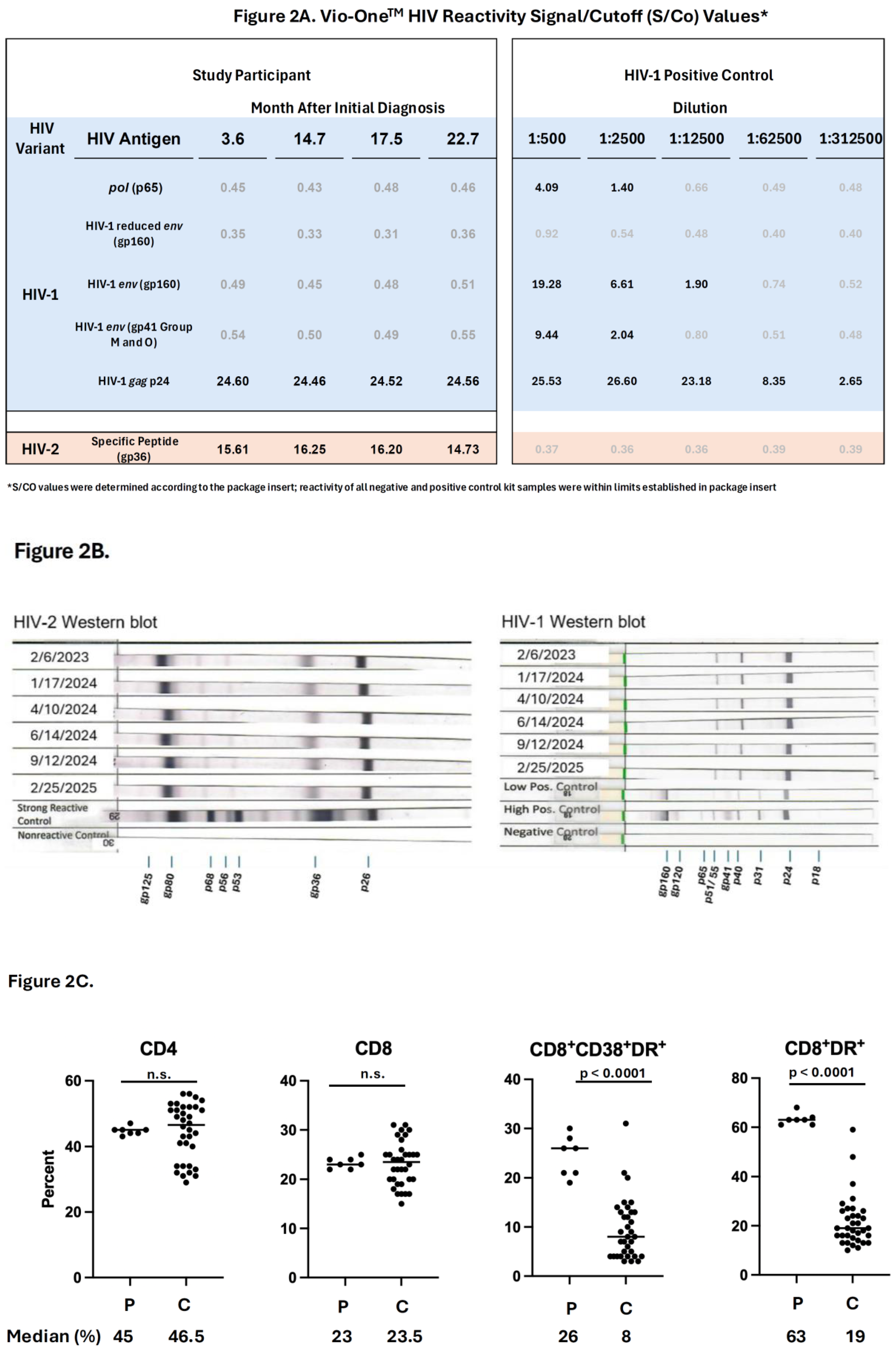
Routine evaluation for over two years (Figure 1) continued to yield inconsistent testing results. HIV-2 gp36 antibody reactivity was inconsistently detected with GeeniusTM while reactivity to HIV-1 gp41 (typically thought to be due to cross-reactivity with gp36) was consistently positive (Figure 1). Repeating GeeniusTM testing in outside laboratories did not resolve discrepancies. Using VioOneTM testing, antibody to HIV-2 gp36 was consistently reactive with high signal-to-cutoff (S/Co) values while no gp41 cross-reactivity was detected (Figure 1, 2A). HIV-1 p24 reactivity was also present (Figure 1, 2A) which is typically a cross reaction with HIV-2 p26. The HIV-2 p26 cross-reactivity was unlikely to be non-specific because HIV-1 p24 S/Co determinations were similar to those identified in individuals with HIV-1 (Figure 2A). The most consistent interpretation of these studies was HIV-2 positive, HIV-1 indeterminate. Additional HIV-2 testing (MP Diagnostics™ HIV-2 Blot 1.2 Western Blot) at the Centers for Disease Control and Prevention’s Division of HIV Prevention Laboratory Branch (CDC DHP) detected antibodies in PWH2 plasma for all three envelope proteins (gp125, gp80, and gp36; Figure 2B) throughout the evaluation period (Figure 1) and for p68, p53, and p26. Bio-Rad HIV-1 Western Blot testing performed at CDC detected Gag p24, antibodies in PWH2 plasma to polyproteins p40 and p51/55, and faint reactivity to gp41 (Figure 2B). The absence of high molecular weight envelope bands implies reactivity is attributed to cross-reactivity and contributes to the difficulty of diagnosing HIV-2. No HIV-1 DNA was detected. Throughout this period, CD4 levels, and CD4/CD8 ratios remained consistently high (Figure 1) and within the range for individuals without HIV.
Sources of potential false positive HIV testing results were evaluated. Analysis of complete blood counts revealed normal hemoglobin, neutrophil, and platelet indices. The participant initially had eosinophilia suggestive of a parasitic infection (Figure 1). An evaluation revealed no ova or parasites in stool. No antibodies were detected against the parasite Schistosoma, which may cause chronic infection, is prevalent in West Africa, and is known to cause falsely reactive HIV screening [14]. Antibodies to a different parasite, Strongyloides, were detected, however (Figure 1). Following two doses of ivermectin starting in month 18, her absolute eosinophil count promptly normalized, and the Strongyloides antibody response resolved over the course of 6 months, as expected (Figure 1). Rheumatologic abnormalities, which can cause false positive HIV testing results, were investigated. Total immunoglobulin and complement levels were normal, antinuclear antibody (ANA) titer was elevated (1:160), but anti-ds DNA levels were not detected. Levels of rheumatoid factor, anti-JO-1, and anti-CCP antibodies were not detected. As such, the rheumatologic profile was not consistent with false positive HIV testing.
Despite the absence of detectable HIV-2 nucleic acid, chronic cellular immune activation typical of HIV-2 infection [33] was evident throughout evaluation, including after resolution of Strongyloides. To determine whether the immune activation was significantly higher than that of otherwise healthy individuals, we quantified levels of CD4, CD8, and the CD8 subsets, CD8+CD38+DR+ and CD8+DR+, from peripheral blood lymphocytes obtained throughout the study and from 11 otherwise healthy individuals matched for age (±5 years), sex, and race. As shown in Fig. 2C, chronically activated CD8+CD38+DR+ and CD8+DR+ percentages (26% and 63%, respectively) were significantly higher (c. 3-fold) than those detected in controls (p<0.0001 for each). No significant differences in levels of CD4 or CD8 percentages were detected. CD8+CD38+DR+ and CD8+DR+ levels were also elevated compared with similarly matched individuals without HIV who have type 2 diabetes, which can be a source of immune activation (N=2; 17 time points; p=0.0086, p<0.0001, respectively). Two unmatched, ART-naïve males with HIV-2 had higher CD8+CD38+DR+ (p=0.0003), but CD8+DR+ percentages were similar to our participant. Evidence of soluble immune activation was not extensively investigated, although D-Dimer levels, which are frequently elevated in HIV-1 infection, were not elevated in this study participant.
Discussion
HIV-2 infection results in lethal immunodeficiency, though it generally progresses more slowly than HIV-1. There are 1–2 million people with HIV-2, residing largely in West Africa and countries with social, cultural, or economic ties to the region. In the United States, there have been approximately 300 imported HIV-2 cases, including 11 dual HIV-1/HIV-2 or probable HIV-2 cases, identified since 2000 [16,34]. This report represents an extreme example of the discordance between viral detection and host response in HIV-2 infection: the absence of detectable viral nucleic acid in peripheral blood in contrast to the presence of a strong immune response to HIV-2 accompanied by elevated immune activation. The discordance between viral detection and immune activation challenges the diagnosis of individuals with suspected HIV-2, especially in low prevalence areas such as the US. Here, the preponderance of serologic evidence, accumulated over a prolonged period, was essential to support the HIV-2 diagnosis. In this setting, it was also necessary to evaluate and eliminate other potential causes of reactive screening assays.
Diagnostic algorithms for HIV infection have evolved over the last 35 years, especially with the development of lateral flow assays focusing on key HIV-1 and -2 components [14,35,36]. Currently, these assays are designed to diagnose HIV-2 by detection of antibodies to HIV-2 Env only, and such supplemental testing algorithms may yield indeterminate or false positive HIV-2 results with GeeniusTM [17]. As Torian, Branson, Gottlieb, Coombs and others pointed out, there is utility of incorporating nucleic acid testing (NAT) in HIV-2 diagnostic algorithms [37,38,39,40,41,42]. In our case, nucleic acid testing was not contributory, but additional serologic testing with VioOneTM uniformly detected gp36 antibodies while GeeniusTM had inconsistent detection of HIV-2 gp36 antibodies, even though it uniformly detected cross-reaction with gp41 of HIV-1. The reasons for cross-reaction detected with GeeniusTM but not with VioOneTM HIV-1 Env peptides are uncertain. HIV-2 Env amino acid sequence shares only approximately 35% homology with HIV-1 [43]. The precise peptides employed as gp41 (group M and O) antigens are not reported for either of these commercial assays but may be distinct. As such, use of both assays in diagnostic dilemmas, as done here, may be informative. Use of FDA and non-FDA approved western blots capable of detecting the presence of highly avid antibodies to linear epitopes provides useful confirmatory results. In our report, antibodies to HIV-2 Gag, RT, and Env proteins were detected.
In this setting,[36], the possibility of infection or dual infection with HIV-1 is an important consideration requiring evaluation. In West Africa, the HIV-1 co-infection rate in PWH2 is relatively high [44]. In this case, some test results suggested co-infection (Figure 1A) and cross reactivity with HIV-1 Gag proteins and gp41 confounded interpretation. Ultimately, the absence of HIV-1 RNA or DNA confirmed HIV-2 monoinfection.
As described, the absence of HIV-2 RNA in plasma is common in PWH2, but the absence of HIV-2 DNA in peripheral blood cells was completely unexpected. Using contemporary assays, HIV-2 DNA has been universally reported in ART-naïve PWH2. Even in early studies using less-sensitive PCR detection assays, absence of HIV-2 DNA detection was infrequent [12]. We were unable to detect HIV-2 nucleic acid using commercial or research assays (<1 copy in c. 50 million peripheral blood lymphocytes). The reasons for the lack of detectable HIV-2 nucleic acid in peripheral blood are uncertain. Previously, Smith, Marx, and colleagues identified two examples of HIV-2 group F in individuals from Sierra Leone which was not detected with standard HIV-2 gag primers but was phylogenetically similar to SIVsmm [45,46]. HIV-2 group F has only been reported in two individuals and only in Sierra Leone. Although formally possible, it is unlikely that our study participant has a rare or potentially new HIV-2 variant and that the absence of detectable HIV-2 in plasma or peripheral blood lymphocytes was due to inability to amplify HIV sequences. Current commercial testing and single-copy assays (Wadsworth) are highly sensitive to all HIV-2 variants; our additional studies using primers specific for SIV did not detect virus. Our study participant did have HLA-B53; epitopes in HIV-2 gag have been reported to bind to HLA-B53, which may contribute to control of HIV-2 [47,48,49,50], perhaps to undetectable levels, as was the case here.
Despite the absence of detectable nucleic acid in blood, we detected elevated levels of immune activation. Previously, levels of activated lymphocytes (CD8+CD38+DR+) have been elevated in PWH2 with high HIV-2 viremia. Elevated lymphocyte activation with low viremia, as found in this case, is unusual [33,51] and has been thought to be associated with the routine presence of HIV-2 infected cells in peripheral blood. Cellular immune activation in the absence of any detectable HIV-2 proviral DNA has not been reported. The source of immune activation and antibody production is uncertain. Chronic immune activation may arise as the consequence of other inflammatory states. In our case, we ruled out the possibility that Strongyloides infection contributed to serologic or cellular immune responses. Although type 2 diabetes (T2D) can result in elevated levels of immune activation, levels measured in our study participant were substantial and higher than in matched T2D controls.
HIV-2 replication in tissues represents an important potential site of replication. Sousa and coworkers have identified marked remodeling in lymph nodes of PWH2 and reported lymphoid-derived follicular helper T cells that may represent an important HIV-2 reservoir [52,53]. Of note, HIV-2 encodes vpx which facilitates replication in mature myeloid-derived lineages. Although mature myeloid cells are absent from peripheral blood, they are abundant in anatomic compartments, such as lung (macrophages), liver (Kupfer cells), gut, as well as brain (microglial cells) and cerebrospinal fluid. HIV-2 has been detected in some of these compartments [54,55]. It is possible that such tissues represent critical HIV-2 reservoirs [56] and are the source of immune activation.
In our analyses, we found no evidence of clinical decline or progressive CD4 lymphopenia due to HIV-2, and CD4 cell numbers and CD4/CD8 ratios remain in the range found in individuals without HIV. Our study participant has no evidence of disease progression, similar to HIV-1 long-term non-progressors (LTNP), and HIV-2 LTNP classification is based on HIV-1 criteria [57]. Approximately 6%–30% of reported PWH2 fulfill these criteria. HIV-2 RNA and/or DNA has been reported in all [58]. In this PWH2, the presence of elevated chronic immune activation makes it difficult to classify her as a typical LTNP. Our findings do indicate, however, that elevated immune activation can persist without evidence of disease progression in HIV-2. It is possible that the elevated levels of immune activation are a component of an effective immune response suppressing infection with levels of HIV-2 RNA and DNA below the limits of detection in commercial and research assays.
Although the PWH2 described consistently declined ART, clinical treatment guidelines recommend ART for all PWH regardless of type or progressor status, and choice of regimen is critical [59]. Most HIV antivirals are highly effective against HIV-2 [60,61] but non-nucleoside reverse transcriptase inhibitors (NNRTI) and enfuvirtide (now withdrawn) have no activity. HIV-2 has reduced sensitivity to lenacapavir and rebound viremia with rapid emergence of capsid mutations has been reported [62,63,64,65], suggesting little utility of this agent for therapy or prophylaxis. ART use has been highly successful in West Africa [66] and lenacapavir prophylaxis efforts are planned worldwide, including these areas of substantial HIV-2 prevalence. Lenacapavir prophylaxis will not prevent HIV-2 transmission, however, and targeted public health efforts will be essential to determine the effects of such prevention efforts on HIV-2 spread.
Conclusions
This report demonstrates that elevated cellular immune activation can persist despite the absence of detectable viral nucleic acid in peripheral blood. We also highlight the obstacles in diagnosing and managing HIV-2 in non-endemic, low-prevalence settings. Additional expertise (CDC, Wadsworth Center, University of Washington) is readily available in such cases and greatly facilitates diagnosis. New methods to analyze HIV infection, including sensitive nucleic acid testing and evaluation of tissue reservoirs, such as reported [67], will help address these challenges. This report also reinforces a clear need for additional studies of HIV-2 as previously described [68]. New studies of epidemiology, diagnostics, and pathogenesis are essential.
Author Contributions
Conceptualization: C.L., P.G., N.S., D.K., F.M.; Methodology: C.L., B.L., F.M.; Formal Analysis: P.G., N.S., B.L., F.M., Investigation: R.D., J.B., P.L., J.A.J., V.S., L.S., C.R., U.S., R.G., P.B., W.S., Resources: A.N., M.E.W., F.M., Data Curation: P.G., N.S., D.K., Writing – Original Draft Preparation: C.L., P.G., N.S., D.K., F.M., Writing – Review & Editing: C.L., P.G., N.S., J.A.J., B.L., M.E.W., F.M., Visualization: P.G., N.S., R.D., P.L., J.H., J.A.J., V.S., F.M., Supervision: C.L., F.M., Funding Acquisition: F.M., Patient Management: C.L., C.N., A.N., D.P., A.W., R.M., J.E., M.M., M.E.W., F.M.
Funding
This research was supported through intramural funding from ZIA BC 011464 and ZIA-NS03130. This research was supported in part by the Intramural Research Program of the National Institutes of Health (NIH). This project has been funded in part with federal funds from the National Cancer Institute, National Institutes of Health, under Contract No. 75N91019D00024.
Institutional Review Board Statement
The study participant was evaluated under the protocols, Diagnosis and Management of Inflammatory and Infectious Diseases (NCT00557726; protocol 78-I-0023, 17 Feb 1978) and Studies of the Pathogenesis of HIV Infection in Human Peripheral Blood Cells and/or Body Fluids in People Living With and Without HIV (NCT00001281; protocol 91-I-0140, 9 Mar 1993). Healthy and diabetic controls were seen under Safety and Immunogenicity of Recombinant Varicella Zoster Virus Vaccine in People With HIV Who Have a CD4 Count Less Than 300 or Greater Than or Equal to 300 and a Healthy Control Population (NCT05580458; protocol 000866-I, 13 Mar 2023), An Evaluation of HIV-associated Neurocognitive Disorders (HAND) in Virologically Controlled Patients (NCT01875588; protocol 13-N-0149, 8 Jul 2013), A Study of Viral Burden in Peripheral Blood Versus Lymphoid and Bone Marrow Tissue in People Living With HIV (NCT00001316; protocol 92-I-0125, 26 Aug 1992), and Studies of the Pathogenesis of HIV Infection in Human Peripheral Blood and/or Body Fluids in People Living With and Without HIV (NCT00001281; protocol 91-I-0140, 9 Mar 1993). PWH2 controls were seen under Leukapheresis Procedures to Obtain Plasma and Lymphocytes for Research Studies on Antiretroviral Naïve HIV-1 Research Participants (NCT00039689; protocol 02-I-0202, 12 Jul 2002), and Blood Markers of Inflammation, Blood Clotting and Blood Vessel Function in HIV-infected Adults (NCT00776412; protocol 09-I-0013, 20 Nov 2008). Studies were conducted in accordance with the Declaration of Helsinki. All protocols were approved by the National Institute of Allergy and Infectious Disease Institutional Review Board.
Informed Consent Statement
Informed consent was obtained under the respective protocol for all subjects involved in the study.
Data Availability Statement
The data presented in this study are available on request from the corresponding author.
Acknowledgments
We are grateful to the study volunteers and their families. We thank R. Coombs and G. Gottlieb for helpful discussions.
Conflicts of Interest
None.
References
- Kleinpeter, A.; Freed, E.O. Human Immunodeficiency Viruses. In Bennett's Principles and Practice of Infectious Diseases, 10th Ed.; Mandell, Douglas, Blaser, M.J., Holland, C.J.S.M., Eds.; Elsevier, 2025. [Google Scholar]
- Williams, A.; Menon, S.; Crowe, M.; Agarwal, N.; Biccler, J.; Bbosa, N.; Ssemwanga, D.; Adungo, F.; Moecklinghoff, C.; Macartney, M.; et al. Geographic and Population Distributions of Human Immunodeficiency Virus (HIV)-1 and HIV-2 Circulating Subtypes: A Systematic Literature Review and Meta-analysis (2010-2021). J. Infect. Dis. 2023, 228, 1583–1591. [Google Scholar] [CrossRef] [PubMed]
- Berzow, D.; Descamps, D.; Obermeier, M.; Charpentier, C.; Kaiser, R.; Guertler, L.; Eberle, J.; Wensing, A.; Sierra, S.; Ruelle, J.; et al. Human Immunodeficiency Virus-2 (HIV-2): A Summary of the Present Standard of Care and Treatment Options for Individuals Living with HIV-2 in Western Europe. Clin. Infect. Dis. 2021, 72, 503–509. [Google Scholar] [CrossRef] [PubMed]
- Ceccarelli, G.; Giovanetti, M.; Sagnelli, C.; Ciccozzi, A.; d'Ettorre, G.; Angeletti, S.; Borsetti, A.; Ciccozzi, M. Human Immunodeficiency Virus Type 2: The Neglected Threat. Pathogens 2021, 10. [Google Scholar] [CrossRef] [PubMed]
- Nyamweya, S.; Hegedus, A.; Jaye, A.; Rowland-Jones, S.; Flanagan, K.L.; Macallan, D.C. Comparing HIV-1 and HIV-2 infection: Lessons for viral immunopathogenesis. Rev. Med. Virol. 2013, 23, 221–240. [Google Scholar] [CrossRef] [PubMed]
- Peeters, M.; Jung, M.; Ayouba, A. The origin and molecular epidemiology of HIV. Expert Rev. Anti Infect. Ther. 2013, 11, 885–896. [Google Scholar] [CrossRef] [PubMed]
- Sedláček, D. Current view on HIV-2 - epidemiology, diagnosis and treatment. Cent. Eur. J. Public Health 2025, 33, 176–180. [Google Scholar] [CrossRef] [PubMed]
- Esbjörnsson, J.; Månsson, F.; Kvist, A.; da Silva, Z.J.; Andersson, S.; Fenyö, E.M.; Isberg, P.E.; Biague, A.J.; Lindman, J.; Palm, A.A.; et al. Long-term follow-up of HIV-2-related AIDS and mortality in Guinea-Bissau: a prospective open cohort study. Lancet HIV 2018. [Google Scholar] [CrossRef] [PubMed]
- Sterling, T.R.; Vlahov, D.; Astemborski, J.; Hoover, D.R.; Margolick, J.B.; Quinn, T.C. Initial plasma HIV-1 RNA levels and progression to AIDS in women and men. N Engl. J. Med. 2001, 344, 720–725. [Google Scholar] [CrossRef] [PubMed]
- Albuquerque, A.S.; Foxall, R.B.; Cortesão, C.S.; Soares, R.S.; Doroana, M.; Ribeiro, A.; Lucas, M.; Antunes, F.; Victorino, R.M.; Sousa, A.E. Low CD4 T-cell counts despite low levels of circulating HIV: insights from the comparison of HIV-1 infected patients with a discordant response to antiretroviral therapy to patients with untreated advanced HIV-2 disease. Clin. Immunol. 2007, 125, 67–75. [Google Scholar] [CrossRef] [PubMed]
- Soares, R.S.; Tendeiro, R.; Foxall, R.B.; Baptista, A.P.; Cavaleiro, R.; Gomes, P.; Camacho, R.; Valadas, E.; Doroana, M.; Lucas, M.; et al. Cell-associated viral burden provides evidence of ongoing viral replication in aviremic HIV-2-infected patients. J. Virol. 2011, 85, 2429–2438. [Google Scholar] [CrossRef] [PubMed]
- Popper, S.J.; Sarr, A.D.; Guèye-Ndiaye, A.; Mboup, S.; Essex, M.E.; Kanki, P.J. Low plasma human immunodeficiency virus type 2 viral load is independent of proviral load: low virus production in vivo. J. Virol. 2000, 74, 1554–1557. [Google Scholar] [CrossRef] [PubMed]
- Rodriguez, S.K.; Sarr, A.D.; MacNeil, A.; Thakore-Meloni, S.; Gueye-Ndiaye, A.; Traoré, I.; Dia, M.C.; Mboup, S.; Kanki, P.J. Comparison of heterologous neutralizing antibody responses of human immunodeficiency virus type 1 (HIV-1)- and HIV-2-infected Senegalese patients: distinct patterns of breadth and magnitude distinguish HIV-1 and HIV-2 infections. J. Virol. 2007, 81, 5331–5338. [Google Scholar] [CrossRef] [PubMed]
- Peters, C.; Pham, P.; Maldarelli, F. Diagnosis of Human Immunodeficiency Virus Infection. In ’s Principles and Practice of Infectious Diseases; Mandell, Douglas, And Bennett, Eds.; Elsevier, 2025. [Google Scholar]
- Guiraud, V.; Bocobza, J.; Desmonet, M.; Damond, F.; Plantier, J.C.; Moreau, G.; Wirden, M.; Stefic, K.; Barin, F.; Gautheret-Dejean, A. Are Confirmatory Assays Reliable for HIV-1/HIV-2 Infection Differentiation? A Multicenter Study. J. Clin. Microbiol. 2023, 61, e0061923. [Google Scholar] [CrossRef] [PubMed]
- Peruski, A.H.; Wesolowski, L.G.; Delaney, K.P.; Chavez, P.R.; Owen, S.M.; Granade, T.C.; Sullivan, V.; Switzer, W.M.; Dong, X.; Brooks, J.T.; et al. Trends in HIV-2 Diagnoses and Use of the HIV-1/HIV-2 Differentiation Test––United States, 2010-2017. Morb. Mortal. Wkly. Rep. 2020, 69, 63–66. [Google Scholar] [CrossRef] [PubMed]
- Wesolowski, L.G.; Chavez, P.R.; Cárdenas, A.M.; Katayev, A.; Slev, P.; Valsamakis, A.; Wang, Y.F.; Yao, J.D.; Dougherty, C.; Gillim-Ross, L.; et al. Routine HIV Test Results in 6 US Clinical Laboratories Using the Recommended Laboratory HIV Testing Algorithm With Geenius HIV 1/2 Supplemental Assay. Sex. Transm. Dis. 2020, 47, S13–s17. [Google Scholar] [CrossRef] [PubMed]
- Orta Resendiz, A.; Petitdemange, C.; Lazzerini, M.; Jacquelin, B.; Beaumont, E.; Perdomo-Celis, F.; Samri, A.; Lambotte, O.; Meyer, L.; Goujard, C.; et al. Expansion of NKG2A(+)CD8(+) T cells with regulatory signatures distinguish individuals living with HIV-2 from those with HIV-1. Med 2026, 7, 101143. [Google Scholar] [CrossRef] [PubMed]
- Scharf, L.; Pedersen, C.B.; Johansson, E.; Lindman, J.; Olsen, L.R.; Buggert, M.; Wilhelmson, S.; Månsson, F.; Esbjörnsson, J.; Biague, A.; et al. Inverted CD8 T-Cell Exhaustion and Co-Stimulation Marker Balance Differentiate Aviremic HIV-2-Infected From Seronegative Individuals. Front Immunol. 2021, 12, 744530. [Google Scholar] [CrossRef] [PubMed]
- Boswell, M.T.; Nazziwa, J.; Kuroki, K.; Palm, A.; Karlson, S.; Månsson, F.; Biague, A.; da Silva, Z.J.; Onyango, C.O.; de Silva, T.I.; et al. Intrahost evolution of the HIV-2 capsid correlates with progression to AIDS. Virus Evol. 2022, 8, veac075. [Google Scholar] [CrossRef] [PubMed]
- Mueller, B.R.; Mehta, M.; Campbell, M.; Neupane, N.; Cedillo, G.; Lee, G.; Coyle, K.; Qi, J.; Chen, Z.; George, M.C.; et al. Autonomic Neuropathy Is Associated with An Increase in Type-1 Cytokines in People Living With HIV. bioRxiv 2025. [Google Scholar] [CrossRef] [PubMed]
- Moysi, E.; Darko, S.; Gea-Mallorquí, E.; Petrovas, C.; Almeida, J.R.; Wolinsky, D.; Peng, Y.; Jaye, A.; Stewart-Jones, G.; Douek, D.C.; et al. Clonotypic architecture of a Gag-specific CD8+ T-cell response in chronic human HIV-2 infection. Eur. J. Immunol. 2021, 51, 2485–2500. [Google Scholar] [CrossRef] [PubMed]
- Nowroozalizadeh, S.; Månsson, F.; da Silva, Z.; Repits, J.; Dabo, B.; Pereira, C.; Biague, A.; Albert, J.; Nielsen, J.; Aaby, P.; et al. Microbial translocation correlates with the severity of both HIV-1 and HIV-2 infections. J. Infect. Dis. 2010, 201, 1150–1154. [Google Scholar] [CrossRef] [PubMed]
- Thiébaut, R.; Charpentier, C.; Damond, F.; Taieb, A.; Antoine, R.; Capeau, J.; Chêne, G.; Collin, G.; Matheron, S.; Descamps, D.; et al. Association of soluble CD14 and inflammatory biomarkers with HIV-2 disease progression. Clin. Infect. Dis. 2012, 55, 1417–1425. [Google Scholar] [CrossRef] [PubMed]
- Salwe, S.; Padwal, V.; Nagar, V.; Patil, P.; Patel, V. T cell functionality in HIV-1, HIV-2 and dually infected individuals: correlates of disease progression and immune restoration. Clin. Exp. Immunol. 2019, 198, 233–250. [Google Scholar] [CrossRef] [PubMed]
- Sousa, A.E.; Carneiro, J.; Meier-Schellersheim, M.; Grossman, Z.; Victorino, R.M. CD4 T cell depletion is linked directly to immune activation in the pathogenesis of HIV-1 and HIV-2 but only indirectly to the viral load. J. Immunol. 2002, 169, 3400–3406. [Google Scholar] [CrossRef] [PubMed]
- Sandler, N.G.; Wand, H.; Roque, A.; Law, M.; Nason, M.C.; Nixon, D.E.; Pedersen, C.; Ruxrungtham, K.; Lewin, S.R.; Emery, S.; et al. Plasma levels of soluble CD14 independently predict mortality in HIV infection. J. Infect. Dis. 2011, 203, 780–790. [Google Scholar] [CrossRef] [PubMed]
- Simonetti, F.R.; Sobolewski, M.D.; Fyne, E.; Shao, W.; Spindler, J.; Hattori, J.; Anderson, E.M.; Watters, S.A.; Hill, S.; Wu, X.; et al. Clonally expanded CD4+ T cells can produce infectious HIV-1 in vivo. Proc. Natl. Acad. Sci. U S A 2016, 113, 1883–1888. [Google Scholar] [CrossRef] [PubMed]
- Anderson, E.M.; Maldarelli, F. Quantification of HIV DNA Using Droplet Digital PCR Techniques. Curr. Protoc. Microbiol. 2018, 51, e62. [Google Scholar] [CrossRef] [PubMed]
- Styer, L.M.; Miller, T.T.; Parker, M.M. Validation and clinical use of a sensitive HIV-2 viral load assay that uses a whole virus internal control. J. Clin. Virol. 2013, 58 Suppl 1, e127-133. [Google Scholar] [CrossRef] [PubMed]
- Cline, A.N.; Bess, J.W.; Piatak, M., Jr.; Lifson, J.D. Highly sensitive SIV plasma viral load assay: practical considerations, realistic performance expectations, and application to reverse engineering of vaccines for AIDS. J. Med. Primatol. 2005, 34, 303–312. [Google Scholar] [CrossRef] [PubMed]
- Higgins, J.; Metcalf, J.A.; Stevens, R.A.; Baseler, M.; Proschan, M.; Lane, H.C.; Sereti, I. Effects of delays in peripheral blood processing, including cryopreservation, on detection of CD31 expression on naive CD4 T cells. Clin. Vaccine Immunol. 2008, 15, 1141–1143. [Google Scholar] [CrossRef] [PubMed]
- Hegedus, A.; Nyamweya, S.; Zhang, Y.; Govind, S.; Aspinall, R.; Mashanova, A.; Jansen, V.A.; Whittle, H.; Jaye, A.; Flanagan, K.L.; et al. Protection versus pathology in aviremic and high viral load HIV-2 infection-the pivotal role of immune activation and T-cell kinetics. J. Infect. Dis. 2014, 210, 752–761. [Google Scholar] [CrossRef] [PubMed]
- Torian, L.V.; Eavey, J.J.; Punsalang, A.P.; Pirillo, R.E.; Forgione, L.A.; Kent, S.A.; Oleszko, W.R. HIV type 2 in New York City, 2000-2008. Clin. Infect. Dis. 2010, 51, 1334–1342. [Google Scholar] [CrossRef] [PubMed]
- Masciotra, S.; Smith, A.J.; Youngpairoj, A.S.; Sprinkle, P.; Miles, I.; Sionean, C.; Paz-Bailey, G.; Johnson, J.A.; Owen, S.M. Evaluation of the CDC proposed laboratory HIV testing algorithm among men who have sex with men (MSM) from five US metropolitan statistical areas using specimens collected in 2011. J. Clin. Virol. 2013, 58 Suppl 1, e8–e12. [Google Scholar] [CrossRef] [PubMed]
- Springer, S.A.; Masciotra, S.; Johnson, J.A.; Campbell, S. Do I Have HIV or Not? Lack of RNA Detection and the Case for Sensitive DNA Testing. Open Forum Infect. Dis. 2020, 7, ofaa478. [Google Scholar] [CrossRef] [PubMed]
- Torian, L.V.; Forgione, L.A.; Punsalang, A.E.; Pirillo, R.E.; Oleszko, W.R. Comparison of Multispot EIA with Western blot for confirmatory serodiagnosis of HIV. J. Clin. Virol. 2011, 52 Suppl 1, S41–44. [Google Scholar] [CrossRef] [PubMed]
- Linley, L.; Ethridge, S.F.; Oraka, E.; Owen, S.M.; Wesolowski, L.G.; Wroblewski, K.; Landgraf, K.M.; Parker, M.M.; Brinson, M.; Branson, B.M. Evaluation of supplemental testing with the Multispot HIV-1/HIV-2 Rapid Test and APTIMA HIV-1 RNA Qualitative Assay to resolve specimens with indeterminate or negative HIV-1 Western blots. J. Clin. Virol. 2013, 58 Suppl 1, e108-112. [Google Scholar] [CrossRef] [PubMed]
- Chang, M.; Steinmetzer, K.; Raugi, D.N.; Smith, R.A.; Ba, S.; Sall, F.; Seydi, M.; Niang, A.; Sall, E.I.; Cisse, O.; et al. Detection and differentiation of HIV-2 using the point-of-care Alere q HIV-1/2 Detect nucleic acid test. J. Clin. Virol. 2017, 97, 22–25. [Google Scholar] [CrossRef] [PubMed]
- Chang, M.; Wong, A.J.; Raugi, D.N.; Smith, R.A.; Seilie, A.M.; Ortega, J.P.; Bogusz, K.M.; Sall, F.; Ba, S.; Seydi, M.; et al. Clinical validation of a novel diagnostic HIV-2 total nucleic acid qualitative assay using the Abbott m2000 platform: Implications for complementary HIV-2 nucleic acid testing for the CDC 4th generation HIV diagnostic testing algorithm. J. Clin. Virol. 2017, 86, 56–61. [Google Scholar] [CrossRef] [PubMed]
- Duncan, D.; Duncan, J.; Kramer, B.; Nilsson, A.Y.; Haile, B.; Butcher, A.; Chugh, S.; Baum, P.; Aldrovandi, G.M.; Young, S.; et al. An HIV Diagnostic Testing Algorithm Using the cobas HIV-1/HIV-2 Qualitative Assay for HIV Type Differentiation and Confirmation. J. Clin. Microbiol. 2021, 59, e0303020. [Google Scholar] [CrossRef] [PubMed]
- Wesolowski, L.G.; Nasrullah, M.; Coombs, R.W.; Rosenberg, E.; Ethridge, S.F.; Hutchinson, A.B.; Dragavon, J.; Rychert, J.; Nolte, F.S.; Madory, J.E.; et al. Acute Infections, Cost per Infection and Turnaround Time in Three United States Hospital Laboratories Using Fourth-Generation Antigen-Antibody Human Immunodeficiency Virus Immunoassays. Open Forum Infect. Dis. 2016, 3, ofv188. [Google Scholar] [CrossRef] [PubMed]
- Huang, M.L.; Essex, M.; Lee, T.H. Localization of immunogenic domains in the human immunodeficiency virus type 2 envelope. J. Virol. 1991, 65, 5073–5079. [Google Scholar] [CrossRef] [PubMed]
- Tchounga, B.K.; Bertine, M.; Damond, F.; Ferre, V.M.; Inwoley, A.; Boni, S.P.; Moisan, A.; Plantier, J.C.; Descamps, D.; Ekouevi, D.K.; et al. Molecular confirmation of HIV-1 and HIV-2 coinfections among initially serologically dually-reactive samples from patients living in West Africa. PLoS ONE 2023, 18, e0283602. [Google Scholar] [CrossRef] [PubMed]
- Smith, S.M.; Christian, D.; de Lame, V.; Shah, U.; Austin, L.; Gautam, R.; Gautam, A.; Apetrei, C.; Marx, P.A. Isolation of a new HIV-2 group in the US. Retrovirology 2008, 5, 103. [Google Scholar] [CrossRef] [PubMed]
- Chen, Z.; Luckay, A.; Sodora, D.L.; Telfer, P.; Reed, P.; Gettie, A.; Kanu, J.M.; Sadek, R.F.; Yee, J.; Ho, D.D.; et al. Human immunodeficiency virus type 2 (HIV-2) seroprevalence and characterization of a distinct HIV-2 genetic subtype from the natural range of simian immunodeficiency virus-infected sooty mangabeys. J. Virol. 1997, 71, 3953–3960. [Google Scholar] [CrossRef] [PubMed]
- Dorrell, L.; Willcox, B.E.; Jones, E.Y.; Gillespie, G.; Njai, H.; Sabally, S.; Jaye, A.; DeGleria, K.; Rostron, T.; Lepin, E.; et al. Cytotoxic T lymphocytes recognize structurally diverse, clade-specific and cross-reactive peptides in human immunodeficiency virus type-1 gag through HLA-B53. Eur. J. Immunol. 2001, 31, 1747–1756. [Google Scholar] [CrossRef] [PubMed]
- Gillespie, G.M.; Pinheiro, S.; Sayeid-Al-Jamee, M.; Alabi, A.; Kaye, S.; Sabally, S.; Sarge-Njie, R.; Njai, H.; Joof, K.; Jaye, A.; et al. CD8+ T cell responses to human immunodeficiency viruses type 2 (HIV-2) and type 1 (HIV-1) gag proteins are distinguishable by magnitude and breadth but not cellular phenotype. Eur. J. Immunol. 2005, 35, 1445–1453. [Google Scholar] [CrossRef] [PubMed]
- Gotch, F.; McAdam, S.N.; Allsopp, C.E.; Gallimore, A.; Elvin, J.; Kieny, M.P.; Hill, A.V.; McMichael, A.J.; Whittle, H.C. Cytotoxic T cells in HIV2 seropositive Gambians. Identification of a virus-specific MHC-restricted peptide epitope. J. Immunol. 1993, 151, 3361–3369. [Google Scholar] [CrossRef]
- Gotch, F.; Nixon, D.; Gallimore, A.; McAdam, S.; McMichael, A. Cytotoxic T lymphocyte epitopes shared between HIV-1, HIV-2, and SIV. J. Med. Primatol. 1993, 22, 119–123. [Google Scholar] [CrossRef]
- Kanki, P.J.; Rowland-Jones, S. The protective effect of HIV-2 infection: implications for understanding HIV-1 immunity. Aids 2014, 28, 1065–1067. [Google Scholar] [CrossRef] [PubMed]
- Fernandes, S.M.; Farias, G.B.; Pires, A.R.; Santos, D.F.; Antão, A.V.; Pires, A.; Moura, R.; Godinho-Santos, A.; Marques, R.T.; Mc Gomes, A.; et al. Lymph node remodelling underlies the attenuated form of HIV/AIDS in people with HIV-2. J. Infect. 2026, 92, 106760. [Google Scholar] [CrossRef] [PubMed]
- Godinho-Santos, A.; Foxall, R.B.; Antão, A.V.; Tavares, B.; Ferreira, T.; Serra-Caetano, A.; Matoso, P.; Sousa, A.E. Follicular Helper T Cells Are Major Human Immunodeficiency Virus-2 Reservoirs and Support Productive Infection. J. Infect. Dis. 2020, 221, 122–126. [Google Scholar] [CrossRef] [PubMed]
- Fernandes, S.M.; Pires, A.R.; Matoso, P.; Ferreira, C.; Nunes-Cabaço, H.; Correia, L.; Valadas, E.; Poças, J.; Pacheco, P.; Veiga-Fernandes, H.; et al. HIV-2 infection is associated with preserved GALT homeostasis and epithelial integrity despite ongoing mucosal viral replication. Mucosal Immunol. 2018, 11, 236–248. [Google Scholar] [CrossRef] [PubMed]
- Wood, B.R.; Klein, J.P.; Lyons, J.L.; Milner, D.A.; Phillips, R.E.; Schutten, M.; Folkerth, R.D.; Ciarlini, P.; Henrich, T.J.; Johnson, J.A. HIV-2 encephalitis: case report and literature review. AIDS Patient Care STDS 2012, 26, 383–387. [Google Scholar] [CrossRef] [PubMed]
- Melandri, M.; Faua, C.; Gantner, P. The HIV-2 reservoir: lessons from HIV-1 and knowledge gaps. Infect. Dis. Now. 2026, 105261. [Google Scholar] [CrossRef] [PubMed]
- Thiebaut, R.; Matheron, S.; Taieb, A.; Brun-Vezinet, F.; Chene, G.; Autran, B. immunology group of the, A.C.O.H.I.V.c. Long-term nonprogressors and elite controllers in the ANRS CO5 HIV-2 cohort. AIDS 2011, 25, 865–867. [Google Scholar] [CrossRef] [PubMed]
- Diallo, M.S.; Samri, A.; Charpentier, C.; Bertine, M.; Cheynier, R.; Thiebaut, R.; Matheron, S.; Collin, F.; Braibant, M.; Candotti, D.; et al. A Comparison of Cell Activation, Exhaustion, and Expression of HIV Coreceptors and Restriction Factors in HIV-1- and HIV-2-Infected Nonprogressors. AIDS Res. Hum. Retroviruses 2021, 37, 214–223. [Google Scholar] [CrossRef] [PubMed]
- Panel on Antiretroviral Guidelines for Adults and Adolescents. Guidelines for the Use of Antiretroviral Agents in Adults and Adolescents with HIV, Department of Health and Human Services. Available online: https://clinicalinfo.hiv.gov/en/guidelines/adult-and-adolescent-arv (accessed on 1 June).
- Moranguinho, I.; Taveira, N.; Bártolo, I. Antiretroviral Treatment of HIV-2 Infection: Available Drugs, Resistance Pathways, and Promising New Compounds. Int. J. Mol. Sci. 2023, 24. [Google Scholar] [CrossRef] [PubMed]
- Wu, V.H.; Smith, R.A.; Masoum, S.; Raugi, D.N.; Ba, S.; Seydi, M.; Grobler, J.A.; Gottlieb, G.S. MK-8591 (4'-Ethynyl-2-Fluoro-2'-Deoxyadenosine) Exhibits Potent Activity against HIV-2 Isolates and Drug-Resistant HIV-2 Mutants in Culture. Antimicrob. Agents Chemother. 2017, 61. [Google Scholar] [CrossRef] [PubMed]
- Montrouge, T.; Bertine, M.; Peytavin, G.; Saint-Joannis, T.; Bachelard, A.; De Truchis, P.; Lariven, S.; Morlat, P.; Pouderoux, C.; Damond, F.; et al. Rapid Selection of HIV-2 Capsid Mutations in Salvage Therapy With Lenacapavir-containing Regimen. Clin. Infect. Dis. 2025, 81, 341–344. [Google Scholar] [CrossRef] [PubMed]
- Delaugerre, C.; Mafi, S.; Zenuni, A.; Peytavin, G.; Amat, K.; Seang, S.; Duvivier, C.; Chirio, D.; Viard, J.P.; Hocqueloux, L.; et al. Effectiveness and Safety of Lenacapavir-Containing Regimens in Highly Experienced HIV-Infected Patients With Multidrug Resistance: Real-world Results From the French Compassionate Use Program (a). Open Forum Infect. Dis. 2026, 13, ofag353. [Google Scholar] [CrossRef] [PubMed]
- Smith, R.A.; Raugi, D.N.; Nixon, R.S.; Seydi, M.; Margot, N.A.; Callebaut, C.; Gottlieb, G.S. Antiviral Activity of Lenacapavir Against Human Immunodeficiency Virus Type 2 (HIV-2) Isolates and Drug-Resistant HIV-2 Mutants. J. Infect. Dis. 2024, 229, 1290–1294. [Google Scholar] [CrossRef] [PubMed]
- van Kampen, J.J.A.; van Nood, E.; Mahmud, R.; Krullaars, Z.; Voskamp, T.; Voskamp, M.; Nijssen, T.; Voermans, J.J.C.; Charpentier, C.; Le Hingrat, Q.; et al. Novel Dolutegravir and Lenacapavir Resistance Patterns in Human Immunodeficiency Virus Type 2 Infection: A Case Report. Open Forum Infect. Dis. 2025, 12, ofae705. [Google Scholar] [CrossRef] [PubMed]
- Raugi, D.N.; Ba, S.; Cisse, O.; Diallo, K.; Tamba, I.T.; Ndour, C.; Badiane, N.M.D.; Fortes, L.; Diallo, M.B.; Faye, D.; et al. Long-term Experience and Outcomes of Programmatic Antiretroviral Therapy for Human Immunodeficiency Virus Type 2 Infection in Senegal, West Africa. Clin. Infect. Dis. 2021, 72, 369–378. [Google Scholar] [CrossRef] [PubMed]
- Lu, M.D.; Telwatte, S.; Kumar, N.; Ferreira, F.; Martin, H.A.; Kadiyala, G.N.; Wedrychowski, A.; Moron-Lopez, S.; Chen, T.H.; Goecker, E.A.; et al. Novel assays to investigate the mechanisms of latent infection with HIV-2. PLoS ONE 2022, 17, e0267402. [Google Scholar] [CrossRef] [PubMed]
- Rowland-Jones, S.; Gea-Mallorquí, E. Closing the equity gap in the treatment of HIV-2 infection. Lancet HIV 2024, 11, e347–e349. [Google Scholar] [CrossRef] [PubMed]
Figure 1.
Summary of study participant’s HIV test results, potential false positive test results evaluation, and lymphocyte subsets from 3.6 to 28.0 months after initial diagnosis.
Figure 1.
Summary of study participant’s HIV test results, potential false positive test results evaluation, and lymphocyte subsets from 3.6 to 28.0 months after initial diagnosis.
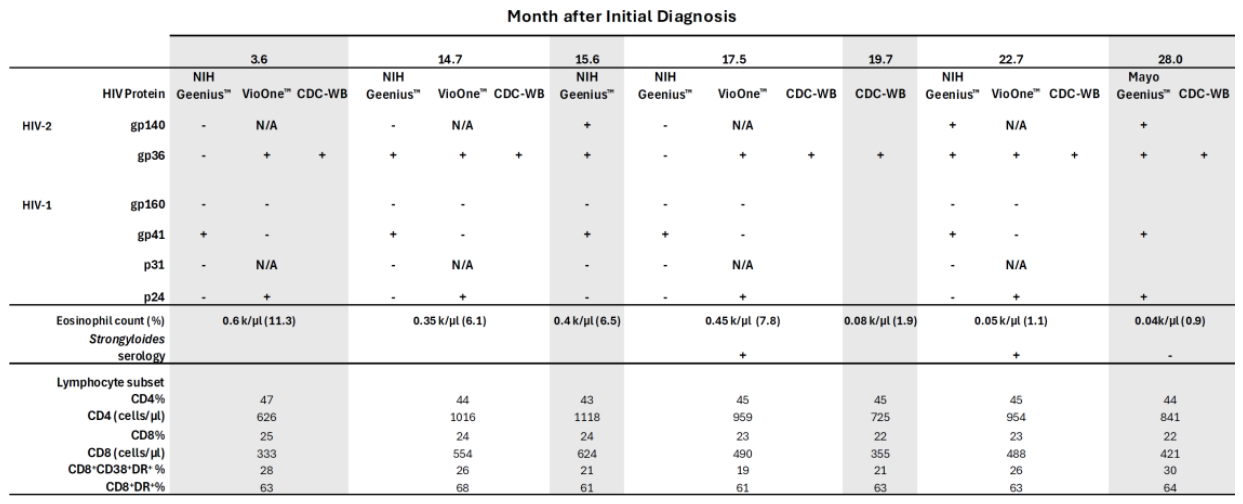
Figure 2.
A. Vio-OneTM HIV reactivity signal/cutoff values are shown for HIV-1 and HIV-2 in the study participant and an HIV-1 positive control. B. Western blot detection of antibodies to HIV-2 and cross-reactivity to HIV-1. Reactivity was observed against HIV-2 proteins p26, gp36, p53, p68, gp80, and weak reactivity to gp125. Reactivity was also observed against HIV-1 proteins p51/55, p40, and possible reactivity to gp41 but not gp120, indicating the cross-reactivity with HIV-1 is largely to Gag products. C. Persistent immune activation in HIV-2 infection despite absence of viremia. Peripheral blood lymphocytes with cell surface markers (CD4, CD8, CD8+CD38+ DR+, CD8+DR+) were quantified by fluorescence-activated cell sorting analyses from 7 time points from study participant and from 11 otherwise healthy volunteers without HIV matched for age (±5 years), sex, and race (34 total time point measurements). In these measurements, there is no assumption of normality, and a Mann-Whitney U test was used to compare percent positive cell proportions. P = Participant, C= Control.
Figure 2.
A. Vio-OneTM HIV reactivity signal/cutoff values are shown for HIV-1 and HIV-2 in the study participant and an HIV-1 positive control. B. Western blot detection of antibodies to HIV-2 and cross-reactivity to HIV-1. Reactivity was observed against HIV-2 proteins p26, gp36, p53, p68, gp80, and weak reactivity to gp125. Reactivity was also observed against HIV-1 proteins p51/55, p40, and possible reactivity to gp41 but not gp120, indicating the cross-reactivity with HIV-1 is largely to Gag products. C. Persistent immune activation in HIV-2 infection despite absence of viremia. Peripheral blood lymphocytes with cell surface markers (CD4, CD8, CD8+CD38+ DR+, CD8+DR+) were quantified by fluorescence-activated cell sorting analyses from 7 time points from study participant and from 11 otherwise healthy volunteers without HIV matched for age (±5 years), sex, and race (34 total time point measurements). In these measurements, there is no assumption of normality, and a Mann-Whitney U test was used to compare percent positive cell proportions. P = Participant, C= Control.
Disclaimer/Publisher’s Note: The statements, opinions and data contained in all publications are solely those of the individual author(s) and contributor(s) and not of MDPI and/or the editor(s). MDPI and/or the editor(s) disclaim responsibility for any injury to people or property resulting from any ideas, methods, instructions or products referred to in the content. |
© 2026 by the authors. Licensee MDPI, Basel, Switzerland. This article is an open access article distributed under the terms and conditions of the Creative Commons Attribution (CC BY) license.
Copyright: This open access article is published under a Creative Commons CC BY 4.0 license, which permit the free download, distribution, and reuse, provided that the author and preprint are cited in any reuse.