
Submitted:
29 July 2026
Posted:
31 July 2026
You are already at the latest version
Abstract
Canine distemper virus (CDV) is a multi-host morbillivirus of domestic dogs and wild carnivores, and early molecular detection is important for treatment, isolation, and wildlife surveillance. However, the most informative ante-mortem specimen type for RT-qPCR screening remains context dependent. This pilot study compared CDV RNA detection and viral load across paired sample matrices from nine dogs and screened archived blood/serum samples from four tigers and one clouded leopard. CDV RNA was extracted using a commercial viral nucleic-acid kit and amplified using a one-step RT-qPCR assay. Five of nine dogs were CDV-positive. Among positive dogs, conjunctival swabs detected CDV RNA in 5/5 animals (100%), followed by nasal swabs (4/5), rectal swabs (3/5), whole blood (2/5), and urine (1/5). Viral load ranged from 9.55 to 213,796.21 RNA copies/μL. Exact Cochran’s Q and Friedman tests did not reach statistical significance, but Kendall’s W suggested a moderate sample-type effect. These results support conjunctival swabs as the most consistent specimen in this dataset, with nasal and rectal swabs as useful complementary matrices.
Keywords:
canine distemper virus
; RT-qPCR
; molecular screening
; conjunctival swab
; wildlife surveillance
; sample matrix
1. Introduction
Canine distemper virus (CDV), also known as canine morbillivirus, is an enveloped, negative-sense, single-stranded RNA virus in the genus Morbillivirus and the family Paramyxoviridae [1,2,3]. CDV infects domestic dogs as well as several wild carnivore species, including felids and other threatened wildlife, and has been repeatedly identified as a conservation and veterinary-health concern [1,4,5,6,7]. The virus has a broad cell and tissue tropism, with infection involving epithelial, lymphoid, and nervous tissues [3,8,9,10,11]. Clinical disease can include respiratory, gastrointestinal, ocular, dermatologic, and neurologic manifestations, while infection outcomes may depend on host immunity, viral strain, and the stage at which the animal is sampled [8,10,11,12].
Early laboratory confirmation is important because there is no specific curative antiviral therapy for canine distemper; treatment is mainly supportive and isolation reduces transmission risk [12,13]. Serological or antigen-based tests may be useful in some settings, but interpretation can be complicated by vaccination history, antibody kinetics, cross-reactivity, or stage of disease [13,14]. Molecular methods, particularly reverse-transcription PCR and real-time quantitative PCR (RT-qPCR), directly target viral RNA and are therefore valuable for early diagnosis and screening [15,16,17,18]. RT-qPCR combines reverse transcription, amplification, and fluorescence-based real-time monitoring in a closed-tube system. Compared to conventional endpoint PCR, it reduces post-amplification handling, provides cycle quantification (Cq) values, and allows the estimation of viral-load when calibrated with standards [16,19,20,21]. Its value for public-health molecular diagnostics became especially visible during SARS-CoV-2 assay deployment, and similar principles support veterinary virology and animal-disease surveillance [17,22,23].
A central practical question in CDV diagnostics is which ante-mortem sample matrix should be prioritized. Previous studies have detected CDV RNA in whole blood, serum, cerebrospinal fluid, urine, conjunctival swabs, nasal swabs, tonsil, rectal swabs, and other tissues [8,15,18,24,25,26,27]. However, viral load can vary with disease stage and clinical presentation, and some matrices may be difficult to collect or may be affected by storage and inhibitors [17,18,28]. For field surveillance and veterinary screening, a suitable matrix should be easy to collect, safe, minimally invasive where possible, and consistently positive in infected animals.
In Nepal, CDV is relevant not only for domestic-dog health but also for wildlife conservation. Human–wildlife interfaces around protected areas create opportunities for contact among domestic dogs and wild carnivores, including tigers and leopards [2,7,13]. Previous evidence of CDV exposure and disease-compatible illness in Nepali wild carnivores highlights the need for practical molecular surveillance strategies [7]. The present pilot study therefore evaluates RT-qPCR detection and viral-load patterns across ante-mortem sample matrices from CDV-suspected dogs and interprets negative wild-carnivore blood/serum results in light of sample availability and assay quality control. The main objective was to identify which sample type provided the most consistent CDV RNA detection among confirmed positive dogs and to generate a quantitative framework for future larger studies.
2. Materials and Methods
2.1. Study Animals and Sample Collection
The study included four tigers, one clouded leopard, eight CDV-suspected dogs, and one dog that had recovered from distemper. Whole blood from the four tigers and the clouded leopard was obtained through the National Trust for Nature Conservation Molecular Biology Diagnostic Laboratory, Chitwan, and stored frozen until analysis. Dog samples were collected after CDV-suspected animals were reported to collaborating collection centers. Whole blood was collected on-site and centrifuged at 2500 rpm for 10 min to obtain the buffy-coat fraction. Swab samples were collected from the conjunctiva, nose, and rectum and placed in sterile collection devices containing 0.9% NaCl or viral transport medium. Urine was collected in sterile tubes when available. Samples were stored at or lower until RNA extraction and RT-qPCR analysis.
2.2. RNA Extraction
Viral RNA was extracted using a GeneDireX Viral Nucleic Acid Isolation Kit (GeneDireX Inc., USA) according to the manufacturer’s protocol. For mucous-membrane swabs, the swab tip was placed in a microcentrifuge tube containing 400 L Buffer V1 and 200 L phosphate-buffered saline. For whole blood, serum, and urine specimens, 200 L of sample was mixed with 400 L Buffer V1. If the available sample volume was below 200 L, phosphate-buffered saline was added to adjust the input volume to 200 L.
2.3. RT-qPCR Assay and Copy-Number Estimation
RT-qPCR was performed using the YouSeq Canine Distemper Virus qPCR test kit (YouSeq, Winchester, UK) on a CFX96 Touch Real-Time PCR Detection System (Bio-Rad). Each 20 L reaction contained 10 L Tetra One-Step 2× RT-qPCR Master Mix, 1 L CDV-specific primer/probe mix, 1 L internal extraction-control primer/probe mix, and 8 L extracted sample RNA. Negative-control reactions replaced sample RNA with DNase/RNase-free water. Positive-control serial dilutions supplied with the kit were used to generate the standard curve. FAM fluorescence was used for CDV target detection, and HEX fluorescence was used for internal extraction-control monitoring.
The quantitative standard curve was constructed by regressing reported Cq values against RNA copy concentration for the quantitative calibration range. Viral RNA copies per microliter were calculated from the standard-curve equation:
where C represents RNA copies/L. Amplification efficiency was estimated as , expressed as a percentage [19,21].
2.4. Statistical Analysis and Graphical Summary
Statistical analysis focused on the five CDV-positive dogs because paired comparisons of sample matrices are meaningful only among animals in which CDV RNA was detected in at least one matrix. Five matrices with complete paired data were analyzed: conjunctival swab, nasal swab, rectal swab, whole blood, and urine. Non-detects were coded as 0 RNA copies/L. Viral loads were transformed as to reduce skewness and to retain non-detects in paired analyses.
Detection proportions were summarized with exact binomial 95% confidence intervals [29]. Paired binary detection outcomes were compared using Cochran’s Q test with an exact conditional/permutation p-value because of the small number of positive dogs [30]. Viral-load ranks across sample matrices were compared using the Friedman test with tie correction [31]. Kendall’s W was calculated from the Friedman statistic as an effect-size estimate, using , where Q is the tie-corrected Friedman statistic, n is the number of dogs, and k is the number of sample types [32]. Pairwise comparisons were not emphasized because the sample size was too small for stable pairwise inference. Calculations were performed using the editable workbook supplied as Supplementary Data 2 and independently reproduced for figure preparation in R.
3. Results
3.1. RT-qPCR Assay Performance and Quality Control
The RT-qPCR standard curve showed a strong linear relationship between RNA copies/L and Cq values across the calibration range used for quantification (Figure 1). The standard-curve equation was , with and an estimated amplification efficiency of 96.3% (Table 1). Internal extraction-control amplification was detected in 25 of 34 reactions (74%). The remaining nine reactions lacked acceptable internal-control amplification, indicating that negative results from those reactions should be interpreted cautiously because RNA loss or PCR inhibition may have occurred.
3.2. Overall CDV Detection Outcome
Among the nine dogs screened by RT-qPCR, five were CDV-positive and four were CDV-negative. All five positive dogs showed detectable CDV RNA in conjunctival swabs. Blood and serum samples obtained from the four tigers and one clouded leopard were negative for CDV RNA. These wild-carnivore results should be interpreted as limited surveillance evidence rather than as definitive evidence of absence, because only blood/serum matrices were available and some archived samples had storage histories that could affect RNA integrity or PCR inhibition.
3.3. Comparative Detection Yield Across Ante-Mortem Sample Matrices
Among the five CDV-positive dogs, detection frequency varied by sample matrix (Figure 2). Conjunctival swabs detected CDV RNA in all five positive dogs (5/5, 100%; exact 95% CI: 47.8–100%). Nasal swabs detected CDV RNA in 4/5 dogs (80%; 95% CI: 28.4–99.5%), rectal swabs in 3/5 dogs (60%; 95% CI: 14.7–94.7%), whole blood in 2/5 dogs (40%; 95% CI: 5.3–85.3%), and urine in 1/5 dog (20%; 95% CI: 0.5–71.6%). The exact confidence intervals were wide because only five positive animals were available, but the observed paired pattern favored conjunctival swabs as the most consistent matrix.
3.4. Viral-Load Pattern Across Sample Matrices
Viral load varied substantially across both animals and sample types, ranging from 9.55 RNA copies/L in whole blood to 213,796.21 RNA copies/L in a conjunctival swab (Table 2). The broad dynamic range supports log-transformed graphical presentation and statistical analysis. Conjunctival swabs had the highest detection consistency and a broad viral-load range, while nasal and rectal swabs provided additional detection in several dogs. Whole blood and urine had lower detection yields in this dataset.
The integrated sampling-matrix plot combines two dimensions relevant to sampling-method selection: how often each specimen type detected CDV among confirmed positive dogs and how strong the viral signal was when detected (Figure 3). Conjunctival swabs occupied the most favorable detection-rate region, nasal and rectal swabs showed intermediate performance, and whole blood and urine showed lower detection yields.
3.5. Exploratory Statistical Comparison
Paired statistical analyses did not show statistically significant differences among sample types at (Table 3). Cochran’s Q test for binary detection yielded with 4 degrees of freedom; the exact conditional/permutation p-value was 0.1428. The Friedman test for log-transformed viral load yielded a tie-corrected statistic of with 4 degrees of freedom and . Kendall’s W was 0.39, suggesting a moderate sample-type effect-size estimate, with conjunctival swabs tending to rank higher across dogs. Because the dataset contained only five positive dogs, these results should be interpreted as preliminary evidence of a favorable conjunctival-swab pattern rather than definitive proof of statistical superiority.
3.6. CDV Detection in Wild Carnivores and the Recovered Dog
Blood and serum specimens obtained from four tigers and one clouded leopard were evaluated by RT-qPCR to assess whether CDV RNA could be detected in the available wildlife samples. None of the wildlife specimens showed CDV-specific amplification above the threshold, whereas the positive-control dilutions amplified as expected. Therefore, CDV RNA was not detected in the tested wild carnivore samples. However, because only blood and/or serum matrices were available, these negative findings should be interpreted cautiously and should not be considered sufficient evidence of absence of CDV infection in the broader wildlife population. The wildlife PCR amplification results are provided in Supplementary Figure S3.
Samples collected from the CDV-recovered dog were also tested to determine whether detectable CDV RNA remained after clinical recovery. All tested sample matrices from the recovered dog were negative for CDV-specific FAM amplification, indicating that CDV RNA was not detected at the time of sampling. Internal-control amplification supported the validity of these negative reactions, suggesting that the absence of CDV signal was not primarily due to PCR inhibition or extraction failure. These findings are consistent with the recovered clinical status of the animal and are presented as supportive evidence that RT-qPCR can be useful for follow-up assessment after recovery. The FAM-labelled CDV amplification curves and HEX-labelled internal-control curves for the recovered dog are shown in Supplementary Figures S4 and S5, respectively.
4. Discussion
This pilot study reorganized the CDV qPCR results around assay validity, paired sample-matrix performance, and cautious statistical interpretation. The main practical finding is that conjunctival swabs were positive in every CDV-positive dog and covered a broad viral-load range. This makes conjunctival swabs a strong candidate matrix for RT-qPCR-based screening in suspected canine distemper, especially when rapid ante-mortem sampling is required. The finding is consistent with the biology of CDV infection, in which ocular and respiratory epithelial involvement can produce detectable viral RNA in conjunctival and nasal secretions [8,13,15,18].
Nasal and rectal swabs should be considered complementary rather than inferior or unnecessary. Nasal swabs detected CDV RNA in 80% of positive dogs, and rectal swabs detected CDV RNA in 60%. Previous work has also shown that CDV RNA can be detected in respiratory, rectal, urine, blood, and other ante-mortem specimens, depending on disease stage and clinical presentation [15,18,24,25,26]. In field conditions, collecting multiple accessible swabs may increase diagnostic coverage, particularly when ocular discharge is absent, when swab quality is uncertain, or when a single specimen type is compromised.
Whole blood and urine showed lower detection yields in the present dataset. Whole blood was positive in only two of five CDV-positive dogs, and urine was positive in only one dog. This does not mean these matrices are never useful; previous studies have reported detection of CDV RNA in blood, buffy coat, and urine [18,26,33]. Rather, the present results indicate that blood or urine alone may miss infected animals in some field settings. This point is especially important for wildlife surveillance, where archived blood or serum may be the only available material. The negative tiger and clouded-leopard results in this study should therefore be interpreted cautiously. They support limited surveillance in the tested specimens, but they do not exclude CDV exposure or infection in the broader wild-carnivore population because conjunctival, nasal, rectal, and urine samples were not available from those animals.
The assay-quality results support the technical reliability of the RT-qPCR standard curve, with an estimated efficiency of 96.3% and . These values fall within generally acceptable performance ranges for qPCR assays [19,21]. However, the internal extraction control failed or was unacceptable in 9 of 34 reactions. Internal-control failure is important because it can reflect RNA loss, extraction inefficiency, or PCR inhibition [17,21,28]. Therefore, negative results from reactions with poor internal-control performance should not be overinterpreted. For future studies, repeated extraction, dilution to reduce inhibitors, or re-sampling may be warranted for clinically suspicious animals with failed internal controls.
The statistical results should be read in the context of sample size. The observed pattern clearly favored conjunctival swabs descriptively, but exact Cochran’s Q and Friedman tests did not reach the conventional 0.05 significance threshold. A nonsignificant p-value should not be interpreted as evidence that all sample types perform equally; rather, it indicates that this small pilot dataset does not provide sufficient statistical evidence to confirm a difference. Kendall’s W of approximately 0.39 suggests a moderate ranked sample-type effect, but this should be reported as an effect-size estimate rather than as a statistically significant effect. Larger studies with more positive animals, complete paired sampling, technical replicates, and sequence confirmation are needed to validate whether conjunctival swabs consistently outperform other matrices.
This study has several limitations. First, only five CDV-positive dogs were available for paired sample-matrix comparison. Second, not all matrices were equally easy to collect, particularly urine, and sample quality may have varied by animal condition. Third, some wild-carnivore samples were archived blood/serum specimens, which limits inference because RNA degradation and PCR inhibitors can affect results. Fourth, the study relied on a commercial kit targeting CDV RNA, but sequencing of amplicons was not performed; sequencing would provide additional confirmation of specificity and would help characterize circulating CDV lineages. Despite these limitations, the study provides a transparent quantitative framework for improving sample selection in CDV molecular screening.
5. Conclusions
In this pilot dataset, conjunctival swabs provided the most consistent ante-mortem detection of CDV RNA among positive dogs and should be prioritized for RT-qPCR-based screening when feasible. Nasal and rectal swabs may improve diagnostic coverage as complementary matrices, whereas blood or serum alone may be insufficient for reliable CDV RNA detection in some surveillance contexts. The lack of statistical significance in paired tests reflects limited power rather than proof of equivalent matrix performance. Larger, balanced studies with complete paired sampling, technical replicates, internal-control troubleshooting, and sequence confirmation are needed to validate these findings and extend them to wildlife surveillance.
Supplementary Materials
The following supporting information can be downloaded at the website of this paper posted on Preprints.org. Supplementary Data 1: sample labels, assay-quality-control data, raw sample-level data, CDV-positive dog viral-load matrix, and detection-rate summaries; Supplementary Data 2: editable calculations for Cochran’s Q test, exact conditional/permutation p-value, Friedman test, Kendall’s W, and formula guide; Supplementary Figures: amplification curves for standards, internal controls, wild-carnivore samples, and the recovered dog.
Author Contributions
Conceptualization, A.O., I.B., S.R. and S.K.U.; methodology, A.O., I.B., S.S., S.R., S.L. and S.K.U.; validation, A.O., I.B., S.S., S.R. and S.L.; formal analysis, A.O., I.B., S.S., S.R. and S.L.; investigation, A.O., I.B., S.S., S.R. and S.L.; resources, S.K.U.; data curation, A.O., I.B., S.S., S.R. and S.L.; writing-original draft preparation, A.O., I.B., S.S., S.R., S.L.; review and editing, A.O., I.B., S.S., S.R., S.L. and S.K.U.; visualization, A.O.; supervision, S.K.U.; project administration, S.K.U.; funding acquisition, S.K.U. All authors have read and agreed to the published version of the manuscript.
Funding
This work was supported by the Department of Biotechnology, Kathmandu University.
Institutional Review Board Statement
Samples were obtained through veterinary and institutional diagnostic/surveillance collaborations. The authors received ethical approval for the use of animal ante-mortem samples from Nepal Veterinary Council Ref. No. Ethical 262/2079/80 and the Department of National Park and Wildlife Conservation Ref. No. 079/080 2201.
Informed Consent Statement
Not applicable. This study did not involve human participants. When any study animals were client-owned, or institution-owned, informed consent for the use of samples was confirmed with client or institution.
Data Availability Statement
The sample-level data, standard-curve calculations, detection-rate summaries, and editable statistical-test calculations are provided as supplementary Excel files.
Acknowledgments
The authors acknowledge Department of Biotechnology at the Kathmandu University, Nepal Technology Innovation Center, Chitwan National Park, National Trust for Nature Conservation, Agriculture and Forestry University, Nepal Polytechnic Institute, Himalayan Animal Rescue Trust, and Sneha’s Care for their support in providing resources. The authors thank Dr. Babu ram Lamichhane, Dr. Pradeepa Silwal, Dr. Himal Luitel, Dr. Wang Pakhrin, Dr. Gopal Dev, Dr. Bhuwan Giri, Dr. Amir Sadaula, Dr. Manoj Paudel, Dr. Sonia Gurung, and Dr. Suditi Paneru for their assistance. The authors are thankful to all the staffs from National Trust for Nature Conservation for their support.
Conflicts of Interest
The authors declare no conflicts of interest.
Abbreviations
The following abbreviations are used in this manuscript:
| CDV | Canine distemper virus |
| CI | Confidence interval |
| Cq | Quantification cycle |
| HEX | Hexachlorofluorescein reporter dye/internal-control channel |
| RT-qPCR | Reverse-transcription quantitative polymerase chain reaction |
| RNA | Ribonucleic acid |
References
- Zhang, H.; Shan, F.; Zhou, X.; Li, B.; Zhai, J. Q.; Zou, S. Z.; et al. Outbreak and genotyping of canine distemper virus in captive Siberian tigers and red pandas. Sci. Rep. 2017, 7, 8132. [Google Scholar] [CrossRef] [PubMed]
- Adhikari, R. B.; Shrestha, M.; Puri, G.; Regmi, G. R.; Ghimire, T. R. Canine Distemper Virus (CDV): an emerging threat to Nepal’s wildlife. Appl. Sci. Technol. Ann. 2020, 1(1), 149–154. [Google Scholar] [CrossRef]
- Duque-Valencia, J.; Sarute, N.; Olarte-Castillo, X. A.; Ruíz-Sáenz, J. Evolution and interspecies transmission of canine distemper virus: an outlook of the diverse evolutionary landscapes of a multi-host virus. Viruses 2019, 11(7), 582. [Google Scholar] [CrossRef] [PubMed]
- Gilbert, M.; Sulikhan, N.; Uphyrkina, O.; Goncharuk, M.; Kerley, L.; Castro, E. H.; et al. Distemper, extinction, and vaccination of the Amur tiger. Proc. Natl. Acad. Sci. 2020, 117(50), 31954–31962. [Google Scholar] [CrossRef] [PubMed]
- Kadam, R. G.; Karikalan, M.; Siddappa, C. M.; Mahendran, K.; Srivastava, G.; Rajak, K. K.; et al. Molecular and pathological screening of canine distemper virus in Asiatic lions, tigers, leopards, snow leopards, clouded leopards, leopard cats, jungle cats, civet cats, fishing cat, and jaguar of different states, India. Infect. Genet. Evol. 2022, 98, 105211. [Google Scholar] [CrossRef] [PubMed]
- Gilbert, M.; Soutyrina, S. V.; Seryodkin, I. V.; Sulikhan, N.; Uphyrkina, O. V.; Goncharuk, M.; et al. Canine distemper virus as a threat to wild tigers in Russia and across their range. Integr. Zool. 2015, 10(4), 329–343. [Google Scholar] [CrossRef] [PubMed]
- Bodgener, J.; Sadaula, A.; Thapa, P. J.; Shrestha, B. K.; Gairhe, K. P.; Subedi, S.; et al. Canine distemper virus in tigers (Panthera tigris) and leopards (P. pardus) in Nepal. Pathogens 2023, 12(2), 203. [Google Scholar] [CrossRef] [PubMed]
- Amude, A. M.; Alfieri, A. A.; Alfieri, A. F. Antemortem diagnosis of CDV infection by RT-PCR in distemper dogs with neurological deficits without the typical clinical presentation. Vet. Res. Commun. 2006, 30(6), 679–687. [Google Scholar] [CrossRef] [PubMed]
- Stettler, M.; Beck, K.; Wagner, A.; Vandevelde, M.; Zurbriggen, A. Determinants of persistence in canine distemper viruses. Vet. Microbiol. 1997, 57(1), 83–93. [Google Scholar] [CrossRef] [PubMed]
- Vandevelde, M.; Zurbriggen, A. Demyelination in canine distemper virus infection: a review. Acta Neuropathol. 2005, 109(1), 56–68. [Google Scholar] [CrossRef] [PubMed]
- Beineke, A.; Puff, C.; Seehusen, F.; Baumgärtner, W. Pathogenesis and immunopathology of systemic and nervous canine distemper. Vet. Immunol. Immunopathol. 2009, 127(1–2), 1–18. [Google Scholar] [CrossRef] [PubMed]
- Evermann, J. F.; Kennedy, M. A. Viral infections. In Small Animal Pediatrics; Elsevier, 2011; pp. 119–129. [Google Scholar]
- Parks, Department of National; Conservation, Wildlife; Forests, Department of; Conservation, Soil. Status of Tigers and Prey in Nepal 2022; Government of Nepal: Nepal, 2022. [Google Scholar]
- Louten, J. Detection and diagnosis of viral infections. In Essential Human Virology; Academic Press, 2016; pp. 111–132. [Google Scholar]
- Elia, G.; Decaro, N.; Martella, V.; Cirone, F.; Lucente, M. S.; Lorusso, E.; et al. Detection of canine distemper virus in dogs by real-time RT-PCR. J. Virol. Methods 2006, 136(1–2), 171–176. [Google Scholar] [CrossRef] [PubMed]
- Mackay, I. M.; Arden, K. E.; Nitsche, A. Real-time PCR in virology. Nucleic Acids Res. 2002, 30(6), 1292–1305. [Google Scholar] [CrossRef] [PubMed]
- Hoffmann, B.; Beer, M.; Reid, S. M.; Mertens, P.; Oura, C. A. L.; van Rijn, P. A.; et al. A review of RT-PCR technologies used in veterinary virology and disease control: sensitive and specific diagnosis of five livestock diseases notifiable to the World Organisation for Animal Health. Vet. Microbiol. 2009, 139(1–2), 1–23. [Google Scholar] [CrossRef] [PubMed]
- Frisk, A. L.; König, M.; Moritz, A.; Baumgärtner, W. Detection of canine distemper virus nucleoprotein RNA by reverse transcription-PCR using serum, whole blood, and cerebrospinal fluid from dogs with distemper. J. Clin. Microbiol. 1999, 37(11), 3634–3643. [Google Scholar] [CrossRef] [PubMed]
- Svec, D.; Tichopad, A.; Novosadova, V.; Pfaffl, M. W.; Kubista, M. How good is a PCR efficiency estimate: recommendations for precise and robust qPCR efficiency assessments. Biomol. Detect. Quantif. 2015, 3, 9–16. [Google Scholar] [CrossRef] [PubMed]
- Deepak, S. A.; Kottapalli, K. R.; Rakwal, R.; Oros, G.; Rangappa, K. S.; Iwahashi, H.; et al. Real-time PCR: revolutionizing detection and expression analysis of genes. Curr. Genom. 2007, 8(4), 234–251. [Google Scholar] [CrossRef] [PubMed]
- Bustin, S. A.; Benes, V.; Garson, J. A.; Hellemans, J.; Huggett, J.; Kubista, M.; et al. The MIQE guidelines: minimum information for publication of quantitative real-time PCR experiments. Clin. Chem. 2009, 55(4), 611–622. [Google Scholar] [CrossRef] [PubMed]
- Corman, V. M.; Landt, O.; Kaiser, M.; Molenkamp, R.; Meijer, A.; Chu, D. K. W.; et al. Detection of 2019 novel coronavirus (2019-nCoV) by real-time RT-PCR. Eurosurveillance 2020, 25(3), 2000045. [Google Scholar] [CrossRef] [PubMed]
- Compton, S. R. PCR and RT-PCR in the diagnosis of laboratory animal infections and in health monitoring. J. Am. Assoc. Lab. Anim. Sci. 2020, 59(5), 458–468. [Google Scholar] [CrossRef] [PubMed]
- Sehata, G.; Sato, H.; Ito, T.; Imaizumi, Y.; Noro, T.; Oishi, E. Use of quantitative real-time RT-PCR to investigate the correlation between viremia and viral shedding of canine distemper virus, and infection outcomes in experimentally infected dogs. J. Vet. Med. Sci. 2015, 77(7), 851–855. [Google Scholar] [CrossRef] [PubMed]
- Sarchahi, A. A.; Madadgar, O.; Mokhtari, A.; Nofouzi, K.; Tabatabaei, M. Detection of canine distemper virus in cerebrospinal fluid, whole blood and conjunctival samples of dogs with neurological signs. Vet. Med. Sci. 2022, 8(4), 1530–1536. [Google Scholar]
- Scagliarini, A.; Dal Pozzo, F.; Gallina, L.; Vaccari, F.; Morganti, L. TaqMan based real time PCR for the quantification of canine distemper virus. Vet. Res. Commun. 2007, 31 (Suppl 1), 261–263. [Google Scholar] [CrossRef] [PubMed]
- Fischer, C. D.; Ikuta, N.; Canal, C. W.; Makiejczuk, A.; Allgayer, M. C.; Cardoso, C. H.; et al. Detection and differentiation of field and vaccine strains of canine distemper virus using reverse transcription followed by nested real time PCR and RFLP analysis. J. Virol. Methods 2013, 194(1–2), 39–45. [Google Scholar] [CrossRef] [PubMed]
- Klein, A.; Barsuk, R.; Dagan, S.; Nusbaum, O.; Shouval, D.; Galun, E. Comparison of methods for extraction of nucleic acid from hemolytic serum for PCR amplification of hepatitis B virus DNA sequences. J. Clin. Microbiol. 1997, 35(7), 1897–1899. [Google Scholar] [CrossRef] [PubMed]
- Clopper, C. J.; Pearson, E. S. The use of confidence or fiducial limits illustrated in the case of the binomial. Biometrika 1934, 26(4), 404–413. [Google Scholar] [CrossRef]
- Cochran, W. G. The comparison of percentages in matched samples. Biometrika 1950, 37(3–4), 256–266. [Google Scholar] [CrossRef]
- Friedman, M. The use of ranks to avoid the assumption of normality implicit in the analysis of variance. J. Am. Stat. Assoc. 1937, 32(200), 675–701. [Google Scholar] [CrossRef]
- Kendall, M. G.; Smith, B. B. The problem of m rankings. Ann. Math. Stat. 1939, 10(3), 275–287. [Google Scholar] [CrossRef]
- Cho, H. S.; Park, N. Y. Detection of canine distemper virus in blood samples by reverse transcription loop-mediated isothermal amplification. J. Vet. Med. Ser. B 2005, 52(9), 410–413. [Google Scholar] [CrossRef] [PubMed]
Figure 1.
RT-qPCR standard curve for CDV RNA copy-number estimation. The fitted line was generated from the quantitative calibration range used for copy-number calculation. Lower-level dilutions are shown as quality-control points but were not used in the reported fit.
Figure 1.
RT-qPCR standard curve for CDV RNA copy-number estimation. The fitted line was generated from the quantitative calibration range used for copy-number calculation. Lower-level dilutions are shown as quality-control points but were not used in the reported fit.
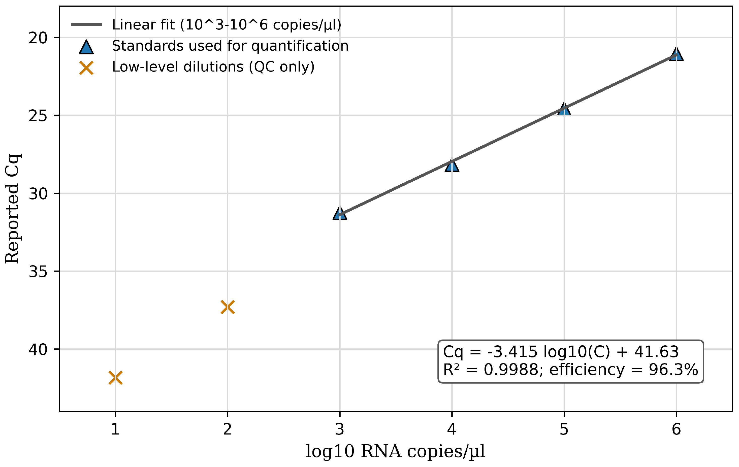
Figure 2.
Detection rate by ante-mortem sample matrix among five CDV-positive dogs. Bars show observed detection proportions.
Figure 2.
Detection rate by ante-mortem sample matrix among five CDV-positive dogs. Bars show observed detection proportions.
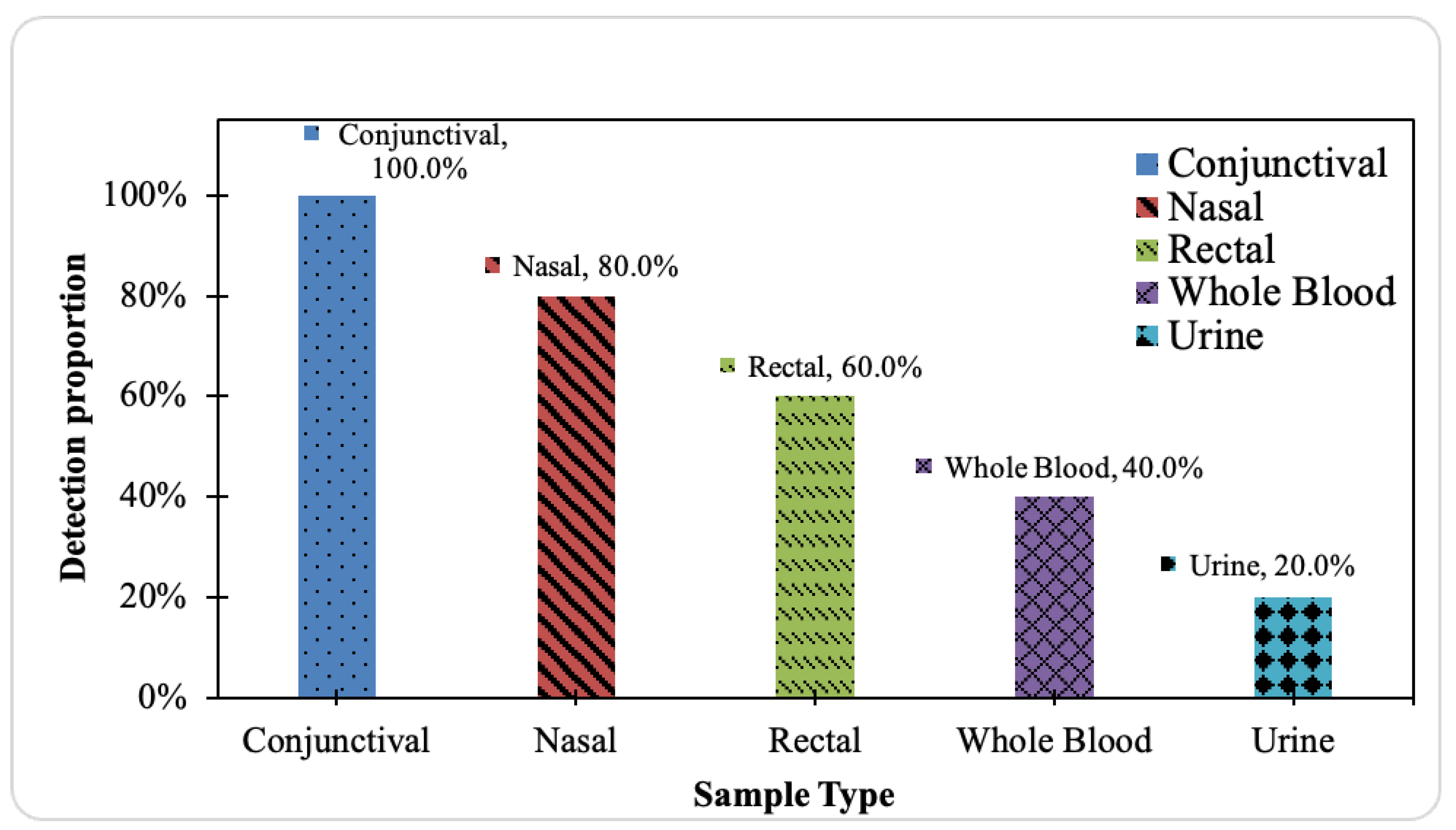
Figure 3.
Integrated sampling-matrix performance among CDV-positive dogs. The x-axis shows detection rate, and the y-axis shows median log10 RNA copies/L among detected samples. Dashed lines mark 50% detection and log10 viral load of 2.
Figure 3.
Integrated sampling-matrix performance among CDV-positive dogs. The x-axis shows detection rate, and the y-axis shows median log10 RNA copies/L among detected samples. Dashed lines mark 50% detection and log10 viral load of 2.
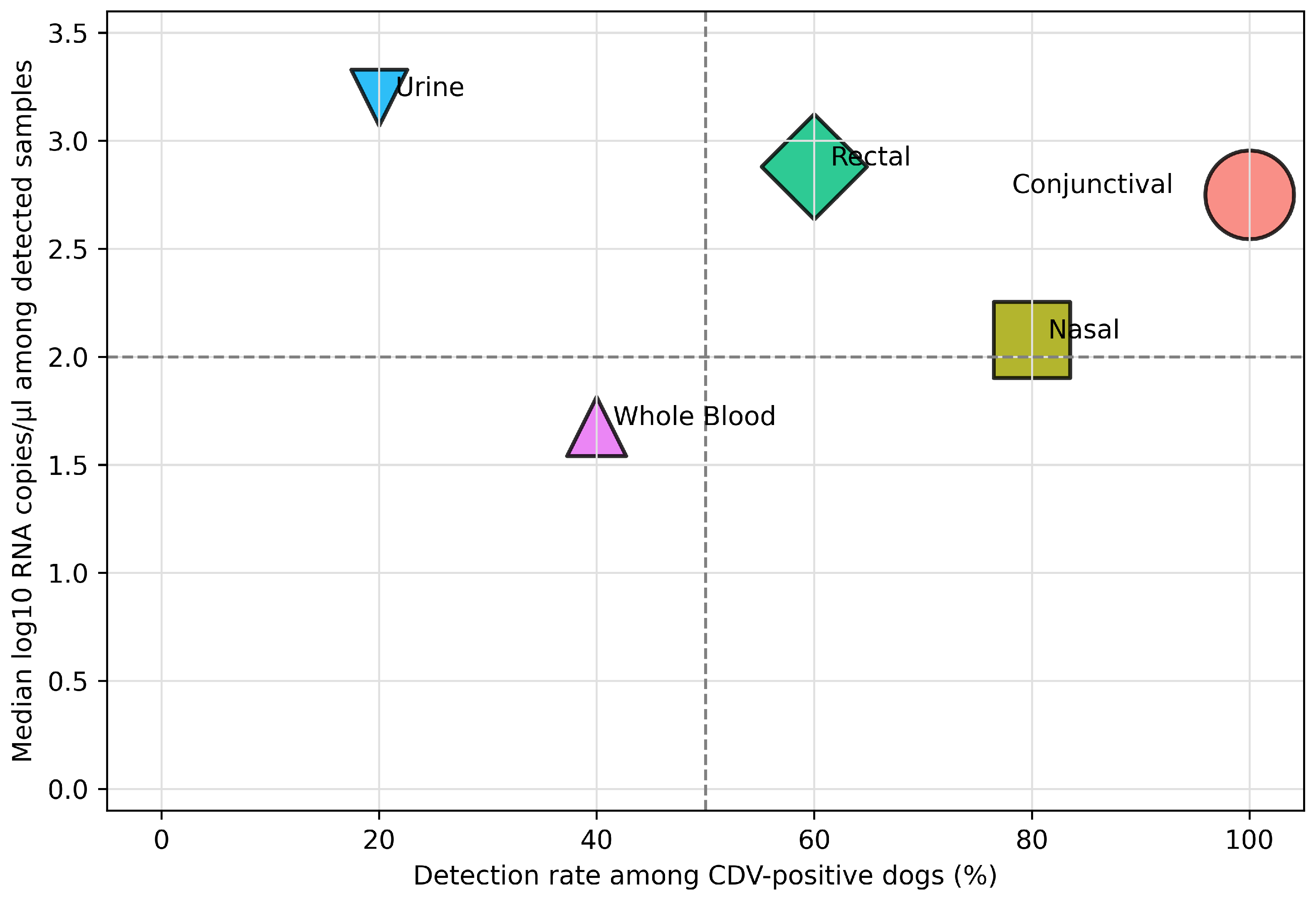

Table 1.
Assay quality-control summary.
| QC measure | Result |
|---|---|
| Standard-curve equation | Cq = -3.415 log10(C) + 41.63 |
| Slope | -3.415 |
| Y-intercept | 41.63 |
| Coefficient of determination | R2 = 0.9988 |
| Estimated amplification efficiency | 96.3% |
| Internal extraction-control pass rate | 25/34 reactions (74%) |
| Quality-control caution | 9/34 reactions lacked acceptable internal-control amplification |
Table 2.
Viral-load matrix for CDV-positive dogs. Non-detects were coded as 0 RNA copies/L for paired statistical analyses.
Table 2.
Viral-load matrix for CDV-positive dogs. Non-detects were coded as 0 RNA copies/L for paired statistical analyses.
| Dog | Conjunctival | Rectal | Nasal | Whole blood | Urine |
|---|---|---|---|---|---|
| Dog 7 | 213,796.21 | 190,546.07 | 0.00 | 0.00 | 0.00 |
| Dog 9 | 213.80 | 758.58 | 87.10 | 9.55 | 0.00 |
| Dog 10 | 44.67 | 54.95 | 25.12 | 213.80 | 0.00 |
| Dog 13 | 131,825.67 | 0.00 | 131,825.67 | 0.00 | 1,584.89 |
| Dog 14 | 562.34 | 0.00 | 162.18 | 0.00 | 0.00 |
Table 3.
Statistical-test summary for paired sample-matrix comparisons among five CDV-positive dogs.
Table 3.
Statistical-test summary for paired sample-matrix comparisons among five CDV-positive dogs.
| Question | Test | Statistic | Interpretation |
|---|---|---|---|
| Do detection rates differ across paired sample types? | Cochran’s Q with exact conditional/permutation p-value | Q = 7.69; df = 4; exact p = 0.1428 | Not statistically significant; observed pattern favors conjunctival swabs but is underpowered. |
| Do viral loads differ across paired sample types? | Friedman test on log10(copies/L + 1) | Q = 7.73; df = 4; p = 0.102 | Not statistically significant; trend favors higher-ranked conjunctival swabs. |
| What is the effect-size estimate for ranked viral-load differences? | Kendall’s W from Friedman statistic | W = 0.39 | Moderate effect-size estimate, not a significant effect claim. |
Disclaimer/Publisher’s Note: The statements, opinions and data contained in all publications are solely those of the individual author(s) and contributor(s) and not of MDPI and/or the editor(s). MDPI and/or the editor(s) disclaim responsibility for any injury to people or property resulting from any ideas, methods, instructions or products referred to in the content. |
© 2026 by the authors. Licensee MDPI, Basel, Switzerland. This article is an open access article distributed under the terms and conditions of the Creative Commons Attribution (CC BY) license (http://creativecommons.org/licenses/by/4.0/).
Copyright: This open access article is published under a Creative Commons CC BY 4.0 license, which permit the free download, distribution, and reuse, provided that the author and preprint are cited in any reuse.


