
Submitted:
29 July 2026
Posted:
30 July 2026
You are already at the latest version
Abstract
Background: Reliable molecular biomarkers that complement cytomorphological assessment for early identification of high-risk oral epithelial lesions remain limited. Although p53 immunocytochemistry (ICC) is widely used as a surrogate marker of TP53 abnormalities, p53 protein expression does not always reflect TP53 transcriptional activity. This study investigated the relationships between p53 ICC, TP53 mRNA expression, and p53 immunohistochemical (IHC) staining patterns in oral high-grade squamous intraepithelial lesions (OHSIL) and oral squamous cell carcinoma (SCC). Methods: A total of 504 liquid-based cytology (LBC) specimens were classified according to the Bethesda System for Reporting Oral Cytology as NILM (n = 394), OLSIL (n = 72), OHSIL (n = 18), and SCC (n = 20). p53 ICC was performed on cytological specimens, TP53 mRNA expression was quantified by quantitative real-time PCR using residual LBC samples, and corresponding biopsy specimens were evaluated for p53 IHC staining patterns. Associations between p53 ICC and clinicopathological variables were also analyzed. Results: The p53 labeling index significantly increased with cytological severity, and TP53 mRNA expression was significantly higher in OHSIL and SCC than in NILM and OLSIL. In OHSIL and SCC, lesions showing a null-type p53 IHC staining pattern showed significantly lower TP53 mRNA expression than lesions with non-null staining patterns. No significant associations between p53 ICC and clinicopathological variables were observed in OHSIL. In contrast, p53 ICC positivity in SCC was significantly associated with the tumor size, depth of invasion, and p53 IHC staining patterns. Conclusions: Negative p53 ICC may result from either sampling limitations or null-type TP53 expression. TP53 mRNA analysis provides complementary molecular information that distinguishes false-negative p53 ICC caused by sampling limitations from biologically reduced TP53 expression. Integrating cytomorphology, p53 ICC, histopathology, and TP53 mRNA analysis may improve diagnostic accuracy and risk stratification of high-risk oral epithelial lesions.
Keywords:
oral liquid-based cytology
; TP53 mRNA
; p53 immunocytochemistry
; oral high-grade squamous intraepithelial lesion
; oral squamous cell carcinoma
1. Introduction
Oral squamous cell carcinoma (SCC) is one of the most common malignancies of the head and neck region and is frequently preceded by oral potentially malignant disorders (OPMDs), including oral epithelial dysplasia (OED). Because the patient’s prognosis is strongly associated with the stage at diagnosis, early detection of high-risk lesions remains a major clinical challenge.
Historically, to improve the standardization and diagnostic accuracy of oral cytology in Japan, the Japanese Society of Clinical Cytology published domestic diagnostic guidelines in 2015 [1]. Building upon these efforts, the diagnostic criteria for oral cytology were subsequently revised to promote more standardized reporting and risk stratification, leading to the introduction of a Bethesda System-based reporting framework in Japan in 2022 [2,3]. This framework established standardized diagnostic categories, including oral low-grade squamous intraepithelial lesion (OLSIL) and oral high-grade squamous intraepithelial lesion (OHSIL), and recommended liquid-based cytology (LBC) for specimen preparation.
Despite these advances, reliable molecular biomarkers that complement cytomorphological assessment and accurately identify high-risk oral epithelial lesions remain limited. In daily practice, distinguishing reactive epithelial atypia from an OHSIL or early SCC based solely on cytomorphological features may be difficult. Consequently, cytopathologists and oral pathologists often encounter challenges in assessing the biological significance and malignant potential of atypical lesions.
Among the molecular biomarkers investigated to date, p53 is one of the most widely used and clinically accepted markers for evaluating of oral epithelial lesions. TP53 is the most frequently altered tumor suppressor gene in head and neck squamous cell carcinoma, with genomic studies demonstrating TP53 alterations in approximately 72% of HNSCCs and in up to 84% of HPV-negative tumors [4]. TP53 alterations represent an early event in oral carcinogenesis and are frequently detected in OPMDs and OSCC. Therefore p53 has been extensively investigated as a biomarker for the early detection and risk assessment of oral epithelial lesions [5,6].
Immunohistochemistry (IHC) and immunocytochemistry (ICC) using anti-p53 antibodies are routinely performed in pathology laboratories and are widely used as surrogate methods for evaluating TP53 abnormalities [7]. However, p53 protein expression does not always reflect TP53 transcriptional activity or mutation-associated biological status. In particular, lesions showing a null immunostaining pattern may retain TP53 mRNA expression despite the absence of detectable p53 protein, whereas other lesions completely lack TP53 mRNA and p53 protein expression. Furthermore, exfoliative oral cytology is inherently subject to sampling limitations because genetically altered cells may remain confined to the basal and parabasal epithelial layers. Consequently, p53 ICC alone may underestimate the molecular abnormalities associated with high-risk oral epithelial lesions.
Recent advances in quantitative real-time PCR (qRT-PCR) have enabled highly sensitive and quantitative assessment of TP53 mRNA expression in clinical specimens [7]. Unlike ICC, which detects protein accumulation, qRT-PCR directly measures TP53 transcriptional activity and may thus provide additional molecular information regarding p53-null lesions. Evaluation of TP53 mRNA expression may help clarify the biological heterogeneity of p53-null lesions and improve the molecular characterization of oral epithelial lesions.
Therefore, this study aimed to investigate the relationships between p53 ICC expression, TP53 mRNA expression, and p53 IHC staining patterns in the categories of OHSIL and SCC diagnosed according to the Bethesda System for Reporting Oral Cytology. By integrating p53 ICC, quantitative TP53 mRNA analysis using qRT-PCR, and histopathological findings, we aimed to clarify the biological characteristics of p53-null lesions and to identify the two principal mechanisms underlying negative p53 ICC results in oral cytology, with particular emphasis on TP53 mRNA expression and sampling limitations.
2. Materials and Methods
2.1. Patients and Study Design
This retrospective study included patients who underwent oral LBC at Niigata University Medical and Dental Hospital between January 2023 and April 2026. A total of 510 oral cytology specimens were collected. Six specimens classified as inadequate were excluded, with 504 satisfactory specimens remaining for analysis.
Cytological diagnoses were classified according to the Bethesda System for Reporting Oral Cytology as negative for an intraepithelial lesion or malignancy (NILM), OLSIL, OHSIL, and SCC. The final cohort comprised 394 NILM, 72 OLSIL, 18 OHSIL, and 20 SCC cases.
A histopathological examination was performed in 154 cases, comprising 44 NILM, 72 OLSIL, 18 OHSIL, and 20 SCC cases. Biopsy diagnoses were used as the reference standard. A TP53 mRNA analysis was performed in all OHSIL and SCC cases (n = 38). The TP53 mRNA analysis was restricted to OHSIL and SCC cases because these high-risk categories represent the primary clinical targets requiring precise molecular characterization and risk stratification. Furthermore, using residual cells from LBC collection vials for all screening-negative specimens (e.g., NILM) was technically and practically challenging because of variations in cell numbers and potential RNA degradation during routine cytological processing.
This study was approved by the Ethics Committee of Niigata University (Approval No. G2022-0027) and was conducted in accordance with the Declaration of Helsinki.
2.2. Histopathological Examination
Biopsy specimens were fixed in 10% neutral-buffered formalin, embedded in paraffin, sectioned at 4 μm, and stained with hematoxylin and eosin. Histological diagnoses were established independently by two experienced oral pathologists according to the WHO Classification of Head and Neck Tumours. Discrepant cases were resolved by consensus.
2.3. p53 ICC
Oral LBC specimens were prepared using the SurePath® liquid-based cytology system (BD Diagnostics, Franklin Lakes, NJ, USA). ICC was performed using a mouse monoclonal anti-p53 antibody (clone DO-7; Dako/Agilent Technologies, Glostrup, Denmark). Antigen retrieval was carried out in EDTA buffer (pH 8.0) using microwave heating, followed by incubation with the primary antibody for 2 h at room temperature. Immunoreactivity was visualized using the EnVision™ detection system (Dako) with 3,3′-diaminobenzidine as the chromogen.
The p53 labeling index (LI) was calculated as the percentage of p53-positive atypical epithelial cells among at least 500 evaluated epithelial cells. Cytological specimens containing distinct nuclear staining in atypical epithelial cells were regarded as p53-positive.
2.4. p53 IHC
IHC was performed on formalin-fixed paraffin-embedded biopsy specimens using the same antibody (clone DO-7) and staining protocol as that used for ICC.
According to the distribution of p53-positive cells, staining patterns were classified into four categories based on the following criteria proposed by Sawada et al. [5].: normal (NM), basal/parabasal (BP), high-expression (HI), and loss (LS). All slides were independently evaluated by two oral pathologists.
2.5. RNA Isolation and qRT- PCR
Total RNA was extracted from the residual cell suspension remaining in the BD SurePath™ LBC collection vial (Becton, Dickinson and Company, Franklin Lakes, NJ, USA) after preparation of cytological slides using the RNAqueous® Kit (Ambion, Grand Island, NY, USA) according to the manufacturer’s instructions. The RNA concentration and purity were assessed using a NanoDrop Lite UV–Vis Spectrophotometer (Thermo Fisher Scientific, Wilmington, DE, USA). After reverse transcription, the concentration of the synthesized cDNA was also determined using the same instrument.
cDNA was synthesized from total RNA using the High-Capacity cDNA Reverse Transcription Kit (Applied Biosystems, Foster City, CA, USA). We performed qRT-PCR using TaqMan™ Universal PCR Master Mix (Applied Biosystems) and TaqMan Gene Expression Assays for TP53 (Assay ID: Hs01034249_m1). We used 18S rRNA (Hs99999901_s1) as the endogenous control for normalization.
Relative TP53 mRNA expression levels were calculated using the 2−ΔΔCt method. TP53 mRNA expression in OLSIL, OHSIL, and SCC samples was normalized to that in NILM specimens, which served as the calibrator. All reactions were performed in triplicate, and the mean values were used for statistical analysis. Relative TP53 mRNA expression was further compared between cases with a null-type p53 IHC staining pattern and those with non-null p53 IHC staining patterns in the OHSIL and SCC groups.
2.6. Statistical Analysis
Continuous variables are presented as the mean ± standard deviation. Comparisons of the p53 LI and TP53 mRNA expression between cytological diagnostic categories were performed using one-way analysis of variance followed by Tukey’s multiple-comparison test.
Associations between p53 ICC findings and clinicopathological variables were analyzed separately in the OHSIL and SCC categories using Fisher’s exact test. The Fisher–Freeman–Halton exact test was applied for variables containing more than two categories.
All statistical analyses were performed using EZR software (Jichi Medical University Saitama Medical Center, version 1.33). All tests were two-sided, and P values < 0.05 were considered statistically significant [8].
3. Results
3.1. Clinicopathological Characteristics
A total of 504 oral LBC specimens were included in this study, comprising 394 that were NILM, 72 OLSIL, 18 OHSIL, and 20 SCC (Table 1).
A histopathological examination was available for all OHSIL and SCC cases. Among the 18 OHSIL cases, histological diagnoses included SCC in 11 (61.1%) cases, severe OED or carcinoma in situ (CIS) in 6 (33.3%) cases, and moderate OED in 1 (5.6%) case. Among the 20 SCC cases, 16 (80.0%) were histologically confirmed as SCC and 4 (20.0%) as severe OED/CIS.
No p53-positive cases were identified in NILM or OLSIL. In contrast, p53 ICC positivity was detected in 2 of 18 (11.1%) OHSIL cases and in 13 of 20 (65.0%) SCC cases (Table 2).
3.2. p53 ICC Expression in Oral Epithelial Lesions
The p53 LI progressively increased according to the cytological severity (Figure 1A). The proportion of p53-positive cases and the mean p53 LI were significantly higher in OHSIL and SCC than in NILM and OLSIL, indicating an association between p53 accumulation and increasing cytological severity. The p53 labeling index was further compared according to the presence or absence of a null-type p53 IHC staining pattern (Figure 1B). Cases showing a null-type p53 IHC staining pattern showed significantly lower p53 labeling indices than cases with a non-null p53 IHC staining pattern in OHSIL and SCC. These findings indicate that lesions with a null-type p53 IHC pattern generally show markedly reduced or absent p53 protein expression in cytological specimens.
3.3. TP53 mRNA Expression According to the Presence or Absence of a Null-Type p53 IHC Staining Pattern
Relative TP53 mRNA expression progressively increased with cytological severity and was significantly higher in OHSIL and SCC than in NILM and OLSIL (Figure 2A). TP53 mRNA expression was further compared between lesions with a null-type p53 IHC staining pattern and those with non-null p53 IHC staining patterns separately in the OHSIL and SCC groups (Figure 2B).
In both OHSIL and SCC, lesions with a null-type p53 IHC staining pattern showed significantly lower TP53 mRNA expression than lesions with a non-null p53 IHC staining pattern. These findings suggest that the null-type p53 IHC staining pattern is associated with reduced TP53 transcription.
3.4. Representative p53 IHC Staining Patterns
Representative cytological, histological, ICC and IHC findings of the four p53 staining patterns are shown in Figure 3. The biological interpretation of these findings is summarized in Figure 4, which illustrates the two principal mechanisms responsible for negative p53 ICC in oral cytology. Negative p53 ICC may result either from sampling limitation, in which atypical cells are not recovered, or from biologically reduced p53 protein expression associated with the null-type staining pattern. The NM and BP patterns showed p53-positive cells confined to the basal or parabasal layers, whereas the HI pattern showed diffuse staining extending into the suprabasal layers. In contrast, the LS (also referred to as null-type) pattern demonstrated complete absence of p53 immunoreactivity despite preservation of epithelial architecture.
3.5. Correlation Between p53 ICC Expression and Clinicopathological Factors
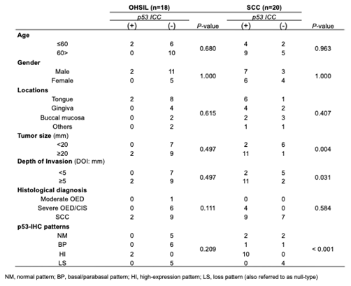
The associations between p53 ICC positivity and clinicopathological variables are shown in Table 2. In patients with an OHSIL (n = 18), no clinicopathological variables showed significant associations with p53 ICC positivity. Specifically, there was no significant association between tumor size and p53 ICC positivity (P = 0.497), and similar results were observed for the depth of invasion (P = 0.497).
In contrast, in patients with SCC (n = 20), p53 ICC positivity was significantly associated with the tumor size (P = 0.004), greater depth of invasion (DOI) (P = 0.031), and p53 IHC staining patterns (P < 0.001). Specifically, the p53 ICC positivity rate was significantly higher in tumors measuring ≥20 mm (91.7%, 11/12 cases) than in those measuring <20 mm (25.0%, 2/8 cases). Similarly, SCCs with a DOI ≥5 mm showed a significantly higher p53 ICC positivity rate (80.0%, 12/15 cases) than those with a DOI <5 mm (20.0%, 1/5 cases). Regarding the p53 IHC staining patterns, all SCCs showing the HI pattern (10/10) were positive for p53 ICC, whereas all SCCs exhibiting the LS pattern (4/4) were negative for p53 ICC. In contrast, both p53 ICC-positive and p53 ICC-negative cases were observed among SCCs with the NM and BP staining patterns.
4. Discussion
The principal finding of this study is that negative p53 ICC arises from two biologically distinct mechanisms: sampling limitation and null-type p53 expression. By integrating p53 ICC, TP53 mRNA expression, and p53 IHC staining patterns, we found that lesions with a null-type p53 IHC staining pattern showed significantly lower TP53 mRNA expression than lesions with non-null p53 IHC staining pattern. These findings indicate that the null-type p53 IHC pattern is associated with reduced TP53 transcription, supporting the concept that the loss-type p53 IHC staining pattern represents a distinct molecular subgroup characterized by reduced TP53 expression. Furthermore, these findings support the complementary use of TP53 mRNA analysis for interpreting negative p53 ICC results in high-risk oral epithelial lesions.
The early detection of malignant transformation in OPMDs remains one of the greatest challenges in oral pathology. Although the introduction of the Japanese Society of Clinical Cytology guidelines and the Bethesda System for Reporting Oral Cytology has markedly improved the standardization and diagnostic accuracy of oral cytology [1,2,3], cytomorphological assessment alone remains insufficient for accurately identifying lesions with malignant potential. Previous studies have shown that the incorporation of molecular biomarkers into LBC improves the diagnostic performance and facilitates the detection of early oral SCC [2,3,7]. Our findings further emphasize that molecular assessment is particularly useful when p53 ICC shows negative results despite morphologically suspicious lesions.
TP53 is the most frequently altered tumor suppressor gene in head and neck squamous cell carcinoma and represents one of the earliest genetic events during oral carcinogenesis [4,6]. Consequently, p53 IHC and ICC have been widely used as surrogate markers of TP53 abnormalities [5,9]. However, these techniques evaluate only protein accumulation and do not necessarily reflect TP53 transcriptional activity.
Lesions showing a null-type p53 IHC staining pattern showed significantly lower TP53 mRNA expression than lesions with a non-null p53 IHC staining pattern, indicating that the loss-type p53 IHC pattern represents a distinct molecular subgroup characterized by reduced TP53 expression. The p53 LI progressively increased from NILM and OLSIL to OHSIL and SCC, consistent with previous reports [2,5,7,10]. This progressive increase supports the concept that TP53-abnormal epithelial cells gradually expand from the basal and parabasal layers toward the epithelial surface during oral carcinogenesis [4,6]. As the proportion of abnormal cells increases, exfoliative cytology is more likely to capture these cells, explaining the substantially higher p53 positivity observed in SCC than in OHSIL.
As summarized in Figure 4, our findings indicate that negative p53 ICC should not be interpreted as representing a single mechanism but rather two biologically distinct mechanisms. The first mechanism is sampling limitation, in which atypical epithelial cells remain confined to the basal or parabasal layers and are therefore not recovered by exfoliative cytology. This phenomenon is particularly relevant in OHSIL, where genetically altered cells frequently occupy only the lower epithelial layers. The second mechanism is null-type p53 expression, in which atypical cells are successfully collected but exhibit complete absence or marked reduction of p53 protein expression, resulting in negative p53 immunostaining despite adequate sampling. Distinguishing these two mechanisms is clinically important because both produce negative p53 ICC findings but reflect fundamentally different biological processes. Accordingly, negative p53 ICC should always be interpreted in conjunction with cytomorphological findings, histopathological evaluation, and TP53 mRNA analysis whenever available.
Another important finding of this study was the concordant reduction in TP53 mRNA expression and p53 labeling indices in lesions showing a null-type p53 IHC staining pattern. Nevertheless, p53 ICC should be interpreted cautiously because the sensitivity of exfoliative cytology depends on adequate recovery of TP53-abnormal cells. Particularly in OHSIL, TP53-abnormal cells are frequently confined to the basal and parabasal epithelial layers, limiting their exfoliation into cytological specimens and increasing the likelihood of false-negative p53 ICC. Therefore, negative p53 ICC should not be interpreted as evidence that TP53-abnormal cells are absent.
Sawada et al. demonstrated that p53 IHC staining patterns accurately predict the TP53 mutation status in OED and proposed the NM, BP, HI, and LS patterns as useful pathological indicators [5]. Our findings further extend these observations by showing that the LS pattern is accompanied by reduced TP53 mRNA expression. In the present study, p53 ICC positivity was significantly associated with p53 IHC staining pattern. While all HI-pattern SCCs were p53 ICC-positive and all LS-pattern SCCs were p53 ICC-negative, both positive and negative ICC findings occurred in the NM and BP patterns. These findings suggest that p53 ICC generally reflects the biological characteristics of p53 IHC staining patterns but may still be influenced by sampling variability, particularly in lesions with limited numbers of TP53-abnormal cells. Integrating p53 IHC with TP53 mRNA analysis therefore provides complementary molecular information for the evaluation of high-risk oral epithelial lesions.
The contrasting findings between OHSIL and SCC are consistent with the biological progression of oral carcinogenesis. In OHSIL, TP53-abnormal cells are frequently confined to the basal and parabasal epithelial layers, limiting their recovery by exfoliative cytology and reducing the sensitivity of p53 ICC. Consequently, p53 ICC findings should be interpreted with caution because the diagnostic performance of exfoliative cytology depends on adequate recovery of TP53-abnormal cells. As lesions progress to SCC, expansion of TP53-abnormal cell populations increases the likelihood of exfoliation into cytological specimens, thereby improving the sensitivity of p53 ICC. This biological progression is consistent with the significant associations observed between p53 ICC positivity and a larger tumor size, greater DOI, and p53 IHC staining patterns in SCC. These findings suggest that false-negative p53 ICC results are more likely to occur in early lesions, particularly OHSIL, than in advanced invasive SCC.
The present findings have direct implications for cytopathological practice. A negative p53 ICC result should not preclude biopsy when cytological atypia is present but should be interpreted together with cytomorphological and, where available, histopathological findings. In selected high-risk cases, TP53 mRNA analysis provides complementary molecular information that distinguishes true biological loss of TP53 expression from false-negative p53 ICC caused by sampling limitations. The presence of both p53 ICC-positive and p53 ICC-negative cases within the NM and BP p53 IHC staining patterns further indicates that p53 ICC results should always be interpreted together with histopathological findings rather than in isolation. Such an integrated diagnostic approach may reduce false-negative interpretation and improve risk stratification of OHSIL and early SCC [10]. Accordingly, integrating cytomorphology, p53 ICC, histopathology, and TP53 mRNA analysis may improve the evaluation of high-risk oral epithelial lesions beyond p53 ICC alone.
There are several limitations to this study. First, the number of OHSIL and SCC cases was relatively small because the TP53 mRNA analysis was performed only in representative lesions. Second, only TP53 mRNA expression was investigated, whereas other molecular alterations associated with oral carcinogenesis were not evaluated. Third, the quality and quantity of the RNA extracted from residual LBC specimens can be affected by the duration of specimen storage and the specific fixation protocols used in routine laboratory workflows. Although we carefully verified RNA concentration and purity using a spectrophotometer, the inherent limitations of RNA preservation in LBC fixatives may affect the amplifiability of long transcripts or lower-abundance mRNAs in some samples. To mitigate these concerns and ensure technical reliability, relative TP53 mRNA expression was strictly normalized using 18S rRNA as an endogenous control, and all qRT-PCR reactions were performed in triplicate.
5. Conclusion
In conclusion, negative p53 immunocytochemistry arises from two distinct mechanisms: sampling limitation and null-type p53 expression. Therefore, negative p53 ICC should not be interpreted as evidence of the absence of a high-risk oral epithelial lesion. Integrating cytomorphology, p53 ICC, histopathology, and TP53 mRNA analysis may improve diagnostic accuracy and reduce false-negative interpretation. These findings provide a practical framework for interpreting negative p53 ICC in the cytological evaluation of high-risk oral epithelial lesions.
Availability of Data and Materials
The datasets used and/or analyzed during the current study are available from the corresponding author on reasonable request.
Patient Consent for Publication
Not applicable.
Author Contributions
J-I.T and N. M. contributed equally to the study’s conceptualization, methodology, and manuscript writing S.O. and K. T. contributed equally to visualization and investigation. M. Y. and T.A. contributed to software, validation, and formal analysis. All authors have read and agreed to the published version of the manuscript.
Funding
This work was supported by JSPS KAKENHI (grant number 23K09150).
Conflicts of Interest
The authors declare that they have no conflicts of interest.
Acknowledgments
We thank Ellen Knapp, PhD, from Edanz (https://jp.edanz.com/ac) for editing a draft of this manuscript.
References
- Tanaka, Y. (Ed.) JSCC Atlas and Guidelines for Cytopathological Diagnosis 5: Digestive Organs; Kanehara: Tokyo, Japan, 2015; pp. 18–79. [Google Scholar]
- Kawaharada, M.; Maruyama, S.; Yamazaki, M.; Abe, T.; Chan, N.N.; Funayama, A.; et al. Clinicopathologic factors influencing the screening accuracy of oral cytology: A retrospective cohort study. Oncol. Lett. 2022, 24, 385. [Google Scholar] [CrossRef] [PubMed]
- Suzuki, T.; Isaka, E.; Hiraga, C.; Akiyama, Y.; Okamura, M.; Oomura, Y.; et al. New oral. cytodiagnostic criteria predict change to oral epithelial dysplasia and cancerization. Oral Sci. Int. 2021, 18, 203–208. [Google Scholar] [CrossRef]
- Cancer Genome Atlas Network. Comprehensive genomic characterization of head and neck. squamous cell carcinomas. Nature 2015, 517, 576–582. [Google Scholar] [CrossRef] [PubMed]
- Sawada, K.; Momose, S.; Kawano, R.; Kohda, M.; Irie, T.; Mishima, K.; et al. Immunohistochemical. staining patterns of p53 predict the mutational status of TP53 in oral epithelial dysplasia. Mod. Pathol. 2022, 35, 177–185. [Google Scholar] [CrossRef] [PubMed]
- Hyodo, T.; Kuribayashi, N.; Fukumoto, C.; Komiyama, Y.; Shiraishi, R.; Kamimura, R.; et al. The. mutational spectrum in whole exon of p53 in oral squamous cell carcinoma and its clinical implications. Sci. Rep. 2022, 12, 21695. [Google Scholar] [CrossRef] [PubMed]
- Kawaharada, M.; Yamazaki, M.; Maruyama, S.; Abe, T.; Chan, N.N.; Kitano, T.; et al. Novel. cytological model for the identification of early oral cancer diagnostic markers: The carcinoma sequence model. Oncol. Lett. 2022, 23, 76. [Google Scholar] [CrossRef] [PubMed]
- Kanda, Y. Investigation of the freely available easy-to-use software “EZR” for medical statistics. Bone Marrow Transplant. 2013, 48, 452–458. [Google Scholar] [CrossRef] [PubMed]
- Köbel, M.; Piskorz, A.M.; Lee, S.; et al. Optimized p53 immunohistochemistry is an accurate. urrogate of TP53 mutation status in ovarian carcinoma. J. Pathol. Clin. Res. 2016, 2, 14–22. [Google Scholar] [CrossRef] [PubMed]
- Akimori, T.; Yamazaki, M.; Abe, T.; Maruyama, S.; Tomihara, K.; Maeda, T.; Tanuma, J. Searching. for new early detection markers of oral epithelial dysplasia and oral squamous cell carcinoma using oral liquid-based cytology. J. Oral Maxillofac. Surg. Med. Pathol. 2024, 36, 624–631. [Google Scholar] [CrossRef]
Figure 1.
(A). Relationship between cytological diagnosis and the p53 labeling index. The p53 labeling index (LI) progressively increased with cytological severity. The proportion of p53-positive cases and the mean p53 LI were significantly higher in the categories of oral high-grade squamous intraepithelial lesion (OHSIL) and squamous cell carcinoma (SCC) than in negative for an intraepithelial lesion or malignancy (NILM) and oral low-grade squamous intraepithelial lesion (OLSIL). These findings indicate an association between p53 accumulation and increasing cytological severity. The boxes indicate the interquartile range (IQR), the horizontal line represents the median, whiskers indicate the minimum and maximum values, and × denotes the mean. P values were calculated using one-way analysis of variance followed by Tukeys multiple-comparison test. *P < 0.05, **P < 0.01, ***P < 0.001. (B). p53 LI according to the presence or absence of a null-type p53 IHC staining pattern in OHSIL and SCC. Cases showing a null-type p53 IHC staining pattern [Null (+)] showed significantly lower p53 labeling indices than cases with non-null p53 IHC staining patterns [Null (−)] in OHSIL and SCC. These findings indicate that lesions with a null-type p53 IHC staining pattern are characterized by markedly reduced p53 protein expression in cytological specimens. The boxes indicate the IQR, the horizontal line represents the median, whiskers indicate the minimum and maximum values, and × denotes the mean. P values were calculated using one-way analysis of variance followed by Tukeys multiple-comparison test.
Figure 1.
(A). Relationship between cytological diagnosis and the p53 labeling index. The p53 labeling index (LI) progressively increased with cytological severity. The proportion of p53-positive cases and the mean p53 LI were significantly higher in the categories of oral high-grade squamous intraepithelial lesion (OHSIL) and squamous cell carcinoma (SCC) than in negative for an intraepithelial lesion or malignancy (NILM) and oral low-grade squamous intraepithelial lesion (OLSIL). These findings indicate an association between p53 accumulation and increasing cytological severity. The boxes indicate the interquartile range (IQR), the horizontal line represents the median, whiskers indicate the minimum and maximum values, and × denotes the mean. P values were calculated using one-way analysis of variance followed by Tukeys multiple-comparison test. *P < 0.05, **P < 0.01, ***P < 0.001. (B). p53 LI according to the presence or absence of a null-type p53 IHC staining pattern in OHSIL and SCC. Cases showing a null-type p53 IHC staining pattern [Null (+)] showed significantly lower p53 labeling indices than cases with non-null p53 IHC staining patterns [Null (−)] in OHSIL and SCC. These findings indicate that lesions with a null-type p53 IHC staining pattern are characterized by markedly reduced p53 protein expression in cytological specimens. The boxes indicate the IQR, the horizontal line represents the median, whiskers indicate the minimum and maximum values, and × denotes the mean. P values were calculated using one-way analysis of variance followed by Tukeys multiple-comparison test.
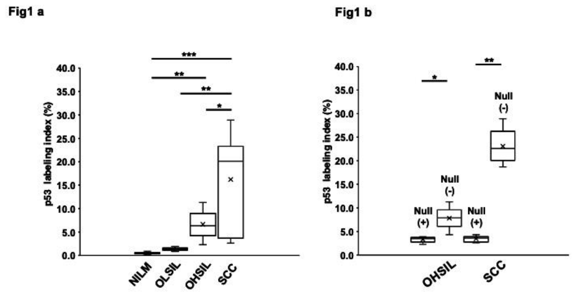
Figure 2.
(A). Relative TP53 mRNA expression according to cytological diagnosis. Relative TP53 mRNA expression was quantified by qRT-PCR in oral LBC specimens classified as negative for NILM, OLSIL, OHSIL, or SCC. TP53 mRNA expression progressively increased with the cytological severity and was significantly higher in OHSIL and SCC than in NILM and OLSIL. The boxes indicate the IQR, the horizontal line represents the median, whiskers indicate the minimum and maximum values, and × denotes the mean. Statistical significance was determined using one-way analysis of variance followed by Tukey’s multiple-comparison test. *P < 0.05, **P < 0.01, ***P < 0.001. (B). Relative TP53 mRNA expression according to the presence or absence of a null-type p53 IHC staining pattern in OHSIL and SCC. Relative TP53 mRNA expression was compared between lesions with a null-type p53 IHC staining pattern [Null (+)] and those with a non-null p53 IHC staining patterns [Null (−)] separately in OHSIL and SCC cases. In OHSIL and SCC, lesions with a null-type p53 IHC staining pattern showed significantly lower TP53 mRNA expression than lesions with a non-null p53 IHC staining pattern. These findings suggest that the null-type p53 IHC staining pattern is associated with reduced TP53 transcription. The boxes indicate the IQR, the horizontal line represents the median, whiskers indicate the minimum and maximum values, and × denotes the mean. *P < 0.05, **P < 0.01.
Figure 2.
(A). Relative TP53 mRNA expression according to cytological diagnosis. Relative TP53 mRNA expression was quantified by qRT-PCR in oral LBC specimens classified as negative for NILM, OLSIL, OHSIL, or SCC. TP53 mRNA expression progressively increased with the cytological severity and was significantly higher in OHSIL and SCC than in NILM and OLSIL. The boxes indicate the IQR, the horizontal line represents the median, whiskers indicate the minimum and maximum values, and × denotes the mean. Statistical significance was determined using one-way analysis of variance followed by Tukey’s multiple-comparison test. *P < 0.05, **P < 0.01, ***P < 0.001. (B). Relative TP53 mRNA expression according to the presence or absence of a null-type p53 IHC staining pattern in OHSIL and SCC. Relative TP53 mRNA expression was compared between lesions with a null-type p53 IHC staining pattern [Null (+)] and those with a non-null p53 IHC staining patterns [Null (−)] separately in OHSIL and SCC cases. In OHSIL and SCC, lesions with a null-type p53 IHC staining pattern showed significantly lower TP53 mRNA expression than lesions with a non-null p53 IHC staining pattern. These findings suggest that the null-type p53 IHC staining pattern is associated with reduced TP53 transcription. The boxes indicate the IQR, the horizontal line represents the median, whiskers indicate the minimum and maximum values, and × denotes the mean. *P < 0.05, **P < 0.01.
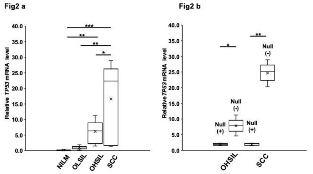
Figure 3.
Representative cytological, histological, and IHC findings of the four p53 staining patterns in OHSIL and SCC. Representative Papanicolaou-stained liquid-based cytology, p53 ICC, hematoxylin and eosin, and p53 IHC images illustrating the four p53 staining patterns. (a–d) Normal pattern: scattered p53-positive cells restricted to the basal or parabasal layers. (e–h) Basal/parabasal pattern: diffuse p53-positive cells involving the basal and parabasal layers. (i–l) High-expression pattern: diffuse p53-positive cells extending into the suprabasal layers. (m–p) Loss pattern (also referred to as null-type): complete absence of p53 immunoreactivity despite preserved epithelial morphology.
Figure 3.
Representative cytological, histological, and IHC findings of the four p53 staining patterns in OHSIL and SCC. Representative Papanicolaou-stained liquid-based cytology, p53 ICC, hematoxylin and eosin, and p53 IHC images illustrating the four p53 staining patterns. (a–d) Normal pattern: scattered p53-positive cells restricted to the basal or parabasal layers. (e–h) Basal/parabasal pattern: diffuse p53-positive cells involving the basal and parabasal layers. (i–l) High-expression pattern: diffuse p53-positive cells extending into the suprabasal layers. (m–p) Loss pattern (also referred to as null-type): complete absence of p53 immunoreactivity despite preserved epithelial morphology.
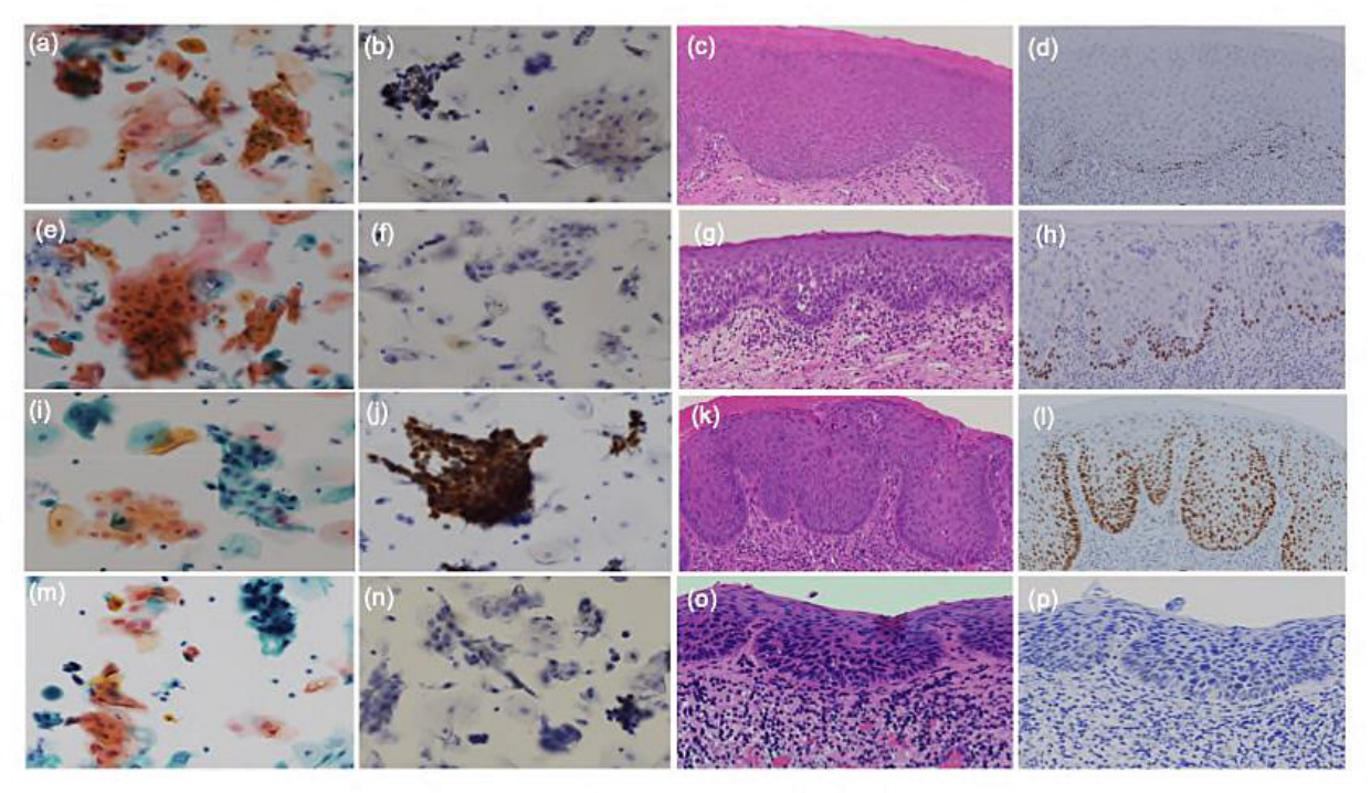
Figure 4.
Two principal mechanisms underlying negative p53 ICC in oral cytology. Negative p53 ICC in oral cytology may result from two biologically distinct mechanisms. (Left) Sampling limitation: atypical epithelial cells remain confined to the basal/parabasal layers and are not recovered by exfoliative cytology. Consequently, only superficial epithelial cells are collected, resulting in false-negative p53 ICC despite the presence of high-risk epithelial lesions. (Right) Null-type p53 expression: atypical epithelial cells are successfully collected; however, they exhibit a null-type p53 staining pattern because of complete absence or marked reduction of p53 protein expression, resulting in negative p53 ICC despite adequate cellular sampling. Together, these mechanisms provide a conceptual framework for interpreting negative p53 immunocytochemistry in oral cytology.
Figure 4.
Two principal mechanisms underlying negative p53 ICC in oral cytology. Negative p53 ICC in oral cytology may result from two biologically distinct mechanisms. (Left) Sampling limitation: atypical epithelial cells remain confined to the basal/parabasal layers and are not recovered by exfoliative cytology. Consequently, only superficial epithelial cells are collected, resulting in false-negative p53 ICC despite the presence of high-risk epithelial lesions. (Right) Null-type p53 expression: atypical epithelial cells are successfully collected; however, they exhibit a null-type p53 staining pattern because of complete absence or marked reduction of p53 protein expression, resulting in negative p53 ICC despite adequate cellular sampling. Together, these mechanisms provide a conceptual framework for interpreting negative p53 immunocytochemistry in oral cytology.
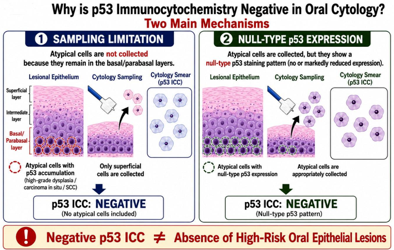

Table 1.
Clinicopathological characteristics and histopathological findings according to oral cytological classification.
Table 1.
Clinicopathological characteristics and histopathological findings according to oral cytological classification.
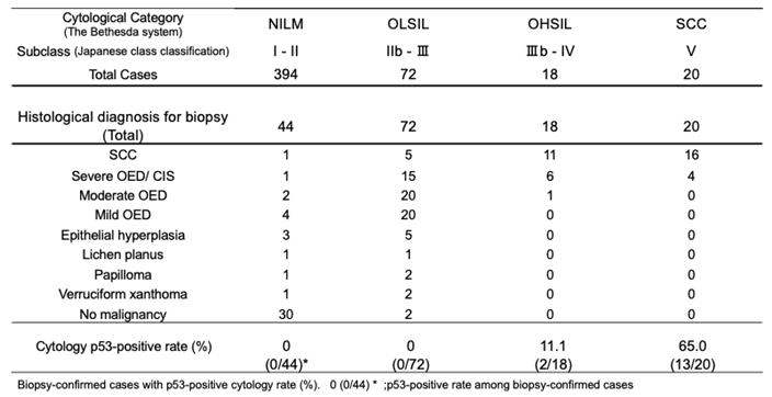 |
Table 2.
Association between p53 ICC expression and clinicopathological characteristics in OHSIL and SCC.
Table 2.
Association between p53 ICC expression and clinicopathological characteristics in OHSIL and SCC.
 |
Disclaimer/Publisher’s Note: The statements, opinions and data contained in all publications are solely those of the individual author(s) and contributor(s) and not of MDPI and/or the editor(s). MDPI and/or the editor(s) disclaim responsibility for any injury to people or property resulting from any ideas, methods, instructions or products referred to in the content. |
© 2026 by the authors. Licensee MDPI, Basel, Switzerland. This article is an open access article distributed under the terms and conditions of the Creative Commons Attribution (CC BY) license (http://creativecommons.org/licenses/by/4.0/).
Copyright: This open access article is published under a Creative Commons CC BY 4.0 license, which permit the free download, distribution, and reuse, provided that the author and preprint are cited in any reuse.


