

Submitted:
25 July 2026
Posted:
30 July 2026
You are already at the latest version
Abstract
Cancer remains a major global health concern, driving the search for safe and effective bioactive compounds from natural sources. Jujube peel red pigment (JP), an anthocyanin-rich extract, has shown preliminary bioactivity, yet its antitumor potential and delivery challenges remain underexplored. This study systematically evaluated the in vitro antitumor activity of JP and developed a thermosensitive hydrogel-based local delivery system (JP-H) to overcome its rapid diffusion and poor retention. JP exhibited selective cytotoxicity against HeLa cervical cancer and B16 melanoma cells, with no obvious toxicity to normal L929 and RAW264.7 cells. Mechanistically, JP induced mitochondrial-dependent apoptosis via upregulating Bax and cleaved Caspase-9/-3, while downregulating Bcl-2, and concurrently triggered G1/S phase arrest through modulation of CCND1, CDK2, CDK4, PCNA, MYC, and TP53. To enable localized delivery, JP was incorporated into an injectable chitosan/gelatin/F127 thermosensitive hydrogel (JP-H), which exhibited rapid sol-gel transition at physiological temperature, shear-thinning behavior, and a porous microstructure. JP-H not only sustained JP release but also significantly enhanced antibacterial activity against E. coli and S. aureus compared to free JP. Furthermore, JP-H markedly inhibited HeLa cell migration and induced superior apoptotic/necrotic cell death in co-culture assays, outperforming free JP. Collectively, this work establishes JP as a multi-target antitumor agent and demonstrates JP-H as a promising local therapeutic platform combining sustained delivery, antibacterial protection, and enhanced anticancer efficacy for cervical cancer treatment.
Keywords:
jujube peel pigment
; thermosensitive hydrogel
; HeLa cells
; mitochondrial apoptosis
; antibacterial activity
; F127
1. Introduction
Cancer remains a major global health challenge. China, in particular, faces a disproportionate cancer burden relative to its population size, alongside a significant shift in the cancer spectrum [1]. Cervical cancer, ranking fourth in incidence among female malignant tumors worldwide and with an increasing incidence among younger individuals, presents a particularly critical challenge [2,3]. The development and progression of cervical cancer are associated with unchecked cell proliferation due to impaired regulation of the cervical cells cycle [4,5]. The transition of cells from the G1 to S phase represents a critical checkpoint in the cell cycle, primarily driven by the Cyclin D1/CDK4/6 complex and regulated by molecules such as proliferating cell nuclear antigen (PCNA), MYC proto-oncogene (MYC), and tumor protein p53 (TP53) [6,7,8,9,10,11]. Dysregulation of this pathway drives tumor proliferation and thus constitutes a key therapeutic target. While conventional antitumor agents have demonstrated efficacy, toxicity, drug resistance, and poor local therapeutic control continue to steer research efforts toward safer naturally derived compounds and more efficient delivery systems [12].
Natural products are a source of structurally diverse molecules with potential antitumor, antioxidant, and anti-inflammatory activities. The traditional Chinese food and medicinal plant jujube (Ziziphus jujuba Mill.) is rich in vitamins and minerals, and has a long history of use as a bioactive plant with diverse characteristics [13]. Jujube extracts have been demonstrated to inhibit proliferation and induce apoptosis in cervical cancer, liver cancer, and other cancer types [14,15]. Jujube peel red pigment (JP), a water-soluble anthocyanin component, exhibits antioxidant activity alongside good stability [16,17], and preliminary studies have demonstrated its potential to inhibit leukemia cell proliferation and suppress tumor cell migration [18]. However, the antitumor activity of JP extract against cervical cancer cells requires systematic evaluation to determine whether it modulates key cell cycle regulators to mediate its anti-cervical cancer activity.
In an attempt to address this, this study first examined the effects of JP on normal cell lines (L929 and RAW264.7) and cancer cell lines (HeLa and B16). Subsequent work focused on the inhibitory effect of JP against HeLa cells and its molecular mechanisms, using migration assays, apoptosis staining, Western blotting, and RT-PCR. However, free natural pigments can diffuse rapidly from the administration site and may not maintain an effective local concentration for long periods. The study therefore subsequently addresses the critical question of translating the biological activity of the free pigment into a practical strategy for localized cervical cancer treatment. Thermosensitive hydrogels that remain flowable before administration, form a gel in situ, improve retention and provide sustained exposure, have low systemic diffusion, and furnish a logical solution.
Accordingly, a local delivery platform was constructed by loading JP into a chitosan/gelatin/F127 thermosensitive hydrogel. Thus, JP serves as the biological antitumor core, while the hydrogel acts as the engineering platform to enhance its local applicability. The preparation route of JP gel and the two core antitumor signaling pathways activated by JP and JP gel have been illustrated in Figure 1. The results confirmed that JP effectively inhibits HeLa cell proliferation and migration in a concentration- and time-dependent manner. The underlying mechanism essentially involves activation of the mitochondrial apoptosis pathway and G1/S phase cell cycle arrest. These findings from this study provide important experimental evidence for the development of JP as a natural product-derived drug candidate against cervical cancer and the design of a hydrogel-based local delivery system.
2. Results and Discussion
2.1. Preparation Process and Component Analysis of JP
The binding between anthocyanins and plant tissues can be effectively disrupted by exposure to an acidified ethanol system, where thermal extraction was carried out at 80 °C to facilitate pigment dissolution (Figure 2a). JP exhibited its maximum absorption peak at 300 nm, with a strong and broad ultraviolet absorption band in the range of 200–350 nm, which is typical of anthocyanin compounds. Due to its conjugated molecular framework, JP exhibits pronounced ultraviolet light absorption, a characteristic that may underlie its antioxidant and photoprotective functions.
A distinct peak at a retention time of approximately 5.140 min was observed during HPLC analysis (Figure 2c), confirming that the main component of the JP sample is the anthocyanin delphinidin-3-O-rutinoside. The remaining low-intensity miscellaneous peaks at 3.943 min and 4.474 min indicated the presence of small amounts of polar impurities. The mutually validated results of UV–vis spectroscopy and HPLC demonstrated that delphinidin-3-O-rutinoside is the main constituent of JB, which also confers its characteristic red color and antioxidant activity [19].
2.2. In Vitro Cytotoxicity and Anti-Migration Activity of JP
JP showed distinct effects on two normal cell lines (Figure 3a). The viability of L929 cells increased slightly with rising JP concentration, whereas that of RAW264.7 cells decreased first and then increased, suggesting an adaptive or defensive cellular response to JP components. JP exhibited no obvious cytotoxicity within a certain dose range, possibly due to the natural origin of anthocyanins, which minimally impact normal cells [20]. In cancer cells, JP inhibited HeLa and B16 proliferation in a concentration-dependent manner, possibly due to oxidative stress and MAPK/PI3K pathways. The effects were slightly stronger on B16 cells than on HeLa. Moreover, JP inhibited HeLa cells in a concentration- and time-dependent manner (Figure 3b), consistent with typical dose–response patterns. High concentrations may induce ROS and mitochondrial damage, while cell cycle arrest or apoptosis are the predominant effects resulting from exposure to low doses [21]. However, these proposed mechanisms require further experimental validation.
Scratch wound healing and Transwell assays were performed to investigate the effect of JP on the migration ability of HeLa cells. In the scratch assay (Figure 3c), JP inhibited migration and wound closure in a concentration-dependent manner at 24 h, with marked suppression at 1.0 mg/mL and virtually no migration at 2.0 mg/mL. At 48 h, the control wounds exhibited nearly complete healing, whereas in the groups treated with 1.0 and 2.0 mg/mL of JB, the scratch remained largely unclosed, confirming sustained inhibition. Transwell assay results (Figure 3d) were also comparable: JP reduced the number of migrated cells dose-dependently, with the strongest effect observed at 2.0 mg/mL. The data demonstrate that JP effectively inhibits HeLa cell migration in a dose-dependent manner.
Tumor metastasis is the primary cause of cancer treatment failure [22]. JP significantly inhibited both the horizontal and vertical migration abilities of HeLa cells in a concentration-dependent fashion. Often, the inhibition of cell migration ability is associated with impaired epithelial-mesenchymal transition (EMT) progression or reduced activity of matrix metalloproteinases (MMPs) [23,24]. MYC serves as a crucial regulator of EMT [25]. While this study did not directly examine the expression of EMT markers or MMPs, the significant suppression of MYC by JP suggests that JP inhibits migration by interfering with the EMT process. This provides a direction for subsequent research to further explore its anti-metastatic mechanism.
2.3. JP Induces Apoptosis in HeLa Cells and Its Molecular Mechanism
Besides migration inhibition, tumor cell apoptosis is a key mechanism of action of anticancer drugs. Hoechst 33258 staining and Western blot were carried out to investigate JP-induced apoptosis in HeLa cells (Figure 4). As shown by the staining results (Figure 4a), the cells exhibited typical apoptotic nuclear features after 48 h treatment with 2.0 mg/mL JP, including chromatin condensation, enhanced fluorescence, and nuclear fragmentation. However, the control nuclei remained intact. Western blot (Figure 4b) showed a JP-induced dose-dependent downregulation of anti-apoptotic Bcl-2 and upregulation of pro-apoptotic Bax, resulting in a significant increase in the Bax/Bcl-2 ratio. Cleavage of Caspase-9 and Caspase-3 was observed. This Bcl-2/Bax imbalance serves as the core switch of the mitochondrial apoptotic pathway and induces programmed cell death by promoting mitochondrial outer membrane permeabilization and subsequent activation of the Caspase-9/-3 cascade.
This shift in the Bax/Bcl-2 ratio leads to increased mitochondrial outer membrane permeability, the release of cytochrome c [26]. These findings are consistent with the reported mechanism of apoptosis induction through the regulation of Bcl-2 family proteins by various natural products [27]. The results of this study show that the cleaved forms of Caspase-9 and Caspase-3 are up-regulated by JP and indicate that it activates the caspase cascade through the intrinsic mitochondrial apoptotic pathway. Activation of this pathway may contribute to greater tumor selectivity than that achieved by conventional cytotoxic agents, although this hypothesis requires further experimental validation. However, the fact that JP may also regulate other apoptosis-related pathways such as endoplasmic reticulum stress- or death receptor-mediated apoptosis cannot be ruled out and requires further mechanistic investigation.
2.4. JP Regulates the Expression of Cell Cycle-Related Genes
In an attempt to probe the molecular mechanism of JP-induced HeLa cell growth inhibition, the expression of cell cycle-related genes was examined (Figure 5a,b). The pro-proliferative genes CCND1, PCNA [28,29], and MYC were downregulated following JP treatment (except for MYC upregulation at 0.1 mg/mL), whereas the tumor suppressor TP53 was upregulated at low concentration (0.1 mg/mL). However, the effect diminished at higher doses. These transcriptional changes collectively favor cell cycle arrest.
To further assess cell cycle execution, the expression of CDKs was analyzed (Figure 5c). Across most concentrations, JP consistently suppressed CDK2 and CDK4, with only slight attenuation observed at 0.3 mg/mL. CDK6, on the contrary, exhibited a non-monotonic response with downregulation at 0.1 mg/mL followed by upregulation at 0.3 mg/mL, and subsequent re-inhibition at 0.4 mg/mL, suggesting a complex, subtype-selective regulation effect of JB on CDKs which may in turn be attributed to possible feedback or compensatory mechanisms among CDK family members.
A key characteristic of malignant tumor proliferation is uncontrolled cell cycle progression [4]. Following the establishment of JP’s pro-apoptotic effect, the study further investigated its cell cycle arrest activity. JP significantly downregulated the expression of CCND1, MYC, and the proliferation marker PCNA. The expression of the tumor suppressor gene TP53 was, however, upregulated. CCND1 complexation with CDK4/6 is the key factor driving cells through the G1 checkpoint; therefore, its downregulation can result directly in G1 phase arrest. The inhibition of MYC, which functions as a global transcriptional regulator, further weakens the cell proliferation signaling network [30]. However, JP exhibited differential regulation of CDK family members, inhibiting CDK2 and CDK4, while showing a concentration-dependent bidirectional regulation of CDK6.
This differential sensitivity observed within the CDK family may originate from a complex regulatory pathway controlling progression through the cell cycle, with individual members of the CDK family having specific functional roles, compensatory pathways, and feedback from other pathway components. Further work is needed to delineate the molecular mechanisms behind this selective regulation. In summary, the transcriptional changes caused by the treatment reflect a coordinated mechanism of cell cycle progression suppression, which is partially consistent with previous reports regarding polyphenols, such as curcumin and quercetin, exerting anti-tumor effects through similar pathways [31,32]. The atypical regulatory pattern exhibited by CDK6 in response to JP indicates that its mechanism of action may involve novel cellular targets.
Compared with natural anti-tumor compounds such as Moscatilin and Celastrol [33,34], JP demonstrates potential for multi-target and multi-pathway synergistic effects. It inhibits tumor growth by regulating classical cell cycle checkpoints and apoptotic switches and also suppresses the migratory behavior of cancer cells. This multi-faceted intervention characteristic reduces the risk of drug resistance associated with single-target drugs [35]. However, the exact primary target, including whether it directly interacts with a specific kinase or receptor, remains unclear and is a key issue that needs to be addressed in future research.
2.5. Physicochemical and Antibacterial Performance of JP-H
To address the key problems associated with the delivery of free JP, including rapid diffusion and poor local retention, JP was incorporated into a thermosensitive hydrogel to achieve sustained release and local enrichment through an in situ gelling injectable drug delivery system possessing both antitumor and antibacterial functions. Temperature-dependent vial inversion experiments demonstrated that both the unloaded and JP-loaded chitosan formulations remained in the sol state across the temperature interval of 15–37 °C and rapidly transitioned into a gel at physiological temperature (37 °C). This thermoresponsive behavior provides a practical handling window for minimally invasive administration while facilitating in situ gel formation following injection (Figure 6a). Additionally, the formulation possesses shear-thinning properties and self-healing capacity, which facilitate facile injection and allow the gel network to rapidly reassemble and recover its structural integrity post-injection(Figure 6b). The injectable precursor solution readily conformed to molds with intricate letter-shaped geometries, illustrating its excellent moldability and adaptability to defects with complex and irregular architectures (Figure 6c).
Such abundant porous architecture provides sufficient space for cell adhesion and proliferation, and also creates transport channels for sustained long-term release of loaded JP pigment, laying a structural foundation for the prolonged antibacterial and antitumor effects of JP-H (Figure 6d). FT-IR analysis (Figure 6e) was performed to confirm the successful incorporation of JP into the hydrogel network and to investigate intermolecular interactions between the pigment and the polymer matrix. The spectrum of the blank hydrogel (C) exhibited characteristic absorption bands of the chitosan/gelatin/F127 composite: a broad O–H/N–H stretching vibration at ~3400 cm−1¹, C–H stretching at ~2920 −1m⁻¹, amide I (C=O stretching) at ~164−1 cm⁻¹, and amide II (N–H bending) at ~1−150 cm⁻¹, consistent with the polysaccharide and protein components. The free JP spectrum (A) displayed typical signals for polyphenolic and glycosidic structures, including O–H stretching (−13400 cm⁻¹), aromatic C=C ring vibrations (~1600 a−1d 1520 cm⁻¹), and C–O–C glycosidic linkag−1s (~1070 cm⁻¹). In the JP-H composite spectrum (B), all major characteristic peaks of both the hydrogel matrix and JP were preserved without the appearance of new bands, indicating that the chemical integrity of each component was maintained during formulation. Notably, the O–H stretching band in JP-H exhibited a slight broadening and a minor redshift compared to that of the blank hydrogel, while the aromatic C=C peaks of JP showed subtle shifts from 1−100 to 1606 cm⁻¹. These spectral changes suggest the formation of intermolecular hydrogen bonds between the hydroxyl groups of JP’s polyphenolic rings and the amino/hydroxyl groups of chitosan and gelatin. Importantly, the absence of new covalent bonds (e.g., ester or Schiff-base linkages) confirms that JP was physically encapsulated within the hydrogel matrix rather than chemically conjugated. This stable physical entrapment is favorable for sustained release and ensures that the bioactivity of JP is not compromised by chemical modification.
The antibacterial activity of free JP, the blank hydrogel, and JP-H was evaluated against the Gram-negative bacterium Escherichia coli and the Gram-positive bacterium Staphylococcus aureus using colony-counting, disk-diffusion, and SEM analyses (Figure 7). Abundant colonies were observed in the PBS control group, indicating unrestricted bacterial growth. Free JP moderately reduced bacterial survival, confirming that the pigment extract possesses intrinsic antibacterial activity. The blank hydrogel also decreased bacterial survival to some extent, which may be associated with the membrane-active properties of protonated amino groups in chitosan. Among all treatments, JP-H produced the lowest bacterial survival rates for both strains, with significantly stronger antibacterial effects than free JP and the blank hydrogel.
The disk-diffusion results were generally consistent with the colony-counting data. JP-H generated the most pronounced inhibition zones against both E. coli and S. aureus, whereas free JP produced smaller inhibition zones and the blank hydrogel showed little or limited diffusion-mediated inhibition. The difference between the colony-counting and disk-diffusion results for the blank hydrogel may reflect the contact-dependent antibacterial activity of chitosan and its limited diffusion through the agar matrix. By contrast, the inhibition zones surrounding JP-H indicate that antibacterial constituents of JP were able to diffuse from the hydrogel into the surrounding medium.
SEM further revealed treatment-dependent alterations in bacterial morphology. Bacteria in the PBS group retained intact cellular contours and relatively smooth surfaces. The blank hydrogel and free JP caused varying degrees of surface roughening, wrinkling, and deformation. More extensive structural damage was observed after JP-H treatment. E. coli displayed pronounced shrinkage, collapse, and disruption of the rod-shaped cellular envelope, whereas S. aureus exhibited surface deformation, aggregation, and loss of its regular spherical morphology. These morphological changes are consistent with compromised bacterial envelope integrity.
Collectively, the results demonstrate that incorporation of JP into the thermosensitive chitosan/gelatin/F127 hydrogel produces stronger antibacterial activity than either free JP or the unloaded hydrogel under the tested conditions. This enhancement may result from the combined antibacterial effects of JP-derived polyphenolic components and the chitosan-containing matrix, together with improved local retention and contact between JP and bacterial cells. However, because JP release kinetics and membrane permeability were not directly measured, the enhanced antibacterial activity should not be attributed conclusively to sustained release or a specific membrane-disruption mechanism.
2.6. Analysis of Migration Inhibition and Cytotoxicity of JP-H Against HeLa Cells
The effects of JP-H on tumor cell migration and survival were evaluated using a wound healing assay and live/dead fluorescence staining in HeLa cells. PBS, blank hydrogel, and free JP were also evaluated in comparison. Following 24 h culture, both the PBS and Blank groups showed substantial closure of the scratch gaps (Figure 8a,b), with no significant difference in migration rates between the two groups. The free JP group demonstrated a slightly higher degree of wound closure in comparison to the controls, indicating that a low JP concentration did not inhibit HeLa cell migration, but rather slightly promoted it. In the JP-H group, the scratch width exhibited almost no change in comparison with the 0 h baseline, demonstrating a significant inhibition of cell migration. In the Live/dead cell staining assay (Figure 8c,e), the PBS and Blank groups displayed bright FITC green fluorescence (live cells) with only sparse PI red fluorescence (dead cells). The live cell fluorescence intensity in the free JP group was comparable to the control, with only a few PI-positive cells. The JP-H group, however, exhibited virtually complete quenching of FITC fluorescence, whereas extensive bright PI red fluorescence pointed towards severe necrosis and apoptosis of HeLa cells, which was consistent with the failure of cells to migrate into the scratch area as demonstrated by the scratch assay.
Quantitative flow cytometry analysis via Annexin V-FITC/PI double staining was adopted to precisely quantify the cellular apoptotic ratio (Figure 8f). The total apoptotic population of the Control and blank hydrogel groups remained at a low level, with only slight apoptosis triggered by free JP treatment. In contrast, JP-H treatment markedly elevated the proportion of early and late apoptotic HeLa cells. Collectively, these results illustrated that JP could simultaneously inhibit tumor cell migration and induce cellular apoptosis, and the thermosensitive hydrogel delivery system further amplified these two antitumor functions, highlighting the great potential of JP-H as a local therapeutic platform for cervical cancer treatment.
The FT-IR and release behavior indicate that long-term sustained release of JP within the three-dimensional hydrogel network mediates the anticancer activity of the hydrogel. The diffusion and dilution of free JP rapidly in the culture medium maintains only a low bioactive concentration, with weak anticancer efficacy. JP-H, on the other hand, gels in situ and maintains a persistently high local concentration of JP in the cellular microenvironment. The disruption of cell membrane integrity and redox homeostasis by exposure to high-dose polyphenols induces apoptosis and necrosis, which is manifested as extensive PI-positive signals and complete inhibition of scratch healing. This is also consistent with the concentration-dependent biological response elicited by JP. JP-H performs dual roles as a broad-spectrum antibacterial and anti-cervical cancer agent, offering potential value for minimally invasive local treatment of cervical cancer with concomitant infection. However, this study has several limitations, including the lack of evaluation of biocompatibility with normal epithelial cells, the absence of toxicity assessment of high concentrations of polyphenols toward normal tissues, and the lack of validation in relevant in vivo animal models. Future work may attempt to modulate crosslinking density or reduce JP loading to balance bioactivity and toxicity. Evaluation of the in vivo antitumor efficacy and biosafety of the JP-loaded hydrogel in appropriate animal models is also an important requisite for further preclinical development.
3. Conclusion
This study combines the antitumor potential of a natural-product jujube peel with an injectable thermosensitive hydrogel-based local delivery. JP extracted from jujube peel inhibited HeLa cell viability and migration, induced Bcl-2/Bax regulation and Caspase-9/-3 activation to trigger mitochondrial apoptosis, and altered the expression of key cell-cycle regulators, including CCND1, CDKs, PCNA, MYC, and TP53, interfering with G1/S phase progression. To overcome the limitations in the localized delivery of free JP, a JP-loaded chitosan/gelatin/F17 thermosensitive hydrogel was fabricated. With injectable delivery, temperature-responsive gelation, local retention potential, bactericidal potential against E. coli and S. aureus, and tumoricidal activity against HeLa cells, JP-H functions as a local antitumor delivery platform. The developed JP-loaded thermoresponsive hydrogel constitutes a promising localized therapeutic platform integrating the sustained drug retention of the hydrogel matrix with the antibacterial and antitumor properties of the naturally derived pigment JP.
4. Experimental Section
Extraction of red pigment from jujube peel: Fresh, washed gray jujubes were oven-dried first at 55 °C for 4 h and then at 65 °C, with intermittent venting to release moisture until the internal humidity reached 70%. The process was repeated 5-8 times. The dried jujubes were soaked in cold water for 24 h, boiled for 5 min, and cooled. Jujube Peels were separated, dried at 40 °C for 15-20 h, pulverized, and sieved through a 60-mesh sieve. The powder was extracted thrice with ethanolic hydrochloric acid solution (pH 0.5) at a solid-to-liquid ratio of 1:30 at 80 °C for 2 h each. Dried crude JP was obtained by combining and concentrating the extracts.
Spectroscopic and chromatographic characterization: In order to determine and record the maximum absorption wavelength, the jujube red pigment solution at 0.5 mg/mL was subjected to full-wavelength scanning. High performance liquid chromatography (HPLC) was also employed for analyzing the constituents of jujube peel red pigment under the following chromatographic conditions: injection volume 20 μL, chromatographic column Eclipse XDB-C18 column, detection wavelength 280 nm, column temperature 30 °C, 10% formic acid aqueous solution as mobile phase A, a mixed solution of methanol, acetonitrile and 10% aqueous formic acid as mobile phase B was, and the ratio of A to B as 67:33 (v/v). Standard delphinidin-3-O-rutinoside and the prepared jujube peel red pigment sample were separately injected and analyzed. Each measurement was carried out in triplicate, and the retention time as well as peak areas were recorded.
Cell lines: HeLa, B16, L929, and RAW264.7 cells, purchased from Huatuo Biotechnology (Shanghai, China), were cultured in Procell special media supplemented with 10% fetal bovine serum (FBS). The cells were maintained in a closed incubator at 37 °C in a 5% CO2 atmosphere. Cells were enzymatically dissociated using EDTA–trypsin and subcultured at 2-day intervals.
Cell viability assay: HeLa, B16, L929, and RAW264.7 cells in the logarithmic phase were trypsinized, resuspended, and seeded into 96-well plates, in a medium containing 5% serum, at a density of 9×10³ cells/well (100 μL/well). When cell confluence exceeded 80%, following a 24 h incubation at 37 °C with 5% CO2, the medium was discarded. Cells were then treated with JP at concentrations of 0.5, 1, and 2 mg/mL (100 μL/well, 5% serum) for 24 h, with three replicate wells per group. Subsequently, 10 μL MTT (5 mg/mL) was added to individual wells. Following 4 h incubation and subsequent removal of the supernatant, 110 μL DMSO was added to individual wells to dissolve formazan crystals. Absorbance was measured at 490 nm. Given that subsequent antitumor mechanism studies were primarily conducted using HeLa cells as the main model, an additional 48 h treatment group was established alongside the 24 h treatment to evaluate the time-dependent anti-proliferative effect of JP. Each experiment was carried out in triplicate. The inhibition rate was calculated as follows:
Inhibition rate (%) = [1 - (Asample / Acontrol)] × 100%
HeLa cell migration assay: HeLa cells were seeded and cultured in 6-well plates until approximately 90% confluence was achieved. Three parallel horizontal guide lines were drawn on the underside of each well using a black marker. Three vertical scratches were then made perpendicular to the guide lines, using a sterile 200 μL pipette tip, to form a grid pattern, with individual scratches spaced at least 0.5 cm apart. The wells were then gently rinsed with PBS to prevent the attachment of detached cells and debris to the scratched area. Each well then received 2 mL of serum-free medium, either without supplementation or containing JP at concentrations of 0.5, 1.0, or 2.0 mg/mL. The plate was then gently swirled to ensure even distribution and left for incubation. At 0, 24, and 48 hours following scratch introduction, cell migration was observed and photographed at the intersections of the guide lines and scratches, using an inverted microscope.
Logarithmic-phase HeLa cells were treated with JP (0, 0.5, 1.0, 2.0 mg/mL) for 24 h, then trypsinized and resuspended in serum-free RPMI-1640 containing corresponding JP concentrations at a density of 105 cells/mL. A Transwell migration assay was used to assess cell migration. A 200 uL aliquot of cells in serum-free medium was replated in the upper compartment of Transwell inserts, and 500 μL RPMI-1640 medium with 10% FBS in the lower compartment served as a chemoattractant. After allowing the system to incubate for 24 h, the non-migrated cells, which were still on the upper surface of the membrane, were gently removed with a cotton swab. Cells migrated to the lower surface were fixed with methanol for 15 minutes, dried, stained with 0.1% crystal violet for 20 minutes, washed with PBS to remove excess crystal violet, and then visually examined by light microscope for quantitative analysis. Hoechst 33258 staining solution staining for apoptosis: Exponentially growing HeLa cells were seeded in 96-well plates at an appropriate density and allowed to adhere for 24 hours. Cells were subjected to 48 hours of treatment with DMEM medium-based JP solutions (0, 0.5, 1.0, and 2.0 mg/mL) (Solarbio, Beijing, China) containing 10% BSA (Solarbio, Beijing, China). The culture medium was gently removed following treatment, and the cells were fixed with 4% paraformaldehyde for 15 minutes at room temperature. After this duration, the fixative was discarded, and the cells were washed twice with PBS. Subsequently, each well received 30 μL of Hoechst 33258 staining solution, followed by a 10-minute incubation of the plates in the dark. The cells were then washed twice with PBS and observed under a fluorescence microscope.
Western Blot Analysis of Mitochondrial Apoptosis-Related Proteins: HeLa cells in the logarithmic phase were seeded in 10 cm dishes at 2×10⁶ cells/mL and treated with JP (0, 1.0, 2.0 mg/mL in DMEM with 10% BSA) for 48 h. RIPA lysis buffer with PMSF was employed for total protein extraction, and concentration was determined by BCA assay. Equal amounts of protein (30 μg/lane) were separated by 10–12% SDS-PAGE and transferred to PVDF membranes. The membranes were blocked with 5% non-fat milk in TBST for 1 h at room temperature before being incubated overnight at 4 °C with primary antibodies against Bax, Bcl-2, Cleaved-caspase 9, and Cleaved-caspase 3. Membranes were washed in TBST and incubated with HRP-conjugated secondary antibody for 1 hour. Protein bands were visualized by ECL and imaged. The intensities of the bands were quantified in Image J and normalized to ACTB.
Analysis of cell cycle-related gene expression by RT-PCR: Primers corresponding to CCND1, CDK2, CDK4, CDK6, PCNA, MYC, TP53, and ACTB were designed and synthesized by Tsingke Biotechnology Co., Ltd. The details are as follows (Table 1):
HeLa cells were treated with 0, 0.1, 0.3, 0.4, 0.5, and 1.0 mg/mL JP in culture medium and then collected into centrifuge tubes. Individual centrifuge tubes then received 1 mL of TRIzol reagent, followed by repeated pipetting to ensure complete cell lysis. The mixture was placed on ice for 15 minutes, following which 200 µL of chloroform was added to each centrifuge tube. The resulting mixture was vigorously shaken for 15 seconds and incubated on ice for 3 minutes. The sample was then centrifuged at 12,000 rpm and 4 °C for 15 minutes. From the aqueous phase, 400 µL was transferred to a new tube, mixed with 400 µL of isopropanol by gentle inversion, and left to stand at room temperature for 20 minutes. After centrifugation of this mixture at 12,000 rpm and 4 °C for 15 minutes, the supernatant was discarded. The pellet was washed with 1 mL of 75% ethanol, centrifuged again under the same conditions, and the supernatant was removed. Following air-drying at room temperature for 3 minutes, the pellet was resuspended in 25 µL of DEPC-treated sterile water to achieve complete dissolution of RNA.
Five microliters of the extracted RNA was transferred to an EP tube, followed by the addition of 1 µL of oligo (dT) 18 primer and 7 µL of H2O. Thermal denaturation was performed by incubating the reaction mixture at 70 °C for 5 min in a PCR thermal cycler, with the heated lid maintained at 105 °C to prevent condensation. Subsequently, 5 µL of 5× MMLV buffer, 2 µL of dNTP mixture, 0.5 µL of RNase inhibitor, 13 µL of the denatured RNA template, and 4.5 µL of H2O were added to the same tube. The reverse transcription reaction was carried out at 37 °C for 90 minutes, and following the completion of the reaction, the cDNA product was stored at –20 °C for subsequent use (Table 2).
Gel electrophoresis of amplified PCR products was performed on 1.5% agarose gels at 110 V for 25 minutes and was then visualized with a gel imaging system. The amplified fragments of the CCND1, CDK2, CDK4, CDK6, PCNA, MYC, TP53, and ACTB (as an internal control) were analyzed by densitometric analysis using Image J software. The signal intensities of each of the target genes were then normalized to the signal intensity of ACTB to determine relative transcript abundance. Data were analyzed and processed by GraphPad Prism version 6.01.
Preparation of JP-loaded thermosensitive hydrogel: Chitosan, gelatin, and F127 were mixed to form an injectable thermosensitive hydrogel matrix. JP was subsequently dispersed into the precursor solution to obtain JP-H. For clarity, the JP group is also denoted as JP and the JP-loaded hydrogel group as JP-H. Sol-gel behavior was evaluated by vial inversion at different temperatures to ensure the injectability of the formulation at lower temperatures and stable gel formation near 37 °C. The interaction among the pigment and hydrogel matrix components was assessed via FTIR analysis. Visual appearance, moldability, and in situ gelation behavior were documented photographically.
Antibacterial assay of JP-H: The hydrogel system was evaluated for its antibacterial activity against E. coli and S. aureus. PBS, blank hydrogel, and free JP and JP-H were incubated with bacterial suspensions. Antibacterial performance was assessed by colony formation on agar plates, whereas bacterial morphology was observed via scanning electron microscopy, wherein bacterial inhibition or bactericidal action was assessed in terms of reduced colony numbers and altered bacterial morphology.
In vitro antitumor activity of JP-H: HeLa cells were used to evaluate the antitumor activity of the JP-loaded hydrogel. For this purpose, cells were treated with PBS, blank hydrogel, free JP, and JP-H. The cell migratory capacity was determined by a wound-healing assay, while the cell viability was assessed with a live/dead staining using FITC and PI fluorescence channels and a Hoechst fluorescence channel. The therapeutic efficacy of the JP-loaded hydrogel was then compared to the free JP formulation to assess whether the hydrogel was more effective at improving the retention of JP and tumor eradication at the diseased site.
Annexin V-FITC/PI double staining flow cytometry assay: HeLa cells were seeded into 6-well culture plates and incubated overnight to reach adherent growth. The cells were divided into four groups: Control, free JP, blank thermosensitive hydrogel, and JP-H. After 24 h of co-culture with corresponding samples, the culture medium was discarded, and cells were digested with trypsin, collected and washed twice with PBS.
Cell pellets were resuspended in 100 μL of Annexin V-FITC binding buffer. Subsequently, 5 μL Annexin V-FITC and 5 μL PI staining solution were added sequentially, mixed gently, and incubated in the dark at room temperature for 15 min. After incubation, another 400 μL binding buffer was supplemented, and the samples were immediately analyzed by flow cytometry. The fluorescence signals of Annexin V-FITC and PI were recorded to distinguish live cells, early apoptotic cells, late apoptotic cells and necrotic cells, and the proportion of apoptotic cells in each group was quantitatively calculated.
Statistical analysis: All data were expressed as mean ± SD. Data were analyzed statistically by SPSS 11.5. ChemBioDraw Ultra 12.0 was used to draw chemical structures. *P<0.05 was regarded as significant and **P<0.01 indicated a high level of statistical significance.
Author Contributions
P.Z.: Writing-original draft, Funding acquisition, Methodology, Data curation. Q.C.: Writing-original draft, Validation, Methodology. S.C.: Validation, Methodology. H.G.: Validation, Methodology. M.M.: Validation, Methodology. Y.P.: Methodology. Z.J.: Methodology. H.L.: Writing-review & editing, Methodology. P.G.: Methodology. X.Z.: Supervision, Methodology. Y.Z.: Writing-review & editing, Funding acquisition, Supervision, Methodology. X.Y.: Writing-review & editing, Validation, Supervision, Conceptualization.
Funding
This work was supported by the National Natural Science Foundation of China (32101150), Key Scientific Research Project Plan in Colleges and Universities of Henan Province (24B180009), and the Training Program for Young Backbone Teachers in Higher Education Institutions of Henan Province (2025GGJS110).
Informed Consent Statement
Not applicable.
Data Availability Statement
The data presented in this study are available upon request from the corresponding author. The data are not publicly available due to ethical reasons.
Conflicts of Interest
The authors declare that they have no known competing financial interests or personal relationships that could have appeared to influence the work reported in this manuscript.
References
- Filho, A.M.; Laversanne, M.; Ferlay, J.; Colombet, M.; Piñeros, M.; Znaor, A.; Parkin; Soerjomataram, I.; Bray, F. The GLOBOCAN 2022 cancer estimates: Data sources, methods, and a snapshot of the cancer burden worldwide. Int. J. Cancer 2025, 156, 1336–1346. [Google Scholar] [CrossRef] [PubMed]
- Sahasrabuddhe, V.V. Cervical Cancer: Precursors and Prevention. Hematol. Oncol. Clin. N 2024, 38, 771–781. [Google Scholar] [CrossRef] [PubMed]
- Xu, M.; Cao, C.; Wu, P.; Huang, X.; Ma, D. Advances in cervical cancer: current insights and future directions. Cancer Commun. 2025, 45, 77–109. [Google Scholar] [CrossRef] [PubMed]
- Wang, Z. Regulation of Cell Cycle Progression by Growth Factor-Induced Cell Signaling. Cells 2021, 10, 3327. [Google Scholar] [CrossRef] [PubMed]
- Tollis, S. The G1/S repressor WHI5 is expressed at similar levels throughout the cell cycle. BMC Res. Notes 2022, 15, 248. [Google Scholar] [CrossRef] [PubMed]
- Palmer, N.; Kaldis, P. Less-well known functions of cyclin/CDK complexes. Semin Cell Dev. biol. 2020, 107, 54–62. [Google Scholar] [CrossRef] [PubMed]
- Wendel, S.O.; Snow, J.A.; Gu, L.; Banerjee, N.S.; Malkas, L.; Wallace, N.A. The potential of PCNA inhibition as a therapeutic strategy in cervical cancer. J. Med. Virol. 2023, 95, e29244. [Google Scholar] [CrossRef] [PubMed]
- Fatma, H.; Maurya, S.K.; Siddique, H.R. Epigenetic modifications of c-MYC: Role in cancer cell reprogramming, progression and chemoresistance. Semin. Cancer Biol. 2022, 83, 166–176. [Google Scholar] [CrossRef] [PubMed]
- Gao, F.Y.; Li, X.T.; Xu, K.; Wang, R.T.; Guan, X.X. c-MYC mediates the crosstalk between breast cancer cells and tumor microenvironment. Cell commun. signal. 2023, 21 28. [Google Scholar] [CrossRef] [PubMed]
- Marvalim, C.; Datta, A.; Lee, S.C. Role of p53 in breast cancer progression: An insight into p53 targeted therapy. Theranostics 2023, 13, 1421–1442. [Google Scholar] [CrossRef] [PubMed]
- Mao, Y.; Jiang, P. The crisscross between p53 and metabolism in cancer. Acta Bioch Bioph Sin. 2023, 55, 914–922. [Google Scholar] [CrossRef]
- Chen, C.; Chen, L.; Mao, C.; Jin, L.; Wu, S.; Zheng, Y.; Cui, Z.; Li, Z.; Zhang, Y.; Zhu, S.; Jiang, H.; Liu, X. Natural Extracts for Antibacterial Applications. Small 2024, 20, e2306553. [Google Scholar] [CrossRef] [PubMed]
- Lu, Y.; Bao, T.; Mo, J.; Ni, J.; Chen, W. Research advances in bioactive components and health benefits of jujube (Ziziphus jujuba Mill.) fruit. J. Zhejiang Univ. Sci. B 2021, 22, 431–449. [Google Scholar] [CrossRef] [PubMed]
- Gou, B.; Chen, G.; Huang, S.; Ning, N.; Gu, Q.; Duan, S.; Du, Y.; Nan, Y.; Yuan, L. Review: performance of jujube and its extracts in cancer: therapeutic, toxicity-reducing and potentiating effects. Front Oncol. 2025, 15, 1489974. [Google Scholar] [CrossRef] [PubMed]
- Zhuang, H.; Jing, N.; Wang, L.; Jiang, G.; Liu, Z. Jujube Powder Enhances Cyclophosphamide Efficiency against Murine Colon Cancer by Enriching CD8+ T Cells While Inhibiting Eosinophilia. Nutrients 2021, 13, 2700. [Google Scholar] [CrossRef] [PubMed]
- Guo, M.; Zhang, Z.; Li, S.; Lian, Q.; Fu, P.; He, Y.; Qiao, J.; Xu, K.; Liu, L.; Wu, M.; Du, Z.; Li, S.; Wang, J.; Shao, P.; Yu, Q.; Xu, G.; Li, D.; Wang, Y.; Tian, S.; Zhao, J.; Zhao, X. Genomic analyses of diverse wild and cultivated accessions provide insights into the evolutionary history of jujube. Plant Biotechnol. J. 2021, 19, 517–531. [Google Scholar] [CrossRef] [PubMed]
- Duan, Y.; Liu, S.; Zhu, Y.; Wang, Y.; Yan, F.; Liu, Z.; Shi, X.; Liu, P.; Liu, M. The Influences of Soil and Meteorological Factors on the Growth and Fruit Quality of Chinese Jujube (Ziziphus jujuba Mill.). Plants 2023, 12, 4107. [Google Scholar] [CrossRef] [PubMed]
- Dou, J.F.; Wu, C.E.; Fan, G.J. Insights into the pigment and non-pigment phenolic profile of polyphenol extracts of jujube peel and their antioxidant and lipid-lowering activities. Food Biosci. 2023, 52, 102493. [Google Scholar] [CrossRef]
- Tang, K.; Karamat, U.; Li, G.; Guo, J.; Jiang, S.; Fu, M.; Yang, X. Integrated metabolome and transcriptome analyses reveal the role of BoGSTF12 in anthocyanin accumulation in Chinese kale (Brassica oleracea var. alboglabra). BMC Plant Biol. 2024, 24, 335. [Google Scholar] [CrossRef] [PubMed]
- Zhu, H.; Wu, X.; Tan, Y.; Shi, L.; Bai, W.; Li, X. Anthocyanin-functionalized selenocysteine nanotherapeutics alleviate cisplatin nephrotoxicity by inhibiting oxidative stress and ferroptosis. J. Nanobiotechnol 2025, 23, 703. [Google Scholar] [CrossRef] [PubMed]
- Han, L.; Yan, Y.; Fan, M.; Gao, S.; Zhang, L.; Xiong, X.; Li, R.; Xiao, X.; Wang, X.; Ni, L.; Tong, D.; Huang, C.; Cao, Y.; Yang, J. Pt3R5G inhibits colon cancer cell proliferation through inducing ferroptosis by down-regulating SLC7A11. Life Sci. 2022, 306, 120859. [Google Scholar] [CrossRef] [PubMed]
- Liang, Y.; Chen, W.M.; Zhang, Y.; Li, L. Remodeling the tumor dormancy ecosystem to prevent recurrence and metastasis. Signal Transduct. Target Ther. 2026, 11 1. [Google Scholar] [CrossRef] [PubMed]
- Din, Z.U.; Cui, B.; Wang, C.; Zhang, X.; Mehmood, A.; Peng, F.; Liu, Q. Crosstalk between lipid metabolism and EMT: emerging mechanisms and cancer therapy. Mol. Cell Biochem. 2025, 480, 103–118. [Google Scholar] [CrossRef] [PubMed]
- Saad, S.H.; Kashanchi, A.; Zadeh, M.A.; Williams, A.; Batrakova, E.V. Exosome-Mediated Crosstalk Between Cancer Cells and Tumor Microenvironment. Cells 2025, 14, 1750. [Google Scholar] [CrossRef] [PubMed]
- Zhang, Q.; Cui, K.; Yang, X.; He, Q.; Yu, J.; Yang, L.; Yao, G.; Guo, W.; Luo, Z.; Liu, Y.; Chen, Y.; He, Z.; Lan, P. c-Myc-IMPDH1/2 axis promotes tumourigenesis by regulating GTP metabolic reprogramming. Clin. Transl. Med. 2023, 13, e1164. [Google Scholar] [CrossRef] [PubMed]
- Hsu, C.M.; Lin, J.J.; Su, J.H.; Liu, C.I. 13-Acetoxysarcocrassolide induces apoptosis in human hepatocellular carcinoma cells through mitochondrial dysfunction and suppression of the PI3K/AKT/mTOR/p70S6K signalling pathway. Pharm. Biol. 2022, 60, 2276–2285. [Google Scholar] [CrossRef] [PubMed]
- Ahmed, Z.S.O.; Khan, E.; Elias, N.; Elshebiny, A.; Dou, Q. Updated Review on Natural Polyphenols: Molecular Mechanisms, Biological Effects, and Clinical Applications for Cancer Management. Biomolecules 2025, 15, 629. [Google Scholar] [CrossRef] [PubMed]
- Yang, F.; Ma, Q.; Huang, B.; Wang, X.; Pan, X.; Yu, T.; Ran, L.; Jiang, S.; Li, H.; Chen, Y.; Liu, Y.; Liang, C.; Ren, J.; Zhang, Y.; Wang, S.; Li, W.; Xiao, B. CircNFATC3 promotes the proliferation of gastric cancer through binding to IGF2BP3 and restricting its ubiquitination to enhance CCND1 mRNA stability. J. Transl. Med. 2023, 21, 402. [Google Scholar] [CrossRef] [PubMed]
- Jiang, H.; Li, H.; Wang, C.; Wang, Y.; Liu, Y. PCNA’s dual legacy in ciliates: Conserved replication scaffold and lineage-specific genome architect. Eur. J. Protistol. 2025, 100, 126162. [Google Scholar] [CrossRef] [PubMed]
- Sun, Z.; Wu, R.; Liang, X.; Shi, T.; Zhang, Y.; Pan, Z.; Zhang, W.; Luan, X. MLCK inhibition induces synthetic lethality in MYC-driven cancer. Cancer Lett. 2025, 625, 217803. [Google Scholar] [CrossRef] [PubMed]
- Ameer, S.F.; Mohamed, M.Y.; Elzubair, Q.A.; Sharif, E.A.M.; Ibrahim, W.N. Curcumin as a novel therapeutic candidate for cancer: can this natural compound revolutionize cancer treatment? Front Oncol. 2024, 14, 1438040. [Google Scholar] [CrossRef] [PubMed]
- Tang, S.M.; Deng, X.T.; Zhou, J.; Li, Q.P.; Ge, X.X.; Miao, L. Pharmacological basis and new insights of quercetin action in respect to its anti-cancer effects. BioMed Pharmacother. 2020, 121, 109604. [Google Scholar] [CrossRef] [PubMed]
- Silva-Reis, R.; Silva, V.L.M.; Cardoso, S.M.; Michalak, I.; Püsküllüoğlu, M.; Calina, D.; Sharifi-Rad, J. Moscatilin, a potential therapeutic agent for cancer treatment: insights into molecular mechanisms and clinical prospects. Med. Oncol. 2024, 41, 228. [Google Scholar] [CrossRef] [PubMed]
- Chen, G.; Zhu, X.; Li, J.; Zhang, Y.; Wang, X.; Zhang, R.; Qin, X.; Chen, X.; Wang, J.; Liao, W.; Wu, Z.; Lu, L.; Wu, W.; Yu, H.; Ma, L. Celastrol inhibits lung cancer growth by triggering histone acetylation and acting synergically with HDAC inhibitors. Pharmacol. Res. 2022, 185, 106487. [Google Scholar] [CrossRef] [PubMed]
- Victoir, B.; Croix, C.; Gouilleux, F.; Prié, G. Targeted Therapeutic Strategies for the Treatment of Cancer. Cancers 2024, 16, 461. [Google Scholar] [CrossRef] [PubMed]
- Wang, F.; Zeng, Y.; Yan, M.; Zhou, Z.; Feng, L.; Yang, F.; Zhao, W.; Hu, Y. Self-transforming hydrogel mimicking tertiary lymph nodes to activate cGAS-STING pathway for enhanced antitumor immunotherapy. Sci. Adv. 2026, 12, eadz5078. [Google Scholar] [CrossRef] [PubMed]
Figure 1.
Schematic diagram of the preparation process of JP gel and the molecular mechanisms of antitumor action by free JP and JP gel.
Figure 1.
Schematic diagram of the preparation process of JP gel and the molecular mechanisms of antitumor action by free JP and JP gel.
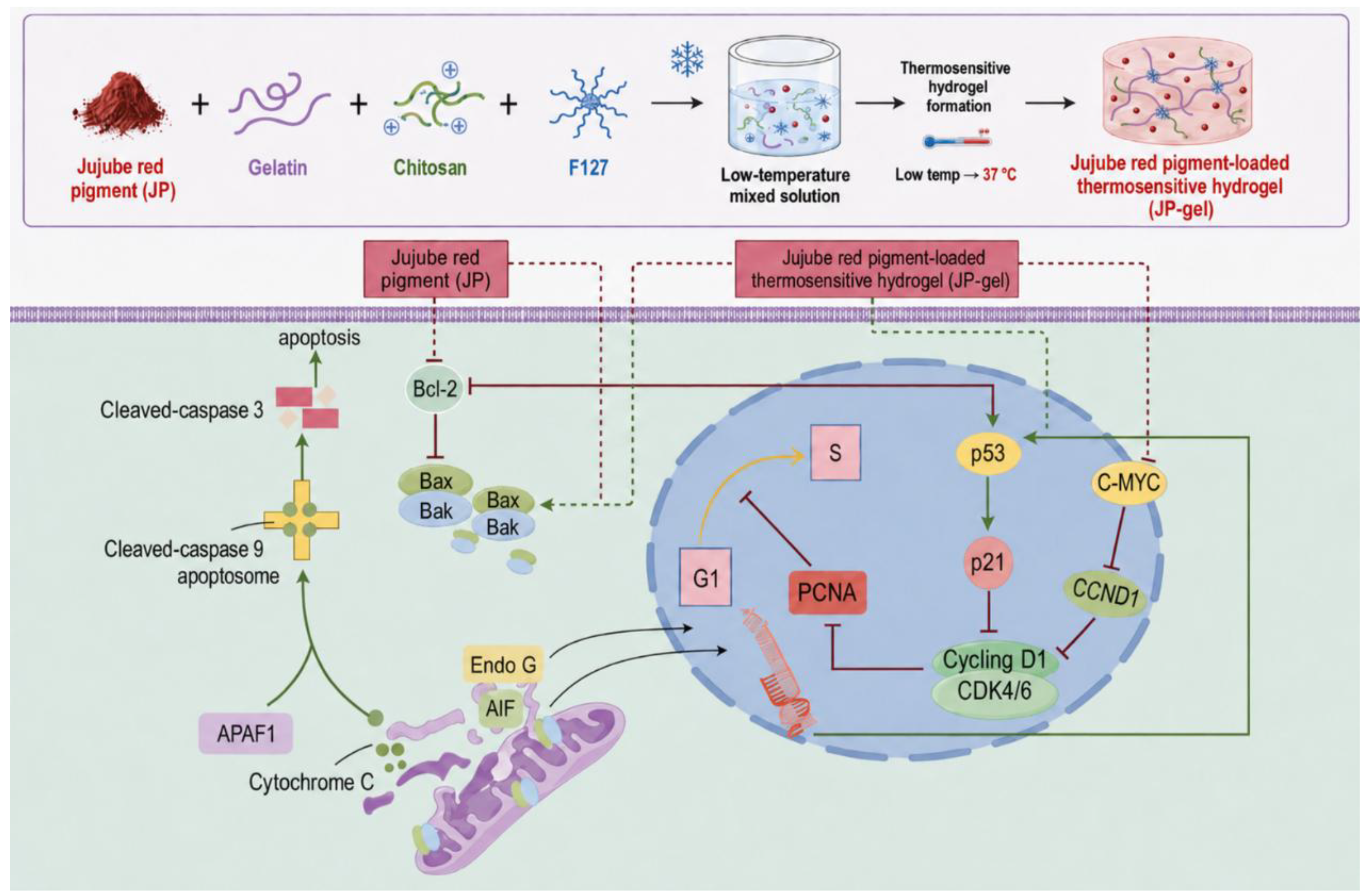
Figure 2.
Preparation and characterization of JP: a) Preparation of JP; b) UV-Vis absorption spectrum of JP; c) HPLC chromatogram of the extracted JP.
Figure 2.
Preparation and characterization of JP: a) Preparation of JP; b) UV-Vis absorption spectrum of JP; c) HPLC chromatogram of the extracted JP.
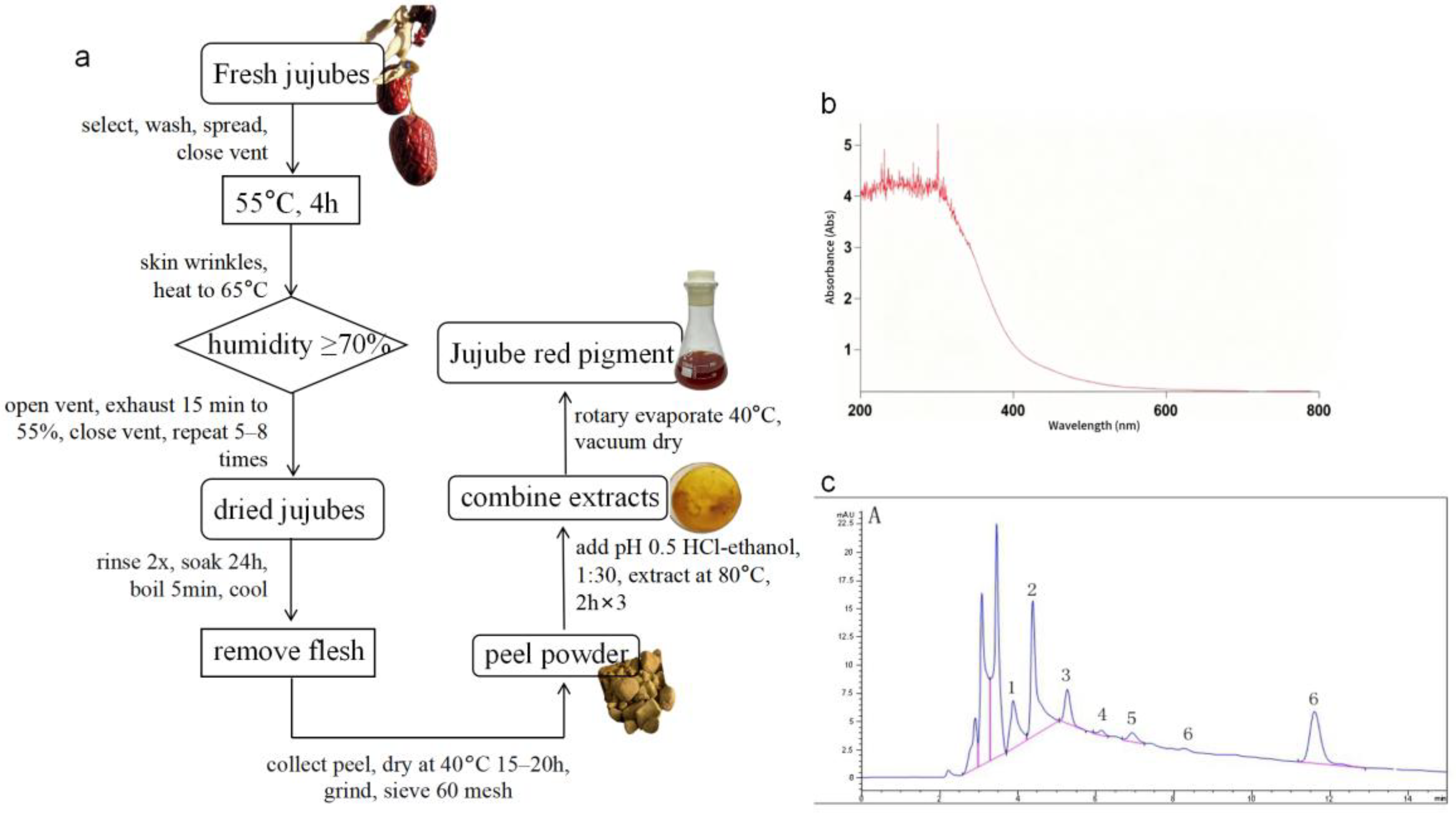
Figure 3.
Cell viability assessment of JP on multiple cell lines and anti-migration performance of JP in HeLa cervical cancer cells: a) Effects of different concentrations of JP (0–2.0 mg/mL) treatment for 24 h on the viability of L929, RAW264.7, HeLa, and B16 cells; b) Effects of different concentrations of JP (0–2.0 mg/mL) treatment for 12 h/48 h on the viability of HeLa cells; c) Wound healing assay showing the migration of HeLa cells following JP treatment at 24 h and 48 h; d) Transwell migration assay quantifying the number of migrated cells under different JP concentrations. Scar bars represent 50 µm.
Figure 3.
Cell viability assessment of JP on multiple cell lines and anti-migration performance of JP in HeLa cervical cancer cells: a) Effects of different concentrations of JP (0–2.0 mg/mL) treatment for 24 h on the viability of L929, RAW264.7, HeLa, and B16 cells; b) Effects of different concentrations of JP (0–2.0 mg/mL) treatment for 12 h/48 h on the viability of HeLa cells; c) Wound healing assay showing the migration of HeLa cells following JP treatment at 24 h and 48 h; d) Transwell migration assay quantifying the number of migrated cells under different JP concentrations. Scar bars represent 50 µm.
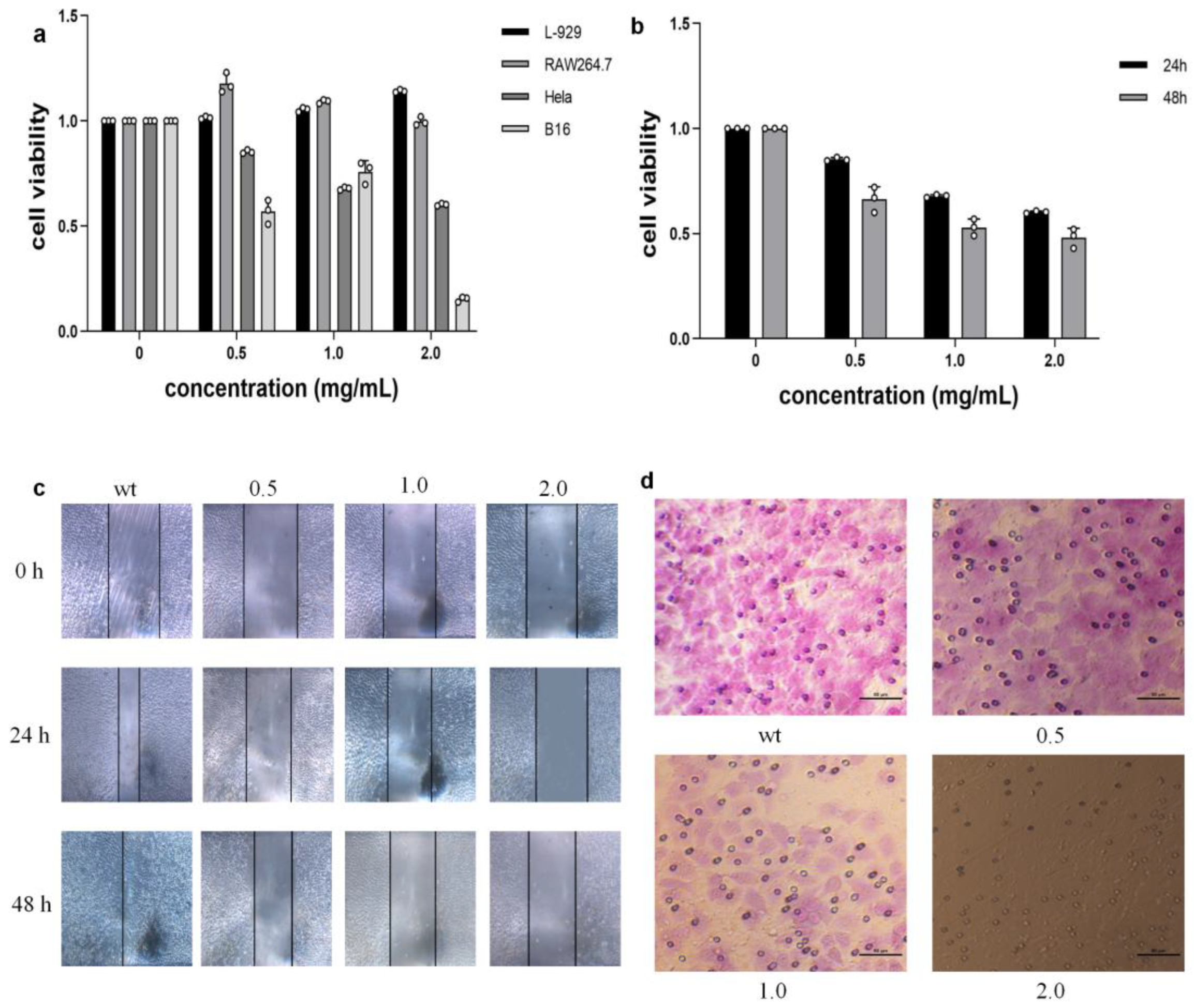
Figure 4.
JP induces apoptosis in HeLa cells and regulates Bax and Bcl-2 expression: a) Representative images of Hoechst 33258 staining showing apoptotic nuclear morphology in HeLa cells treated with 0 mg/mL or 2.0 mg/mL JP; b) Western blot analysis of Bax and Bcl-2 expression in HeLa cells after JP treatment. Data presented as mean ± SD, n = 3. Statistically significant *p < 0.05, **p < 0.01,***p < 0.01.
Figure 4.
JP induces apoptosis in HeLa cells and regulates Bax and Bcl-2 expression: a) Representative images of Hoechst 33258 staining showing apoptotic nuclear morphology in HeLa cells treated with 0 mg/mL or 2.0 mg/mL JP; b) Western blot analysis of Bax and Bcl-2 expression in HeLa cells after JP treatment. Data presented as mean ± SD, n = 3. Statistically significant *p < 0.05, **p < 0.01,***p < 0.01.
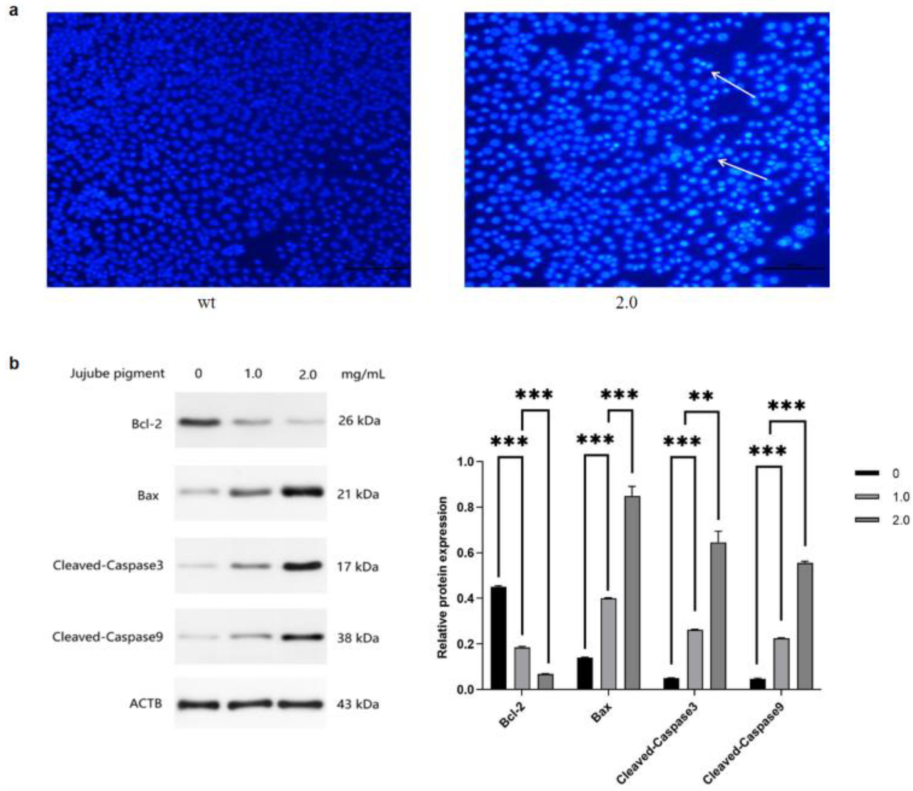
Figure 5.
JP modulates the cell cycle regulatory network in HeLa cells: a) qPCR analysis of key cell cycle regulators (CCND1, MYC, PCNA, and TP53); b) qPCR analysis of CCND1, MYC, PCNA, and TP53; c) qPCR analysis of CDK2, CDK4, and CDK6. Data are presented as mean ± SD, n = 3. *p < 0.05, **p < 0.01.
Figure 5.
JP modulates the cell cycle regulatory network in HeLa cells: a) qPCR analysis of key cell cycle regulators (CCND1, MYC, PCNA, and TP53); b) qPCR analysis of CCND1, MYC, PCNA, and TP53; c) qPCR analysis of CDK2, CDK4, and CDK6. Data are presented as mean ± SD, n = 3. *p < 0.05, **p < 0.01.
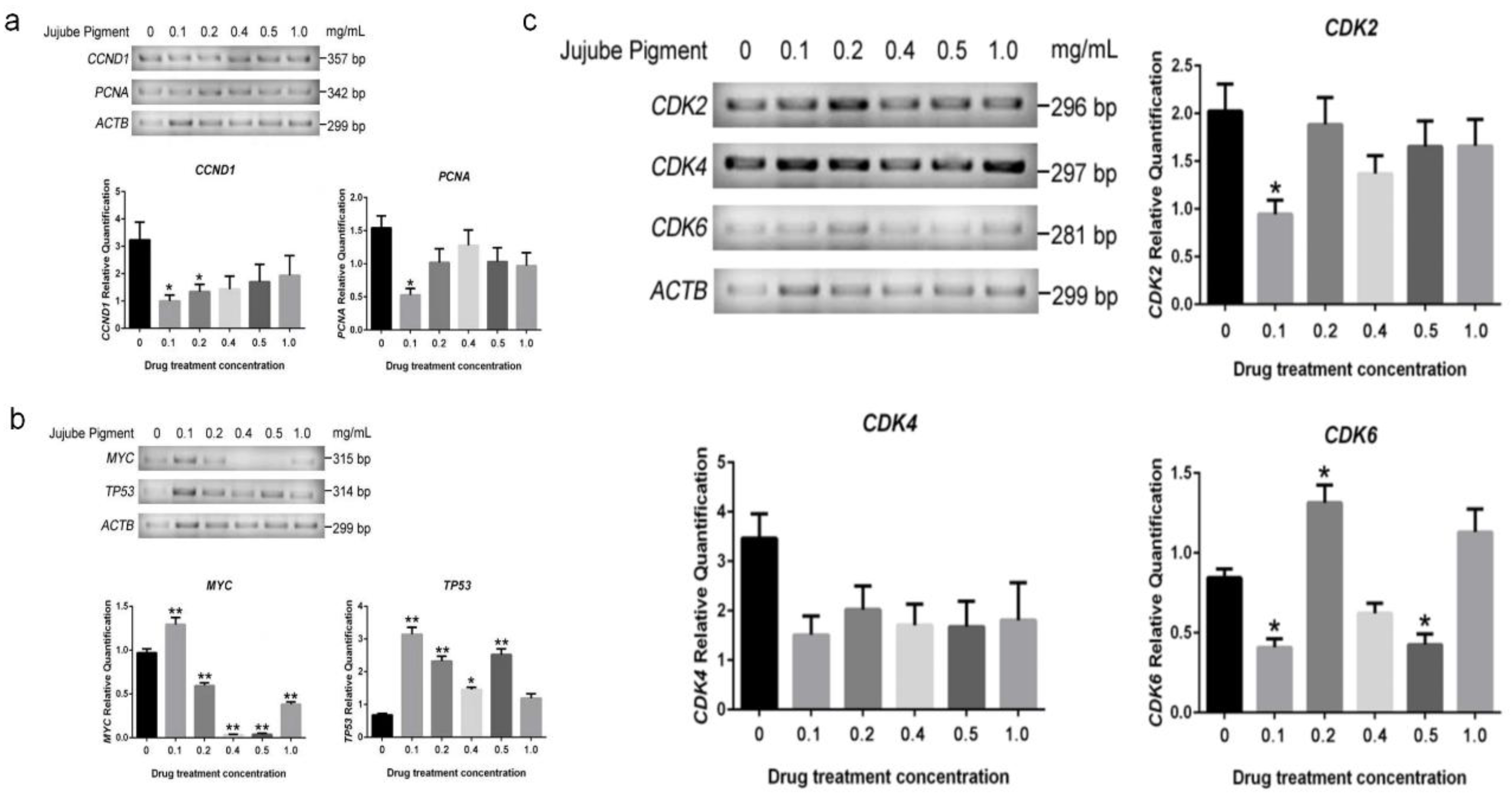
Figure 6.
Thermoresponsive sol–gel transition, macroscopic morphology, self-healing performance, microstructure and chemical characterization of JP-H: a) Digital photographs showing the temperature-dependent sol–gel behavior of JP-H precursor solution at 15, 20, 25, 30, 35 and 37 °C; b) Macroscopic photographs of the as-prepared JP-H, including intact hydrogel, cut cross-section, and self-healing performance after fracture and reconnection; c) Injectable and moldable performance of JP-H precursor: the solution was cast into letter-shaped molds at 25 °C (sol state) and transformed into solid hydrogel at 37 °C. (d) SEM images displaying the porous internal microstructure of lyophilized JP-H with different magnifications; e) FT-IR spectra of (A) free JP, (B) JP-H composite hydrogel, and (C) blank hydrogel matrix.
Figure 6.
Thermoresponsive sol–gel transition, macroscopic morphology, self-healing performance, microstructure and chemical characterization of JP-H: a) Digital photographs showing the temperature-dependent sol–gel behavior of JP-H precursor solution at 15, 20, 25, 30, 35 and 37 °C; b) Macroscopic photographs of the as-prepared JP-H, including intact hydrogel, cut cross-section, and self-healing performance after fracture and reconnection; c) Injectable and moldable performance of JP-H precursor: the solution was cast into letter-shaped molds at 25 °C (sol state) and transformed into solid hydrogel at 37 °C. (d) SEM images displaying the porous internal microstructure of lyophilized JP-H with different magnifications; e) FT-IR spectra of (A) free JP, (B) JP-H composite hydrogel, and (C) blank hydrogel matrix.
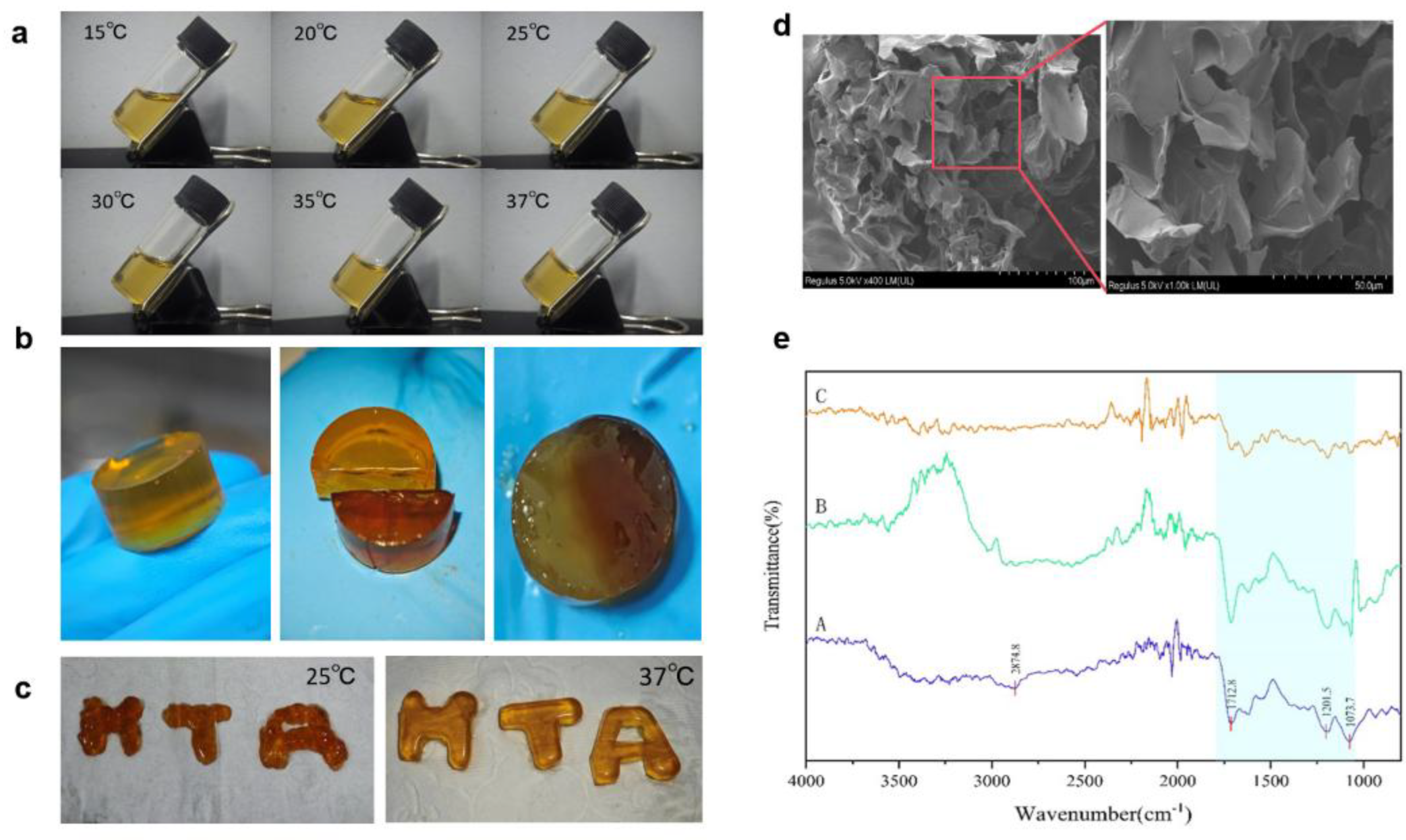
Figure 7.
antibacterial activity of free JP and JP-H against Escherichia coli and Staphylococcus aureus. (a) Schematic illustration of the antibacterial assay. (b) Representative colony-counting images and SEM micrographs of E. coli and S. aureus following treatment with PBS, free JP, blank hydrogel, or JP-H. (c) Quantitative analysis of relative bacterial survival determined by colony counting. (d) Representative disk-diffusion images and quantitative analysis of inhibition-zone diameters for free JP, blank hydrogel, and JP-H. Data are presented as mean ± SD, n = [6]. Different symbols indicate statistically significant differences as defined in the figure.
Figure 7.
antibacterial activity of free JP and JP-H against Escherichia coli and Staphylococcus aureus. (a) Schematic illustration of the antibacterial assay. (b) Representative colony-counting images and SEM micrographs of E. coli and S. aureus following treatment with PBS, free JP, blank hydrogel, or JP-H. (c) Quantitative analysis of relative bacterial survival determined by colony counting. (d) Representative disk-diffusion images and quantitative analysis of inhibition-zone diameters for free JP, blank hydrogel, and JP-H. Data are presented as mean ± SD, n = [6]. Different symbols indicate statistically significant differences as defined in the figure.
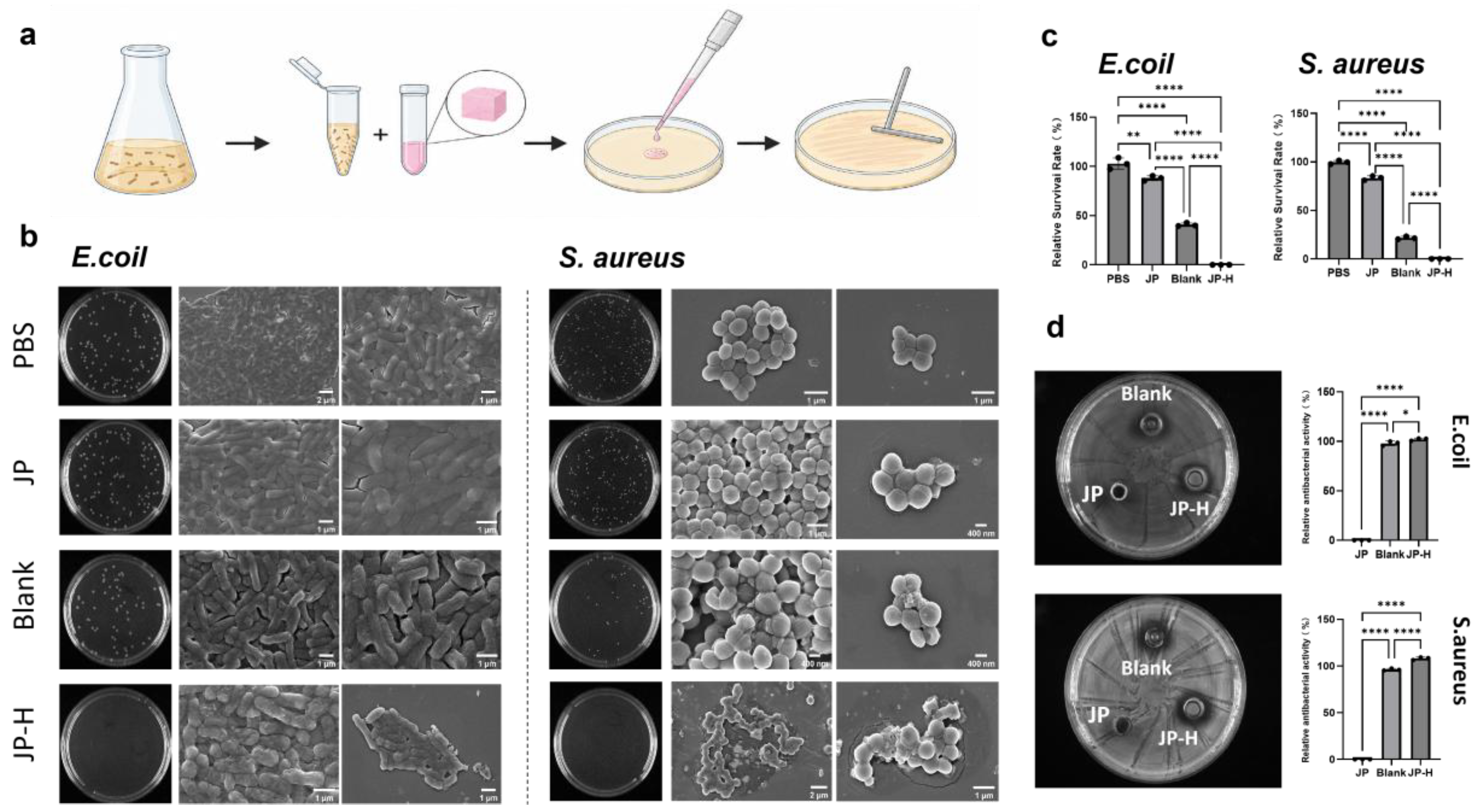
Figure 8.
anti-migration and pro-apoptotic effects of free JP and JP-H on HeLa tumor cells: a) Bright-field scratch wound images of HeLa cells treated with Control, free JP, blank hydrogel, and JP-H at 0h, 12h, 24h and 36h; b) Quantitative statistical analysis of cell migration rates corresponding to the scratch wound assay in panel a); c) Relative viability of HeLa cells after treatment with serial concentrations of JP; d) Schematic workflow of the co-culture system of thermosensitive hydrogel and tumor cells, followed by multi-channel fluorescence apoptosis staining; e) Corresponding bright field (BF), FITC, PI and Hoechst 33342 fluorescence micrographs of HeLa cells in each group after 24 h incubation; arrows indicate apoptotic cell nuclei; f) Flow cytometry scatter plots of Annexin V-FITC/PI double staining for quantitative detection of apoptosis ratios in HeLa cells from Control, Blank, free JP and JP-H groups.
Figure 8.
anti-migration and pro-apoptotic effects of free JP and JP-H on HeLa tumor cells: a) Bright-field scratch wound images of HeLa cells treated with Control, free JP, blank hydrogel, and JP-H at 0h, 12h, 24h and 36h; b) Quantitative statistical analysis of cell migration rates corresponding to the scratch wound assay in panel a); c) Relative viability of HeLa cells after treatment with serial concentrations of JP; d) Schematic workflow of the co-culture system of thermosensitive hydrogel and tumor cells, followed by multi-channel fluorescence apoptosis staining; e) Corresponding bright field (BF), FITC, PI and Hoechst 33342 fluorescence micrographs of HeLa cells in each group after 24 h incubation; arrows indicate apoptotic cell nuclei; f) Flow cytometry scatter plots of Annexin V-FITC/PI double staining for quantitative detection of apoptosis ratios in HeLa cells from Control, Blank, free JP and JP-H groups.
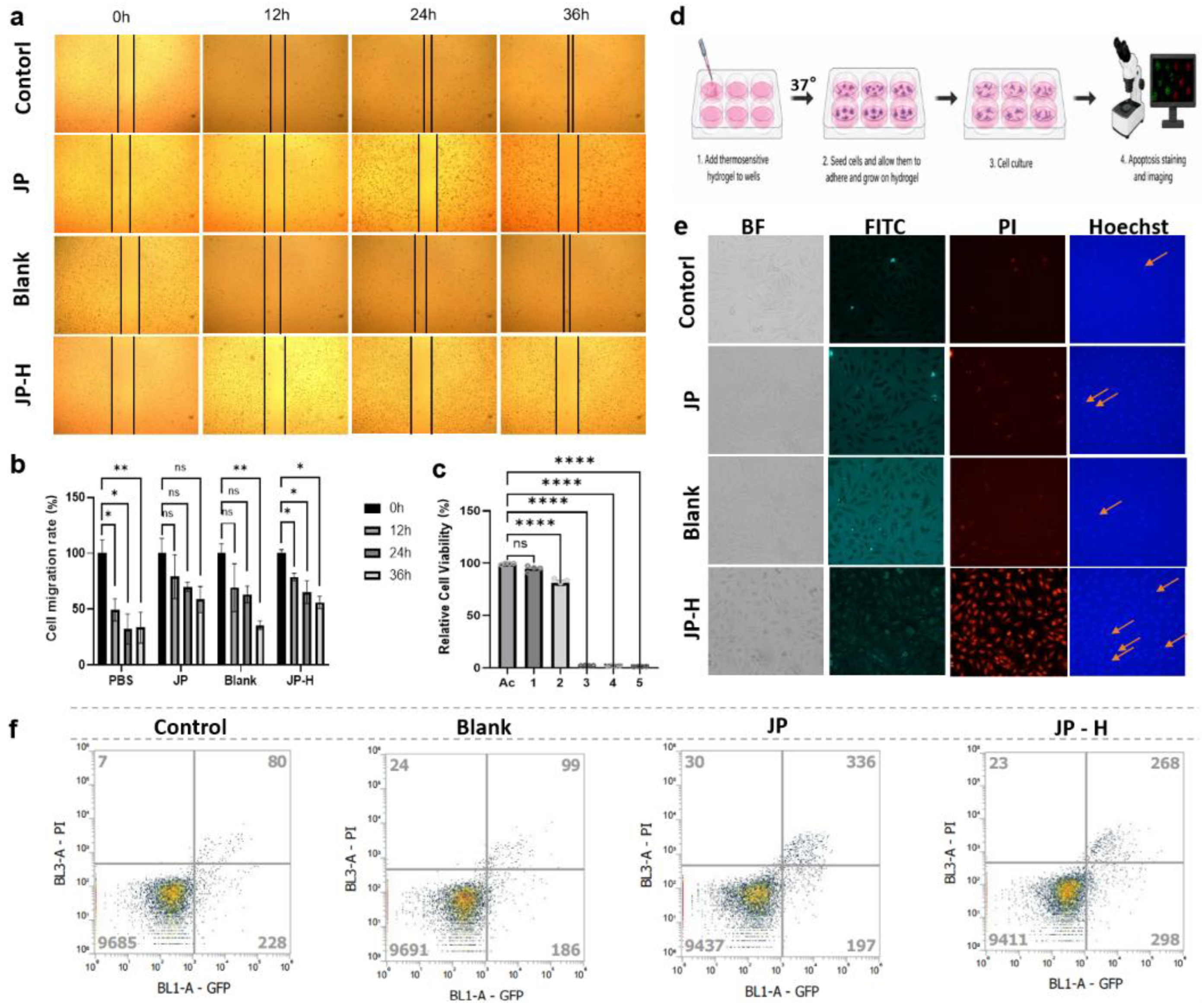

Table 1.
Sequences of primers used for RT-PCR analysis of cell cycle-related gene expression.
| Gene | Orientation and Sequences |
|---|---|
| CCND1 | Forward:5’TTCGTGGCCTCTAAGATGAAG 3’ |
| Reverse:5’GTAGGACAGGAAGTTGTTGGG 3’ | |
| CDK2 | Forward:5’TCAAGCTAGCAGACTTTGGAC 3’ |
| Reverse:5’ACTTGGCTTGTAATCAGGCAT 3’ | |
| CDK4 | Forward:5’AATTGCATCGTTCACCGAGAT 3’ |
| Reverse:5’CAGCCCAATCAGGTCAAAGAT 3’ | |
| CDK6 | Forward:5’CAGTGTCACGAACAGACAGAG 3’ |
| Reverse:5’GGTTAGAGCCATCTGGAAACT 3’ | |
| PCNA | Forward:5’TTGGCGCTAGTATTTGAAGCA 3’ |
| Reverse:5’CAGGTACCTCAGTGCAAAAGT 3’ | |
| MYC | Forward:5’CTTCTCTCCGTCCTCGGATTC 3’ |
| Reverse:5’GGATAGTCCTTCCGAGTGGAG 3’ | |
| TP53 | Forward:5’CCTGTCATCTTCTGTCCCTTC3’ |
| Reverse:5’CTCGGATAAGATGCTGAGGAG3’ | |
| ACTB | Forward:5’TCTACAATGAGCTGCGTGTGG3’ |
| Reverse:5’GAGGTAGTCAGTCAGGTCCCG3’ |
Table 2.
PCR reaction components.
| Component | Volume (µL) |
|---|---|
| 2×F8 PCR Master MIX | 10 |
| Forward Primer(10 μ M) | 0.5 |
| Reverse Primer(10 μ M) | 0.5 |
| cDNA Template | 0.8 |
| DMSO | 5 |
| Nuclease-free Water | To 20 |
Disclaimer/Publisher’s Note: The statements, opinions and data contained in all publications are solely those of the individual author(s) and contributor(s) and not of MDPI and/or the editor(s). MDPI and/or the editor(s) disclaim responsibility for any injury to people or property resulting from any ideas, methods, instructions or products referred to in the content. |
© 2026 by the authors. Licensee MDPI, Basel, Switzerland. This article is an open access article distributed under the terms and conditions of the Creative Commons Attribution (CC BY) license (http://creativecommons.org/licenses/by/4.0/).
Copyright: This open access article is published under a Creative Commons CC BY 4.0 license, which permit the free download, distribution, and reuse, provided that the author and preprint are cited in any reuse.


