
Submitted:
28 July 2026
Posted:
30 July 2026
You are already at the latest version
Abstract
Chronic psychological stress is common among patients with breast cancer and has been linked to poorer outcomes, yet how it weakens antitumor immunity is still incompletely defined. Stress acts through two neuroendocrine arms. The sympathetic nervous system releases catecholamines that signal via β-adrenergic receptors, and the hypothalamic–pituitary–adrenal axis releases glucocorticoids that signal via the glucocorticoid receptor. These arms are usually studied apart. Integrating mechanistic, preclinical, and clinical evidence, we argue that in breast cancer they converge on one immunological endpoint, an exhausted CD8⁺ T cell and natural killer cell phenotype with high inhibitory-receptor expression, and that they reinforce each other through a feed-forward loop in which glucocorticoids sustain catecholaminergic tone. Stress hormones therefore behave as a convergent, druggable neuroendocrine checkpoint. The framework explains why single-arm interventions such as β-blockers or glucocorticoid receptor antagonists have given inconsistent results, and it predicts that co-targeting both arms together with behavioral stress reduction could potentiate immune checkpoint blockade. We assess the supporting evidence, address the paradox that glucocorticoids remain essential for managing immunotherapy toxicity, and set out the models, biomarkers, and trial designs needed to test whether easing the neuroendocrine stress burden restores durable antitumor immunity in breast cancer.
Keywords:
breast cancer
; chronic stress
; β-adrenergic signaling
; glucocorticoid receptor
; CD8⁺ T cell exhaustion
; immune checkpoint blockade
; β-blocker
; neuro-immune axis
1. Introduction
Breast cancer remains the most frequently diagnosed malignancy in women worldwide, and a substantial fraction of patients carry sustained psychological distress through diagnosis, treatment, and survivorship. Epidemiological syntheses associate depression and anxiety with higher recurrence and cancer-specific mortality in breast cancer [1], and pooled cohort analyses link stressful life events to a modest increase in incidence [2]. These associations are not uniform. A large prospective United Kingdom cohort detected no relationship between the frequency of perceived stress and breast cancer risk once recall bias was minimized [3]. The discrepancy points to a mechanistic rather than a purely correlative question: once chronic stress is present, how does it reshape tumor biology and, in particular, the antitumor immune response?
Chronic stress is transduced by two neuroendocrine effector arms. The sympathetic nervous system releases the catecholamines norepinephrine and epinephrine, whereas the hypothalamic–pituitary–adrenal axis releases glucocorticoids [4,5]. Both hormone classes act on receptors expressed by tumor cells and by essentially every immune lineage. Foundational work in cancer neuroscience showed that catecholamine signaling through β-adrenergic receptors accelerates tumor growth, angiogenesis, and metastasis [6,7], and that sympathetic input remodels the tumor microenvironment toward an immunosuppressive, pro-metastatic state [8].
Mechanistic studies have since resolved this picture at the level of individual immune cells. β-adrenergic signaling constrains the metabolic fitness and effector program of CD8⁺ T cells, and catecholamines acting through the β1-adrenergic receptor were recently shown to drive terminal T cell exhaustion, positioning the receptor itself as an actionable checkpoint [9]. Elevated adrenergic tone raises inhibitory-receptor expression on tumor-infiltrating lymphocytes and blunts the efficacy of PD-1 blockade in preclinical models [10]. The catecholamine arm therefore does more than promote generic tumor growth; it directly weakens the cytotoxic compartment that immunotherapy depends on.
The glucocorticoid arm converges on strikingly similar endpoints. Tumor-derived and stress-induced glucocorticoids act through the glucocorticoid receptor to transactivate PD-1, TIM-3, and LAG-3 and to impose a dysfunctional CD8⁺ T cell state that predicts failure of checkpoint blockade [11], while glucocorticoid receptor activity within tumor cells can upregulate PD-L1 and repress MHC class I [12]. Stress-elevated glucocorticoids additionally disable dendritic cell priming through induction of TSC22D3 [13]. These observations carry direct clinical weight, because immune checkpoint inhibitors are now standard of care for triple-negative breast cancer [14,15], yet durable responses remain the exception, and efforts to repurpose β-blockers as immunotherapy adjuncts have yielded encouraging but inconsistent clinical results [16].
Despite this convergence, the sympathetic and glucocorticoid arms are almost always reviewed in isolation, and recent broad surveys of neuro-immune crosstalk in breast cancer treat them as parallel contributors to a generally pro-tumor state. We propose a different framing. Catecholamines and glucocorticoids funnel onto one shared molecular endpoint, the checkpoint-high, exhausted CD8⁺ and natural killer cell phenotype, and the two arms reinforce each other, because glucocorticoids induce the catecholamine-synthesizing enzymes that sustain adrenergic tone [17,18]. On this view, stress hormones behave as a single, druggable neuroendocrine checkpoint rather than as two separate risk factors. The framing yields a specific, falsifiable prediction that sets it apart from existing surveys: because the arms converge and amplify one another, blockade of a single arm should be insufficient, whereas co-targeting both should more reliably restore antitumor immunity. In this review we integrate the cell-type-specific mechanisms of both arms in breast cancer, define their convergent checkpoint, explain how it sustains immunotherapy resistance, and evaluate dual-axis pharmacological co-targeting, combining β-blockers with glucocorticoid receptor modulators alongside behavioral stress reduction, as a strategy to restore antitumor immunity and potentiate checkpoint blockade.
2. The Neuroendocrine Stress Response and Its Immune Interface in the Breast Tumor Microenvironment
Chronic psychological stress is relayed to peripheral tissues by two coordinated neuroendocrine effector systems whose outputs, catecholamines and glucocorticoids, act on receptors that are broadly expressed across tumor cells and immune lineages. Understanding how stress shapes antitumor immunity in breast cancer therefore begins with the anatomy and receptor logic of these two arms and with the ways in which they intersect instead of operating in parallel (Figure 1).
The sympathetic nervous system is the faster arm. Postganglionic sympathetic fibers innervate lymphoid organs and, in solid tumors, the tumor bed itself, releasing norepinephrine locally while the adrenal medulla adds circulating epinephrine [5,19]. These catecholamines engage α- and β-adrenergic receptors, of which the β2 subtype dominates on lymphocytes, natural killer cells, dendritic cells, and myeloid populations and signals through Gαs, adenylyl cyclase, cyclic AMP, and protein kinase A [20]. The functional consequence is not fixed. β2-adrenergic receptors can switch between Gαs- and Gαi-biased signaling, so that adrenergic input enhances or restrains a given immune cell depending on activation state and local receptor density [21]. This context dependence is a recurring theme that later sections revisit at the level of individual cell types.
The hypothalamic–pituitary–adrenal axis is the slower, more sustained arm. Perceived stressors drive pituitary adrenocorticotropic hormone release and adrenal cortical secretion of glucocorticoids, principally cortisol in humans, which diffuse freely across membranes to activate the cytoplasmic glucocorticoid receptor [4]. Ligand-bound glucocorticoid receptor reprograms transcription in T cells, dendritic cells, macrophages, and neutrophils, and its net effect on immunity is generally suppressive but, like adrenergic signaling, is conditioned by dose, timing, and target cell [22]. Acute, transient glucocorticoid and catecholamine surges can transiently mobilize and even enhance immune effector function, whereas chronic elevation biases the system toward suppression, a distinction formalized in the acute-versus-chronic framework of stress immunology [23].
A central and often overlooked point is that the two arms are not independent. Glucocorticoids exert permissive, amplifying control over catecholaminergic output. Classic endocrine work showed that adrenal cortical steroids, delivered at high concentration through the intra-adrenal portal circulation, induce phenylethanolamine N-methyltransferase and thereby the conversion of norepinephrine to epinephrine [17]. More recent studies confirm that corticosterone upregulates catecholamine-synthesizing enzymes, including tyrosine hydroxylase, at both transcript and protein levels [18], and glucocorticoids further sustain β2-adrenergic receptor density and cyclic AMP responsiveness on lymphocytes [24]. Chronic stress thus does not present the immune system with two separable signals but with a reinforcing circuit in which HPA activity potentiates the sympathetic arm.
At the level of the tumor, sympathetic and glucocorticoid signaling converge on a microenvironment that favors progression. Sympathetic input remodels the breast tumor microenvironment by recruiting and polarizing macrophages, promoting angiogenesis and inflammation, and constraining cytotoxic immunity, a program summarized in the concept of neural regulation of the tumor microenvironment [8,25]. This program overlaps substantially with the transcriptional signature of chronic adversity, in which sustained β-adrenergic tone upregulates pro-inflammatory genes while suppressing type I interferon responses [26]. The breast is a densely innervated, hormone-responsive organ, which makes its tumor microenvironment particularly exposed to these neuroendocrine inputs [27].
Framing the two arms as a coupled system reshapes the questions worth pursuing. Most human data remain associative, and the local concentrations, temporal dynamics, and receptor occupancy of catecholamines and glucocorticoids within the breast tumor microenvironment are still poorly quantified, particularly across molecular subtypes and treatment states. Spatially resolved measurement of hormone gradients, receptor expression on defined immune populations, and the degree to which HPA activity amplifies adrenergic tone in patient tumors are the key gaps. Resolving them is a prerequisite for interpreting the cell-type-specific mechanisms examined next, first for the β-adrenergic arm and then for the glucocorticoid arm, before the two are integrated into a single convergent framework.
3. The Sympathetic β-Adrenergic Arm Reprograms Antitumor Immunity in Breast Cancer
Sympathetic outflow reaches the breast tumor microenvironment as locally released norepinephrine and systemic epinephrine, and its immunological footprint is broad: β-adrenergic signaling simultaneously blunts the effector arm of adaptive and innate immunity and expands the suppressive myeloid compartment. Across the major intratumoral populations, catecholamines converge on a cyclic AMP–protein kinase A program that lowers cytotoxic output, raises inhibitory-receptor expression, and shifts myeloid cells toward immunosuppression. This section examines that footprint cell type by cell type, emphasizing breast cancer evidence where it exists and mechanistic evidence from other tumors where it is more mature (Figure 2).
CD8⁺ T cells are the most consequential target. β-adrenergic signaling restrains their metabolic fitness by blocking the glycolytic and oxidative reprogramming that activated T cells require, reducing glucose uptake and effector cytokine production during priming [28,29,30]. Within tumors, chronic adrenergic input lowers mitochondrial mass and CD28 expression in tumor-infiltrating lymphocytes and drives them toward an exhausted, metabolically insufficient state that pharmacological β-blockade can partially reverse [31]. The most direct demonstration that this constitutes a checkpoint came from work showing that exhausted CD8⁺ T cells upregulate the β1-adrenergic receptor and that catecholamines acting through this receptor accelerate the transition to terminal exhaustion, so that receptor ablation preserves progenitor-exhausted cells and restores effector function [9]. Consistent with these mechanisms, sustained β-adrenergic tone raises PD-1 on intratumoral CD8⁺ T cells and undermines the efficacy of anti–PD-1 therapy, an effect reproduced genetically, pharmacologically, and by manipulating housing temperature [10]. The catecholamine arm therefore functions as a direct brake on the cytotoxic compartment that immunotherapy mobilizes, well beyond a generic growth-promoting role [32].
Natural killer cells are similarly suppressed. Early in vivo work established that β-adrenergic stimulation reduces NK cytotoxicity in a dose-dependent manner and compromises resistance to tumor metastasis, and that β-blockade with agents such as nadolol reverses this suppression [33]. Subsequent studies attributed stress-induced loss of NK-mediated metastatic control specifically to adrenal catecholamines acting through β-adrenergic receptors, linking a physiological stressor to a defined receptor mechanism [34]. Because NK cells provide early surveillance against disseminating breast tumor cells, adrenergic suppression of this compartment is particularly relevant to the metastatic phase of disease [35].
Dendritic cells occupy the interface between innate sensing and T cell priming, and β-adrenergic signaling degrades their stimulatory capacity. β2-adrenergic receptor activation constrains dendritic cell activation, in part by rewiring CD40-driven signaling, and β-blockade restores their capacity to prime effector T cells and to support antitumor responses [36,37]. Impaired antigen presentation upstream compounds the cell-intrinsic defects imposed on CD8⁺ and NK cells, so that the adrenergic arm constrains antitumor immunity at both the induction and effector stages.
The clearest breast cancer–specific evidence concerns the myeloid compartment. In an orthotopic model, sympathetic activation induced a metastatic switch in primary breast cancer by recruiting CD11b⁺F4/80⁺ macrophages and driving their M2 polarization, increasing distant metastasis roughly thirty-fold with little effect on primary tumor growth; propranolol and macrophage depletion abrogated the effect [38]. Adrenergic signaling also acts directly on myeloid-derived suppressor cells: β2-adrenergic receptor expression rises with tumor burden and, through STAT3, sustains MDSC survival and their expression of arginase-1 and PD-L1 [39], while further shaping the fatty-acid oxidation and oxidative metabolism that underpin MDSC suppressive function [40]. In breast cancer specifically, chronic stress expands myeloid-derived suppressor cells through a norepinephrine–β-adrenergic–IL-6/STAT3 axis to promote pulmonary metastasis [41], and chronic psychological stress mobilizes splenic CXCR2⁺ MDSCs to seed a pre-metastatic niche via tumor-associated macrophage–derived CXCL1, suppressing CD8⁺ T cell cytotoxicity at the metastatic site [42]. Norepinephrine can also polarize macrophages toward the M2 state through β2-adrenergic signaling on breast tumor cells, amplifying local IL-6 production [43]. Together these studies place adrenergic remodeling of myeloid cells at the center of stress-driven breast cancer progression.
Human correlative data, though limited, are consistent with the preclinical mechanisms. In estrogen receptor–negative breast cancer, β2-adrenergic receptor expression associates with biomarkers of tumor immunity and predicts poorer prognosis, suggesting that adrenergic tone marks tumors with a more suppressed immune contexture [44]. This observation nominates β2-adrenergic receptor expression as a candidate stratifier, a theme developed when immunotherapy resistance and dual-axis targeting are considered.
At the translational level, the β-adrenergic arm is attractive precisely because it is druggable and because its blockade repeatedly restores immune function across models, lifting CD8⁺ effector programs, preserving NK cytotoxicity, restraining MDSC suppression, and enhancing responses to checkpoint blockade [10,31]. Important gaps remain, however. Most mechanistic data derive from murine models or from cells other than human breast tumor infiltrates, the relative contributions of β1 versus β2 receptors across immune populations are incompletely resolved, and the receptor's Gαs-to-Gαi switching implies that outcomes may depend on chronicity and cell state in ways not yet mapped in patient tumors. Whether β2-adrenergic receptor or intratumoral catecholamine levels can serve as predictive biomarkers, and how adrenergic blockade should be timed relative to immunotherapy, are the questions that most directly bridge this arm to therapeutic application and to its integration with glucocorticoid signaling, addressed next.
4. The HPA Glucocorticoid Arm and Glucocorticoid-Receptor-Driven Immune Suppression in Breast Cancer
The glucocorticoid arm suppresses antitumor immunity through a single receptor with unusually broad reach. Acting on the glucocorticoid receptor expressed in T cells, natural killer cells, dendritic cells, regulatory T cells, and the tumor cells themselves, stress-elevated and locally generated glucocorticoids reach the same functional endpoint as the adrenergic arm: an exhausted, checkpoint-high cytotoxic compartment set against a tumor rendered less visible to it. In breast cancer this arm carries an additional layer of significance because glucocorticoid receptor activity is itself an established prognostic determinant, particularly in receptor-negative disease (Figure 3).
Glucocorticoid control of CD8⁺ T cell fate is direct and transcriptional. A gradient of glucocorticoid receptor activity runs from naive to exhausted tumor-infiltrating CD8⁺ T cells, and receptor engagement transactivates multiple inhibitory receptors, including PD-1, TIM-3, and LAG-3, while imposing a dysfunctional differentiation state; glucocorticoids of tumor monocyte–macrophage origin are sufficient to drive this program, and high glucocorticoid signaling marks patients, including those with melanoma, who fail checkpoint blockade [11]. The endpoint is therefore the same exhausted phenotype produced by β-adrenergic signaling, reached through an independent receptor, a parallelism that motivates the convergence framework developed later.
Glucocorticoid receptor signaling also acts cell-intrinsically within tumor cells to lower their immunogenicity. In pancreatic cancer, glucocorticoid receptor activity upregulates PD-L1 while transcriptionally repressing MHC class I, simultaneously raising an inhibitory ligand and hiding tumor antigen; genetic or pharmacological interruption of this axis restores cytotoxic T cell infiltration and reverses resistance to checkpoint blockade, and high glucocorticoid receptor expression tracks with poorer patient survival [12]. Although defined outside the breast, this mechanism illustrates how a single arm can suppress the effector cell and disarm its target in tandem.
Innate cytotoxicity is likewise curtailed. Endogenous glucocorticoids selectively and tissue-specifically induce PD-1 on natural killer cells, a pathway first defined in the control of viral immunity that constrains interferon-γ output [45]. In the tumor context, glucocorticoids cooperate with the microenvironment-enriched cytokines IL-12, IL-15, and IL-18 to upregulate PD-1 on human natural killer cells through combined transcriptional and translational mechanisms [46]. A clinical correlate is provided by depression-associated hypercortisolism, which upregulates PD-1 on tumor-infiltrating natural killer cells and blunts their cytotoxicity to accelerate hepatocellular carcinoma progression [47], and glucocorticoid receptor activation can further reprogram natural killer cells toward an immunosuppressive, amphiregulin-driven state across several tumor types [48].
Beyond suppressing effectors, the glucocorticoid arm actively expands and empowers immunosuppressive populations. Tumors generate active glucocorticoids not only from adrenal output but by recycling inactive metabolites through 11β-hydroxysteroid dehydrogenase, and these locally produced hormones activate regulatory T cells to enhance their suppressive capacity and promote tumor growth, an axis whose interruption sensitizes tumors to checkpoint blockade [49,50,51]. In parallel, stress-induced glucocorticoids disable dendritic cell priming by inducing the glucocorticoid-responsive factor TSC22D3, which blocks type I interferon responses and interferon-γ⁺ T cell activation and thereby abrogates the antitumor immunity elicited by chemotherapy and immunotherapy; circulating TSC22D3 and plasma cortisol in patients correlate with negative affect [13].
In breast cancer the glucocorticoid receptor is both an immune modulator and a prognostic axis in its own right. In estrogen receptor–negative disease, glucocorticoid receptor activation associates with shorter relapse-free survival and with transcriptional programs governing epithelial–mesenchymal transition, adhesion, and inflammation [52]. The prognostic direction is context dependent: glucocorticoid receptor expression predicts worse outcome under taxane-based therapy yet can associate with better outcome under anthracycline-based regimens [53], and triple-negative tumors in which glucocorticoid receptor is expressed by immune rather than malignant cells show longer survival, indicating that the cell type carrying the receptor shapes its prognostic meaning [54]. Mechanistically, microenvironmental cues such as TGF-β1 drive p38 MAPK phosphorylation of the receptor at Ser134, and this phosphorylated species is required for anchorage-independent growth, migration, and invasion and defines a signature predicting shorter survival in triple-negative breast cancer [55]. The systemic consequences are equally direct: rising glucocorticoids during breast cancer progression activate the glucocorticoid receptor at distant sites and upregulate the kinase ROR1 to promote colonization and reduce survival, a finding that prompted explicit caution about glucocorticoid use in this setting [56]. Chronic stress further increases metastasis through glucocorticoid-dependent, neutrophil-mediated remodeling of the pre-metastatic microenvironment, an effect abolished by neutrophil-specific deletion of the glucocorticoid receptor [57].
Translationally, the glucocorticoid arm presents a sharper dilemma than the adrenergic arm. Its receptor is eminently druggable, and antagonism restores immune function and chemosensitivity in preclinical breast cancer models, yet glucocorticoids remain indispensable for managing immune-related adverse events and for supportive care, so systemic blockade cannot be pursued naively. The prognostic and immunological effects are strongly conditioned by cell type, tumor subtype, and concurrent therapy, and human data linking endogenous glucocorticoid tone to intratumoral immune states in breast cancer remain sparse. Defining where glucocorticoid receptor activity is immunosuppressive versus dispensable, and in which breast cancer subtypes its blockade would help rather than harm, is the central unresolved question. Because this arm reaches the same exhausted, checkpoint-high endpoint as the sympathetic arm and is amplified by it, the two are most usefully considered together, which the following section does.
- 5.
- Convergence: The Two Arms as a Single Druggable Neuroendocrine Checkpoint
The preceding sections describe two neuroendocrine arms that suppress antitumor immunity through separate receptors. Read together, they make a stronger claim than either alone: catecholamines and glucocorticoids operate as a single functional unit rather than two independent risk factors; they converge on one immunological endpoint and reinforce each other. This section develops that unit, the convergent neuroendocrine checkpoint, along three axes of convergence, phenotypic, molecular, and systemic, and then draws out its reciprocal amplification, which is what distinguishes a coupled circuit from a simple sum of two pathways (Figure 4).
Phenotypic convergence is the most conspicuous. Adrenergic and glucocorticoid signaling arrive at the same exhausted, checkpoint-high state in the cytotoxic compartment despite engaging different receptors. Catecholamines acting through the β1-adrenergic receptor accelerate terminal CD8⁺ T cell exhaustion [9], while glucocorticoids acting through the glucocorticoid receptor transactivate the same inhibitory receptors, namely PD-1, TIM-3, and LAG-3, and impose a parallel dysfunctional program [11]. The convergence extends beyond CD8⁺ T cells. Both arms raise PD-1 on natural killer cells and blunt their cytotoxicity [45], both weaken dendritic cell priming, and both expand and empower suppressive myeloid and regulatory populations. A cell-by-cell tabulation of these parallel effects (Table 1) shows that for essentially every intratumoral immune lineage, the catecholamine and glucocorticoid arms push in the same direction, which is why chronic stress produces a coherent immunosuppressive contexture rather than a patchwork of opposing effects.
This convergence is best understood as action on a differentiation trajectory rather than on a fixed state. Exhausted CD8⁺ T cells span a continuum from stem-like, TCF1⁺ progenitor cells, which retain proliferative potential and mediate the response to checkpoint blockade, to terminally exhausted, TCF1⁻ cells that resist reinvigoration [58], and it is the progenitor pool whose proliferative burst underlies successful immunotherapy [59]. Both neuroendocrine arms bias this trajectory toward its terminal end. Catecholamines acting through the β1-adrenergic receptor accelerate the progenitor-to-terminal transition, and receptor ablation preserves progenitor-exhausted cells and restores effector function [9], whereas the glucocorticoid receptor imposes its dysfunctional program along the same gradient, its activity rising from naive toward exhausted cells [11]. Framing the two arms as forces that push cytotoxic cells along a shared exhaustion trajectory, rather than as producers of a static phenotype, sharpens the convergence claim and yields a concrete prediction: relieving neuroendocrine tone should shift the balance back toward the checkpoint-responsive progenitor pool.
Molecular convergence underlies the phenotypic overlap, and it can be traced to a shared transcriptional endpoint rather than to a shared proximal pathway. The dysfunctional cytotoxic state is programmed by the HMG-box factor TOX, in whose absence exhausted CD8⁺ T cells fail to form [60], acting with TOX2 and the NR4A family of orphan nuclear receptors downstream of NFAT to enforce high inhibitory-receptor expression and to suppress cytokine output [61]. Both neuroendocrine arms feed this module from different directions. The glucocorticoid receptor, itself a ligand-activated transcription factor, transactivates PD-1, TIM-3, and LAG-3 and imposes the dysfunctional program [11], whereas the adrenergic arm signals through Gαs, cyclic AMP, protein kinase A, and the transcriptional modulators CREB and CREM [9,62], and NR4A is a recognized protein kinase A and CREB target [63], which places the catecholamine second-messenger cascade immediately upstream of the same exhaustion-driving nuclear receptors. A glucocorticoid transcriptional node and an adrenergic cyclic AMP–PKA–CREB node thus meet at the NFAT–TOX/NR4A module and at the inhibitory-receptor loci it controls, so the two arms reach one endpoint without sharing their proximal signaling.
Direct evidence that a cyclic AMP–PKA–CREB node can drive this checkpoint output comes from an analogous Gαs-coupled receptor. In colorectal cancer, thyroid-stimulating hormone receptor signaling in CD8⁺ tumor-infiltrating lymphocytes raised PD-1 and TIM-3 through protein kinase A and CREB, and deletion of the receptor restored effector differentiation [64]. Because β-adrenergic receptors engage the same Gαs–cyclic AMP–PKA–CREB cascade, this offers a mechanistic precedent for how catecholamines could install the exhaustion transcriptional program, converging on the endpoint that glucocorticoids reach through the receptor. Convergence extends to the inhibitory ligand: glucocorticoid receptor activity upregulates PD-L1 and represses MHC class I in tumor cells [12], while adrenergic signaling sustains interferon-γ–dependent PD-L1 through the β3-adrenergic receptor [65], so both arms raise the same ligand from different cellular compartments. This shared logic is the reason steroid and catecholamine signaling are increasingly considered together as modulators of antitumor immunity [66]. Two links in the model remain to be closed experimentally: whether β-adrenergic input induces TOX or NR4A within exhausted CD8⁺ T cells, and whether the glucocorticoid receptor and CREB co-occupy the same inhibitory-receptor loci in a single T cell. Both are predictions rather than established facts, although glucocorticoid–protein kinase A crosstalk documented in other cell systems makes them plausible [67].
Reciprocal amplification makes the two arms a coupled circuit rather than a coincidence. Glucocorticoids exert permissive control over the catecholaminergic arm at multiple levels. They induce phenylethanolamine N-methyltransferase, the enzyme that converts norepinephrine to epinephrine [17], upregulate the rate-limiting catecholamine-synthesizing enzyme tyrosine hydroxylase [18], and sustain β-adrenergic receptor density and cyclic AMP responsiveness on lymphocytes [24]. HPA activation therefore does not merely add glucocorticoid-mediated suppression on top of adrenergic suppression; it increases the synthesis of catecholamines and the sensitivity of their receptors, so that the two arms operate as a feed-forward loop. This coupling predicts that the immunosuppressive output of chronic stress is supra-additive and that interrupting one arm may be partially compensated by the other, a prediction with direct therapeutic consequences.
Systemic convergence operates at the level of the whole organism. Both arms are driven by the same upstream stressor and are engaged together during chronic psychological stress, cold stress, and social isolation, each of which suppresses antitumor immunity and can be traced to catecholamine and glucocorticoid output [68]. At the transcriptional level, sustained adversity elicits a conserved response that upregulates pro-inflammatory genes and downregulates type I interferon, a program aligned with the immunosuppressive effects of both arms [26]. In breast cancer specifically, chronic stress mobilizes suppressive myeloid cells and remodels pre-metastatic niches through mechanisms attributable to adrenergic signaling and to glucocorticoid signaling in different studies, consistent with two arms converging on a shared myeloid-driven, cytotoxic-suppressed microenvironment.
Framing stress hormones as one druggable neuroendocrine checkpoint reorganizes how the field should think about intervention. If the two arms converge on a common endpoint and amplify one another, then blockade of a single arm should be incompletely effective, because the unblocked arm continues to drive the shared phenotype and, through reciprocal amplification, may intensify in compensation. This is the review's central, falsifiable prediction, and it offers a mechanistic explanation for a pattern that recurs in the clinical literature: β-blocker monotherapy or glucocorticoid receptor antagonism alone produces inconsistent effects on immune-related outcomes. It predicts, conversely, that co-targeting both arms, or a shared downstream node, should yield more durable restoration of antitumor immunity than either alone. The convergent checkpoint is therefore both a conceptual synthesis and a testable hypothesis, developed in the sections that follow. Two gaps must be acknowledged: direct experimental evidence that combined adrenergic and glucocorticoid blockade outperforms single-arm blockade remains limited, particularly in breast cancer models, and the magnitude and directionality of the feed-forward loop have not been quantified within human breast tumors. How this convergent unit sustains resistance to immune checkpoint blockade is examined next.

Table 1.
Convergent effects of the sympathetic β-adrenergic and HPA glucocorticoid arms across the breast tumor immune microenvironment.
Table 1.
Convergent effects of the sympathetic β-adrenergic and HPA glucocorticoid arms across the breast tumor immune microenvironment.
| Immune population | Catecholamine / β-adrenergic (SNS) effect | Glucocorticoid / GR (HPA) effect | Shared convergent endpoint |
|---|---|---|---|
| CD8⁺ T cell | β1-adrenergic receptor → cAMP/PKA/CREM drives terminal exhaustion; blocks metabolic reprogramming; ↑PD-1, undermines PD-1 blockade [9,10,28,31] | GR transactivates PD-1/TIM-3/LAG-3 and imposes dysfunction along the naive→exhausted gradient [11] | Exhausted, checkpoint-high, metabolically impaired CD8⁺ T cells |
| Natural killer cell | β-adrenergic stimulation ↓cytotoxicity and compromises resistance to metastasis [33,34] | GC induce PD-1, ↓IFN-γ, and drive an AREG-mediated suppressive state [45,46,47,48] | Cytotoxicity-low, PD-1-high NK cells |
| Dendritic cell | β2-adrenergic receptor constrains DC activation and T cell priming (CD40 rewiring) [36,37] | GC induce TSC22D3, blocking type I IFN responses and T cell priming [13] | Impaired antigen presentation and priming |
| Myeloid-derived suppressor cell | β2-adrenergic receptor–STAT3 ↑survival, arginase-1, PD-L1; shapes FAO/OXPHOS; expands MDSCs in breast cancer [39,40,41,42] | Tumor-derived GC support myeloid suppression and recruitment [13,51,57] | Expanded, more suppressive myeloid compartment |
| Tumor-associated macrophage | β2-adrenergic receptor → M2 polarization and the metastatic switch in breast cancer [38,43] | GC-dependent neutrophil/myeloid remodeling of the (pre-)metastatic niche [57] | M2-skewed, pro-metastatic myeloid milieu |
| Regulatory T cell | Adrenergic tone supports a suppressive milieu | Tumor GC recycling via 11β-HSD activates Tregs and enhances suppression [49,50] | Enhanced Treg-mediated suppression |
| Tumor cell (antigenicity) | β3-adrenergic receptor sustains IFN-γ–dependent PD-L1 [65] | GR ↑PD-L1 and ↓MHC class I [12] | ↑Inhibitory ligand / ↓antigen visibility |
6. Stress-Driven Resistance to Immune Checkpoint Blockade in Breast Cancer
Immune checkpoint blockade has become part of standard breast cancer care, yet its benefit is confined to a minority of patients, and the convergent neuroendocrine checkpoint offers a mechanistic and modifiable explanation for part of this shortfall. By driving the same exhausted, checkpoint-high state that these therapies attempt to reverse, chronic stress signaling is positioned to blunt their efficacy from the outset. This section connects the mechanisms of the preceding sections to the clinical problem of resistance and to the biomarkers that might identify it.
The clinical baseline defines both the opportunity and the ceiling. In advanced triple-negative breast cancer, adding pembrolizumab to chemotherapy prolongs survival specifically in tumors with a PD-L1 combined positive score of at least ten [14], and in early high-risk triple-negative disease neoadjuvant pembrolizumab improves event-free and overall survival largely independent of PD-L1 status [15]. First-line atezolizumab plus nab-paclitaxel likewise benefits the PD-L1-positive subgroup, although its overall-survival advantage did not reach significance in the intention-to-treat population [69,70]. These results establish checkpoint blockade as effective yet selective, leaving a large population whose tumors either lack PD-L1 or progress despite it. Whether host stress physiology contributes to that resistance is a question the mechanistic literature answers with increasing clarity.
Preclinical evidence indicates that each arm of the convergent checkpoint can undermine checkpoint blockade and that relieving stress signaling restores it. Sustained β-adrenergic tone raises PD-1 on tumor-infiltrating CD8⁺ T cells and reduces the efficacy of anti–PD-1 therapy, an effect reversed by β-blockade or by lowering physiological adrenergic stress [10]. Blockade of the β1-adrenergic receptor synergizes with checkpoint blockade in models of melanoma and of checkpoint-resistant pancreatic cancer, restoring effector function and generating tissue-resident memory–like T cells [9], and pharmacological β2-adrenergic blockade reverses the immune evasion imposed by social isolation and improves responses to anti–PD-1 [71]. The glucocorticoid arm shows the same pattern: stress-induced glucocorticoids acting through dendritic-cell TSC22D3 abrogate the antitumor immunity elicited by immunotherapy, and interrupting this axis restores responsiveness [13], while high endogenous glucocorticoid signaling marks tumors that fail checkpoint blockade [11].
Clinical data, drawn mostly from other tumor types, corroborate the glucocorticoid mechanism. Baseline corticosteroid exposure at or above ten milligrams of prednisone equivalent associates with reduced response, progression-free survival, and overall survival on PD-1 and PD-L1 blockade in non–small-cell lung cancer [72]. This association is partly confounded by indication, because the detrimental effect concentrates in patients receiving steroids for cancer-related, palliative reasons rather than for incidental conditions, indicating that disease burden contributes to the signal [73]. Direct evidence linking endogenous, stress-derived glucocorticoid or catecholamine tone to checkpoint-blockade outcomes specifically in breast cancer remains sparse, which is itself an important gap rather than evidence of absence.
Several candidate biomarkers emerge for identifying stress-driven resistance. β2-adrenergic receptor expression associates with an immunosuppressed contexture and poor prognosis in estrogen receptor–negative breast cancer [44], glucocorticoid receptor activation predicts poor outcome in the same population [52], and roughly a quarter of triple-negative tumors express glucocorticoid receptor, defining a subgroup in which its activity could drive resistance [74]. Because both arms drive cytotoxic cells toward the terminal, checkpoint-refractory end of the exhaustion trajectory, the balance between progenitor and terminally exhausted CD8⁺ T cells is itself a candidate readout of stress-driven resistance, and in triple-negative breast cancer the fraction of proliferating CD8⁺TCF1⁺ T cells was a dominant spatial predictor of response to neoadjuvant checkpoint blockade [75]. Beyond receptor expression, host-level mediators are being identified: chronic stress represses a Blautia–acetate immunological axis that otherwise supports tumor-infiltrating CD8⁺ T cells, and restoring acetate reverses the stress-associated impairment [76]. Whether these markers, alone or combined with PD-L1, can prospectively stratify patients likely to benefit from stress-axis intervention is untested.
The central limitation of this literature is the near-absence of prospective breast cancer trials that measure stress-axis activity and checkpoint-blockade outcomes together, compounded by the confounding between glucocorticoid use and disease severity and by the retrospective nature of most β-blocker analyses. The mechanistic case that the convergent checkpoint drives immunotherapy resistance is strong and internally consistent, but its clinical magnitude in breast cancer is not yet quantified. Closing this gap requires biomarker-guided studies that stratify by adrenergic and glucocorticoid signaling, and it motivates the therapeutic strategies examined in the following section.
7. Dual-Axis Targeting to Potentiate Immunotherapy in Breast Cancer
The convergent checkpoint carries a direct therapeutic corollary. If catecholamines and glucocorticoids funnel onto one immunosuppressive endpoint and amplify one another, then durable restoration of antitumor immunity should require relieving both arms, not one, and it should be achievable through agents already in clinical use. Three levers follow from this logic: β-adrenergic blockade, glucocorticoid receptor modulation, and behavioral reduction of the upstream stressor, each evaluated below as a partner for checkpoint blockade instead of as a standalone antitumor therapy. A recurring constraint, the indispensability of glucocorticoids in routine oncology care, shapes how aggressively the glucocorticoid arm can be targeted (Figure 5).
β-adrenergic blockade is the most mature lever. In breast cancer, short-course preoperative propranolol downregulated mesenchymal and inflammatory tumor gene programs and improved markers of cellular immunity in a randomized window-of-opportunity trial [77], and perioperative propranolol combined with a COX-2 inhibitor reduced metastasis-associated biomarkers, including markers of epithelial–mesenchymal transition [78]. Observational and pooled analyses suggest the benefit is subtype-restricted: a large cohort with meta-analysis found no overall association between β-blocker use and breast cancer survival but a protective signal confined to triple-negative disease [79], consistent with earlier reviews reporting improved recurrence-free survival concentrated in that subtype [80]. Direct combination with immunotherapy has entered the clinic: a phase I trial of propranolol plus pembrolizumab in melanoma was well tolerated, produced an objective response rate of seventy-eight percent, and was associated with rising interferon-γ and falling IL-6 in responders [16], and retrospective analyses report that non-selective β-blockers modulate immunity more strongly than β1-selective agents [81]. The evidence is not uniformly positive: a real-world cohort found β-blocker exposure at checkpoint-inhibitor initiation associated with worse survival [82], and breast cancer meta-analyses report small, frequently non-significant effects vulnerable to immortal-time bias [83]. These discrepancies, far from refuting the mechanism, underline that timing, β-blocker selectivity, and patient selection determine outcome.
Glucocorticoid receptor modulation is the second lever and is best developed in triple-negative disease, where roughly a quarter of tumors express the receptor. Preclinical work established that glucocorticoid receptor antagonism with mifepristone relieves receptor-mediated chemoresistance and restores taxane-induced apoptosis [74]. Clinical translation has been mixed: a randomized phase II trial of nab-paclitaxel with or without mifepristone in advanced triple-negative breast cancer closed early for insufficient accrual and showed no improvement in progression-free survival or response [84]. Selective glucocorticoid receptor modulators that spare androgen and progesterone receptors offer a cleaner pharmacology; relacorilant combined with nab-paclitaxel overcame taxane resistance in preclinical and phase I studies including triple-negative expansion cohorts [85], and a randomized phase II trial in platinum-resistant ovarian cancer improved progression-free survival, providing proof of principle that can be extrapolated to breast cancer [86]. None of these agents has yet been combined with checkpoint blockade in breast cancer, so the immunological rationale for glucocorticoid receptor modulation as an immunotherapy adjunct remains to be tested clinically.
Behavioral stress reduction targets the shared upstream driver and therefore relieves both arms at once. A randomized trial of a psychological intervention in surgically treated breast cancer reduced recurrence and both breast-cancer-specific and all-cause mortality [87], and follow-up showed that intervention participants maintained better immune function, including natural killer cytotoxicity and T cell proliferation, whereas controls experienced sustained stress and immune decline [88]. Because behavioral intervention lowers catecholamine and glucocorticoid output simultaneously and carries minimal toxicity, it is conceptually well matched to a convergent checkpoint, though it has never been formally tested as an adjunct to checkpoint blockade.
The glucocorticoid arm imposes a paradox that constrains all glucocorticoid-directed strategies. Glucocorticoids are first-line therapy for moderate-to-severe immune-related adverse events and are widely used for supportive care, yet the same signaling suppresses dendritic-cell antigen presentation and T cell effector function and, in clinical series, associates with reduced checkpoint-blockade efficacy [89]. Baseline and early on-treatment glucocorticoid exposure correlates with worse outcomes on checkpoint blockade [72], although the association is partly driven by indication and disease burden [73]. Any attempt to target the glucocorticoid arm must therefore preserve the capacity to manage toxicity, which argues for tumor-selective or cell-type-selective receptor modulation, careful timing relative to immunotherapy, and avoidance of blanket systemic glucocorticoid exposure during the window when antitumor priming matters most.
Integrating these levers, the convergent checkpoint predicts that co-targeting both arms, or a shared downstream node, combined with behavioral stress reduction, should potentiate checkpoint blockade more reliably than any single intervention. A rational trial framework would stratify patients by adrenergic and glucocorticoid signaling, for example β2-adrenergic and glucocorticoid receptor expression together with PD-L1, pair a non-selective β-blocker with a selective glucocorticoid receptor modulator, schedule dosing to precede and accompany checkpoint blockade while protecting the priming window, and incorporate behavioral intervention as a low-toxicity backbone. Such designs remain hypothetical; the principal gaps are the absence of any breast cancer trial combining dual-axis targeting with checkpoint blockade, unresolved questions of β-receptor selectivity and glucocorticoid timing, and the safety tension between glucocorticoid receptor blockade and toxicity management.
- 8.
- Challenges and Future Perspectives
Translating the convergent neuroendocrine checkpoint from framework to clinical strategy raises a connected set of challenges that span experimental models, data integration, emerging methods, and computational analysis. These are not independent obstacles; each conditions the next, and progress on all four is required before dual-axis targeting can be tested rationally in breast cancer.
The first challenge is experimental fidelity. Much of the mechanistic evidence derives from murine models of restraint, social isolation, or subthermoneutral housing, each of which activates the neuroendocrine axes but imperfectly reproduces human chronic psychological stress [68]. Species differences compound the problem, because the enzymatic and receptor architecture that couples the glucocorticoid and catecholamine arms, including adrenal expression of phenylethanolamine N-methyltransferase, differs between mouse and human [17]. Models that better capture human disease are needed: orthotopic and patient-derived breast tumors carrying defined molecular subtypes, systems that permit manipulation of tumor innervation, and designs that measure endogenous catecholamine and glucocorticoid tone directly instead of inferring it.
The second challenge is data integration. The field spans associative epidemiology, mechanistic tumor immunology, and pharmacoepidemiology, and these strands are difficult to reconcile. Observational β-blocker studies in breast cancer are heterogeneous and vulnerable to immortal-time bias, differences in receptor selectivity, and confounding by indication, which is why their pooled effects are small and inconsistent even though the mechanistic case is strong. Reconciling them requires harmonized endpoints, prespecified subtype analyses, and integration of psychosocial, endocrine, and tumor-immune measurements within the same cohorts, so that host stress physiology and intratumoral immune states can be linked directly rather than inferred across separate studies.
The third challenge is methodological. Defining where the two arms converge within the human breast tumor microenvironment demands single-cell and spatial resolution. Single-cell and spatial transcriptomics can map β-adrenergic and glucocorticoid receptor expression against exhaustion programs across CD8⁺ T cells, natural killer cells, dendritic cells, and suppressive myeloid populations, and can test whether the checkpoint-high phenotype predicted by convergence is spatially organized around innervation or hormone gradients [9]. Complementary approaches, including neural tracing, chemogenetic or optogenetic control of tumor-associated sympathetic input, intravital imaging, and biosensors capable of reporting local hormone concentrations, would move the field from correlation toward causal, spatially resolved mechanism in intact tumors.
The fourth challenge is computational, and it is where the frontier now sits. The convergent checkpoint is inherently a multivariable, dynamic system, and the feed-forward coupling between the arms is not amenable to intuition alone. Machine-learning integration of psychosocial phenotyping, endocrine measurements, wearable-derived physiological signals, and tumor-immune profiling could identify patients whose resistance is stress-driven and therefore modifiable. In silico modeling of the reciprocal amplification loop could predict which combinations, and which sequencing relative to checkpoint blockade, are most likely to succeed, and multimodal classifiers combining β2-adrenergic receptor, glucocorticoid receptor, and PD-L1 status could operationalize the biomarker-guided stratification the framework requires.
Addressing these challenges feeds back onto application. Faithful models and integrated data enable the biomarker-guided, dual-axis combination trials outlined earlier; spatial and causal methods define where and when to intervene; and computational tools select the patients and schedules most likely to benefit. Each successful trial, in turn, tests the central claim of this review, that the two arms act as one convergent, self-amplifying checkpoint, and will either validate the framework, refine its boundaries by identifying contexts where a single arm dominates, or refute it where co-targeting confers no advantage. The framework is therefore a hypothesis engineered to be tested.
9. Conclusions
Chronic psychological stress engages two neuroendocrine arms, the sympathetic catecholaminergic and the HPA glucocorticoid, that in breast cancer converge on a single immunological endpoint, the checkpoint-high, exhausted cytotoxic phenotype, and reinforce one another through a feed-forward loop in which glucocorticoids sustain catecholaminergic tone. We have argued that these arms are most usefully understood not as separate risk factors but as one convergent, druggable neuroendocrine checkpoint. The value of this framing is practical: it explains why single-arm interventions such as β-blockade or glucocorticoid receptor antagonism have produced inconsistent results, it nominates combined adrenergic and glucocorticoid receptor signaling as candidate biomarkers of stress-driven immunotherapy resistance, and it predicts that co-targeting both arms alongside behavioral stress reduction should potentiate checkpoint blockade more reliably than any single lever. Realizing this potential requires better human-relevant models, integration of psychosocial, endocrine, and tumor-immune data, spatially resolved and causal methods, and computational tools capable of stratifying patients and modeling the amplification loop. As these capabilities mature and enable biomarker-guided combination trials, the resulting evidence will refine, extend, or overturn the convergent checkpoint framework, and in doing so will clarify whether relieving the neuroendocrine burden of chronic stress can be translated into durable antitumor immunity in breast cancer.
Author Contributions
Conceptualization, W.P.; investigation, W.P. and Z.D.; writing—original draft preparation, W.P. and Z.D.; visualization, Y.Z. and Z.L.; writing—review and editing, L.S. All authors have read and agreed to the published version of the manuscript.
Funding
This research received no external funding.
Institutional Review Board Statement
Not applicable.
Informed Consent Statement
Not applicable.
Data Availability Statement
No new data were created or analyzed in this study. Data sharing is not applicable to this article.
Acknowledgments
Not applicable.
Conflicts of Interest
The authors declare no conflicts of interest. The funders had no role in the design of the study; in the collection, analyses, or interpretation of data; in the writing of the manuscript; or in the decision to publish the results.
References
- Wang, X.; Wang, N.; Zhong, L.; Wang, S.; Zheng, Y.; Yang, B.; Zhang, J.; Lin, Y.; Wang, Z. Prognostic value of depression and anxiety on breast cancer recurrence and mortality: a systematic review and meta-analysis of 282,203 patients. Mol. Psychiatry 2020, 25, 3186–3197. [Google Scholar] [CrossRef] [PubMed]
- Bahri, N.; Fathi Najafi, T.; Homaei Shandiz, F.; Tohidinik, H.R.; Khajavi, A. The relation between stressful life events and breast cancer: a systematic review and meta-analysis of cohort studies. Breast Cancer Res. Treat. 2019, 176, 53–61. [Google Scholar] [CrossRef] [PubMed]
- Schoemaker, M.J.; Jones, M.E.; Wright, L.B.; Griffin, J.; McFadden, E.; Ashworth, A.; Swerdlow, A.J. Psychological stress, adverse life events and breast cancer incidence: a cohort investigation in 106,000 women in the United Kingdom. Breast Cancer Res. 2016, 18, 72. [Google Scholar] [CrossRef] [PubMed]
- Sapolsky, R.M.; Romero, L.M.; Munck, A.U. How do glucocorticoids influence stress responses? Integrating permissive, suppressive, stimulatory, and preparative actions. Endocr. Rev. 2000, 21, 55–89. [Google Scholar] [CrossRef] [PubMed]
- Pu, T.; Sun, J.; Ren, G.; Li, H. Neuro-immune crosstalk in cancer: mechanisms and therapeutic implications. Signal Transduct. Target. Ther. 2025, 10, 176. [Google Scholar] [CrossRef] [PubMed]
- Thaker, P.H.; Han, L.Y.; Kamat, A.A.; Arevalo, J.M.; Takahashi, R.; Lu, C.; Jennings, N.B.; Armaiz-Pena, G.; Bankson, J.A.; Ravoori, M.; Merritt, W.M.; Lin, Y.G.; Mangala, L.S.; Kim, T.J.; Coleman, R.L.; Landen, C.N.; Li, Y.; Felix, E.; Sanguino, A.M.; Newman, R.A.; Lloyd, M.; Gershenson, D.M.; Kundra, V.; Lopez-Berestein, G.; Lutgendorf, S.K.; Cole, S.W.; Sood, A.K. Chronic stress promotes tumor growth and angiogenesis in a mouse model of ovarian carcinoma. Nat. Med. 2006, 12, 939–44. [Google Scholar] [CrossRef] [PubMed]
- Winkler, F.; Venkatesh, H.S.; Amit, M.; Batchelor, T.; Demir, I.E.; Deneen, B.; Gutmann, D.H.; Hervey-Jumper, S.; Kuner, T.; Mabbott, D.; Platten, M.; Rolls, A.; Sloan, E.K.; Wang, T.C.; Wick, W.; Venkataramani, V.; Monje, M. Cancer neuroscience: State of the field, emerging directions. Cell 2023, 186, 1689–1707. [Google Scholar] [CrossRef] [PubMed]
- Cole, S.W.; Nagaraja, A.S.; Lutgendorf, S.K.; Green, P.A.; Sood, A.K. Sympathetic nervous system regulation of the tumour microenvironment. Nat. Rev. Cancer 2015, 15, 563–72. [Google Scholar] [CrossRef] [PubMed]
- Globig, A.M.; Zhao, S.; Roginsky, J.; Maltez, V.I.; Guiza, J.; Avina-Ochoa, N.; Heeg, M.; Araujo Hoffmann, F.; Chaudhary, O.; Wang, J.; Senturk, G.; Chen, D.; O'Connor, C.; Pfaff, S.; Germain, R.N.; Schalper, K.A.; Emu, B.; Kaech, S.M. The β1-adrenergic receptor links sympathetic nerves to T cell exhaustion. Nature 2023, 622, 383–392. [Google Scholar] [CrossRef] [PubMed]
- Bucsek, M.J.; Qiao, G.; MacDonald, C.R.; Giridharan, T.; Evans, L.; Niedzwecki, B.; Liu, H.; Kokolus, K.M.; Eng, J.W.; Messmer, M.N.; Attwood, K.; Abrams, S.I.; Hylander, B.L.; Repasky, E.A. β-Adrenergic Signaling in Mice Housed at Standard Temperatures Suppresses an Effector Phenotype in CD8⁺ T Cells and Undermines Checkpoint Inhibitor Therapy. Cancer Res. 2017, 77, 5639–5651. [Google Scholar] [CrossRef] [PubMed]
- Acharya, N.; Madi, A.; Zhang, H.; Klapholz, M.; Escobar, G.; Dulberg, S.; Christian, E.; Ferreira, M.; Dixon, K.O.; Fell, G.; Tooley, K.; Mangani, D.; Xia, J.; Singer, M.; Bosenberg, M.; Neuberg, D.; Rozenblatt-Rosen, O.; Regev, A.; Kuchroo, V.K.; Anderson, A.C. Endogenous Glucocorticoid Signaling Regulates CD8⁺ T Cell Differentiation and Development of Dysfunction in the Tumor Microenvironment. Immunity 2020, 53, 658–671.e6. [Google Scholar] [CrossRef] [PubMed]
- Deng, Y.; Xia, X.; Zhao, Y.; Zhao, Z.; Martinez, C.; Yin, W.; Yao, J.; Hang, Q.; Wu, W.; Zhang, J.; Yu, Y.; Xia, W.; Yao, F.; Zhao, D.; Sun, Y.; Ying, H.; Hung, M.C.; Ma, L. Glucocorticoid receptor regulates PD-L1 and MHC-I in pancreatic cancer cells to promote immune evasion and immunotherapy resistance. Nat. Commun. 2021, 12, 7041. [Google Scholar] [CrossRef] [PubMed]
- Yang, H.; Xia, L.; Chen, J.; Zhang, S.; Martin, V.; Li, Q.; Lin, S.; Chen, J.; Calmette, J.; Lu, M.; Fu, L.; Yang, J.; Pan, Z.; Yu, K.; He, J.; Morand, E.; Schlecht-Louf, G.; Krzysiek, R.; Zitvogel, L.; Kang, B.; Zhang, Z.; Leader, A.; Zhou, P.; Lanfumey, L.; Shi, M.; Kroemer, G.; Ma, Y. Stress-glucocorticoid-TSC22D3 axis compromises therapy-induced antitumor immunity. Nat. Med. 2019, 25, 1428–1441. [Google Scholar] [CrossRef] [PubMed]
- Cortes, J.; Rugo, H.S.; Cescon, D.W.; Im, S.A.; Yusof, M.M.; Gallardo, C.; Lipatov, O.; Barrios, C.H.; Perez-Garcia, J.; Iwata, H.; Masuda, N.; Torregroza Otero, M.; Gokmen, E.; Loi, S.; Guo, Z.; Zhou, X.; Karantza, V.; Pan, W.; Schmid, P. Pembrolizumab plus Chemotherapy in Advanced Triple-Negative Breast Cancer. N. Engl. J. Med. 2022, 387, 217–226. [Google Scholar] [CrossRef] [PubMed]
- Schmid, P.; Cortes, J.; Dent, R.; Pusztai, L.; McArthur, H.; Kummel, S.; Bergh, J.; Denkert, C.; Park, Y.H.; Hui, R.; Harbeck, N.; Takahashi, M.; Untch, M.; Fasching, P.A.; Cardoso, F.; Andersen, J.; Patt, D.; Danso, M.; Ferreira, M.; Mouret-Reynier, M.A.; Im, S.A.; Ahn, J.H.; Gion, M.; Baron-Hay, S.; Boileau, J.F.; Ding, Y.; Tryfonidis, K.; Aktan, G.; Karantza, V.; O'Shaughnessy, J. Event-free Survival with Pembrolizumab in Early Triple-Negative Breast Cancer. N. Engl. J. Med. 2022, 386, 556–567. [Google Scholar] [CrossRef] [PubMed]
- Gandhi, S.; Pandey, M.R.; Attwood, K.; Ji, W.; Witkiewicz, A.K.; Knudsen, E.S.; Allen, C.; Tario, J.D.; Wallace, P.K.; Cedeno, C.D.; Levis, M.; Stack, S.; Funchain, P.; Drabick, J.J.; Bucsek, M.J.; Puzanov, I.; Mohammadpour, H.; Repasky, E.A.; Ernstoff, M.S. Phase I Clinical Trial of Combination Propranolol and Pembrolizumab in Locally Advanced and Metastatic Melanoma: Safety, Tolerability, and Preliminary Evidence of Antitumor Activity. Clin. Cancer Res. 2021, 27, 87–95. [Google Scholar] [CrossRef] [PubMed]
- Wurtman, R.J.; Axelrod, J. Control of enzymatic synthesis of adrenaline in the adrenal medulla by adrenal cortical steroids. J. Biol. Chem. 1966, 241, 2301–5. [Google Scholar] [CrossRef]
- Busceti, C.L.; Ferese, R.; Bucci, D.; Ryskalin, L.; Gambardella, S.; Madonna, M.; Nicoletti, F.; Fornai, F. Corticosterone Upregulates Gene and Protein Expression of Catecholamine Markers in Organotypic Brainstem Cultures. Int. J. Mol. Sci. 2019, 20, 2901. [Google Scholar] [CrossRef] [PubMed]
- Magnon, C.; Hall, S.J.; Lin, J.; Xue, X.; Gerber, L.; Freedland, S.J.; Frenette, P.S. Autonomic nerve development contributes to prostate cancer progression. Science 2013, 341, 1236361. [Google Scholar] [CrossRef] [PubMed]
- Farooq, M.A.; Ajmal, I.; Hui, X.; Chen, Y.; Ren, Y.; Jiang, W. β2-Adrenergic Receptor Mediated Inhibition of T Cell Function and Its Implications for CAR-T Cell Therapy. Int. J. Mol. Sci. 2023, 24, 12837. [Google Scholar] [CrossRef] [PubMed]
- Wu, L.; Tai, Y.; Hu, S.; Zhang, M.; Wang, R.; Zhou, W.; Tao, J.; Han, Y.; Wang, Q.; Wei, W. Bidirectional Role of β2-Adrenergic Receptor in Autoimmune Diseases. Front. Pharmacol. 2018, 9, 1313. [Google Scholar] [CrossRef] [PubMed]
- Liberman, A.C.; Budzinski, M.L.; Sokn, C.; Gobbini, R.P.; Steininger, A.; Arzt, E. Regulatory and Mechanistic Actions of Glucocorticoids on T and Inflammatory Cells. Front. Endocrinol. 2018, 9, 235. [Google Scholar] [CrossRef] [PubMed]
- Alotiby, A. Immunology of Stress: A Review Article. J. Clin. Med. 2024, 13, 6394. [Google Scholar] [CrossRef] [PubMed]
- Allolio, B.; Ehses, W.; Steffen, H.M.; Muller, R. Reduced lymphocyte β2-adrenoceptor density and impaired diastolic left ventricular function in patients with glucocorticoid deficiency. Clin. Endocrinol. 1994, 40, 769–75. [Google Scholar] [CrossRef] [PubMed]
- Zahalka, A.H.; Frenette, P.S. Nerves in cancer. Nat. Rev. Cancer 2020, 20, 143–157. [Google Scholar] [CrossRef] [PubMed]
- Powell, N.D.; Sloan, E.K.; Bailey, M.T.; Arevalo, J.M.; Miller, G.E.; Chen, E.; Kobor, M.S.; Reader, B.F.; Sheridan, J.F.; Cole, S.W. Social stress up-regulates inflammatory gene expression in the leukocyte transcriptome via β-adrenergic induction of myelopoiesis. Proc. Natl. Acad. Sci. USA 2013, 110, 16574–9. [Google Scholar] [CrossRef] [PubMed]
- Hu, J.; Chen, W.; Shen, L.; Chen, Z.; Huang, J. Crosstalk between the peripheral nervous system and breast cancer influences tumor progression. Biochim. Biophys. Acta Rev. Cancer 2022, 1877, 188828. [Google Scholar] [CrossRef] [PubMed]
- Qiao, G.; Bucsek, M.J.; Winder, N.M.; Chen, M.; Giridharan, T.; Olejniczak, S.H.; Hylander, B.L.; Repasky, E.A. β-Adrenergic signaling blocks murine CD8⁺ T-cell metabolic reprogramming during activation: a mechanism for immunosuppression by adrenergic stress. Cancer Immunol. Immunother. 2019, 68, 11–22. [Google Scholar] [CrossRef] [PubMed]
- Estrada, L.D.; Agac, D.; Farrar, J.D. Sympathetic neural signaling via the β2-adrenergic receptor suppresses T-cell receptor-mediated human and mouse CD8⁺ T-cell effector function. Eur. J. Immunol. 2016, 46, 1948–58. [Google Scholar] [CrossRef] [PubMed]
- Slota, C.; Shi, A.; Chen, G.; Bevans, M.; Weng, N.P. Norepinephrine preferentially modulates memory CD8 T cell function inducing inflammatory cytokine production and reducing proliferation in response to activation. Brain Behav. Immun. 2015, 46, 168–79. [Google Scholar] [CrossRef] [PubMed]
- Qiao, G.; Chen, M.; Mohammadpour, H.; MacDonald, C.R.; Bucsek, M.J.; Hylander, B.L.; Barbi, J.J.; Repasky, E.A. Chronic Adrenergic Stress Contributes to Metabolic Dysfunction and an Exhausted Phenotype in T Cells in the Tumor Microenvironment. Cancer Immunol. Res. 2021, 9, 651–664. [Google Scholar] [CrossRef] [PubMed]
- Paijens, S.T.; Vledder, A.; de Bruyn, M.; Nijman, H.W. Tumor-infiltrating lymphocytes in the immunotherapy era. Cell. Mol. Immunol. 2021, 18, 842–859. [Google Scholar] [CrossRef] [PubMed]
- Shakhar, G.; Ben-Eliyahu, S. In vivo β-adrenergic stimulation suppresses natural killer activity and compromises resistance to tumor metastasis in rats. J. Immunol. 1998, 160, 3251–8. [Google Scholar] [CrossRef]
- Ben-Eliyahu, S.; Shakhar, G.; Page, G.G.; Stefanski, V.; Shakhar, K. Suppression of NK cell activity and of resistance to metastasis by stress: a role for adrenal catecholamines and β-adrenoceptors. Neuroimmunomodulation 2000, 8, 154–64. [Google Scholar] [CrossRef] [PubMed]
- Gergues, M.; Bari, R.; Koppisetti, S.; Gosiewska, A.; Kang, L.; Hariri, R.J. Senescence, NK Cells, and Cancer: Navigating the Crossroads of Aging and Disease. Front. Immunol. 2025, 16, 1565278. [Google Scholar] [CrossRef] [PubMed]
- Singh, A.; Ranjan, A. Adrenergic receptor signaling regulates the CD40-receptor mediated anti-tumor immunity. Front. Immunol. 2023, 14, 1141712. [Google Scholar] [CrossRef] [PubMed]
- Mohammadpour, H.; O'Neil, R.; Qiu, J.; McCarthy, P.L.; Repasky, E.A.; Cao, X. Blockade of Host β2-Adrenergic Receptor Enhances Graft-versus-Tumor Effect through Modulating APCs. J. Immunol. 2018, 200, 2479–2488. [Google Scholar] [CrossRef] [PubMed]
- Sloan, E.K.; Priceman, S.J.; Cox, B.F.; Yu, S.; Pimentel, M.A.; Tangkanangnukul, V.; Arevalo, J.M.; Morizono, K.; Karanikolas, B.D.; Wu, L.; Sood, A.K.; Cole, S.W. The sympathetic nervous system induces a metastatic switch in primary breast cancer. Cancer Res. 2010, 70, 7042–52. [Google Scholar] [CrossRef] [PubMed]
- Mohammadpour, H.; MacDonald, C.R.; Qiao, G.; Chen, M.; Dong, B.; Hylander, B.L.; McCarthy, P.L.; Abrams, S.I.; Repasky, E.A. β2 adrenergic receptor-mediated signaling regulates the immunosuppressive potential of myeloid-derived suppressor cells. J. Clin. Investig. 2019, 129, 5537–5552. [Google Scholar] [CrossRef] [PubMed]
- Mohammadpour, H.; MacDonald, C.R.; McCarthy, P.L.; Abrams, S.I.; Repasky, E.A. β2-adrenergic receptor signaling regulates metabolic pathways critical to myeloid-derived suppressor cell function within the TME. Cell Rep. 2021, 37, 109883. [Google Scholar] [CrossRef] [PubMed]
- An, J.; Feng, L.; Ren, J.; Li, Y.; Li, G.; Liu, C.; Yao, Y.; Yao, Y.; Jiang, Z.; Gao, Y.; Xu, Y.; Wang, Y.; Li, J.; Liu, J.; Cao, L.; Qi, Z.; Yang, L. Chronic stress promotes breast carcinoma metastasis by accumulating myeloid-derived suppressor cells through activating β-adrenergic signaling. Oncoimmunology 2021, 10, 2004659. [Google Scholar] [CrossRef] [PubMed]
- Zheng, Y.; Wang, N.; Wang, S.; Zhang, J.; Yang, B.; Wang, Z. Chronic psychological stress promotes breast cancer pre-metastatic niche formation by mobilizing splenic MDSCs via TAM/CXCL1 signaling. J. Exp. Clin. Cancer Res. 2023, 42, 129. [Google Scholar] [CrossRef] [PubMed]
- Park, H.J.; Lee, S.C.; Park, S.H. Norepinephrine stimulates M2 macrophage polarization via β2-adrenergic receptor-mediated IL-6 production in breast cancer cells. Biochem. Biophys. Res. Commun. 2024, 741, 151087. [Google Scholar] [CrossRef] [PubMed]
- Kurozumi, S.; Kaira, K.; Matsumoto, H.; Hirakata, T.; Yokobori, T.; Inoue, K.; Horiguchi, J.; Katayama, A.; Koshi, H.; Shimizu, A.; Oyama, T.; Sloan, E.K.; Kurosumi, M.; Fujii, T.; Shirabe, K. β2-Adrenergic receptor expression is associated with biomarkers of tumor immunity and predicts poor prognosis in estrogen receptor-negative breast cancer. Breast Cancer Res. Treat. 2019, 177, 603–610. [Google Scholar] [CrossRef] [PubMed]
- Quatrini, L.; Wieduwild, E.; Escaliere, B.; Filtjens, J.; Chasson, L.; Laprie, C.; Vivier, E.; Ugolini, S. Endogenous glucocorticoids control host resistance to viral infection through the tissue-specific regulation of PD-1 expression on NK cells. Nat. Immunol. 2018, 19, 954–962. [Google Scholar] [CrossRef] [PubMed]
- Quatrini, L.; Vacca, P.; Tumino, N.; Besi, F.; Di Pace, A.L.; Scordamaglia, F.; Martini, S.; Munari, E.; Mingari, M.C.; Ugolini, S.; Moretta, L. Glucocorticoids and the cytokines IL-12, IL-15, and IL-18 present in the tumor microenvironment induce PD-1 expression on human natural killer cells. J. Allergy Clin. Immunol. 2021, 147, 349–360. [Google Scholar] [CrossRef] [PubMed]
- Zhao, Y.; Jia, Y.; Shi, T.; Wang, W.; Shao, D.; Zheng, X.; Sun, M.; He, K.; Chen, L. Depression promotes hepatocellular carcinoma progression through a glucocorticoid-mediated upregulation of PD-1 expression in tumor-infiltrating NK cells. Carcinogenesis 2019, 40, 1132–1141. [Google Scholar] [CrossRef] [PubMed]
- Wei, Q.; Liang, G.; Zeng, R.; Li, Y.; Hong, A.; Wang, H.; Feng, S.; Wang, Y.; Wang, Y. Glucocorticoid Receptor Activation Reprograms NK Cells to Drive AREG-Mediated Immunosuppression: A Pan-Cancer Role for AREG. Adv. Sci. 2026, 13, e12620. [Google Scholar] [CrossRef] [PubMed]
- Taves, M.D.; Otsuka, S.; Taylor, M.A.; Donahue, K.M.; Meyer, T.J.; Cam, M.C.; Ashwell, J.D. Tumors produce glucocorticoids by metabolite recycling, not synthesis, and activate Tregs to promote growth. J. Clin. Investig. 2023, 133, e164599. [Google Scholar] [CrossRef] [PubMed]
- Matossian, M.D.; Shiang, C.; Dolcen, D.N.; Dreyer, M.; Hatogai, K.; Hall, K.; Saha, P.; Biernacka, A.; Sweis, R.F.; Karrison, T.; Chen, N.; Nanda, R.; Conzen, S.D. High tumor glucocorticoid receptor expression in early-stage, triple-negative breast cancer is associated with increased T-regulatory cell infiltration. Breast Cancer Res. Treat. 2025, 209, 563–572. [Google Scholar] [CrossRef] [PubMed]
- Kobayashi, M.; Konno, C.; Shimizu, A.; Kobayashi, M.; Hori, T. Tumor-associated glucocorticoid inhibition by mifepristone reduces polymorphonuclear myeloid-derived suppressor cells and promotes antitumor immunity in triple-negative breast cancer-bearing mice. Oncol. Rep. 2026, 55, 60. [Google Scholar] [CrossRef] [PubMed]
- Pan, D.; Kocherginsky, M.; Conzen, S.D. Activation of the glucocorticoid receptor is associated with poor prognosis in estrogen receptor-negative breast cancer. Cancer Res. 2011, 71, 6360–70. [Google Scholar] [CrossRef] [PubMed]
- Elkashif, A.; Bingham, V.; Haddock, P.; Humphries, M.P.; McQuaid, S.; Mullan, P.B.; McCarthy, H.O.; Buckley, N.E. Glucocorticoid Receptor Expression Predicts Good Outcome in response to Taxane-Free, Anthracycline-Based Therapy in Triple Negative Breast Cancer. J. Oncol. 2020, 2020, 3712825. [Google Scholar] [CrossRef] [PubMed]
- Prabhu, J.S.; Patil, S.; Rajarajan, S.; Ce, A.; Nair, M.; Alexander, A.; Ramesh, R.; Bs, S.; Sridhar, T. Triple-negative breast cancers with expression of glucocorticoid receptor in immune cells show better prognosis. Ann. Oncol. 2021, 32. [Google Scholar] [CrossRef] [PubMed]
- Perez Kerkvliet, C.; Dwyer, A.R.; Diep, C.H.; Oakley, R.H.; Liddle, C.; Cidlowski, J.A.; Lange, C.A. Glucocorticoid receptors are required effectors of TGFβ1-induced p38 MAPK signaling to advanced cancer phenotypes in triple-negative breast cancer. Breast Cancer Res. 2020, 22, 39. [Google Scholar] [CrossRef] [PubMed]
- Obradovic, M.M.S.; Hamelin, B.; Manevski, N.; Couto, J.P.; Sethi, A.; Coissieux, M.M.; Munst, S.; Okamoto, R.; Kohler, H.; Schmidt, A.; Bentires-Alj, M. Glucocorticoids promote breast cancer metastasis. Nature 2019, 567, 540–544. [Google Scholar] [CrossRef] [PubMed]
- He, X.Y.; Gao, Y.; Ng, D.; Michalopoulou, E.; George, S.; Adrover, J.M.; Sun, L.; Albrengues, J.; Dassler-Plenker, J.; Han, X.; Wan, L.; Wu, X.S.; Shui, L.S.; Huang, Y.H.; Liu, B.; Su, C.; Spector, D.L.; Vakoc, C.R.; Van Aelst, L.; Egeblad, M. Chronic stress increases metastasis via neutrophil-mediated changes to the microenvironment. Cancer Cell 2024, 42, 474–486.e12. [Google Scholar] [CrossRef] [PubMed]
- Miller, B.C.; Sen, D.R.; Al Abosy, R.; Bi, K.; Virkud, Y.V.; LaFleur, M.W.; Yates, K.B.; Lako, A.; Felt, K.; Naik, G.S.; Manos, M.; Gjini, E.; Kuchroo, J.R.; Ishizuka, J.J.; Collier, J.L.; Griffin, G.K.; Maleri, S.; Comstock, D.E.; Weiss, S.A.; Brown, F.D.; Panda, A.; Zimmer, M.D.; Manguso, R.T.; Hodi, F.S.; Rodig, S.J.; Sharpe, A.H.; Haining, W.N. Subsets of exhausted CD8⁺ T cells differentially mediate tumor control and respond to checkpoint blockade. Nat. Immunol. 2019, 20, 326–336. [Google Scholar] [CrossRef] [PubMed]
- Siddiqui, I.; Schaeuble, K.; Chennupati, V.; Fuertes Marraco, S.A.; Calderon-Copete, S.; Pais Ferreira, D.; Carmona, S.J.; Scarpellino, L.; Gfeller, D.; Pradervand, S.; Luther, S.A.; Speiser, D.E.; Held, W. Intratumoral Tcf1⁺PD-1⁺CD8⁺ T Cells with Stem-like Properties Promote Tumor Control in Response to Vaccination and Checkpoint Blockade Immunotherapy. Immunity 2019, 50, 195–211.e10. [Google Scholar] [CrossRef] [PubMed]
- Khan, O.; Giles, J.R.; McDonald, S.; Manne, S.; Ngiow, S.F.; Patel, K.P.; Werner, M.T.; Huang, A.C.; Alexander, K.A.; Wu, J.E.; Attanasio, J.; Yan, P.; George, S.M.; Bengsch, B.; Staupe, R.P.; Donahue, G.; Xu, W.; Amaravadi, R.K.; Xu, X.; Karakousis, G.C.; Mitchell, T.C.; Schuchter, L.M.; Kaye, J.; Berger, S.L.; Wherry, E.J. TOX transcriptionally and epigenetically programs CD8⁺ T cell exhaustion. Nature 2019, 571, 211–218. [Google Scholar] [CrossRef] [PubMed]
- Seo, H.; Chen, J.; González-Avalos, E.; Samaniego-Castruita, D.; Das, A.; Wang, Y.H.; López-Moyado, I.F.; Georges, R.O.; Zhang, W.; Onodera, A.; Wu, C.J.; Lu, L.F.; Hogan, P.G.; Bhandoola, A.; Rao, A. TOX and TOX2 transcription factors cooperate with NR4A transcription factors to impose CD8⁺ T cell exhaustion. Proc. Natl. Acad. Sci. USA 2019, 116, 12410–12415. [Google Scholar] [CrossRef] [PubMed]
- Postler, T.S. A Most Versatile Kinase: The Catalytic Subunit of PKA in T-Cell Biology. Int. Rev. Cell Mol. Biol. 2021, 361, 301–318. [Google Scholar] [CrossRef] [PubMed]
- Mohan, H.M.; Aherne, C.M.; Rogers, A.C.; Baird, A.W.; Winter, D.C.; Murphy, E.P. Molecular pathways: the role of NR4A orphan nuclear receptors in cancer. Clin. Cancer Res. 2012, 18, 3223–3228. [Google Scholar] [CrossRef] [PubMed]
- Zeng, S.; Hu, H.; Li, Z.; Hu, Q.; Shen, R.; Li, M.; Liang, Y.; Mao, Z.; Zhang, Y.; Zhan, W.; Zhu, Q.; Wang, F.; Xiao, J.; Xu, B.; Liu, G.; Wang, Y.; Li, B.; Xu, S.; Zhang, Z.; Zhang, C.; Wang, Z.; Liang, L. Local TSH/TSHR signaling promotes CD8⁺ T cell exhaustion and immune evasion in colorectal carcinoma. Cancer Commun. 2024, 44, 1287–1310. [Google Scholar] [CrossRef] [PubMed]
- Bruno, G.; Nastasi, N.; Subbiani, A.; Boaretto, A.; Ciullini Mannurita, S.; Mattei, G.; Nardini, P.; Della Bella, C.; Magi, A.; Pini, A.; De Marco, E.; Tondo, A.; Favre, C.; Calvani, M. β3-adrenergic receptor on tumor-infiltrating lymphocytes sustains IFN-γ-dependent PD-L1 expression and impairs anti-tumor immunity in neuroblastoma. Cancer Gene Ther. 2023, 30, 890–904. [Google Scholar] [CrossRef] [PubMed]
- Schwarzlmueller, P.; Triebig, A.; Assie, G.; Jouinot, A.; Theurich, S.; Maier, T.; Beuschlein, F.; Kobold, S.; Kroiss, M. Steroid hormones as modulators of anti-tumoural immunity. Nat. Rev. Endocrinol. 2025, 21, 331–343. [Google Scholar] [CrossRef] [PubMed]
- Kim, J.; Lei, Y.; Lu, X.Y.; Kim, C.S. Glucocorticoid–glucocorticoid receptor–HCN1 channels reduce neuronal excitability in dorsal hippocampal CA1 neurons. Mol. Psychiatry 2022, 27, 4035–4049. [Google Scholar] [CrossRef] [PubMed]
- Kokolus, K.M.; Capitano, M.L.; Lee, C.T.; Eng, J.W.; Waight, J.D.; Hylander, B.L.; Sexton, S.; Hong, C.C.; Gordon, C.J.; Abrams, S.I.; Repasky, E.A. Baseline tumor growth and immune control in laboratory mice are significantly influenced by subthermoneutral housing temperature. Proc. Natl. Acad. Sci. USA 2013, 110, 20176–81. [Google Scholar] [CrossRef] [PubMed]
- Schmid, P.; Adams, S.; Rugo, H.S.; Schneeweiss, A.; Barrios, C.H.; Iwata, H.; Dieras, V.; Hegg, R.; Im, S.A.; Shaw Wright, G.; Henschel, V.; Molinero, L.; Chui, S.Y.; Funke, R.; Husain, A.; Winer, E.P.; Loi, S.; Emens, L.A. Atezolizumab and Nab-Paclitaxel in Advanced Triple-Negative Breast Cancer. N. Engl. J. Med. 2018, 379, 2108–2121. [Google Scholar] [CrossRef] [PubMed]
- Emens, L.A.; Adams, S.; Barrios, C.H.; Dieras, V.; Iwata, H.; Loi, S.; Rugo, H.S.; Schneeweiss, A.; Winer, E.P.; Patel, S.; Henschel, V.; Swat, A.; Kaul, M.; Molinero, L.; Patel, S.; Chui, S.Y.; Schmid, P. First-line atezolizumab plus nab-paclitaxel for unresectable, locally advanced, or metastatic triple-negative breast cancer: IMpassion130 final overall survival analysis. Ann. Oncol. 2021, 32, 983–993. [Google Scholar] [CrossRef] [PubMed]
- Zhao, X.; Li, F.; Cheng, C.; Bi, M.; Li, J.; Cong, J.; Wang, X. Social isolation promotes tumor immune evasion via β2-adrenergic receptor. Brain Behav. Immun. 2025, 123, 607–618. [Google Scholar] [CrossRef] [PubMed]
- Arbour, K.C.; Mezquita, L.; Long, N.; Rizvi, H.; Auclin, E.; Ni, A.; Martinez-Bernal, G.; Ferrara, R.; Lai, W.V.; Hendriks, L.E.L.; Sabari, J.K.; Caramella, C.; Plodkowski, A.J.; Halpenny, D.; Chaft, J.E.; Planchard, D.; Riely, G.J.; Besse, B.; Hellmann, M.D. Impact of Baseline Steroids on Efficacy of Programmed Cell Death-1 and Programmed Death-Ligand 1 Blockade in Patients With Non-Small-Cell Lung Cancer. J. Clin. Oncol. 2018, 36, 2872–2878. [Google Scholar] [CrossRef] [PubMed]
- Ricciuti, B.; Dahlberg, S.E.; Adeni, A.; Sholl, L.M.; Nishino, M.; Awad, M.M. Immune Checkpoint Inhibitor Outcomes for Patients With Non-Small-Cell Lung Cancer Receiving Baseline Corticosteroids for Palliative Versus Nonpalliative Indications. J. Clin. Oncol. 2019, 37, 1927–1934. [Google Scholar] [CrossRef] [PubMed]
- Skor, M.N.; Wonder, E.L.; Kocherginsky, M.; Goyal, A.; Hall, B.A.; Cai, Y.; Conzen, S.D. Glucocorticoid receptor antagonism as a novel therapy for triple-negative breast cancer. Clin. Cancer Res. 2013, 19, 6163–72. [Google Scholar] [CrossRef] [PubMed]
- Wang, X.Q.; Danenberg, E.; Huang, C.S.; Egle, D.; Callari, M.; Bermejo, B.; Dugo, M.; Zamagni, C.; Thill, M.; Anton, A.; Zambelli, S.; Russo, S.; Ciruelos, E.M.; Greil, R.; Győrffy, B.; Semiglazov, V.; Colleoni, M.; Kelly, C.M.; Mariani, G.; Del Mastro, L.; Biasi, O.; Seitz, R.S.; Valagussa, P.; Viale, G.; Gianni, L.; Bianchini, G.; Ali, H.R. Spatial predictors of immunotherapy response in triple-negative breast cancer. Nature 2023, 621, 868–876. [Google Scholar] [CrossRef] [PubMed]
- Ye, L.; Hou, Y.; Hu, W.; Wang, H.; Yang, R.; Zhang, Q.; Feng, Q.; Zheng, X.; Yao, G.; Hao, H. Repressed Blautia-acetate immunological axis underlies breast cancer progression promoted by chronic stress. Nat. Commun. 2023, 14, 6160. [Google Scholar] [CrossRef] [PubMed]
- Hiller, J.G.; Cole, S.W.; Crone, E.M.; Byrne, D.J.; Shackleford, D.M.; Pang, J.B.; Henderson, M.A.; Nightingale, S.S.; Ho, K.M.; Myles, P.S.; Fox, S.; Riedel, B.; Sloan, E.K. Preoperative β-Blockade with Propranolol Reduces Biomarkers of Metastasis in Breast Cancer: A Phase II Randomized Trial. Clin. Cancer Res. 2020, 26, 1803–1811. [Google Scholar] [CrossRef] [PubMed]
- Shaashua, L.; Shabat-Simon, M.; Haldar, R.; Matzner, P.; Zmora, O.; Shabtai, M.; Sharon, E.; Allweis, T.; Barshack, I.; Hayman, L.; Arevalo, J.; Ma, J.; Horowitz, M.; Cole, S.; Ben-Eliyahu, S. Perioperative COX-2 and β-Adrenergic Blockade Improves Metastatic Biomarkers in Breast Cancer Patients in a Phase-II Randomized Trial. Clin. Cancer Res. 2017, 23, 4651–4661. [Google Scholar] [CrossRef] [PubMed]
- Lofling, L.L.; Stoer, N.C.; Sloan, E.K.; Chang, A.; Gandini, S.; Ursin, G.; Botteri, E. β-blockers and breast cancer survival by molecular subtypes: a population-based cohort study and meta-analysis. Br. J. Cancer 2022, 127, 1086–1096. [Google Scholar] [CrossRef] [PubMed]
- Caparica, R.; Bruzzone, M.; Agostinetto, E.; De Angelis, C.; Fede, A.; Ceppi, M.; de Azambuja, E. β-blockers in early-stage breast cancer: a systematic review and meta-analysis. ESMO Open 2021, 6, 100066. [Google Scholar] [CrossRef] [PubMed]
- Kokolus, K.M.; Zhang, Y.; Sivik, J.M.; Schmeck, C.; Zhu, J.; Repasky, E.A.; Drabick, J.J.; Schell, T.D. β blocker use correlates with better overall survival in metastatic melanoma patients and improves the efficacy of immunotherapies in mice. Oncoimmunology 2018, 7, e1405205. [Google Scholar] [CrossRef] [PubMed]
- Syed, F.F.; Jafry, B.; Collins, J.; Kamran, A. β-blocker use at immune checkpoint inhibitor initiation and survival in advanced solid tumors: A multi-institutional real-world cohort study. J. Clin. Oncol. 2026, 44 (16_suppl), e14544. [Google Scholar] [CrossRef]
- Scott, O.W.; TinTin, S.; Cavadino, A.; Elwood, J.M. β-blocker use and breast cancer outcomes: a meta-analysis. Breast Cancer Res. Treat. 2024, 206, 443–463. [Google Scholar] [CrossRef] [PubMed]
- Chen, N.; Matossian, M.; Saha, P.; Rampurwala, M.; Kamaraju, S.; Hahn, O.; Howard, F.M.; Fleming, G.F.; Freeman, J.Q.; Karrison, T.; Conzen, S.; Nanda, R.; Stringer-Reasor, E.M. A randomized phase II trial of nab-paclitaxel with or without mifepristone for advanced triple-negative breast cancer. Breast Cancer Res. Treat. 2025, 211, 111–119. [Google Scholar] [CrossRef] [PubMed]
- Munster, P.N.; Greenstein, A.E.; Fleming, G.F.; Borazanci, E.; Sharma, M.R.; Custodio, J.M.; Tudor, I.C.; Pashova, H.I.; Shepherd, S.P.; Grauer, A.; Sachdev, J.C. Overcoming Taxane Resistance: Preclinical and Phase 1 Studies of Relacorilant, a Selective Glucocorticoid Receptor Modulator, with Nab-Paclitaxel in Solid Tumors. Clin. Cancer Res. 2022, 28, 3214–3224. [Google Scholar] [CrossRef] [PubMed]
- Colombo, N.; Van Gorp, T.; Matulonis, U.A.; Oaknin, A.; Grisham, R.N.; Fleming, G.F.; Olawaiye, A.B.; Nguyen, D.D.; Greenstein, A.E.; Custodio, J.M.; Pashova, H.I.; Tudor, I.C.; Lorusso, D. Relacorilant + Nab-Paclitaxel in Patients With Recurrent, Platinum-Resistant Ovarian Cancer: A Three-Arm, Randomized, Controlled, Open-Label Phase II Study. J. Clin. Oncol. 2023, 41, 4779–4789. [Google Scholar] [CrossRef] [PubMed]
- Andersen, B.L.; Yang, H.C.; Farrar, W.B.; Golden-Kreutz, D.M.; Emery, C.F.; Thornton, L.M.; Young, D.C.; Carson, W.E., 3rd. Psychologic intervention improves survival for breast cancer patients: a randomized clinical trial. Cancer 2008, 113, 3450–8. [Google Scholar] [CrossRef] [PubMed]
- Andersen, B.L.; Thornton, L.M.; Shapiro, C.L.; Farrar, W.B.; Mundy, B.L.; Yang, H.C.; Carson, W.E., 3rd. Biobehavioral, immune, and health benefits following recurrence for psychological intervention participants. Clin. Cancer Res. 2010, 16, 3270–8. [Google Scholar] [CrossRef] [PubMed]
- Wang, H.; Zhou, J.; Guo, X.; Li, Y.; Duan, L.; Si, X.; Zhang, L. Use of glucocorticoids in the management of immunotherapy-related adverse effects. Thorac. Cancer 2020, 11, 3047–3052. [Google Scholar] [CrossRef] [PubMed]
Figure 1.
The two neuroendocrine stress arms and their receptor interface in the breast tumor microenvironment. Chronic psychological stress is relayed to the breast tumor by two effector arms. The sympathetic nervous system (left) delivers norepinephrine from postganglionic fibers that innervate the tumor bed and epinephrine from the adrenal medulla; these catecholamines act mainly through the β2-adrenergic receptor, which dominates on lymphocytes, natural killer cells, dendritic cells, and myeloid cells and signals through Gαs, cyclic AMP, and protein kinase A. The hypothalamic–pituitary–adrenal axis (right) releases cortisol, which crosses membranes to activate the cytoplasmic glucocorticoid receptor expressed across the same immune lineages and in the tumor cells themselves. Both receptor systems are broadly represented on CD8⁺ T cells, natural killer cells, dendritic cells, myeloid-derived suppressor cells, tumor-associated macrophages, regulatory T cells, and malignant cells. A permissive feed-forward interaction links the two arms: glucocorticoids induce the catecholamine-synthesizing enzymes phenylethanolamine N-methyltransferase and tyrosine hydroxylase and sustain β-adrenergic receptor density, so they enter the microenvironment as a coupled system rather than in parallel. This panel establishes the anatomy and receptor logic for the cell-type mechanisms in Figure 2 and Figure 3.
Figure 1.
The two neuroendocrine stress arms and their receptor interface in the breast tumor microenvironment. Chronic psychological stress is relayed to the breast tumor by two effector arms. The sympathetic nervous system (left) delivers norepinephrine from postganglionic fibers that innervate the tumor bed and epinephrine from the adrenal medulla; these catecholamines act mainly through the β2-adrenergic receptor, which dominates on lymphocytes, natural killer cells, dendritic cells, and myeloid cells and signals through Gαs, cyclic AMP, and protein kinase A. The hypothalamic–pituitary–adrenal axis (right) releases cortisol, which crosses membranes to activate the cytoplasmic glucocorticoid receptor expressed across the same immune lineages and in the tumor cells themselves. Both receptor systems are broadly represented on CD8⁺ T cells, natural killer cells, dendritic cells, myeloid-derived suppressor cells, tumor-associated macrophages, regulatory T cells, and malignant cells. A permissive feed-forward interaction links the two arms: glucocorticoids induce the catecholamine-synthesizing enzymes phenylethanolamine N-methyltransferase and tyrosine hydroxylase and sustain β-adrenergic receptor density, so they enter the microenvironment as a coupled system rather than in parallel. This panel establishes the anatomy and receptor logic for the cell-type mechanisms in Figure 2 and Figure 3.
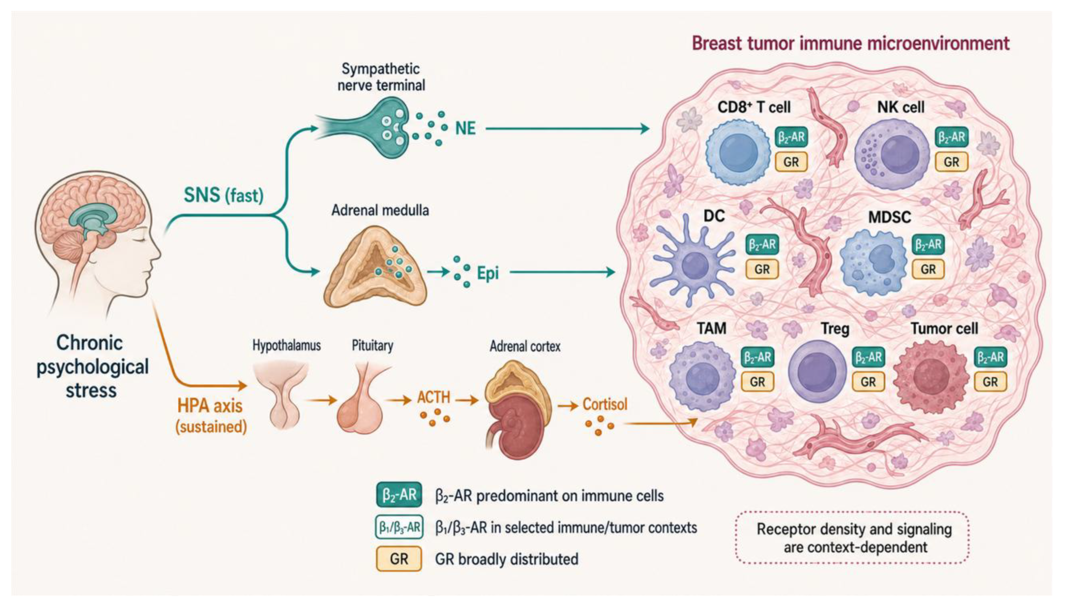
Figure 2.
The sympathetic β-adrenergic arm reprograms antitumor immunity through a shared cyclic AMP–protein kinase A program. Locally released norepinephrine and systemic epinephrine engage β-adrenergic receptors on several intratumoral populations, converging on a Gαs–cyclic AMP–protein kinase A cascade that lowers cytotoxic output and expands immunosuppression. In CD8⁺ T cells, β-adrenergic signaling blocks the metabolic reprogramming that effector function requires, and catecholamines acting through the β1-adrenergic receptor accelerate the transition to terminal exhaustion and raise PD-1, blunting responses to PD-1 blockade. Natural killer cell cytotoxicity falls, weakening control of metastatic spread. Dendritic cell activation and T cell priming are constrained through β2-adrenergic receptor signaling. Myeloid-derived suppressor cells expand and, through a β2-adrenergic receptor–STAT3 axis, raise arginase-1 and PD-L1, while macrophages are driven toward an M2, pro-metastatic state that underlies the sympathetic metastatic switch in breast cancer. On tumor cells, the β3-adrenergic receptor sustains interferon-γ–dependent PD-L1. Two nodes are emphasized: the β1-adrenergic receptor driving terminal exhaustion, and the β3-adrenergic receptor sustaining PD-L1.
Figure 2.
The sympathetic β-adrenergic arm reprograms antitumor immunity through a shared cyclic AMP–protein kinase A program. Locally released norepinephrine and systemic epinephrine engage β-adrenergic receptors on several intratumoral populations, converging on a Gαs–cyclic AMP–protein kinase A cascade that lowers cytotoxic output and expands immunosuppression. In CD8⁺ T cells, β-adrenergic signaling blocks the metabolic reprogramming that effector function requires, and catecholamines acting through the β1-adrenergic receptor accelerate the transition to terminal exhaustion and raise PD-1, blunting responses to PD-1 blockade. Natural killer cell cytotoxicity falls, weakening control of metastatic spread. Dendritic cell activation and T cell priming are constrained through β2-adrenergic receptor signaling. Myeloid-derived suppressor cells expand and, through a β2-adrenergic receptor–STAT3 axis, raise arginase-1 and PD-L1, while macrophages are driven toward an M2, pro-metastatic state that underlies the sympathetic metastatic switch in breast cancer. On tumor cells, the β3-adrenergic receptor sustains interferon-γ–dependent PD-L1. Two nodes are emphasized: the β1-adrenergic receptor driving terminal exhaustion, and the β3-adrenergic receptor sustaining PD-L1.
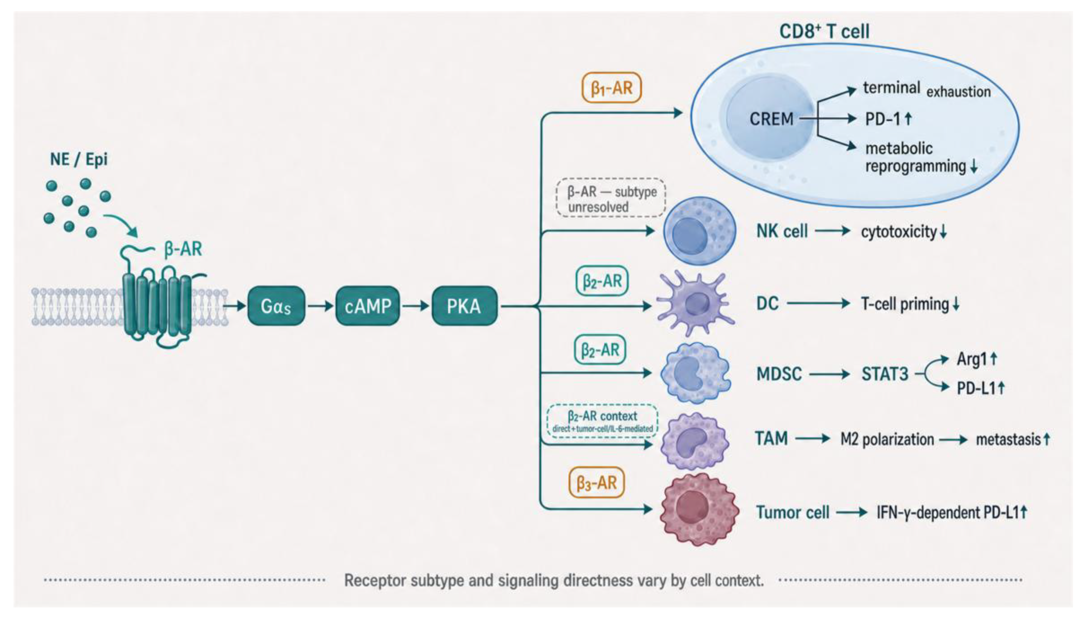
Figure 3.
The HPA glucocorticoid arm suppresses antitumor immunity through the glucocorticoid receptor. Stress-elevated and locally generated glucocorticoids activate the glucocorticoid receptor across immune lineages and in tumor cells, reaching the same immunosuppressive endpoint as the adrenergic arm through an independent receptor. In CD8⁺ T cells, the glucocorticoid receptor transactivates the inhibitory receptors PD-1, TIM-3, and LAG-3 and imposes a dysfunctional differentiation program whose intensity rises from naive toward exhausted cells. On tumor cells, glucocorticoid receptor activity raises PD-L1 and represses MHC class I, at once increasing an inhibitory ligand and reducing antigen visibility. Natural killer cells acquire PD-1 and lose interferon-γ output, with a further shift toward an amphiregulin-driven suppressive state. Dendritic cell priming is disabled through induction of TSC22D3, which blocks type I interferon responses. Regulatory T cells are activated by glucocorticoids that tumors regenerate through 11β-hydroxysteroid dehydrogenase, and glucocorticoid-dependent neutrophil and myeloid remodeling reshapes the pre-metastatic niche. A single receptor thus produces broad, coordinated suppression that mirrors the adrenergic arm.
Figure 3.
The HPA glucocorticoid arm suppresses antitumor immunity through the glucocorticoid receptor. Stress-elevated and locally generated glucocorticoids activate the glucocorticoid receptor across immune lineages and in tumor cells, reaching the same immunosuppressive endpoint as the adrenergic arm through an independent receptor. In CD8⁺ T cells, the glucocorticoid receptor transactivates the inhibitory receptors PD-1, TIM-3, and LAG-3 and imposes a dysfunctional differentiation program whose intensity rises from naive toward exhausted cells. On tumor cells, glucocorticoid receptor activity raises PD-L1 and represses MHC class I, at once increasing an inhibitory ligand and reducing antigen visibility. Natural killer cells acquire PD-1 and lose interferon-γ output, with a further shift toward an amphiregulin-driven suppressive state. Dendritic cell priming is disabled through induction of TSC22D3, which blocks type I interferon responses. Regulatory T cells are activated by glucocorticoids that tumors regenerate through 11β-hydroxysteroid dehydrogenase, and glucocorticoid-dependent neutrophil and myeloid remodeling reshapes the pre-metastatic niche. A single receptor thus produces broad, coordinated suppression that mirrors the adrenergic arm.
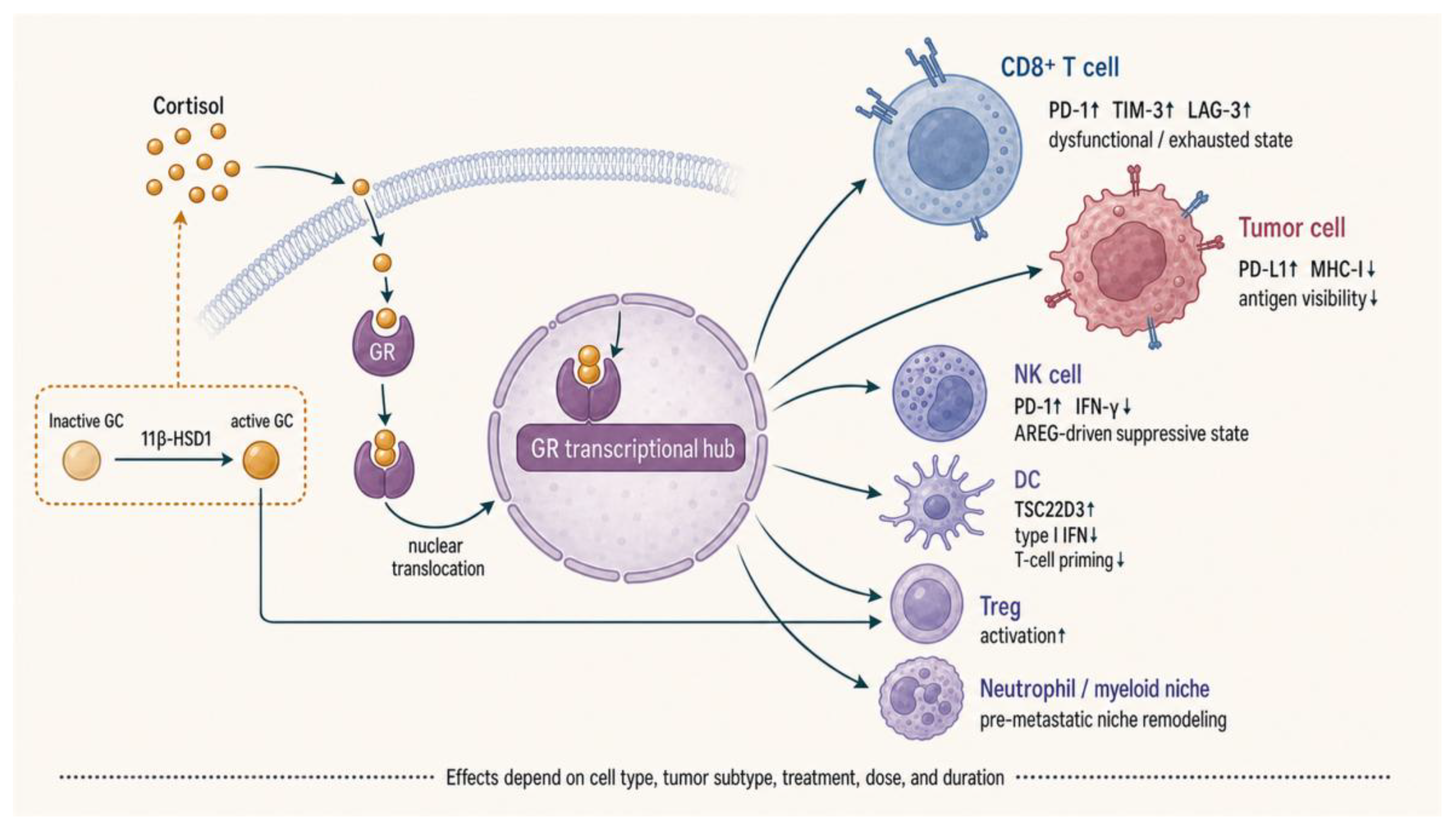
Figure 4.
Stress hormones as a convergent, self-amplifying neuroendocrine checkpoint. The central proposition of the review. The β-adrenergic and glucocorticoid arms enter from opposite sides and converge on one output, an exhausted, checkpoint-high CD8⁺ and natural killer cell phenotype marked by PD-1, TIM-3, and LAG-3. Convergence operates at three levels. Phenotypically, both arms push cytotoxic cells along the differentiation trajectory from TCF1⁺ progenitor-exhausted toward terminally exhausted states. Molecularly, an adrenergic cyclic AMP–protein kinase A–CREB node and a glucocorticoid receptor transcriptional node meet at the NFAT–TOX/NR4A exhaustion module and at the inhibitory-receptor loci it controls, while both arms converge on PD-L1 from different cellular compartments. Systemically, a single upstream stressor engages both arms together. A feed-forward loop is drawn as a bold arrow: glucocorticoids induce phenylethanolamine N-methyltransferase and tyrosine hydroxylase and sustain β-adrenergic receptor density, so the arms reinforce one another and their combined output is supra-additive. This coupling predicts that blocking one arm alone is insufficient. Dashed arrows mark the two links that remain to be closed experimentally: whether β-adrenergic input induces TOX or NR4A, and whether the glucocorticoid receptor and CREB co-occupy the same inhibitory-receptor loci.
Figure 4.
Stress hormones as a convergent, self-amplifying neuroendocrine checkpoint. The central proposition of the review. The β-adrenergic and glucocorticoid arms enter from opposite sides and converge on one output, an exhausted, checkpoint-high CD8⁺ and natural killer cell phenotype marked by PD-1, TIM-3, and LAG-3. Convergence operates at three levels. Phenotypically, both arms push cytotoxic cells along the differentiation trajectory from TCF1⁺ progenitor-exhausted toward terminally exhausted states. Molecularly, an adrenergic cyclic AMP–protein kinase A–CREB node and a glucocorticoid receptor transcriptional node meet at the NFAT–TOX/NR4A exhaustion module and at the inhibitory-receptor loci it controls, while both arms converge on PD-L1 from different cellular compartments. Systemically, a single upstream stressor engages both arms together. A feed-forward loop is drawn as a bold arrow: glucocorticoids induce phenylethanolamine N-methyltransferase and tyrosine hydroxylase and sustain β-adrenergic receptor density, so the arms reinforce one another and their combined output is supra-additive. This coupling predicts that blocking one arm alone is insufficient. Dashed arrows mark the two links that remain to be closed experimentally: whether β-adrenergic input induces TOX or NR4A, and whether the glucocorticoid receptor and CREB co-occupy the same inhibitory-receptor loci.
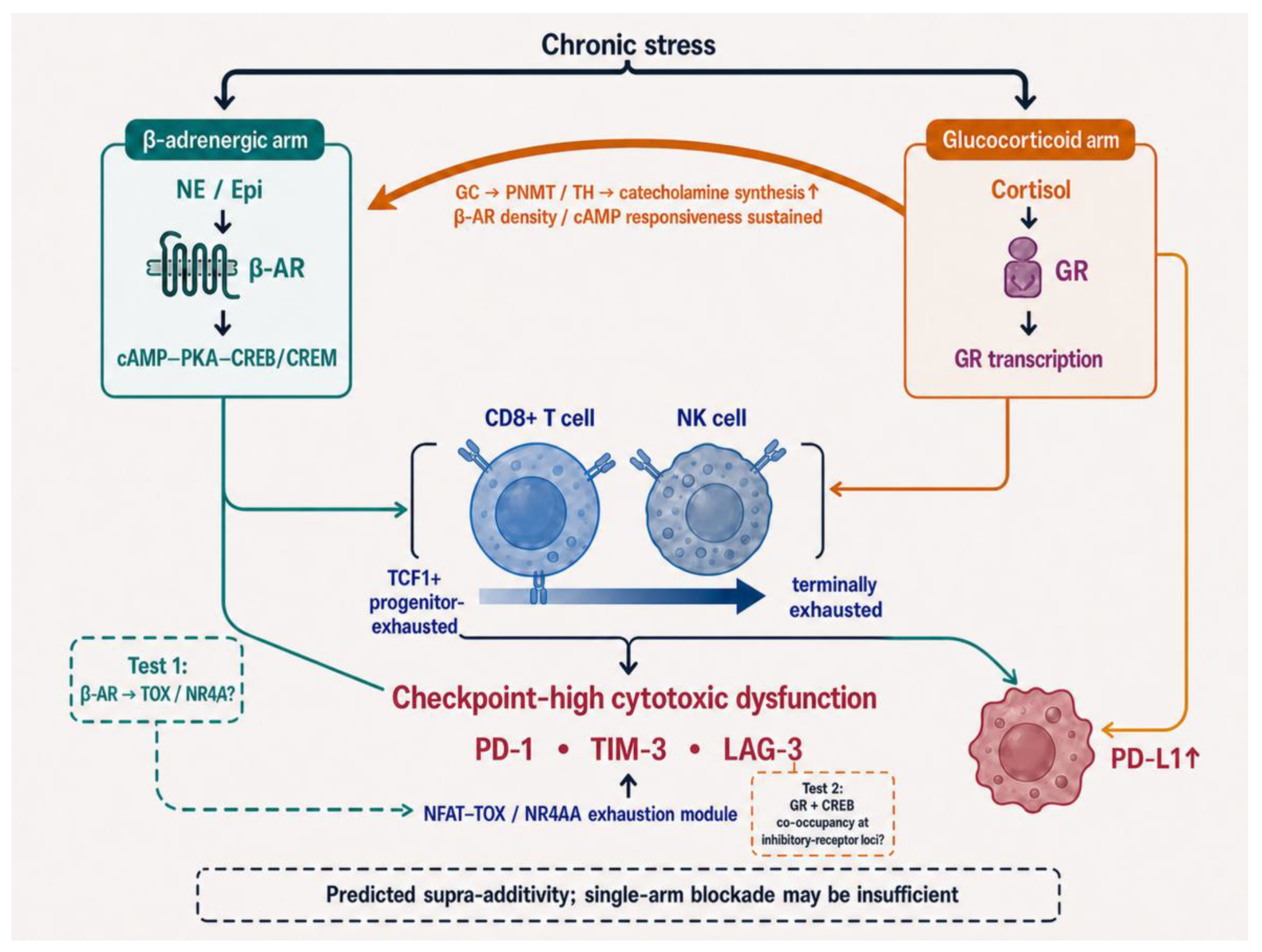
Figure 5.
Dual-axis co-targeting to relieve the convergent checkpoint and potentiate immunotherapy. The therapeutic logic that follows from convergence. Because the two arms funnel onto one endpoint and amplify each other, durable restoration of antitumor immunity is proposed to require relieving both, using agents already in clinical use. Three levers act at different levels: β-adrenergic blockade with a non-selective β-blocker relieves the sympathetic arm; a selective glucocorticoid receptor modulator relieves the glucocorticoid arm while sparing the capacity to manage toxicity; and behavioral stress reduction lowers the shared upstream driver and therefore relieves both arms at once. Together these are intended to shift exhausted CD8⁺ and natural killer cells back toward the checkpoint-responsive progenitor pool and to potentiate anti–PD-1 and anti–PD-L1 therapy. Biomarker-guided stratification that combines β2-adrenergic receptor, glucocorticoid receptor, and PD-L1 status is shown selecting patients whose resistance is stress-driven. Two constraints are flagged: dosing should be timed to protect the antitumor priming window, and the glucocorticoid paradox, namely that glucocorticoids remain essential for managing immune-related adverse events, limits how aggressively the glucocorticoid arm can be blocked.
Figure 5.
Dual-axis co-targeting to relieve the convergent checkpoint and potentiate immunotherapy. The therapeutic logic that follows from convergence. Because the two arms funnel onto one endpoint and amplify each other, durable restoration of antitumor immunity is proposed to require relieving both, using agents already in clinical use. Three levers act at different levels: β-adrenergic blockade with a non-selective β-blocker relieves the sympathetic arm; a selective glucocorticoid receptor modulator relieves the glucocorticoid arm while sparing the capacity to manage toxicity; and behavioral stress reduction lowers the shared upstream driver and therefore relieves both arms at once. Together these are intended to shift exhausted CD8⁺ and natural killer cells back toward the checkpoint-responsive progenitor pool and to potentiate anti–PD-1 and anti–PD-L1 therapy. Biomarker-guided stratification that combines β2-adrenergic receptor, glucocorticoid receptor, and PD-L1 status is shown selecting patients whose resistance is stress-driven. Two constraints are flagged: dosing should be timed to protect the antitumor priming window, and the glucocorticoid paradox, namely that glucocorticoids remain essential for managing immune-related adverse events, limits how aggressively the glucocorticoid arm can be blocked.
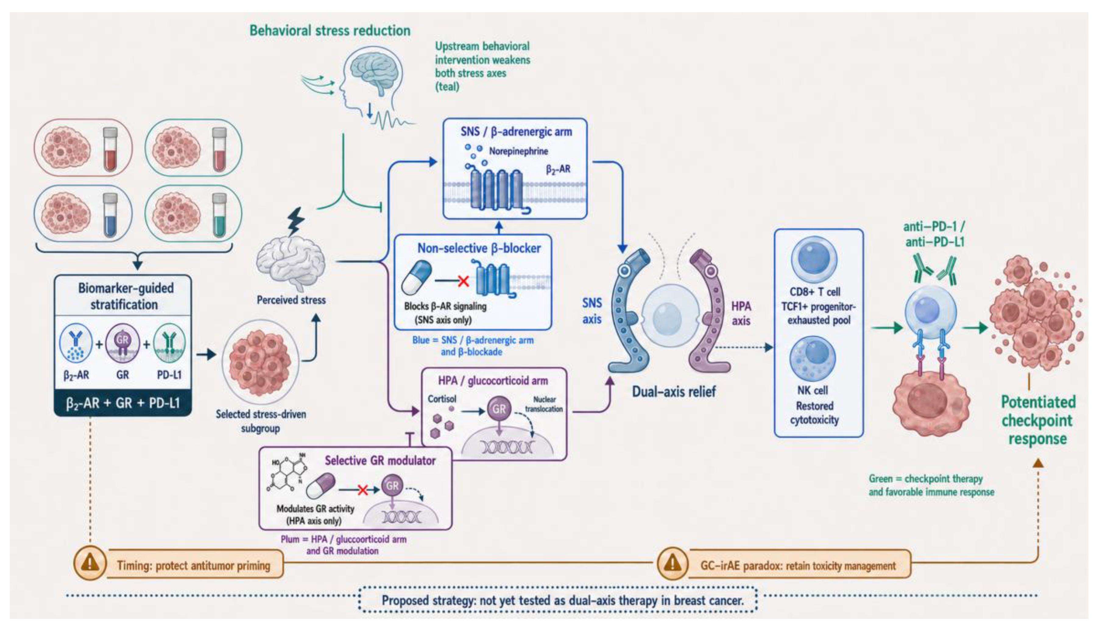
Disclaimer/Publisher’s Note: The statements, opinions and data contained in all publications are solely those of the individual author(s) and contributor(s) and not of MDPI and/or the editor(s). MDPI and/or the editor(s) disclaim responsibility for any injury to people or property resulting from any ideas, methods, instructions or products referred to in the content. |
© 2026 by the authors. Licensee MDPI, Basel, Switzerland. This article is an open access article distributed under the terms and conditions of the Creative Commons Attribution (CC BY) license.
Copyright: This open access article is published under a Creative Commons CC BY 4.0 license, which permit the free download, distribution, and reuse, provided that the author and preprint are cited in any reuse.


