
Submitted:
28 July 2026
Posted:
30 July 2026
You are already at the latest version
Abstract
A microcirculation was developed to study live endothelium under peristaltic flow. Vascular channel molds were fabricated with photolithography. Channels were formed with polydimethylsiloxane and seeded with human endothelial cells. A peristaltic pump circulated growth media over cells for 24 hours with varying shear stress and volumetric flow. After 24 hours, cells were live-imaged with differential interference contrast microscopy or fixed and stained with fluorophore-conjugated phalloidin and 4’,6-diamidino-2’-phenylindole,dihydrochloride. Endothelial cell alignment was measured with purpose-written MATLAB code. Endothelial cell length and nucleus size were measured with semi-automated Zen and Image J analysis. Compared to a static no flow condition (0 Pa shear stress), significant endothelial cell alignment was observed during flow of 0.23 mL/min (2 Pa, p=0.0011) and flow of 0.46 mL/min (4 Pa, p=0.0004). Cell length (0 Pa, 50±14 µm; 2 Pa, 99±26 µm; 4 Pa, 110±30 µm, p< 0.0001) and nucleus area (0 Pa, 0.10±0.02 pixels2; 2 Pa, 11±0.01 pixels2; 4 Pa, 13±0.02 pixels2, p< 0.0001) increased significantly whereas nucleus circularity (0 Pa, 0.86±0.06; 2 Pa, 0.84±0.04; 4 Pa, 0.85±0.03, p=0.07) trended toward decrease. An ex vivo microcirculation was developed for laboratory experimentation. This relatively simple and cost-effective system may have translational utility to investigate human endothelial cell biology during flow.
Keywords:
artificial circulation
; microcirculation
; microfluidic
; endothelial cell
; shear stress
; polydimethylsiloxane (PDMS)
1. Introduction
Artificial microcirculations with live cells and flow are useful in laboratory investigation. Ex vivo circuits seeded with endothelial cells may be used to study vascular biology, rheology, and biophysics [1,2,3,4,5,6,7,8,9,10,11,12,13,14,15,16,17,18,19,20,21,22,23,24,25]. Varied experimental models provide opportunities to study artificial circulation, vascular biology, endothelial cell pharmacology, angiogenesis, and thrombosis and hemostasis in tightly controlled conditions.
In this article, we describe and characterize a relatively simple and cost-effective ex vivo microcirculation with live human endothelial cells. The model, research utility, and potential clinical translation are discussed herein.
2. Methods
2.1. Vascular Channel Photolithography
A photomask template with desired channel dimensions was fabricated with Inkscape (Scalable Vector Graphics) open-source graphics editor. The photomask template included 15 channel patterns (Figure 1A). Channel patterns were printed (Micro Lithography Services Limited) for use on 3-inch silicon wafers (University Wafers) (Figure 1B). Molds were made on silicon wafers with epoxy-based, negative-tone permanent photoresist material as we previously described [26]. SU-8 2035 (Kayaku Advanced Materials) was chosen to reach patterned channel heights between 80 and 100 µm. Silicon wafers were rinsed with acetone and dried with a stream of nitrogen. After drying, SU-8 was poured on the substrate surface, which was spin coated at 600 revolutions per minute (RPM) for 100 seconds with an initial spread cycle of 10 seconds at 500 RPM. Next, the wafer was soft baked at 65 °C for 5 minutes, 95 °C for 30 minutes, and exposed to 365 nm ultraviolet light for 4 minutes. Post-exposure bake was performed at 65 °C for 10 minutes and 95 °C for 20 minutes. The wafer was immersed in SU-8 developer and agitated until channel patterns were clearly visible. The develop cycle lasted 3 minutes and was terminated by isopropyl alcohol rinse and blow drying with nitrogen. This process yielded repeatable SU-8 patterns to mold polydimethylsiloxane (PDMS) microvascular channels.
2.2. Vascular Channel Preparation
Straight 35 mm long vascular channels were produced in PDMS. It was possible to generate channels that were approximately 85 µm tall x 300 to 1,100 µm wide. For this study, channel size of 85 x 1,100 µm was selected, manufactured, and used in the microcirculation. Replicas were made with approximately 22 g of Sylgard 184 (DOW) with a mixture of 90% Elastomer Base and 10% Elastomer Curing Agent by weight. The mixture was placed in a desiccator to prevent bubbles and other channel defects upon curing. Circumferential tape around wafers retained PDMS within tape borders (Figure 1C). PDMS coated wafers were cured at 60 °C overnight. After curing, PDMS was carefully peeled off wafers, channel shapes cut out with an X-ACTO knife, and placed channel side down on tape to ensure no debris. To form vascular channels, a plasma cleaner (Harrick Plasma) bonded the PDMS to 24 x 50 mm #1.5 rectangular coverslips (Corning). Before each experiment, coverslips were soaked in ethanol for 5 minutes, dried, and cleaned with air plasma for 10 minutes. Inlet and outlet channel holes were fashioned with 18-gauge blunt syringe tips (Jensen Global) at channel beginning and end. The coverslip and PDMS channel were cleaned with air plasma for 30 seconds before bonding together by rapid and careful flipping of the PDMS channel on to the coverslip and application of topical pressure (Figure 1D). After bonding, deionized water was injected to preserve coverslip surface hydrophilicity.
2.3. Human Endothelial Cell Culture
Study approval was obtained from Geisinger Medical Center Institutional Review Board (IRB# 2021-0441, approved 01/05/2022) to use human cells and blood products in laboratory experiments.
Human umbilical vein endothelial cells (HUVECs, Lonza) were cultured in buffered endothelial growth media (EGM, Lonza) made with 5 mL penicillin streptomycin (Gibco) and EGM SingleQuots Supplements (Lonza) which contained 5 mL fetal bovine serum, 2 mL recombinant human basic fibroblast growth factor, 0.5 mL ascorbic acid, 0.5 mL heparin, 0.5 mL vascular endothelial growth factor, 0.5 mL gentamicin sulfate-amphotericin, 0.5 mL recombinant human epidermal growth factor, 0.5 mL long arginine 3-insulin-like growth factor-1, and 0.2 mL hydrocortisone in 500 mL endothelial basal medium (Lonza) in T75 culture flasks (Corning) at 37oC with 5% CO2. Fourth or fifth passage cells were used.
2.4. Seeding of Vascular Channels with Endothelial Cells
PDMS channels were sterilized with ethanol. Vascular channels were coated with fibronectin (Sigma-Aldrich) at 100 µg/mL and dried in a laminar flow hood. HUVECs were seeded at approximately 1.03x10⁶ cells/mL in EGM. Cells were seeded in channels and allowed to reach at least 80% confluence before applying flow. Channels of this size held approximately 3.39x10⁴ cells. After seeding, channel inlets and outlets were sealed with vacuum grease to prevent bubble formation or media evaporation from channels. Endothelial cells settled, adhered, and spread for 24 hours prior to initiation of flow.
2.5. Circuit Design
The model was designed to investigate human endothelial cell behavior during flow and shear stress conditions. The circuit was constructed inside a cell incubator. The circuit consisted of a peristaltic pump (Longer Precision Pump Co.), tygon tubing (approximately 750 cm), and a 100 mL reservoir (Figure 2). Tygon tubing (0.51 mm 3-stop, Darwin Microfluidics) connected the media reservoir to the pump and to the channel inlet. Tygon tubing (1.02 mm, VWR) connected the channel outlet to the media reservoir. Inlet and outlet tubing were inserted into the reservoir to recirculate media through the circuit.
2.6. Circuit Function with Live Cells and Flow
At the beginning of an experiment, low flow of 0.17 mL/min (1.5 Pa) was applied for approximately two hours to deair the system and flush out non-adherent cells. Afterward, cells were examined under light microscopy to verify healthy cells with at least 80% confluence were adherent within the channel. Flow was then increased to achieve the desired flow rate or shear stress target for 24 hours.
Three experimental conditions were examined after 24 hours: 1. Static condition with no shear stress (no flow), 2. 2 Pa shear stress (0.23 mL/min), 3. 4 Pa shear stress (0.46 mL/min).
2.7. Estimation of Flow
Flow rate through the circuit was determined by direct volumetric measurement of fluid output after 10 minutes at fixed RPM. Flow rate for each condition was selected to achieve shear stress of 0 Pa, 2 Pa, and 4 Pa within channels.
2.8. Estimation of Shear Stress
Shear stress was estimated with the method below [27]:
where η is viscosity, Q is flow rate, h is channel height, w is channel width, and z is distance from channel center perpendicular to flow.
2.9. Endothelial Cell Imaging
Endothelial cells were imaged with differential interference contrast microscopy or fixed and stained with 4’ 6-diamidino-2’-phenylindole, dihydrochloride (DAPI, Thermo Fisher Scientific) and Alexa Fluor 488 phalloidin (Thermo Fisher Scientific). To fix cells, channels were infused with 3.7% formaldehyde (Thermo Fisher Scientific) at room temperature for 15 min. Channels were rinsed twice with phosphate buffered saline (PBS, Sigma-Aldrich) with 3% bovine serum albumin (BSA, Sigma-Aldrich) followed by 0.5% Triton X-100 (Sigma-Aldrich) in PBS, incubated at room temperature for 20 min, and again rinsed with PBS with 3% BSA. Next, phalloidin (1:100 in PBS) was infused into the channel at room temperature for 15 min. After rinsing with Dulbecco’s PBS (DPBS, Sigma-Aldrich), DAPI (1:1,000 in PBS) was infused into the channel at room temperature for 30 min. A final DPBS rinse was performed. The channel was imaged on a Zeiss LSM 710 confocal microscope (Zeiss) with 405 and 488 nm wavelength lasers.
2.10. Endothelial Cell Alignment Analysis
2.11. Endothelial Cell Morphology Analysis
Confocal images of cells within channels were analyzed to determine cell length, nucleus size, and nucleus circularity during each condition. Zen software (Zeiss) was used to measure cell length. The longest length of phalloidin-stained cells was measured and recorded.
ImageJ software was used to determine nucleus size and nucleus circularity. Images of DAPI stained cells were converted to 8-bit images before thresholding to highlight nuclei. Nucleus area was recorded. The Shape Descriptors feature provided a measurement of circularity. Circularity was measured on a scale of 0 to 1 with 1 being a perfect circle and 0 being a line or single point.
2.12. Statistics
GraphPad Prism software was used to perform statistical analyses. Cell alignment histogram shape distributions were compared with Nonparametric Kolmogorov-Smirnov Tests. For cell length, nucleus area, and nucleus circularity, n=50 cells were randomly measured from each condition. Non-repeated measures ANOVA with Tukey post-test was performed. A p<0.05 was considered statistically significant.
3. Results
As a static control, zero flow produced zero shear stress. During flow conditions, 0.23 mL/min produced 2 Pa shear stress, and 0.46 mL/min flow produced 4 Pa shear stress.
After 24 hours, control endothelial cells exhibited classic cobblestone morphology without prominent actin filaments, and cells did not align (Figure 2D). In contrast, cells exposed to 2 Pa and 4 Pa shear stress for 24 hours elongated in the direction of flow, exhibited prominent actin filaments, and significantly aligned (p=0.0004, Figure 2E,F).
Endothelial cell length (0 Pa, 50±14 µm; 2 Pa, 99±26 µm; 4 Pa, 110±30 µm, p<0.0001) and nucleus area (0 Pa, 0.10±0.02 pixels2; 2 Pa, 11±0.01 pixels2; 4 Pa, 13±0.02 pixels2, p<0.0001) increased significantly during flow conditions (Figure 4A,B). Nucleus circularity (0 Pa, 0.86±0.06; 2 Pa, 0.84±0.04; 4 Pa, 0.85±0.03, p=0.07) trended toward a decrease that was not significant.
The total cost to build the system and buy start-up supplies was approximately $17,177. The cost to run each experiment was approximately $60 worth of disposables and cell culture materials. For reference, Table 1 lists costs of system components.
4. Discussion
We developed and tested an ex vivo microcirculation with live human endothelial cells and flow. Specifically, the circuit includes PDMS-based vascular channels and a peristaltic pump that is 1) simple to image live cells, 2) relatively inexpensive to construct (<$18,000) and run each experiment (<$100), and 3) customizable for experimental goals. Preliminary experiments characterized 1) circuit flow and shear stress, 2) endothelial cellular alignment, and 3) endothelial cell and nucleus architecture. This simple and inexpensive system may be used to study live endothelial cell biology during flow and shear stress conditions.
Biomechanical forces from blood pressure, flow, and shear stress influence vascular architecture and function [28,29]. Vascular remodeling is a natural consequence of pathologic mechanical loading [28,29]. Many clinical examples exist. Non-pulsatile mechanical circulatory support activates cellular programs involved in endothelial cell size, metabolism, protein manufacturing, matrix deposition, vasoreactivity, life cycle, and microcirculatory dysfunction [30,31]. Chronic hypertension causes endothelial dysfunction, hyperplastic remodeling, and accelerated atherogenesis [32]. Deranged neurohormonal signaling in heart failure causes endothelial dysfunction and arterial stiffening [33]. Increased flow in arteriovenous fistulas for hemodialysis cause intimal hyperplasia and vascular dilation [32]. In these pathologies, abnormal mechanical forces influence endothelial cell architecture and function with important clinical implications. Endothelial cell remodeling may alter barrier and vasomotor function and contribute to angiodysplasia [34,35] and bleeding [36,37], refractory hypertension [37,38] and hemorrhagic stroke [37,38]. However, mechanistic basis and biophysical thresholds for vascular changes are poorly characterized.
Ex vivo microcirculations with live endothelial cells provide experimental models to investigate mechanisms of vascular remodeling and test novel therapies. Circuits may be designed to model either physiologic or pathophysiologic flow and shear stress conditions. Multiple ex vivo circuit types have been described that vary from large, complex circulations [1,2,3] to organ-on-a-chip configurations [4]. These have included parallel plate flow chambers [5,6,7,8,9], circular impinging flow chambers [10], orbital shakers [11,12,13], cone and plate devices [13], and commercially available channels [13,14,15]. Three-dimensional cell culture techniques [18], compliance [1], and hemodynamic sensors may expand experimental capabilities [19,20]. These systems allow investigation of cell migration and alignment [5,6,10,13,14,21], effects of wall shear stress [6,12], shear stress gradients [6,10,22], turbulence and disturbed flow [21], flow directionality change [9], cell signaling [5,8,13,14], and angiogenesis and neovascularization during various experimental conditions. These systems have been reviewed in detail [16,17].
PDMS vascular channels are commonly used in ex vivo microcirculations [21,23,24]. PDMS is inexpensive, offers design versatility to mimic vessel sizes and geometries, and does not require specialized or expensive equipment or machining. PDMS surface treatment facilitates cell adhesion for many cell lines [25]. PDMS is optically transparent for high magnification imaging and is gas permeable for long-term cell culture in an incubator.
The total cost to construct the system and buy start-up supplies was less than $18,000. Each experiment cost approximately $60 in disposable supplies and cell culture materials. Equipment purchased to perform experiments included a peristaltic pump, plasma cleaner, and vacuum pump. Other equipment such as a cell culture incubator, laminar flow hood, microscope, desiccator, hot plates, and oven (optional) are common in university and industry laboratories. A custom photomask, silicon wafers, glass coverslips, tubing, syringe tips, endothelial cells, cell culture and staining supplies, and disposables modestly increase costs. Photolithography may be performed in a clean room, or a simple system may be organized in a laboratory setting [39,40]. Low-cost ultraviolet exposure for photolithography is also easily achieved with simple electronics and minimal supplies. For less than $250 [39], a system may be constructed with an Arduino Uno microcontroller (Arduino), board of 161 ultraviolet light-emitting diodes (DigiKey), IRFP460 power metal-oxide-semiconductor field-effect transistor (DigiKey), liquid crystal display (DigiKey), 4x4 keypad (DigiKey), potentiometer (DigiKey), and an external power supply (DigiKey). Electronic component configuration and code to program and run the microcontroller are previously reported [40]. Simple housing in which to mount components and a sample stage to hold wafers are easily machined. A spin coater (Setcas), hotplates, photoresist and accompanying developer, and other common laboratory chemicals are required.
5. Limitations
Limitations of an ex vivo system are inherent in this model. Hemodynamics are not identical to native physiology. A single layer of endothelial cells seeded on PDMS does not recreate intima-media interactions of an intact vessel. Modification of the system with a more sophisticated pump, sensors, and endothelial cell-smooth muscle cell co-culture or a live, intact vessel may increase biological accuracy and clinical relevance of the system.
Twenty-four hours of flow does not provide chronic, steady state homeostasis. Endothelial cell metabolism depletes media nutrients, and during prolonged experiments, recirculated media should be replaced.
Endothelial cell media does not contain blood cells. Circulation of human blood products or anticoagulated whole human blood is an option in this model that may further increase clinical relevance. Low circuit priming volume may limit identification of peptides secreted from endothelial cells into supernatant in low concentration. Larger channels with more cells may mitigate this limitation.
Silicon wafer molds are fragile and may develop defects or break. Batches of PDMS channels may vary slightly in volume and surface contour based on minor variations in preparation technique and curing time. Appropriate deairing and avoidance of air entrainment are necessary to prevent bubbles in the circuit that may undermine cell adhesion when flow is applied.
6. Conclusions
We describe a simple and cost-effective ex vivo microcirculation with live endothelial cells and peristaltic flow. This system has applications to investigate endothelial biology during tightly controlled flow and shear stress conditions. This model may be useful to study mechanical influence over endothelial cell architecture, function, and signaling. Modification of the circuit may facilitate investigator specific goals.
Author Contributions
Conceptualization, AAA and CRB; methodology, AAA and CRB; validation, AAA; formal analysis, AAA; investigation, AAA and RDD; resources, CRB; data curation, AAA; writing—original draft preparation, AAA and CRB; writing—review and editing, AAA, RDD and CRB; supervision, CRB; project administration, CRB; funding acquisition, AAA and CRB. All authors have read and agreed to the published version of this manuscript.
Funding
Funds were provided by NIH R01 HL172022-01, a grant from Geisinger Janet Weis Children’s Hospital Beyond the Bricks Foundation, and a grant from the Bucknell-Geisinger Research Initiative.
Institutional Review Board Statement
Study approval was obtained from Geisinger Medical Center Institutional Review Board (IRB# 2021-0441, approved 01/05/2022) to use human cells and blood products in laboratory experiments.
Informed Consent Statement
Not applicable.
Data Availability Statement
The datasets in this article are available from the corresponding author upon reasonable request.
Acknowledgments
The authors acknowledge and thank Dr. Aurelia Honerkamp-Smith at Lehigh University, Bethlehem PA for use of photolithography facilities.
Conflicts of Interest
The authors declare no disclosures or conflict of interest.
References
- Bartoli CR, Spence PA, Giridharan GA. Novel J stents reduce the risk of embolic stroke in vitro. Neuroradiology. 2010;52(5):345–7. [CrossRef]
- Restle DJ, Zhang DM, Hung G, Howard JL, Kallel F, Acker MA, et al. Preclinical Models for Translational Investigations of Left Ventricular Assist Device-Associated von Willebrand Factor Degradation. Artif Organs. 2015;39(7):569–75. [CrossRef]
- Bartoli CR, Kang J, Zhang D, Howard J, Acker M, Atluri P, et al. Left Ventricular Assist Device Design Reduces von Willebrand Factor Degradation: A Comparative Study Between the HeartMate II and the EVAHEART Left Ventricular Assist System. Ann Thorac Surg. 2017;103(4):1239–44. [CrossRef]
- Cao UMN, Zhang Y, Chen J, Sayson D, Pillai S, Tran SD. Microfluidic Organ-on-A-chip: A Guide to Biomaterial Choice and Fabrication. Int J Mol Sci. 2023;24(4). [CrossRef]
- Wang C, Baker BM, Chen CS, Schwartz MA. Endothelial cell sensing of flow direction. Arterioscler Thromb Vasc Biol. 2013;33(9):2130–6. [CrossRef]
- Yoshino D, Sakamoto N, Sato M. Fluid shear stress combined with shear stress spatial gradients regulates vascular endothelial morphology. Integr Biol (Camb). 2017;9(7):584–94. [CrossRef]
- Karlon WJ, Hsu P-P, Li S, Chien S, McCulloch AD, Omens JH. Measurement of Orientation and Distribution of Cellular Alignment and Cytoskeletal Organization. Annals of Biomedical Engineering. 1999;27(6):712–20. [CrossRef]
- Ebong EE, Lopez-Quintero SV, Rizzo V, Spray DC, Tarbell JM. Shear-induced endothelial NOS activation and remodeling via heparan sulfate, glypican-1, and syndecan-1. Integr Biol (Camb). 2014;6(3):338–47. [CrossRef]
- Wang C, Lu H, Schwartz MA. A novel in vitro flow system for changing flow direction on endothelial cells. J Biomech. 2012;45(7):1212–8. [CrossRef]
- Ostrowski MA, Huang NF, Walker TW, Verwijlen T, Poplawski C, Khoo AS, et al. Microvascular endothelial cells migrate upstream and align against the shear stress field created by impinging flow. Biophys J. 2014;106(2):366–74. [CrossRef]
- Fernandes A, Hosseini V, Vogel V, Lovchik RD. Engineering solutions for biological studies of flow-exposed endothelial cells on orbital shakers. PLoS ONE. 2022;17(1):e0262044. [CrossRef]
- Feng S, Bowden N, Fragiadaki M, Souilhol C, Hsiao S, Mahmoud M, et al. Mechanical Activation of Hypoxia-Inducible Factor 1α Drives Endothelial Dysfunction at Atheroprone Sites. Arteriosclerosis, Thrombosis, and Vascular Biology. 2017;37(11):2087–101. [CrossRef]
- Ghimire K, Zaric J, Alday-Parejo B, Seebach J, Bousquenaud M, Stalin J, et al. MAGI1 Mediates eNOS Activation and NO Production in Endothelial Cells in Response to Fluid Shear Stress. Cells [Internet]. 2019; 8(5):[388 p.]. [CrossRef]
- ‘t Hart DC, van der Vlag J, Nijenhuis T. Laminar flow substantially affects the morphology and functional phenotype of glomerular endothelial cells. PLoS ONE. 2021;16(5):e0251129. [CrossRef]
- Volpatti LR, Yetisen AK. Commercialization of microfluidic devices. Trends in Biotechnology. 2014;32(7):347–50. [CrossRef]
- Meng F, Cheng H, Qian J, Dai X, Huang Y, Fan Y. In vitro fluidic systems: Applying shear stress on endothelial cells. Medicine in Novel Technology and Devices. 2022;15:100143. [CrossRef]
- Gharib G, Bütün İ, Muganlı Z, Kozalak G, Namlı İ, Sarraf SS, et al. Biomedical Applications of Microfluidic Devices: A Review. Biosensors [Internet]. 2022; 12(11):[1023 p.]. [CrossRef]
- Gupta N, Liu JR, Patel B, Solomon DE, Vaidya B, Gupta V. Microfluidics-based 3D cell culture models: Utility in novel drug discovery and delivery research. Bioeng Transl Med. 2016;1(1):63–81. [CrossRef]
- Buttkewitz MA, Heuer C, Bahnemann J. Sensor integration into microfluidic systems: trends and challenges. Current Opinion in Biotechnology. 2023;83:102978. [CrossRef]
- Islam MN, Doria SM, Fu X, Gagnon ZR. Piezoresistive Conductive Microfluidic Membranes for Low-Cost On-Chip Pressure and Flow Sensing. Sensors (Basel). 2022;22(4). [CrossRef]
- Tovar-Lopez F, Thurgood P, Gilliam C, Nguyen N, Pirogova E, Khoshmanesh K, et al. A Microfluidic System for Studying the Effects of Disturbed Flow on Endothelial Cells. Front Bioeng Biotechnol. 2019;7:81. [CrossRef]
- Sakamoto N, Saito N, Han X, Ohashi T, Sato M. Effect of spatial gradient in fluid shear stress on morphological changes in endothelial cells in response to flow. Biochemical and Biophysical Research Communications. 2010;395(2):264–9. [CrossRef]
- Shih H-C, Lee T-A, Wu H-M, Ko P-L, Liao W-H, Tung Y-C. Microfluidic Collective Cell Migration Assay for Study of Endothelial Cell Proliferation and Migration under Combinations of Oxygen Gradients, Tensions, and Drug Treatments. Sci Rep. 2019;9(1):8234. [CrossRef]
- Thomas A, Daniel Ou-Yang H, Lowe-Krentz L, Muzykantov VR, Liu Y. Biomimetic channel modeling local vascular dynamics of pro-inflammatory endothelial changes. Biomicrofluidics. 2016;10(1):014101. [CrossRef]
- Halldorsson S, Lucumi E, Gómez-Sjöberg R, Fleming RMT. Advantages and challenges of microfluidic cell culture in polydimethylsiloxane devices. Biosensors and Bioelectronics. 2015;63:218–31. [CrossRef]
- Anthony AA, Sahin O, Yapici MK, Rogers D, Honerkamp-Smith AR. Systematic measurements of interleaflet friction in supported bilayers. Biophys J. 2022;121(15):2981–93. [CrossRef]
- Jönsson P, Beech JP, Tegenfeldt JO, Höök F. Mechanical Behavior of a Supported Lipid Bilayer under External Shear Forces. Langmuir. 2009;25(11):6279–86. [CrossRef]
- Haga JH, Li Y-SJ, Chien S. Molecular basis of the effects of mechanical stretch on vascular smooth muscle cells. Journal of Biomechanics. 2007;40(5):947–60. [CrossRef]
- Li YS, Haga JH, Chien S. Molecular basis of the effects of shear stress on vascular endothelial cells. J Biomech. 2005;38(10):1949–71. [CrossRef]
- Bartoli CR, Spence PA, Siess T, Raess DH, Koenig SC, Dowling RD. Nonphysiologic blood flow triggers endothelial and arterial remodeling in vivo: implications for novel left ventricular assist devices with a peripheral anastomosis. J Thorac Cardiovasc Surg. 2014;148(1):311–21. [CrossRef]
- Bruegger D, Schwartz L, Chappell D, Jacob M, Rehm M, Vogeser M, et al. Release of atrial natriuretic peptide precedes shedding of the endothelial glycocalyx equally in patients undergoing on- and off-pump coronary artery bypass surgery. Basic Res Cardiol. 2011;106(6):1111–21. [CrossRef]
- Leitschuh M, Chohanian A. Vascular Changes in Hyertension. Medical Clinics of North America. 1987;71(5):827–41. [CrossRef]
- Nakamura M. Peripheral vascular remodeling in chronic heart failure: Clinical relevance and new conceptualization of its mechanisms. Journal of Cardiac Failure. 1999;5(2):127–38. [CrossRef]
- Kang J, Hennessy-Strahs S, Kwiatkowski P, Bermudez CA, Acker MA, Atluri P, et al. Continuous-Flow LVAD Support Causes a Distinct Form of Intestinal Angiodysplasia. Circulation Research. 2017;121(8):963–9. [CrossRef]
- Bartoli CR, Zhang DM, Hennessy-Strahs S, Kang J, Restle DJ, Bermudez C, et al. Clinical and In Vitro Evidence That Left Ventricular Assist Device-Induced von Willebrand Factor Degradation Alters Angiogenesis. Circ Heart Fail. 2018;11(9):e004638. [CrossRef]
- Crow S, Chen D, Milano C, Thomas W, Joyce L, Piacentino V, 3rd, et al. Acquired von Willebrand syndrome in continuous-flow ventricular assist device recipients. Ann Thorac Surg. 2010;90(4):1263–9; discussion 9. [CrossRef]
- Mehra MR, Uriel N, Naka Y, Cleveland JC, Jr., Yuzefpolskaya M, Salerno CT, et al. A Fully Magnetically Levitated Left Ventricular Assist Device - Final Report. N Engl J Med. 2019;380(17):1618–27. [CrossRef]
- Markham DW, Fu Q, Palmer MD, Drazner MH, Meyer DM, Bethea BT, et al. Sympathetic neural and hemodynamic responses to upright tilt in patients with pulsatile and nonpulsatile left ventricular assist devices. Circ Heart Fail. 2013;6(2):293–9. [CrossRef]
- Murat Kaya Y, Ilyas F, editors. UV-LED exposure system for low-cost photolithography. ProcSPIE; 2014.
- Anthony A. Systematic Measurements of Interleaflet Friction in Supported Bilayers: Lehigh University; 2022.
Figure 1.
A: A photomask template with vascular channel patterns is shown. B: A wafer is shown after a straight channel pattern was applied via photolithography. C: A wafer is shown with a layer of PDMS before curing. D: A cured PDMS channel is shown with inlet and outlet holes plasma bonded to a glass coverslip.
Figure 1.
A: A photomask template with vascular channel patterns is shown. B: A wafer is shown after a straight channel pattern was applied via photolithography. C: A wafer is shown with a layer of PDMS before curing. D: A cured PDMS channel is shown with inlet and outlet holes plasma bonded to a glass coverslip.
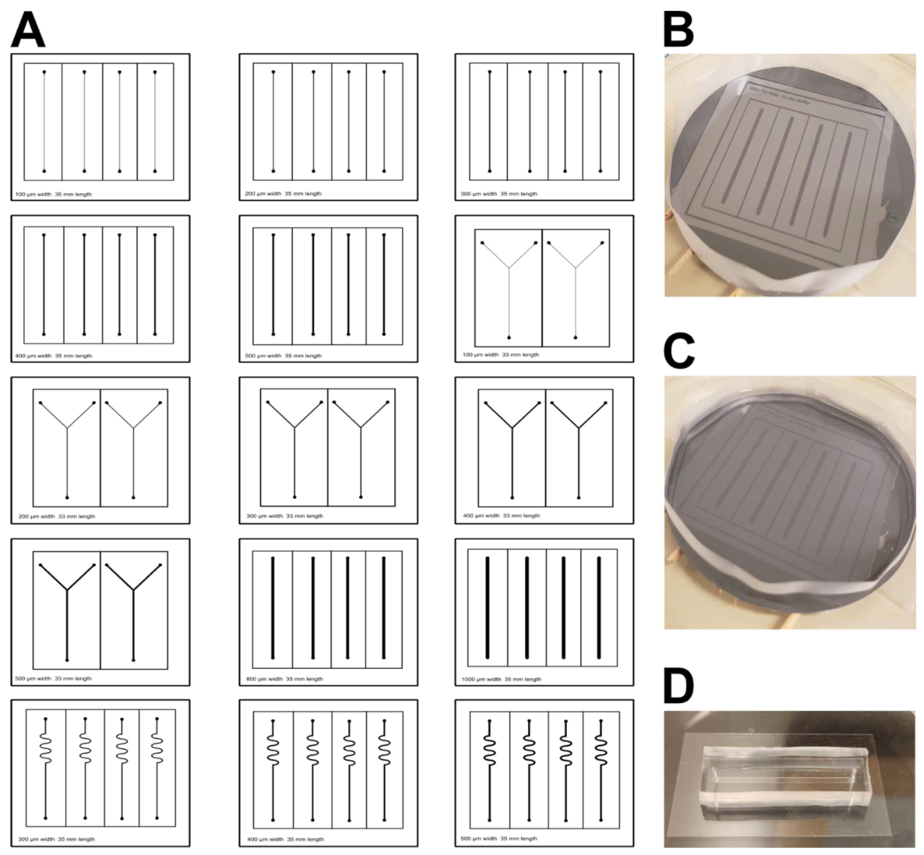
Figure 2.
A: The ex vivo microcirculation is shown. B: A cartoon schematic is shown.
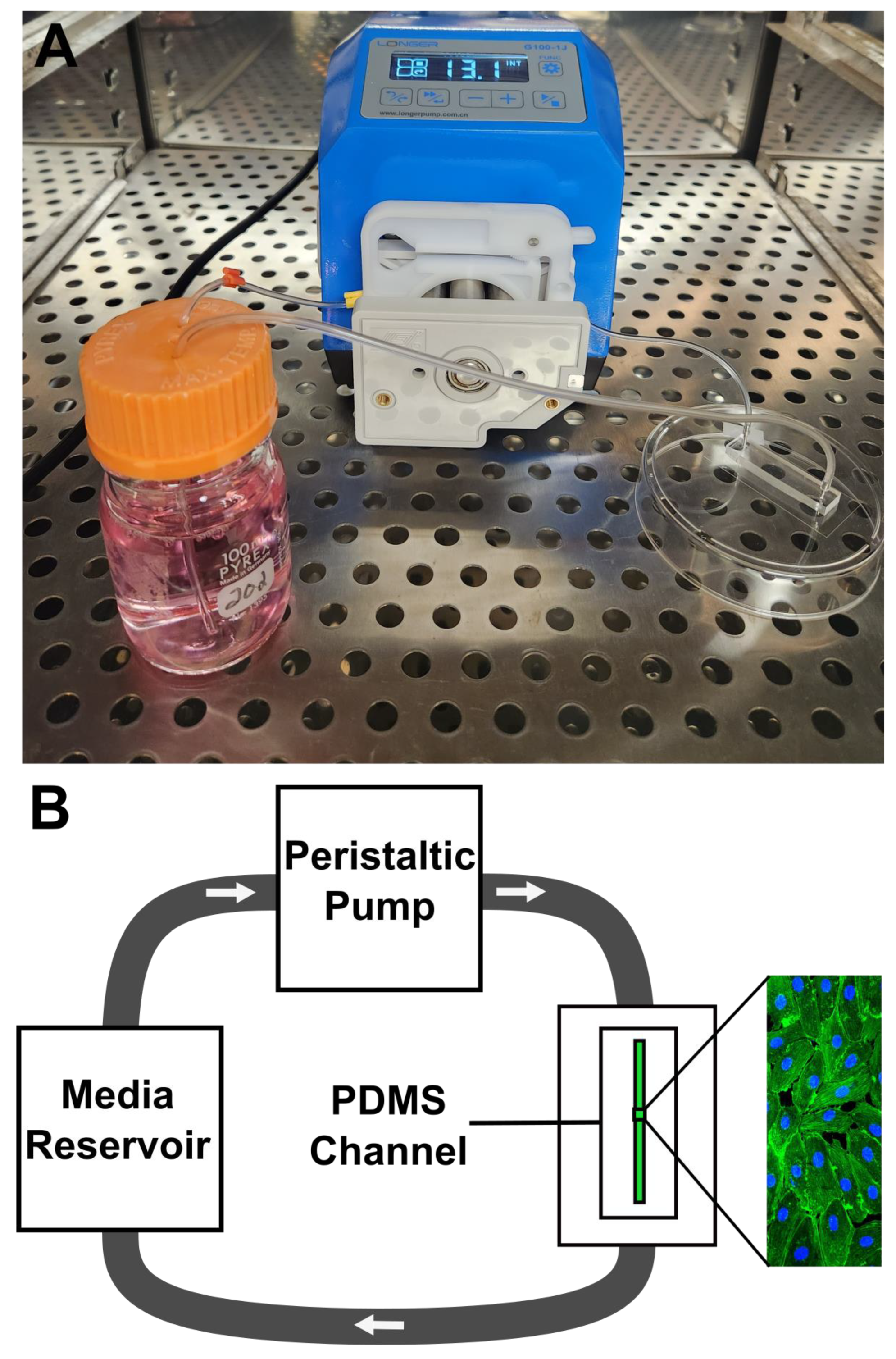
Figure 3.
Endothelial cells fixed with 4’ 6-diamidino-2’-phenylindole, dihydrochloride and phalloidin imaged at 20x after 24 hours of A: 0 Pa shear stress (no flow), B: 2 Pa shear stress (0.23 mL/min flow), and C: 4 Pa shear stress (0.46 mL/min flow). D–F: Cell orientation analysis demonstrated significant cellular alignment in the direction of flow with 2 Pa and 4 Pa shear stress.
Figure 3.
Endothelial cells fixed with 4’ 6-diamidino-2’-phenylindole, dihydrochloride and phalloidin imaged at 20x after 24 hours of A: 0 Pa shear stress (no flow), B: 2 Pa shear stress (0.23 mL/min flow), and C: 4 Pa shear stress (0.46 mL/min flow). D–F: Cell orientation analysis demonstrated significant cellular alignment in the direction of flow with 2 Pa and 4 Pa shear stress.
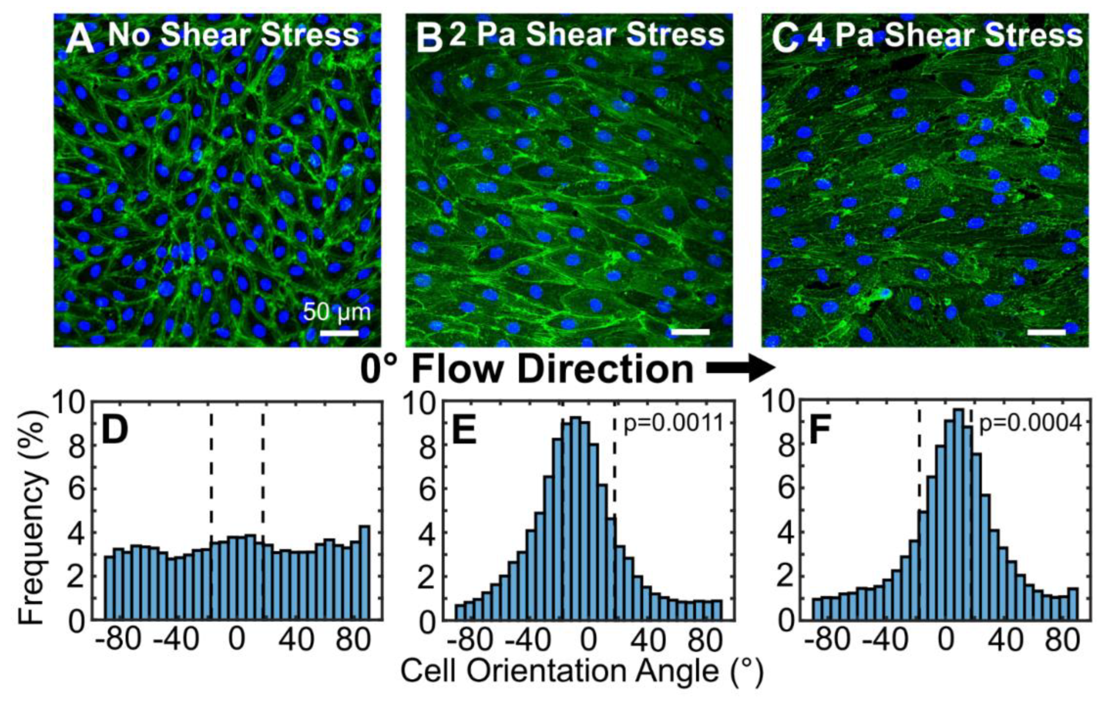
Figure 4.
A: Cell length increased significantly with application of flow and shear stress. B: Nucleus area increased significantly with application of flow and shear stress. C: Nucleus circularity did not change with application of flow and shear stress.
Figure 4.
A: Cell length increased significantly with application of flow and shear stress. B: Nucleus area increased significantly with application of flow and shear stress. C: Nucleus circularity did not change with application of flow and shear stress.
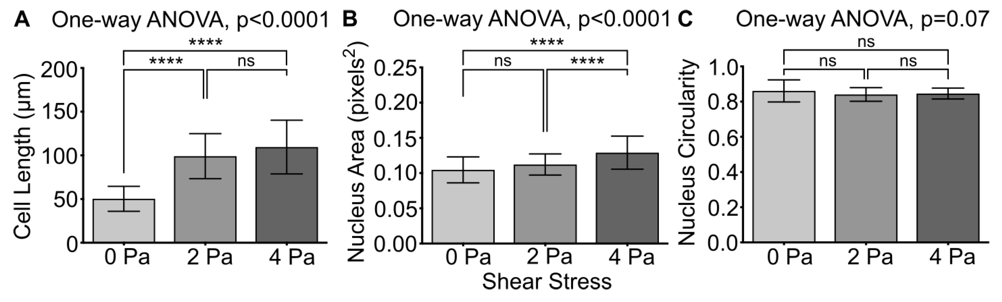

Table 1.
Ex vivo microcirculation equipment, supplies, and costs.
| Supply | Company | Quantity | Cost |
|---|---|---|---|
| Peristaltic pump | Longer Precision Pump Co | 1 | $1,162 |
| Plasma cleaner | Harrick Plasma | 1 | $4,720 |
| Vacuum pump | Harrick Plasma | 1 | $3,470 |
| Spin coater | Setcas | 1 | $2,000 |
| Photomask | Micro Lithography Services | 1 | $150 |
| Silicon wafers | University Wafer | 25 | $224 |
| SU-8 2035 | Kayaku Advanced Materials | 500 mL | $550 |
| Arduino UNO microcontroller | Arduino | 1 | $28 |
| UV light-emitting diodes | DigiKey | 250 | $55 |
| IRFP460 power metal-oxide-semiconductor field-effect transistor | DigiKey | 1 | $6 |
| Liquid crystal display | DigiKey | 1 | $7 |
| 4x4 keypad | DigiKey | 1 | $6 |
| Potentiometer | DigiKey | 1 | $4 |
| External power supply | DigiKey | 1 | $10 |
| Glass coverslips | Corning | 1,000 | $380 |
| 3-stop Tygon tubing | Darwin Microfluidics | 12 | $200 |
| Tygon tubing | VWR | 50 ft | $172 |
| 18-gauge syringe tips | Jensen Global | 500 | $13 |
| Sylgard 184 kit | DOW | 3.9 kg | $729 |
| HUVECs | Lonza | 1 vial | $443 |
| EGM Bullet Kit (SingleQuot Supplements and EMB) | Lonza | 1 kit | $216 |
| Penicillin streptomycin | Gibco | 100 mL | $28 |
| T75 tissue culture flasks | Corning | 100 | $424 |
| Fibronectin | Sigma-Aldrich | 1 mg | $316 |
| DAPI | Thermo Fisher Scientific | 1 mL | $200 |
| Alexa Fluor 488 Phalloidin | Thermo Fisher Scientific | 1 vial | $642 |
| Paraformaldehyde | Thermo Fisher Scientific | 1 L | $47 |
| PBS powder | Sigma-Aldrich | 10 pack | $79 |
| BSA | Sigma-Aldrich | 50 g | $526 |
| Triton X-100 | Sigma-Aldrich | 5 mL | $20 |
| DPBS | Sigma-Aldrich | 100 mL | $12 |
| Ethanol | Thermo Fisher Scientific | 1 L | $191 |
| Acetone | Thermo Fisher Scientific | 1 L | $30 |
| Isopropyl alcohol | Sigma-Aldrich | 100 mL | $47 |
| Nitrogen gas cylinder | Airgas | 1 tank | $35 |
| Carbon dioxide gas cylinder | Airgas | 1 tank | $35 |
Disclaimer/Publisher’s Note: The statements, opinions and data contained in all publications are solely those of the individual author(s) and contributor(s) and not of MDPI and/or the editor(s). MDPI and/or the editor(s) disclaim responsibility for any injury to people or property resulting from any ideas, methods, instructions or products referred to in the content. |
© 2026 by the authors. Licensee MDPI, Basel, Switzerland. This article is an open access article distributed under the terms and conditions of the Creative Commons Attribution (CC BY) license (http://creativecommons.org/licenses/by/4.0/).
Copyright: This open access article is published under a Creative Commons CC BY 4.0 license, which permit the free download, distribution, and reuse, provided that the author and preprint are cited in any reuse.


