
Submitted:
27 July 2026
Posted:
29 July 2026
You are already at the latest version
Abstract
Oleanolic acid (OA) is a pentacyclic triterpenoid with broad biological activity and a chemically accessible scaffold that has yielded numerous semisynthetic derivatives. Many of these compounds alter phosphorylation-dependent pathways, including PI3K/AKT/mTOR, AMPK/mTOR, MAPK, JAK/STAT3, NF-κB, and Nrf2 signaling. However, such changes are commonly reported as isolated pathway effects or down-stream responses to cellular stress, with less attention paid to the balance between kinase and phosphatase activities. This review examines OA derivatives at the kinase–phosphatase interface, with emphasis on protein tyrosine phosphatase 1B (PTP1B), re-ceptor tyrosine kinases, EGFR/AKT signaling, and phosphorylation-dependent transi-tions from stress adaptation to cancer cell death. The available evidence links OA and selected derivatives with PTP1B inhibition, receptor-proximal signaling, AKT/mTOR and AMPK/mTOR regulation, stress- and inflammation-related pathways, autophagy, and apoptosis. We also introduce the concept of structure–signaling relationships, in which chemical modification may influence not only potency, but also the point at which a derivative first perturbs the signaling network. Finally, we propose an evidence framework for distinguishing direct target modulation from secondary phosphorylation changes associated with oxidative stress, organelle dysfunction, or advanced cellular injury. Viewing OA derivatives through the kinase–phosphatase interface may provide a more precise basis for their mechanistic evaluation and future development in cancer research.
Keywords:
oleanolic acid
; oleanolic acid derivatives
; protein tyrosine phosphatase 1B
; PTP1B
; receptor tyrosine kinases
; EGFR
; PI3K/AKT/mTOR
; AMPK/mTOR
; phosphorylation homeostasis
; kinase–phosphatase balance
; cancer cell death
; autophagy
; apoptosis
1. Introduction
Protein phosphorylation is one of the central regulatory mechanisms controlling cancer cell behavior. Aberrant phosphorylation-dependent signaling contributes to sustained proliferation, resistance to apoptosis, metabolic rewiring, inflammatory adaptation, invasion, metastasis, and therapy resistance [1,2]. This central role of phosphorylation has led cancer biology and anticancer drug discovery to adopt a predominantly kinase-centered perspective. Receptor tyrosine kinases (RTKs), PI3K/AKT/mTOR signaling, MAPK cascades, JAK/STAT signaling, and stress-activated kinases have become major conceptual and therapeutic frameworks in modern oncology [1,2]. The clinical success of several kinase inhibitors has further reinforced this view.
However, phosphorylation-dependent signaling is not determined by kinase activity alone. Protein phosphatases actively regulate the amplitude, duration, localization, and reversibility of phosphorylation signals [2,3,4]. They are not passive “off-switches” for kinase-driven pathways, but dynamic regulators of cellular decision-making. Depending on substrate identity, subcellular localization, timing, and network context, phosphatases may terminate, redirect, sustain, or reshape signaling output. The biological consequences of phosphorylation in cancer cells are therefore better understood as the product of a dynamic kinase–phosphatase balance than as the result of kinase activation alone [2,3,4,5].
Among phosphatase-related targets, protein tyrosine phosphatase 1B (PTP1B, encoded by PTPN1) is particularly relevant. PTP1B has been studied extensively as a negative regulator of insulin and leptin signaling and as a therapeutic target in metabolic diseases [3]. At the same time, its role extends beyond metabolism. PTP1B participates in RTK regulation, endoplasmic reticulum-associated signaling, cytokine pathways, inflammation, immune regulation, and cancer biology [2,3,4]. This dual metabolic and oncological relevance makes PTP1B an attractive node for reinterpreting natural-product pharmacology from the perspective of kinase–phosphatase signaling balance.
Oleanolic acid (OA) is a naturally occurring pentacyclic triterpenoid present in numerous edible and medicinal plants. It has been investigated for anticancer, antioxidant, anti-inflammatory, hepatoprotective, neuroprotective, antidiabetic, and cytoprotective effects [6,7]. From a medicinal chemistry perspective, OA is also a chemically tractable scaffold. The hydroxyl group at C-3, the double bond between C-12 and C-13, and the carboxyl group at C-28 provide key sites for semisynthetic modification. These positions have been used to generate esters, amides, oximes, lactones, bromolactones, glycosides, glycoconjugates, dimers, acetylated derivatives, and other analogues with altered biological and physicochemical properties [8,9,10,11,12,13,14].
Many OA derivatives have been developed with the aim of improving potency, selectivity, solubility, cellular uptake, and predicted pharmacological behavior. In cancer models, OA and its derivatives have frequently been reported to influence PI3K/AKT/mTOR, AMPK/mTOR, MAPK, JAK/STAT3, NF-κB, and Nrf2-associated pathways [6,15]. These observations are usually described through changes in phosphorylation markers such as p-AKT, p-mTOR, p-AMPK, p-ERK, p-JNK, p-p38, p-STAT3, p-JAK2, or p-NF-κB p65. Such markers are valuable, but their interpretation is often limited. A change in phosphorylation can indicate direct modulation of a kinase or phosphatase, but it can also reflect altered receptor trafficking, oxidative stress, mitochondrial dysfunction, ER stress, autophagy, apoptosis, or secondary collapse of survival signaling.
This interpretive problem is especially relevant to OA, which was identified as a natural inhibitor of PTP1B and later served as a scaffold for the development of PTP1B-directed derivatives [8,9]. More recently, OA glycoside derivatives were reported to target the PTP1B/PI3K/AKT signaling pathway in breast cancer models [10]. These findings suggest that the phosphatase-related pharmacology of OA derivatives may be relevant not only to metabolic disease, but also to cancer signaling. In parallel, selected OA derivatives have been linked with receptor-proximal kinase pathways. For example, K73-03 was reported to inhibit pancreatic cancer cell proliferation in vitro and in vivo through blockade of the EGFR/AKT pathway [16]. In hepatocellular carcinoma cells, K73-03 was also associated with mitochondrial dysfunction, ROS accumulation, apoptosis, and suppression of JAK2/STAT3 and NF-κB/p65 signaling [17].
These studies place OA derivatives within a broader signaling landscape than is usually acknowledged. Their effects need not arise from a single shared target: chemically distinct derivatives may perturb PTP1B and related phosphatases, RTKs such as EGFR and HER2, AKT/mTOR and AMPK/mTOR signaling, stress-activated MAPKs, STAT3/NF-κB-dependent survival programs, Nrf2-mediated antioxidant responses, or organelle-associated stress pathways [6,13]. OA derivatives may therefore be viewed as candidate modulators of phosphorylation homeostasis rather than only as cytotoxic, antioxidant, anti-inflammatory, or metabolically active triterpenoids.
These interconnected signaling entry points and their possible consequences for stress adaptation and cancer cell death are summarized in Figure 1.
The aim of this review is to discuss OA derivatives at the kinase–phosphatase interface. Particular emphasis is placed on PTP1B, RTKs, EGFR/AKT signaling, and phosphorylation-dependent transitions from stress adaptation to cancer cell death. The review does not attempt to provide another broad catalogue of all anticancer effects of OA. Instead, it focuses on a narrower question: whether OA derivatives can be interpreted as modulators of kinase–phosphatase signaling balance and how this interpretation may improve mechanistic understanding of their anticancer activity.
Against this background, the discussion begins with PTP1B, the best-supported phosphatase-related target of OA-based compounds, and then follows the signaling network through RTKs, AKT/mTOR, AMPK/mTOR, and stress-responsive transcriptional pathways. The final sections ask whether chemical modification can bias the point of network entry and what experimental evidence is needed to separate direct target engagement from secondary effects of cellular injury.
2. PTP1B as a Phosphatase-Related Entry Point for Oleanolic Acid Derivatives
PTP1B is one of the most relevant phosphatase-related nodes for discussing OA derivatives at the kinase–phosphatase interface. Historically, PTP1B was investigated mainly as a negative regulator of insulin and leptin signaling and therefore as a therapeutic target in type 2 diabetes, obesity, and related metabolic disorders [3]. This metabolic origin remains relevant to OA research because the compound has also been investigated in relation to insulin resistance, glucose metabolism, oxidative stress, hepatometabolic regulation, and formulation-dependent translation [18]. However, PTP1B biology is no longer restricted to metabolic disease. The enzyme is now recognized as a context-dependent regulator of receptor signaling, cytokine signaling, immune responses, inflammation, and tumor biology [2,3,4].
PTP1B is especially interesting because its function cannot be reduced to simple termination of signaling. In some settings, PTP1B attenuates phosphorylation-dependent responses by dephosphorylating receptor or downstream substrates. In other settings, it may support oncogenic signaling indirectly, for example by regulating receptor trafficking, adaptor-protein signaling, or feedback loops. This apparent paradox reflects a general feature of phosphatase biology. Dephosphorylation does not necessarily mean biological inhibition. Depending on the substrate and cellular context, phosphatases may suppress, redirect, stabilize, or amplify signaling output [2,3,4].
The spatial organization of PTP1B is central to its function. PTP1B is anchored to the cytosolic face of the endoplasmic reticulum. This localization allows it to interact with substrates at membrane contact sites, during receptor internalization, and in endosomal trafficking pathways. RTK phosphorylation is not confined to the plasma membrane. Activated EGFR and other RTKs can continue signaling after endocytosis, and their downstream output depends on receptor sorting, recycling, degradation, and dephosphorylation. ER-associated PTP1B has been shown to interact with endocytosed EGFR at ER–endosome contact sites, contributing to receptor dephosphorylation and sorting [19,20]. This spatial relationship is highly relevant for OA derivatives, many of which are lipophilic and may influence membrane-associated or organelle-associated signaling.
The connection between OA and PTP1B is supported by experimental evidence. OA was identified as a natural triterpenoid inhibitor of PTP1B during screening of plant-derived compounds [8]. This finding provided a medicinal chemistry entry point for OA-based phosphatase-directed design. The OA scaffold combines a rigid hydrophobic pentacyclic core with modifiable functional groups. The C-28 carboxyl group may participate in electrostatic or hydrogen-bonding interactions and can be transformed into esters, amides, or conjugates. The C-3 hydroxyl group provides a convenient site for acylation, glycosylation, and other modifications. The hydrophobic triterpenoid core may contribute to interactions with non-polar regions outside the conserved catalytic pocket of PTP1B [8,9].
These structural features are important because selective PTP1B inhibition remains challenging. The catalytic domains of PTP family members are highly conserved, especially between PTP1B and T-cell protein tyrosine phosphatase (TCPTP). Potency against PTP1B alone is not sufficient. An OA-derived inhibitor intended for mechanistic or translational studies should also demonstrate selectivity over related phosphatases and activity in a relevant cellular model [3,4,9,21]. This requirement is particularly important in cancer, where different phosphatases may have distinct and sometimes opposing effects on RTK signaling, STAT3 activation, AKT phosphorylation, and apoptosis resistance.
OA glycoconjugates and glycoside derivatives represent one of the most important structural classes linking OA chemistry with PTP1B inhibition. Liu and co-workers synthesized OA-derived glycoconjugates and evaluated their inhibitory activity against PTP1B, including selectivity over TCPTP [9]. This work is relevant because it moved beyond the parent compound and addressed a key medicinal chemistry question: whether C-3 glycosylation or glycoconjugation can improve the phosphatase-related profile of the OA scaffold. Such modifications may alter polarity, hydrogen-bonding capacity, interaction with peripheral binding regions, and cellular distribution.
More recently, Song and co-workers described OA glycoside derivatives designed around the PTP1B/PI3K/AKT axis in breast cancer [10]. This work extends OA-based PTP1B chemistry beyond its traditional metabolic setting and places it within a cancer-relevant survival pathway. Earlier PTP1B studies were largely framed around insulin signaling and glucose homeostasis; the breast cancer model instead raises the question of how phosphatase inhibition affects receptor-dependent growth and adaptation.
Breast cancer provides a meaningful biological setting for this discussion. PTP1B has been implicated in HER2/Neu-driven mammary tumorigenesis, and cellular studies have linked increased PTP1B expression with breast cancer cell proliferation, migration, and resistance to apoptosis [22,23]. OA derivatives that modulate PTP1B may consequently be relevant in tumors in which this phosphatase supports receptor-driven growth, AKT activation, inflammatory signaling, or resistance to apoptosis. However, this interpretation requires caution. PTP1B may support oncogenic signaling in some breast cancer models, but it can also participate in negative regulation of RTKs such as EGFR by dephosphorylation and receptor sorting [19,20,23]. The biological consequence of PTP1B inhibition by an OA derivative will likely depend on tumor type, receptor status, phosphatase abundance, subcellular localization, and the dominant survival pathway [24].
Rather than reducing its relevance, this context dependence explains why PTP1B must be examined within defined receptor and tumor backgrounds [5,24]. The context-dependent biology of PTP1B is precisely what makes it a useful model for OA derivative research. By linking metabolic regulation with RTK signaling and cellular stress responses, PTP1B brings together two established areas of OA pharmacology: metabolic modulation and anticancer activity. This overlap is relevant because many cancer cells combine growth-factor dependence with metabolic reprogramming, oxidative stress, and inflammatory adaptation [5,24].
The PTP1B–RTK–AKT axis provides a practical conceptual bridge. RTKs such as EGFR and HER2 activate PI3K/AKT/mTOR and MAPK signaling, thereby supporting proliferation, survival, migration, and therapy resistance. PTP1B can interact with this signaling landscape by dephosphorylating receptor substrates, regulating receptor trafficking, modulating adaptor-protein signaling, or influencing pathway duration [19,20]. Accordingly, a decrease in p-AKT after treatment with an OA derivative should not be interpreted automatically as evidence of direct AKT inhibition. It may reflect upstream RTK attenuation, altered receptor internalization, phosphatase modulation, oxidative stress, or a later consequence of cellular stress.
Physicochemical differences also complicate comparisons across OA derivative classes. Glycosylated compounds may differ from the parent scaffold in polarity and intracellular access, whereas dimerization and acetylation can increase hydrophobic surface area and alter membrane partitioning. Lactonization and bromolactonization modify the triterpenoid skeleton and may change its interactions with proteins, membranes, or organelles. Similar downstream readouts, such as reduced p-AKT or p-mTOR, therefore need not reflect the same upstream mechanism [11,12,13,14].
PTP1B may also be affected indirectly through redox regulation. Classical PTPs depend on a highly reactive catalytic cysteine residue that can undergo reversible oxidation and loss of catalytic activity during redox signaling [3,4,25]. OA and its derivatives are widely discussed in the context of oxidative stress, ROS modulation, mitochondrial function, ER stress, and antioxidant defense [6,15]. In cancer cells, where basal ROS levels are often elevated, OA derivatives may influence PTP1B and other phosphatases not only by direct binding but also by changing the redox state of catalytic cysteines. This adds another layer of complexity. Reduced phosphorylation of AKT, ERK, STAT3, or NF-κB after OA derivative treatment may result from altered kinase activity, altered phosphatase activity, receptor-level changes, redox-sensitive phosphatase regulation, or general stress responses.
Claims of a PTP1B-dependent mechanism therefore require validation beyond downstream phosphorylation markers. Demonstrating reduced p-AKT or altered PI3K/AKT signaling is not sufficient to prove a PTP1B-dependent mechanism. Stronger evidence should include biochemical PTP1B activity assays, selectivity profiling against TCPTP and other phosphatases, cellular target-engagement studies, and functional approaches such as PTP1B knockdown, knockout, overexpression, or rescue. Substrate-trapping PTP mutants may also help identify candidate substrates whose phosphorylation changes after OA derivative exposure. Such approaches would be particularly valuable for OA glycosides and glycoconjugates, where phosphatase-directed activity is already plausible.
PTP1B should not be treated as a universal mechanism for OA derivatives or as the only phosphatase that may be involved. Depending on the model, TCPTP, SHP2, PTEN, PP2A, and dual-specificity phosphatases may also shape the observed response [2,4,5,24]. At present, however, PTP1B remains the best-supported phosphatase-related entry point linking OA chemistry with metabolic regulation, RTK biology, and AKT-associated cancer signaling [8,9,10].
Reframing OA derivatives through PTP1B also helps organize otherwise scattered pathway observations. Instead of treating changes in EGFR/AKT, PI3K/AKT/mTOR, AMPK/mTOR, MAPK, STAT3, NF-κB, and Nrf2 as unrelated findings, they can be viewed as parts of a broader phosphorylation network. Within this network, OA derivatives may act at different entry points depending on structure, physicochemical properties, and cellular context. PTP1B provides one of the clearest starting points for this interpretation and justifies a broader discussion of OA derivatives at the kinase–phosphatase interface.
B-directed OA chemistry, together with derivative classes that still require phosphatase-centered validation, is summarized in Table 1.
3. Receptor Tyrosine Kinases: EGFR, HER2, and PTP1B-Dependent Signal Regulation
Receptor tyrosine kinases (RTKs) are among the most important kinase families in cancer biology. By converting extracellular growth factor signals into intracellular phosphorylation cascades, RTKs regulate proliferation, survival, migration, metabolism, angiogenesis, and therapy resistance [26]. EGFR and HER2 are particularly relevant in the context of OA derivatives because they organize receptor-proximal networks connected with PI3K/AKT/mTOR, MAPK/ERK, JAK/STAT3, and NF-κB signaling [27]. Their signaling output is shaped not only by ligand binding and kinase-domain activity, but also by receptor internalization, endosomal sorting, degradation, recycling, and phosphatase-mediated dephosphorylation [19,20,26,28].
In conventional anticancer pharmacology, RTKs are usually treated as targets for direct kinase inhibition, antibody-mediated receptor blockade, or ligand–receptor interference. This view has been highly productive, but it does not capture the full spatial complexity of receptor signaling. Activated EGFR or HER2 can continue signaling after internalization, and the strength and duration of downstream pathway activation depend on the fate of the receptor after endocytosis. Whether a receptor is recycled, degraded, retained in endosomal compartments, or dephosphorylated can strongly influence AKT, ERK, STAT3, and mTOR signaling output [19,20,26].
This spatial organization is especially relevant for PTP1B. As an ER-anchored phosphatase, PTP1B can interact with internalized EGFR at ER–endosome contact sites [19,20]. This interaction contributes to receptor dephosphorylation and sorting, linking phosphatase activity not only with signal termination but also with receptor fate. Thus, the relationship between RTKs and PTP1B should not be understood as a simple opposition between kinase activation and phosphatase inhibition. Instead, it represents a spatially organized signaling interface in which receptor phosphorylation, trafficking, and dephosphorylation are functionally connected.
OA derivatives may influence this interface through several non-exclusive mechanisms. They may directly affect receptor phosphorylation, alter membrane organization, change receptor internalization, influence ER or endosomal function, modify PTP1B activity, or induce cellular stress that secondarily reshapes RTK signaling. This is particularly relevant because OA derivatives differ substantially in polarity, lipophilicity, molecular size, and intracellular distribution. A glycosylated OA derivative, a lactone, a dimer, and an acetylated dimer may produce similar downstream changes in p-AKT or p-mTOR while acting through different upstream mechanisms.
Among OA derivatives, K73-03 is currently one of the clearest examples linking an OA-based scaffold with receptor-proximal kinase signaling. Zhou and co-workers reported that K73-03 inhibited pancreatic cancer cell proliferation in vitro and in vivo and proposed blockade of the EGFR/AKT pathway as a major mechanism [16]. In that study, reverse pharmacophore screening and molecular dynamics simulation suggested EGFR as a potential target, while cellular experiments showed reduced phosphorylation of EGFR and AKT after K73-03 treatment. These observations place at least one OA derivative close to the receptor level rather than restricting its reported activity to late markers of stress or apoptosis [16].
Nevertheless, the interpretation of K73-03 and related derivatives requires caution. A decrease in p-EGFR and p-AKT supports involvement of the EGFR/AKT axis, but it does not by itself prove direct EGFR inhibition in living cells. Reduced receptor phosphorylation may result from direct receptor binding, impaired ligand-induced activation, altered receptor trafficking, increased receptor dephosphorylation, oxidative stress, changes in membrane organization, or secondary effects caused by reduced cell viability. K73-03 is therefore best regarded as a well-supported starting point for RTK-proximal OA pharmacology, rather than as a universal mechanistic model for the entire derivative class.
In HER2/Neu-driven mammary tumor models, PTP1B deficiency delayed or prevented tumor development, indicating that the phosphatase can support HER2-associated oncogenesis in defined settings [23]. This provides a rationale for testing PTP1B-directed OA derivatives in receptor-driven breast cancer. At the same time, PTP1B can attenuate EGFR signaling through receptor dephosphorylation and sorting [19,20], so the direction of its biological effect cannot be assumed without considering receptor status and cellular context. PTP1B cannot be classified consistently as either an oncogenic facilitator or a tumor-suppressive brake. Its output depends on receptor background, substrate availability, localization, feedback wiring, and tumor type.
This variability can be exploited experimentally by comparing structurally distinct OA derivatives in receptor-defined cancer models. OA glycosides and glycoconjugates may be prioritized for PTP1B-directed studies. K73-03-like derivatives and selected lactones may be evaluated for EGFR or broader RTK-proximal signaling. Dimers and acetylated dimers may be compared for effects on membrane-proximal signaling, receptor trafficking, organelle stress, and integrated phosphorylation readouts. Such comparisons would move the field beyond simple cytotoxic potency and toward structure–signaling relationships.
RTKs are also important because they determine how cancer cells respond to stress. EGFR, HER2, and other RTKs activate AKT/mTOR, MAPK/ERK, STAT3, and NF-κB modules, which collectively regulate mitochondrial integrity, protein synthesis, redox buffering, autophagy, and apoptosis resistance [26,29]. If an OA derivative weakens receptor-proximal survival signaling while simultaneously inducing metabolic or oxidative stress, the cell may cross a threshold from adaptation to death. In this model, RTK modulation is not isolated from stress signaling. It is one of the upstream determinants of whether cancer cells can tolerate OA derivative-induced pressure.
K73-03 again illustrates this layered response. In pancreatic cancer models, K73-03 was associated with inhibition of EGFR/AKT signaling and reduced proliferation [16]. In hepatocellular carcinoma cells, K73-03 induced mitochondrial dysfunction, excessive ROS production, apoptosis, and inhibition of JAK2/STAT3 and NF-κB/p65 signaling [17]. These findings suggest that OA derivatives may affect receptor-proximal kinase signaling and downstream stress-response pathways either in parallel or sequentially. It remains unclear whether receptor modulation initiates the subsequent stress-response and cell-death pathways, whether mitochondrial or redox injury suppresses survival signaling secondarily, or whether both processes develop in parallel.
Resolving this sequence requires time-resolved analysis. Early decreases in p-EGFR or p-AKT, observed before loss of viability, mitochondrial depolarization, or caspase cleavage, would support a receptor-proximal mechanism. In contrast, phosphorylation changes observed only after mitochondrial damage, caspase activation, or severe oxidative stress should be interpreted as secondary consequences of established cellular injury rather than as primary target effects. RTK arrays, ligand-stimulation assays, receptor internalization studies, and early time-point phosphorylation analysis would be particularly useful for OA derivatives proposed to act near the receptor level.
A cautious evidence hierarchy should be applied when interpreting RTK modulation by OA derivatives. The lowest level of evidence is a downstream change in p-AKT or p-ERK. Stronger evidence includes reduced phosphorylation of the receptor itself, such as p-EGFR or p-HER2. Still stronger evidence includes ligand-response experiments, kinase activity assays, receptor trafficking studies, target-engagement methods, and comparison with established RTK inhibitors. The strongest evidence would combine receptor-level effects with functional rescue or genetic approaches showing that the receptor is required for the biological response.
Overall, RTKs represent the kinase-side counterpart to the phosphatase-related activity of OA derivatives. EGFR and HER2 are not only upstream kinases but also spatially regulated signaling platforms whose output depends on receptor activation, trafficking, degradation, and phosphatase access. OA derivatives may influence this system through direct receptor modulation, PTP1B-related dephosphorylation, membrane-proximal effects, or stress-driven network remodeling. Understanding which of these mechanisms predominates for each derivative class will be essential for transforming OA derivative research from descriptive pathway reporting into mechanism-guided anticancer development.
4. AKT/mTOR and AMPK/mTOR as the Survival–Stress Switch
The PI3K/AKT/mTOR and AMPK/mTOR axes represent two interconnected signaling systems that determine how cancer cells integrate growth factor stimulation, nutrient availability, energy status, protein synthesis, autophagy, and survival. In receptor-driven tumors, RTK activation commonly promotes PI3K-dependent AKT phosphorylation, followed by mTOR-associated anabolic signaling [26,29,30,31]. This supports proliferation, protein synthesis, glucose metabolism, resistance to apoptosis, and adaptation to unfavorable microenvironmental conditions [26,29]. In contrast, AMPK is activated under metabolic stress and energy imbalance and can suppress mTOR-dependent growth signaling while promoting catabolic adaptation, autophagy, and stress tolerance [32].
Although PI3K/AKT/mTOR and AMPK/mTOR are often described as separate pathways, in cancer cells they function as a coupled survival–stress switch. AKT/mTOR signaling generally favors growth and biosynthesis, whereas AMPK activation reflects energetic pressure and may either promote adaptive autophagy or contribute to cell death when stress becomes excessive [29,32]. Changes in p-AKT, p-mTOR, p-AMPK, p-p70S6K, and p-4EBP1 after OA derivative exposure should be considered together, because their combined pattern is more informative than any single marker for distinguishing continued survival, adaptive autophagy, and progression toward regulated cell death.
AKT/mTOR signaling is one of the most frequently reported phosphorylation modules affected by OA and its derivatives. This is expected, because AKT integrates inputs from RTKs, PI3K, phosphoinositide metabolism, nutrient availability, and feedback loops [26,29]. In cancer cells, sustained AKT/mTOR activity supports proliferation, inhibits pro-apoptotic signaling, maintains metabolic fitness, and contributes to therapy resistance. Therefore, attenuation of AKT/mTOR phosphorylation is often interpreted as a plausible anticancer mechanism for natural products and their derivatives.
However, reduced p-AKT or p-mTOR after OA derivative treatment requires careful interpretation. These changes may result from several upstream events: inhibition of RTK activation, altered receptor trafficking, modulation of PI3K activity, altered phosphatase function, oxidative stress, mitochondrial damage, or loss of cell viability. The same derivative may also reduce AKT/mTOR phosphorylation through different mechanisms depending on cancer type, receptor background, mutation status, exposure time, and intracellular accumulation. Thus, AKT/mTOR should be treated not only as a downstream survival pathway but also as a readout of upstream receptor, kinase, phosphatase, and stress-related network changes.
K73-03 illustrates this interpretive problem. In pancreatic cancer models, this OA derivative reduced phosphorylation of EGFR and AKT and suppressed tumor cell proliferation [16]. These observations place AKT downstream of receptor-proximal events. However, in hepatocellular carcinoma cells, K73-03 was linked with mitochondrial dysfunction, ROS accumulation, apoptosis, and inhibition of JAK2/STAT3 and NF-κB/p65 signaling [17]. Together, these findings indicate that K73-03 can affect both receptor-associated survival signaling and downstream stress responses, although the temporal order of these events remains unresolved. What remains unclear is whether AKT attenuation is an initiating event, a consequence of receptor-level modulation, or part of a later collapse of survival signaling during cellular stress.
AMPK/mTOR signaling is one of the best-described stress-related axes in OA research. In colon cancer cells, OA was reported to inhibit proliferation and viability while inducing autophagy and apoptosis through AMPK activation and mTOR inhibition [33]. The study links OA-induced cytotoxicity with a defined metabolic stress pathway rather than with a nonspecific loss of viability alone [33]. Activation of AMPK and suppression of mTOR can reduce anabolic signaling, inhibit protein synthesis, promote autophagy, and sensitize cells to death when stress exceeds the adaptive capacity of the cell [32,33].
The biological meaning of AMPK activation depends strongly on context. In non-malignant cells, AMPK activation may support survival by restoring energy homeostasis, reducing oxidative damage, and promoting adaptive autophagy. In cancer cells, especially those already exposed to oncogenic, metabolic, and oxidative stress, further activation of AMPK combined with mTOR inhibition may contribute to growth arrest or death. This context dependence is relevant to OA derivatives because their effects frequently extend to mitochondrial function, ROS production, ER stress, and autophagy-associated processes [6,15,17,33,34]. AMPK/mTOR should therefore be interpreted as a stress-threshold module rather than as a uniformly cytoprotective or uniformly pro-death pathway.
In the framework proposed here, AMPK/mTOR occupies a special position. It is not primarily an RTK-proximal module, but it receives input from metabolic stress, mitochondrial function, nutrient sensing, and upstream kinase networks. OA derivatives that influence RTKs, PTP1B, mitochondrial function, or redox balance may converge on AMPK/mTOR as a downstream integrator. Conversely, early AMPK activation may reshape AKT/mTOR, autophagy, and apoptosis signaling. Time-resolved analysis is essential: AMPK activation observed early after treatment may indicate metabolic sensing, whereas late AMPK activation may reflect secondary stress during cytotoxic injury.
AKT/mTOR and AMPK/mTOR are often considered opposing axes, but their relationship is more complex. AKT promotes anabolic growth signaling, whereas AMPK suppresses energy-consuming processes and can inhibit mTORC1. Nevertheless, cancer cells contain multiple feedback loops connecting RTKs, PI3K, AKT, mTORC1, mTORC2, MAPK, AMPK, and stress kinases [29,32]. Inhibition of one node may produce compensatory activation or suppression of another. This is particularly important for OA derivatives, which may simultaneously alter receptor signaling, redox state, mitochondrial function, and phosphatase activity.
For example, a decrease in p-mTOR after OA derivative treatment may result from upstream AKT suppression, AMPK-mediated mTORC1 inhibition, reduced nutrient signaling, oxidative stress, or late-stage cytotoxicity. Similarly, increased p-AMPK may reflect genuine energetic stress, mitochondrial dysfunction, CaMKKβ-dependent signaling, or adaptive compensation. Without early time points, dose-response analysis, and functional rescue experiments, the directionality of these events remains uncertain.
Comparison across structural classes is particularly informative in this setting. Glycoside derivatives, lactones, dimers, acetylated dimers, and K73-03-like molecules may all reduce p-AKT or p-mTOR, but they may not share the same upstream mechanism. A glycoside derivative designed toward PTP1B may influence AKT through phosphatase-related signaling, whereas a more lipophilic dimer may alter membrane or organelle-associated stress responses before downstream mTOR inhibition becomes visible. Similar downstream phosphorylation readouts should therefore not be mistaken for identical mechanisms of action.
Autophagy is one of the principal biological outputs of mTOR suppression and AMPK activation. In cancer cells, autophagy has a dual role. It may support survival by removing damaged organelles, recycling nutrients, and buffering metabolic stress, but it may also contribute to cell death or sensitize cells to apoptosis when stress is excessive or prolonged [33,35]. This duality is important for OA and its derivatives because many studies report increased LC3-II, Beclin-1, autophagosome formation, or changes in p62/SQSTM1 after treatment, yet the functional meaning of these markers is not always clear.
Distinguishing autophagy induction from changes in autophagic flux is essential [36]. Increased LC3-II may indicate enhanced autophagosome formation, but it may also reflect impaired autophagosome degradation. Similarly, increased Beclin-1 does not automatically prove that autophagy is responsible for cell death. For OA derivatives, especially those that affect lysosomes, mitochondria, ER stress, or membrane organization, autophagy markers should be interpreted together with flux assays and pharmacological or genetic modulation of autophagy.
From the perspective of phosphorylation signaling, autophagy can be viewed as a stress adaptation program controlled by kinase networks. AKT/mTOR inhibition and AMPK activation favor autophagy, while MAPK, JNK, p38, and STAT3 may modulate autophagy depending on cellular context. OA derivatives may therefore promote autophagy through multiple routes: suppression of RTK/AKT/mTOR survival signaling, activation of AMPK by metabolic stress, ROS-mediated stress kinase activation, ER stress, or mitochondrial damage. The key mechanistic question is whether autophagy is protective, cytotoxic, or a parallel response to compound-induced stress.
Apoptosis is frequently reported as a major anticancer outcome of OA and OA derivatives. It is often associated with mitochondrial dysfunction, ROS accumulation, altered Bax/Bcl-2 ratio, cytochrome c release, caspase activation, and PARP cleavage [6,15,17,33]. These markers indicate execution of cell death, but they do not always identify the upstream phosphorylation events that initiate or facilitate the process. AKT/mTOR attenuation, AMPK activation, JNK/p38 activation, STAT3 inhibition, and NF-κB suppression can all contribute to lowering the apoptotic threshold.
One way to reconcile these findings is to view OA derivatives as compounds that lower the phosphorylation-dependent threshold separating stress adaptation from irreversible cell death. Under basal conditions, RTK/AKT/mTOR, STAT3, and NF-κB signaling help cancer cells tolerate oxidative, metabolic, and proteotoxic stress. If OA derivatives simultaneously weaken survival signaling and enhance AMPK activation, mitochondrial stress, ER stress, or ROS accumulation, the balance may shift from adaptation to apoptosis. This model does not require a single universal target for all OA derivatives. Instead, it explains how structurally diverse derivatives may converge on related phenotypes through partially different upstream events.
Compared with apoptosis and autophagy, ferroptosis remains poorly established in OA derivative research. Recent reviews have proposed possible links with lipid peroxidation, redox imbalance, and AKT/mTOR-associated signaling, but the current evidence does not support treating ferroptosis as a general mechanism of OA derivatives [37]. Claims of ferroptotic activity should therefore require lipid ROS measurements, analysis of GPX4 and SLC7A11, evidence of iron dependence, and rescue with appropriate ferroptosis inhibitors [38,39].
The survival–stress switch perspective has several practical implications. First, phosphorylation markers should be measured at early and late time points to distinguish initiating events from secondary cytotoxicity. Second, p-AKT, p-mTOR, and p-AMPK should be interpreted together rather than as unrelated markers. Third, autophagy should be evaluated by flux rather than by static LC3-II or Beclin-1 measurements alone. Fourth, apoptosis and ferroptosis should be distinguished experimentally rather than inferred from ROS accumulation. Finally, structural classes of OA derivatives should be compared using harmonized signaling readouts, because differences in lipophilicity, polarity, dimerization, and intracellular distribution may determine which upstream module is engaged first.
In this framework, AKT/mTOR and AMPK/mTOR are not merely downstream pathways affected by OA derivatives. They are integrative signaling modules that connect RTK activity, phosphatase regulation, metabolic stress, mitochondrial function, autophagy, and cell death. Understanding how different OA derivatives influence this survival–stress switch may help transform OA research from descriptive pathway reporting into mechanism-guided development of anticancer triterpenoids.
5. MAPK, STAT3, NF-κB, and Nrf2 in Stress- and Inflammation-Related Signaling
Cancer cells do not respond to pharmacological pressure only through classical survival pathways such as AKT/mTOR or AMPK/mTOR. They also activate stress- and inflammation-related signaling systems that determine whether damage is buffered, tolerated, or converted into cell death. Among these systems, MAPK cascades, JAK/STAT3 signaling, NF-κB, and Nrf2 are especially relevant for OA and its derivatives. These modules connect phosphorylation with redox regulation, cytokine signaling, mitochondrial function, transcriptional adaptation, and therapy resistance [40,41,42,43,44].
In OA derivative research, these pathways are often used as mechanistic markers. Decreased p-STAT3, reduced NF-κB p65 phosphorylation, increased p-JNK, altered p-p38, or changes in Nrf2 nuclear accumulation are frequently interpreted as evidence of pathway-specific activity. Such data are useful, but they need context. The same marker may reflect direct signaling modulation, oxidative stress, inflammatory suppression, mitochondrial damage, ER stress, protective adaptation, or late-stage apoptosis. The more informative question is when these changes occur, what precedes them, and whether they contribute to adaptation or to loss of viability.
MAPK signaling illustrates this problem clearly. ERK1/2 is often associated with proliferation, migration, and adaptive survival, whereas JNK and p38 are more commonly linked with stress responses, inflammation, and mitochondrial apoptosis [40]. However, this division is not absolute. ERK activation may protect cancer cells under some conditions, whereas ERK suppression may reduce migration or invasion in others. JNK and p38 may contribute to apoptosis, but they may also participate in adaptive autophagy or stress tolerance depending on timing, dose, and cell type.
OA has been reported to activate p38, JNK, and ERK in cancer cells. In one study, MAPK activation was associated with mitochondrial apoptosis, cytochrome c release, caspase cleavage, and PARP cleavage; inhibition of p38 partly rescued the pro-apoptotic effect of OA, supporting a functional role for p38 in this model [45]. This type of evidence is valuable because it connects phosphorylation changes with functional outcome. In contrast, marker changes alone provide only pathway association.
The role of ERK is particularly context-dependent. OA suppressed migration and invasion of malignant glioma cells by inhibiting MAPK/ERK signaling; reactivation of MEK/ERK partially rescued the inhibitory effect on migration and invasion [46]. In another setting, however, OA-induced ERK activation promoted Nrf2 expression, reduced ROS accumulation, and protected cancer cells against apoptosis; ERK inhibition or Nrf2 suppression increased OA-induced apoptotic death [47]. Thus, ERK may function either as a suppressed pro-migratory pathway or as an adaptive anti-apoptotic response, depending on the biological endpoint.
JNK also requires careful interpretation. OA was reported to induce protective autophagy through JNK and mTOR-related mechanisms, and inhibition of this autophagy sensitized cancer cells to apoptosis [48]. Increased p-JNK should consequently not be classified automatically as a pro-apoptotic signal. It may represent an early stress response, a driver of protective autophagy, or a component of later death signaling. For OA derivatives, this means that MAPK readouts should be interpreted together with apoptosis assays, autophagic flux, ROS measurements, and functional inhibition or rescue experiments.
STAT3 represents another important phosphorylation-dependent survival module. Activated STAT3 integrates cytokine signaling, RTK-associated inputs, inflammation, immune escape, stemness, invasion, and resistance to apoptosis [41]. In cancer cells, persistent STAT3 activation supports transcription of genes involved in proliferation and survival. OA derivatives have been linked with STAT3 modulation in several contexts. A morpholide derivative of an oleanolic oxime–succinic acid conjugate was reported to diminish NF-κB and STAT activity in human hepatocellular carcinoma cells [49]. K73-03 also suppressed JAK2/STAT3 signaling in hepatocellular carcinoma cells, together with ROS accumulation, mitochondrial dysfunction, and apoptosis [17].
These observations suggest that OA derivatives may weaken transcriptional survival programs that help cancer cells tolerate stress. However, STAT3 inhibition should not be interpreted in isolation. Reduced p-STAT3 may result from upstream JAK inhibition, RTK attenuation, oxidative stress, phosphatase modulation, cytokine signaling changes, or late apoptosis-associated collapse of survival signaling. Since STAT3 is functionally connected with EGFR, IL-6/JAK signaling, NF-κB, and mitochondrial stress responses, its phosphorylation state can reflect both primary signaling interference and broader cellular injury.
NF-κB is similarly important. It regulates inflammation, immune signaling, survival, invasion, angiogenesis, and therapy resistance [42]. Canonical NF-κB activation depends on phosphorylation and degradation of IκB proteins, followed by nuclear translocation of NF-κB subunits such as p65/RelA. Because OA is widely recognized as an anti-inflammatory triterpenoid, NF-κB has been one of the most frequently investigated pathways in OA-related pharmacology [6,49,50].
Some OA derivatives were designed to combine triterpenoid pharmacology with anti-inflammatory or anticancer properties. Oleanolic acid oxime derivatives conjugated with nonsteroidal anti-inflammatory drugs were reported to modulate Nrf2-ARE and NF-κB signaling in HepG2 hepatoma cells and normal THLE-2 hepatocytes [50]. Importantly, these derivatives showed cell-context-dependent effects, reducing stress-adaptive signaling in hepatoma cells while supporting a more protective response in normal hepatocytes [50]. Such selectivity is relevant for OA derivative development because it suggests that the same scaffold may suppress cancer-cell stress tolerance while preserving protective signaling in non-malignant cells.
Nrf2 adds another layer to this stress-inflammatory network. As a master regulator of antioxidant and electrophile-response genes, Nrf2 supports glutathione metabolism, detoxification, heme oxygenase-1 expression, NADPH regeneration, and redox balance [43]. In normal tissues, Nrf2 activation is generally cytoprotective. In cancer, persistent Nrf2 activity may support antioxidant buffering, metabolic adaptation, chemoresistance, and survival under stress.
OA has been associated with Nrf2 activation, in keeping with its antioxidant and cytoprotective profile [7,43,44,51]. However, in cancer cells exposed to OA, Nrf2 activation may also protect against excessive ROS accumulation and apoptosis. ERK-dependent Nrf2 induction was reported to reduce OA-induced ROS and apoptotic death, whereas ERK inhibition or Nrf2 suppression increased apoptosis [47]. This result shows that a phosphorylation-dependent stress response can limit, rather than enhance, the anticancer effect of OA [47].
Nrf2 consequently represents a context-dependent node in OA derivative pharmacology. In normal cells, Nrf2 activation may contribute to safety and cytoprotection. In cancer cells, especially those with high basal oxidative stress, Nrf2 activation may improve stress tolerance and reduce compound efficacy. Conversely, derivatives that suppress Nrf2 in cancer cells may weaken antioxidant defenses and increase vulnerability to ROS-mediated apoptosis or ferroptosis-like damage. The biological meaning of Nrf2 modulation depends on cell type, basal redox state, dose, and timing.
Viewed together, MAPK, STAT3, NF-κB, and Nrf2 form an interconnected stress- and inflammation-responsive layer rather than four independent pathways. Moderate stress may activate ERK/Nrf2 and autophagy, supporting adaptation. Stronger or prolonged stress may activate p38, suppress STAT3 and NF-κB, impair antioxidant buffering, and promote apoptosis or other forms of regulated cell death. The final outcome depends on receptor background, basal ROS level, mitochondrial reserve, inflammatory state, and the structural properties of the derivative.
This integrated view helps explain why OA derivatives may show different effects across cancer models. A derivative that suppresses NF-κB and STAT3 in hepatocellular carcinoma cells may not behave identically in EGFR-dependent pancreatic cancer or HER2-positive breast cancer. A compound that induces protective ERK/Nrf2 signaling in one model may trigger p38-dependent apoptosis in another. These model-dependent effects mean that stress- and inflammation-related signaling must be interpreted together with tumor type, exposure time, dose, and functional phenotype.
Taken together, these pathways form the downstream layer through which receptor-proximal and metabolic perturbations are translated into redox adaptation, inflammatory survival, or cell death [34,44]. MAPK, STAT3, NF-κB, and Nrf2 shape the transcriptional and redox response to stress. OA derivatives may act at one or more of these levels. The challenge is to determine which level is primary for each structural class.
6. Structure–Signaling Relationships of OA Derivatives
A major advantage of OA as a medicinal chemistry scaffold is that its biological profile can be modified without losing the characteristic pentacyclic triterpenoid framework. The C-3 hydroxyl group, the C-12/C-13 double bond, and the C-28 carboxyl group provide chemically accessible positions for semisynthetic transformation [8,9,10,11,12,13,14]. These modifications are usually discussed in terms of cytotoxic potency, antioxidant activity, solubility, or ADMET-related properties. From the perspective of this review, an additional question is important: can specific structural modifications bias OA derivatives toward distinct phosphorylation-dependent signaling outputs?
In this review, a structure–signaling relationship denotes a change in the point at which a derivative first perturbs the signaling network, rather than a change in potency alone. The concept extends conventional SAR analysis by linking chemical modification with candidate mechanisms, including PTP1B inhibition, receptor-proximal effects, AKT/mTOR or AMPK/mTOR regulation, stress-activated MAPKs, NF-κB/STAT3/Nrf2 signaling, and organelle-associated responses [8,9,10,11,12,13,14,16,17,49,50].
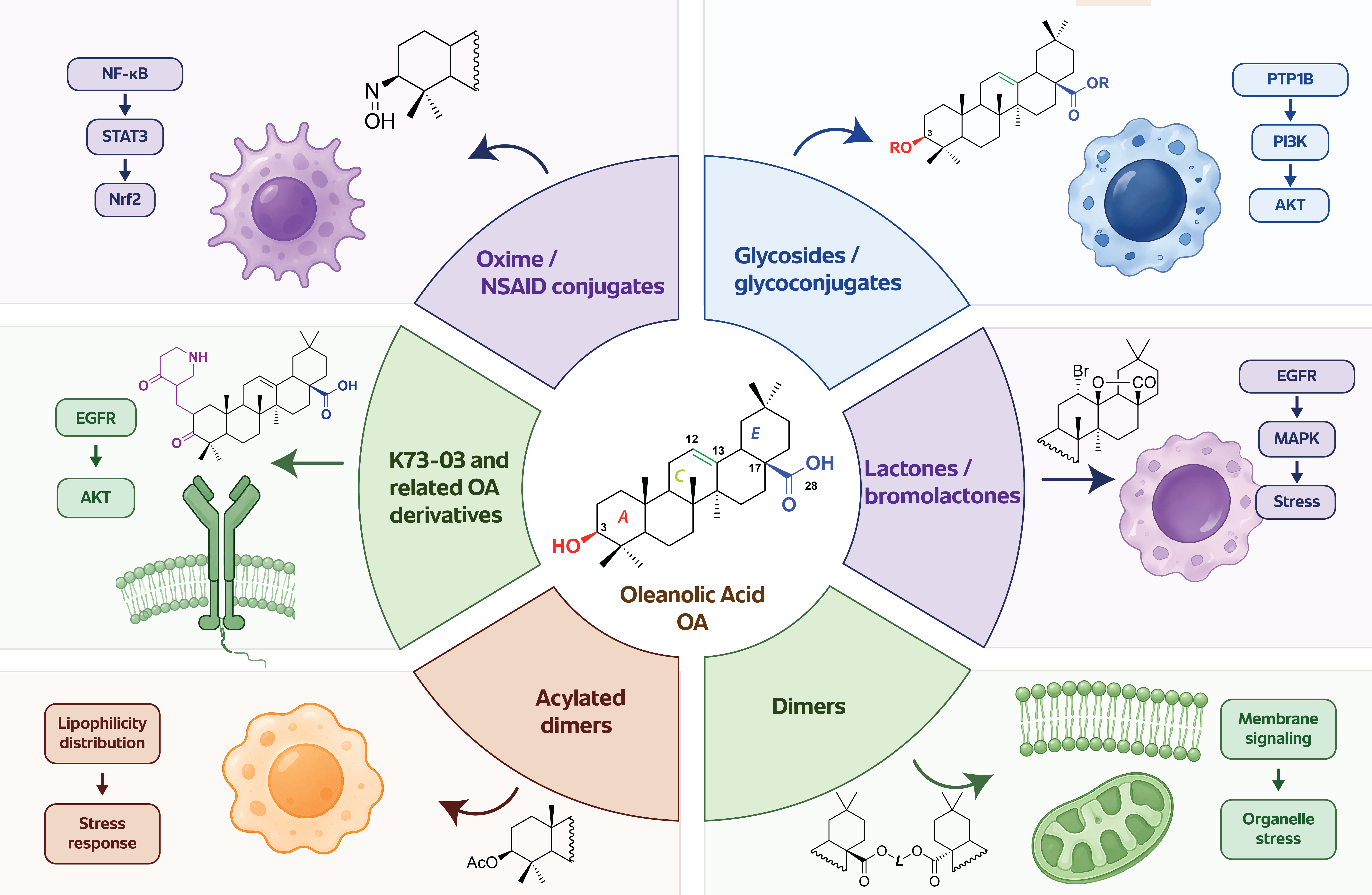
The principal modification sites of the OA scaffold and the signaling hypotheses associated with the resulting derivative classes are shown in Figure 2.
The C-3 hydroxyl group is one of the most frequently modified positions of the OA scaffold. Acetylation, esterification, glycosylation, and glycoconjugation at this site can alter polarity, membrane affinity, steric interactions, and hydrogen-bonding capacity. In PTP1B-directed OA chemistry, C-3 glycosylation and glycoconjugation are especially relevant because several OA glycoconjugates and glycoside derivatives have been evaluated as PTP1B inhibitors [8,9,10].
C-3 glycosylation may influence PTP1B-related activity in several ways. Glycosylated or glycoconjugated derivatives may improve interactions outside the conserved catalytic pocket, alter aqueous compatibility, and change cellular distribution. These features are important because selectivity remains a central problem in PTP1B inhibitor development. The catalytic domains of PTP1B and closely related phosphatases, especially TCPTP, are highly conserved; therefore, substituents that interact with peripheral or allosteric regions may be required to improve selectivity [3,4,9].
C-3 acylation may produce a different pharmacological profile. Acetylation or esterification can increase lipophilicity and membrane passage, while also favoring membrane-associated or organelle-associated stress responses over direct phosphatase modulation [52]. This contrast becomes particularly clear when OA glycosides are compared with acetylated monomeric or dimeric derivatives. Similar downstream effects, such as reduced p-AKT or p-mTOR, may arise from different upstream mechanisms: phosphatase-directed activity in one case and altered cellular uptake, membrane interaction, or mitochondrial stress in another.
The C-28 carboxyl group is another major site for OA derivatization. It can be transformed into esters, amides, salts, conjugates, or used as a point of dimerization. C-28 modification can strongly affect charge, lipophilicity, molecular size, and interaction with proteins or membranes. In OA dimers, two triterpenoid units are connected through linker systems, generating molecules with expanded hydrophobic surface area and altered three-dimensional architecture [11,12,13,53].
OA dimers merit separate consideration because dimerization changes more than molecular mass. Their high cytotoxic potency cannot be attributed automatically to the simple presence of two OA units. Dimerization may change cellular uptake, membrane partitioning, intracellular localization, protein-binding geometry, and the ability to affect receptor-proximal or organelle-associated signaling. Linker length, saturation, flexibility, and orientation may determine whether a dimer behaves mainly as a bulky hydrophobic stressor, a membrane-interacting molecule, a scaffold capable of multivalent interactions, or a compound with altered access to intracellular signaling proteins [11,12,13].
At present, the kinase–phosphatase consequences of OA dimerization remain largely unexplored. OA dimers have been evaluated for cytotoxicity, antioxidant potential, SAR, docking, and ADMETox-related properties [11,12,13], but systematic studies of PTP1B activity, RTK phosphorylation, phosphotyrosine signaling, AKT/mTOR dynamics, phosphatase selectivity, and early stress signaling are still lacking. This gap creates a clear opportunity: OA dimers could be compared with monomeric derivatives to determine whether dimerization shifts the dominant mechanism from classical pathway modulation toward membrane-proximal, organelle-stress, or phosphatase-associated signaling.
For OA dimers, the linker should be considered more than a spacer. It may determine molecular flexibility, distance between triterpenoid units, conformational accessibility, and the ability to interact with membranes or protein surfaces. Short linkers may produce compact structures with restricted flexibility, whereas longer linkers may allow broader conformational sampling. Unsaturated linkers may increase rigidity and influence the spatial presentation of both OA units [11,12,13]. These structural features may have signaling consequences. A more rigid or spatially defined dimer may interact differently with membrane domains, receptor complexes, or hydrophobic protein pockets than a flexible dimer.
Lactones and bromolactones of OA represent another structurally distinct derivative class. These modifications alter the E-ring region and may change molecular shape, polarity, electrophilic character, and interaction potential. OA lactones and bromolactones have been evaluated for cytotoxicity, antioxidant activity, SAR, molecular docking, and ADMETox properties, with EGFR tyrosine kinase domain docking used as one interpretive tool [14]. From the perspective of this review, lactones are relevant because they provide a bridge between structural modification and RTK-proximal hypotheses.
However, docking alone cannot establish RTK modulation. If a lactone derivative shows favorable docking to EGFR and cytotoxicity in an EGFR-dependent model, it becomes reasonable to test receptor phosphorylation, receptor internalization, and downstream AKT/mTOR signaling at early time points. Such docking should be treated as hypothesis-generating and should be followed by receptor-level validation, kinase assays, ligand-response experiments, and comparison with established EGFR inhibitors. This caution is important because OA derivatives may also affect RTK-associated readouts indirectly through membrane effects, oxidative stress, or organelle dysfunction.
K73-03 occupies a special position among OA derivatives because it has been linked more directly with EGFR/AKT signaling than most other OA-based compounds. It was reported to suppress pancreatic cancer cell proliferation in vitro and in vivo and to reduce phosphorylation of EGFR and AKT [16]. In hepatocellular carcinoma cells, K73-03 was associated with mitochondrial dysfunction, ROS accumulation, apoptosis, and suppression of JAK2/STAT3 and NF-κB/p65 signaling [17]. The value of K73-03 is not limited to this single compound. It provides a model for how OA derivatives might be developed toward receptor-proximal pharmacology.
Direct comparison under matched experimental conditions could test whether K73-03-like derivatives produce early RTK-associated effects, whether glycosides preferentially influence PTP1B-related signaling, and whether dimers or acetylated dimers more readily induce membrane or organelle stress. Such comparisons would require common cellular models, matched exposure conditions, and harmonized signaling readouts. Otherwise, structurally different derivatives may appear mechanistically similar simply because they converge on the same late cytotoxic markers.
Lipophilicity and cellular distribution are important hidden variables in this field. Highly lipophilic compounds may accumulate in membranes, mitochondria, ER, or lysosome-associated compartments. This can produce strong biological effects without requiring high-affinity binding to a soluble protein target. For triterpenoid derivatives, such compartmental effects should not be dismissed as experimental noise. They may be part of the mechanism.
Membrane and organelle effects can strongly influence phosphorylation signaling. Perturbation of plasma membrane organization may affect receptor clustering, ligand responsiveness, and RTK activation. Effects on endosomal or ER membranes may alter EGFR trafficking and the accessibility of ER-anchored PTP1B to receptor substrates. Mitochondrial and ER stress can reshape AMPK, JNK, p38, autophagy, and apoptosis-associated signaling, whereas lysosomal dysfunction may alter mTOR activity and autophagic flux [34]. Thus, an OA derivative may produce changes in p-AKT, p-mTOR, p-AMPK, p-JNK, or p-STAT3 through altered organelle function rather than direct kinase or phosphatase binding.
This issue is especially relevant for dimeric and acetylated derivatives. Increased lipophilicity may improve uptake and cytotoxicity, but it may also increase the likelihood of membrane-associated stress. Conversely, glycosylation may increase polarity and alter uptake route or target access. These physicochemical differences should be treated as mechanistic variables in OA derivative research. Reporting only cytotoxic IC50 values without parallel physicochemical and signaling characterization may obscure the true relationship between structure and biological outcome.
The current literature does not yet allow a definitive classification of OA derivatives by primary signaling entry point. Nevertheless, a working classification may be useful. OA glycosides and glycoconjugates may be prioritized for PTP1B-related and phosphatase-selectivity studies. K73-03-like derivatives and selected lactones may be prioritized for RTK/EGFR/AKT validation. Dimers and acetylated dimers may be prioritized for membrane-proximal signaling, mitochondrial and ER stress, and integrated kinase–phosphatase profiling. Oxime and anti-inflammatory conjugates may be especially relevant to NF-κB, STAT3, and Nrf2-centered stress-inflammatory signaling [49,50].
A practical comparison of these derivative classes, the signaling entry points that merit priority, and the main interpretive cautions is provided in Table 2.
This classification should be considered a hypothesis rather than a conclusion. The same derivative may affect multiple modules, and the dominant mechanism may depend on cancer model, dose, exposure time, formulation, and intracellular distribution. Still, organizing OA derivatives by possible signaling entry point may make future studies more rational. Instead of testing every derivative against the same limited cytotoxicity panel, researchers could select signaling assays based on structural features and predicted biological behavior.
Ultimately, the structure–signaling perspective may help bridge medicinal chemistry and cancer signaling. OA derivative research has already shown that chemical modification can improve cytotoxicity, antioxidant activity, and predicted pharmacokinetic properties [11,12,13,14]. The next step is to determine whether structural modification also changes the pathway by which a compound enters the phosphorylation network. This would allow OA derivatives to be developed not only as more potent cytotoxic agents, but also as better-defined modulators of receptor, phosphatase, metabolic, and stress-response pathways that govern adaptation and cell death.
7. Experimental Framework for Demonstrating Kinase–Phosphatase Rewiring by OA Derivatives
Interpreting phosphorylation changes induced by OA derivatives requires a clear hierarchy of evidence. Many studies report that a compound decreases p-AKT, p-mTOR, p-STAT3, p-NF-κB, or p-ERK, or increases p-AMPK, p-JNK, or p-p38. These observations are informative, but they do not identify the primary point of action by themselves. A phosphorylation marker can change because a compound directly modulates a kinase, inhibits or activates a phosphatase, affects receptor trafficking, alters membrane organization, induces oxidative stress, damages mitochondria, or reduces cell viability. OA derivative studies should distinguish pathway association from mechanistic causality and align each claim with the strength of the supporting evidence [5].
A practical framework should separate several levels of evidence: marker modulation, pathway association, enzyme-level activity, receptor-level validation, cellular target engagement, and functional causality. Marker modulation means that a phosphorylation signal changes after treatment. Pathway association means that this change is consistent with a known signaling cascade. Enzyme-level activity shows that a kinase or phosphatase is directly affected in a biochemical assay. Receptor-level validation demonstrates that receptor phosphorylation, activation, internalization, or degradation is altered. Target engagement indicates that the compound interacts with a candidate target in a cellular context. Functional causality requires rescue, knockdown, overexpression, genetic editing, or pharmacological comparison showing that the candidate node contributes to the biological phenotype.
This progression from descriptive pathway readouts to causal mechanistic evidence is illustrated in Figure 3.
A single late phosphorylation measurement rarely identifies the initiating mechanism. A decrease in p-AKT after 24 h of treatment may reflect early pathway inhibition, but it may also result from mitochondrial collapse, caspase activation, nutrient depletion, or reduced cell number. Similarly, increased p-AMPK at a late time point may indicate primary energetic stress or secondary damage. For OA derivatives, which may affect redox balance, membranes, mitochondria, ER, and lysosomes, timing is not a technical detail but a mechanistic variable.
Priority should be given to early signaling readouts obtained before substantial loss of viability. If p-EGFR, p-AKT, p-STAT3, or PTP1B activity changes within minutes to a few hours, before major loss of viability, mitochondrial depolarization, or caspase cleavage, the evidence for upstream signaling modulation is stronger. If changes appear only after cytotoxicity is already evident, they should be interpreted as secondary consequences of established cellular injury rather than as primary target effects. Dose-response analysis is equally important, because high concentrations may generate nonspecific stress, whereas lower concentrations may reveal more selective signaling events.
For the topic of this review, phosphatase-aware validation is especially important. If an OA derivative is proposed to act through PTP1B, the study should include more than downstream p-AKT or p-PI3K readouts. At minimum, it should assess PTP1B enzymatic activity, selectivity over TCPTP, and cellular consequences of PTP1B modulation [3,5,21,54]. Depending on the model, additional phosphatases such as SHP2, PTEN-related signaling modules, PP2A, or dual-specificity phosphatases may also be relevant. This broader view is necessary because different phosphatases may produce distinct, and sometimes opposite, effects on receptor signaling, STAT3 activity, AKT phosphorylation, and apoptosis resistance.
Genetic tools can strengthen causal interpretation. PTP1B knockdown, knockout, overexpression, or rescue with catalytically inactive mutants can help determine whether an OA derivative requires PTP1B to produce its signaling or cytotoxic effect. Substrate-trapping PTP mutants may also help identify candidate substrates whose phosphorylation changes after treatment. Such approaches would be particularly useful for OA glycosides, glycoconjugates, and other derivatives proposed to act through PTP1B/PI3K/AKT signaling.
When OA derivatives are proposed to affect EGFR, HER2, or other RTKs, receptor-level assays should be included. Measuring p-AKT alone is insufficient to infer RTK modulation. A stronger approach would assess receptor phosphorylation, ligand-induced receptor activation, receptor internalization, receptor degradation, and downstream propagation to AKT, ERK, STAT3, and mTOR. RTK phosphorylation arrays may provide a useful first screen, especially when the relevant receptor is unknown.
For EGFR-related hypotheses, ligand-stimulation experiments are particularly useful. Cells can be serum-starved and then stimulated with EGF in the presence or absence of an OA derivative. If the derivative prevents ligand-induced EGFR phosphorylation at early time points, receptor-proximal activity becomes more plausible. If receptor phosphorylation is unchanged but AKT or ERK decreases later, the compound may act downstream or through cellular stress. If receptor phosphorylation increases while signaling output decreases, altered receptor trafficking, degradation, or phosphatase accessibility should be considered.
Target-engagement methods can help bridge the gap between biochemical activity and cellular mechanism. The cellular thermal shift assay can test whether a compound stabilizes a candidate protein in cells or lysates [55]. Drug affinity responsive target stability can assess whether ligand binding protects a target protein from proteolysis [56]. Thermal proteome profiling extends thermal stability analysis to the proteome and may identify both direct targets and downstream stability changes [57]. These methods are relevant when OA derivatives are proposed to interact with PTP1B, EGFR, AKT-associated components, or other signaling proteins.
However, target-engagement methods also require careful interpretation. Thermal stabilization or protease protection can support target engagement, but changes may also reflect protein complex remodeling, membrane association, aggregation, or downstream stress effects. This is particularly important for highly lipophilic or dimeric OA derivatives. Ideally, target engagement should be combined with enzyme activity, early signaling readouts, and functional rescue.
Temporal ordering provides a practical way to distinguish receptor-proximal signaling from secondary stress responses. One OA derivative may first reduce p-EGFR and p-AKT, then suppress mTOR, activate AMPK, induce autophagy, and finally trigger mitochondrial apoptosis. Another derivative may first induce mitochondrial ROS, then activate AMPK and p38, suppress STAT3 and NF-κB, and finally cause apoptosis. These two patterns may lead to similar cytotoxicity but reflect different mechanisms. Without temporal resolution, such differences remain hidden.
For future studies, a minimal signaling panel should include both upstream and downstream readouts. For receptor-proximal activity, p-EGFR, p-HER2, or broader RTK arrays may be useful depending on the model. For survival signaling, p-AKT, p-mTOR, p-p70S6K, and p-4EBP1 should be considered. For metabolic stress, p-AMPK and mitochondrial readouts are important. For stress signaling, p-ERK, p-JNK, p-p38, p-STAT3, NF-κB p65 phosphorylation, and Nrf2 localization may be informative. For phosphatase-related hypotheses, PTP1B activity and selectivity over TCPTP should be included whenever possible.
This panel should be interpreted together with functional outcomes, including viability, apoptosis, autophagic flux, ROS, mitochondrial membrane potential, and—where relevant—experimentally validated ferroptosis markers [36,38,39]. The purpose is not to test every possible pathway in every study, but to avoid isolated marker interpretation. Structural class should guide assay selection. Glycosides and glycoconjugates may justify stronger emphasis on PTP1B and TCPTP. Lactones and K73-03-like derivatives may justify RTK/EGFR readouts. Dimers and acetylated dimers may require stronger attention to membrane, mitochondrial, ER, and lysosomal stress.
The language used in OA derivative studies should match the strength of the evidence. If only p-AKT or p-mTOR changes are shown, the appropriate claim is pathway association. If enzyme inhibition is demonstrated in vitro, the claim may be biochemical activity. If cellular target engagement is shown, the claim becomes stronger. If genetic or pharmacological rescue confirms that the candidate node is required for the phenotype, the study may reasonably claim mechanistic causality. Such discipline is especially important for natural-product-derived compounds, where pleiotropy is common and may be biologically valuable, but should not be confused with an undefined mechanism.
Table 3 expands this hierarchy by matching each evidence level with the conclusions that can, and cannot, reasonably be drawn from it, together with wording appropriate to the strength of the data.
8. Translational and Interpretive Challenges
The development of OA derivatives as modulators of kinase–phosphatase signaling faces several translational and interpretive challenges. These challenges do not diminish the value of the OA scaffold, but they must be addressed if the field is to move beyond descriptive cytotoxicity and pathway-marker studies. The most important limitations include poor aqueous solubility, formulation-dependent exposure, derivative-specific uptake, possible membrane-associated effects, context-dependent signaling, and insufficient separation between primary signaling events and secondary stress responses.
OA is a hydrophobic pentacyclic triterpenoid with low aqueous solubility, and its systemic exposure depends strongly on dose, formulation, delivery matrix, and postprandial lipid handling [7,18]. Human studies with OA-enriched functional olive oil have demonstrated measurable systemic exposure, but they also show why mechanistic effects observed in vitro cannot be extrapolated without formulation-specific pharmacokinetic and pharmacodynamic bridging [18]. Many semisynthetic modifications are designed to improve potency, but improved cytotoxicity in cell culture does not necessarily translate into favorable pharmacokinetics. A derivative with strong activity in vitro may still show poor absorption, rapid metabolism, low systemic exposure, nonspecific tissue accumulation, or formulation-dependent behavior.
This issue is directly relevant to signaling interpretation. Cellular phosphorylation changes depend not only on molecular affinity for a candidate target but also on intracellular concentration, subcellular distribution, and exposure time. A derivative that accumulates strongly in membranes or organelles may produce rapid stress signaling. A more polar derivative may reach different compartments or interact preferentially with extracellular, membrane-proximal, or cytosolic targets. Accordingly, signaling profiles should be analyzed alongside physicochemical properties, intracellular exposure, and pharmacokinetic behavior whenever such data are available.
Lipophilicity is both an advantage and a limitation. It may support membrane passage and interaction with hydrophobic protein regions, but it may also increase nonspecific membrane partitioning, aggregation, or organelle accumulation. This is especially relevant for dimeric and acetylated derivatives, which can have expanded hydrophobic surface area and altered intracellular distribution [11,12,13]. Such effects should not automatically be treated as artifacts. For triterpenoid derivatives, membrane and organelle behavior may be part of the mechanism.
Membrane-associated effects can influence phosphorylation signaling without direct kinase or phosphatase inhibition. Perturbation of plasma membrane organization may affect receptor clustering, RTK activation, and ligand responsiveness. Effects on endosomal or ER membranes may alter EGFR trafficking and the accessibility of ER-anchored PTP1B to receptor substrates. Mitochondrial or lysosomal accumulation may activate AMPK, JNK, p38, autophagy, or apoptosis-associated pathways. Thus, membrane behavior should be considered a mechanistic variable rather than an experimental nuisance.
A central interpretive difficulty is the distinction between direct signaling modulation and secondary responses to cellular stress. A decrease in p-AKT, p-mTOR, p-STAT3, or NF-κB activation may reflect direct interference with receptor, kinase, or phosphatase systems. The same pattern may also appear after mitochondrial depolarization, ROS overload, ER stress, caspase activation, or general loss of viability. Timing is central to this distinction. Early events observed before major viability loss are more likely to represent upstream signaling effects. Late events observed after apoptosis or severe oxidative damage should be interpreted as features of an established stress-associated cell-death response.
Context dependence across cancer models is another major challenge. OA derivatives may behave differently depending on tumor type, receptor expression, mutation status, basal oxidative stress, metabolic state, and phosphatase expression. EGFR-dependent pancreatic cancer cells, HER2-positive breast cancer cells, hepatocellular carcinoma cells, colon cancer cells, glioma cells, and prostate cancer cells may not respond through the same dominant signaling route. A derivative that suppresses EGFR/AKT signaling in one model may primarily induce mitochondrial stress in another. A compound that activates protective ERK/Nrf2 signaling in one cell line may activate p38-dependent apoptosis in another [45,46,47].
Selectivity is also critical. PTP1B is an attractive target, but the catalytic sites of PTP family members are highly conserved, and selectivity over related phosphatases, especially TCPTP, is difficult to achieve [3,4,9]. Without selectivity profiling, it is difficult to determine whether cellular effects result from PTP1B modulation, broader PTP inhibition, or unrelated stress responses. Selectivity should be considered at biochemical, cellular, and functional levels. Biochemical selectivity assesses activity against purified phosphatases. Cellular selectivity asks whether the relevant phosphatase is engaged in the correct compartment. Functional selectivity asks whether modulation of that phosphatase explains the biological outcome.
Overreliance on docking is another limitation. Molecular docking is useful for hypothesis generation, especially when selecting candidate targets for experimental validation. However, docking cannot establish cellular mechanism. This is particularly important for OA derivatives because their hydrophobic scaffolds may fit into several protein pockets, and predicted binding does not necessarily mean target engagement under biological conditions. Docking-based hypotheses should therefore be followed by enzyme assays, receptor-level validation, target-engagement studies, or functional rescue experiments.
Most OA derivative studies still rely on conventional two-dimensional cultures. These models remain useful for screening, SAR analysis, and early signaling studies, but they do not reproduce tumor architecture, oxygen and nutrient gradients, extracellular-matrix interactions, immune components, or penetration barriers [58]. Because OA derivatives differ markedly in lipophilicity and cellular uptake, their apparent potency and signaling profile may change in spheroids, organoids, or co-culture systems.
These models are useful for screening, SAR analysis, and initial signaling studies, but they do not fully reproduce tumor architecture, oxygen gradients, nutrient limitations, extracellular matrix interactions, immune components, or drug penetration barriers. Since OA derivatives may be strongly influenced by lipophilicity, uptake, and local concentration, more complex models could significantly change their apparent potency and signaling profile.
Spheroids, organoids, and co-culture systems may help determine whether kinase–phosphatase effects observed in 2D cultures persist under more physiologically relevant conditions. They may also reveal whether highly lipophilic dimers penetrate tumor-like structures efficiently or remain limited to outer cell layers. For compounds proposed to affect RTK signaling, 3D models may be especially relevant because receptor activation and downstream signaling are strongly influenced by cell–cell contacts, extracellular matrix interactions, and spatial gradients.
A final challenge is the lack of standardized signaling panels across OA derivative studies. Different studies examine different markers, time points, concentrations, and cell lines, making direct comparison difficult. Some focus on apoptosis markers, others on ROS, NF-κB, STAT3, AKT/mTOR, AMPK/mTOR, docking, or antioxidant assays. As a result, it remains difficult to compare structural classes or identify consistent structure–signaling relationships.
A practical solution would be to implement shared minimal panels for matched derivative series. Such panels should include early and late readouts of RTK/AKT/mTOR, AMPK/mTOR, MAPK, STAT3, NF-κB, Nrf2, PTP1B activity, ROS, mitochondrial function, autophagic flux, and apoptosis. Not every study must test every marker, but structurally related compounds should be evaluated using harmonized assays. This would allow the field to move from isolated reports toward comparative, mechanism-guided development.
The next stage of OA derivative research should not focus only on producing compounds with lower IC50 values. Potency remains important, but it is no longer sufficient. The field now needs to understand how different OA derivative classes enter cancer signaling networks, which nodes they affect first, and whether their activity depends on direct kinase modulation, phosphatase regulation, receptor-proximal effects, membrane behavior, organelle stress, or transcriptional stress responses.
The most informative next step would be a systematic comparison of OA derivative classes under matched biological and analytical conditions. OA glycosides and glycoconjugates could be evaluated for PTP1B potency, TCPTP selectivity, and PTP1B/PI3K/AKT signaling. K73-03-like derivatives and selected lactones could be tested for EGFR or broader RTK-proximal effects. Dimers and acetylated dimers could be investigated for membrane-associated signaling, mitochondrial and ER stress, AMPK/mTOR activation, and integrated phospho-signaling profiles. Oxime and anti-inflammatory conjugates could be prioritized for NF-κB, STAT3, and Nrf2-centered signaling.
Such a strategy would allow OA derivatives to be grouped not only by chemical structure or cytotoxic potency, but also by signaling entry point. This may reveal whether certain structural motifs consistently bias compounds toward phosphatase modulation, receptor-proximal signaling, metabolic stress, or inflammatory transcriptional control. If confirmed, this approach could help design OA derivatives with more predictable mechanisms and better-defined translational potential.
9. Conclusions
OA and its derivatives have traditionally been discussed as cytotoxic, antioxidant, anti-inflammatory, or metabolically active triterpenoids. This view remains valid, but it is incomplete. Increasing evidence indicates that OA derivatives influence phosphorylation-dependent signaling pathways that regulate receptor activity, metabolic adaptation, stress responses, autophagy, apoptosis, and inflammatory survival programs. The central challenge is to determine whether these effects reflect direct modulation of kinases or phosphatases, altered receptor trafficking, membrane-associated behavior, organelle stress, or secondary consequences of cytotoxicity.
This review proposes that OA derivatives should be considered potential modulators of phosphorylation homeostasis at the interface between PTP1B, RTKs, AKT/mTOR, AMPK/mTOR, MAPK, STAT3, NF-κB, and Nrf2 signaling. PTP1B is especially important because it connects metabolic regulation, RTK signaling, phosphatase biology, and cancer-related survival pathways. However, PTP1B should not be viewed as the only possible target. Rather, it provides a model node for understanding how OA derivatives may reshape kinase–phosphatase signaling balance.
The most promising future strategy is to move from descriptive pathway reporting toward structure–signaling relationships. OA glycosides, lactones, dimers, acetylated dimers, K73-03-like derivatives, and oxime conjugates should be compared using harmonized early signaling readouts, phosphatase assays, RTK-level validation, target-engagement tools, and functional rescue experiments. Such an approach may clarify which structural modifications drive specific signaling outcomes.
By reframing OA derivatives as natural-product-derived modulators of kinase–phosphatase networks, the field may gain a more precise mechanistic foundation for anticancer development. This perspective does not replace classical cytotoxicity or SAR studies. Instead, it adds a mechanistic layer that may help explain why structurally distinct OA derivatives show different biological profiles and how they can be rationally optimized to influence cancer-relevant stress adaptation and regulated cell death.
Author Contributions
Conceptualization, A.G. Methodology, A.G.; Writing—original draft preparation, A.G. and B.B.-C.; Writing—review and editing, B.B.-C.; Visualization, B.B.-C.; Supervision, A.G. All authors have read and agreed to the published version of the manuscript.
Funding
This research received no external funding.
Data Availability Statement
All data supporting the findings of this study are contained within the article. No additional data were generated or are available.
Acknowledgments
The authors would like to thank Mariusz Malinowski, mariusz.malinowski@student.wsb.edu.pl a student of English Philology at WSB University, for his assistance with English-language editing during the preparation of this manuscript.
Conflicts of Interest
The authors declare no conflicts of interest.
Abbreviations
The following abbreviations are used in this manuscript:
| ADMETox absorption, distribution, metabolism, excretion, and toxicity |
| AKT protein kinase B |
| AMPK AMP-activated protein kinase |
| CETSA cellular thermal shift assay |
| CUPRAC cupric reducing antioxidant capacity |
| DARTS drug affinity responsive target stability |
| DPPH 2,2-diphenyl-1-picrylhydrazyl |
| EGFR epidermal growth factor receptor |
| ER endoplasmic reticulum |
| ERK extracellular signal-regulated kinase |
| HER2 human epidermal growth factor receptor 2 |
| JAK Janus kinase |
| JNK c-Jun N-terminal kinase |
| MAPK mitogen-activated protein kinase |
| mTOR mechanistic target of rapamycin |
| NF-κB nuclear factor kappa B |
| Nrf2 nuclear factor erythroid 2-related factor 2 |
| OA oleanolic acid |
| PI3K phosphoinositide 3-kinase |
| PTP protein tyrosine phosphatase |
| PTP1B protein tyrosine phosphatase 1B |
| ROS reactive oxygen species |
| RTK receptor tyrosine kinase |
| SAR structure–activity relationship |
| SI selectivity index |
| STAT3 signal transducer and activator of transcription 3 |
| TCPTP T-cell protein tyrosine phosphatase |
| TPP thermal proteome profiling |
References
- Hunter, T. Tyrosine Phosphorylation: Thirty Years and Counting. Curr. Opin. Cell Biol. 2009, 21, 140–146. [Google Scholar] [CrossRef] [PubMed]
- Östman, A.; Hellberg, C.; Böhmer, F.D. Protein-Tyrosine Phosphatases and Cancer. Nat. Rev. Cancer 2006, 6, 307–320. [Google Scholar] [CrossRef] [PubMed]
- Delibegović, M.; Dall’Angelo, S.; Dekeryte, R. Protein Tyrosine Phosphatase 1B in Metabolic Diseases and Drug Development. Nat. Rev. Endocrinol. 2024, 20, 366–378. [Google Scholar] [CrossRef] [PubMed]
- Stanford, S.M.; Bottini, N. Targeting Protein Phosphatases in Cancer Immunotherapy and Autoimmune Disorders. Nat. Rev. Drug Discov. 2023, 22, 273–294. [Google Scholar] [CrossRef] [PubMed]
- Tiganis, T.; Tonks, N.K. Mechanisms, Functions and Therapeutic Targeting of Protein Tyrosine Phosphatases. Nat. Rev. Mol. Cell Biol. 2026, 27, 129–152. [Google Scholar] [CrossRef] [PubMed]
- Žiberna, L.; Šamec, D.; Mocan, A.; Nabavi, S.; Bishayee, A.; Farooqi, A.; Sureda, A.; Nabavi, S. Oleanolic Acid Alters Multiple Cell Signaling Pathways: Implication in Cancer Prevention and Therapy. Int. J. Mol. Sci. 2017, 18, 643. [Google Scholar] [CrossRef] [PubMed]
- Günther, A.; Bednarczyk-Cwynar, B. Oleanolic Acid: A Promising Antioxidant—Sources, Mechanisms of Action, Therapeutic Potential, and Enhancement of Bioactivity. Antioxidants 2025, 14, 598. [Google Scholar] [CrossRef] [PubMed]
- Zhang, Y.-N.; Zhang, W.; Hong, D.; Shi, L.; Shen, Q.; Li, J.-Y.; Li, J.; Hu, L.-H. Oleanolic Acid and Its Derivatives: New Inhibitor of Protein Tyrosine Phosphatase 1B with Cellular Activities. Bioorg. Med. Chem. 2008, 16, 8697–8705. [Google Scholar] [CrossRef] [PubMed]
- Liu, Q.; Guo, T.; Li, D.; Li, F.; Li, W. Synthesis and Evaluation of Several Oleanolic Acid Glycoconjugates as Protein Tyrosine Phosphatase 1B Inhibitors. Eur. J. Med. Chem. 2014, 79, 34–46. [Google Scholar] [CrossRef] [PubMed]
- Song, X.; Wang, L.; Liu, Y.; Xu, K.; Cai, P.; Liu, J.; Liu, Q.; Fan, D. Discovery of Novel Oleanolic Acid Glycoside Derivatives Targeting PTP1B/PI3K/AKT Signaling Pathway for the Treatment of Breast Cancer. Bioorganic Chem. 2025, 157, 108296. [Google Scholar] [CrossRef] [PubMed]
- Günther, A.; Zalewski, P.; Sip, S.; Ruszkowski, P.; Bednarczyk-Cwynar, B. Oleanolic Acid Dimers with Potential Application in Medicine—Design, Synthesis, Physico-Chemical Characteristics, Cytotoxic and Antioxidant Activity. Int. J. Mol. Sci. 2024, 25, 6989. [Google Scholar] [CrossRef] [PubMed]
- Günther, A.; Zalewski, P.; Sip, S.; Bednarczyk-Cwynar, B. Exploring the Potential of Oleanolic Acid Dimers–Cytostatic and Antioxidant Activities, Molecular Docking, and ADMETox Profile. Molecules 2024, 29, 3623. [Google Scholar] [CrossRef] [PubMed]
- Günther, A.; Zalewski, P.; Sip, S.; Ruszkowski, P.; Bednarczyk-Cwynar, B. Acetylation of Oleanolic Acid Dimers as a Method of Synthesis of Powerful Cytotoxic Agents. Molecules 2024, 29, 4291. [Google Scholar] [CrossRef] [PubMed]
- Bednarczyk-Cwynar, B.; Günther, A.; Ruszkowski, P.; Sip, S.; Zalewski, P. Oleanolic Acid Lactones as Effective Agents in the Combat with Cancers—Cytotoxic and Antioxidant Activity, SAR Analysis, Molecular Docking and ADMETox Profile. Int. J. Mol. Sci. 2025, 26, 4099. [Google Scholar] [CrossRef] [PubMed]
- Günther, A.; Bednarczyk-Cwynar, B. Phosphoproteomics and Multi-Omics for Oleanolic Acid Target Deconvolution: From Phosphorylation Signatures to Mechanistic Validation. Kinases Phosphatases 2026, 4, 14. [Google Scholar] [CrossRef]
- Zhou, Z.; Dong, Y.; Li, N.; Niu, M.; Wang, S.; Zhou, Y.; Sun, Z.; Chu, P.; Tang, Z. An Oleanolic Acid Derivative, K73-03, Inhibits Pancreatic Cancer Cells Proliferation in Vitro and in Vivo via Blocking EGFR/Akt Pathway. Cell Biol. Int. 2022, 46, 1801–1813. [Google Scholar] [CrossRef] [PubMed]
- Wang, J.; Ma, C.; Tang, Z.; Sun, Z.; Qaed, E.; Chi, X.; Wang, J.; Jamalat, Y.; Geng, Z.; Tang, Z.; et al. Mechanism Study of Oleanolic Acid Derivative, K73-03, Inducing Cell Apoptosis in Hepatocellular Carcinoma. Cancer Cell Int. 2024, 24, 17. [Google Scholar] [CrossRef] [PubMed]
- Günther, A.; Bednarczyk-Cwynar, B.; Malinowski, M. Oleanolic Acid as an Adjunct in Type 2 Diabetes Mellitus. J. Clin. Transl. Res. 2026, 0, 026220050. [Google Scholar] [CrossRef]
- Eden, E.R.; White, I.J.; Tsapara, A.; Futter, C.E. Membrane Contacts between Endosomes and ER Provide Sites for PTP1B–Epidermal Growth Factor Receptor Interaction. Nat. Cell Biol. 2010, 12, 267–272. [Google Scholar] [CrossRef] [PubMed]
- Hanafusa, H.; Fujita, K.; Kamio, M.; Iida, S.; Tamura, Y.; Hisamoto, N.; Matsumoto, K. LRRK1 Functions as a Scaffold for PTP1B-Mediated EGFR Sorting into ILVs at the ER–Endosome Contact Site. J. Cell Sci. 2023, 136, jcs260566. [Google Scholar] [CrossRef] [PubMed]
- Wiesmann, C.; Barr, K.J.; Kung, J.; Zhu, J.; Erlanson, D.A.; Shen, W.; Fahr, B.J.; Zhong, M.; Taylor, L.; Randal, M.; et al. Allosteric Inhibition of Protein Tyrosine Phosphatase 1B. Nat. Struct. Mol. Biol. 2004, 11, 730–737. [Google Scholar] [CrossRef] [PubMed]
- Yu, M.; Liu, Z.; Liu, Y.; Zhou, X.; Sun, F.; Liu, Y.; Li, L.; Hua, S.; Zhao, Y.; Gao, H.; et al. PTP 1B Markedly Promotes Breast Cancer Progression and Is Regulated by miR-193a-3p. FEBS J. 2019, 286, 1136–1153. [Google Scholar] [CrossRef] [PubMed]
- Bentires-Alj, M.; Neel, B.G. Protein-Tyrosine Phosphatase 1B Is Required for HER2/ Neu –Induced Breast Cancer. Cancer Res. 2007, 67, 2420–2424. [Google Scholar] [CrossRef] [PubMed]
- Julien, S.G.; Dubé, N.; Hardy, S.; Tremblay, M.L. Inside the Human Cancer Tyrosine Phosphatome. Nat. Rev. Cancer 2011, 11, 35–49. [Google Scholar] [CrossRef] [PubMed]
- Meng, T.-C.; Fukada, T.; Tonks, N.K. Reversible Oxidation and Inactivation of Protein Tyrosine Phosphatases In Vivo. Mol. Cell 2002, 9, 387–399. [Google Scholar] [CrossRef] [PubMed]
- Lemmon, M.A.; Schlessinger, J. Cell Signaling by Receptor Tyrosine Kinases. Cell 2010, 141, 1117–1134. [Google Scholar] [CrossRef] [PubMed]
- Yarden, Y.; Sliwkowski, M.X. Untangling the ErbB Signalling Network. Nat. Rev. Mol. Cell Biol. 2001, 2, 127–137. [Google Scholar] [CrossRef] [PubMed]
- Casaletto, J.B.; McClatchey, A.I. Spatial Regulation of Receptor Tyrosine Kinases in Development and Cancer. Nat. Rev. Cancer 2012, 12, 387–400. [Google Scholar] [CrossRef] [PubMed]
- Manning, B.D.; Toker, A. AKT/PKB Signaling: Navigating the Network. Cell 2017, 169, 381–405. [Google Scholar] [CrossRef] [PubMed]
- Fruman, D.A.; Chiu, H.; Hopkins, B.D.; Bagrodia, S.; Cantley, L.C.; Abraham, R.T. The PI3K Pathway in Human Disease. Cell 2017, 170, 605–635. [Google Scholar] [CrossRef] [PubMed]
- Saxton, R.A.; Sabatini, D.M. mTOR Signaling in Growth, Metabolism, and Disease. Cell 2017, 169, 361–371. [Google Scholar] [CrossRef] [PubMed]
- Herzig, S.; Shaw, R.J. AMPK: Guardian of Metabolism and Mitochondrial Homeostasis. Nat. Rev. Mol. Cell Biol. 2018, 19, 121–135. [Google Scholar] [CrossRef] [PubMed]
- Hu, C.; Cao, Y.; Li, P.; Tang, X.; Yang, M.; Gu, S.; Xiong, K.; Li, T.; Xiao, T. Oleanolic Acid Induces Autophagy and Apoptosis via the AMPK-mTOR Signaling Pathway in Colon Cancer. J. Oncol. 2021, 2021, 1–17. [Google Scholar] [CrossRef] [PubMed]
- Günther, A.; Bednarczyk-Cwynar, B. Oleanolic Acid in Organelle Stress: Mitochondrial Dysfunction, Endoplasmic Reticulum Stress, Autophagy, and Apoptosis. Stresses 2026, 6, 22. [Google Scholar] [CrossRef]
- Dikic, I.; Elazar, Z. Mechanism and Medical Implications of Mammalian Autophagy. Nat. Rev. Mol. Cell Biol. 2018, 19, 349–364. [Google Scholar] [CrossRef] [PubMed]
- Klionsky, D.J.; Abeliovich, H.; Agostinis, P.; Agrawal, D.K.; Aliev, G.; Askew, D.S.; Baba, M.; Baehrecke, E.H.; Bahr, B.A.; Ballabio, A.; et al. Guidelines for the Use and Interpretation of Assays for Monitoring Autophagy in Higher Eukaryotes. Autophagy 2008, 4, 151–175. [Google Scholar] [CrossRef] [PubMed]
- Hu, X.; Qin, Y. Oleanolic Acid and Its Derivatives in Breast Cancer Therapy: Mechanistic Insights, Structural Modifications, and Novel Delivery Strategies. Front. Pharmacol. 2026, 17, 1755987. [Google Scholar] [CrossRef] [PubMed]
- Stockwell, B.R.; Friedmann Angeli, J.P.; Bayir, H.; Bush, A.I.; Conrad, M.; Dixon, S.J.; Fulda, S.; Gascón, S.; Hatzios, S.K.; Kagan, V.E.; et al. Ferroptosis: A Regulated Cell Death Nexus Linking Metabolism, Redox Biology, and Disease. Cell 2017, 171, 273–285. [Google Scholar] [CrossRef] [PubMed]
- Galluzzi, L.; Vitale, I.; Aaronson, S.A.; Abrams, J.M.; Adam, D.; Agostinis, P.; Alnemri, E.S.; Altucci, L.; Amelio, I.; Andrews, D.W.; et al. Molecular Mechanisms of Cell Death: Recommendations of the Nomenclature Committee on Cell Death 2018. Cell Death Differ. 2018, 25, 486–541. [Google Scholar] [CrossRef] [PubMed]
- Cargnello, M.; Roux, P.P. Activation and Function of the MAPKs and Their Substrates, the MAPK-Activated Protein Kinases. Microbiol. Mol. Biol. Rev. 2011, 75, 50–83. [Google Scholar] [CrossRef] [PubMed]
- Yu, H.; Lee, H.; Herrmann, A.; Buettner, R.; Jove, R. Revisiting STAT3 Signalling in Cancer: New and Unexpected Biological Functions. Nat. Rev. Cancer 2014, 14, 736–746. [Google Scholar] [CrossRef] [PubMed]
- Taniguchi, K.; Karin, M. NF-κB, Inflammation, Immunity and Cancer: Coming of Age. Nat. Rev. Immunol. 2018, 18, 309–324. [Google Scholar] [CrossRef] [PubMed]
- He, F.; Ru, X.; Wen, T. NRF2, a Transcription Factor for Stress Response and Beyond. Int. J. Mol. Sci. 2020, 21, 4777. [Google Scholar] [CrossRef] [PubMed]
- Günther, A.; Kulawik, M.; Sip, S.; Zalewski, P.; Jarmołowska-Jurczyszyn, D.; Stawicki, P.; Bednarczyk-Cwynar, B. Targeting Oxidative Stress in Carcinogenesis: Oleanolic Acid and Its Molecular Pathways. Antioxidants 2026, 15, 67. [Google Scholar] [CrossRef] [PubMed]
- Liu, J.; Wu, N.; Ma, L.-N.; Zhong, J.-T.; Liu, G.; Zheng, L.-H.; Lin, X.-K. P38 MAPK Signaling Mediates Mitochondrial Apoptosis in Cancer Cells Induced by Oleanolic Acid. Asian Pac. J. Cancer Prev. 2014, 15, 4519–4525. [Google Scholar] [CrossRef] [PubMed]
- Guo, G.; Yao, W.; Zhang, Q.; Bo, Y. Oleanolic Acid Suppresses Migration and Invasion of Malignant Glioma Cells by Inactivating MAPK/ERK Signaling Pathway. PLoS ONE 2013, 8, e72079. [Google Scholar] [CrossRef] [PubMed]
- Liu, J.; Ma, L.; Chen, X.; Wang, J.; Yu, T.; Gong, Y.; Ma, A.; Zheng, L.; Liang, H. ERK Inhibition Sensitizes Cancer Cells to Oleanolic Acid-Induced Apoptosis through ERK/Nrf2/ROS Pathway. Tumor Biol. 2016, 37, 8181–8187. [Google Scholar] [CrossRef] [PubMed]
- Liu, J.; Zheng, L.; Zhong, J.; Wu, N.; Liu, G.; Lin, X. Oleanolic Acid Induces Protective Autophagy in Cancer Cells through the JNK and mTOR Pathways. Oncol. Rep. 2014, 32, 567–572. [Google Scholar] [CrossRef] [PubMed]
- Krajka-Kuźniak, V.; Bednarczyk-Cwynar, B.; Narożna, M.; Szaefer, H.; Baer-Dubowska, W. Morpholide Derivative of the Novel Oleanolic Oxime and Succinic Acid Conjugate Diminish the Expression and Activity of NF-κB and STATs in Human Hepatocellular Carcinoma Cells. Chem. Biol. Interact. 2019, 311, 108786. [Google Scholar] [CrossRef] [PubMed]
- Narożna, M.; Krajka-Kuźniak, V.; Bednarczyk-Cwynar, B.; Kleszcz, R.; Baer-Dubowska, W. The Effect of Novel Oleanolic Acid Oximes Conjugated with Indomethacin on the Nrf2-ARE And NF-κB Signaling Pathways in Normal Hepatocytes and Human Hepatocellular Cancer Cells. Pharmaceuticals 2020, 14, 32. [Google Scholar] [CrossRef] [PubMed]
- Wang, X.; Ye, X.; Liu, R.; Chen, H.-L.; Bai, H.; Liang, X.; Zhang, X.-D.; Wang, Z.; Li, W.; Hai, C.-X. Antioxidant Activities of Oleanolic Acid in Vitro: Possible Role of Nrf2 and MAP Kinases. Chem. Biol. Interact. 2010, 184, 328–337. [Google Scholar] [CrossRef] [PubMed]
- Bednarczyk-Cwynar, B.; Ruszkowski, P.; Günther, A.; Sip, S.; Bednarek-Rajewska, K.; Zalewski, P. Enhancing the Pharmacological Properties of Triterpenes Through Acetylation: An Anticancer and Antioxidant Perspective. Molecules 2025, 30, 2661. [Google Scholar] [CrossRef] [PubMed]
- Happi, G.M.; Ntabo, V.K.; Tcho, A.T.; Wansi, J.D. Naturally Occurring Dimeric Triterpenoids: Occurrence, Chemistry and Bioactivities. Phytochemistry 2022, 200, 113242. [Google Scholar] [CrossRef] [PubMed]
- Liu, Q.; Guo, T.; Li, D.; Li, F.; Li, W. Synthesis and Evaluation of Several Oleanolic Acid Glycoconjugates as Protein Tyrosine Phosphatase 1B Inhibitors. Eur. J. Med. Chem. 2014, 79, 34–46. [Google Scholar] [CrossRef] [PubMed]
- Molina, D.M.; Jafari, R.; Ignatushchenko, M.; Seki, T.; Larsson, E.A.; Dan, C.; Sreekumar, L.; Cao, Y.; Nordlund, P. Monitoring Drug Target Engagement in Cells and Tissues Using the Cellular Thermal Shift Assay. Science 2013, 341, 84–87. [Google Scholar] [CrossRef] [PubMed]
- Lomenick, B.; Hao, R.; Jonai, N.; Chin, R.M.; Aghajan, M.; Warburton, S.; Wang, J.; Wu, R.P.; Gomez, F.; Loo, J.A.; et al. Target Identification Using Drug Affinity Responsive Target Stability (DARTS). Proc. Natl. Acad. Sci. 2009, 106, 21984–21989. [Google Scholar] [CrossRef] [PubMed]
- Savitski, M.M.; Reinhard, F.B.M.; Franken, H.; Werner, T.; Savitski, M.F.; Eberhard, D.; Molina, D.M.; Jafari, R.; Dovega, R.B.; Klaeger, S.; et al. Tracking Cancer Drugs in Living Cells by Thermal Profiling of the Proteome. Science 2014, 346, 1255784. [Google Scholar] [CrossRef] [PubMed]
- Drost, J.; Clevers, H. Organoids in Cancer Research. Nat. Rev. Cancer 2018, 18, 407–418. [Google Scholar] [CrossRef] [PubMed]
Figure 1.
Oleanolic acid derivatives at the kinase–phosphatase interface. OA-based compounds may influence phosphorylation homeostasis through phosphatase-related, receptor-proximal, metabolic, stress-responsive, and transcriptional signaling nodes. The resulting cellular response may range from transient adaptation and autophagy to apoptosis, ferroptosis-like damage, or reduced proliferation and invasion. The arrows indicate signaling relationships and do not imply that every OA derivative acts directly on all presented targets.
Figure 1.
Oleanolic acid derivatives at the kinase–phosphatase interface. OA-based compounds may influence phosphorylation homeostasis through phosphatase-related, receptor-proximal, metabolic, stress-responsive, and transcriptional signaling nodes. The resulting cellular response may range from transient adaptation and autophagy to apoptosis, ferroptosis-like damage, or reduced proliferation and invasion. The arrows indicate signaling relationships and do not imply that every OA derivative acts directly on all presented targets.
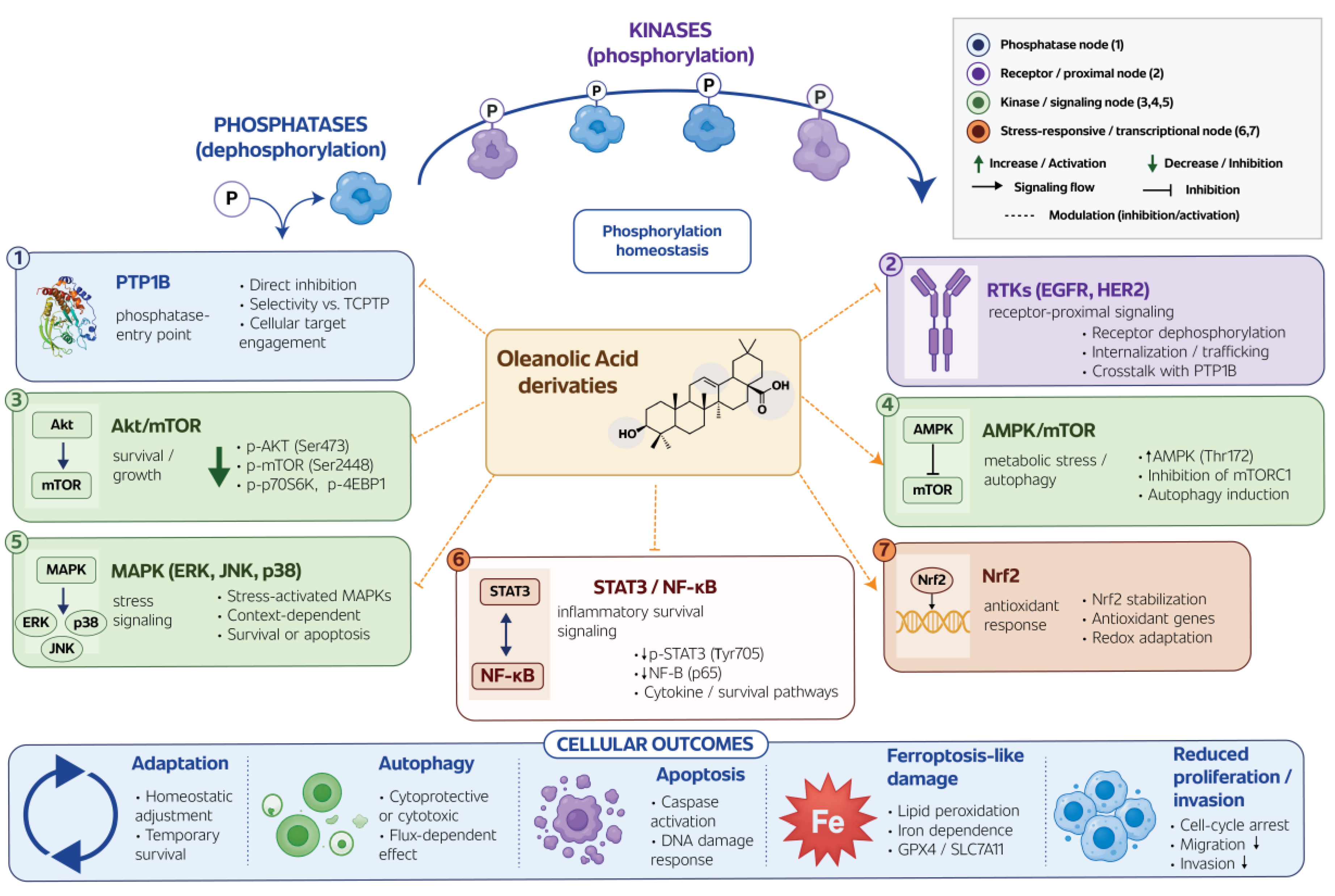
Figure 2.
Structural modification map of oleanolic acid and candidate signaling entry points. The C-3 hydroxyl group, the C-12/C-13 double bond, and the C-28 carboxyl group provide the principal sites for semisynthetic modification. Glycosylation, lactonization, dimerization, acetylation, oxime formation, and conjugation may alter polarity, lipophilicity, cellular distribution, and target accessibility, thereby biasing individual derivative classes toward different signaling profiles. The indicated associations are working hypotheses and should not be interpreted as established target assignments.
Figure 2.
Structural modification map of oleanolic acid and candidate signaling entry points. The C-3 hydroxyl group, the C-12/C-13 double bond, and the C-28 carboxyl group provide the principal sites for semisynthetic modification. Glycosylation, lactonization, dimerization, acetylation, oxime formation, and conjugation may alter polarity, lipophilicity, cellular distribution, and target accessibility, thereby biasing individual derivative classes toward different signaling profiles. The indicated associations are working hypotheses and should not be interpreted as established target assignments.
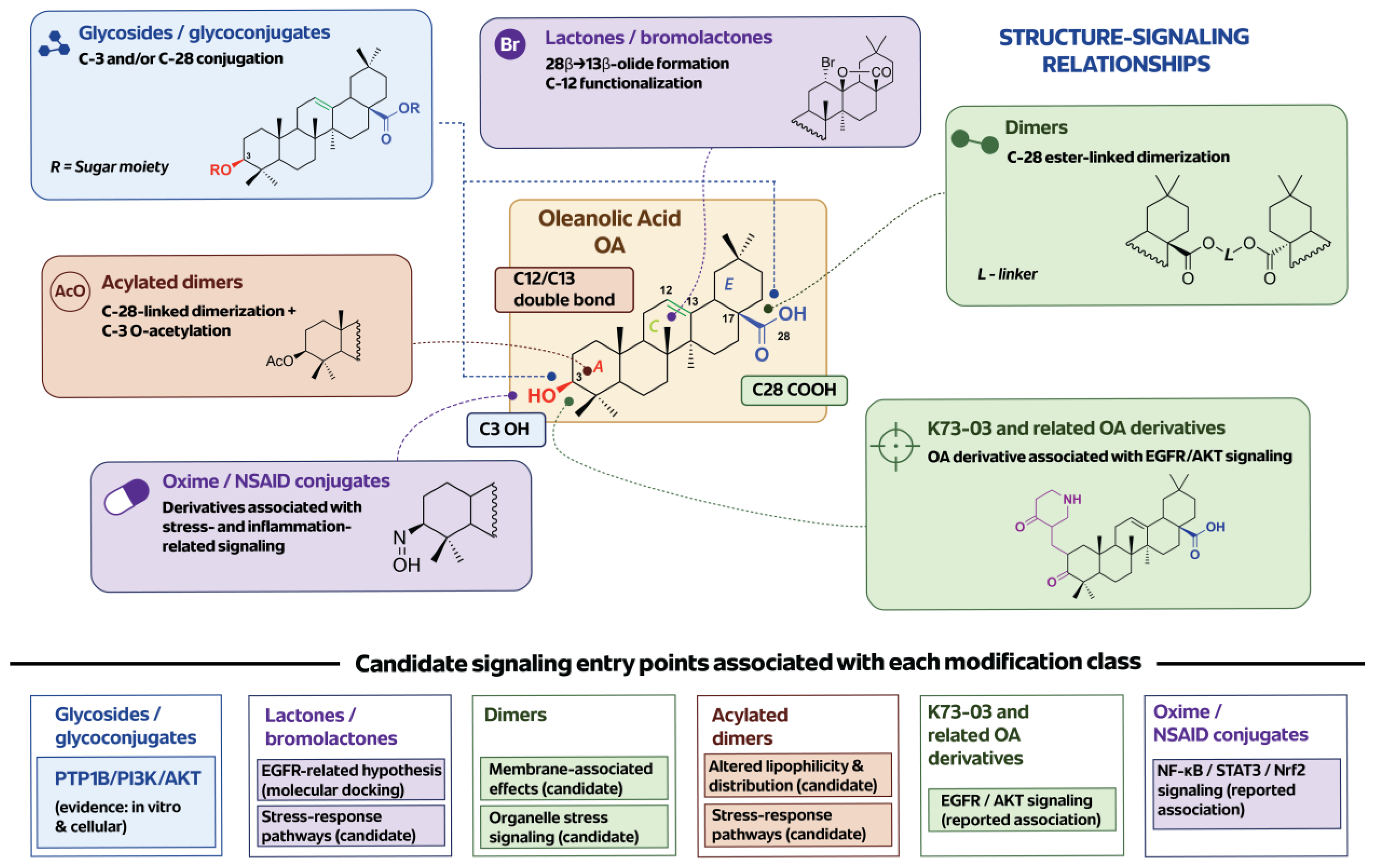
Figure 3.
Evidence hierarchy for mechanistic claims in studies of OA derivatives. Changes in individual phosphorylation markers represent pathway-associated observations rather than proof of direct target modulation. Mechanistic confidence increases when marker analysis is complemented by enzyme assays, receptor-level validation, cellular target-engagement methods, genetic or pharmacological rescue, and comparative testing of structurally related derivatives.
Figure 3.
Evidence hierarchy for mechanistic claims in studies of OA derivatives. Changes in individual phosphorylation markers represent pathway-associated observations rather than proof of direct target modulation. Mechanistic confidence increases when marker analysis is complemented by enzyme assays, receptor-level validation, cellular target-engagement methods, genetic or pharmacological rescue, and comparative testing of structurally related derivatives.
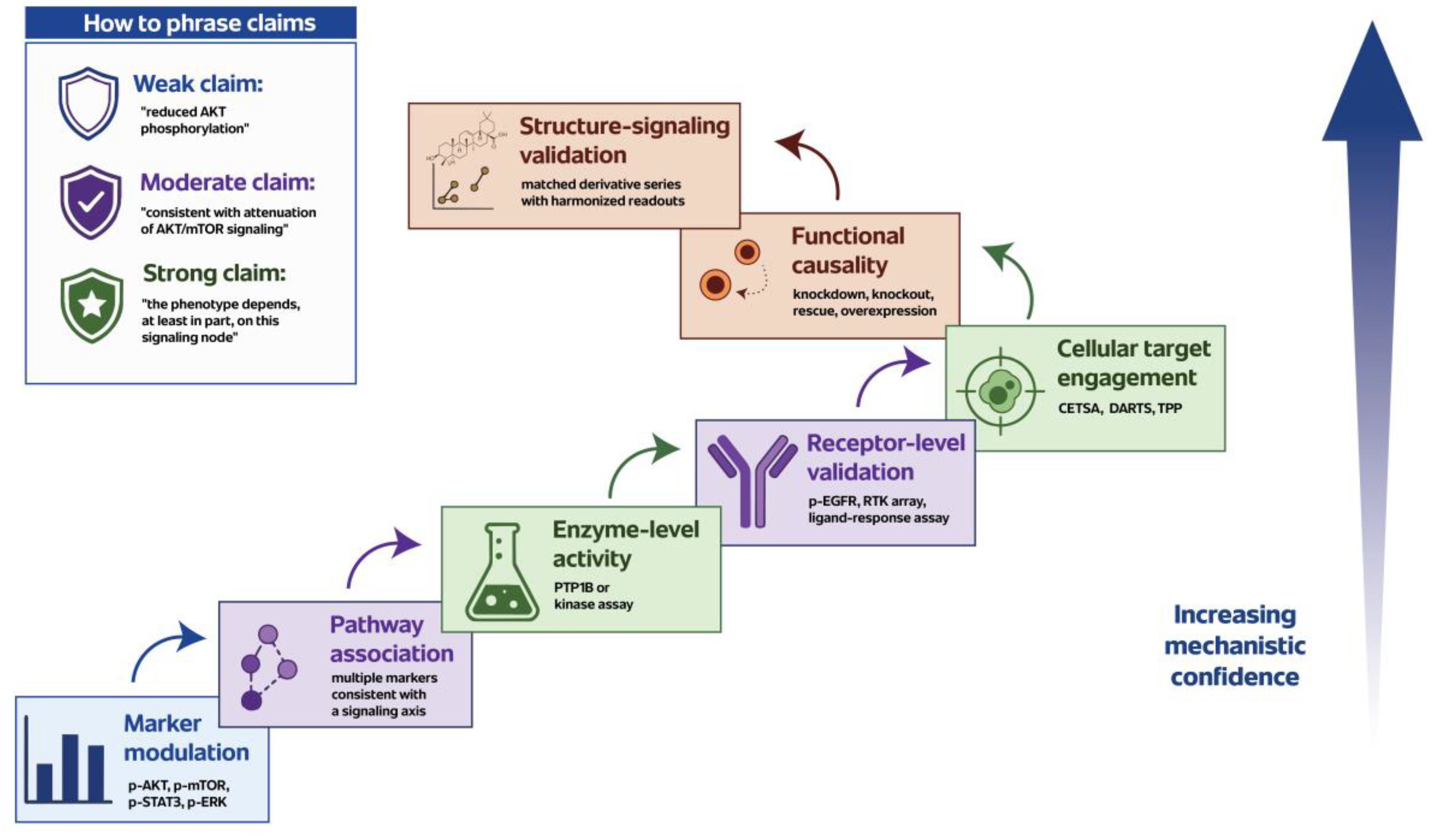

Table 1.
PTP1B-Directed OA Derivatives and Phosphatase-Relevant Evidence.
| Compound / derivative class | Main modification |
PTP1B-related evidence |
Selectivity information |
Cellular / biological context | Interpretive value |
|---|---|---|---|---|---|
| OA parent compound [8] |
Unmodified oleanane scaffold | Identified as a natural triterpenoid inhibitor of PTP1B | Limited selectivity information in early studies | Initial natural-product-based PTP1B inhibition studies | Establishes OA as a phosphatase-relevant natural-product scaffold |
| OA derivatives optimized from parent scaffold [8] | Semisynthetic modification of OA core | Competitive PTP1B inhibition and cellular activity reported | Requires careful comparison with related phosphatases | PTP1B inhibitor development and cellular activity assays | Shows that OA can be chemically optimized toward PTP1B-directed activity |
| OA glycoconjugates / triterpenoid saponins [9] | Glycosylation / glycoconjugation, often involving C-3 |
In vitro PTP1B inhibitory activity evaluated | TCPTP selectivity assessed |
Enzyme-level phosphatase inhibition studies | Introduces selectivity as a key medicinal chemistry variable for OA-derived PTP1B inhibitors |
| OA glycoside derivatives [10] | Glycoside-based semisynthetic derivatives | Designed to target PTP1B/PI3K/AKT signaling | PTP1B and TCPTP comparison reported | Breast cancer models, including pathway and biological activity readouts | Connects OA–PTP1B pharmacology with cancer-related AKT signaling |
| OA-Br-1 / related brominated OA glycoside derivative [10] |
OA glycoside derivative with brominated sugar-related substitution and C-28 modification | PTP1B inhibition reported with downstream PI3K/AKT pathway modulation | Selectivity over TCPTP reported | Breast cancer models and in vivo xenograft evaluation | One of the strongest examples linking OA derivative chemistry, PTP1B inhibition, and cancer-relevant signaling |
| OA dimers [11,12] | C-28-linked dimeric derivatives |
PTP1B not yet systematically validated | Not established |
Cytostatic, antioxidant, docking, and ADMETox studies | Candidate class for future phosphatase-oriented screening and phosphotyrosine signaling analysis |
| Acetylated OA dimers [13] | C-28-linked dimers with C-3 acetylation |
PTP1B not yet systematically validated | Not established |
Cytotoxicity, SI, DPPH, CUPRAC, and QSAR-related studies | Useful for testing whether acetylation and dimerization shift signaling toward stress-associated rather than phosphatase-directed mechanisms |
| OA lactones / bromolactones [14] | Lactonization or bromolactonization |
PTP1B not established as a primary target | Not established |
Cytotoxicity, EGFR docking, antioxidant activity, and ADMETox profiling | More suitable for RTK-proximal hypotheses than PTP1B-centered claims unless phosphatase assays are performed |
Table 2.
OA Derivative Classes and Candidate Signaling Entry Points.
| OA derivative class | Main structural feature |
Candidate signaling entry point to prioritize |
Suggested early readouts |
Main caution |
|---|---|---|---|---|
| OA parent compound [6,8,33] |
Unmodified oleanane scaffold with C-3 hydroxyl group, C-12/C-13 double bond, and C-28 carboxyl group | Broad stress, metabolic, antioxidant, and phosphorylation-associated signaling | p-AMPK, p-mTOR, ROS, mitochondrial membrane potential, apoptosis markers, PTP1B activity | Low solubility, pleiotropic activity, and difficulty distinguishing direct target effects from secondary stress responses |
| OA glycosides / glycoconjugates [8,9,10] | Sugar or glycoconjugate modification, commonly involving C-3 and/or C-28-derived substituents | PTP1B-related signaling and phosphatase selectivity | PTP1B activity, TCPTP selectivity, p-AKT, p-mTOR, PI3K/AKT readouts | Cellular uptake and intracellular target access may differ markedly from parent OA |
| OA glycoside derivatives targeting PTP1B/PI3K/AKT [10] | Glycoside-based semisynthetic OA derivatives optimized toward PTP1B-related activity | PTP1B/PI3K/AKT signaling in breast cancer models | PTP1B inhibition, TCPTP comparison, p-AKT, p-PI3K, apoptosis and proliferation markers | Downstream AKT changes do not alone prove PTP1B-dependent causality |
| K73-03-like derivatives [16,17] | Semisynthetic OA derivatives linked with EGFR/AKT modulation | RTK-proximal signaling, especially EGFR/AKT | p-EGFR, p-AKT, RTK arrays, ligand-stimulation assays, receptor internalization | Reduced p-EGFR and p-AKT support pathway involvement but do not alone prove direct EGFR inhibition |
| OA lactones / bromolactones [14] | Lactone or bromolactone formation with modification of the E-ring region and C-12-associated functionality | RTK hypotheses, EGFR-associated signaling, and stress signaling | p-EGFR, p-AKT, p-ERK, ROS, mitochondrial membrane potential, early viability-independent readouts | Docking should be treated as hypothesis-generating and requires receptor-level validation |
| OA dimers [11,12] | Two OA units connected through C-28-derived linker systems | Membrane-proximal signaling, organelle stress, and integrated phospho-signaling | p-AKT, p-mTOR, p-AMPK, ROS, mitochondrial stress, autophagic flux, broad phospho-panel | High cytotoxicity may reflect uptake, lipophilicity, or membrane/organelle effects rather than one defined target |
| Acetylated OA dimers [13] | Dimeric OA derivatives with C-3 acetylation | Membrane- and organelle-associated signaling, altered cellular distribution, and stress-response pathways | Early phospho-panel, mitochondrial membrane potential, ER stress markers, ROS, autophagic flux | Late phosphorylation changes may reflect cytotoxic collapse; comparison with non-acetylated dimers is essential |
| OA oxime / NSAID conjugates [49,50] | Oxime-based or anti-inflammatory conjugates, including NSAID-linked derivatives | NF-κB, STAT3, Nrf2, and inflammatory stress signaling |
p-NF-κB p65, p-STAT3, Nrf2 nuclear localization, target gene expression | Effects may differ between cancer and non-malignant cells; Nrf2 activation can be protective or pro-survival depending on context |
Table 3.
Evidence Levels for Kinase–Phosphatase Mechanistic Claims in OA Derivative Studies.
| Evidence level |
Typical experiment |
What can be concluded |
What cannot be concluded |
Recommended language |
|---|---|---|---|---|
| Marker modulation |
Western blot or immunoassay for p-AKT, p-mTOR, p-AMPK, p-ERK, p-JNK, p-p38, p-STAT3, p-NF-κB p65 | The compound changes phosphorylation-associated signaling under the tested conditions | Direct target identity, primary mechanism, or kinase/phosphatase causality | “The compound reduced AKT phosphorylation” or “altered mTOR-associated signaling markers” |
| Pathway association |
Multiple markers consistent with a known pathway, e.g., p-AKT + p-mTOR + p-p70S6K |
A pathway is associated with the cellular response | Direct inhibition of the pathway or primary target engagement | “The data are consistent with attenuation of AKT/mTOR signaling” |
| Enzyme-level activity |
In vitro kinase or phosphatase assay, e.g., PTP1B inhibition assay |
The compound can affect a purified enzyme biochemically |
Cellular target engagement or relevance to the phenotype |
“The compound inhibited PTP1B in vitro” |
| Phosphatase selectivity |
PTP1B assay with TCPTP and other phosphatases | Selectivity or lack of selectivity among related phosphatases | Functional dependence on a specific phosphatase in cells | “The compound showed PTP1B inhibition with selectivity over TCPTP” |
| Receptor-level validation |
p-EGFR, p-HER2, RTK array, ligand-stimulation assay, receptor internalization or degradation assay | The compound affects receptor-proximal signaling | Direct receptor binding or ATP-competitive RTK inhibition unless separately tested | “The compound altered EGFR phosphorylation or ligand-induced receptor signaling” |
| Cellular target engagement |
CETSA, DARTS, TPP, pull-down, SPR in relevant context, or related methods | The compound engages or stabilizes a candidate target in a cellular or biochemical context | That the target is required for the phenotype | “The data support cellular engagement of the candidate target” |
| Functional causality |
Knockdown, knockout, rescue, overexpression, catalytically inactive mutants, pathway rescue, comparison with reference inhibitors | The candidate node contributes to the biological outcome | Universal mechanism across all derivatives, doses, or cancer models | “The phenotype depends, at least in part, on this signaling node” |
| Structure–signaling validation | Matched derivative series tested with harmonized early signaling readouts | Structural features correlate with distinct signaling entry points | Definitive target identity without additional target-engagement and rescue data | “This derivative class preferentially engages a receptor-proximal / phosphatase-related / stress-associated signaling profile” |
| Translational validation |
3D models, organoids, xenografts, formulation-aware exposure analysis, pharmacokinetics, safety readouts | The signaling mechanism is more likely to persist under biologically relevant conditions | Clinical efficacy or patient benefit without further studies | “The mechanism remains detectable in more complex preclinical models” |
Disclaimer/Publisher’s Note: The statements, opinions and data contained in all publications are solely those of the individual author(s) and contributor(s) and not of MDPI and/or the editor(s). MDPI and/or the editor(s) disclaim responsibility for any injury to people or property resulting from any ideas, methods, instructions or products referred to in the content. |
© 2026 by the authors. Licensee MDPI, Basel, Switzerland. This article is an open access article distributed under the terms and conditions of the Creative Commons Attribution (CC BY) license.
Copyright: This open access article is published under a Creative Commons CC BY 4.0 license, which permit the free download, distribution, and reuse, provided that the author and preprint are cited in any reuse.


