

Submitted:
27 July 2026
Posted:
29 July 2026
You are already at the latest version
Abstract
Heart-rate variability (HRV) tracks sleep stages in real time, with parasympathetic dominance concentrated in slow-wave sleep (SWS) and attenuated during REM sleep. Evidence indicates that this coupling is causally, not merely correlationally, linked to post-exercise cardiac autonomic recovery: enhancing slow-wave activity increases parasympathetic HRV, sleep restriction disrupts nocturnal autonomic state directly, and post-exercise vagal reactivation is depressed on nights following intense exercise, localized to the SWS stage. This review synthesizes the literature into a framework in which SWS functions as a physiological gate for post-exercise autonomic recovery, integrating brainstem circuitry and neurotransmitter systems, causal manipulations (acoustic, pharmacological, deprivation-based), exercise dose-response, and boundary conditions (age, sex, training status, sleep, and cardiovascular disorders) under which the coupling is preserved, attenuated, or absent. The gating effect is graded and window-specific rather than uniform: robust in the early post-exercise reactivation phase and during nocturnal SWS, but not established across the full multi-hour recovery curve. The framework is translated into falsifiable predictions and candidate study designs, including a critical appraisal of the validity of wearable and nearable sleep-tracking. Two evidentiary gaps are transparently addressed: the human circuit-level mechanism remains largely inferred from rodent work, and independent citation-network verification is not feasible for all foundational sources.
Keywords:
slow-wave sleep
; heart-rate variability
; autonomic nervous system
; exercise recovery
; parasympathetic reactivation
; baroreflex
; sleep architecture
; cardiac vagal control
; wearable sleep monitoring
1. Introduction
1.1. Background and Rationale
Cardiac parasympathetic tone, most commonly indexed by heart rate variability (HRV), is widely used as a marker of postexercise recovery, typically summarized as a single nocturnal average or morning spot measurement. This convention treats sleep as a passive backdrop against which autonomic recovery unfolds according to its own schedule. However, a separate body of evidence indicates that autonomic state tracks sleep architecture in real time: parasympathetic activity is highest during slow-wave sleep (SWS) and is measurably attenuated during rapid-eye-movement sleep, a relationship that extends to functional connectivity within central autonomic brain regions specifically during SWS [1,2].
Critically, this relationship appears to be causal rather than correlational. Experimentally enhancing slow-wave activity with closed-loop acoustic stimulation increases parasympathetic HRV power and reduces sympathetic drive, establishing that the direction of causation runs from sleep architecture to the autonomic state [3]. This mechanism plausibly underlies postexercise autonomic recovery: vagal-related HRV indices measured during SWS are depressed on the night following intense exercise and recover over subsequent nights [4], SWS-phase HRV discriminates training-load state more sensitively than whole-night measurements [5], and functionally overreached athletes show a selective reduction in parasympathetic control specifically during SWS, without a corresponding change in the whole-night average [6]. Sleep restriction directly disrupts this mechanism: polysomnographically confirmed partial sleep restriction increases sympathetic tone and decreases vagal tone during the sleep period itself, not merely upon waking [7].
Despite this convergent evidence, the sleep-stage-specific basis of post-exercise autonomic recovery has not, to our knowledge, been synthesized into a dedicated conceptual framework. Existing reviews of postexercise cardiac parasympathetic reactivation frame recovery primarily as a function of elapsed time — placing full recovery at up to 24 h after low-intensity exercise, 24–48 h after threshold-intensity exercise, and at least 48 h after high-intensity exercise [8] — without distinguishing the specific contribution of sleep architecture, particularly SWS, from the passage of time itself. This distinction is not merely semantic: if SWS is the physiological substrate of nocturnal autonomic recovery, then interventions or conditions that alter SWS quantity or quality (age, sex, training status, sleep or cardiovascular disorders, or sleep-tracking-guided behavior change) should be expected to alter recovery independently of elapsed time, a prediction that a purely time-based framework does not make.
This review addresses this gap by proposing slow-wave sleep as a physiological gate for postexercise cardiac autonomic recovery: a conceptual framework in which the strength and even the detectability of postexercise autonomic effects depend on whether and how the intervening sleep period engages the SWS-autonomic coupling rather than on elapsed time alone. Consistent with the broader literature on sleep-exercise interactions, this gating effect is not proposed as uniform or absolute; the evidence reviewed below indicates that it is graded and window-specific, with the strongest support in the early post-exercise reactivation phase and the SWS period itself, and considerably weaker support across the intermediate, multi-hour recovery window [9].
1.2. Objectives
The specific objectives of this review are as follows: (1) to synthesize the brainstem circuitry and neurotransmitter systems implicated in mediating the coupling between slow-wave sleep and autonomic tone, distinguishing human correlational evidence from causal circuit-level evidence derived predominantly from animal models (Section 2); (2) to evaluate the causal evidence linking experimental manipulation of slow-wave sleep — acoustic, pharmacological, and deprivation-based — to autonomic and cardiovascular outcomes (Section 3); (3) to characterize the dose-response relationship between exercise intensity and duration and the magnitude of SWS-phase autonomic disruption, including differences between trained and sedentary individuals (Section 4); (4) to identify the boundary conditions — age, sex, clinical sleep and cardiovascular disorders, and sleep-monitoring technology — under which the SWS-autonomic gating effect is preserved, attenuated, or untested (Section 5, Section 6 and Section 7); and (5) to translate the resulting framework into falsifiable predictions and candidate study designs for future empirical testing (Section 8).
1.3. Guiding Research Questions
Q1. Which brainstem nuclei and neurotransmitter systems mediate the coupling between slow-wave sleep and autonomic tone, and to what extent is this circuitry established in humans versus that inferred from animal models?
Q2. Beyond closed-loop acoustic enhancement, which experimental manipulations of slow-wave sleep have been shown to causally alter autonomic or cardiovascular parameters, and in which direction?
Q3. How does the magnitude of slow-wave sleep phase autonomic disruption following exercise scale with exercise intensity and duration, and does this relationship differ between trained and sedentary individuals?
Q4. Do age-related declines in slow-wave sleep and documented sex differences in sleep architecture alter the strength of the slow-wave-sleep-autonomic coupling, and through which mechanisms?
Q5. In clinical populations with disrupted slow-wave sleep, is autonomic recovery measurably impaired relative to healthy sleepers, and does treating the underlying sleep disorder restore autonomic recovery capacity?
Q6. Are consumer wearable and nearable sleep-tracking devices sufficiently valid for either heart-rate-variability output or slow-wave-sleep staging to support research or applied monitoring of this coupling in a post-exercise context?
1.4. Scope and Manuscript Structure
This review is conceptual and hypothesis-generating in nature. Slow-wave sleep is proposed as a physiological gate for post-exercise autonomic recovery in the sense of a graded, window-specific modulator of recovery, not as a strict, all-or-none requirement, or as a validated diagnostic or monitoring index. The literature was identified using a targeted, question-driven strategy, as detailed in the Literature Review Strategy and Scope subsection immediately following this Introduction, which also reports the outcome of an independent citation-network verification step applied to several of the foundational sources discussed in Section 2 and Section 3.
The remainder of the manuscript is organized as follows. Section 2 reviews the brainstem circuitry and neurotransmitter systems implicated in SWS-autonomic coupling. Section 3 reviews the causal manipulations of SWS and their autonomic and cardiovascular consequences. Section 4 addresses the dose-response relationship between exercise and SWS-phase autonomic disruption. Section 5 addresses age and sex as sources of variation in coupling. Section 6 addresses clinical populations with disrupted SWS. Section 7 critically appraises wearable and nearable technologies for monitoring this coupling. Section 8 translates the framework into falsifiable predictions and candidate research designs. Section 9 provides a general discussion and conclusion.
1.5. Literature Review Strategy and Scope
Evidence for this review was identified using a two-stage, targeted, question-driven strategy rather than a fixed database systematic search protocol. In the first stage, six specific, mechanistically framed questions — corresponding to Section 2, Section 3, Section 4, Section 5, Section 6 and Section 7 of this review — were submitted individually to Elicit, an AI-assisted literature discovery and synthesis tool that queries a large indexed corpus of scholarly publications. Consistent with the approach adopted in related work by the authors, priority was given to studies reporting serial or interventional data over single-timepoint or purely observational findings, and both positive and null or contrasting findings were retained and reported, with the boundary conditions distinguishing them made explicit in the narrative rather than resolved by selective citation.
In the second stage, an independent citation network verification step was applied to four foundational sources identified as load-bearing for the causal claims developed in Section 2 and Section 3, using Scite, a tool that classifies papers citing a given source as supporting, contrasting, or mentioning its findings. This verification step yielded a mixed and instructive outcome that is reported transparently here rather than being silently incorporated into the narrative. For one source (Grimaldi et al. [3]), independent verification identified a direct, well-characterized extension of the finding in an independent sample [10], and this cross-confirmation is noted at the relevant point in Section 3. For the second source (Tasali et al. [11]), citation-network verification, after confirming the correct target paper by title and DOI, found no citing paper meeting a strict quotable-evidence criterion for replication, extension, or contradiction. This is reported in Section 3 as a confirmed citation-tracking gap rather than as either a replication or contradiction, and the relevant contrasting evidence discussed in Section 3 is drawn directly from the original literature search [12]. For a third source (Sayk et al. [13]), the verification tool returned a set of citing papers that, on inspection, included several with only tangential thematic relevance to the specific SWS deprivation mechanism; these were not judged reliable enough to characterize the replication landscape and are not reported as such. Therefore, the primary findings of Sayk et al. are presented in Section 3 based on the original study alone. For the fourth source (Yao et al. [14]), a mouse study of brainstem baroreflex-sleep circuitry, citation-network verification could not be completed due to an indexing failure in the verification tool that persisted across repeated attempts with corrected identifiers; this source is therefore presented in Section 2 as a circuit-level mechanistic account from the original literature, without independent citation-network confirmation, and this limitation is noted again in Section 9.
This review does not follow a PRISMA-based systematic-review protocol. No fixed set of databases, Boolean query strings, or pre-registered inclusion/exclusion criteria were applied, and no independent dual reviewer screening was conducted. Accordingly, the evidence base underlying this review should be understood as a structured, mechanistically prioritized narrative synthesis supplemented by a partial, transparently reported citation network verification step rather than an exhaustive or fully reproducible systematic review.
The resulting evidence base is heterogeneous in study type and species. Priority was given to human experimental and interventional studies; where a specific mechanistic claim rests primarily or exclusively on rodent evidence, this is stated explicitly in the surrounding text (see, in particular, Section 2). The evidence base also includes a small number of conference and meeting abstracts, which have not undergone full peer review and are noted as such at the first mention.
2. Brainstem Circuitry and Neurotransmitter Systems Mediating the Slow-Wave-Sleep–Autonomic Coupling
One framing point governs how this section should be read: in humans, the slow-wave sleep (SWS)–autonomic link is established almost entirely through correlational evidence — coordinated cortical and cardiac oscillations whose direction of causation is difficult to establish from observational recordings alone [15]. The circuit-level, causal dissection of this coupling comes almost entirely from rodent (and some cat) studies, and much of that work characterizes NREM sleep broadly or REM sleep specifically rather than SWS in isolation. Where a finding concerns REM rather than SWS, or derives from a non-human species, this is stated explicitly below rather than being left to be inferred.
2.1. The Autonomic Signature of SWS in Humans
The consistent human finding is that deepening non-REM sleep is a state of parasympathetic dominance and sympathetic withdrawal. Progression into SWS is marked by increased high-frequency (parasympathetic) HRV [3], and across sleep stages, parasympathetic tone tracks the depth of sleep, whereas sympathetic tone tracks time asleep [16]. A more quantitatively specific dissociation refines this picture: electroencephalographic delta power correlates negatively with the sympathetic HRV index (the low-frequency-to-high-frequency ratio) but not with the vagal index, indicating that deeper SWS chiefly reflects graded sympathetic withdrawal rather than a further increase in vagal tone [17]. The nocturnal blood pressure “dipping” characteristic of non-REM sleep is understood to arise from generalized cardiovascular deactivation together with baroreflex resetting, driven by central autonomic commands rather than passive mechanical factors [18].
2.2. Brainstem and Autonomic Nuclei
Nucleus tractus solitarius (NTS). The medullary NTS is the first central relay for baroreceptors and other vagal afferents and constitutes a node at which cardiovascular and sleep-state control converge. In a rat model, chemogenetic and optogenetic stimulation of NTS neurons and the NTS→parabrachial nucleus pathway increased delta electroencephalographic power specifically during NREM sleep [19]. More directly relevant to this review, a 2022 mouse study demonstrated that the cardiovascular baroreflex circuit itself “moonlights” in sleep control: activity-tagged, barosensitive NTS neurons, when chemogenetically or optogenetically activated, promote non-REM sleep while simultaneously lowering blood pressure and heart rate [14]. This finding provides the most direct mechanistic bridge identified in this review between the baroreflex circuit and the sleep state that carries parasympathetic dominance, although it remains, at present, a mouse finding without a demonstrated human counterpart (see Literature Review Strategy and Scope for the status of independent citation-network verification of this source).
Rostral ventrolateral medulla (RVLM), and C1 neurons. The RVLM is the principal sympathoexcitatory premotor source of the cardiovascular system. Its surface activity reflects both the early baroreflex response and later compensatory activity during sleep, with response latencies longer in REM sleep than in quiet sleep [20]. Sympathoexcitatory, adrenergic C1 neurons within the RVLM have been linked to the sympathetic limb of the arterial baroreflex, with a rat study reporting that C1-neuron activation during REM sleep is specifically dependent on baroreflex input [21]. These neurons are not merely passive during sleep: reviews of the interface between autonomic and arousal systems describe C1 neurons as responsive to hypoxia and capable of promoting arousal from non-REM sleep [22], and a separate line of rat work showed that adrenergic ventrolateral medullary cells are activated during REM sleep even as pontine noradrenergic cells are suppressed [23]. Yao et al. linked these observations: GABAergic neurons of the caudal ventrolateral medulla were found to promote non-REM sleep partly by inhibiting the sympathoexcitatory, wake-promoting adrenergic neurons of the RVLM [14]. Because the C1/RVLM mechanisms in this literature are characterized predominantly in the context of REM sleep and arousal rather than SWS specifically, they are presented here as contextual circuitry rather than as an established SWS-specific mechanism.
Nucleus ambiguus (NA) and cardiac vagal neurons. The nucleus ambiguus houses most parasympathetic cardiac vagal preganglionic neurons. In a mouse model, genetically defined (Tbx3-positive) NA cardiovagal neurons project directly onto the heart and, when activated, immediately and dramatically slow the heart rate, an effect abolished by atropine, confirming a muscarinic mechanism [14]. Because these cardiac vagal neurons are intrinsically silent, their firing and thus resting vagal tone are set entirely by synaptic drive from glutamatergic, GABAergic, glycinergic, and serotonergic inputs [24]. In the Yao et al. mouse study, cholinergic NA neurons also independently promoted non-REM sleep [14], again coupling the cardiac-vagal limb of the circuit to the sleep state itself, not merely to its downstream cardiac effectors.
Lateral paragigantocellular nucleus (LPGi) — an REM-specific gate. For state-dependent withdrawal of vagal tone, the clearest characterized circuit concerns REM sleep and not SWS. In rat studies, stimulation of the LPGi evokes GABAergic inhibition of cardiac vagal neurons in the nucleus ambiguus, providing a candidate mechanism for the reduced parasympathetic cardiac activity and heart-rate elevation characteristic of REM sleep; this GABAergic pathway is itself modulated by α7-nicotinic (cholinergic) transmission [25]. This mechanism is included here explicitly for contrast rather than as evidence bearing on the SWS-specific gating proposed in this review, as it describes REM-phase vagal withdrawal, the opposite sleep stage, and the opposite direction of the autonomic effect.
2.3. Higher-Order Central Autonomic Network
Above the brainstem, a forebrain “central autonomic network” comprising the insula, anterior and midcingulate cortex, amygdala, hypothalamus, periaqueductal gray, and parabrachial nucleus, together with the medullary NTS, NA, ventrolateral medulla, and raphe, overlaps anatomically with arousal circuitry [22]. Human evidence linking this network specifically to SWS, rather than to sleep or arousal in general, comes from a study in older adults at risk of dementia: parasympathetic (high-frequency) HRV measured specifically during SWS was associated with functional connectivity in core central-autonomic regions (right anterior insula, posterior midcingulate cortex) and with amygdala–thalamus connectivity, with no equivalent association during REM sleep or wakefulness [2]. This dissociation suggests that SWS constitutes a privileged window for brain–heart coupling rather than being one instance of a general sleep-related phenomenon. Consistent with this, loss of the SWS-specific parasympathetic signal has been observed as a form of autonomic “hypervigilance” in chronic fatigue syndrome, in which vagally mediated HRV is reduced specifically during deeper sleep stages but not during resting wakefulness [26].
2.4. Neurotransmitter Systems
Cholinergic signaling. Cholinergic systems appear to play two distinct roles in coupling. First, in state control, wake-active cholinergic neurons of the basal forebrain and pedunculopontine tegmentum, together with monoaminergic neurons, provide state-dependent input onto the glutamatergic and GABAergic command neurons that ultimately set sympathetic versus cardiovagal output, and locus-coeruleus activation drives sympathetic activation with concurrent cardiovagal inhibition [22,27]. Second, in direct autonomic modulation, nicotinic (α7) transmission gates the LPGi→cardiac-vagal-neuron inhibitory pathway described above [25], and the vagal end-organ effect of NA cardiovagal neurons is muscarinic and atropine-sensitive [14].
Adenosinergic signaling. The coupling here is best characterized as indirect; adenosine functions as the homeostatic sleep-pressure signal that builds the SWS state carrying the parasympathetic shift, rather than as a demonstrated direct autonomic transmitter in its own right. Adenosine accumulates within the cholinergic basal forebrain during wakefulness and acts via A1 receptors to inhibit wake-active neurons to induce recovery NREM sleep and delta activity [28]. Selective lesioning of cholinergic basal forebrain neurons abolishes both the adenosine rise and the homeostatic sleep response that follows sleep deprivation [29]. A glial–neuronal circuit, in which glial adenosine kinase sets extracellular adenosine concentration by acting on neuronal A1 receptors, controls the magnitude of slow-wave activity [30]. Therefore, adenosine’s relationship with autonomic tone is best understood as an inferred chain: adenosine promotes deeper SWS, which in turn carries parasympathetic dominance, rather than a directly mapped autonomic pathway.
Glutamate and GABA are effector systems. It is worth naming explicitly that the actual command neurons for both sympathetic and cardiovagal output are glutamatergic and GABAergic; cholinergic, monoaminergic, and orexinergic systems act as state-dependent modulators superimposed on this glutamate/GABA architecture rather than as the primary effectors [27]. Hypothalamic orexin has been identified as a key permissive signal for sleep-related blood pressure changes [18].
2.5. Phasic Events as a Causal-Sequence Probe: K-Complexes
Because the tonic SWS–autonomic association is fundamentally correlational in humans, some of the clearest evidence of the temporal directionality of central-to-autonomic coupling comes from phasic, rather than tonic, non-REM events. Spontaneous K-complexes are followed by transient rises in muscle sympathetic nerve activity and blood pressure, a pattern interpreted as cortical activation transiently and gradedly facilitating sympathetic outflow [31]; this pressor response has been shown to be larger in men than in women [32]. Such evoked-event paradigms currently represent the primary approach available for probing the temporal directionality of central-to-autonomic coupling in humans in the absence of the causal circuit manipulations available in animal models [15].
2.6. Synthesis: A Circuit-Level Account with an Explicit Species and Evidence-Type Gap
Taken together, the evidence reviewed in this section indicates that a plausible multi-nodal brainstem circuit spanning the NTS, RVLM/C1 neurons, and NA cardiac vagal neurons, modulated by cholinergic and adenosinergic systems acting on an underlying glutamate/GABA effector architecture, could in principle mediate a causal link between SWS and autonomic tone. However, to the best of our knowledge, a dedicated, causal, SWS-specific circuit tracing from a defined brainstem nucleus to a measured autonomic output has not been established in humans. Human data in this domain consist of HRV correlations, while the elegant optogenetic and chemogenetic circuit dissections available (NTS, C1/RVLM, LPGi→cardiac-vagal-neuron, cholinergic NA) are derived from rodents and frequently characterize NREM sleep in general or REM sleep specifically rather than SWS. The LPGi and C1 mechanisms, in particular, are REM-phase phenomena, included here for mechanistic contrast rather than as direct SWS evidence. Therefore, this circuit-level account should be read as a plausible mechanistic scaffold consistent with the human correlational and causal manipulation evidence reviewed in Section 3, not as a demonstrated human mechanism in its own right.
3. Causal Manipulations of Slow-Wave Sleep and Their Autonomic and Cardiovascular Consequences
This section addresses which experimental manipulations of SWS, beyond closed-loop acoustic enhancement, have been shown to causally alter autonomic or cardiovascular parameters and in which direction. The evidence is markedly uneven across manipulation types: strongest and most consistent for deprivation and suppression paradigms, informative but confounded for pharmacological approaches, and largely untested for transcranial neuromodulation (Figure 1).
3.1. Closed-Loop Acoustic Enhancement: The Reference Causal Tool
Closed-loop acoustic stimulation remains the cleanest available causal tool for experimentally enhancing SWS while reading autonomic consequences. Automated closed-loop stimulation in middle-aged men increased high-frequency HRV and the standard deviation of NN intervals specifically during N3 sleep [3], and this finding was independently reproduced and extended in a separate randomized crossover sample, providing genuine cross-study confirmation of the effect rather than reliance on a single study [10] (see Literature Review Strategy and Scope). A related study found that auditory slow-wave enhancement improved next-day left ventricular systolic and diastolic function [33]. An important qualification, however, is that each individual evoked slow wave carries a transient cardiovascular activation — a short blood-pressure rise and a biphasic heart-rate response occurring during the stimulation itself [33] — and an earlier rat study found that acoustic stimuli delivered during non-REM sleep increased blood-pressure and heart-period variability to a degree that could plausibly favor arrhythmia in a susceptible substrate [34]. Therefore, acoustic SWS enhancement should be understood as producing a net parasympathetic gain over the study period, superimposed on transient, stimulus-locked activation events, rather than as a uniformly calming intervention at every timescale.
3.2. Selective SWS Deprivation and Suppression
Selective SWS deprivation → higher nocturnal blood pressure, blunted dipping. The landmark deprivation manipulation was that of Sayk et al.: healthy participants were selectively stripped of SWS using EEG-triggered acoustic arousals, without shortening total sleep time, in a crossover design against undisturbed sleep, with microneurographic muscle sympathetic nerve activity (MSNA) and HRV as readouts [13]. Depriving SWS significantly attenuated mean arterial blood pressure dipping during the first SWS-dominated half of the night, although some dipping still occurred, and the second REM-dominated half of the night was unaffected [13]. Notably, the effect was circumscribed rather than global: nighttime catecholamine excretion, morning MSNA, morning HRV, and daytime ambulatory blood pressure were all unchanged, indicating that a single night of SWS loss did not reset baroreflex-mediated sympathetic regulation on a more lasting basis [13]. As detailed in the Literature Review Strategy and Scope, independent citation network verification of this source identified several candidate supporting citations that, on inspection, addressed only tangential aspects of sleep and cardiovascular physiology rather than this specific deprivation-dipping mechanism; therefore, this finding is presented here on the basis of the original study alone, and its independent replication status should be considered unresolved rather than established.
Selective SWS suppression → sympathetic and metabolic shift. A second, metabolically oriented suppression paradigm comes from Tasali and colleagues, who suppressed SWS throughout the night using acoustic tones while holding total sleep time constant and found marked reductions in insulin sensitivity without compensatory insulin release, with the effect size proportional to the amount of SWS removed [11]. This finding is widely interpreted as reflecting increased sympathetic and reduced parasympathetic drive during a night of shallow sleep [11]. An important caveat on attribution, identified in the original literature search, comes from Ukraintseva et al., who reproduced glucose tolerance and melatonin changes after one night of SWS suppression but found that these effects tracked total sleep time, REM sleep duration, and awakenings more closely than SWS loss per se [12]. As detailed in the Literature Review Strategy and Scope, independent citation-network verification for Tasali et al. ’sfinding — after confirming the correct target paper by title and DOI — returned no citing paper meeting a strict quotable-evidence criterion for replication, extension, or contradiction. This is reported here as a confirmed citation-tracking gap: the absence of a directly quotable citing statement should not be read as either a replication or a refutation of the original finding, and the contrast discussed above, while genuinely relevant, was identified through the original topic-based literature search rather than through citation-network tracing of Tasali et al. itself.
3.3. Pharmacological Enhancement: The Sodium-Oxybate Cautionary Tale
Pharmacological SWS enhancement provides a mechanistically instructive case in which the direction of the autonomic effect is reversed relative to acoustic enhancement. Sodium oxybate robustly deepens SWS but is associated with increased blood pressure and sympathetic activity. Microneurography in patients with narcolepsy showed that both MSNA and blood pressure increased after six months of treatment, suggestive of sympathetic hyperactivity [35], and a large real-world cohort study found that initiating sodium oxybate was associated with an increased risk of new-onset hypertension (odds ratio ≈ 1.6) [36]. However, the corrective finding is important: switching patients from a high-sodium to a low-sodium oxybate formulation at an equivalent dose lowered 24-hour ambulatory systolic blood pressure by approximately 4 mmHg [37], indicating that the hemodynamic signal associated with oxybate is driven substantially by the sodium load of the formulation rather than by SWS enhancement itself. Therefore, sodium oxybate does not constitute clean evidence that pharmacologically deepening SWS raises blood pressure; if anything, once the sodium confound is accounted for, the finding is consistent with the parasympathetic-gain direction seen with acoustic enhancement. γ-Aminobutyric-acid-ergic SWS enhancers such as tiagabine reliably increase slow-wave activity [38], but essentially no autonomic or cardiovascular endpoints have been tested with this drug class, which constitutes a genuine evidence gap rather than a null finding.
3.4. Transcranial and Emerging Neuromodulation Approaches
Slow-oscillatory transcranial direct-current stimulation delivered during NREM sleep at approximately 0.75 Hz stabilizes sleep and increases N3 duration in patients with chronic insomnia, mimicking the effect of slow-wave-enhancing pharmacology [39]; however, this pilot study did not report any autonomic outcome. Closed-loop transcranial magnetic stimulation and closed-loop transcutaneous vagus nerve stimulation phase-locked to slow oscillations are emerging tools for modulating SWS, but no autonomic or cardiovascular readouts for these specific approaches were identified in this review. Transcranial neuromodulation methods are therefore established as capable of shifting SWS, while their downstream autonomic consequences remain largely uncharacterized, an explicit evidence gap rather than a documented null effect.
3.5. Synthesis: A Directional Map Across Manipulation Types
Across the manipulations reviewed in this section, a consistent directional pattern emerged, along with clearly demarcated gaps. Removing SWS, whether through selective deprivation or suppression, pushes toward higher nocturnal blood pressure, blunted dipping, and reduced insulin sensitivity [11,13]. Acoustically adding SWS pushes toward greater overnight parasympathetic tone and improves next-day cardiac function, with transient stimulus-locked activation superimposed on this net effect [3,10,33]. Pharmacological “enhancement” via sodium oxybate paradoxically raises blood pressure, but for reasons attributable to sodium load rather than SWS enhancement [35,36,37]. Transcranial neuromodulation methods remain largely unmeasured for autonomic endpoints [39]. This directional consistency across independent deprivation, suppression, and enhancement paradigms constitutes the strongest available evidence, short of a single unified causal experiment, that SWS functions as a physiological gate for autonomic tone, while the explicit gaps noted throughout (the pharmacological confound, the untested transcranial route, and the unresolved citation-network status of the Sayk and Tasali findings) define the boundaries of that claim.
4. Exercise Dose-Response and the Magnitude of Slow-Wave-Sleep-Phase Autonomic Disruption
A measurement caveat governs how this section should be read: most of the studies reviewed here quantify whole-night or multi-hour nocturnal HRV as a proxy for autonomic tone during deep sleep rather than segmenting the SWS stage itself. Only a small subset of studies isolates SWS specifically; these are identified explicitly below. Therefore, the genuinely SWS-phase-specific dose-response evidence is considerably thinner than the broader nocturnal-autonomic-recovery evidence.
4.1. Dose-Response: Scaling with Session Magnitude, with Intensity and Duration Dissociating
The core finding was that post-exercise suppression of nocturnal parasympathetic activity scales with the overall magnitude of the exercise session, although intensity and duration do not contribute equally. A controlled comparison found that the average nocturnal heart rate rose to approximately 109% of rest-day values after moderate endurance exercise but to approximately 130% after a marathon, while vagally mediated high-frequency HRV power fell to approximately 77% after moderate exercise and to only approximately 34% after the marathon, a pattern explicitly interpreted as a dose-dependent effect on autonomic modulation [40]. Intensity and duration dissociate at sub-maximal loads: a controlled matrix varying intensity (45%, 60%, 75% of VO2max for 30 min) and duration (30, 60, 90 min) found that higher intensity elevated nocturnal heart rate but did not change nocturnal HRV, whereas HRV declined only after the 90-minute session, with neither manipulation degrading overall sleep quality [41]. At the extreme-intensity end, supramaximal intermittent exercise suppressed vagal HRV measured in the first estimated SWS period, with the depression persisting into the second night and recovering only by the third, indicating that intensity can extend the recovery tail even when the total work is comparatively modest [4].
The largest-scale confirmation of this dose-response relationship comes from a wearable-based analysis of approximately four million person-nights, in which higher exercise strain and later exercise timing were both associated with higher nocturnal resting heart rate and lower nocturnal HRV; critically, exercise bouts ending four or more hours before sleep onset showed no association with either measure [42]. This timing-by-intensity interaction is mechanistically echoed by a controlled study in which morning exercise enhanced parasympathetic activity during subsequent sleep, whereas evening exercise shifted the balance toward sympathetic dominance (higher nocturnal heart rate) [43].
4.2. SWS-Stage-Specific Evidence: A Small but Informative Subset
The most stage-specific finding available indicates that even mild physical activity within an hour of bedtime leaves the overall sleep architecture intact but raises heart-rate variance and the low-frequency-to-high-frequency ratio specifically during SWS, indicating reduced parasympathetic dominance within the deep-sleep stage itself [44]. A separate study similarly found that evening endurance exercise partially suppressed parasympathetic activity (pNN50) across the sleep period, whether performed under normoxic or hypoxic conditions [45]. A recurring dissociation is worth emphasizing throughout this evidence: autonomic disruption of this kind does not translate into disrupted sleep as conventionally measured. Vigorous late-night exercise to exhaustion raised heart rate through the first sleeping hours, yet increased the proportion of NREM sleep and left subjective and actigraphic sleep quality unchanged [46]. In ultra-endurance runners, exercise intensity was more important than exercise timing for subjective sleep quality, even though nocturnal HRV did not differ across conditions [47].
4.3. Trained Versus Sedentary Individuals: Characterized Separately, Not in a Matched Comparison
Baseline differences between trained and untrained individuals are large: endurance-trained men show approximately four-fold higher nocturnal high-frequency (vagal) HRV power than untrained men, indicating that trained individuals begin sleep from a substantially higher parasympathetic set-point [48], and meta-analytic evidence confirms that exercise training raises resting parasympathetic HRV in previously sedentary individuals [49]. Trained athletes also appear relatively resilient to acute evening exercise loads: in highly trained female soccer players, late-night training sessions did not alter SWS-episode or hour-by-hour nocturnal HRV relative to rest days [50], and in endurance runners, an evening high-intensity interval session raised nocturnal heart rate but left HRV unchanged and modestly improved sleep continuity [51]. On the sedentary side, a small pilot study found that morning walking or running produced no consistent nocturnal HRV change, although this used morning timing, which the dose-response evidence above predicts should be comparatively benign [52]. Notably, even severely overtrained athletes showed no HRV difference from controls during sleep; the parasympathetic deficit emerged only after awakening, suggesting that the sleeping brain may protect vagal tone even under accumulated training stress, with the autonomic cost surfacing on waking challenge rather than during deep sleep [53].
4.4. Synthesis: An Inferred Rather Than Directly Demonstrated Dose-Response Curve for SWS Specifically
No study identified in this review segmented the SWS stage specifically, parametrically varied exercise intensity and duration, and did so in matched trained-versus-sedentary groups within a single design. The dose-response matrix work is conducted almost entirely in trained or physically active men; the trained-versus-untrained comparisons available largely address baseline autonomic tone rather than post-exercise dose-response; and only a small number of studies isolate SWS rather than whole-night HRV. The scaling of SWS-phase autonomic disruption with exercise dose, contrasted by training status, is therefore best described as inferred by combining these separate strands of evidence rather than directly demonstrated by a single study, a gap that directly motivates the study design recommendations proposed in Section 8.
5. Age and Sex as Sources of Variation in the Slow-Wave-Sleep–Autonomic Coupling
No study identified in this review tested the full three-way interaction of age-related SWS decline, sex-related differences in sleep architecture, and SWS-coupled post-exercise autonomic recovery. The following synthesizes four separate evidence streams that bear on this question, with the seams between them made explicit rather than smoothed over; cardiorespiratory fitness recurs as a confounder throughout the sex comparisons in particular.
5.1. Aging Degrades the SWS–Autonomic Coupling Itself
This is the best-supported component in this section. Slow-wave sleep and cardiac autonomic control during sleep decline with age. In older adults, normalized high-frequency (parasympathetic) HRV during sleep is broadly reduced, a loss linked to decreased SWS duration, with the normal sleep stage-dependent modulation of HRV flattening out [54]. More mechanistically specific evidence indicates that young adults show a discrete coupling event in which a burst of slow-wave activity precedes a heart-rate burst followed by a vagal (high-frequency) surge, and that in older adults, this coupling is essentially abolished, with neither the slow-wave boost nor the vagal modulation observed during SWS [55]. Therefore, aging appears to not merely reduce SWS quantity but also uncouple SWS from its autonomic correlate.
5.2. Aging and Post-Exercise Recovery: Real, but Fitness-Dependent
Aging slows post-exercise heart-rate recovery, and in women, this appears to track slower sympathetic withdrawal rather than impaired vagal reactivation, with the recovery time constant correlating strongly with age [56]. However, this age-related penalty largely disappears in individuals who remain physically active; in both active young and active older men, high-intensity interval exercise depressed vagal HRV and baroreflex sensitivity to a comparable degree, with recovery by 60 min in both groups and no age-associated difference in the response [57]. This qualifier is important: “aging blunts post-exercise autonomic recovery” is, on the available evidence, more precisely described as “aging combined with deconditioning blunts recovery.”
5.3. Sex Differences in Sleep Architecture and Sleep Autonomics
Women objectively show more SWS and greater electroencephalographic slow-wave power than men across a wide age range, a difference thought to arise partly from the organizational and activational effects of sex steroids [58]. The aging trajectories for SWS also diverge by sex: SWS begins to decline in men during their thirties but is comparatively preserved in women across the same age window, indicating that the age-related decline addressed in Section 5.1 is itself sex-dependent [59]. On the autonomic side, men show lower vagal tone and higher sympathovagal balance than women during sleep, especially during REM [60], and these sex differences emerge as early as adolescence, with within-night autonomic recovery blunted in girls relative to boys [61].
5.4. Sex Differences in Post-Exercise Recovery: Contested and Fitness-Confounded
The literature on sex differences in post-exercise autonomic recovery genuinely disagrees, and the disagreement is informative. Some studies have reported that men show faster heart rate recovery and greater vagal reactivation after exercise [62], while other studies have framed premenopausal women as having a cardioprotective profile, characterized by lower resting sympathetic tone and faster vagal reactivation with higher post-exercise high-frequency power [63]. The reconciling finding is that the apparent male advantage in heart-rate recovery disappears once aerobic capacity is statistically controlled: matching men and women for peak oxygen uptake eliminates the sex difference, indicating that much of what appears to be a sex effect is more accurately a fitness effect [63].
5.5. Attribution: Hormones, Autonomic Baseline, or Sleep Architecture
The available evidence permits a reasonably clear, albeit provisional, attribution. Hormonal factors matter for baseline reflex physiology but appear weak and secondary for the specific outcomes addressed in this review: exogenous estradiol increases cardiovagal baroreflex sensitivity to a hypertensive stimulus in young women [63], and menopausal status is associated with reduced HRV [64]; yet, estradiol did not explain autonomic modulation during exercise recovery in a direct test (age did) [56], no association emerged between HRV and circulating estradiol levels in a separate study [64], and the sleep-related sympathovagal sex difference persisted after statistically accounting for reproductive hormone levels [60]. The autonomic baseline appears to be the more consistent explanation: women’s higher resting vagal tone and lower sympathetic tone propagate into both sleep and post-exercise states, and sex differences in HRV are better predicted by this baseline (and by fitness) than by circulating hormone levels [63]. Sleep architecture is plausible but essentially untested as a mediating variable: women have more SWS, and SWS–vagal coupling is precisely what aging degrades (Section 5.1), so architecture should plausibly matter, yet no study identified in this review used SWS as the explicit mediating variable between sex and post-exercise autonomic recovery. A suggestive but indirect hint that the pathway runs through sleep comes from the finding that adequate sleep duration improved HRV in middle-aged women but not in men [65].
Table 1 consolidates the six determinants addressed in this section so that the direction of effect and the proposed primary explanation for each can be compared at a glance before the integrative synthesis below.
5.6. Synthesis: An Integrative Study Design Is the Explicit Gap
Taken together, aging weakens the SWS-autonomic coupling and slows post-exercise recovery, predominantly via deconditioning rather than age per se; women show more SWS, higher vagal tone, and a slower age-related SWS decline than men; and sex differences in post-exercise recovery are real but are substantially explained by fitness and baseline autonomic tone rather than by circulating hormones. To our knowledge, an integrative study tracking SWS-stage-specific autonomic recovery after a standardized exercise dose in age- and fitness-matched men and women across their lifespan has not yet been conducted. Until such a study exists, the question of whether age and sex modulate the coupling proposed in this review is answered by inference across separate literatures rather than by direct data, a gap made explicit in the study design recommendations in Section 8.
6. Clinical Populations with Disrupted Slow-Wave Sleep
This section addresses whether autonomic recovery is impaired in clinical populations with disrupted SWS and whether treating the underlying sleep disorder restores autonomic recovery capacity. One conceptual point applies throughout: in obstructive sleep apnea (OSA), the dominant clinical model addressed here, autonomic disruption is driven principally by intermittent hypoxia and repetitive arousals rather than by the loss of SWS. Therefore, Disrupted SWS → impaired autonomic recovery” in this population is best read as a correlate of a broader physiological disruption rather than as a clean, SWS-specific causal chain, and this qualification should be kept in mind when relating this section back to the SWS-specific gating framework proposed elsewhere in this review.
6.1. Obstructive Sleep Apnea: Robust Impairment That Persists Through Sleep
Foundational microneurographic work has demonstrated that patients with OSA have elevated sympathetic nerve activity even while awake and, critically, that blood pressure and sympathetic activity fail to fall during sleep, instead surging with each apneic event [66]. A meta-analysis spanning 850 patients across 26 microneurographic studies found that muscle sympathetic nerve activity increased monotonically with OSA severity, from approximately 42 bursts per 100 heartbeats in healthy controls to approximately 71 in severe OSA, correlating with the apnea-hypopnea index and oxygen desaturation rather than with body weight [67]. Therefore, the normal restorative, parasympathetic-dominant, blood-pressure-dipping state of deep sleep is essentially abolished in untreated OSA.
6.2. Continuous Positive Airway Pressure: Autonomic Recovery Restored at Multiple Levels
Treatment with continuous positive airway pressure (CPAP) reverses the OSA autonomic signature across all readouts examined in this study. CPAP acutely lowers sympathetic activity and blood pressure during sleep [66], and long-term therapy reduces daytime muscle sympathetic traffic, although this effect emerges only after extended treatment of six to 12 months rather than immediately [68]. At the level of the central mechanism, a longitudinal study combining functional imaging with microneurography found that six months of CPAP reduced muscle sympathetic nerve activity by roughly half (from approximately 54 to 23 bursts per minute) and returned abnormal activity in the RVLM, medullary raphe, and dorsolateral pons to control levels, indicating that the brainstem sympathoexcitatory circuitry itself normalizes with treatment [69]. CPAP also improved baroreflex control of heart rate during stage 2 NREM sleep by nearly 70% relative to pretreatment values [70] and converted the majority of non-dipping OSA patients back to a normal nocturnal blood pressure dipping pattern [71], with even a single night of CPAP improving HRV specifically during non-REM sleep [72]. The most direct causal evidence available runs in the reverse direction: in a randomized controlled withdrawal trial, discontinuing CPAP for two weeks caused OSA to recur and produced a significant rise in morning systolic blood pressure (approximately +8.5 mmHg), heart rate, and urinary catecholamines relative to patients who continued therapy [73], providing the strongest available evidence that the autonomic benefit of CPAP is genuinely treatment-dependent rather than a stable trait change.
6.3. Insomnia: A Real but Weaker, Phenotype-Dependent Signal
Insomnia is associated with a hyperarousal pattern; however, the supporting evidence is considerably weaker than that for OSA. Young adults with primary insomnia display sustained nighttime cardiac sympathetic hyperactivation (a shortened pre-ejection period) across the whole night [74], and adults with chronic insomnia show blunted sympathetic baroreflex sensitivity and exaggerated cardiovascular reactivity to stress, even when resting blood pressure, heart rate, and sympathetic activity do not differ from those of good sleepers [75]. The deficit appears to be concentrated in a specific phenotype: only insomnia with objective short sleep duration was associated with dampened parasympathetic (high-frequency) activity and sympathovagal imbalance, while a near-normal-duration insomnia phenotype was not [76]. However, two caveats temper this picture considerably. A systematic review and meta-analysis of 17 studies concluded that the current evidence does not reliably confirm impaired HRV in insomnia disorder [77], and a focused review noted that the supporting studies have mostly relied on sympathetic measures with limited specificity and reproducibility [78]. No adequate evidence has been identified on whether treating insomnia (for example, with cognitive behavioral therapy for insomnia) restores autonomic function, which stands as a genuine gap in contrast to the well-characterized CPAP literature for OSA.
6.4. Sleep Fragmentation: Inconsistent in Humans, with the Clearest SWS-Specific Data Coming from Animal Models
Evidence on pure sleep fragmentation, distinct from sleep restriction or OSA-related hypoxia, is mixed. In a controlled crossover design, partial sleep restriction increased sympathetic tone and decreased vagal tone overnight, but experimental sleep fragmentation via hourly awakenings did not significantly change any cardiac autonomic parameter [7], suggesting that fragmentation per se may be less autonomically disruptive than curtailed sleep duration. The most SWS-specific evidence of fragmentation comes from a chronic rat model, in which fragmentation raised mean arterial pressure specifically during SWS, accompanied by a compensatory rise in parasympathetic (high-frequency) power that normalized during subsequent recovery sleep [79].
Table 2 arranges the three clinical conditions addressed in this section along a gradient of evidentiary strength, from the robust and multiply reversible OSA literature to the largely untested question of pure sleep fragmentation in humans.
6.5. Synthesis
For OSA, the evidence supports a confident conclusion on both halves of the question addressed in this section: autonomic recovery is clearly impaired relative to healthy sleepers, and CPAP restores sympathetic tone, baroreflex sensitivity, blood pressure dipping, and brainstem function, with treatment withdrawal reversing these gains. For insomnia, impairment is real but modest, phenotype-dependent, and not consistently demonstrated across studies, and the question of whether treatment restores autonomic function remains largely unstudied. For pure sleep fragmentation, the human autonomic signal is inconsistent, and the cleanest SWS-specific evidence is derived from animal models. Because OSA-related autonomic disruption is driven substantially by intermittent hypoxia rather than SWS loss per se, this clinical literature should be read as an informative context for, rather than a direct confirmation of, the SWS-specific gating framework proposed in this review.
7. Wearable and Nearable Technology for Monitoring the Slow-Wave-Sleep–Autonomic Coupling
Consumer sleep-tracking devices perform two distinct measurement tasks with markedly different validity: measuring HRV, which they do well, and classifying the SWS stage, which they do only moderately well. The post-exercise recovery context specifically stresses the conditions under which the staging performance is weakest.
7.1. Heart-Rate-Variability Measurement: Strong Validity
On the cardiac side, the validity evidence is strong. The nocturnal heart rate and HRV of a wrist-worn ring agreed almost perfectly with simultaneous electrocardiography (r2 ≈ 0.996 for heart rate and 0.980 for HRV, with sub-beat and approximately 1 ms bias) across a wide age range of healthy adults [80]. Among nearables, an under-mattress ballistocardiographic sensor showed only trivial mean bias in heart rate and root-mean-square-of-successive-differences HRV relative to electrocardiography (r ≈ 0.90 and 0.89, respectively) when averaged over the full sleep period, although approximately 28% of its epochs were erroneous or missing compared with approximately 1% for the reference system, indicating reliability at the night-level mean but greater noise epoch-to-epoch [81]. Wrist- or ring-based photoplethysmography and under-mattress ballistocardiography are therefore valid for nocturnal HRV at the night-average level, which is the level at which most recovery-monitoring applications operate [80,81].
7.2. Slow-Wave-Sleep Staging: Only Fair-to-Moderate
Sleep-stage classification is the weaker link, and deep sleep, although detected somewhat better than light sleep, remains imperfectly classified. Across validation studies against polysomnography, multi-state agreement for consumer wrist devices has been reported in the range of kappa 0.20–0.53, with deep and REM sleep generally better identified than wake or light sleep [82,83]; epoch-level sensitivity for detecting sleep can exceed 0.93, while specificity remains as low as 0.18–0.54, leading to a characterization of overall stage assessment as inconsistent [84]. The best-performing device identified in this study, a ring-based sensor using an updated staging algorithm, showed no significant difference from polysomnography for deep sleep time, with a per-stage accuracy of 75.5–90.6% [85]. A broader eleven-tracker validation study reported macro-F1 scores of only 0.26–0.69 across devices for deep sleep classification [86]. Photoplethysmography-plus-accelerometer staging algorithms reach a four-class kappa of only approximately 0.42, even in healthy adults [87], and an under-mattress nearable achieved 83% sleep-wake accuracy with 95% sensitivity but only 37% specificity, overestimating total sleep time and performing worse in individuals with sleep disorders [88].
A structural reason underlies this limitation: photoplethysmography and ballistocardiography lack electroencephalographic input and therefore infer SWS from heart rate, HRV, and movement features — the very autonomic signals under study in this review — which caps how independently these devices can validate the SWS-autonomic coupling itself, as opposed to assuming it.
Table 3 restores the device-level detail underlying Section 7.1 and Section 7.2 in comparative form, since the pattern across studies — strong HRV validity, fair-to-moderate staging validity — is easier to assess when the individual validation studies are placed side by side rather than embedded in narrative.
7.3. The Post-Exercise Recovery Context: Two Compounding Problems
Two considerations make the post-exercise recovery context more difficult to monitor than a generic validation night. First, device staging accuracy degrades during disrupted sleep, with consumer trackers performing worse on nights with poorer, more fragmented sleep [84]. Because intense or late exercise tends to elevate nocturnal heart rate and fragment sleep (Section 4), this is precisely the regime in which the staging accuracy is weakest. Because staging algorithms rely on heart rate and HRV features, post-exercise heart-rate elevation can bias the stage classification itself — the same physiological disturbance under study can distort the label assigned to it. Second, and more encouragingly, the recovery-monitoring literature has largely avoided this problem by using nocturnal HRV directly rather than depending on precise stage classification in the domain where these devices are strong. In world-class alpine skiers, HRV measured during deep sleep was found to be a valid, time-efficient alternative to morning supine HRV measurement, with deep sleep segments identified from RR intervals corresponding well to electroencephalographic delta power [89]. Nocturnal heart rate and HRV indices show high night-to-night reliability (intraclass correlation coefficients of 0.91–0.98), and, directly relevant to this review, a maximal running test depressed HRV most systematically in four-hour and full-night segments rather than in a single morning spot measurement [90]. Home wrist photoplethysmography has also detected post-concussion changes in nocturnal parasympathetic activity in athletes during their return to sports, illustrating a further applied use case for this technology [91].
7.4. Synthesis and Practical Guidance
For post-exercise recovery applications, the two measurement functions of consumer sleep technology should be treated separately. For HRV and autonomic recovery monitoring, consumer wrist photoplethysmography, ring sensors, and under-mattress ballistocardiography are genuinely valid at the night-average level, and nocturnal, particularly four-hour, HRV appears to capture post-exercise autonomic disturbance at least as well as a morning spot measurement. For SWS staging, device-reported “deep sleep” should be treated as a moderate approximation (kappa approximately 0.2–0.5 across devices, with the best-performing ring sensor at the upper end of this range) rather than as a polysomnography substitute, and should be expected to degrade further on the fragmented, elevated heart rate nights that follow hard exercise. No validation study identified in this review specifically tested deep sleep staging accuracy in a post-exercise paradigm against polysomnography. The recovery-monitoring literature validates the HRV output while assuming the accuracy of staging, and the staging-validation literature does not incorporate a post-exercise stressor. Therefore, the extent to which exercise-induced sleep disruption erodes wearable SWS-staging accuracy remains unquantified, which is a specific and tractable target for the study designs proposed in Section 8.
8. Testable Predictions and Minimum Study-Design Requirements
8.1. Rationale and Scope of Operationalization
Section 2, Section 3, Section 4, Section 5, Section 6 and Section 7 identified a consistent but domain-specific pattern: strong and, in one case, causally established evidence for a nocturnal, SWS-locked autonomic gate operating in the early post-exercise reactivation window and during the SWS period itself, alongside explicit, named gaps in the human circuit-level mechanism, the pharmacological and transcranial manipulation literature, the exercise dose-response curve for SWS specifically, age-by-sex-by-training interactions, the SWS-specificity of clinical autonomic impairment, and the post-exercise validity of wearable SWS staging. This section translates that pattern into six falsifiable predictions, one corresponding to each guiding research question introduced in Section 1.3, together with the minimum study design requirements. Consistent with the position taken throughout this review, no composite index or diagnostic score is proposed; each prediction specifies the direction of effect, the conditions required for its detection, and an explicit falsification criterion.
8.2. Testable Predictions (P1–P6)
P1 — A human SWS-specific causal circuit is detectable with simultaneous EEG and autonomic recording. If the rodent-derived circuit reviewed in Section 2 (NTS → caudal ventrolateral medulla → RVLM/NA) has a human functional analog, then non-invasive manipulation of vagal afferent traffic (for example, transcutaneous auricular vagus nerve stimulation) timed to SWS onset, with concurrent polysomnography and beat-to-beat autonomic recording, should alter subsequent HRV within the same night in a dose-dependent manner. Falsification: A well-powered, EEG-confirmed trial finding no relationship between the timing or intensity of vagal afferent stimulation during SWS and subsequent HRV would indicate that the rodent circuitry reviewed in Section 2 does not generalize to an accessible human mechanism, at least via this route. Basis: [14,19,21,22,23,24,25].
P2 — Autonomic direction under SWS manipulation depends on the route, not merely on SWS quantity. Because acoustic enhancement and sodium-oxybate-based pharmacological enhancement of SWS produce opposite apparent autonomic directions (Section 3), the deciding factor is predicted to be the manipulation route and its confounds (transient stimulus-locked activation for acoustic; sodium load for oxybate), rather than SWS quantity itself. A GABAergic SWS enhancer without sodium or comparable confounds (for example, tiagabine) is predicted to reproduce the parasympathetic-gain direction seen with acoustic enhancement, not the pressor direction seen with oxybate. Falsification: A trial of a confound-free pharmacological SWS enhancer that reproduces the oxybate-like pressor direction would indicate that route-specific confounds are not sufficient to explain the current directional discrepancy and that a more direct SWS-dose relationship with sympathetic tone should be considered instead. Basis: [3,10,33,35,36,37,38].
P3 — A discrete post-exercise timing threshold gates SWS-phase autonomic disruption. Building on the finding that exercise bouts ending four or more hours before sleep onset show no detectable association with nocturnal HRV or heart rate [42], SWS-phase-specific autonomic disruption (not only whole-night HRV) is predicted to show a comparable and possibly sharper timing threshold when the SWS stage is isolated with polysomnography rather than inferred from whole-night recordings. Falsification: A polysomnography-confirmed, SWS-segmented study finding a continuous, threshold-free dose-response relationship between exercise-to-sleep interval and SWS-phase autonomic disruption, with no discernible inflection near the four-hour mark, would disconfirm the discrete-threshold form of this prediction, though it would not disconfirm dose-dependence itself. Basis: [40,41,42,43,44,45,46,47].
P4 — Fitness-matching substantially attenuates apparent age and sex differences in post-exercise SWS-phase autonomic recovery. Consistent with the finding that fitness-matching eliminates the apparent male advantage in post-exercise heart-rate recovery [63] and that active older men show recovery indistinguishable from active young men [57], a study that fitness-matches (rather than merely age- or sex-matches) participants is predicted to find that most of the apparent age and sex variation in SWS-phase autonomic recovery is attenuated relative to unmatched comparisons. Falsification: A fitness-matched design that still finds a large, statistically robust residual age or sex effect on SWS-phase autonomic recovery after controlling for aerobic capacity would indicate that fitness is not the dominant confound in this domain, contrary to the pattern seen in the broader (non-SWS-segmented) heart rate recovery literature. Basis: [54,55,56,57,58,59,60,61,62,63,64,65].
P5 — In patients with OSA, autonomic impairment during sleep tracks the hypoxic burden more closely than SWS quantity. Given that OSA-related sympathetic activation correlates with the apnea-hypopnea index and oxygen desaturation rather than with body weight [67], and that CPAP normalizes brainstem sympathoexcitatory activity together with hypoxia correction [69], within-patient variation in nocturnal autonomic impairment is predicted to track hypoxic burden (apnea-hypopnea index, oxygen desaturation index) more closely than it tracks SWS quantity or percentage once the hypoxic burden is statistically accounted for. Falsification: A study finding that SWS quantity independently predicts nocturnal autonomic impairment in OSA after fully accounting for hypoxic burden would indicate a genuine SWS-specific contribution in this clinical population, distinct from the hypoxia-driven mechanism proposed as dominant in Section 6. Basis: [66,67,68,69,70,71,72,73].
P6 — Wearable SWS-staging accuracy degrades specifically on post-exercise nights relative to matched control nights. Because consumer devices infer SWS from heart rate, HRV, and movement features that are themselves altered by preceding exercise [84], and because no validation study identified in this review has directly tested this scenario (Section 7.4), within-subject comparison of device-classified SWS against polysomnography is predicted to show measurably lower staging agreement (lower kappa) on nights following moderate-to-hard exercise than on matched non-exercise control nights in the same individuals. Falsification: A within-subject, polysomnography-referenced comparison finding no meaningful difference in staging agreement between post-exercise and control nights would indicate that wearable SWS staging is more robust to exercise-induced physiological perturbation than the mechanism proposed here. Basis: [80,81,82,83,84,85,86,87,88,89,90,91].
8.3. Candidate Study Designs
The six predictions above cluster into three testable groups, each requiring a distinct study architecture: P1–P3 concern the circuit-level mechanism, manipulation route, and exercise timing threshold, and can be addressed within a single combined design (Box 1); P4 and P5 concern two structurally similar confounding problems — fitness masking an apparent age/sex effect, and hypoxic burden masking an apparent SWS-specific effect — and are presented as parallel, confound-controlled designs (Box 2); and P6 concerns the specific technological question of wearable validity under post-exercise conditions, which requires its own dedicated comparison against polysomnography (Box 3). None of these designs presupposes the others; they are intended as independent, separately fundable studies that jointly test the framework proposed in this review.
Table 4 maps each prediction to its corresponding design group and the primary outcome tested, before the detailed minimum requirements for each design are presented in Box 1, Box 2 and Box 3.
Box 1. Minimum Requirements for a Combined Mechanism-and-Dose-Response Study (P1–P3).
Core design — within-subject, repeated-measures design combining full polysomnography (to isolate SWS from whole-night sleep), beat-to-beat autonomic recording (ECG-derived HRV, and where feasible microneurography or continuous blood pressure), and a standardized, quantified exercise stimulus varied orthogonally in intensity, duration, and time-to-sleep-onset.
Minimum exercise-arm structure — at least three time-to-sleep-onset conditions spanning the hypothesized four-hour threshold (e.g., ≤1 h, ≈2–3 h, ≥4 h before sleep onset), crossed with at least two intensity levels, to directly test P3 rather than relying on wearable-derived, non-EEG-confirmed sleep staging as in the largest existing dataset [42].
Circuit-probe arm (optional extension for P1) — addition of a non-invasive vagal-afferent stimulation condition timed to polysomnographically confirmed SWS onset, powered to detect a dose-dependent HRV effect.
Minimum reporting — SWS percentage and continuity metrics (not only total sleep time), exact stimulus-to-sleep timing, and pre-registered primary autonomic endpoints for the early-reactivation, nocturnal SWS, and intermediate multi-hour windows analyzed separately, given the demonstrated window-dependence of this literature.
Box 1 addresses the mechanism and dose, while the next pair of designs addresses two structurally parallel confounding problems identified in Section 5 and Section 6: an apparent demographic effect that may instead reflect fitness, and an apparent SWS-specific clinical effect that may instead reflect hypoxic burden. Both were structured as confounder-controlled comparisons rather than simple group comparisons.
Box 2. Minimum Requirements for a Fitness-Matched Age/Sex Design (P4) and a Hypoxia-Controlled Clinical Design (P5).
P4 design — factorial recruitment across age (young vs. older) and sex (male vs. female), with participants matched on directly measured aerobic capacity (peak oxygen uptake) rather than on age or training history alone, and with SWS-segmented autonomic recovery as the primary outcome following a standardized exercise stimulus.
P5 design — within-patient, repeated-night observational or CPAP-titration design in OSA, with simultaneous polysomnographic SWS quantification, continuous hypoxic-burden metrics (apnea-hypopnea index, oxygen desaturation index, time below a defined saturation threshold), and beat-to-beat autonomic recording, analyzed with hypoxic burden and SWS quantity entered as independent predictors of nocturnal autonomic impairment.
Shared minimum reporting — explicit statistical control for the confound each design is intended to isolate (aerobic capacity for P4; hypoxic burden for P5), since the central contribution of both predictions is separating a plausible confound from a genuine age/sex or SWS-specific effect, not merely re-demonstrating the confounded association.
Box 3. Minimum Requirements for a Post-Exercise Wearable Validation Study (P6).
Core design — within-subject comparison of at least one validated consumer wearable or nearable device against simultaneous polysomnography, across matched post-exercise and non-exercise control nights in the same individuals, with exercise dose standardized and quantified as in Box 1.
Primary outcome — epoch-level staging agreement (kappa) for SWS specifically, compared between post-exercise and control nights, powered to detect the degradation proposed in P6 rather than only overall night-level agreement.
Secondary outcome — device-derived nocturnal HRV accuracy under the same conditions, to determine whether HRV measurement validity (Section 7.1) degrades alongside, or independently of, staging accuracy on post-exercise nights.
The final design addresses a narrower, purely methodological question left open by Section 7: whether the wearable technology increasingly used to operationalize the first five predictions is valid under the specific conditions those predictions require.
8.4. Falsification Criteria, Consolidated
Taken together, the framework proposed in this review would be substantially weakened by any of the following outcomes: (i) a well-powered, EEG-confirmed vagal-afferent stimulation study finding no dose-dependent HRV effect when timed to SWS, undermining the proposed human relevance of the rodent circuitry reviewed in Section 2; (ii) a confound-free pharmacological SWS enhancer reproducing a pressor rather than parasympathetic-gain direction, undermining the route-specific attribution proposed in P2; (iii) an EEG-segmented dose-response study finding no discernible timing threshold near four hours, undermining the discrete-gating form of P3; (iv) a fitness-matched design retaining a large residual age or sex effect on SWS-phase recovery, undermining the fitness-attribution proposed in P4; (v) an SWS-quantity effect on OSA-related autonomic impairment that survives full statistical control for hypoxic burden, undermining the hypoxia-primacy account proposed in P5; and (vi) no measurable degradation in wearable SWS-staging accuracy on post-exercise relative to control nights, undermining the specific technological limitation proposed in P6. None of these outcomes would necessarily invalidate the broader distinction, established in Section 2 and Section 3, between SWS as a correlate of autonomic state and SWS as a causal gate for it; they would specifically narrow or revise the corresponding domain-specific claim.
9. Discussion and Conclusions
9.1. Synthesis: Revisiting the Guiding Research Questions
The preceding sections developed and operationalized a conceptual framework addressing the six guiding questions introduced in Section 1.3. This section synthesizes the evidence for each question.
Regarding Q1 (circuit-level mechanism), a plausible multi-nodal brainstem circuit spanning the NTS, RVLM/C1 neurons, and NA cardiac vagal neurons modulated by cholinergic and adenosinergic systems is well characterized in rodents but has not, to our knowledge, been established as an SWS-specific causal pathway in humans [14,19,20,21,22,23,24,25]. Human evidence remains correlational and is anchored primarily in HRV recordings and functional connectivity studies [1,2,15,16,17,18]. In our assessment, this species gap is the single most consequential limitation of the mechanistic account offered in Section 2.
Regarding Q2 (causal manipulations beyond acoustic enhancement), the evidence is strongest for deprivation and suppression paradigms, which consistently push toward higher nocturnal blood pressure and reduced insulin sensitivity when SWS is removed [11,13], and weakest for pharmacological and transcranial approaches, where a genuine confound (sodium load) reverses the apparent direction of one major manipulation (sodium oxybate) [35,36,37] and where autonomic endpoints are largely untested for others (GABAergic enhancers, transcranial stimulation) [38,39]. Closed-loop acoustic enhancement remains the cleanest available causal tool, with cross-study confirmation of its parasympathetic gain direction [3,10].
Regarding Q3 (exercise dose-response), SWS-phase autonomic disruption scales with exercise magnitude but dissociates by intensity, duration, and timing, with the clearest evidence indicating that exercise completed four or more hours before sleep produces no detectable nocturnal autonomic disturbance [40,41,42,43]. Genuinely SWS-segmented dose-response data, as opposed to whole-night HRV used as a proxy, remain comparatively sparse [44,45,46,47], and no study has parametric combination of dose, timing, and training status within a single design [48,49,50,51,52,53].
Regarding Q4 (age and sex), aging measurably degrades the SWS-autonomic coupling itself [54,55], but the associated slowing of post-exercise recovery is substantially attenuated in individuals who remain physically active, indicating a fitness confound rather than an age effect per se [56,57]. Sex differences in sleep architecture and sleep autonomics are well documented [58,59,60,61], but sex differences in post-exercise autonomic recovery are contested and are likewise substantially explained by aerobic fitness rather than by circulating hormones once fitness is accounted for [62,63,64,65].
Regarding Q5 (clinical populations), the evidence is strong and multiple convergent for OSA, where autonomic impairment during sleep is well established and substantially reversed by CPAP across sympathetic, baroreflex, brainstem, and blood pressure-dipping readouts, with reversibility on treatment withdrawal providing the clearest causal evidence in this section [66,67,68,69,70,71,72,73]. However, this impairment is understood to be driven predominantly by intermittent hypoxia rather than by SWS loss per se, which limits the direct impact of the OSA literature on the SWS-specific gating framework proposed in this review. Evidence for insomnia and pure sleep fragmentation is considerably weaker and inconsistent [74,75,76,77,78,79].
Regarding Q6 (wearable technology), consumer devices are genuinely valid for nocturnal HRV at the night-average level [80,81] but only fair-to-moderate for SWS staging specifically [82,83,84,85,86,87,88], and no validation study has directly tested staging accuracy in a post-exercise paradigm, despite mechanistic reasons to expect degradation under these conditions [84]. Recovery-monitoring applications that rely directly on HRV, rather than on precise stage classification, appear to sidestep this limitation successfully [89,90,91].
9.2. Implications for Practice
Although this review is conceptual rather than interventional, two provisional implications follow from the evidence synthesized above. First, post-exercise autonomic monitoring that relies on nocturnal or four-hour HRV, rather than precise SWS-stage classification, is better supported by current validation evidence and should be preferred in applied recovery-monitoring contexts using consumer devices (Section 7). Second, the discrete exercise-timing threshold identified in Section 4 (bouts ending approximately four or more hours before sleep onset showing no detectable nocturnal autonomic association) offers a specific, actionable scheduling heuristic, although it awaits direct confirmation in an SWS-segmented design (P3, Section 8). Both implications should be treated as provisional pending the empirical tests proposed in Section 8 below.
9.3. Limitations
First, literature was identified using a targeted, question-driven strategy via Elicit, supplemented by a partial citation-network verification step via Scite, rather than a PRISMA-based systematic review, as detailed in the Literature Review Strategy and Scope Section. No formal risk-of-bias assessment was applied, and the evidence base included a number of conference and meeting abstracts, identified as such at first mention, whose findings should be weighted accordingly.
Second, the circuit-level mechanism proposed in Section 2 rests predominantly on rodent evidence; the corresponding human causal circuit remains, at present, inferred rather than demonstrated, as explicitly stated in Section 2.6.
Third, independent citation-network verification, attempted for four foundational sources, produced a mixed and only partially informative outcome: genuine cross-confirmation for one source (Grimaldi et al. [3], via Diep et al. [10]), a confirmed absence of directly quotable citing evidence for a second (Tasali et al. [11]), citations judged too tangential to characterize a replication landscape for a third (Sayk et al. [13]), and a technical indexing failure preventing verification of a fourth (Yao et al. [14]). This should be read as a transparent account of a partially successful verification effort, not as a blanket validation of the sources involved.
Fourth, the OSA literature reviewed in Section 6, while the strongest clinical evidence available, reflects a mechanism (intermittent hypoxia and repetitive arousal) that is only partially specific to SWS loss, limiting the directness with which it can be read as confirming the SWS-specific gating framework proposed in this review.
Finally, the framework proposed here — SWS as a physiological gate for post-exercise autonomic recovery — remains a conceptual synthesis rather than a validated mechanism, biomarker, or monitoring index; its value will be determined by whether the six predictions and associated study designs proposed in Section 8 are supported or refuted by future empirical work.
9.4. Conclusions
This review aimed to synthesize the fragmented literature on the relationship between slow-wave sleep and post-exercise cardiac autonomic recovery into a single conceptual framework. Three conclusions follow from the synthesis presented here.
First, heart rate variability is not a passive overnight average but tracks sleep stage in real time, with the strongest and most causally grounded evidence indicating that SWS specifically, rather than sleep in general, carries the parasympathetic-dominant, sympathetic-withdrawal signature associated with cardiovascular recovery.
Second, this SWS-autonomic coupling functions as a graded, window-specific gate on post-exercise recovery rather than as a uniform requirement: the strongest evidence concentrates in the early post-exercise reactivation phase and the nocturnal SWS period itself, with considerably weaker support across the intermediate, multi-hour recovery window, and with domain-specific boundary conditions — exercise timing, training status, age, sex, and the presence of hypoxic sleep disorders — each modulating whether and how strongly the gate operates.
Third, the circuit-level mechanism underlying this coupling remains substantially inferred from rodent studies rather than being demonstrated in humans, and several evidentiary gaps — the pharmacological and transcranial manipulation literature, the SWS-segmented exercise dose-response curve, the integrative age-by-sex-by-fitness design, and the post-exercise validity of wearable SWS staging — define clear priorities for future research rather than settled questions.
Taken together, the framework proposed in this review — slow-wave sleep as a physiological gate for post-exercise cardiac autonomic recovery — is offered as a testable hypothesis, not an established mechanism. Its value will be determined by whether the six falsifiable predictions and associated study designs proposed in Section 8 are supported or refuted by future empirical work.
Author Contributions
Conceptualization, methodology, validation, formal analysis, investigation, data curation, writing—original draft preparation, writing—review and editing, visualization, supervision: Teodora Dominteanu, Amelia Elena Stan and Andreea Voinea. All authors have read and agreed to the published version of the manuscript.
Funding
This review received no external funding.
Data Availability Statement
No new data were created or analyzed in this study.
Acknowledgments
During the preparation of this manuscript, the authors used Elicit for literature identification, Scite for citation-network verification, and Paperpal (Cactus Communications, Mumbai, India) for English translation, grammar improvement, and proofreading support. The authors have carefully reviewed and edited the output, and take full responsibility for the content of this publication.
Conflicts of Interest
The authors declare no conflicts of interest.
Abbreviations
The following abbreviations are used in this manuscript:
| AI | Artificial Intelligence |
| CPAP | Continuous Positive Airway Pressure |
| ECG | Electrocardiography/Electrocardiogram |
| EEG | Electroencephalography/Electroencephalogram |
| GABA | Gamma-Aminobutyric Acid |
| HRV | Heart-Rate Variability |
| LPGi | Lateral Paragigantocellular Nucleus |
| MSNA | Muscle Sympathetic Nerve Activity |
| N3 | Stage 3 Non-Rapid-Eye-Movement Sleep (a Slow-Wave-Sleep Stage) |
| NA | Nucleus Ambiguus |
| NREM | Non-Rapid-Eye-Movement (Sleep) |
| NTS | Nucleus Tractus Solitarius (Nucleus of the Solitary Tract) |
| OSA | Obstructive Sleep Apnea |
| NN50 | Percentage of Successive NN Intervals Differing by More Than 50 ms |
| PRISMA | Preferred Reporting Items for Systematic Reviews and Meta-Analyses |
| REM | Rapid-Eye-Movement (Sleep) |
| RVLM | Rostral Ventrolateral Medulla |
| SWS | Slow-Wave Sleep |
| VO2max/VO2peak | Maximal/Peak Oxygen Uptake |
References
- Berlad, I.; Shlitner, A.; Ben-Haim, S.; Lavie, P. Power spectrum analysis and heart rate variability in Stage 4 and REM sleep: evidence for state-specific changes in autonomic dominance. J. Sleep Res. 1993, 2, 88–90. [Google Scholar] [CrossRef] [PubMed]
- Kong, S.D.X.; Gordon, C.J.; Hoyos, C.M.; Wassing, R.; D’Rozario, A.; Mowszowski, L.; Ireland, C.; Palmer, J.R.; Grunstein, R.R.; Shine, J.M.; et al. Heart rate variability during slow wave sleep is linked to functional connectivity in the central autonomic network. Brain Commun. 2023, 5, fcad129. [Google Scholar] [CrossRef] [PubMed]
- Grimaldi, D.; Papalambros, N.A.; Reid, K.J.; Abbott, S.M.; Malkani, R.G.; Gendy, M.; Iwanaszko, M.; Braun, R.I.; Sanchez, D.J.; Paller, K.A.; et al. Strengthening sleep–autonomic interaction via acoustic enhancement of slow oscillations. Sleep 2019, 42, zsz036. [Google Scholar] [CrossRef] [PubMed]
- Hani, A.H.; Laursen, P.B.; Said, A.; Martin, B. Nocturnal Heart Rate Variability Following Supramaximal Intermittent Exercise. Int. J. Sports Physiol. Perform. 2009, 4, 435–447. [Google Scholar] [CrossRef] [PubMed]
- Buchheit, M.; Simon, C.; Piquard, F.; Ehrhart, J.; Brandenberger, G. Effects of increased training load on vagal-related indexes of heart rate variability: a novel sleep approach. Am. J. Physiol. Heart Circ. Physiol. 2004, 287, H2813–H2818. [Google Scholar] [CrossRef] [PubMed]
- Dupuy, O.; Bérard, L.; Audiffren, M.; Bosquet, L. Night and postexercise cardiac autonomic control in functional overreaching. Appl. Physiol. Nutr. Metab. 2013, 38, 200–208. [Google Scholar] [CrossRef] [PubMed]
- Schlagintweit, J.; Laharnar, N.; Glos, M.; et al. Effects of sleep fragmentation and partial sleep restriction on heart rate variability during night. Sci. Rep. 2023, 13, 6202. [Google Scholar] [CrossRef] [PubMed]
- Stanley, J.; Peake, J.M.; Buchheit, M. Cardiac Parasympathetic Reactivation Following Exercise: Implications for Training Prescription. Sports Med. 2013, 43, 1259–1277. [Google Scholar] [CrossRef] [PubMed]
- Papadakis, Z.; Forsse, J.S.; Peterson, M.N. Effects of High-Intensity Interval Exercise and Acute Partial Sleep Deprivation on Cardiac Autonomic Modulation. Res. Q. Exerc. Sport 2021, 92, 824–842. [Google Scholar] [CrossRef] [PubMed]
- Diep, C.; Ftouni, S.; Drummond, S.P.A.; Garcia-Molina, G.; Anderson, C. Heart rate variability increases following automated acoustic slow wave sleep enhancement. J. Sleep Res. 2022, 31, e13545. [Google Scholar] [CrossRef] [PubMed]
- Tasali, E.; Leproult, R.; Ehrmann, D.A.; Van Cauter, E. Slow-wave sleep and the risk of type 2 diabetes in humans. Proc. Natl. Acad. Sci. USA 2008, 105, 1044–1049. [Google Scholar] [CrossRef] [PubMed]
- Ukraintseva, Y.V.; Liaukovich, K.M.; Saltykov, K.A.; Belov, D.A.; Nizhnik, A.N. Selective slow-wave sleep suppression affects glucose tolerance and melatonin secretion. The role of sleep architecture. Sleep Med. [Represents the closest available contrast to the SWS-specificity of Tasali et al. 2008 identified in the original literature search; attributes glucose-tolerance and melatonin changes primarily to REM sleep duration and nocturnal awakenings rather than to SWS suppression per se. This citation was located through the original Elicit-based search, not through Scite citation-network tracing of Tasali et al., which returned no qualifying quotable citation (see Literature Review Strategy and Scope).]. 2020, 67, 171–183. [Google Scholar] [CrossRef] [PubMed]
- Sayk, F.; Teckentrup, C.; Becker, C.; et al. Effects of selective slow-wave sleep deprivation on nocturnal blood pressure dipping and daytime blood pressure regulation. Am. J. Physiol. Regul. Integr. Comp. Physiol. 2010, 298, R191–R197. [Google Scholar] [CrossRef] [PubMed]
- Yao, Y.; Barger, Z.; Saffari Doost, M.; Tso, C.F.; Darmohray, D.; Silverman, D.; Liu, D.; Ma, C.; Cetin, A.; Yao, S.; et al. Cardiovascular baroreflex circuit moonlights in sleep control. Neuron 2022, 110, 3986–3999.e6. [Google Scholar] [CrossRef] [PubMed]
- de Zambotti, M.; Trinder, J.; Silvani, A.; Colrain, I.M.; Baker, F.C. Dynamic coupling between the central and autonomic nervous systems during sleep: A review. Neurosci. Biobehav. Rev. 2018, 90, 84–103. [Google Scholar] [CrossRef] [PubMed]
- van Eekelen, A.P.J.; Varkevisser, M.; Kerkhof, G.A. Cardiac Autonomic Activity During Human Sleep: Analysis of Sleep Stages and Sleep Cycles. Biol. Rhythm Res. 2003, 34, 493–502. [Google Scholar] [CrossRef]
- Yang, C.C.H.; Lai, C.-W.; Lai, H.Y.; Kuo, T.B.J. Relationship between electroencephalogram slow-wave magnitude and heart rate variability during sleep in humans. Neurosci. Lett. 2002, 329, 213–216. [Google Scholar] [CrossRef] [PubMed]
- Silvani, A.; Dampney, R.A.L. Central control of cardiovascular function during sleep. Am. J. Physiol. Heart Circ. Physiol. 2013, 305, H1683–H1692. [Google Scholar] [CrossRef] [PubMed]
- Zhai, F.; Lv, Y.; Shi, F.; Li, S.; Guo, Z.; Yang, Y.; Chen, J.; Lu, J. The nucleus of solitary tract (NTS) synchronizes sleep-wake-state-dependent cortical activity through the parabrachial nucleus (PB) in rat. Sleep Med. 2024, 122, 45–50. [Google Scholar] [CrossRef] [PubMed]
- Richard, C.A.; Rector, D.M.; Macey, P.M.; Ali, N.; Harper, R.M. Late-developing rostral ventrolateral medullary surface responses to cardiovascular challenges during sleep. Brain Res. 2003, 985, 65–77. [Google Scholar] [CrossRef] [PubMed]
- Souza, G.M.; Stornetta, D.S.; Abbott, S.B. The activation of C1 neurons during REM sleep is dependent on the arterial baroreflex. FASEB J. 2022, 36. [Google Scholar] [CrossRef]
- Benarroch, E.E. Interface between the autonomic and arousal systems. Auton. Neurosci. 2015, 192, 38. [Google Scholar] [CrossRef]
- Stettner, G.M.; Lei, Y.; Herr, K.B.; Kubin, L. Evidence That Adrenergic Ventrolateral Medullary Cells Are Activated whereas Precerebellar Lateral Reticular Nucleus Neurons Are Suppressed during REM Sleep. PLoS ONE 2013, 8, e62410. [Google Scholar] [CrossRef] [PubMed]
- Dergacheva, O.; Weigand, L.A.; Dyavanapalli, J.; Mares, J.; Wang, X.; Mendelowitz, D. Synaptic pathways that mediate respiratory-cardiovascular interactions and their vulnerability to disruption. Prog. Brain Res. 2014, 212, 39–58. [Google Scholar] [CrossRef] [PubMed]
- Dergacheva, O.; Wang, X.; Lovett-Barr, M.R.; Jameson, H.; Mendelowitz, D. The Lateral Paragigantocellular Nucleus Modulates Parasympathetic Cardiac Neurons: A Mechanism for Rapid Eye Movement Sleep-Dependent Changes in Heart Rate. J. Neurophysiol. 2010, 104, 685–694. [Google Scholar] [CrossRef] [PubMed]
- Fatt, S.J.; Beilharz, J.E.; Joubert, M.; et al. Parasympathetic activity is reduced during slow-wave sleep, but not resting wakefulness, in patients with chronic fatigue syndrome. J. Clin. Sleep Med. 2020, 16, 19–28. [Google Scholar] [CrossRef] [PubMed]
- Benarroch, E.E. Control of the cardiovascular and respiratory systems during sleep. Auton. Neurosci. 2019, 218, 54–63. [Google Scholar] [CrossRef] [PubMed]
- Porkka-Heiskanen, T.; Kalinchuk, A.V. Adenosine, energy metabolism and sleep homeostasis. Sleep Med. Rev. 2011, 15, 123–135. [Google Scholar] [CrossRef] [PubMed]
- Kalinchuk, A.V.; McCarley, R.W.; Stenberg, D.; Porkka-Heiskanen, T.; Basheer, R. The role of cholinergic basal forebrain neurons in adenosine-mediated homeostatic control of sleep: lessons from 192 IgG-saporin lesions. Neuroscience 2008, 157, 238–253. [Google Scholar] [CrossRef] [PubMed]
- Bjorness, T.E.; Dale, N.; Mettlach, G.; Sonneborn, A.; Sahin, B.; Fienberg, A.A.; Yanagisawa, M.; Bibb, J.A.; Greene, R.W. An Adenosine-Mediated Glial-Neuronal Circuit for Homeostatic Sleep. J. Neurosci. 2016, 36, 3709–3721. [Google Scholar] [CrossRef] [PubMed]
- Tank, J.; Diedrich, A.; Hale, N.; Niaz, F.E.; Furlan, R.; Robertson, R.M.; Mosqueda-Garcia, R. Relationship between blood pressure, sleep K-complexes, and muscle sympathetic nerve activity in humans. Am. J. Physiol. Regul. Integr. Comp. Physiol. 2003, 285, R208–R214. [Google Scholar] [CrossRef] [PubMed]
- Greenlund, I.M.; Smoot, C.A.; Carter, J.R. Sex differences in blood pressure responsiveness to spontaneous K-complexes during stage II sleep. J. Appl. Physiol. 2020, 130, 491–497. [Google Scholar] [CrossRef] [PubMed]
- Huwiler, S.; Carro-Domínguez, M.; Stich, F.; Sala, R.; Aziri, F.; Trippel, A.; Ryf, T.; Markendorf, S.; Niederseer, D.; Bohm, P.; et al. Slow waves during deep sleep support cardiac function. Curr. Issues Sport Sci. 2024, 9, 004. [Google Scholar] [CrossRef]
- Silvani, A.; Bojic, T.; Cianci, T.; Franzini, C.; Lodi, C.A.; Predieri, S.; Zoccoli, G.; Lenzi, P. Effects of Acoustic Stimulation on Cardiovascular Regulation During Sleep. Sleep 2003, 26, 201–205. [Google Scholar] [CrossRef] [PubMed]
- Giannoccaro, M.P.; Donadio, V.; Plazzi, G.; Pizza, F.; Vandi, S.; Leta, V.; Liguori, R. Sympathetic and cardiovascular changes induced by Sodium oxybate treatment in patients with narcolepsy and cataplexy. Clin. Neurophysiol. 2013, 124, e218. [Google Scholar] [CrossRef]
- Ben-Joseph, R.H.; Somers, V.K.; Black, J.; D’Agostino, R.B., Jr.; Davis, M.; Macfadden, W.; Mues, K.E.; Jackson, C.; Ni, W.; Cook, M.N.; et al. Increased Risk of New-Onset Hypertension in Patients With Narcolepsy Initiating Sodium Oxybate: A Real-World Study. Mayo Clin. Proc. 2024, 99, 1710–1721. [Google Scholar] [CrossRef] [PubMed]
- White, W.B.; Kovacs, R.J.; Alexander, J.K.; Baranak, C.; Nichols, D.A.; Fuller, D.S.; Dai, J.; Whalen, M.; Ajayi, A.; Hutchinson, B.; et al. Effects of High- Versus Low-Sodium Oxybate on Blood Pressure in Patients With Narcolepsy. Hypertension 2025, 82. [Google Scholar] [CrossRef] [PubMed]
- Taranto-Montemurro, L.; Sands, S.A.; Edwards, B.A.; Azarbarzin, A.; Marques, M.; de Melo, C.M.; Eckert, D.J.; White, D.P.; Wellman, A. Effects of Tiagabine on Slow Wave Sleep and Arousal Threshold in Patients with Obstructive Sleep Apnea. Sleep 2017, 40, zsw052. [Google Scholar] [CrossRef] [PubMed]
- Saebipour, M.R.; Joghataei, M.T.; Yoonessi, A.; Sadeghniiat-Haghighi, K.; Khalighinejad, N.; Khademi, S. Slow oscillating transcranial direct current stimulation during sleep has a sleep-stabilizing effect in chronic insomnia: a pilot study. J. Sleep Res. 2015, 24, 518–525. [Google Scholar] [CrossRef] [PubMed]
- Hynynen, E.; Vesterinen, V.; Rusko, H.; Nummela, A. Effects of Moderate and Heavy Endurance Exercise on Nocturnal HRV. Int. J. Sports Med. 2010, 31, 428–432. [Google Scholar] [CrossRef] [PubMed]
- Myllymäki, T.; Rusko, H.; Sysväoja, H.; et al. Effects of exercise intensity and duration on nocturnal heart rate variability and sleep quality. Eur. J. Appl. Physiol. 2012, 112, 801–809. [Google Scholar] [CrossRef] [PubMed]
- Leota, J.; Presby, D.M.; Le, F.; et al. Dose-response relationship between evening exercise and sleep. Nat. Commun. 2025, 16, 3297. [Google Scholar] [CrossRef] [PubMed]
- Yamanaka, Y.; Hashimoto, S.; Takasu, N.N.; et al. Morning and evening physical exercise differentially regulate the autonomic nervous system during nocturnal sleep in humans. Am. J. Physiol. Regul. Integr. Comp. Physiol. 2015, 309, R1112–R1121. [Google Scholar] [CrossRef] [PubMed]
- Bulckaert, A.; Exadaktylos, V.; Haex, B.; De Valck, E.; Verbraecken, J.; Berckmans, D. Elevated Variance in Heart Rate During Slow-Wave Sleep After Late-Night Physical Activity. Chronobiol. Int. 2011, 28, 282–284. [Google Scholar] [CrossRef] [PubMed]
- Kobayashi, M.; Kasahara, N.; Imai, A.; Goto, K. Partial reduction of parasympathetic nerve activity during sleep after endurance exercise under hypoxic conditions. Phys. Act. Nutr. 2025, 29, 35–40. [Google Scholar] [CrossRef] [PubMed]
- Myllymäki, T.; Kyröläinen, H.; Savolainen, K.; Hokka, L.; Jakonen, R.; Juuti, T.; Martinmäki, K.; Kaartinen, J.; Kinnunen, M.-L.; Rusko, H. Effects of vigorous late-night exercise on sleep quality and cardiac autonomic activity. J. Sleep Res. 2011, 20, 146–153. [Google Scholar] [CrossRef] [PubMed]
- Ramos-Campo, D.J.; Ávila-Gandia, V.; Luque, A.J.; Rubio-Arias, J.Á. Effects of hour of training and exercise intensity on nocturnal autonomic modulation and sleep quality of amateur ultra-endurance runners. Physiol. Behav. 2019, 198, 134–139. [Google Scholar] [CrossRef] [PubMed]
- Goldsmith, R.; Bigger, J.; Steinman, R.; et al. Comparison of 24-hour parasympathetic activity in endurance-trained and untrained young men. J. Am. Coll. Cardiol. 1992, 20, 552–558. [Google Scholar] [CrossRef] [PubMed]
- Casanova-Lizón, A.; Manresa-Rocamora, A.; Flatt, A.A.; Sarabia, J.M.; Moya-Ramón, M. Does Exercise Training Improve Cardiac-Parasympathetic Nervous System Activity in Sedentary People? A Systematic Review with Meta-Analysis. Int. J. Environ. Res. Public Health 2022, 19, 13899. [Google Scholar] [CrossRef] [PubMed]
- Costa, J.A.; Brito, J.; Nakamura, F.Y.; Oliveira, E.M.; Rebelo, A.N. Effects of Late-Night Training on “Slow-Wave Sleep Episode” and Hour-by-Hour-Derived Nocturnal Cardiac Autonomic Activity in Female Soccer Players. Int. J. Sports Physiol. Perform. 2018, 13, 638–644. [Google Scholar] [CrossRef] [PubMed]
- Thomas, C.; Jones, H.; Whitworth-Turner, C.; et al. High-intensity exercise in the evening does not disrupt sleep in endurance runners. Eur. J. Appl. Physiol. 2020, 120, 359–368. [Google Scholar] [CrossRef] [PubMed]
- Yuda, E.; Moriyama, Y.; Mori, T.; Yoshida, Y.; Kawahara, M.; Hayano, J. Acute effects of endurance exercise on nocturnal autonomic functions in sedentary subjects: a pilot study. J. Exerc. Rehabil. 2018, 14, 113–117. [Google Scholar] [CrossRef] [PubMed]
- Rusko, H.; Konttinen, N.; Uusitalo, A.; Hynynen, E. Heart Rate Variability during Night Sleep and after Awakening in Overtrained Athletes. Med. Sci. Sports Exerc. 2006, 38, 313–317. [Google Scholar] [CrossRef] [PubMed]
- Brandenberger, G.; Viola, A.U.; Ehrhart, J.; Charloux, A.; Geny, B.; Piquard, F.; Simon, C. Age-related changes in cardiac autonomic control during sleep. J. Sleep Res. 2003, 12, 173–180. [Google Scholar] [CrossRef] [PubMed]
- Chen, P.; Naji, M.; Sattari, N.; Whitehurst, L.N.; Mednick, S.C. 0063 Age Related Changes in Central Autonomic Couplings During Sleep. Sleep 2020, 43, A26. [Google Scholar] [CrossRef]
- Beltrame, T.; Catai, A.M.; Rebelo, A.C.; Tamburus, N.Y.; Zuttin, R.S.; Takahashi, A.C.; Da Silva, E. Associations Between Heart Rate Recovery Dynamics With Estradiol Levels in 20 to 60 Year-Old Sedentary Women. Front. Physiol. 2018, 9, 360133. [Google Scholar] [CrossRef] [PubMed]
- Marôco, J.L.; Pinto, M.; Laranjo, S.; Santa-Clara, H.; Fernhall, B.; Melo, X. Cardiovagal Modulation in Young and Older Male Adults Following Acute Aerobic Exercise. Int. J. Sports Med. 2022, 43, 931–940. [Google Scholar] [CrossRef] [PubMed]
- Baker, F.C.; Yuksel, D.; de Zambotti, M. Sex differences in sleep. In Encyclopedia of Sleep and Circadian Rhythms, 2nd ed.; Kushida, C.A., Ed.; Academic Press: Cambridge, MA, USA, 2023; pp. 138–145. [Google Scholar] [CrossRef]
- Ehlers, C.; Kupfer, D. Slow-wave sleep: do young adult men and women age differently? J. Sleep Res. 1997, 6, 211–215. [Google Scholar] [CrossRef] [PubMed]
- Valladares, E.M.; Eljammal, S.M.; Motivala, S.; Ehlers, C.L.; Irwin, M.R. Sex differences in cardiac sympathovagal balance and vagal tone during nocturnal sleep. Sleep Med. 2008, 9, 310–316. [Google Scholar] [CrossRef] [PubMed]
- de Zambotti, M.; Javitz, H.; Franzen, P.L.; Brumback, T.; Clark, D.B.; Colrain, I.M.; Baker, F.C. Sex- and Age-Dependent Differences in Autonomic Nervous System Functioning in Adolescents. J. Adolesc. Health 2018, 62, 184–190. [Google Scholar] [CrossRef] [PubMed]
- Liu, C.-H.; Wang, J.-S. Gender-related Difference in Cardiac Autonomic Nervous Activity during Post-Exercise Recovery. FASEB J. 2016, 30, 1291.6. [Google Scholar] [CrossRef]
- Richey, R.E.; Miner, J.A.; Miner, J.C.; Brunt, V.E.; Kaplan, P.F.; Halliwill, J.R.; Minson, C.T. Exogenous estradiol increases cardiovagal baroreflex sensitivity during a hypertensive stimulus in premenopausal young women. Am. J. Physiol. Heart Circ. Physiol. 2025. [Google Scholar] [CrossRef] [PubMed]
- Ramesh, S.; James, M.T.; Holroyd-Leduc, J.M.; Wilton, S.B.; Sola, D.Y.; Ahmed, S.B. Heart rate variability as a function of menopausal status, menstrual cycle phase, and estradiol level. Physiol. Rep. 2022, 10, e15298. [Google Scholar] [CrossRef] [PubMed]
- Gonzales, J.U.; Elavsky, S.; Cipryan, L.; Jandackova, V.; Burda, M.; Jandacka, D. Influence of sleep duration and sex on age-related differences in heart rate variability: Findings from program 4 of the HAIE study. Sleep Med. 2023, 106, 69–77. [Google Scholar] [CrossRef] [PubMed]
- Somers, V.K.; et al. Sympathetic Neural Mechanisms in Obstructive Sleep Apnea. Am. J. Hypertens. 1996, 9, 180A. [Google Scholar] [CrossRef]
- Grassi, G.; Mancia, G.; Corrao, G.; Seravalle, G.; Bonzani, M.; Biffi, A.; Quarti-Trevano, F. Neuroadrenergic activation in obstructive sleep apnea syndrome: a systematic review and meta-analysis. J. Hypertens. 2021, 39, 2281–2289. [Google Scholar] [CrossRef] [PubMed]
- Narkiewicz, K.; Kato, M.; Phillips, B.G.; Pesek, C.A.; Davison, D.E.; Somers, V.K. Nocturnal Continuous Positive Airway Pressure Decreases Daytime Sympathetic Traffic in Obstructive Sleep Apnea. Circulation 1999, 100, 2332–2335. [Google Scholar] [CrossRef] [PubMed]
- Lundblad, L.C.; Fatouleh, R.H.; McKenzie, D.K.; Macefield, V.G.; Henderson, L.A. Brainstem activity changes associated with restored sympathetic drive following CPAP treatment in OSA subjects: a longitudinal investigation. J. Neurophysiol. 2015, 114, 893–901. [Google Scholar] [CrossRef] [PubMed]
- Bonsignore, M.R.; Parati, G.; Insalaco, G.; Marrone, O.; Castiglioni, P.; Romano, S.; Di Rienzo, M.; Mancia, G.; Bonsignore, G. Continuous Positive Airway Pressure Treatment Improves Baroreflex Control of Heart Rate during Sleep in Severe Obstructive Sleep Apnea Syndrome. Am. J. Respir. Crit. Care Med. 2002, 166, 279–286. [Google Scholar] [CrossRef] [PubMed]
- Akashiba, T.; Minemura, H.; Yamamoto, H.; Kosaka, N.; Saito, O.; Horie, T. Nasal Continuous Positive Airway Pressure Changes Blood Pressure “Non-dippers” to “Dippers” in Patients With Obstructive Sleep Apnea. Sleep 1999, 22, 849–853. [Google Scholar] [CrossRef] [PubMed]
- Kufoy, E.; Palma, J.A.; Lopez, J.; Alegre, M.; Urrestarazu, E.; Artieda, J.; Iriarte, J. Changes in the Heart Rate Variability in Patients with Obstructive Sleep Apnea and Its Response to Acute CPAP Treatment. PLoS ONE 2012, 7, e33769. [Google Scholar] [CrossRef] [PubMed]
- Kohler, M.; Stoewhas, A.-C.; Ayers, L.; Senn, O.; Bloch, K.E.; Russi, E.W.; Stradling, J.R. Effects of Continuous Positive Airway Pressure Therapy Withdrawal in Patients with Obstructive Sleep Apnea: A Randomized Controlled Trial. Am. J. Respir. Crit. Care Med. 2011, 184, 1192–1199. [Google Scholar] [CrossRef] [PubMed]
- de Zambotti, M.; Covassin, N.; Sarlo, M.; et al. Nighttime cardiac sympathetic hyper-activation in young primary insomniacs. Clin. Auton. Res. 2013, 23, 49–56. [Google Scholar] [CrossRef] [PubMed]
- Carter, J.R.; Grimaldi, D.; Fonkoue, I.T.; Medalie, L.; Mokhlesi, B.; Van Cauter, E. Assessment of sympathetic neural activity in chronic insomnia: evidence for elevated cardiovascular risk. Sleep 2018, 41, zsy048. [Google Scholar] [CrossRef] [PubMed]
- Jarrin, D.C.; Ivers, H.; Lamy, M.; Chen, I.Y.; Harvey, A.G.; Morin, C.M. Cardiovascular autonomic dysfunction in insomnia patients with objective short sleep duration. J. Sleep Res. 2018, 27, e12663. [Google Scholar] [CrossRef] [PubMed]
- Zhao, W.; Jiang, B. Heart rate variability in patients with insomnia disorder: a systematic review and meta-analysis. Sleep Breath 2023, 27, 1309–1313. [Google Scholar] [CrossRef] [PubMed]
- Grimaldi, D.; Goldstein, M.; Carter, J. Insomnia and cardiovascular autonomic control. Auton. Neurosci. 2019, 220, 102554. [Google Scholar] [CrossRef]
- Xu, S.; Hentig, L.; Lawler, S.; Yang, L.; Barb, J.; Fink, A.; Maki, K. 0110 Sleep Fragmentation Elevates Blood Pressure and Alters Parasympathetic Activity During Slow Wave Sleep in Rats. Sleep 2026, 49, A48. [Google Scholar] [CrossRef]
- Kinnunen, H.; et al. Feasible assessment of recovery and cardiovascular health: accuracy of nocturnal HR and HRV assessed via ring PPG in comparison to medical grade ECG. Physiol. Meas. 2020, 41, 04NT01. [Google Scholar] [CrossRef] [PubMed]
- Vesterinen, V.; Rinkinen, N.; Nummela, A. A Contact-Free, Ballistocardiography-Based Monitoring System (Emfit QS) for Measuring Nocturnal Heart Rate and Heart Rate Variability: Validation Study. JMIR Biomed. Eng. 2020, 5, e16620. [Google Scholar] [CrossRef] [PubMed]
- Schyvens, A.-M.; Van Oost, N.; Peters, B.; Aerts, J.-M.; Masci, F.; Neven, A.; Dirix, H.; Wets, G.; Ross, V.; Verbraecken, J. A performance validation of six commercial wrist-worn wearable devices for sleep stage scoring compared to polysomnography. Eur. Respir. J. 2025, 66, PA5621. [Google Scholar] [CrossRef]
- Miller, D.J.; Sargent, C.; Roach, G.D. A Validation of Six Wearable Devices for Estimating Sleep, Heart Rate and Heart Rate Variability in Healthy Adults. Sensors 2022, 22, 6317. [Google Scholar] [CrossRef] [PubMed]
- Chinoy, E.D.; Cuellar, J.A.; Huwa, K.E.; Jameson, J.T.; Watson, C.H.; Bessman, S.C.; Hirsch, D.A.; Cooper, A.D.; Drummond, S.P.A.; Markwald, R.R. Performance of seven consumer sleep-tracking devices compared with polysomnography. Sleep 2021, 44, zsaa291. [Google Scholar] [CrossRef] [PubMed]
- Svensson, T.; Madhawa, K.; Nt, H.; Chung, U.; Kishi Svensson, A. Validity and reliability of the Oura Ring Generation 3 (Gen3) with Oura sleep staging algorithm 2.0 (OSSA 2.0) when compared to multi-night ambulatory polysomnography. Sleep Med. 2024, 115, 251–263. [Google Scholar] [CrossRef] [PubMed]
- Lee, T.; Cho, Y.; Cha, K.S.; Jung, J.; Cho, J.; Kim, H.; Kim, D.; Hong, J.; Lee, D.; Keum, M.; et al. Accuracy of 11 Wearable, Nearable, and Airable Consumer Sleep Trackers: Prospective Multicenter Validation Study. JMIR mHealth uHealth 2023, 11, e50983. [Google Scholar] [CrossRef] [PubMed]
- Fonseca, P.; Weysen, T.; Goelema, M.; Møst, E.I.; Radha, M.; Scheurleer, C.L.; Van Den Heuvel, L.; Aarts, R.M. Validation of Photoplethysmography-Based Sleep Staging Compared With Polysomnography in Healthy Middle-Aged Adults. Sleep 2017, 40, zsx097. [Google Scholar] [CrossRef]
- Manners, J.; Kemps, E.; Lechat, B.; Catcheside, P.; Eckert, D.J.; Scott, H. Performance evaluation of an under-mattress sleep sensor versus polysomnography in >400 nights with healthy and unhealthy sleep. J. Sleep Res. 2025, 34, e14480. [Google Scholar] [CrossRef] [PubMed]
- Herzig, D.; Testorelli, M.; Olstad, D.S.; Erlacher, D.; Achermann, P.; Eser, P.; Wilhelm, M. Heart-Rate Variability During Deep Sleep in World-Class Alpine Skiers: A Time-Efficient Alternative to Morning Supine Measurements. Int. J. Sports Physiol. Perform. 2017, 12, 648–654. [Google Scholar] [CrossRef] [PubMed]
- Nuuttila, O.; Seipäjärvi, S.; Kyröläinen, H.; Nummela, A. Reliability and Sensitivity of Nocturnal Heart Rate and Heart-Rate Variability in Monitoring Individual Responses to Training Load. Int. J. Sports Physiol. Perform. 2022, 17, 1296–1303. [Google Scholar] [CrossRef] [PubMed]
- Delling, A.C.; Jakobsmeyer, R.; Coenen, J.; Christiansen, N.; Reinsberger, C. Home-Based Measurements of Nocturnal Cardiac Parasympathetic Activity in Athletes during Return to Sport after Sport-Related Concussion. Sensors 2023, 23, 4190. [Google Scholar] [CrossRef] [PubMed]
Figure 1.
The slow-wave sleep (SWS)–autonomic gate is proposed in this review. Exercise generates a transient, dose-dependent post-exercise autonomic perturbation (Section 4). Following sleep onset, the autonomic state tracks the sleep stage: SWS is associated with a parasympathetic-dominant, “gate open” state supported by both correlational human evidence and causal manipulation (Section 2 and Section 3), whereas REM is associated with vagal withdrawal via a distinct, contrasting rodent circuit that is not part of the proposed SWS gate. Age, sex/fitness, exercise timing and dose, and hypoxic sleep disorders modulate the strength of this gate (Section 4, Section 5 and Section 6). The resulting autonomic recovery is strongest and most directly supported in the early post-exercise reactivation window and during nocturnal SWS itself, but is not established across the intermediate multi-hour recovery window, consistent with a graded rather than uniform gating effect.
Figure 1.
The slow-wave sleep (SWS)–autonomic gate is proposed in this review. Exercise generates a transient, dose-dependent post-exercise autonomic perturbation (Section 4). Following sleep onset, the autonomic state tracks the sleep stage: SWS is associated with a parasympathetic-dominant, “gate open” state supported by both correlational human evidence and causal manipulation (Section 2 and Section 3), whereas REM is associated with vagal withdrawal via a distinct, contrasting rodent circuit that is not part of the proposed SWS gate. Age, sex/fitness, exercise timing and dose, and hypoxic sleep disorders modulate the strength of this gate (Section 4, Section 5 and Section 6). The resulting autonomic recovery is strongest and most directly supported in the early post-exercise reactivation window and during nocturnal SWS itself, but is not established across the intermediate multi-hour recovery window, consistent with a graded rather than uniform gating effect.
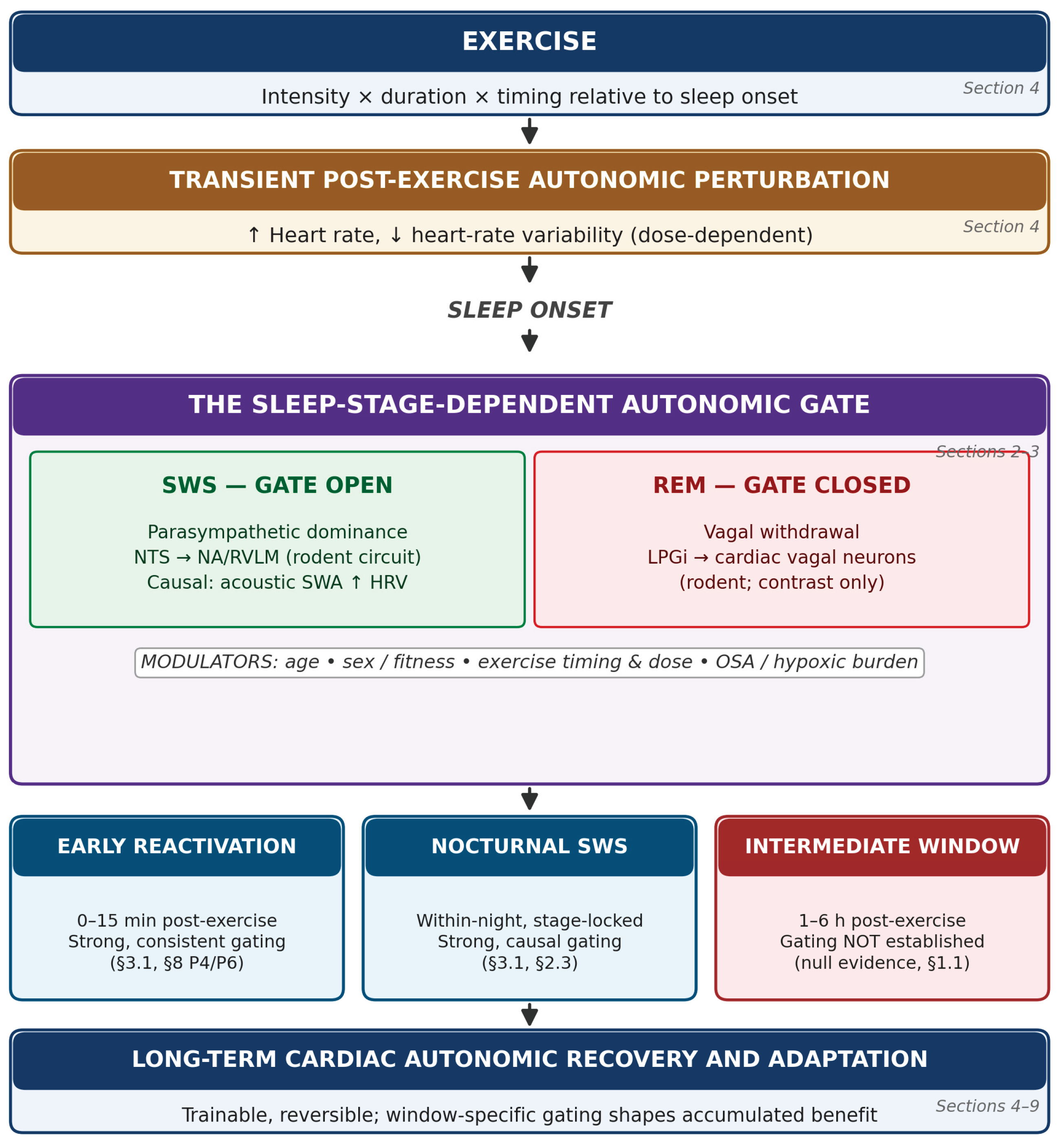

Table 1.
Age and sex as sources of variation in the SWS–autonomic coupling and post-exercise recovery. SWS, slow-wave sleep; VO2peak, peak oxygen uptake. The directions of effect reflect the studies cited and should not be read as a claim that any single determinant has been tested in a design that isolates it from the others (Section 5.6).
Table 1.
Age and sex as sources of variation in the SWS–autonomic coupling and post-exercise recovery. SWS, slow-wave sleep; VO2peak, peak oxygen uptake. The directions of effect reflect the studies cited and should not be read as a claim that any single determinant has been tested in a design that isolates it from the others (Section 5.6).
| Determinant | Direction of effect | Primary explanation | Basis |
|---|---|---|---|
| Aging → SWS–autonomic coupling | Coupling weakened; discrete slow-wave/heart-rate/vagal burst sequence essentially abolished in older adults | Loss of the coupling event itself, not merely reduced SWS quantity | [54,55] |
| Aging → post-exercise recovery | Slower recovery in sedentary older adults; recovery comparable to young adults in active older adults | Deconditioning, not age per se | [56,57] |
| Sex → sleep architecture | Women show more SWS and a slower age-related SWS decline than men | Organizational/activational effects of sex steroids (proposed, not directly tested) | [58,59] |
| Sex → sleep autonomics | Men show lower vagal tone and higher sympathovagal balance during sleep, especially REM | Emerges by adolescence; persists after covarying for reproductive hormones | [60,61] |
| Sex → post-exercise recovery | Contested; apparent male advantage in heart-rate recovery reported in some studies | Advantage disappears once aerobic capacity (VO2peak) is matched | [62,63] |
| Attribution (hormones vs. baseline vs. architecture) | Autonomic baseline tone is the most consistent predictor; hormonal effects weak/inconsistent for these outcomes; architecture untested as mediator | Estradiol did not explain exercise-recovery modulation directly; no study used SWS as an explicit mediating variable | [56,60,63,64,65] |
Table 2.
Evidence gradient for autonomic impairment and its reversibility across three clinical populations with disrupted sleep. CPAP, continuous positive airway pressure; CBT-I, cognitive behavioral therapy for insomnia; RCT, randomized controlled trial; BP, blood pressure; HRV, heart rate variability. In OSA, impairment is understood to be driven predominantly by intermittent hypoxia rather than SWS loss per se (Section 6, introductory paragraph), a qualification that applies to the entire row.
Table 2.
Evidence gradient for autonomic impairment and its reversibility across three clinical populations with disrupted sleep. CPAP, continuous positive airway pressure; CBT-I, cognitive behavioral therapy for insomnia; RCT, randomized controlled trial; BP, blood pressure; HRV, heart rate variability. In OSA, impairment is understood to be driven predominantly by intermittent hypoxia rather than SWS loss per se (Section 6, introductory paragraph), a qualification that applies to the entire row.
| Disorder | Impairment evidence | Treatment reversibility | Overall confidence | Basis |
|---|---|---|---|---|
| Obstructive sleep apnea (OSA) | Sympathetic activity elevated even while awake; fails to fall during sleep; surges with each apnea; scales monotonically with severity | CPAP restores sympathetic tone, brainstem activity, baroreflex sensitivity, and BP dipping; withdrawal reverses gains within two weeks | Strong; multiply convergent across microneurography, imaging, and RCT-level withdrawal evidence | [66,67,68,69,70,71,72,73] |
| Insomnia | Nighttime sympathetic hyperactivation reported in some studies; deficit concentrated in the objective-short-sleep-duration phenotype | No adequate evidence identified on whether treatment (e.g., CBT-I) restores autonomic function | Real but modest; a 17-study meta-analysis found no reliable overall HRV impairment | [74,75,76,77,78] |
| Pure sleep fragmentation (without restriction or hypoxia) | Human evidence inconsistent; experimental fragmentation alone did not significantly alter cardiac autonomic parameters in a controlled crossover | Not applicable/untested in humans | Weak in humans; the only SWS-specific evidence (raised BP with compensatory parasympathetic rise) comes from a chronic rat model | [7,79] |
Table 3.
Validation of wearable and nearable devices for heart rate/HRV measurement (top two rows) and slow-wave sleep staging (remaining rows) against reference standards. HR, heart rate; HRV, heart rate variability; ECG, electrocardiography; RMSSD, root mean square of successive differences; PSG, polysomnography; PPG, photoplethysmography. None of the studies listed here incorporated a post-exercise stressor (Section 7.3 and Section 7.4).
Table 3.
Validation of wearable and nearable devices for heart rate/HRV measurement (top two rows) and slow-wave sleep staging (remaining rows) against reference standards. HR, heart rate; HRV, heart rate variability; ECG, electrocardiography; RMSSD, root mean square of successive differences; PSG, polysomnography; PPG, photoplethysmography. None of the studies listed here incorporated a post-exercise stressor (Section 7.3 and Section 7.4).
| Device/study | Function tested | Performance vs. reference | Basis |
|---|---|---|---|
| Wrist-worn ring (Kinnunen et al.) | Nocturnal HR and HRV vs. ECG | r2 ≈ 0.996 (HR), 0.980 (HRV); sub-beat, ~1 ms bias | [80] |
| Under-mattress ballistocardiography, Emfit QS (Vesterinen et al.) | Nocturnal HR and RMSSD vs. ECG | r ≈ 0.90 (HR), 0.89 (RMSSD) at night-level mean; ~28% erroneous/missing epochs | [81] |
| Six wrist devices (Schyvens et al.) | Multi-state sleep-stage scoring vs. PSG | Multi-state κ 0.21–0.53; deep and REM better identified than wake/light | [82] |
| Six devices (Miller et al.) | Sleep, HR, and HRV estimation vs. PSG/ECG | Multi-state agreement 50–65%; κ 0.20–0.52 | [83] |
| Seven consumer devices (Chinoy et al.) | Sleep-stage scoring vs. PSG | Epoch sensitivity ≥0.93; specificity 0.18–0.54; degrades on fragmented-sleep nights | [84] |
| Ring sensor, Oura Gen3 (Svensson et al.) | Deep-sleep time and staging vs. PSG | No significant difference from PSG for deep-sleep time; per-stage accuracy 75.5–90.6% | [85] |
| Eleven trackers (Lee et al.) | Deep-stage classification vs. PSG | Macro-F1 0.26–0.69 across devices | [86] |
| PPG + accelerometer algorithm (Fonseca et al.) | Four-class sleep staging vs. PSG | κ ≈ 0.42 in healthy middle-aged adults | [87] |
| Under-mattress sensor, Withings Sleep Analyzer (Manners et al.) | Sleep-wake classification vs. PSG | 83% accuracy; 95% sensitivity; 37% specificity; overestimates total sleep time | [88] |
Table 4.
Correspondence between predictions P1–P6, the design group intended to test each, and its primary outcome. HRV, heart-rate variability; SWS, slow-wave sleep; OSA, obstructive sleep apnea. Full study-design requirements for each group are given in Box 1, Box 2 and Box 3 below.
| Prediction | Design group | Core question tested | Primary outcome |
|---|---|---|---|
| P1 | Box 1 | Whether a human functional analog exists for the rodent NTS→NA/RVLM circuit | Dose-dependent HRV response to SWS-timed vagal-afferent stimulation |
| P2 | Box 1 | Whether manipulation route, not SWS quantity, determines autonomic direction | Autonomic direction under a confound-free pharmacological SWS enhancer |
| P3 | Box 1 | Whether a discrete exercise-timing threshold gates SWS-phase disruption | SWS-segmented autonomic disruption across time-to-sleep-onset conditions |
| P4 | Box 2 | Whether fitness, rather than age or sex, explains apparent recovery differences | SWS-segmented autonomic recovery in fitness-matched age/sex groups |
| P5 | Box 2 | Whether hypoxic burden, rather than SWS quantity, explains OSA autonomic impairment | Nocturnal autonomic impairment vs. hypoxic burden and SWS quantity |
| P6 | Box 3 | Whether wearable SWS-staging accuracy degrades on post-exercise nights | Epoch-level staging agreement (kappa) on post-exercise vs. control nights |
Disclaimer/Publisher’s Note: The statements, opinions and data contained in all publications are solely those of the individual author(s) and contributor(s) and not of MDPI and/or the editor(s). MDPI and/or the editor(s) disclaim responsibility for any injury to people or property resulting from any ideas, methods, instructions or products referred to in the content. |
© 2026 by the authors. Licensee MDPI, Basel, Switzerland. This article is an open access article distributed under the terms and conditions of the Creative Commons Attribution (CC BY) license.
Copyright: This open access article is published under a Creative Commons CC BY 4.0 license, which permit the free download, distribution, and reuse, provided that the author and preprint are cited in any reuse.


