

Submitted:
27 July 2026
Posted:
28 July 2026
You are already at the latest version
Preprints on COVID-19 and SARS-CoV-2
Abstract
Introduction: Cognitive dysfunction ("brain fog") is a common manifestation of post-acute COVID-19 syndrome (PACS) and may persist long after the acute infection. While cross-sectional studies have described cognitive deficits, longitudinal evidence on recov-ery trajectories remains limited. Methods: We conducted a longitudinal observational study of neurocognitive performance and neuropsychiatric symptoms in patients with PACS. Participants underwent assessment with 20 standardized tests covering five cogni-tive domains (memory, attention, language, executive functions, psychomotor processing speed); anxiety, depression, and sleep quality were assessed at three time points. Changes were analysed using the Friedman test. Results: Forty-two patients were included (median age 57 years; 35.7% female) from a predominantly hospitalized cohort (81% hospitalised; 66.7% requiring respiratory support). Comparison with non-completers (n=544) showed that completers were more severely ill during the acute phase rather than healthier or more motivated. Significant improvements over time were observed in verbal short-term learning, visuospatial memory, working memory, constructional praxis, phonological verbal fluency, and psychomotor processing speed (all p≤0.05). Sleep quality also im-proved (p< 0.0001). Conclusion: Patients with PACS may show gradual, heterogeneous and domain-specific cognitive improvement, with persistent deficits in a proportion of in-dividuals. These findings highlight the importance of long-term neuropsychological mon-itoring and integrated cognitive-psychiatric evaluation in post-COVID care. Given the small, predominantly hospitalized sample, improvements should be interpreted cau-tiously and confirmed in larger controlled studies, although alternate test forms make practice effects unlikely.
Keywords:
post-COVID19
; long-COVID
; PACS
; cognitive outcomes
; neuropsychiatric symptoms
; longitudinal neuropsychological evaluation
1. Introduction
Post-acute COVID-19 syndrome (PACS), commonly referred to as long COVID, is characterized by the persistence of symptoms following the acute phase of SARS-CoV-2 infection. These symptoms may affect multiple organ systems and frequently include fatigue, cognitive difficulties, mood disturbances and sleep disorders, representing an increasingly recognized challenge for healthcare systems worldwide [1,2]. Although early case definitions (e.g., NICE 2021) considered symptoms persisting beyond four weeks from the acute infection, more recent and conservative definitions endorsed by the World Health Organization (WHO), the National Academies of Sciences, Engineering, and Medicine (NASEM) and the Centers for Disease Control and Prevention (CDC) require symptom persistence for at least three months, a criterion that improves specificity and reduces the inclusion of patients with self-limiting subacute symptoms [2,3,4].
Among the neurological manifestations of PACS, cognitive dysfunction—often described by patients as “brain fog”—is one of the most commonly reported symptoms and may substantially impair daily functioning and quality of life [5]. Cognitive disturbances may arise during the subacute phase of the infection or shortly after hospital discharge and can persist for several months following recovery from acute COVID-19 [6,7,8].
The pathophysiological mechanisms underlying cognitive impairment in patients recovering from COVID-19 are likely multifactorial. Proposed mechanisms include neuroinflammation, immune dysregulation, endothelial dysfunction and microvascular injury, as well as indirect effects of systemic illness such as hypoxia or widespread cytokine release [9,10,11,12,13]. Previous studies have identified impairments across multiple cognitive domains, particularly executive functions, attention, memory and processing speed [14].
Although cross-sectional investigations have consistently documented cognitive deficits in individuals recovering from COVID-19, longitudinal evidence describing the trajectory of cognitive recovery remains limited and heterogeneous. Some studies have shown that a substantial proportion of patients continue to exhibit pathological performance in at least one cognitive domain up to one year after infection [15,16]. Moreover, persistent cognitive slowing and mental fatigue have been reported even in individuals who experienced mild forms of the disease [17]
Cognitive impairment in PACS is also frequently associated with neuropsychiatric symptoms, including anxiety, depression and sleep disturbances, which may further contribute to reduced quality of life and functional limitations [18,19]. Understanding the interaction between neurocognitive deficits and psychiatric symptoms may therefore be crucial for improving long-term management strategies in these patients.
The aim of this study was to investigate longitudinal changes in neurocognitive performance and neuropsychiatric symptoms in a cohort of patients with PACS undergoing comprehensive neuropsychological assessment during follow-up.
2. Materials and Methods
2.1. Study design and setting
The Neuro-COVID Study is an observational, longitudinal, monocentric study conducted at the Post-COVID outpatient service of the National Institute for Infectious Diseases Lazzaro Spallanzani (IRCCS), Rome, Italy.
The study was approved by the Ethics Committee of INMI Lazzaro Spallanzani (approval number 119/2020, dated 20 May 2020) and conducted in accordance with the Declaration of Helsinki. All participants provided written informed consent prior to participation.
2.2. Participants
Patients with a documented history of SARS-CoV-2 infection and persistent neurological or cognitive symptoms were eligible for inclusion. Participants were either referred by clinicians or self-referred to the Post-COVID outpatient service for symptoms occurring at least four weeks after the acute infection, consistent with the definition of post-acute COVID-19 syndrome. We acknowledge that this four-week threshold reflects the earlier NICE definition and is broader than the more recent three-month criterion currently recommended by the WHO, NASEM and CDC; the implications of this inclusive window are addressed in the Discussion.
Patients who underwent neurocognitive assessment within seven months after resolution of the acute infection were considered eligible for the present analysis. The study period extended from September 2020 to January 2023. Because the baseline (t0) assessment could be performed at any time up to seven months after the resolution of the acute infection rather than at a fixed interval, the timing of the first evaluation varied across participants. This variability in the baseline window represents a potential source of heterogeneity, as patients assessed earlier and those assessed later may have been at different stages of spontaneous recovery at study entry.
A flow diagram illustrating the study population and patient selection process is shown in Figure 1.
2.3. Neurocognitive assessment
Neurocognitive assessment (NCA) was performed at three time points during follow-up: approximately three months (t0), six months (t1) and twelve months (t2) after the resolution of the acute infection.
Participants underwent a comprehensive standardized neuropsychological battery including 20 tests covering five cognitive domains: memory, attention, language, executive functions and psychomotor processing speed.
The following tests were administered:
Mini-Mental State Examination (MMSE) for global cognitive screening [20]
Rey Auditory Verbal Learning Test (RAVLT-ST, RAVLT-DR, RAVLT-REC) for verbal memory [21]
Digit Span Forward and Backward (DSF, DSB) for verbal short-term and working memory [22]
Corsi Span Forward and Backward (CSF, CSB) for visuospatial memory [22]
Rey-Osterrieth Complex Figure (ROCF-Copy and ROCF-Delayed Recall) for visuospatial constructional abilities and visual memory [23]
Trail Making Test A and B (TMTA, TMTB) for processing speed and executive functioning [24]
WAIS-R Digit Symbol (DS) for psychomotor processing speed [25]
Stroop Test (ST) for inhibitory control and cognitive flexibility [26]
Multiple Features Target Cancellation (MFTC) for attention [27]
Phonological and categorical verbal fluency tests (PVF, CVF) for language production [28]
Functional autonomy was evaluated using the Instrumental Activities of Daily Living scale (IADL) [29].
Raw test scores were adjusted for age, sex and educational level and converted into equivalent scores according to Italian normative standards [30,31]. Equivalent scores range from 0 to 4, where 0 indicates pathological performance and 4 indicates performance within the upper normal range. Cognitive impairment was defined as performance below the normative cut-off (equivalent score = 0).
2.4. Neuropsychiatric assessment
The following questionnaires were administered to assess the presence of neuropsychiatric symptoms and their possible influence on neurocognitive performance: the Beck Anxiety Inventory (BAI) [32] which assess the cognitive and physiological symptoms of anxiety [range = 0–63 were classified as in: 0–7 = no symptoms, 8–15 = mild symptoms, 16–25 = moderate symptoms; 26–63 = severe symptoms]; the Beck Depression Inventory (BDI-II) [33], which assess cognitive, affective, and physiological symptoms of depression (Somatic-affective SA/ Cognitive C dimensions) [range = 0–63 were classified as in: 0–13 = no symptoms, 14–19 = mild symptoms, 20–28 = moderate symptoms; 29–63 = severe symptoms]; the Pittsburgh Sleep Quality Index (PSQI) [34] for sleep quality assessment (if score >5 indicates the presence of poor sleep quality). It should also be noted that several BDI-II items (e.g., fatigue, loss of energy, changes in sleep, appetite or weight, difficulty concentrating and reduced interest in activities) overlap substantially with the somatic and cognitive manifestations of PACS itself. Consequently, BDI-II scores in this population may partly capture the somatic burden of long COVID rather than depression per se, and depression severity may be overestimated. The use of the separate somatic-affective (SA) and cognitive (C) dimensions was intended to partially mitigate this issue.
2.5. Statistical analysis
Data were summarized using descriptive statistics. Continuous variables are presented as medians and interquartile ranges (IQR), while categorical variables are expressed as frequencies and percentages.
Comparisons between categorical variables were performed using the Chi-square test or Fisher’s exact test when appropriate. Longitudinal changes in cognitive and psychiatric variables across the three time points were analysed using the non-parametric Friedman test.
Statistical significance was defined as p < 0.05. To account for the multiple comparisons inherent in the cognitive–neuropsychiatric association analyses, p-values were adjusted using the Benjamini–Hochberg false discovery rate (FDR) procedure, with a significance threshold set at FDR < 0.05. Effect sizes for longitudinal changes were quantified using Kendall’s W (interpreted as small ≥0.10, medium ≥0.30, large ≥0.50). All analyses were conducted using SPSS version 29.0 (IBM Corp., Chicago, IL, USA).
To minimize practice (test–retest) effects arising from repeated assessment, alternate (parallel) forms of the neuropsychological tests were administered across the three time points whenever available. The use of alternate forms reduces the influence of task-specific learning on serial performance, so that the longitudinal changes observed are less likely to be attributable to mere re-exposure to the same test material.
3. Results
3.1. Participant characteristics
A total of 42 patients were included in the analysis. The median age was 57 years (IQR 38–81), and 35.7% of participants were female. The median educational level was 13 years (IQR 8–18). Most patients had been previously hospitalized during the acute phase of COVID-19 (81%), and 66.7% required oxygen therapy. Of the 967 patients initially enrolled at the Post-COVID outpatient service, only 42 (4.3%) completed all three neurocognitive assessments and were included in the present longitudinal analysis (see Figure 1). Non-completion of follow-up assessments was attributed to multiple factors, including perceived cognitive recovery leading to voluntary dropout, lack of motivation to return in the absence of ongoing symptoms, severe psychological distress precluding continued participation, and work or logistical constraints limiting availability for repeated testing.
To characterise potential selection bias, baseline demographic and clinical characteristics of study completers were formally compared with those of non-completers (n=544; Table S1). Age (completers: median 57 years, IQR 51–60; non-completers: median 54 years, IQR 47–61; p=0.273) and educational level (both groups: median 13 years; p=0.359) did not differ significantly between groups. Notably, however, completers had a significantly higher prevalence of acute-phase respiratory support (66.7% vs 33.6%; p<0.001), pulmonary embolism (16.7% vs 2.9%; p<0.001), corticosteroid use (66.7% vs 47.4%; p=0.016), and tocilizumab/sarilumab treatment (7.1% vs 1.1%; p=0.021), and were more frequently female (64.3% vs 44.7%; p=0.014). These findings indicate that completers were, if anything, more severely ill during the acute phase than non-completers, rather than representing a healthier or more motivated subgroup. The direction of any resulting selection bias is therefore likely to be towards overrepresentation of severe acute disease and its sequelae, which should be considered when interpreting the observed cognitive trajectories.
Baseline demographic and clinical characteristics of the study population are summarized in Table 1.
3.2. Longitudinal neurocognitive outcomes
Longitudinal analyses revealed significant changes in several cognitive domains across the three assessments.
Improvements in verbal short-term learning (RAVLT-ST) were observed over time, with the proportion of impaired patients decreasing from 23.8% at baseline (t0) to 11.9% at t1 and 9.5% at t2 (p = 0.032).
Similarly, visuospatial short-term memory (CSF) improved significantly, with impairment rates decreasing from 31% at t0 to 2.4% at t1 and 0% at t2 (p < 0.0001). Improvements were also observed in visuospatial working memory (CSB) (p = 0.002).
Significant improvements were additionally observed in constructional praxis (ROCF-C; p = 0.018), phonological verbal fluency (PVF; p = 0.05) and psychomotor processing speed (Digit Symbol; p = 0.002).
Sleep quality also significantly improved across follow-up evaluations, with the proportion of patients reporting poor sleep quality decreasing from 31% at baseline to 7.1% at t1 and 2.4% at t2 (p < 0.0001).
Detailed results of the longitudinal neurocognitive and neuropsychiatric analyses are presented in Table 2.
3.3. Association between neurocognitive performance and neuropsychiatric symptoms
The following analyses of associations between neurocognitive performance and neuropsychiatric symptoms are considered exploratory, given the multiple comparisons and small sample size. P-values were adjusted using the Benjamini–Hochberg FDR procedure; all reported associations survived FDR correction (FDR < 0.05).
At baseline (t0), increased anxiety symptoms were significantly associated with poorer performance in verbal short-term memory (Digit Span Forward; p = 0.040) and psychomotor processing speed (Digit Symbol; p = 0.020). Sleep disturbances were also associated with reduced processing speed (p = 0.018).
At the second assessment (t1), anxiety symptoms were associated with poorer verbal learning performance (RAVLT-ST; p = 0.035), while sleep disturbances were associated with poorer performance in both verbal learning (RAVLT-ST; p = 0.033) and verbal long-term memory (RAVLT-DR; p = 0.010).
At the final assessment (t2), depressive symptoms were associated with poorer performance in verbal long-term memory (RAVLT-DR).
A detailed representation of these associations is provided in Figure 2A–C.
4. Discussion
To our knowledge, this study represents one of the few longitudinal investigations providing detailed neuropsychological monitoring across multiple cognitive domains in patients with PACS using in-person standardized assessment.
This longitudinal study investigated the evolution of neurocognitive performance and neuropsychiatric symptoms in patients with post-acute COVID-19 syndrome (PACS) undergoing comprehensive neuropsychological assessment over a one-year follow-up period. Our findings indicate that cognitive performance may gradually improve over time in several domains, particularly verbal short-term learning, visuospatial memory, working memory, constructional praxis, phonological verbal fluency and psychomotor processing speed. However, recovery was heterogeneous across cognitive domains, and a subset of individuals continued to show impaired or borderline performance during follow-up.
These findings are consistent with previous studies reporting partial recovery of cognitive function after COVID-19, in which deficits persisting for months tend to improve progressively in a proportion of patients [35,36,37,38], particularly in attentional and executive functions, while residual impairments remain in some individuals [8,16]. These improvements should nonetheless be interpreted with caution. The use of alternate (parallel) test forms across assessments makes test–retest learning an unlikely explanation, supporting a genuine change in performance. However, most participants had been hospitalized, frequently with oxygen and steroid treatment, and substantial cognitive improvement during the year after hospital discharge is well documented across critically ill populations independently of SARS-CoV-2; as detailed in the Limitations, the attrition pattern in our cohort (Table S1) selectively overrepresents patients with more severe acute illness. The early entry criterion (from four weeks after infection) further increases the likelihood that part of the improvement reflects natural resolution of acute illness rather than a chronic PACS-specific process, contributions our design cannot fully disentangle. Recent large longitudinal studies are consistent with this pattern: in a prospective in-person cohort, most cognitive domains improved progressively while processing speed and executive functioning remained below the normative mean [39], and serial assessments in the COVID and Cognition Study similarly described gradual symptom and cognitive change over time [40].
The mechanisms underlying cognitive dysfunction in PACS are likely multifactorial. Neuroinflammatory responses triggered by SARS-CoV-2 have been proposed as a major contributor to persistent neurological symptoms [13], potentially affecting neuronal function, synaptic transmission and cerebral microcirculation, while systemic factors such as hypoxia, endothelial dysfunction and metabolic disturbances during acute infection may also contribute to neural injury [9,10,11,12]. However, as the present study did not include neuroimaging or inflammatory biomarkers, these pathways were not measured and no causal link can be established. The gradual improvement seen in our cohort is compatible with, but cannot be attributed to, the progressive resolution of inflammatory processes; alternative explanations, including expected post-hospitalization recovery and practice effects, are at least equally plausible.
Improvements were not uniform across domains: while several functions recovered, others remained stable and a small proportion of individuals showed worsening of previously normal scores. Similar heterogeneity has been reported previously, with cognitive trajectories varying substantially between individuals [8,40]. A recent 36-month study of hospitalized COVID-19 patients identified four distinct trajectories—stable normal function, recovery, persistent impairment and delayed decline [41]—while other long-term data reported essentially negligible change in most patients [42], confirming that the magnitude and direction of cognitive change remain heterogeneous across studies. These patterns may reflect differences in disease severity, individual vulnerability, pre-existing conditions or psychological factors.
Sleep quality improved significantly over time. Sleep disturbances are among the most prevalent symptoms in PACS [43,44] and may contribute to cognitive complaints by affecting attention, memory consolidation and executive functioning; the observed improvement may therefore have played a role in the recovery of certain cognitive functions, as sleep disturbances and mental fatigue are closely related to performance in tasks involving processing speed and attentional resources [17,45].
Our results also highlight the complex relationship between neurocognitive performance and neuropsychiatric symptoms in patients with PACS. Anxiety symptoms were associated with poorer performance in verbal short-term memory and psychomotor processing speed during the first assessment. In addition, sleep disturbances were associated with reduced performance in processing speed and verbal memory tasks during follow-up. These findings are consistent with previous research indicating that psychological distress may contribute to cognitive impairment in patients recovering from COVID-19 [40,46,47,48].
The relationship between depressive symptoms and cognition changed across assessments: at baseline, depressive symptoms were unexpectedly associated with better visuospatial memory (CSF/CSB), whereas later evaluations showed an association with poorer verbal long-term memory. The counter-intuitive baseline association is most likely spurious, given the small sample, the many cognitive–neuropsychiatric pairs tested without correction for multiple comparisons, and dichotomized categories with few impaired cases. All associations were tested using Chi-square or Fisher’s exact test (expected counts <5) on dichotomised variables, with n=42 at each time point, and should be regarded as hypothesis-generating only. These fluctuations may reflect the bidirectional interaction between mood and cognition, whereby psychological distress influences cognition through reduced motivation, impaired attentional control or mental fatigue, while persistent cognitive difficulties may maintain anxiety and depressive symptoms.
From a clinical perspective, these findings emphasize the importance of comprehensive neuropsychological assessment in PACS, as cognitive complaints may reflect both objective alterations and psychological factors. Long-term monitoring may help identify individuals at risk of persistent impairment and guide targeted rehabilitation, and integrated approaches addressing both cognitive and emotional symptoms may improve quality of life.
Several limitations should be considered. First, the study did not include pre-infection neurocognitive assessments, limiting estimation of cognitive decline attributable to SARS-CoV-2. Second, the absence of a matched control group prevents comparison with individuals without prior COVID-19. Third, the small sample size limits statistical power and generalizability: only 42 of 967 enrolled patients (4.3%) completed all three assessments. As shown in Table S1, completers did not differ from non-completers in age or education but were more severely ill acutely (respiratory support 66.7% vs 33.6%, p<0.001; pulmonary embolism 16.7% vs 2.9%, p<0.001; corticosteroids 66.7% vs 47.4%, p=0.016), so the sample is enriched for hypoxia-related and post-critical-illness trajectories rather than healthier individuals. Fourth, the cohort was older than the general PACS population and predominantly hospitalized, two-thirds receiving oxygen and steroids; cognitive dysfunction and its improvement may be driven largely by hypoxia-related injury, critical illness and expected post-hospitalization recovery common across non-COVID populations, rather than PACS-specific mechanisms, while the high prevalence of hypertension (42.9%) and vascular risk factors may have contributed through a vascular pathway. These features limit generalizability to the broader, mostly non-hospitalized long-COVID population with brain fog. Fifth, no neuroimaging or inflammatory biomarker data were available, so mechanistic interpretations remain speculative. Finally, the inclusion criterion of symptoms from four weeks after infection reflects an earlier, broader definition of PACS than the three-month criterion now recommended by the WHO, NASEM and CDC, and the variable baseline window (up to seven months) adds heterogeneity in the timing of first evaluation.
Despite these limitations, the present study provides valuable longitudinal data based on detailed in-person neuropsychological assessments across multiple cognitive domains. The use of a comprehensive neuropsychological battery and repeated evaluations allowed a more nuanced characterization of cognitive trajectories in patients with PACS.
Future studies should aim to include larger multicentre cohorts and appropriate control groups in order to better clarify the mechanisms and long-term trajectory of cognitive impairment following COVID-19. In addition, further research integrating neuroimaging, inflammatory biomarkers and neuropsychological assessment may help elucidate the biological processes underlying cognitive dysfunction in PACS.
5. Conclusions
Our results provide descriptive longitudinal evidence suggesting that, in a small and predominantly hospitalized cohort with post-acute COVID-19 syndrome, performance on several cognitive measures tended to improve over the first year of follow-up, while deficits in a subset of cognitive functions persisted. Critically, a formal comparison showed that study completers were more severely ill during the acute phase than non-completers, indicating that the sample selectively represents patients with more severe acute disease rather than those who recovered most easily; the observed improvements may therefore partly reflect the expected post-hospitalisation recovery trajectory rather than PACS-specific processes. The use of alternate test forms across assessments makes a substantial contribution of practice effects unlikely. Exploratory analyses indicated that anxiety, depressive symptoms and sleep disturbances were associated with poorer performance across several cognitive domains; these findings should nonetheless be regarded as hypothesis-generating only. Integrated neuropsychological and psychiatric monitoring appears warranted in this population. Larger controlled studies with non-hospitalised comparison groups are needed to clarify the specific contribution of PACS to the observed cognitive trajectories.
Supplementary Materials
The following supporting information can be downloaded at the website of this paper posted on Preprints.org, Figure S1: title; Table S1: title; Video S1: title.
Author Contributions
MC, AA and CP conceived and designed the study. MC, CP, GDD and ACB developed the study protocol; GDD, MM and ACB performed all the neurocognitive assessment (NCA) and are responsible for data curation; IS performed the data analysis. MC, IM and VM clinically evaluated patients and refer to NCA; GDD, ACB and MC wrote the first draft. AA supervised this work. All authors read and approved the final manuscript.
Funding
This research received no external funding.
Institutional Review Board Statement
The study was conducted in accordance with the Declaration of Helsinki, and approved by the Ethics Committee of INMI Lazzaro Spallanzani (approval number 119/2020, dated 20 May 2020) for studies involving humans.
Informed Consent Statement
Informed consent was obtained from all subjects involved in the study.”.
Data Availability Statement
The datasets generated and/or analyzed during the current study are not publicly available due to patient privacy reasons, but are available from the corresponding author on reasonable request.
Conflicts of Interest
The authors declare no conflicts of interest.
Acknowledgments
The author gratefully acknowledges all the participants, the statistician and the clinical study assistants of the National Institute for Infectious Diseases, “Lazzaro Spallanzani” (Sperduti I., Brita A.C., Maresca M., Camici M., Pinnetti C., Mastrorosa I., Mazzotta V., Antinori).
A preliminary version of this work was previously deposited as a preprint
Del Duca G, et al. Prolonged clinical monitoring of cognitive performance and psychiatric symptoms among PACS. Research Square, 2026. Available at: https://www.researchsquare.com/article/rs-6817736/v1.
Abbreviations
The following abbreviations are used in this manuscript:
| BAI | Beck Anxiety Inventory |
| BDI-II | Beck Depression Inventory-II |
| C | Cognitive (BDI-II subscale) |
| CDC | Centers for Disease Control and Prevention |
| CSB | Corsi Span Backward |
| CSF | Corsi Span Forward |
| CVF | Categorical Verbal Fluency |
| DS | Digit Symbol (WAIS-R) |
| DSB | Digit Span Backward |
| DSF | Digit Span Forward |
| FDR | False Discovery Rate |
| IADL | Instrumental Activities of Daily Living |
| IQR | Interquartile Range |
| MFTC | Multiple Features Target Cancellation |
| MFTC-T | Multiple Features Target Cancellation—Time |
| MMSE | Mini-Mental State Examination |
| NASEM | National Academies of Sciences, Engineering, and Medicine |
| NCA | Neurocognitive Assessment |
| NICE | National Institute for Health and Care Excellence |
| PACC | Post-Acute COVID-19 Condition |
| PACS | Post-Acute COVID-19 Syndrome |
| PSQI | Pittsburgh Sleep Quality Index |
| PVF | Phonological Verbal Fluency |
| RAVLT | Rey Auditory Verbal Learning Test |
| RAVLT-DR | Rey Auditory Verbal Learning Test—Delayed Recall |
| RAVLT-REC | Rey Auditory Verbal Learning Test—Recognition |
| RAVLT-ST | Rey Auditory Verbal Learning Test—Short-Term learning |
| ROCF | Rey-Osterrieth Complex Figure |
| ROCF-C | Rey-Osterrieth Complex Figure—Copy |
| SA | Somatic-Affective (BDI-II subscale) |
| SARS-CoV-2 | Severe Acute Respiratory Syndrome Coronavirus 2 |
| ST | Stroop Test |
| ST-ERR | Stroop Test—Errors |
| ST-T | Stroop Test—Time |
| TMTA | Trail Making Test, Part A |
| TMTB | Trail Making Test, Part B |
| WAIS-R | Wechsler Adult Intelligence Scale—Revised |
| WHO | World Health Organization |
References
- NICE. COVID-19 rapid guideline: managing the long-term effects of COVID-19. 11 November 2021. Available online: https://www.nice.org.uk/guidance/ng188 (accessed on June 2026).
- Soriano, J.B.; Murthy, S.; Marshall, J.C.; Relan, P.; Diaz, J.V. WHO Clinical Case Definition Working Group on Post-COVID-19 Condition. A clinical case definition of post-COVID-19 condition by a Delphi consensus. Lancet Infect. Dis. 2022, 22(4), e102–e107. [Google Scholar] [CrossRef] [PubMed]
- National Academies of Sciences, Engineering, and Medicine. A Long COVID Definition: A Chronic, Systemic Disease State with Profound Consequences; The National Academies Press: Washington, DC, 2024. [Google Scholar] [CrossRef] [PubMed]
- Centers for Disease Control and Prevention. Clinical Overview of Long COVID. Atlanta, GA: CDC; last updated March 9, 2026. Available online: https://www.cdc.gov/long-covid/hcp/clinical-overview/index.html.
- Jaywant, A.; Gunning, F.M.; Oberlin, L.E.; et al. Cognitive Symptoms of Post–COVID-19 Condition and Daily Functioning. JAMA Netw. Open 2024, 7(2), e2356098. [Google Scholar] [CrossRef] [PubMed]
- Alemanno, F.; Houdayer, E.; Parma, A.; Spina, A.; Del Forno, A.; Scatolini, A.; Iannaccone, S. COVID-19 cognitive deficits after respiratory assistance in the subacute phase: a COVID-rehabilitation unit experience. PLoS ONE 2021, 16(2), e0246590. [Google Scholar] [CrossRef] [PubMed]
- Beaud, V.; Crottaz-Herbette, S.; Dunet, V.; Vaucher, J.; Bernard-Valnet, R.; Du Pasquier, R.; Clarke, S. Pattern of cognitive deficits in severe COVID-19. J. Neurol. Neurosurg. Psychiatry 2021, 92(5), 567–568. [Google Scholar] [PubMed]
- Lorenzo Diana, Rossana Regazzoni, Matteo Sozzi, Stefania Piconi, Luca Borghesi, Elisa Lazzaroni, Paola Basilico, Angelo Aliprandi, Nadia Bolognini, Daniela R. Bonardi, Daniele Colombo, Andrea Salmaggi, Monitoring cognitive and psychological alterations in COVID-19 patients: A longitudinal neuropsychological study, Journal of the Neurological Sciences, Volume 444, 2023, 120511, ISSN 0022-510X. [CrossRef] [PubMed]
- Seeble, J.; Waterboer, T.; Hippchen, T.; Simon, J.; Kirchner, M.; Lim, A.; et al. Persistent symptoms in adult patients 1 year after coronavirus disease 2019 (COVID-19): a prospective cohort study. Clin. Infect. Dis. 2022, 74(7), 1191–8. [Google Scholar] [CrossRef]
- Hampshire, A.; Trender, W.; Chamberlain, S.R.; Jolly, A.E.; Grant, J.E.; Patrick, F.; et al. Cognitive deficits in people who have recovered from COVID-19. EClinicalMedicine 2021, 39, 101044. [Google Scholar] [CrossRef]
- Koralnik, I.J.; Tyler, K.L. COVID-19: a global threat to the nervous system. Ann. Neurol. 2020, 88(1), 1–11. [Google Scholar] [CrossRef]
- Zhou, H.; Lu, S.; Chen, J.; Wei, N.; Wang, D.; Lyu, H.; et al. The landscape of cognitive function in recovered COVID-19 patients. J. Psychiatr. Res. 2020, 129, 98–102. [Google Scholar] [CrossRef]
- Han, Y.; Yuan, K.; Wang, Z.; Liu, W.J.; Lu, Z.A.; Liu, L.; Lu, L. Neuropsychiatric manifestations of COVID-19, potential neurotropic mechanisms, and therapeutic interventions. Transl. Psychiatry 2021, 11(1), 1–20. [Google Scholar] [CrossRef] [PubMed]
- Blazhenets, G.; Schroeter, N.; Bormann, T.; Thurow, J.; Wagner, D.; Frings, L.; et al. Slow but Evident Recovery from Neocortical Dysfunction and Cognitive Impairment in a series of chronic COVID-19 patients. J. Nucl. Med. 2021, 62(7), 910–5. [Google Scholar] [CrossRef] [PubMed]
- Poletti, S.; Palladini, M.; Mazza, M.G.; De Lorenzo, R.; Furlan, R.; Ciceri, F.; Benedetti, F. Long-term consequences of COVID-19 on cognitive functioning up to 6 months after discharge: role of depression and impact on quality of life. Eur. Arch. Psychiatry Clin. Neurosci. 2022, 272(5), 773–782. [Google Scholar] [PubMed]
- Ferrucci, R.; Dini, M.; Rosci, C.; Capozza, A.; Groppo, E.; Reitano, M.R.; Priori, A. One-year cognitive follow-up of COVID-19 hospitalized patients. Eur. J. Neurol. 2022, 29(7), 2006–2014. [Google Scholar] [CrossRef] [PubMed]
- Martin, E.M.; Srowig, A.; Utech, I.; et al. Persistent cognitive slowing in post-COVID patients: longitudinal study over 6 months. J. Neurol. 2024, 271, 46–58. [Google Scholar] [CrossRef] [PubMed]
- Mastrorosa, I.; Del Duca, G.; Pinnetti, C.; Lorenzini, P.; Vergori, A.; Brita, A.C.; Camici, M.; Mazzotta, V.; Baldini, F.; Chinello, P.; Mencarini, P.; Giancola, M.L.; Abdeddaim, A.; Girardi, E.; Vaia, F.; Antinori, A. What is the impact of post-COVID-19 syndrome on health-related quality of life and associated factors: a cross-sectional analysis. Health Qual. Life Outcomes 2023, 21(1), 28. [Google Scholar] [CrossRef] [PubMed] [PubMed Central]
- Perlis, R.H.; Lunz Trujillo, K.; Safarpour, A.; et al. Association of post-COVID-19 condition symptoms and employment status. JAMA Netw. Open 2023, 6(2), e2256152. [Google Scholar] [CrossRef] [PubMed]
- Measso, G.; et al. The Mini-Mental State Examination: normative study of an italian random sample. Dev. Neuropsycol 1993, 9, 77–85. [Google Scholar] [CrossRef] [PubMed]
- Carlesimo, G.A.; Caltagirone, C.; Gainotti, G. The mental deterioration battery: normative data diagnostic reliability and qualitative analyses of cognitive impariment. The group for the standardization of the mental deterioration battery. Eur. Neurol. 1996, 36(6), 378–84. [Google Scholar] [CrossRef] [PubMed]
- Monaco, M.; et al. Forward and backward span for verbal and visuo-spatial data: standardization and normative data from an italian adult population. Neurol. Sci. [CrossRef] [PubMed]
- Caffarra, P.; et al. Rey-Osterrieth complex figure: normative values in an italian population sample. Neurol. sci. 2002, 22(6), 443–7. [Google Scholar] [CrossRef] [PubMed]
- Giovagnoli, A.R.; et al. Trail making test: normative values from 287 normal adult controls. Ital. J. Neurol. Sci. 1996, 17(4), 305. [Google Scholar] [CrossRef] [PubMed]
- Orsini, A.; Laicardi, C. WAIS-R. Contributo Alla Taratura Italiana; Giunti O.S. Organizzazioni Speciali: Firenze, 1997. [Google Scholar]
- Caffarra, P.; et al. Una versione abbreviata del test di Stroop: dati normativi nella popolazione italiana. Riv. Di Neurol. 2002, 12, n4. [Google Scholar]
- Marra, C.; et al. The multiple features target cancellation (MFTC): an attenzional visual conjunction search test. Normative values for the italian population. Neurol. Sci. 2012. [Google Scholar] [CrossRef] [PubMed]
- Marra, C.; Ferraccioli, M.; Gainotti, G. Gender-related dissociations of categorical fluency in normal subjects and in subjects with Alzheimer’s disease. Neuropsychology 2007, 21(2), 207–11. [Google Scholar] [CrossRef] [PubMed]
- Lawton, M.P.; Brody, E.M. Assessment of older people: self-maintaining and instrumental activities of daily living. Gerontologist 1969, 9(3), 179–86. [Google Scholar] [CrossRef] [PubMed]
- Capitani, E.; Laiacona, M. Composite neuropsychological batteries and demographic correction: standardization based on equivalent scores, with a review of published data. J. Clin. Exp. Neuropsychol. 1997, 19(6), 795–809. [Google Scholar] [CrossRef] [PubMed]
- Capitani, E.; Laiacona, M. Outer and inner tolerance limits: their usefulness for the construction of norms and the standardization of neuropsychological tests. Clin. Neuropsychol. 2017, 31(6–7), 1219–1230. [Google Scholar] [CrossRef] [PubMed]
- Beck, A. T.; Steer, R. A. Manual for the Beck anxiety inventory; Psychological Corporation: San Antonio, TX, 1990. [Google Scholar]
- Beck, A.; Steer, R.; Brown, G. Beck Depression Inventory, 2nd edition; The Psychological Corporation: San Antonio, Tex, USA, 1996. [Google Scholar]
- Curcio, G.; Tempesta, D.; Scarlata, S.; et al. Validity of the Italian version of the Pittsburgh Sleep Quality Index (PSQI). Neurol. Sci. 2013, 34(4), 511–51. [Google Scholar] [CrossRef] [PubMed]
- Almeria, M.; Cejudo, J.C.; Deus, J.; Krupinski, J. Long Neurocognitive and Neuropsychiatric Sequelae in Participants with Post-COVID-19 Infection: A Longitudinal Study. Neurol. Int. 2024, 16(4), 853–868. [Google Scholar] [CrossRef] [PubMed] [PubMed Central]
- Ferrando, S.J.; Dornbush, R.; Lynch, S.; et al. Neuropsychological, medical, and psychiatric findings after recovery from acute COVID-19: a cross-sectional study. J. Acad. Consult Liaison Psychiatry 2022, 63, 474–484. [Google Scholar] [CrossRef] [PubMed]
- Prabhakaran, D.; Day, G.S.; Munipalli, B.; et al. Neurophenotypes of covid-19: risk factors and recovery outcomes. Brain Behav. Immun. Health 2023, 30, 100648. [Google Scholar] [CrossRef] [PubMed]
- Becker, J.H.; Li, J.; Lin, J.J.; Federman, A.; Bagiella, E.; Kale, M.S.; Fierer, D.; Bartram, L.; Wisnivesky, J.P. Neurocognitive trajectories in long COVID: evidence from longitudinal analyses. Brain Behav. Immun. Health 2025, 48, 101093. [Google Scholar] [CrossRef] [PubMed]
- Yeung, S. P., Guo, P., Adlard, F. L., Zhang, S. R., Bhagat, V., Cho, J., Curtis, L., Kaser, M., Haggard, M. P., & Cheke, L. G. (2025). COVCOG 3—Trajectory of Long COVID: Longitudinal Changes in Symptoms and Cognitive Impairment. A Third Publication From the COVID and Cognition Study. [CrossRef]
- Maziero MP, Lee EA, Colpo GD, Couture L, Merrill LC, Baskin L, Cahuiche AE, Petway A, Fan H, Reese E, Anderson KM, McCullough LD, Schulz PE, Ortiz GJ. Cognitive trajectories after hospitalization for COVID-19: a 36-month longitudinal study. J Neuropsychiatry Clin Neurosci. 2026 (published online April 7, 2026). [CrossRef] [PubMed]
- Koch, D.W.; Klinkhammer, S.; Verveen, A.; Visser, D.; Nieuwkerk, P.T.; Verwijk, E.; van Berckel, B.N.M.; Horn, J.; Tolboom, N.; van Heugten, C.M.; Verfaillie, S.C.J.; Knoop, H. Long-term cognitive functioning following COVID-19: Negligible neuropsychological changes over time. Clin. Neuropsychol. 2026, 40(1), 109–127, Epub 2025 May 2. [Google Scholar] [CrossRef] [PubMed]
- Taquet, M.; Sillett, R.; Zhu, L.; et al. Neurological and psychiatric risk trajectories after SARS-COV-2 infection: an analysis of 2-year retrospective cohort studies including 1 284 437 patients. Lancet Psychiatry 2022, 9, 815–827. [Google Scholar] [CrossRef] [PubMed]
- Vergori, A.; Del Duca, G.; Borrelli, P.; Brita, A.C.; Pinnetti, C.; Mastrorosa, I.; Camici, M.; Mondi, A.; Mazzotta, V.; Chinello, P.; Mencarini, P.; Giancola, M.L.; Abdeddaim, A.; Girardi, E.; Antinori, A. Cognitive outcomes and psychological symptoms in an Italian cohort with post-acute COVID-19 condition (PACC). Heliyon 2024, 10(20), e39431. [Google Scholar] [CrossRef] [PubMed] [PubMed Central]
- Pilotto, A.; Cristillo, V.; Cotti Piccinelli, S.; Zoppi, N.; Bonzi, G.; Sattin, D.; Schiavolin, S.; Raggi, A.; Canale, A.; Gipponi, S.; Libri, I.; Frigerio, M.; Bezzi, M.; Leonardi, M.; Padovani, A. Long-term neurological manifestations of COVID-19: prevalence and predictive factors. Neurol. Sci. 2021, 42(12), 4903–4907, Epub 2021 Sep 15. [Google Scholar] [CrossRef] [PubMed] [PubMed Central]
- Almeria, M.; Cejudo, J.C.; Sotoca, J.; Deus, J.; Krupinski, J. Cognitive profile following COVID-19 infection: Clinical predictors leading to neuropsychological impairment. Brain Behav. Immun. Health 2020, 9, 100163. [Google Scholar] [CrossRef] [PubMed]
- Hampshire, A.; Azor, A.; Atchison, C.; Trender, W.; Hellyer, P.J.; Giunchiglia, V.; Husain, M.; Cooke, G.S.; Cooper, E.; Lound, A.; Donnelly, C.A.; Chadeau-Hyam, M.; Ward, H.; Elliott, P. Cognition and memory after covid-19 in a large community sample. N. Engl. J. Med. 2024, 390(9), 806–818. [Google Scholar] [CrossRef] [PubMed] [PubMed Central]
- Jaywant, A.; Vanderlind, W.M.; Alexopoulos, G.S.; et al. Frequency and profile of objective cognitive deficits in hospitalized patients recovering from COVID-19. Neuropsychopharmacol. 2021, 46, 2235–2240. [Google Scholar] [CrossRef] [PubMed]
- Gebhard, C.E.; Sütsch, C.; Bengs, S.; Deforth, M.; Buehler, K.P.; Hamouda, N.; et al. Sex- and gender-specific risk factors of post-COVID-19 syndrome: a population-based cohort study in Switzerland. medRxiv 2021, 06.30. [Google Scholar]
Figure 1.
Flow chart.
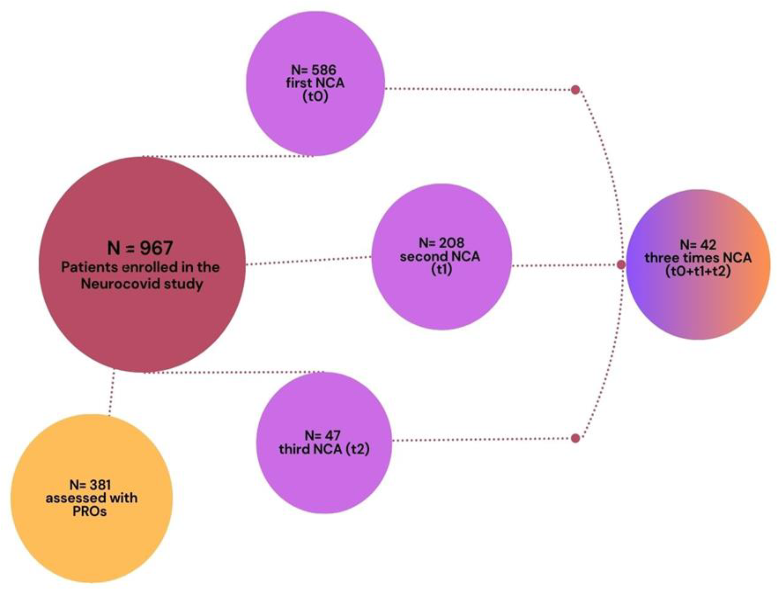
Figure 2.
A Longitudinal association between neurocognitive performance and neuropsychiatric symptoms at t0. B Longitudinal association between neurocognitive performance and neuropsychiatric symptoms at t0. C Longitudinal association between neurocognitive performance and neuropsychiatric symptoms at t1.
Figure 2.
A Longitudinal association between neurocognitive performance and neuropsychiatric symptoms at t0. B Longitudinal association between neurocognitive performance and neuropsychiatric symptoms at t0. C Longitudinal association between neurocognitive performance and neuropsychiatric symptoms at t1.
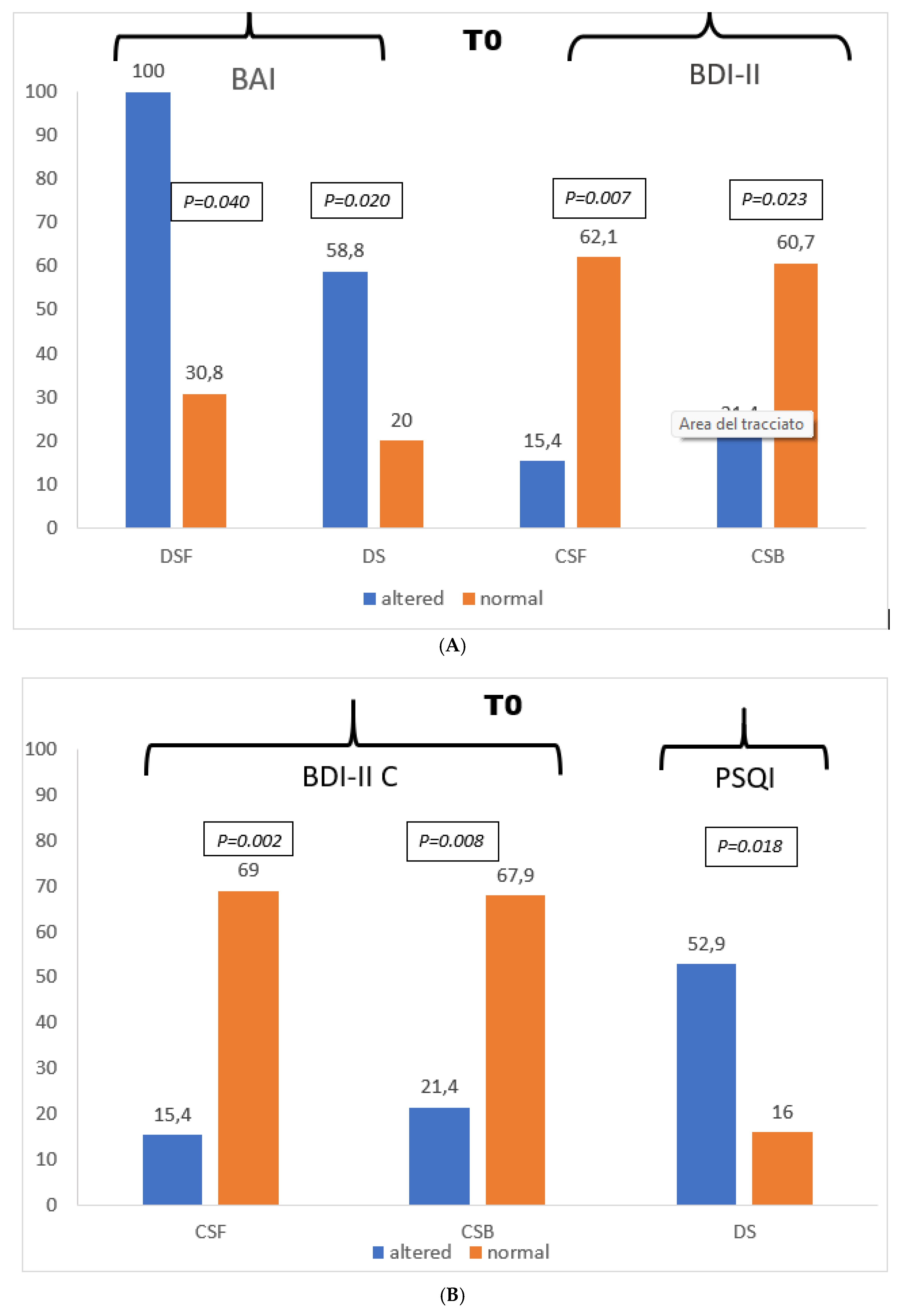
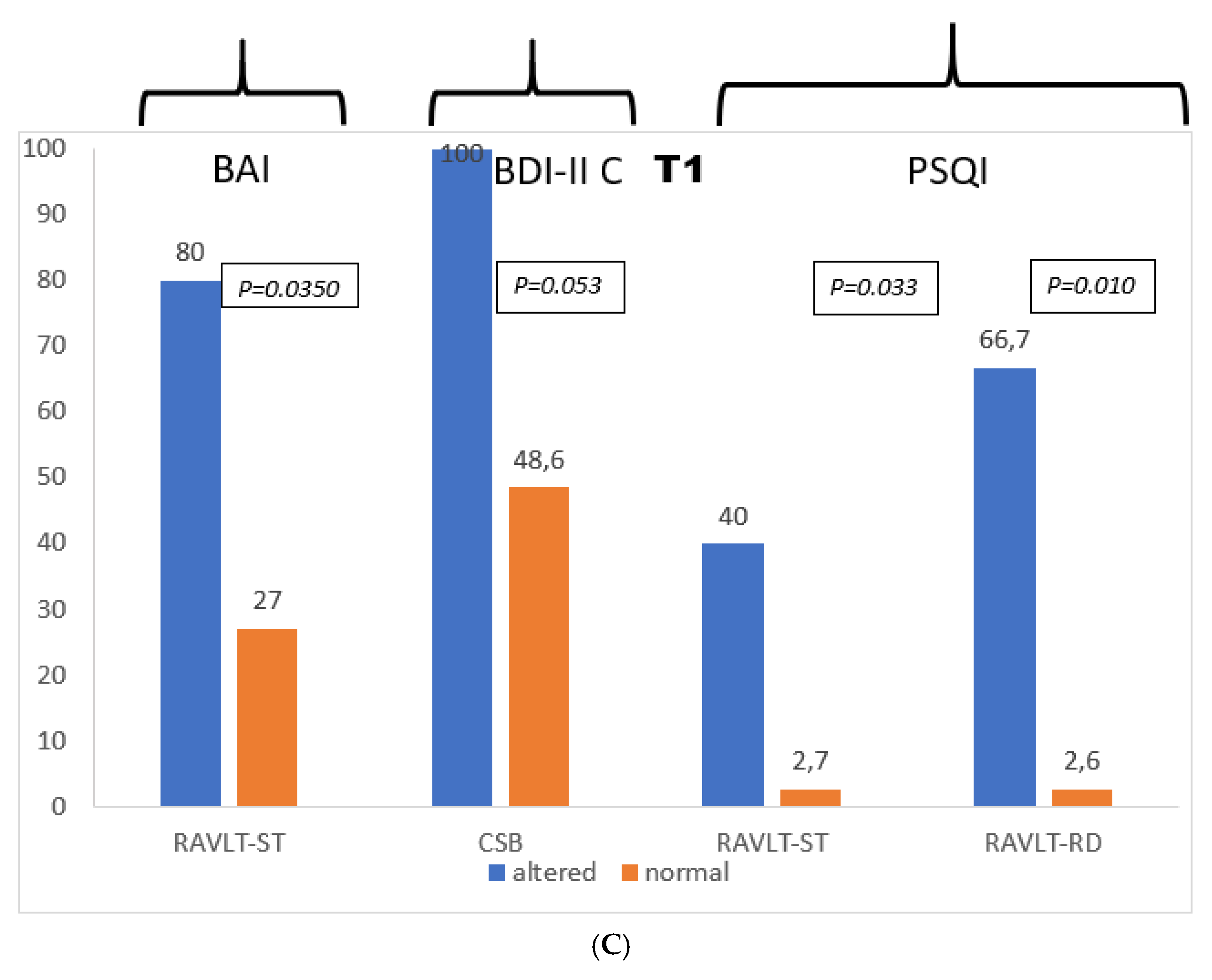

Table 1.
Baseline clinical characteristics for the study population.
| Variables | Sample N= 42 |
|---|---|
| Age, median (IQR) | 57 (38-81) |
| Female | 15 (35.7%) |
| Education, median (IQR) | 13 (8-18) |
| Smoking | 2 (4.8%) |
| Comorbidities | |
| Previous acute myocardial infarction | 2 (4.8%) |
| Hypertension | 18 (42.9%) |
| Diabetes | 5 (11.9%) |
| Cardiac Disease | 3 (7.1%) |
| Neurological disease | 2 (4.8%) |
| Respiratory diseases | 0 (0%) |
| Cancer | 0 (0%) |
| Neuropsychological symptoms | 31 (73.8%) |
| Oxygen therapy | 28 (66.7%) |
| Pulmonary Embolism | 7 (16.7%) |
| Previous hospitalization (PH) | 34 (81.0%) |
| Anti-SARS-CoV-2 therapy | |
| Tocilizumab/sarilumab | 3 (7.1%) |
| Kaletra | 4 (9.5%) |
| Steroids | 28 (66.7%) |
| Remdesivir | 20 (47.6%) |
| Plasma | 1 (2.4%) |
| Hydroxychloroquine | 9 (21.4%) |
| LMWH | 28 (66.7%) |
| ACE_I | 6 (14.3%) |
| ARB | 2 (4.8%) |
| Statine | 5 (11.9%) |
Table 2.
Temporal changes in cognitive functions and neuropsychiatric symptoms (t0-t1-t2).
| T0 | T1 | T2 | P value | ||||
|---|---|---|---|---|---|---|---|
| NCA | normal | impaired | normal | impaired | normal | impaired | |
| MMSE | 42 (100%) | 0 | 42 (100%) | 0 | 42 (100%) | 0 | 1 |
| RAVLT-ST | 32 (76.2%) | 10 (23.8%) | 37 (88.1%) | 5 (11.9%) | 38 (90.5%) | 4 (9.5%) | 0.032 |
| RAVLT-RD | 36 (85.7%) | 6 (14.3%) | 39 (92.9%) | 3 (7.1%) | 39 (92.9%) | 3 (7.1%) | 0.276 |
| RAVLT-REC | 30 (71.4%) | 12 (28.6%) | 31 (73.8%) | 11 (26.2%) | 34 (81%) | 8 (19%) | 0.486 |
| ROCF-DR | 27 (64.3%) | 15 (35.7%) | 31 (73.8%) | 11 (26.2%) | 32 (76.2%) | 10 (23.8%) | 0.148 |
| DSF | 39 (92.9%) | 3 (7.1%) | 37 (88.1%) | 5 (11.9%) | 35 (83.3%) | 7 (16.7%) | 0.223 |
| DSB | 38 (90.5%) | 4 (9.5%) | 36 (85.7%) | 6 (14.3%) | 40 (95.2%) | 2 (4.8%) | 0.264 |
| CSF | 29 (69%) | 13 (31%) | 41 (97.6%) | 1 (2.4%) | 42 (100%) | 0 | <0.0001 |
| CSB | 28 (66.7%) | 14 (33.3%) | 37 (88.1%) | 5 (11.9%) | 39 (92.9%) | 3 (7.1%) | 0.002 |
| ROCF-C | 35 (83.3%) | 7 (16.7%) | 39 (92.9%) | 3 (7.1%) | 41 (97.6%) | 1 (2.4%) | 0.018 |
| MFTC-ACC | 30 (71.4%) | 12 (28.6%) | 36 (85.7%) | 6 (14.3%) | 36 (85.7%) | 6 (14.3%) | 0.105 |
| MFTC-ERR | 42 (100%) | 0 | 42 (100%) | 0 | 42 (100%) | 0 | 1 |
| MFTC-T | 42 (100%) | 0 | 42 (100%) | 0 | 42 (100%) | 0 | 1 |
| PVF | 34 (81%) | 8 (19%) | 36 (85.7%) | 6 (14.3%) | 38 (90.5%) | 4 (9.5%) | 0.05 |
| CVF | 26 (61.9%) | 16 (38.1%) | 32 (76.2%) | 10 (23.8%) | 30 (71.4%) | 12 (28.6%) | 0.116 |
| ST-T | 41 (97.6%) | 1 (2.4%) | 39 (92.9%) | 3 (7.1%) | 41 (97.6%) | 1 (2.4%) | 0.368 |
| ST-ERR | 40 (95.2%) | 2 (4.8%) | 42 (100%) | 0 | 42 (100%) | 0 | 0.135 |
| TMTA | 41 (97.6%) | 1 (2.4%) | 42 (100%) | 0 | 42 (100%) | 0 | 0.368 |
| TMTB | 42 (100%) | 0 | 42 (100%) | 0 | 42 (100%) | 0 | 1 |
| DS | 25 (59.5%) | 17 (40.5%) | 36 (85.7%) | 6 (14.3%) | 36 (85.7%) | 6 (14.3%) | 0.002 |
| BAI | 27 (64.3%) | 15 (35.7%) | 28 (66.7%) | 14 (33.3%) | 30 (71.4%) | 12 (28.6%) | 0.705 |
| BDI-II | 22 (52.4%) | 20 (47.6%) | 26 (61.9%) | 16 (38.1%) | 25 (59.5%) | 17 (40.5%) | 0.395 |
| BDI-II SA | 24 (57.1%) | 18 (42.9%) | 29 (69%) | 13 (31%) | 29 (69%) | 13 (31%) | 0.249 |
| BDI-II C | 20 (47.6%) | 22 (52.4%) | 19 (45.2%) | 23 (54.8%) | 23 (54.8%) | 19 (45.2%) | 0.504 |
| PSQI | 29 (69%) | 13 (31%) | 39 (92.9%) | 3 (7.1%) | 41 (97.6%) | 1 (2.4%) | <0.0001 |
Abbreviations: Mini Mentale State Examination (MMSE); Rey Auditory Verbal Learning Test-Short Term (RAVLT-ST); Rey Auditory Verbal Learning Test-Delayed Recall (RAVLT-RD); Rey Auditory Verbal Learning Test-Recognition (RAVLT-REC); Rey-Osterrieth Complex Figure- Delayed Recall (ROCF-DR); Digit Span Forward (DSF); Digit Span Backward (DSB); Corsi Span Forward (CSF); Corsi Span Backward (CSB); Rey-Osterrieth Complex Figure- Copy (ROCF-C); Multiple Features Target Cancellation-Accuracy (MFTC-ACC); Multiple Features Target Cancellation-Errors (MFTC-ERR); Multiple Features Target Cancellation-Time (MFTC-T); Phonological Verbal Fluency (PVF); Categorical Verbal Fluency (CVF); Stroop test Color Word- Time (ST-T); Stroop test Color Word- Errors (ST-ERR); Trial Making Test A (TMTA); Trial Making Test B (TMTB);Digit Symbol Test (DS); Beck Anxiety Inventory (BAI); Beck Depression Inventory (BDI-II); Beck Depression Inventory Somatic-Affective (BDI-II SA); Beck Depression Inventory Cognitive (BDI-II C); Pittsburgh Sleep Quality Index (PSQI).
Disclaimer/Publisher’s Note: The statements, opinions and data contained in all publications are solely those of the individual author(s) and contributor(s) and not of MDPI and/or the editor(s). MDPI and/or the editor(s) disclaim responsibility for any injury to people or property resulting from any ideas, methods, instructions or products referred to in the content. |
© 2026 by the authors. Licensee MDPI, Basel, Switzerland. This article is an open access article distributed under the terms and conditions of the Creative Commons Attribution (CC BY) license (http://creativecommons.org/licenses/by/4.0/).
Copyright: This open access article is published under a Creative Commons CC BY 4.0 license, which permit the free download, distribution, and reuse, provided that the author and preprint are cited in any reuse.


