

Submitted:
24 July 2026
Posted:
27 July 2026
You are already at the latest version
Abstract
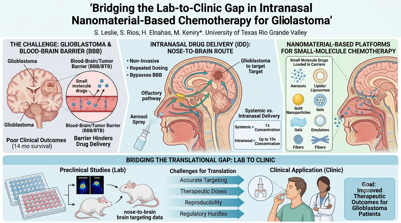
Clinical outcomes for brain cancer are often poor because the blood–brain/tumor barrier hinders effective drug delivery to malignant tissue. Glioblastoma, the most common primary brain malignancy in adults, has an average survival of approximately fourteen months [1]. Here, we discuss novel strategies that our research group and others are developing to deliver chemotherapy to the brain via the nasal cavity. Although significant hurdles remain, intranasal delivery holds substantial promise for improving outcomes for patients with brain cancer. Intranasal delivery is non‑invasive, permits repeated dosing, and has been shown to enable direct nose‑to‑brain transport that bypasses the blood–brain barrier.
Challenges such as accurately targeting drugs to the appropriate region of the nasal cavity at therapeutically relevant doses, while maintaining reproducibility, make this cutting-edge approach a regulatory challenge. The prolonged path to clinical translation discourages many researchers from pursuing this potentially life‑saving strategy. Nevertheless, preclinical studies demonstrate that intranasal delivery can achieve up to ten‑fold higher concentrations of select drugs in the brain [2].
Cancer chemotherapeutics span a wide range of molecular formats, from small molecules to 150‑kilodalton antibodies. Accordingly, delivery strategies must be carefully matched to the molecular properties of each therapeutic. Here, we focus on the intranasal delivery of small‑molecule inhibitors using nanomaterial‑based platforms, including aerosols, lipids, gold nanoparticles, gels, emulsions, fibers, and their combinations. Ultimately, we hope that intranasal delivery approaches will be translated to provide patients with better therapeutic outcomes.
Keywords:
intranasal delivery
; glioblastoma
; nose-to-brain
; nanomaterials
; translational research
; drug delivery
1. Clinical Rationale: Why This Field Exists
Brain Tumors Remain Limited by Delivery, Not Just Potency
The incidence of brain disease has increased in recent years. CNS therapeutics have low success rates due to complex biological, technical, clinical, and regulatory challenges. The blood-brain barrier remains the primary obstacle to effective drug delivery. Studies tracking fabricated nanoparticles show that not even 20 percent of the expected drug concentration was found at the target, along with substantial off-target accumulation in preclinical rat trials [3].
Why Nose-to-Brain Delivery Is Attractive
Conventional oral administration commonly does not achieve therapeutic levels in the brain due to the structures of the blood-brain barrier [2]. Nose-to-brain delivery is noninvasive and repeatable. Noninvasive routes include lipid nanoparticles, polymer-based nanoparticles, cell-derived exosomes, hydrogel delivery systems, micron delivery systems, protein nanoparticles, and inorganic nanoparticles. Common intranasal strategies use direct drug absorption and permeation enhancers, but suffer from poor drug stability, limited availability to target, and quick clearance [3,4,5,6,7]. New nano delivery methods are being investigated to functionalize nanoparticles for enhanced drug stability, increased nasal residence, and increased mucosal penetration. Recent advances in liposomes, polymeric nanoparticles, and nanogels have been investigated to encapsulate therapeutics and incorporate ligands that would help target [3,4,5,6,7]. Current challenges impede clinical translation: safety concerns, manufacturing scalability, and regulatory barriers. Emerging trends include AI-driven formulation design.
2. Translational Hurdles in Nose-to-Brain Chemotherapy
The clinical translation of intranasal drug delivery systems for brain tumors is limited by many interconnected physiological, pharmacokinetic, and regulatory challenges. Even though preclinical studies often demonstrate promising drug accumulation in the brain, these results do not consistently translate to in vivo success due to differences in anatomy, dosing issues, and long-term safety considerations [4,5,6,7]. Identifying and addressing these translational hurdles is important for advancing nose-to-brain nanomedicine toward clinical applications (Figure 1).
Anatomical Variability and Deposition
Reliable nose-to-brain therapeutic delivery depends on consistent drug deposition in the upper regions of the nasal cavity, specifically the olfactory epithelium [4,5]. Yet, human nasal anatomy is notoriously variable. This variability alters airflow patterns and drug distribution, making targeted delivery difficult in practice [6]. Conventional nasal sprays, for instance, are often unable to reliably reach these critical regions, limiting their therapeutic efficiency [7]. Even when deposition is achieved, other physiological barriers further complicate effective delivery (Figure 2).
Mucociliary Clearance and Dose Constraints
Mucociliary clearance is a particularly formidable obstacle. As an innate defense, this process quickly removes foreign particles from the nasal cavity, significantly decreasing the drug residence time and limiting absorption [8,9]. While mucoadhesive materials can extend this residence time, they often create a new problem: a trade-off where excessive adhesion prevents the drug from actually penetrating the mucus layer [10,11]. In addition to these challenges, the nasal cavity can only accommodate small administration volumes, usually between 0.2 and 0.3 mL per nostril [12]. To be effective, delivery systems must therefore utilize highly potent drugs or achieve exceptionally high drug loading [10].
Preclinical-to-Human Gap
Beyond delivery mechanics, translational challenges are further amplified by discrepancies between preclinical models and human physiology. The gap is largely anatomical. Preclinical success in rodent models often overestimates delivery efficiency in humans, since rodents have a proportionally larger olfactory region [2,13]. This anatomical difference facilitates a more direct transport route that simply does not exist in humans.
Even successful delivery to the brain does not guarantee therapeutic efficacy. Drug presence within the brain alone is insufficient; effective treatment requires adequate drug concentrations within tumor tissue and its infiltrative margins, which are often poorly reached [14,15]. Too many studies focus on whole-brain distribution while neglecting tumor-specific pharmacokinetics, which limits their translational relevance.
Long-Term Safety and Manufacturing
Safety concerns also grow with repeated administration. Chronic dosing may lead to local toxicity issues, including epithelial damage, irritation, and inflammation [16]. Moreover, the impact of long-term exposure to nanomaterials raises concerns regarding central nervous system safety, particularly under repeated dosing regimens [17].
In parallel, successful clinical translation requires extensive manufacturing and quality control. Nanomaterial systems must demonstrate reproducibility in parameters including particle size, surface charge, drug loading, and release behavior [18,19]. Stability during storage and administration is also very important, as variability can significantly impact both efficacy and safety of the drug delivery system [20,21].
Regulatory Complexity
These technical challenges are further compounded by regulatory complexities. Intranasal nanomedicines are often classified as combination products, which require evaluation of both the drug formulation and the delivery device [22,23]. This increases the regulatory burden and adds a layer of required comprehensive safety and performance data [24]. Ultimately, clinical adoption depends on demonstrating clear advantages over existing standards of care, including systemic chemotherapy and alternative delivery routes [25]. Without meaningful comparative benefits, successful translation to clinical practice remains very unlikely [26].
Intranasal Deposition Barrier for Accurate Cancer Chemotherapeutic Dosing
Aerosols are commonly used for the nasal delivery of anti-inflammatories (e.g., fluticasone, mometasone) [27]. This method is familiar and scalable with possibility of repeated dosing. Aerosols have been examined for the ability to deliver cancer chemotherapeutics to the brain via the nasal cavity (e.g., NEO100, which has begun Phase I and II clinical trials) [28]. However, in practical terms, there is tremendous human variability with using nasal sprays (e.g., a computational study across four ethnic groups found wide variation in nasal proportions and spray uptake) [29]. Another hurdle is that low-viscosity aerosols are quickly cleared from the nasal cavity. Targeting the olfactory nerve region of the nasal cavity reproducibly may prove challenging, in part because sprays can vary widely in droplet size and content (particles below ~10 µm tend to agglomerate owing to their high surface area, whereas larger particles deposit preferentially in the anterior nasal passage) [30]. The variability aspect of aerosols makes this method almost unfeasible, as variations in cancer chemotherapeutic drug dosing are not acceptable.
Powders such as Amorphous Solid Dispersions (ASDs) hold promise as a chemotherapy delivery method due to greater drug dosing, stability and longer resistance in the nasal cavity. Many small molecule inhibitors are poorly soluble in aqueous solutions such as those found in aerosols but can be embedded into powders at much higher dosages [31]. Although the residence time of ASDs is typically far greater than aerosols, the powder can get trapped in mucus, leading to inconsistent drug delivery.
Hydrogels are much more viscous than aerosols, leading to greater nasal retention and a much higher likelihood of chemotherapeutics reaching the appropriate area of the nasal cavity for delivery to the brain. However, the issue of mucus trapping remains, and the problem of drug degradation is greater compared to powders with increased molecular mobility and the presence of water in hydrogels. There is an added issue that even if the drug is stably delivered to the appropriate area of the nasal cavity, will it reproducibly transit the nerves/intercellular spaces to transit into the brain to reach the tumors? More testing is needed to evaluate intranasal drug delivery efficiency and reproducibility using highly promising hydrogels before their clinical deployment.
Fibers embedded with chemotherapeutics can be placed as an insert into the nasal cavity near the trigeminal and olfactory nerves. For example, Propel is an FDA-approved sinus implant that delivers steroids to target inflammation [32]. This nasal insert underwent several clinical trials that demonstrated improved patient outcomes following sinus surgery. Fibers can be fabricated from an array of materials including Polycaprolactone (PCL), Chitosan (CS), Polyvinyl Alcohol (PVA), Poly (lactic-co-glycolic acid) (PLGA), Polyvinylpyrrolidone (PVP) [33] and Nylon-6. PLGA is already FDA approved. These inserts are nasally placed in an outpatient setting and biodegrade within two months. This leads to sustained drug delivery that can be repeated. However, the drugs that are combined with the fibers must be stable and not cause irritation.
3. Nanomaterial Platforms
A wide range of nanomaterial-based delivery systems have been explored to improve the efficiency of intranasal drug delivery for brain tumors. While many nanomaterials demonstrate promising clinical performance, their translational success depends not only on their ability to enhance drug delivery, but also on their stability, manufacturability, and regulatory feasibility.
Each nanomaterial platform offers specific advantages based on its physiochemical properties and design strategy. The clinical potential of these platforms is best evaluated by how effectively they address existing hurdles without creating significant new ones. In this section, we assess major nanomaterial systems based on their ability to overcome key translational challenges, as well as their first-in-human (FIH) readiness.
Tier assignments correspond to the tier classification framework in Section 5, in which Tier 1 platforms combine high FIH readiness with broad coverage of translational hurdles, Tier 2 platforms are realistic candidates requiring additional development, and Tier 3 platforms face significant safety, manufacturing, or regulatory barriers before near-term clinical translation is viable. References are the manuscript entries that most directly support each platform's characterization; complete citation details appear in the References section. FIH readiness reflects the authors' interpretation of manufacturing simplicity, regulatory precedent, and toxicology track record; placements may shift as new clinical data emerge. ᵃ Reported in multiple LNP intranasal formulation studies; magnitude depends on lipid composition and dosing frequency. ᵇ Fold-improvement in maximum tolerated dose (MTD) is model-dependent and observed in preclinical GBM studies; clinical validation pending.
3.1. Polymeric Nanoparticles
Polymeric nanoparticles, specifically those composed of biodegradable polymers such as poly(lactic-co-glycolic acid) (PLGA), are one of the most extensively studied nanomaterial platforms for drug delivery [34,35]. These systems are designed to protect encapsulated drugs from degradation while enabling controlled and sustained drug release, which makes them especially attractive for applications in brain tumor therapy [36]. In addition to PLGA, other polymeric systems like chitosan, alginate, and gelatin-based nanoparticles have also been explored because of their biocompatibility and tunable physical and chemical properties allowing for the modification of interactions with the nasal mucus layer and epithelial tissues [37,38].
One of the main advantages of polymeric nanoparticles is their ability to enhance nasal residence time through mucoadhesive surface modifications [39]. For example, chitosan-based nanoparticles have been studied for their inherent mucoadhesive properties [40], which promote longer interaction of the drug with the nasal mucosa and improve drug retention [41]. Surface modification strategies such as PEGylation have also been used to reduce mucus binding and improve particle diffusion [42]. This process may reduce retention time, which highlights the trade-off between mucus penetration and adhesion [43]. Polymeric systems benefit from more well-established manufacturing processes that support their reproducibility and quality control compared to other complex nanomaterial systems [44]. These systems can achieve high drug loading, making them suitable for addressing dose limitations associated with the nasal cavity's small volume [45]. The use of biocompatible and clinically familiar polymers often results in manageable safety profiles, which enhances patient tolerance for repeated dosing.
Despite these advantages, nanoparticles do not fully overcome translational challenges. Deposition remains largely dependent on the delivery device instead of the nanoparticle itself, meaning that even well-designed systems may fail to consistently reach the olfactory region [46]. Also, while polymeric nanoparticles can improve drug stability and residence time, there is limited evidence of adequate tumor and margin distribution that would lead to any meaningful tumor pharmacokinetics [47]. Targeting strategies, such as ligand-functionalized nanoparticles (e.g., transferrin or lactoferrin conjugated systems [45]), have been explored to enhance tumor uptake. However, these approaches require robust validation in human-relevant models [48,49]. Strategies aimed at increasing cohesion can also reduce penetration through the mucus membranes.
3.2. Polymeric Micelles (PEGylated and Stimulus-Responsive Variants)
Current functionalized nanoparticles (FNPs) are usually made in organic phases, which results in hydrophobic surfaces. This does not work well for biomedical applications since they would need to be water-dispersible and biocompatible. Techniques such as ligand exchange and surface modification could be used to solve this problem, but polymer micelle encapsulation proves a solution that does not depend on functional groups, which typical FNPs usually lack [50]. Micelles have a core-shell structure that would encapsulate the hydrophobic FNP. Self-assembling polymer micelles are fabricated in an aqueous solution by amphiphilic copolymers and produce a hydrophobic core and hydrophilic shell that stabilizes the nanoparticle [51].
Hydrophilic and Hydrophobic Components
Polyethylene glycol (PEG) is commonly used for the hydrophilic segments. It increases water solubility and biocompatibility, which promote circulation time and add more functional groups for further surface modification. PEG has an abundance of hydroxyl groups which offer excellent water solubility, strong biocompatibility, and lack of toxicity, along with solid physical stability and prolonged biological activity. Other common hydrophilic polymers are methoxy PEG and acid-sensitive poly(2-ethyl-2-oxazoline) [52].
Hydrophobic components commonly used are PCL, PLGA, poly(benzyl aspartate), and PLA. PLA is a highly researched polymer due to its sustainable, biocompatible, and biodegradable properties. PLGA is also highly researched, has FDA approval, and is a beneficial polymer because in addition to the properties of PLA, PLGA also adds mechanical strength. An emerging area of research in hydrophobic components is polyamino acids such as polyglutamic acid, polylysine, and polyaspartic acid. These are useful because they have extremely reactive carboxyl and amino groups which degrade into amino acid monomers and easily integrate hydrazone bonds, phenylimine bonds, and acetals. Polylysine is most promising in gene carrier materials due to its free amino acids and its ability to dissociate into cationic ions that bond with anionic nucleic acids via robust electrostatic interactions. These can be optimized for encapsulation efficiency, loading capacity, and delivery efficacy and further modified as stimuli-responsive polymeric micelles to enhance in vivo stability, biocompatibility, and targeted delivery [53].
Formation Techniques
There are three main techniques used in forming the nanoparticles for PEGylation, which all have certain strengths based on formulation. Physical embedding is solubilizing drugs in polymer micelles via hydrophobic interactions and hydrogen bonds via emulsion, solid dispersion, or dialysis. Chemical binding is based on covalent bonding of the drug molecules onto hydrophobic polymer chains. These types of formulation avoid dilution once the nanoparticles have entered the bloodstream and thus increase bioavailability. Since the micelles fabricated using this method are formed by strong covalent bonds, they can carry high drug load. The caveat is that these bonds are formed through electrostatic interaction and thus need the right charges.
Stability Challenges
Challenges for polymer micelles are the structural changes faced from pH, temperature, dilution, and ionic strength. Without optimization to correct these issues, premature release of the drug may occur [54]. Crosslinking does increase stability and can be achieved through the hydrophilic shell, the hydrophobic core, or a core/shell interaction. For example, core-crosslinked micelle designs can support high drug loading (on the order of 50% by weight) while resisting dilution. Photoinduced crosslinking was used in a hydrophilic gemcitabine (GEM) maleimidenediyne (EDY) hydrophobic segment using Bergman cyclization polymerization that could also be 50% drug loaded by weight. Core-crosslinked nanoparticles compared to non-crosslinked were more stable under dilution. Drug release is also lower at neutral pH for core crosslinking. Another study using PEG-PMPC fabricated micelles that were small and maintained stability during dilution and had the capability to encapsulate DOX. Ionic bonds can also be used to crosslink [55].
3.3. Lipid Nanoparticles (LNPs) and LNP-Loaded Mucoadhesive Fibers
What they solve best: potent encapsulation for certain payload classes; manufacturing precedent in other indications.
LNPs with Mucoadhesive Properties
Lipid nanoparticles are FDA approved in therapeutics such as the Pfizer and Moderna COVID-19 mRNA vaccines and GSK shingles vaccine Shingrix [56]. Current LNP formulations are made with ionizable lipids, which retain a near-neutral pH 7.4 (while in circulation) and are better tolerated than cationic formulations. In acidic environments (such as in endosomes), ionizable lipids are protonated, leading to a lower pH of 5-6.5 and therefore endosomal escape allowing for payload delivery. This pH-responsive behavior is governed by the lipid’s pKa and is arguably the most critical parameter when designing LNPs. LNPs most commonly have a hydrophilic aqueous core which is ideal for nucleic acids. However, some LNPs have a solid core that can entrap hydrophobic molecules. In the context of cancer, ionizable LNPs can be amenable to repeated dosing, given the lower toxicity and can accommodate many different types of cargo such as nucleic acids and small molecules. Strengths with using LNPs include the potential for high levels of payload and increased cellular uptake of reagents.
Polymeric Micelle-Incorporated Fibers (and Other Fiber-Based Hybrids)
Polymeric micelles are particularly useful for hydrophobic drug delivery and are already FDA approved in the instance of Genexol, which assists with paclitaxel drug delivery for breast cancer [50]. Polymeric micelles are nanoscopic (10–100 nm) core-shell structures formed by the self-assembly of amphiphilic block copolymers in an aqueous solution. They consist of a hydrophobic core that solubilizes poorly soluble drugs and a hydrophilic shell (or corona) that stabilizes the structure in water, typically formed from PEG. These drug-loaded polymeric micelles can be spun into fibers such as PGLA or PVDV using electrospinning or Forcespinnig to increase their stability. Embedding micelles into the nanofibers also promotes the proper placement of drug within the nasal cavity with a potential for controlled release.
3.4. Hydrogels (Including Nanoparticle-in-Gel Hybrids)
The hydrogel-based delivery system represents a practical strategy that is often used to mucociliary clearance (H2) [57,59,60,61]. Typically characterized by high biocompatibility, hydrogels are hydrophilic, three-dimensional polymer networks that can retain large amounts of water while maintaining their structural integrity [61,62,63,64]. In intranasal delivery, in situ gelling systems, such as thermoresponsive (e.g., poloxamers) and ion-sensitive (e.g., chitosan) polymers allow for liquid-to-gel transitions upon their administration [65]. This phase transition allows for improved retention at the nasal epithelium and prolonged drug exposure, directly addressing the challenge of insufficient residence time, which often limits drug absorption and therapeutic efficacy in GBM treatments [65,66].
One advantage of hydrogel systems is their ability to enhance nasal residence time (H2) and their ability to sustain drug release [59]. Through mucoadhesion to the nasal epithelium, hydrogels reduce rapid clearance of intranasal drug administration and maintain the drug concentration gradient, which favors transportation along the trigeminal and olfactory pathways [59]. Several preliminary studies have shown that hydrogel-based systems significantly increase drug retention and enhance delivery to the brain compared to conventional liquid systems [67,68]. Additionally, hydrogels have the ability to inhibit enzymatic degradation of encapsulated drug treatments within the nasal cavity, thus improving drug stability and bioavailability [59]. From an engineering perspective, polymer composition, gelation temperature, viscosity, and crosslinking density need to be optimized to ensure effective nasal administration and sufficient in situ gel formation [69]. When these systems are combined with nanoparticles, they allow for the integration of controlled drug release with enhanced uptake and targeting potential, offering a multifunctional approach to intranasal delivery [70].
Utilizing hydrogels has drawbacks such as the crosslinking density, and polymer composition can significantly limit the penetration of larger molecules [69]. In addition to this limitation, the variability of hydrogel formulation poses a great challenge for ensuring quality control (H7). The gelation behavior, viscosity, and rheological properties are known to be highly sensitive to changes in environmental conditions such as pH, temperature, and ionic strength [71,72]. These changes can severely complicate manufacturing standardization. From a safety perspective (H6), repeated intranasal exposure to certain polymers can lead to irritation and epithelial damage, which necessitates thorough evaluation of efficacy and safety.
When considering a translational perspective, hydrogel systems exhibit high to moderate first-in-human (FIH) readiness, especially when these systems are formulated using biocompatible polymers that have predictable gelation mechanisms [72]. When compared to complex nanoparticles, hydrogels are reliable candidates for clinical development due to their easy manufacturing and established regulations, especially when using polymers that have prior clinical use (e.g., chitosan derivatives) [70,74]. The translational potential of hydrogels has been shown to be the strongest when the system is supported with pharmacokinetic data, which demonstrates improved drug distributions to tumor regions [75]. Due to these characteristics, hydrogels provide a balance between practical applications and enhanced delivery, making them a promising strategy for clinical translation in intranasal GBM therapies [67,75].
3.5. Amorphous Solid Dispersions (ASDs)
Amorphous solid dispersions increase solubility and bioavailability of poorly water-soluble drugs. Up to 40% of marketed drugs and 90% of pipeline drugs are poorly soluble. ASD has active drug dispersed in amorphous form within a polymer matrix. The amorphous state requires no energy to break the crystal lattice of the molecule, which increases the apparent solubility. There are many techniques as it is a growing field for drug delivery. For the formulation to be efficient, physical stability must be achieved. Some of the barriers that prevent physical stability include formulation, equipment, process variables, and downstream processing. There are a few different techniques employed, such as spray drying, electrospraying, and rotary evaporation [76].
Physical Stability
Amorphous phase separation (APS) reduces stability and occurs where the polymer-rich phase separates from the drug-rich phase.
Crystallization is affected by formulation, environmental factors, and processing. These lead to nucleation and crystal growth and are highly affected by temperature, moisture, and mechanical stress. Higher polymer concentration can kinetically stabilize ASD but will not prevent phase separation. Hydrogen bonding in the drug-polymer interaction can lower the chemical potential and reduce the tendency to form a lower energy ordered crystalline structure [77].
Polymers that have a high glass transition temperature (Tg) increase kinetic barriers [31]. Polymers usually follow a phenomenon where molecular mobility becomes negligible at 50°C below Tg. Common polymers are PVP, PVP/PVA, HPMC, HPMCAS, and Eudragit series [31,76]. Surfactants and additives are used to improve solubility but could also increase nucleation and crystallization. Environmental factors such as moisture, temperature, and mechanical stress during processing and storage matter as well. Moisture, Tg, and increase in molecular mobility promote crystallization and decrease the amorphous stability. This method works by breaking the crystal lattice of the drug and quickly treating to attain the amorphous state. Other methods include melting the polymer-drug mixture and rapid solidification. Key considerations for solvent evaporation include solvent choice, evaporation rate, residual solvent levels, and environmental impact. Common solvents are acetone, dichloromethane, ethanol, methanol, ethyl acetate, and water. Class III solvents are preferred as residual solvent levels must comply with guidelines [76].
The olfactory and trigeminal regions only make up 5-7% of the epithelium in the nasal passage [10], which is why delivery systems primarily leave particles in the anterior and respiratory regions. This leads to mucociliary clearance where the formulation is eliminated before the drug reaches the brain [9]. The challenges such as attaining specific rheological properties in atomizing formulations into mist can lead to excess liquid dropping out of the nasal cavity or throat deposition [30]. Physical and chemical stability could be affected due to the moisture in the nasal cavity shifting the formulation to favor recrystallization or phase separation, which may allow the drug to precipitate out of solution and lower bioavailability. The nanoparticles must be small enough to not be trapped in nasal mucus in the sub 150-200 nm range. The appropriate surface charge also needs to be attained for this route [37].
FIH Realism: HIGH–MODERATE
ASDs are effective for poorly water-soluble BCS class II and IV drugs and usually generate a “spring and parachute” mechanism which temporarily induces a highly concentrated and supersaturated state [78]. This does increase oral absorption, proved mathematically by the area under the curve. There is difficulty translating these therapeutics from preclinical to human models because of inherent stability concerns of the amorphous form of the drug.
3.6. Gold Nanoparticles (AuNPs)
As a unique class of nanoparticles, gold nanoparticles (AuNPs) are characterized by high physiochemical stability, malleable surface chemistry, and imaging capability, and have been explored for intranasal drug delivery [79,80]. Due to their inorganic nature, the size, shape, and surface functionalization of AuNPs can be engineered precisely to fit special accommodations, allowing for reproducible and stable formulations [79,80]. These characteristics make AuNPs especially attractive for theranostic applications, which combine both drug delivery systems and imaging into a single platform [79,80,81]. Within the GBM context, AuNPs have been investigated regarding their abilities to enhance drug stability, biodistribution, and targeted delivery through ligand conjugations [82,83,84].
One of the main advantages of AuNP systems is their rigid structure and degradation resistance, which allows them to exhibit consistent physiochemical properties, such as particle size distribution and surface charge [79]. These characteristics are critical for ensuring reproducible research, enabling stable formulations, and meeting regulatory standards (H7). Additionally, interaction with the nasal epithelium can be enhanced by functionalizing AuNPs with targeting ligands, polymers, and/or peptides [85]. Furthermore, their ability to be tracked in real time via computed tomography or photoacoustic imaging is due in part to their optical and electronic properties [86,87]. This directly supports the goal of improving the evaluation of tumor pharmacokinetics (H5) and biodistribution [87,88]. The ability of AuNPs to be tracked is a key advantage in comparison to many organic nanocarriers, which may often lack imaging functionality.
However, despite these advantages there are significant translational challenges which limit the ability of AuNPs to reach clinical standards [89]. One major concern is long-term safety (H6), as inorganic particles are not typically biodegradable, which raises the concern of accumulation due to repeated administration [89]. Some studies have shown that the size of the nanoparticle, its surface chemistry, and the dose influence biodistribution and clearance within the mucosal membranes, with smaller nanoparticles showing a greater distribution and potential off-target accumulation [90,91]. AuNPs can remain in biological systems and require extensive safety regulations and long-term toxicity studies [92]. These safety regulations are then further complicated by the need to establish solid impurity control and clearance (H7) the smallest variation in synthesis can greatly affect particle behavior and biological interactions [92]. Thus, these factors increase the difficulty of manufacturing and gaining approval compared to simpler delivery systems.
From a clinical perspective, there is a significant risk-benefit challenge (H6, H8) when considering AuNPs. Despite their ability to offer advanced functionalities such as imaging and targeting systems, these abilities must be considered against increased safety burdens and development complexities [87]. In several cases, simpler delivery systems such as polymeric nanoparticles or hydrogels can achieve sufficient therapeutic outcomes with fewer translational barriers compared to AuNPs [26,69]. Thus, as a result, AuNP systems must be justified by clear and substantial improvements in therapeutic efficacy or diagnostic capability.
Overall, when taken into consideration, AuNPs demonstrate low FIH readiness for intranasal oncology applications [87]. Clinical translation is most likely to be dependent upon the development of strategies that address and overcome long-term safety and clearance concerns, such as particle designs, biodegradable coatings, or even hybrid systems that easily facilitate elimination [89,90]. Thus, AuNPs are most likely to remain secondary to simpler, more clinically ready delivery systems for intranasal GBM therapy, unless they are supported by strong safety data and/or compelling therapeutic and diagnostic advantages [88,93].
3.7. Targeted Protein-Based Nanomaterials (Ferritin Nanocages)
Targeted protein-based nanomaterials, such as ferritin nanocages, represent a biologically driven approach to drug delivery that differs from traditional synthetic nanoparticle systems [97]. Protein cages afford fine precision through genetic engineering at distinct locations, which is not typically possible with the surfaces of synthetic particles [98]. Ferritin is a naturally occurring iron storage protein that self-assembles into nanoscale cage-like structures, which makes it a promising platform for encapsulating and delivering drugs [97,99]. Unlike traditional nanocarriers that depend on passive transport systems, ferritin-based systems offer the potential for biological targeting, specifically through receptor-mediated uptake pathways such as the Transferrin Receptor 1 (TfR1), SCARA5, and TIM-2 [48,101,102].
One of the main advantages of ferritin nanocages is that they have the potential to improve tumor pharmacokinetics by using selective targeting mechanisms. Many tumor cells, including glioblastoma, overexpress specific receptors such as the transferrin receptor 1, a characteristic that can be exploited and targeted by ferritin-based systems [49,103]. In one study, H-ferritin-caged doxorubicin demonstrated more than 10-fold higher drug concentration inside of the tumor compared to free doxorubicin after a single injection [103]. Because ferritin uses receptor-mediated endocytosis instead of just passive movement, it accumulates more precisely in tumors while avoiding healthy organs, which can improve the maximum tolerated dose by up to fourfold, as seen in recent studies [104]. This receptor-driven approach allows for more precise delivery to the tumor tissue and its infiltrative margins, which addresses a large limitation of traditional intranasal delivery systems [105].
Ferritin-based systems also introduce some significant translational challenges. Manufacturing and quality control are very complex compared to synthetic polymer systems, as protein-based nanomaterials require biological grade consistency and are very sensitive to variations between particles in production and purification processes [106,107,108]. Also, repeated dosing raises concerns regarding immunogenicity and long-term safety, as protein-based carriers may trigger an immune response over time [109,110]. Regulatory complexity also becomes an issue, since these systems can fall under both biological and combination product pathways, requiring more extensive safety and validation of data [111,112]. Variability in receptor expression across patients and tumor types also introduces uncertainty, since targeting efficiency might not be consistent in large clinical populations [113,114]. Even though their biologically driven targeting strategy is very promising, their clinical success depends on the validation of receptor targets in human tumors as well as the ability to lower immunogenicity and ensure consistent manufacturing. So, ferritin-based systems are usually more likely to succeed in scenarios where strong biological justification and extensive preclinical data support their use.
4. Minimum Reporting Framework for Preclinical Studies
Successful clinical translation of an intranasal nanomaterial system depends on innovative design, but also on the quality and relevance of its preclinical evidence. Although many studies demonstrate promising results, inconsistent experimental design and incomplete reporting often limit their translational value. So, establishing a minimum reporting framework is essential to ensure that preclinical findings are both reproducible and clinically meaningful.
Verifying Drug Deposition
One of the most important requirements is to verify drug deposition in the relevant region in the nose. Instead of assuming sufficient delivery to the olfactory region, studies should directly show deposition using imaging or anatomical validation techniques, such as gamma scintigraphy, [115] PET, SPECT, MRI, or fluorescence imaging techniques. Without this validation, conclusions about nose-to-brain drug transport remain uncertain.
Repeated-Dose Nasal Tolerability and Disease Models
In addition to deposition, repeated-dose nasal tolerability must be evaluated extensively. Chronic administration studies should include histological analysis of nasal tissue to assess potential epithelial damage, inflammation, or long-term toxicity. This is very important for nanomaterial-based systems, where cumulative exposure can introduce safety concerns. In addition, it is important to use appropriate disease models for drug delivery. Orthotopic tumor models, which more accurately replicate tumor growth in the brain environment, should be preferred over simpler models. These studies also include survival endpoints alongside delivery concentration data to confirm therapeutic efficacy. Research indicates that high drug delivery rates can fail to translate into clinical success if the agent lacks the potency to improve survival in models.
Tumor and Margin Pharmacokinetics
Another important factor is the evaluation of tumor and tumor margin pharmacokinetics. As we mentioned before, many studies report drug distribution in the whole brain. This, however, is insufficient for assessing therapeutic relevance. Effective translation must show adequate drug concentrations inside of the tumor and its infiltrative margins, where residual disease often stays. Preclinical studies should also include clinically meaningful comparisons. These may be systemic administration of the same drug, intranasal delivery of free drug, or appropriate device controls. Without these comparisons, it is difficult to determine whether a given system actually offers a true advantage over existing approaches.
Manufacturability Indicators
Finally, early indicators of manufacturability are equally important. Studies should report batch reproducibility and stability under storage and administration. These factors are all essential for scaling and regulatory approval, and their lack can greatly hinder a drug's translation.
5. Tier Classification of Nanomaterial Platforms
To advance intranasal nanomaterial-based therapies for GBM, it is crucial that platforms which demonstrate realistic FIH potential must be identified [116,117]. Despite several nanocarrier systems demonstrating promising efficiency, only a small portion of these systems are able to effectively balance an approach that considers manufacturability, regulatory feasibility, safety, and clinical practicality [117]. Thus, these nanomaterial platforms can be categorized into three tiers which reflect their near-term clinical readiness and address translational obstacles including residence time (H2), tumor pharmacokinetics (H5), safety (H6), and quality control (H7). These sit alongside anatomical deposition (H1) mucus penetration (H3), dose and nasal-volume constraints (H4), and regulatory pathway and comparative-benefit complexity (H8), which together make up the complete H1–H8 translational-hurdle framework shown in Figure 1 (Section 2) and referenced throughout this review.
Tier Assignment Methodology: Tier placement for each platform reflects two independent axes, each scored qualitatively as High, Moderate, or Low. The first axis, FIH readiness, reflects existing manufacturing, regulatory, or clinical precedent (e.g., FDA-approved analogs using the same platform class) and demonstrated formulation stability under storage and administration. The second axis, hurdles addressed, reflects how many of the eight translational hurdles defined in Section 2 (H1–H8) a platform mitigates without introducing new ones. A platform's tier follows directly from how many of these two axes it scores High on: Tier 1 platforms score High on both axes, Tier 2 platforms score High on one axis, and Tier 3 platforms score High on neither. Table 1 (Section 3) summarizes FIH readiness, tier, and the specific hurdles (H1–H8) each platform addresses or leaves unresolved.
Tier 1: Most Realistic Candidates for Near-Term Translation
In Tier 1, nanomaterial systems are characterized as the most realistic candidates for near-term clinical translation [118,119]. This is due to their established safety protocols and manufacturing simplicity. One system that falls into this category is simple, biodegradable polymeric nanoparticles (BPNPs), which provide a strong balance between flexibility in drug loading and manageable safety, manufacturing, and controls (CMC) [120,121]. (see Table 1, Polymeric nanoparticles).
Additionally, hydrogels and in situ gelling nanoparticles fall into this tier as they offer a direct solution to the barrier of limited nasal residence time (H2). (see Table 1, Hydrogels). Furthermore, amorphous solid dispersion (ASD)-based powders have also demonstrated strong translational potential, especially in the case of poorly soluble drugs, since they can enable high dose efficiency within the limited intranasal volume while simultaneously maintaining shelf stability [76,77] (see Table 1, Amorphous solid dispersions).
Tier 2: Realistic but Requiring Additional Development
In Tier 2, nanomaterial systems are considered realistic, but require additional development to address translational barriers and move from bench to clinical settings [52,55]. For example, polymeric micelles—specifically those with simplified architectures, such as diblock (AB) or triblock (ABA) copolymers—are well-suited for hydrophobic drug delivery, but must still meet stability standards related to dilution within the bloodstream and addressing physical stress in aerosolization conditions [51,54]. (see Table 1, Polymeric micelles).
LNP sprays and LNP-loaded mucoadhesive fibers are also included within this tier due to their advantages in payload versatility and manufacturing control; however, their effectiveness depends on efficient nasal tolerability as well as their ability to navigate regulatory pathways [122]. Studies have shown that residence time can be improved by including mucoadhesive fibers and/or coatings to LNPs [123,124]. However, challenges consist of side effects, damaging cilia, and generating contact time that can conquer the mucociliary clearance mechanism [123,124] (See Table 1, Lipid nanoparticles).
Furthermore, mucoadhesive fibers (or inserts), especially those which utilize micelles or nanoparticles, provide the advantage of improved deposition control and increased residence time [125]. These fibers use polymers which interact with mucin, which are glycosylated proteins that are responsible for mucosal structure [123,124]. By interacting with mucin, these fibers are able to reduce dose frequency by increasing drug contact time, ultimately enhancing drug bioavailability through adhesion to the mucosal layer [123,124]. These systems, overall, are promising but require additional comprehensive developmental adjustments to support FIH studies. In order to be considered for testing in FIH studies, these mucoadhesive fibers currently lack standardized methodology and long-term studies that focus on stability and toxicity, which are needed for clinical translation [33,126].
Tier 3: Least Realistic for Near-Term Intranasal Oncology Trials
In Tier 3, these nanoparticle systems are considered the least realistic for near-term intranasal oncology trials due to their significant safety and translational challenges. AuNPs fall into this category due to their inorganic nature, which raises concerns of long-term accumulation and chronic toxicity (H6), as well as clearance and combination-product regulatory burden (H8). These concerns increase the burden and risk of not obtaining regulatory approval (see Table 1, Gold nanoparticles).
Similarly, protein-based nanocages (such as ferritin) offer the advantage of strong biological targeting potential yet are severely limited due to their issues related to structural heterogeneity, complex manufacturing, and immunogenicity [127]. Protein-based nanocage methodology includes expression systems, such as E. coli and mammalian cells, but requires tedious optimization leading to constant extensions of development timelines [98,128]. These characteristics significantly affect the reproducibility of protein-based nanocages and add to complications in quality control (H7) [127]. Overall, these systems are generally unlikely to advance into early-phase clinical trials without first addressing safety and manufacturability concerns (see Table 1, Ferritin nanocages).
Summary
As a result, nanoparticle systems which prioritize reproducibility and execute simple manufacturing processes are more likely to meet safety standards and be suitable for early clinical trials. Advancing research within this field heavily depends on addressing translational challenges and aligning nanomaterial designs with regulatory expectations, which would progress these systems towards achieving therapeutic tumor exposure.
6. Practical Conclusions
Nose to brain nanoparticle drug delivery is a promising area of research due to its noninvasive method of treatment for central nervous system diseases by overcoming the hurdles of the blood brain barrier and reducing the systemic toxicity of current methods. There are many different techniques in this emerging field and have been discussed in this review detailing the respective strengths and weaknesses and have come to the conclusion that each application has a unique advantage to either drug solubility, stability, targeting, and bioavailability.
Despite encouraging preclinical outcomes, clinical translation remains limited due to challenges including variability in nasal anatomy and physiology, mucociliary clearance, limited dose capacity, formulation stability, scalability, as well as a lack of ability to accurately predict human nose to brain transport. Intranasal delivery methods offer much potential for delivering therapeutics to the central nervous system. A translation focused approach that can integrate efficacy, safety, and scalability offers the most credible path to successful clinical trials for intranasal nose-to-brain chemotherapy.
Author Contributions
Conceptualization, M.K.; Writing—Original Draft Preparation, S.L., S.R., H.E., and M.K.; Writing—Review & Editing, S.L., S.R., H.E., and M.K.; Visualization, M.K.; Supervision, M.K.; Project Administration, M.K.; Funding Acquisition, M.K. All authors have read and agreed to the published version of the manuscript.
Funding
This research was funded by the National Institutes of Health, grant number R16GM153687.
Institutional Review Board Statement
Not applicable.
Informed Consent Statement
Not applicable.
Data Availability Statement
No new data were created or analyzed in this study. Data sharing is not applicable to this article.
Acknowledgments
The authors thank members of the Keniry laboratory and the School of Integrative Biological and Chemical Sciences at the University of Texas Rio Grande Valley for their support during manuscript preparation.
Conflicts of Interest
The authors declare no conflicts of interest.
References
- Stupp, R.; Mason, W.P.; van den Bent, M.J.; Weller, M.; Fisher, B.; Taphoorn, M.J.B.; Belanger, K.; Brandes, A.A.; Marosi, C.; Bogdahn, U.; Curschmann, J.; et al. Radiotherapy plus concomitant and adjuvant temozolomide for glioblastoma. N Engl. J. Med. 2005, 352(10), 987–996. [Google Scholar] [CrossRef]
- Crowe, T.P.; Hsu, W.H. Evaluation of recent intranasal drug delivery systems to the central nervous system. Pharmaceutics 2022, 14(3), 629. [Google Scholar] [CrossRef] [PubMed]
- Madani, F.; Morovvati, H.; Webster, T.J.; Najaf Asaadi, S.; Rezayat, S.M.; Hadjighassem, M.; Khosravani, M.; Adabi, M. Combination chemotherapy via poloxamer 188 surface-modified PLGA nanoparticles that traverse the blood-brain barrier in a glioblastoma model. Sci. Rep. 2024, 14(1), 19516. [Google Scholar] [CrossRef] [PubMed]
- Shah, P.; Lalan, M.; Barve, K. Intranasal delivery: An attractive route for the administration of nucleic acid based therapeutics for CNS disorders. Front Pharmacol. 2022, 13, 974666. [Google Scholar] [CrossRef] [PubMed]
- Djupesland, P.G. Nasal drug delivery devices: characteristics and performance in a clinical perspective-a review. Drug Deliv. Transl. Res. 2013, 3(1), 42–62. [Google Scholar] [CrossRef] [PubMed]
- Liu, G.; Martin, W.J.; Mirmozaffari, Y.; Ni, R.; Li, Z. Computational Modeling of Nasal Cavity Aerodynamics: Implications for Surgical Outcomes and Targeted Drug Administration. Ear Nose Throat J. 2025, 1455613251335109. [Google Scholar] [CrossRef] [PubMed]
- Liu, G.; Doranehgard, M.H.; Ruan, X.; Chen, B.; Senior, B.; Kimple, A.; Ni, R.; Li, Z. Comparative analysis of drug deposition patterns among three commercial nasal spray brands: A computational and experimental study. J. Mol. Liq. 2025, 425, 127276. [Google Scholar] [CrossRef] [PubMed]
- Inoue, D.; Seto, Y.; To, H. Nasal Residence Depending on the Administered Dosage Form: Impact of Formulation Type on the In Vivo Nasal Retention Time of Drugs in Rats. Pharmaceutics 2025, 17(7), 863. [Google Scholar] [CrossRef] [PubMed]
- Roth, D.; Şahin, A.T.; Ling, F.; et al. Structure and function relationships of mucociliary clearance in human and rat airways. Nat. Commun. 2025, 16, 2446. [Google Scholar] [CrossRef] [PubMed]
- Formica, M.L.; Real, D.A.; Picchio, M.L.; Catlin, E.; Donnelly, R.F.; Paredes, A.J. On a highway to the brain: A review on nose-to-brain drug delivery using nanoparticles. Appl. Mater. Today 2022, 29, 101631. [Google Scholar] [CrossRef]
- Porfiryeva, N.N.; Moustafine, R.I.; Khutoryanskiy, V.V. Advances in mucoadhesive and mucus-penetrating materials, nano-formulations, and in situ gelling systems for nasal drug delivery. Expert Opin. Drug Deliv. 2026, 23(5), 797–822. [Google Scholar] [CrossRef] [PubMed]
- Illum, L. Nasal drug delivery—possibilities, problems and solutions. J. Control Release 2003, 87(1-3), 187–198. [Google Scholar] [CrossRef] [PubMed]
- Ruigrok, M.J.; de Lange, E.C. Emerging insights for translational pharmacokinetic and pharmacokinetic-pharmacodynamic studies: towards prediction of nose-to-brain transport in humans. AAPS J. 2015, 17(3), 493–505. [Google Scholar] [CrossRef] [PubMed]
- Noorani, I.; de la Rosa, J. Breaking barriers for glioblastoma with a path to enhanced drug delivery. Nat. Commun. 2023, 14, 5909. [Google Scholar] [CrossRef] [PubMed]
- Sarkaria, J.N.; Hu, L.S.; Parney, I.F.; Pafundi, D.H.; Brinkmann, D.H.; Laack, N.N.; Giannini, C.; Burns, T.C.; Kizilbash, S.H.; Laramy, J.K.; Swanson, K.R.; Kaufmann, T.J.; Brown, P.D.; Agar, N.Y.R.; Galanis, E.; Buckner, J.C.; Elmquist, W.F. Is the blood-brain barrier really disrupted in all glioblastomas? A critical assessment of existing clinical data. Neuro Oncol. 2018, 20(2), 184–191. [Google Scholar] [CrossRef] [PubMed]
- Costantino, H.R.; Illum, L.; Brandt, G.; Johnson, P.H.; Quay, S.C. Intranasal delivery: physicochemical and therapeutic aspects. Int. J. Pharm. 2007, 337(1-2), 1–24. [Google Scholar] [CrossRef] [PubMed]
- Zia, S.; Islam Aqib, A.; Muneer, A.; Fatima, M.; Atta, K.; Kausar, T.; Zaheer, C.-N.F.; Ahmad, I.; Saeed, M.; Shafique, A. Insights into nanoparticles-induced neurotoxicity and cope up strategies. Front Neurosci. 2023, 17, 1127460. [Google Scholar] [CrossRef] [PubMed]
- Paliwal, R.; Babu, R.J.; Palakurthi, S. Nanomedicine scale-up technologies: feasibilities and challenges. AAPS PharmSciTech 2014, 15(6), 1527–34. [Google Scholar] [CrossRef] [PubMed]
- Desai, N. Challenges in development of nanoparticle-based therapeutics. AAPS J. 2012, 14(2), 282–95. [Google Scholar] [CrossRef] [PubMed]
- Izak-Nau, E.; Huk, A.; Reidy, B.; Uggerud, H.; Vadset, M.; Eiden, S.; Voetz, M.; Himly, M.; Duschl, A.; Dusinska, M.; Lynch, I. Impact of storage conditions and storage time on silver nanoparticles' physicochemical properties and implications for their biological effects. RSC Adv. 2015, 5, 84172–84185. [Google Scholar] [CrossRef]
- Egwu, C.O.; Aloke, C.; Onwe, K.T.; Umoke, C.I.; Nwafor, J.; Eyo, R.A.; Chukwu, J.A.; Ufebe, G.O.; Ladokun, J.; Audu, D.T.; Agwu, A.O.; Obasi, D.C.; Okoro, C.O. Nanomaterials in Drug Delivery: Strengths and Opportunities in Medicine. Molecules 2024, 29(11), 2584. [Google Scholar] [CrossRef] [PubMed]
- MB, B.; NC, V.; HA, D.; VS, K. Regulatory requirements for nasal spray drug products: comparative insights from the USFDA, EMA, PMDA, and TGA toward global harmonization. Int. J. Newgen Res. Pharm. Healthc. 2025, 3(2), 202–209. [Google Scholar] [CrossRef]
- Rodríguez-Gómez, F.D.; Monferrer, D.; Penon, O.; Rivera-Gil, P. Regulatory pathways and guidelines for nanotechnology-enabled health products: a comparative review of EU and US frameworks. Front Med. 2025, 12, 1544393. [Google Scholar] [CrossRef] [PubMed]
- Reis, M.E.; Bettencourt, A.; Ribeiro, H.M. The regulatory challenges of innovative customized combination products. Front Med. 2022, 9, 821094. [Google Scholar] [CrossRef] [PubMed]
- Agrahari, V.; Hiremath, P. Challenges Associated and Approaches for Successful Translation of Nanomedicines Into Commercial Products. Nanomedicine 2017, 12(8), 819–823. [Google Scholar] [CrossRef] [PubMed]
- Hertig, J.B.; Shah, V.P.; Flühmann, B.; Mühlebach, S.; Stemer, G.; Surugue, J.; Moss, R.; Di Francesco, T. Tackling the challenges of nanomedicines: are we ready? Am. J. Health Syst. Pharm. 2021, 78(12), 1047–1056. [Google Scholar] [CrossRef] [PubMed]
- Aneeza, W.H.; Husain, S.; Rahman, R.A.; Van Dort, D.; Abdullah, A.; Gendeh, B.S. Efficacy of mometasone furoate and fluticasone furoate on persistent allergic rhinoconjunctivitis. Allergy Rhinol. 2013, 4(3), e142–e146. [Google Scholar] [CrossRef]
- Schönthal, A.H.; Peereboom, D.M.; Wagle, N.; Lai, R.; Mathew, A.J.; Hurth, K.M.; Simmon, V.F.; Howard, S.P.; Taylor, L.P.; Chow, F.; da Fonseca, C.O. Phase I trial of intranasal NEO100, highly purified perillyl alcohol, in adult patients with recurrent glioblastoma. Neurooncol Adv. 2021, 3(1), vdab005. [Google Scholar] [CrossRef] [PubMed]
- Keeler, J.A.; Patki, A.; Woodard, C.R.; Frank-Ito, D.O. A computational study of nasal spray deposition pattern in four ethnic groups. J. Aerosol Med. Pulm. Drug Deliv. 2016, 29(2), 153–166. [Google Scholar] [PubMed]
- Maaz, A.; Blagbrough, I.S.; De Bank, P.A. In vitro evaluation of nasal aerosol depositions: an insight for direct nose to brain drug delivery. Pharmaceutics 2021, 13(7), 1079. [Google Scholar] [CrossRef] [PubMed]
- Baghel, S.; Cathcart, H.; O'Reilly, N.J. Polymeric Amorphous Solid Dispersions: A Review of Amorphization, Crystallization, Stabilization, Solid-State Characterization, and Aqueous Solubilization of Biopharmaceutical Classification System Class II Drugs. J. Pharm. Sci. 2016, 105(9), 2527–2544. [Google Scholar] [CrossRef] [PubMed]
- Marple, B.F.; Smith, T.L.; Han, J.K.; Gould, A.R.; Jampel, H.D.; Stambaugh, J.W.; Mugglin, A.S. Advance II: a prospective, randomized study assessing safety and efficacy of bioabsorbable steroid-releasing sinus implants. Otolaryngol. Head. Neck Surg. 2012, 146(6), 1004–1011. [Google Scholar] [CrossRef] [PubMed]
- Sofi, H.S.; Abdal-Hay, A.; Ivanovski, S.; Zhang, Y.S.; Sheikh, F.A. Electrospun nanofibers for the delivery of active drugs through nasal, oral and vaginal mucosa: current status and future perspectives. Mater. Sci. Eng. C 2020, 111, 110756. [Google Scholar] [CrossRef] [PubMed]
- Yang, J.; Zeng, H.; Luo, Y.; Chen, Y.; Wang, M.; Wu, C.; Hu, P. Recent Applications of PLGA in Drug Delivery Systems. Polymers 2024, 16(18), 2606. [Google Scholar] [CrossRef] [PubMed]
- Makadia, H.K.; Siegel, S.J. Poly Lactic-co-Glycolic Acid (PLGA) as Biodegradable Controlled Drug Delivery Carrier. Polymers 2011, 3(3), 1377–1397. [Google Scholar] [CrossRef] [PubMed]
- Mahmoud, B.S.; AlAmri, A.H.; McConville, C. Polymeric Nanoparticles for the Treatment of Malignant Gliomas. Cancers 2020, 12(1), 175. [Google Scholar] [CrossRef] [PubMed]
- Naki, T.; Peter, S.; Alven, S. Polymeric Nanocarrier-Based Drug Formulations for Enhancing Nose-to-Brain Delivery. Pharmaceutics 2025, 17(10), 1242. [Google Scholar] [CrossRef] [PubMed]
- Illum, L. Chitosan and its use as a pharmaceutical excipient Why is chitosan mucoadhesive? In Pharm Res; 1998; Volume 15, 9, pp. 1326–1331. Also consider Sogias IA, Williams AC, Khutoryanskiy VV. Why is chitosan mucoadhesive? Biomacromolecules. 2008;9(7):1837-1842. https://doi.org/10.1021/bm800276d. [Google Scholar] [CrossRef]
- Shim, S.; Yoo, H.S. The Application of Mucoadhesive Chitosan Nanoparticles in Nasal Drug Delivery. Mar. Drugs 2020, 18(12), 605. [Google Scholar] [CrossRef] [PubMed]
- Casettari, L.; Illum, L. Chitosan in nasal delivery systems for therapeutic drugs. J. Control Release 2014, 190, 189–200. [Google Scholar] [CrossRef] [PubMed]
- Ways, T.M.M.; Lau, W.M.; Khutoryanskiy, V.V. Chitosan and Its Derivatives for Application in Mucoadhesive Drug Delivery Systems. Polymers 2018, 10(3), 267. [Google Scholar] [CrossRef] [PubMed]
- Suk, J.S.; Xu, Q.; Kim, N.; Hanes, J.; Ensign, L.M. PEGylation as a strategy for improving nanoparticle-based drug and gene delivery. Adv. Drug Deliv. Rev. 2016, 99 Pt A, 28–51. [Google Scholar] [CrossRef] [PubMed]
- Alghareeb, S.; Asare-Addo, K.; Conway, B.R.; Adebisi, A.O. PLGA nanoparticles for nasal drug delivery. J. Drug Deliv. Sci. Technol. 2024, 95, 105564. [Google Scholar] [CrossRef]
- Vauthier, C.; Bouchemal, K. Methods for the preparation and manufacture of polymeric nanoparticles. Pharm. Res. 2009, 26(5), 1025–1058. [Google Scholar] [CrossRef] [PubMed]
- Deshmukh, V.; Pathan, N.S.; Haldar, N.; Nalawade, S.; Narwade, M.; Gajbhiye, K.R.; Gajbhiye, V. Exploring intranasal drug delivery via nanocarriers: A promising glioblastoma therapy. Colloids Surf. B Biointerfaces 2025, 245, 114285. [Google Scholar] [CrossRef] [PubMed]
- Hu, K.; Shi, Y.; Jiang, W.; Han, J.; Huang, S.; Jiang, X. Lactoferrin conjugated PEG-PLGA nanoparticles for brain delivery: preparation, characterization and efficacy in Parkinson's disease. Int. J. Pharm. 2011, 415(1-2), 273–283. [Google Scholar] [CrossRef] [PubMed]
- Zhang, W.; Mehta, A.; Tong, Z.; Esser, L.; Voelcker, N.H. Development of Polymeric Nanoparticles for Blood-Brain Barrier Transfer-Strategies and Challenges. Adv. Sci. (Weinh) 2021, 8(10), 2003937. [Google Scholar] [CrossRef] [PubMed]
- Li, J.; Zhang, Z.; Zhang, B.; Yan, X.; Fan, K. Transferrin receptor 1 targeted nanomedicine for brain tumor therapy. Biomater. Sci. 2023, 11(10), 3394–3413. [Google Scholar] [CrossRef] [PubMed]
- Ramalho, M.J.; Loureiro, J.A.; Coelho, M.A.N.; Pereira, M.C. Transferrin receptor-targeted nanocarriers: overcoming barriers to treat glioblastoma. Pharmaceutics 2022, 14(2), 279. [Google Scholar] [CrossRef] [PubMed]
- Kim, T.Y.; Kim, D.W.; Chung, J.Y.; Shin, S.G.; Kim, S.C.; Heo, D.S.; Kim, N.K.; Bang, Y.J. Phase I and pharmacokinetic study of Genexol-PM, a cremophor-free, polymeric micelle-formulated paclitaxel, in patients with advanced malignancies. Clin. Cancer Res. 2004, 10(11), 3708–3716. [Google Scholar] [CrossRef] [PubMed]
- Cabral, H.; Miyata, K.; Osada, K.; Kataoka, K. Block Copolymer Micelles in Nanomedicine Applications. Chem. Rev. 2018, 118(14), 6844–6892. [Google Scholar] [CrossRef] [PubMed]
- Verma, P.; et al. A Critical Sojourn of Polymeric Micelles: Technological Concepts, Recent Advances, and Future Prospects. Assay. Drug Dev. Technol. 2023, 21(2), 31–47. [Google Scholar] [CrossRef] [PubMed]
- Cao, Z.; Zuo, X.; Liu, X.; Xu, G.; Yong, K.T. Recent progress in stimuli-responsive polymeric micelles for targeted delivery of functional nanoparticles. Adv. Colloid Interface Sci. Epub 2024 May 29. 2024, 330, 103206. [Google Scholar] [CrossRef] [PubMed]
- Owen, S.C.; Chan, D.P.Y.; Shoichet, M.S. Polymeric micelle stability. Nano Today 2012, 7(1), 53–65. [Google Scholar] [CrossRef]
- Polat, H.; et al. Comparative Stability of Synthetic and Natural Polymeric Micelles in Physiological Environments: Implications for Drug Delivery. Pharmaceutics 2025, 17(11). [Google Scholar] [CrossRef] [PubMed]
- Hou, X.; Zaks, T.; Langer, R.; Dong, Y. Lipid nanoparticles for mRNA delivery. Nat. Rev. Mater. 2021, 6(12), 1078–1094. [Google Scholar] [CrossRef] [PubMed]
- Yeruva, T.; et al. Hydrogels for Mucosal Drug Delivery. ACS Appl. Bio Mater. 2023, 6(5), 1684–1700. [Google Scholar] [CrossRef] [PubMed]
- Vashist, A.; et al. Recent trends on hydrogel based drug delivery systems for infectious diseases. Biomater. Sci. 2016, 4(11), 1535–1553. [Google Scholar] [CrossRef] [PubMed]
- Karavasili, C.; Fatouros, D.G. Smart materials: in situ gel-forming systems for nasal delivery. Drug Discov. Today 2016, 21(1), 157–166. [Google Scholar] [CrossRef] [PubMed]
- Karoyo, A.H.; Wilson, L.D. A Review on the Design and Hydration Properties of Natural Polymer-Based Hydrogels. Materials 2021, 14(5). [Google Scholar] [CrossRef] [PubMed]
- Ho, T.C.; et al. Hydrogels: Properties and Applications in Biomedicine. Molecules 2022, 27(9). [Google Scholar] [CrossRef] [PubMed]
- Segneanu, A.E.; et al. Advancements in Hydrogels: A Comprehensive Review of Natural and Synthetic Innovations for Biomedical Applications. Polymers 2025, 17(15). [Google Scholar] [CrossRef] [PubMed]
- Fan, R.; et al. Thermosensitive Hydrogels and Advances in Their Application in Disease Therapy. Polymers 2022, 14(12). [Google Scholar] [CrossRef] [PubMed]
- Basso, J.; et al. Hydrogel-Based Drug Delivery Nanosystems for the Treatment of Brain Tumors. Gels 2018, 4(3). [Google Scholar] [CrossRef] [PubMed]
- Vigata, M.; et al. Hydrogels as Drug Delivery Systems: A Review of Current Characterization and Evaluation Techniques. Pharmaceutics 2020, 12(12). [Google Scholar] [CrossRef] [PubMed]
- Yeruva, T.; et al. Rapid in situ forming PEG hydrogels for mucosal drug delivery. Biomater. Sci. 2025, 13(16), 4390–4399. [Google Scholar] [CrossRef] [PubMed]
- Thang, N.H.; Chien, T.B.; Cuong, D.X. Polymer-Based Hydrogels Applied in Drug Delivery: An Overview. Gels 2023, 9(7). [Google Scholar] [CrossRef] [PubMed]
- Delgado-Pujol, E.J.; et al. Hydrogels and Nanogels: Pioneering the Future of Advanced Drug Delivery Systems. Pharmaceutics 2025, 17(2). [Google Scholar] [CrossRef] [PubMed]
- Bashir, S.; et al. Fundamental Concepts of Hydrogels: Synthesis, Properties, and Their Applications. Polymers 2020, 12(11). [Google Scholar] [CrossRef] [PubMed]
- Correa, S.; et al. Translational Applications of Hydrogels. Chem. Rev. 2021, 121(18), 11385–11457. [Google Scholar] [CrossRef] [PubMed]
- Qiu, Y.; Park, K. Environment-sensitive hydrogels for drug delivery. Adv. Drug Deliv. Rev. 2001, 53(3), 321–339. [Google Scholar] [CrossRef] [PubMed]
- Drath, I.; Richter, F.; Feja, M. Nose-to-brain drug delivery: from bench to bedside. Transl. Neurodegener. 2025, 14(1), 23. [Google Scholar] [CrossRef] [PubMed]
- Taokaew, S.; Kaewkong, W.; Kriangkrai, W. Recent Development of Functional Chitosan-Based Hydrogels for Pharmaceutical and Biomedical Applications. Gels 2023, 9(4). [Google Scholar] [CrossRef] [PubMed]
- Bhattarai, N.; Gunn, J.; Zhang, M. Chitosan-based hydrogels for controlled, localized drug delivery. Adv. Drug Deliv. Rev. 2010, 62(1), 83–99. [Google Scholar] [CrossRef] [PubMed]
- Norouzi, M.; Nazari, B.; Miller, D.W. Injectable hydrogel-based drug delivery systems for local cancer therapy. Drug Discov. Today 2016, 21(11), 1835–1849. [Google Scholar] [CrossRef] [PubMed]
- Pandi, P.; et al. Amorphous solid dispersions: An update for preparation, characterization, mechanism on bioavailability, stability, regulatory considerations and marketed products. Int. J. Pharm. 2020, 586, 119560. [Google Scholar] [CrossRef] [PubMed]
- Budiman, A.; et al. Biocompatible Natural Polymer-Based Amorphous Solid Dispersion System Improving Drug Physicochemical Properties, Stability, and Efficacy. Polymers 2025, 17(15). [Google Scholar] [CrossRef] [PubMed]
- Brouwers, J.; Brewster, M.E.; Augustijns, P. Supersaturating drug delivery systems: the answer to solubility-limited oral bioavailability? J. Pharm. Sci. 2009, 98(8), 2549–2572. [Google Scholar] [CrossRef] [PubMed]
- Zahdeh, M.; Karaman, R. Gold Nanoparticles in Biomedical Applications: Synthesis, Functionalization, and Recent Advances. Molecules 2025, 31(1). [Google Scholar] [CrossRef] [PubMed]
- Eker, F.; et al. Gold Nanoparticles in Nanomedicine: Unique Properties and Therapeutic Potential. Nanomaterials 2024, 14(22). [Google Scholar] [CrossRef] [PubMed]
- Kong, F.Y.; Zhang, J.W.; Li, R.F.; Wang, Z.X.; Wang, W.J.; Wang, W. Unique Roles of Gold Nanoparticles in Drug Delivery, Targeting and Imaging Applications. Molecules 2017, 22(9), 1445. [Google Scholar] [CrossRef] [PubMed]
- Coluccia, D.; Figueiredo, C.A.; Wu, M.Y.; Riemenschneider, A.N.; Diaz, R.; Luck, A.; Smith, C.; Das, S.; Ackerley, C.; O'Reilly, M.; Hynynen, K.; Rutka, J.T. Enhancing glioblastoma treatment using cisplatin-gold-nanoparticle conjugates and targeted delivery with magnetic resonance-guided focused ultrasound. Nanomedicine 2018, 14(4), 1137–1148. [Google Scholar] [CrossRef] [PubMed]
- Choi, W.; Kohane, D.S. Hybrid Nanoparticle-Hydrogel Systems for Drug Delivery Depots and Other Biomedical Applications. ACS Nano 2024, 18(34), 22780–22792. [Google Scholar] [CrossRef] [PubMed]
- Velasco-Aguirre, C.; et al. Peptides and proteins used to enhance gold nanoparticle delivery to the brain: preclinical approaches. Int. J. Nanomed. 2015, 10, 4919–36. [Google Scholar] [CrossRef]
- Hu, X.; et al. Multifunctional Gold Nanoparticles: A Novel Nanomaterial for Various Medical Applications and Biological Activities. Front Bioeng. Biotechnol. 2020, 8, 990. [Google Scholar] [CrossRef] [PubMed]
- Zhuang, L.; Lian, Y.; Zhu, T. Multifunctional gold nanoparticles: bridging detection, diagnosis, and targeted therapy in cancer. Mol. Cancer 2025, 24(1), 228. [Google Scholar] [CrossRef] [PubMed]
- Bai, X.; et al. The Basic Properties of Gold Nanoparticles and their Applications in Tumor Diagnosis and Treatment. Int. J. Mol. Sci. 2020, 21(7). [Google Scholar] [CrossRef] [PubMed]
- Anselmo, A.C.; Mitragotri, S. A Review of Clinical Translation of Inorganic Nanoparticles. AAPS J. 2015, 17(5), 1041–54. [Google Scholar] [CrossRef] [PubMed]
- Singh, P.; et al. Medical importance and pharmacokinetics of gold nanoparticles in the human body. Mol. Cancer 2025, 24(1), 252. [Google Scholar] [CrossRef] [PubMed]
- Hoshyar, N.; et al. The effect of nanoparticle size on in vivo pharmacokinetics and cellular interaction. Nanomedicine (Lond) 2016, 11(6), 673–92. [Google Scholar] [CrossRef] [PubMed]
- Kus-Liskiewicz, M.; Fickers, P.; Ben Tahar, I. Biocompatibility and Cytotoxicity of Gold Nanoparticles: Recent Advances in Methodologies and Regulations. Int. J. Mol. Sci. 2021, 22(20). [Google Scholar] [CrossRef] [PubMed]
- Niznik, L.; et al. Gold Nanoparticles (AuNPs)-Toxicity, Safety and Green Synthesis: A Critical Review. Int. J. Mol. Sci. 2024, 25(7). [Google Scholar] [PubMed]
- Arvizo, R.; Bhattacharya, R.; Mukherjee, P. Gold nanoparticles: opportunities and challenges in nanomedicine. Expert Opin. Drug Deliv. 2010, 7(6), 753–63. [Google Scholar] [CrossRef] [PubMed]
- Begines, B.; et al. Polymeric Nanoparticles for Drug Delivery: Recent Developments and Future Prospects. Nanomaterials 2020, 10(7). [Google Scholar] [CrossRef] [PubMed]
- Rahman, M.A.; et al. Gold Nanoparticle-Based Precision Medicine Strategies for Glioblastoma: Current Biomedical Applications and Future Outlook. Molecules 2026, 31(4). [Google Scholar] [CrossRef] [PubMed]
- Truffi, M.; Fiandra, L.; Sorrentino, L.; Monieri, M.; Corsi, F.; Mazzucchelli, S. Ferritin nanocages: a biological platform for drug delivery, imaging and theranostics in cancer. Pharmacol. Res. 2016, 107, 57–65. [Google Scholar] [CrossRef] [PubMed]
- Molino, N.M.; Wang, S.W. Caged protein nanoparticles for drug delivery. Curr. Opin. Biotechnol. 2014, 28, 75–82. [Google Scholar] [CrossRef] [PubMed]
- Zhu, Y.; Zhu, Y.; Cao, T.; Liu, X.; Liu, X.; Yan, Y.; Shi, Y.; Wang, J.C. Ferritin-based nanomedicine for disease treatment. Med. Rev. (2021) 2023, 3(1), 49–74. [Google Scholar] [CrossRef] [PubMed]
- Khan, M.I.; Liu, L.; Yuan, X.; Sani, A.; Sun, X.; Ying, B. Engineered ferritin nanocage as tunable vehicles for biomedical applications. Chem. Eng. J. 2025, 524, 168840. [Google Scholar] [CrossRef]
- Li, L.; Fang, C.J.; Ryan, J.C.; Niemi, E.C.; Lebrón, J.A.; Björkman, P.J.; Arase, H.; Torti, F.M.; Torti, S.V.; Nakamura, M.C.; Seaman, W.E. Binding and uptake of H-ferritin are mediated by human transferrin receptor-1. Proc. Natl. Acad. Sci. U S A 2010, 107(8), 3505–3510. [Google Scholar] [CrossRef] [PubMed]
- Li, J.Y.; Paragas, N.; Ned, R.M.; Qiu, A.; Viltard, M.; Leete, T.; Drexler, I.R.; Chen, X.; Sanna-Cherchi, S.; Mohammed, F.; Williams, D.; Lin, C.S.; Schmidt-Ott, K.M.; Andrews, N.C.; Barasch, J. Scara5 is a ferritin receptor mediating non-transferrin iron delivery. Dev. Cell 2009, 16(1), 35–46. [Google Scholar] [CrossRef] [PubMed]
- Han, J.; Seaman, W.E.; Di, X.; Wang, W.; Willingham, M.; Torti, F.M.; Torti, S.V. Iron uptake mediated by binding of H-ferritin to the TIM-2 receptor in mouse cells. PLoS ONE 2011, 6(8), e23800. [Google Scholar] [CrossRef] [PubMed]
- Liang, M.; Fan, K.; Zhou, M.; Duan, D.; Zheng, J.; Yang, D.; Feng, J.; Yan, X. H-ferritin-nanocaged doxorubicin nanoparticles specifically target and kill tumors with a single-dose injection. Proc. Natl. Acad. Sci. U S A 2014, 111(41), 14900–14905. [Google Scholar] [CrossRef] [PubMed]
- Sevieri, M.; Pinori, M.; Chesi, A.; Bonizzi, A.; Sitia, L.; Truffi, M.; Morasso, C.; Corsi, F.; Mazzucchelli, S. Novel Bioengineering Strategies to Improve Bioavailability and In Vivo Circulation of H-Ferritin Nanocages by Surface Functionalization. ACS Omega 2023, 8(8), 7244–7251. [Google Scholar] [CrossRef] [PubMed]
- Marrocco, F.; Falvo, E.; Mosca, L.; Tisci, G.; Arcovito, A.; Reccagni, A.; Limatola, C.; Bernardini, R.; Ceci, P.; D’Alessandro, G.; Colotti, G. Nose-to-brain selective drug delivery to glioma via ferritin-based nanovectors reduces tumor growth and improves survival rate. Cell Death Dis. 2024, 15(4), 262. [Google Scholar] [CrossRef] [PubMed]
- Wang, Z.; Wang, X.; Xu, W.; Li, Y.; Lai, R.; Qiu, X.; Chen, X.; Chen, Z.; Mi, B.; Wu, M.; Wang, J. Translational Challenges and Prospective Solutions in the Implementation of Biomimetic Delivery Systems. Pharmaceutics 2023, 15(11), 2623. [Google Scholar] [CrossRef] [PubMed]
- Ahire, E.D.; Savaliya, N.; Makwana, K.V.; Salave, S.; Banth, M.K.; Bhavsar, B.; Khunt, D.; Prajapati, B.G. Protein-bound nano-injectable suspension: unveiling the promises and challenges. Appl. Nano 2025, 6(2), 9. [Google Scholar] [CrossRef]
- João, J.; Prazeres, D.M.F. Manufacturing of non-viral protein nanocages for biotechnological and biomedical applications. Front Bioeng. Biotechnol. 2023, 11, 1200729. [Google Scholar] [CrossRef] [PubMed]
- Xu, Y.; Zhao, W.; Zhu, Y.; Sun, B.; Wu, C.; Yin, Y. Preparation and Immunogenicity Evaluation of a Ferritin-Based GnRH Nanoparticle Vaccine. Vaccines 2025, 13(8), 781. [Google Scholar] [CrossRef] [PubMed]
- He, J.; Fan, K.; Yan, X. Ferritin drug carrier (FDC) for tumor targeting therapy. J. Control Release 2019, 311-312, 288–300. [Google Scholar] [CrossRef] [PubMed]
- Ehmann, F.; Sakai-Kato, K.; Duncan, R.; Hernán Pérez de la Ossa, D.; Pita, R.; Vidal, J.M.; Kohli, A.; Tothfalusi, L.; Sanh, A.; Tinton, S.; Robert, J.L.; Silva Lima, B.; Amati, M.P. Next-generation nanomedicines and nanosimilars: EU regulators' initiatives relating to the development and evaluation of nanomedicines. Nanomedicine (Lond) 2013, 8(5), 849–856. [Google Scholar] [CrossRef] [PubMed]
- Foulkes, R.; Man, E.; Thind, J.; Yeung, S.; Joy, A.; Hoskins, C. The regulation of nanomaterials and nanomedicines for clinical application: current and future perspectives. Biomater. Sci. 2020, 8(17), 4653–4664. [Google Scholar] [CrossRef] [PubMed]
- Voth, B.; Nagasawa, D.T.; Pelargos, P.E.; Chung, L.K.; Ung, N.; Gopen, Q.; Tenn, S.; Kamei, D.T.; Yang, I. Transferrin receptors and glioblastoma multiforme: current findings and potential for treatment. J. Clin. Neurosci. 2015, 22(7), 1071–1076. [Google Scholar] [CrossRef] [PubMed]
- Agnihotri, T.G.; et al. Nanosystems at Nexus: Navigating Nose-to-Brain Delivery for Glioblastoma Treatment. Mol. Pharm. 2025, 22(2), 599–619. [Google Scholar] [CrossRef] [PubMed]
- Djupesland, P.G.; Skretting, A. Nasal deposition and clearance in man: comparison of a bidirectional powder device and a traditional liquid spray pump. J. Aerosol Med. Pulm. Drug Deliv. 2012, 25(5), 280–289. [Google Scholar] [CrossRef] [PubMed]
- Joseph, T.M.; et al. Nanoparticles: Taking a Unique Position in Medicine. Nanomaterials 2023, 13(3). [Google Scholar] [CrossRef] [PubMed]
- Kumar, A.; et al. Clinical Applications of Targeted Nanomaterials. Pharmaceutics 2025, 17(3). [Google Scholar] [CrossRef] [PubMed]
- Geszke-Moritz, M.; Moritz, M. Biodegradable Polymeric Nanoparticle-Based Drug Delivery Systems: Comprehensive Overview, Perspectives and Challenges. Polymers 2024, 16(17). [Google Scholar] [CrossRef] [PubMed]
- Abdulsalam, L.; et al. Advanced Biocompatible and Biodegradable Polymers: A Review of Functionalization, Smart Systems, and Sustainable Applications. Polymers 2025, 17(21). [Google Scholar] [CrossRef] [PubMed]
- Zielinska, A.; Carreiro, F.; Oliveira, A.M.; Neves, A.; Pires, B.; Venkatesh, D.N.; Durazzo, A.; Lucarini, M.; Eder, P.; Silva, A.M.; Santini, A.; Souto, E.B. Polymeric Nanoparticles: Production, Characterization, Toxicology and Ecotoxicology. Molecules 2020, 25(16), 3731. [Google Scholar] [CrossRef] [PubMed]
- Mitchell, M.J.; Billingsley, M.M.; Haley, R.M.; Wechsler, M.E.; Peppas, N.A.; Langer, R. Engineering precision nanoparticles for drug delivery. Nat. Rev. Drug Discov. 2021, 20(2), 101–124. [Google Scholar] [CrossRef] [PubMed]
- Maniyamgama, N.; et al. Muco-Penetrating Lipid Nanoparticles Having a Liquid Core for Enhanced Intranasal mRNA Delivery. Adv. Sci. (Weinh) 2025, 12(11), e2407383. [Google Scholar] [CrossRef] [PubMed]
- Shaikh, R.; et al. Mucoadhesive drug delivery systems. J. Pharm. Bioallied Sci. 2011, 3(1), 89–100. [Google Scholar] [CrossRef] [PubMed]
- Perez-Gonzalez, G.L.; et al. Mucoadhesive electrospun nanofibers for drug delivery systems: applications of polymers and the parameters’ roles. Int. J. Nanomed. 2019, 14, 5271–5285. [Google Scholar] [CrossRef]
- Desai, N.; et al. Mucoadhesive nanofibers for ocular drug delivery: mechanisms, design strategies, and applications. Drug Deliv. Transl. Res. 2026, 16(4), 1096–1135. [Google Scholar] [CrossRef] [PubMed]
- Bassi da Silva, J.; et al. A critical review about methodologies for the analysis of mucoadhesive properties of drug delivery systems. Drug Dev. Ind. Pharm. 2017, 43(7), 1053–1070. [Google Scholar] [CrossRef] [PubMed]
- Macone, A.; et al. Challenges in Exploiting Human H Ferritin Nanoparticles for Drug Delivery: Navigating Physiological Constraints. Wiley Interdiscip. Rev. Nanomed. Nanobiotechnol 2024, 16(6), e2016. [Google Scholar] [CrossRef] [PubMed]
- Rodrigues, M.Q.; Alves, P.M.; Roldao, A. Functionalizing Ferritin Nanoparticles for Vaccine Development. Pharmaceutics 2021, 13(10). [Google Scholar] [CrossRef] [PubMed]
Figure 1.
Translational hurdles for intranasal nose-to-brain chemotherapy in glioblastoma. Eight hurdles (H1–H8) are grouped by the stage at which they arise. Anatomical hurdles (H1–H4, red) reflect physical constraints on drug deposition and residence in the nasal cavity; biological hurdles (H5–H6, blue) reflect pharmacokinetic and safety challenges once the drug reaches its target; translational hurdles (H7–H8, gray) apply throughout development and regulatory review. Nanomaterial platforms are evaluated in later sections by which hurdles they address (see Table 1 and Section 5).
Figure 1.
Translational hurdles for intranasal nose-to-brain chemotherapy in glioblastoma. Eight hurdles (H1–H8) are grouped by the stage at which they arise. Anatomical hurdles (H1–H4, red) reflect physical constraints on drug deposition and residence in the nasal cavity; biological hurdles (H5–H6, blue) reflect pharmacokinetic and safety challenges once the drug reaches its target; translational hurdles (H7–H8, gray) apply throughout development and regulatory review. Nanomaterial platforms are evaluated in later sections by which hurdles they address (see Table 1 and Section 5).
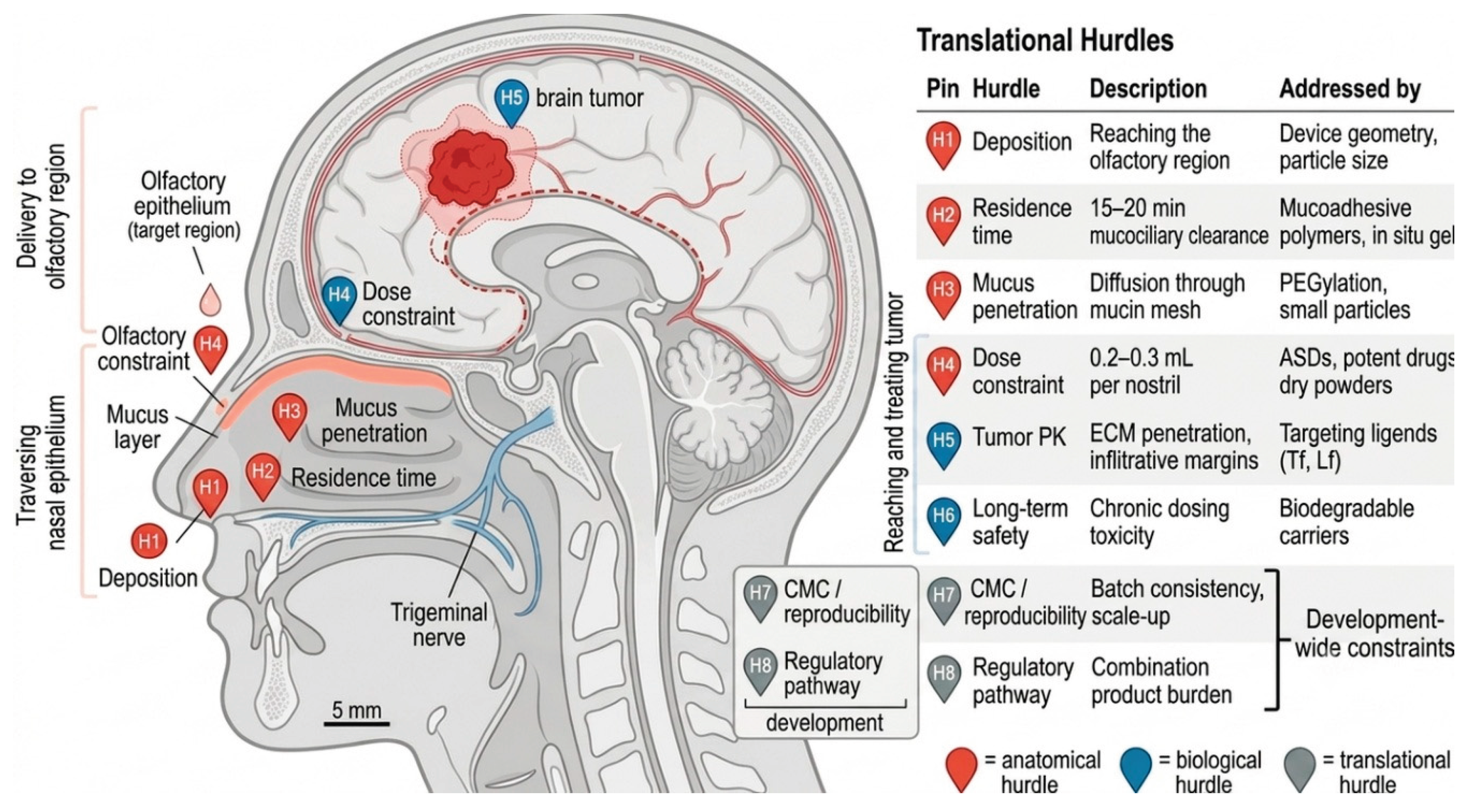
Figure 2.
Anatomy of the nasal cavity and nose-to-brain transport routes. Drugs administered intranasally can reach the CNS via the olfactory pathway (green arrow: olfactory epithelium → cribriform plate → olfactory bulb → forebrain) or via the trigeminal pathway (blue arrow: nasal mucosa → trigeminal nerve branches → brainstem). Both routes bypass the systemic blood–brain barrier (BBB, inset).
Figure 2.
Anatomy of the nasal cavity and nose-to-brain transport routes. Drugs administered intranasally can reach the CNS via the olfactory pathway (green arrow: olfactory epithelium → cribriform plate → olfactory bulb → forebrain) or via the trigeminal pathway (blue arrow: nasal mucosa → trigeminal nerve branches → brainstem). Both routes bypass the systemic blood–brain barrier (BBB, inset).
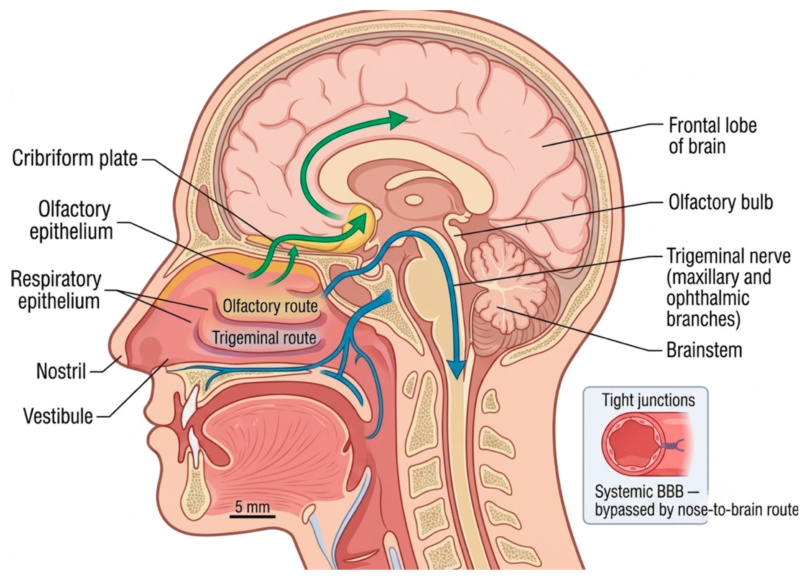

Table 1.
Summary of nanomaterial delivery platforms for intranasal nose-to-brain chemotherapy of glioblastoma, characterized by structure, mechanism, key advantages and limitations, first-in-human (FIH) readiness, tier classification, and representative manuscript references.
Table 1.
Summary of nanomaterial delivery platforms for intranasal nose-to-brain chemotherapy of glioblastoma, characterized by structure, mechanism, key advantages and limitations, first-in-human (FIH) readiness, tier classification, and representative manuscript references.
| Platform | Structure | Primary mechanism | Key advantages | Key limitations | FIH readiness | Tier | References |
|---|---|---|---|---|---|---|---|
|
Polymeric nanoparticles PLGA, chitosan |
Solid biodegradable polymer core, 100–300 nm | Encapsulation with controlled release; surface modification for mucoadhesion or targeting | Biocompatible; mucoadhesive (chitosan); established CMC precedent; biodegrades to non-toxic byproducts (lactic/glycolic acid) | Mucus binding vs. penetration trade-off; batch variability; limited tumor PK without targeting ligands | High | 1 | [48,49,103,104] |
|
Polymeric micelles PEGylated, stimulus-responsive |
Amphiphilic block copolymer core-shell, 10–100 nm | Self-assembly around hydrophobic drugs; stimulus-triggered release | Excellent for hydrophobic drugs; Genexol-PM clinical precedent for paclitaxel delivery | Dilution instability below CMC; aerosolization stress; limited intranasal precedent | Moderate | 2 | [105,106,107,108,109,110,111,112,113,114] |
|
Lipid nanoparticles (LNPs) with mucoadhesive fibers |
Ionizable lipid + PEG-lipid + cholesterol + helper lipid, 40–200 nm | Endosomal escape; mucoadhesive fiber coating extends residence time | mRNA vaccine precedent (Pfizer/Moderna/Shingrix) provides regulatory pathway; high payload versatility | Cilia damage riskᵃ; intranasal chemotherapy application immature; combination-product regulatory burden with fibers | Moderate | 2 | [50,115,122,123,124] |
|
Hydrogels in situ gelling systems |
3D polymer network; thermoresponsive (poloxamer) or ion-sensitive (chitosan) | Liquid-to-gel transition on administration; sustained release from swollen matrix | Directly addresses residence time (H2) and dose (H4); mucoadhesive; well-tolerated | Restricted diffusion through nasal mucosa (H5); gelation sensitive to pH/temperature; standardization challenges (H7) | High | 1 | [58,59,60,61,62,63,64,65,67,68,69,70,71,72,73,74,82] |
|
Amorphous solid dispersions ASDs |
Drug dispersed in amorphous state within polymer matrix (HPMC-AS, PVP) | “Spring and parachute” solubility enhancement; hydrogen-bond crystallization inhibition | Solves H4 (dose constraint) via high loading; strong regulatory precedent; suitable for poorly soluble small molecules | Physical stability during storage; mucus trapping in powder form; recrystallization risk | High | 1 | [40,75] |
|
Gold nanoparticles AuNPs |
Rigid inorganic core with functionalizable surface, 5–100 nm | Surface conjugation of drugs, imaging agents, or targeting ligands; photothermal capability | Precise size and shape control; multifunctional (theranostic potential); rigid structure resists degradation | Chronic accumulation in liver/kidney (H6); non-biodegradable; combination-product regulatory burden (H8); intranasal precedent lacking | Low | 3 | [79,80,81,83,84,85,86,87,88,89,90,91,94,95] |
|
Ferritin nanocages protein-based targeting |
24-subunit self-assembling protein cage; 12 nm outer / 8 nm inner | Native TfR1/SCARA5/TIM-2 receptor binding for tumor targeting; drug encapsulation in cage lumen | Excellent tumor PK (H5) via native receptor targeting; up to 4× MTD improvement in preclinical studiesᵇ | Immunogenicity risk on repeated dosing (H6); manufacturing heterogeneity (H7); no intranasal precedent (H8) | Low | 3 | [48,49,96,97,98,99,100,101,102,103,104,105,106,110,111] |
Disclaimer/Publisher’s Note: The statements, opinions and data contained in all publications are solely those of the individual author(s) and contributor(s) and not of MDPI and/or the editor(s). MDPI and/or the editor(s) disclaim responsibility for any injury to people or property resulting from any ideas, methods, instructions or products referred to in the content. |
© 2026 by the authors. Licensee MDPI, Basel, Switzerland. This article is an open access article distributed under the terms and conditions of the Creative Commons Attribution (CC BY) license (http://creativecommons.org/licenses/by/4.0/).
Copyright: This open access article is published under a Creative Commons CC BY 4.0 license, which permit the free download, distribution, and reuse, provided that the author and preprint are cited in any reuse.


