

Submitted:
21 July 2026
Posted:
27 July 2026
You are already at the latest version
Abstract
Donor-derived cell-free DNA (dd-cfDNA) is an emerging non-invasive biomarker of kidney allograft injury. Elevated dd-cfDNA has been associated with acute rejection, particularly antibody-mediated rejection; however, dd-cfDNA reflects graft-derived tissue injury rather than a specific alloimmune mechanism. Its interpretation may therefore be challenging in conditions characterized by endothelial and microvascular injury, such as atypical hemolytic uremic syndrome (aHUS). This retrospective observational study included 14 kidney transplant recipients who underwent their first kidney transplantation at Wroclaw Medical University between April and December 2022. Twelve non-aHUS recipients with stable renal function and two recipients with aHUS were assessed. Plasma samples were collected at four early post-transplant time points: day 7 (D7), day 14 (D14), month 1 (M1), and month 2 (M2). dd-cfDNA was measured using next-generation sequencing with the AlloSeq cfDNA assay. Donor-specific anti-HLA antibodies (DSA) were assessed using single-antigen bead testing. In non-aHUS recipients, median %dd-cfDNA declined from 0.72% [0.28%, 2.33%] at D7 to 0.12% [0.05%, 0.34%] by M1. In contrast, recipients with aHUS demonstrated persistently elevated dd-cfDNA values fluctuating around 1% [0.88%, 1.14%] during the first month of follow-up. No DSA were detected in any patient during the observation period. No apparent correlation was observed between %dd-cfDNA and serum creatinine in this small cohort. In this preliminary observational study, kidney transplant recipients with aHUS showed persistently elevated early post-transplant dd-cfDNA despite the absence of detectable DSA, absence of clinically documented rejection, and preserved graft function during follow-up. These findings suggest that universal dd-cfDNA thresholds may require cautious interpretation in aHUS. Larger biopsy-anchored studies integrating complement and endothelial injury markers are needed to clarify whether dd-cfDNA elevation in aHUS reflects alloimmune injury, complement-mediated microvascular injury, or overlapping mechanisms.
Keywords:
donor-derived cell-free DNA
; dd-cfDNA
; atypical hemolytic uremic syndrome
; aHUS
; kidney transplantation
; thrombotic microangiopathy
; complement
; rejection
; donor-specific antibodies
1. Introduction
1.1. Donor-Derived Cell-Free DNA: A Promising Non-Invasive Biomarker
Kidney transplantation remains the preferred treatment for patients with end-stage renal disease, offering improved survival and quality of life compared with dialysis [1]. Despite advances in immunosuppression and post-transplant care, allograft rejection continues to threaten long-term graft survival and function. Acute rejection may lead to irreversible graft injury if not identified and treated promptly [2].
The current diagnostic reference standard for rejection is histopathological evaluation of kidney allograft biopsy specimens. Although biopsies provide essential diagnostic information, they are invasive, carry procedural risks such as bleeding or infection, and may be affected by sampling error and interobserver variability [3,4]. These limitations have stimulated the search for safer, repeatable, and more objective diagnostic adjuncts.
One of the most promising approaches is donor-derived cell-free DNA, a fragment of DNA released into the recipient circulation as a result of allograft cell injury and turnover. Cell-free DNA is generated predominantly during apoptosis and necrosis, and its presence in circulation may reflect tissue injury. In transplant recipients, the proportion of cfDNA originating from donor tissue provides a quantitative measure of allograft-derived injury [5].
Numerous studies have demonstrated an association between elevated dd-cfDNA and episodes of acute rejection, with increased levels indicating heightened cell turnover and injury in the transplanted organ [6,7]. Before detecting changes in conventional clinical markers like serum creatinine or proteinuria, dd-cfDNA levels may rise, potentially allowing for earlier clinical intervention [8]. This has positioned dd-cfDNA as a useful adjunct in post-transplant surveillance and in the assessment of immunological risk [9].
Beyond rejection, dd-cfDNA has also shown promise in identifying other forms of graft injury, including BK virus nephropathy and ischemia-reperfusion injury, although specificity remains a major concern. Overall, dd-cfDNA offers an appealing, non-invasive tool for post-transplant monitoring, yet important diagnostic caveats remain, particularly in recipients with complex comorbidities or rare conditions such as atypical hemolytic uremic syndrome.
1.2. Diagnostic Challenges of dd-cfDNA in Kidney Transplant Recipients with aHUS
Atypical hemolytic uremic syndrome is a rare, life-threatening disease characterized by complement dysregulation leading to thrombotic microangiopathy, endothelial damage, and multiorgan injury, with the kidneys being among the most commonly affected organs [10,11]. Patients with aHUS frequently progress to end-stage renal disease and may require kidney transplantation. However, transplant outcomes in this population remain complex because of the risk of disease recurrence and persistent microvascular injury after transplantation [12].
These pathophysiological mechanisms pose a unique challenge for the interpretation of dd-cfDNA. Chronic, recurrent, or subclinical endothelial injury associated with aHUS, even in the absence of overt alloimmune activation, may independently enhance graft-derived cell injury and the release of dd-cfDNA. This raises concerns about the specificity of dd-cfDNA as a marker of rejection in this context, because elevated values may not necessarily reflect immune-mediated allograft injury but may instead reflect complement-mediated endothelial injury and associated cell turnover [13].
1.3. Aim of the Study
This study sought to address this knowledge gap by examining the dynamics of plasma dd-cfDNA after kidney transplantation in recipients with and without aHUS. By evaluating the relationship between dd-cfDNA levels, DSA status, serum creatinine, and clinical course, this study aimed to assess the reliability and limitations of dd-cfDNA as a biomarker of graft injury in the specific context of aHUS.
2. Results
Recipient-level peri-transplant characteristics are presented in Table 1. Non-aHUS and aHUS recipients are shown as separate patient-level rows.
Overall, maintenance immunosuppression was based on corticosteroids, tacrolimus, and mycophenolate mofetil in all recipients. Delayed graft function was recorded in one non-aHUS recipient and in one aHUS recipient.

Table 2.
Cohort characteristics and testing schedule in kidney transplant recipients with and without aHUS. Available cohort and testing characteristics are summarized in Table 2.
Table 2.
Cohort characteristics and testing schedule in kidney transplant recipients with and without aHUS. Available cohort and testing characteristics are summarized in Table 2.
| Variable | Non-aHUS recipients | aHUS recipients |
|---|---|---|
| Number of recipients | 12 | 2 |
| First kidney transplantation | 12/12 | 2/2 |
| Study period | April–December 2022 | April–December 2022 |
| Plasma collection time points | D7, D14, M1, M2 | D7, D14, M1, M2 |
| Clinical graft status during observation | Stable renal function during follow-up | Preserved graft function during follow-up |
| Clinically documented acute rejection | None documented | None documented |
| Protocol biopsies | Not performed systematically | Not performed systematically |
| DSA detected during follow-up | 0/12 | 0/2 |
| dd-cfDNA assay | AlloSeq cfDNA, NGS | AlloSeq cfDNA, NGS |
| DSA assay | LABScreen Single Antigen Beads | LABScreen Single Antigen Beads |
In non-aHUS kidney transplant recipients, median %dd-cfDNA values showed a gradual decline over time, ranging from 0.72% [0.28%, 2.33%] at day 7 to 0.12% [0.05%, 0.34%] at M1, consistent with stabilization of graft function. In contrast, recipients with aHUS demonstrated persistently elevated dd-cfDNA levels, with median values fluctuating around 1% [0.88%, 1.14%] during the first month of follow-up (Figure 1).
No donor-specific anti-HLA antibodies were detected in any patient throughout the observation period. No apparent correlation was observed between %dd-cfDNA levels and serum creatinine levels across the cohort. Given the small sample size and repeated measurements within individuals, this observation should be interpreted descriptively rather than as definitive evidence of independence between these markers.
3. Discussion
This study examined early dd-cfDNA kinetics in kidney transplant recipients with and without aHUS. In non-aHUS recipients, dd-cfDNA levels declined within the first month after transplantation, consistent with recovery from perioperative injury and stabilization of graft function. By contrast, recipients with aHUS showed persistently elevated dd-cfDNA around the commonly used 1% threshold despite the absence of detectable DSA, absence of clinically documented rejection, and preserved graft function during follow-up.
These findings raise important questions regarding the interpretation of dd-cfDNA in the context of aHUS. Previous studies in broader kidney transplant populations have consistently shown that dd-cfDNA levels around or above 1% are associated with acute rejection and poorer graft outcomes [6,7,8]. However, our data suggest that in aHUS recipients, elevated dd-cfDNA may not carry the same predictive meaning as in unselected kidney transplant populations. Instead, the observed elevations may reflect ongoing endothelial injury and cell turnover related to underlying complement-mediated pathology, rather than exclusively alloimmune-mediated graft damage [10,11,12,13]. This highlights a potential limitation of applying universal dd-cfDNA thresholds across biologically diverse transplant populations.
The role of dd-cfDNA as a biomarker of allograft rejection has been progressively refined since its initial clinical validation [5,14]. A 1% threshold discriminates active rejection with high negative but limited positive predictive value, reflecting that dd-cfDNA rises with any source of graft injury rather than with rejection specifically [5]. This signal is strongest for antibody-mediated rejection, in which microvascular inflammation produces greater endothelial DNA release than the predominantly tubulointerstitial injury of T-cell-mediated rejection [9,15]. A recent meta-analysis confirmed higher pooled diagnostic accuracy for antibody-mediated than for any-cause rejection [8]. Even low-grade or borderline T-cell-mediated rejection may, however, be accompanied by elevated dd-cfDNA and an increased risk of subsequent graft injury [16], underscoring that the biomarker reports tissue injury along a continuum rather than a binary rejection state.
Beyond a single cross-sectional measurement, the temporal behavior of dd-cfDNA carries additional prognostic information. In the longitudinal ADMIRAL cohort, persistently elevated values—rather than isolated peaks—predicted future de novo donor-specific antibodies and a clinically meaningful decline in glomerular filtration rate, and elevations may precede biopsy-proven rejection by several weeks [17,18]. Surveillance studies using the same next-generation sequencing assay applied here have similarly linked persistent early post-transplant elevation to non-improving graft function [19]. This kinetic perspective is directly relevant to the present cohort: a transient peri-operative rise that resolves is expected, whereas values that remain elevated beyond the first month are more concerning. In aHUS, ongoing complement-mediated endothelial injury can sustain dd-cfDNA release even without rejection, complicating this phenomenon. Distinguishing persistent elevation due to alloimmune injury from that due to underlying microvascular disease is, therefore, the central interpretive challenge in this population and one that a fractional threshold alone cannot resolve.
The lack of published information on dd-cfDNA changes in aHUS patients makes it harder to understand the clinical situation. It remains unclear whether the biomarker behaves similarly to other transplant populations or whether elevated dd-cfDNA in aHUS reflects baseline vascular injury, recurrent or subclinical thrombotic microangiopathy, subclinical rejection, or overlapping mechanisms. This knowledge gap calls for studies specifically addressing rare transplant populations, because universal dd-cfDNA thresholds may not be appropriate in all clinical contexts.
None of the patients in our cohort developed detectable DSA during follow-up, and no apparent correlation was observed between dd-cfDNA and serum creatinine. In a cohort without documented rejection and with stable graft function, the absence of such a correlation is expected and should be interpreted cautiously rather than as positive evidence that dd-cfDNA captures occult injury. This disconnect demonstrates the complexity of graft monitoring in aHUS recipients. Serum creatinine is an insensitive and delayed marker of injury, whereas dd-cfDNA may capture biological processes not reflected in routine biochemical testing. Importantly, recent evidence shows that antibody-mediated rejection can occur even in the absence of detectable DSA, with DSA-negative antibody-mediated rejection sharing molecular signatures and dd-cfDNA release patterns with DSA-positive cases [20]. These findings emphasize that a negative DSA result does not fully exclude alloimmune injury.
In the context of aHUS, persistently elevated dd-cfDNA without detectable DSA may reflect non-alloimmune endothelial injury, low-grade immune activation below the threshold of serological detection, or a combination of both. Accordingly, dd-cfDNA should not be interpreted as a stand-alone diagnostic test in this setting. A multimodal approach integrating DSA, graft function, proteinuria, infection status, hemolysis and thrombotic microangiopathy markers, complement activation markers, and biopsy findings when clinically indicated is likely to provide a more reliable assessment.
Our study adds to a limited body of literature addressing dd-cfDNA kinetics in rare transplant populations. The persistently elevated levels observed in aHUS recipients highlight the need for more nuanced interpretation of this biomarker in the setting of chronic microvascular injury. Future research should investigate disease-specific thresholds or integrate dd-cfDNA with complementary biomarkers, such as complement activation products, endothelial injury markers, and gene expression profiling. Such approaches may help disentangle alloimmune from non-alloimmune injury and improve risk stratification in complex patient groups.
This study has important limitations. First, only two aHUS recipients were included; therefore, the findings should be interpreted as hypothesis-generating. Second, the study was retrospective and single-centre. Third, protocol biopsies and molecular biopsy assessment were not systematically available, so subclinical rejection could not be definitively excluded. Fourth, detailed markers of complement activation and thrombotic microangiopathy were not available for integrated analysis. Fifth, dd-cfDNA was analyzed as a fractional value; absolute dd-cfDNA quantification may provide additional information in future studies. Finally, follow-up was limited to the early post-transplant period, and longer-term graft outcomes were not assessed.
4. Materials and Methods
This retrospective observational study included 14 kidney transplant recipients: 12 non-aHUS recipients with stable renal function and 2 recipients with aHUS. The aHUS subgroup consisted of two female kidney transplant recipients aged between 20 and 40 years. Both patients received a kidney transplant from a deceased donor. In one patient, aHUS was the primary cause of kidney failure, whereas in the other patient, aHUS was considered most likely secondary to primary focal segmental glomerulosclerosis.
In the first patient, genetic testing performed as part of the diagnostic work-up for aHUS revealed a heterozygous pathogenic variant in the CFH gene. In addition, the C3 GGTA haplotype, which has been associated with dense deposit disease, was identified. The patient also carried the CFH H3 risk haplotype (TGTGT), associated with a less favorable disease course and an increased risk of kidney allograft loss, as well as one copy of the MCP/CD46 aHUS risk haplotype (GGAAC). Genetic analysis also demonstrated heterozygous ADAMTS13 variants associated with familial thrombotic thrombocytopenic purpura.
In the second patient, genetic testing revealed heterozygous CD46 variants c.946+23G>T and c.*783T>C. The role of the c.946+23G>T variant in modulating disease risk remains uncertain; however, its presence has been associated with alternative splicing of the CD46 transcript, involving selective inclusion of exon 8 and resulting in impaired functionality of the gene product. The c.*783T>C variant has been described as a risk variant for hemolytic uremic syndrome. In addition, the patient carried the CFH H3 risk haplotype (TGTGT), associated with a less favorable disease course and an increased risk of kidney allograft loss.
In both patients, eculizumab therapy was continued after kidney transplantation and was subsequently converted to ravulizumab. No recurrence of aHUS was observed after transplantation in either patient. Both patients received basiliximab induction and standard triple maintenance immunosuppression consisting of corticosteroids, mycophenolate mofetil, and tacrolimus.
All patients underwent their first kidney transplantation in the Department and Clinic of Nephrology, Transplant Medicine and Internal Medicine at Wroclaw Medical University between April and December 2022. Protocol biopsies were not performed systematically as part of this study.
Blood samples were collected at four time points: one week after transplantation (D7), two weeks after transplantation (D14), one month after transplantation (M1), and two months after transplantation (M2). Samples were collected into tubes dedicated to cfDNA stabilization (Cell-Free DNA BCT RUO, Streck, La Vista, NE, USA). cfDNA was isolated from 4 mL of plasma using the MagMAX Cell-Free DNA Isolation Kit (Applied Biosystems, Thermo Fisher Scientific Inc., Waltham, MA, USA), according to the manufacturer's protocol.
cfDNA concentration was measured using fluorescence-based methods with the Quantus Fluorometer (Promega Corporation, Madison, WI, USA; RRID:SCR_026279) and QuantiFluor ONE dsDNA System (Promega Corporation, Madison, WI, USA). cfDNA quality was verified using automated electrophoresis with the 4200 TapeStation System (Agilent, Santa Clara, CA, USA; RRID:SCR_018435) and High Sensitivity D1000 ScreenTape (Agilent, Santa Clara, CA, USA).
dd-cfDNA in kidney transplant recipients was evaluated by next-generation sequencing using the AlloSeq cfDNA Kit (CareDx Inc., Brisbane, CA, USA). Sequencing was performed on a MiSeq instrument using the MiSeq Reagent v3 150-cycle Kit (Illumina Inc., San Diego, CA, USA). Sequencing results were analyzed with AlloSeq cfDNA Software (CareDx).
Donor-specific anti-HLA antibody profiling was performed using LABScreen Single Antigen Beads (One Lambda, Inc., Thermo Fisher Scientific, Canoga Park, CA, USA) according to the manufacturer's instructions. Samples were analyzed on a Luminex FlexMAP 3D platform (Luminex Corporation, Austin, TX, USA), and results were interpreted using HLA Fusion Software, version 4.7 (One Lambda, Inc.).
Donor-specific anti-HLA antibody positivity was defined as the presence of at least one anti-HLA class I and/or class II antibody directed against the corresponding donor HLA antigen. Anti-HLA antibody specificity testing was performed only in samples with a positive screening result for anti-HLA antibodies of the respective HLA class. The final classification of antibody specificity was based on the mean normalized fluorescence intensity value (MFI_norm) obtained for bead populations corresponding to a given serological specificity with a positive or borderline signal.
For anti-HLA class I antibodies, the laboratory-defined cut-off value was MFI_norm = 1100, with a reported measurement uncertainty of 55%. Results were classified as positive when MFI_norm was >=1700, borderline when MFI_norm was >=500 and <1700, and negative when MFI_norm was <500. For anti-HLA class II antibodies, the laboratory-defined cut-off value was MFI_norm = 1250, with a reported measurement uncertainty of 40%. Results were classified as positive when MFI_norm was >=1750, borderline when MFI_norm was >=750 and <1750, and negative when MFI_norm was <750. The cut-off values were established with reference to a non-immunized male population, defined as individuals without a history of transplantation or blood transfusion.
For the purposes of the present analysis, patients were considered DSA-positive when at least one donor-specific anti-HLA antibody met the laboratory criteria for a positive result.
Statistical analyses were conducted using STATISTICA version 13 (TIBCO Software Inc., 2017, Dell, OK, USA; RRID:SCR_014213). Given the small sample size and the exploratory nature of the study, analyses were primarily descriptive. dd-cfDNA values are presented as medians with interquartile ranges where appropriate. Associations between %dd-cfDNA and serum creatinine were assessed descriptively; formal inferential conclusions were avoided because of the limited cohort size.
The study protocol was reviewed and approved by the Bioethical Committee of Wroclaw Medical University (approval no. 37/2022). All participants provided written informed consent before inclusion in the study, in accordance with the Declaration of Helsinki.
An AI-based language model (ChatGPT, OpenAI, San Francisco, CA, USA) was used to assist in language refinement. No AI tool was used for data analysis or generation of scientific conclusions.
The authors made and verified all scientific content, interpretations, and conclusions.
5. Conclusions
In this preliminary observational study, kidney transplant recipients with aHUS demonstrated persistently elevated early post-transplant dd-cfDNA despite the absence of detectable DSA, absence of clinically documented rejection, and preserved graft function during follow-up, whereas non-aHUS recipients showed a decline in dd-cfDNA during early follow-up. These findings suggest that aHUS may represent a biologically distinct setting in which universal dd-cfDNA thresholds require cautious interpretation. Larger biopsy-anchored studies integrating dd-cfDNA with DSA, complement activation markers, endothelial injury biomarkers, and molecular graft assessment are needed to clarify the clinical meaning of dd-cfDNA elevation in this rare transplant population.
Author Contributions
Conceptualization, P.W., W.S., M.U., A.D. and M.B.; methodology, K.G., W.S., A.D. and K.Gr.; investigation, P.W., M.B. and M.U.; formal analysis, K.G., W.S., K.Gr. and K.M.B.; data curation, K.G., W.S., P.J., M.K.-K. and K.M.B.; writing—original draft preparation, P.W., K.G. and W.S.; writing—review and editing, P.W., K.G., K.M.B. and W.S.; supervision, M.B. and M.U.; funding acquisition, M.B. and M.U.; project administration, M.B. and M.U. All authors have read and agreed to the published version of the manuscript.
Funding
The research was supported by grant no. MINI.C200.21.008 from the National Science Centre, Poland, and by subsidy no. SUBZ.A500.25.019 from Wroclaw Medical University.
Institutional Review Board Statement
The study was conducted in accordance with the Declaration of Helsinki and approved by the Bioethical Committee of Wroclaw Medical University, approval no. 37/2022, approved on 26 January 2022.
Informed Consent Statement
Informed consent was obtained from all subjects involved in the study.
Data Availability Statement
The data presented in this study are available on reasonable request from the corresponding author due to privacy and ethical restrictions.
Acknowledgments
The authors thank the staff of the Department and Clinic of Nephrology, Transplant Medicine, and Internal Medicine at Wroclaw Medical University for their assistance in patient care and sample collection. The authors are also grateful to the laboratory team for technical support with cfDNA isolation, next-generation sequencing, and Luminex analyses. Finally, the authors acknowledge the patients and their families for their participation and trust, which made this study possible.
Conflicts of Interest
The authors declare no conflicts of interest.
References
- Wolfe, R.A.; Ashby, V.B.; Milford, E.L.; Ojo, A.O.; Ettenger, R.E.; Agodoa, L.Y.C.; Held, P.J.; Port, F.K. Comparison of Mortality in All Patients on Dialysis, Patients on Dialysis Awaiting Transplantation, and Recipients of a First Cadaveric Transplant. N. Engl. J. Med. 1999, 341, 1725–1730. [Google Scholar] [CrossRef] [PubMed]
- Hariharan, S.; Israni, A.K.; Danovitch, G. Long-Term Survival after Kidney Transplantation. N. Engl. J. Med. 2021, 385, 729–743. [Google Scholar] [CrossRef] [PubMed]
- Furness, P.N.; Taub, N. International Variation in the Interpretation of Renal Transplant Biopsies: Report of the CERTPAP Project. Kidney Int. 2001, 60, 1998–2012. [Google Scholar] [CrossRef] [PubMed]
- Sellarés, J.; De Freitas, D.G.; Mengel, M.; Reeve, J.; Einecke, G.; Sis, B.; Hidalgo, L.G.; Famulski, K.; Matas, A.; Halloran, P.F. Understanding the Causes of Kidney Transplant Failure: The Dominant Role of Antibody-Mediated Rejection and Nonadherence. Am. J. Transplant. 2012, 12, 388–399. [Google Scholar] [CrossRef] [PubMed]
- Bloom, R.D.; Bromberg, J.S.; Poggio, E.D.; Bunnapradist, S.; Langone, A.J.; Sood, P.; Matas, A.J.; Mehta, S.; Mannon, R.B.; Sharfuddin, A.; et al. Cell-Free DNA and Active Rejection in Kidney Allografts. J. Am. Soc. Nephrol. 2017, 28, 2221–2232. [Google Scholar] [CrossRef] [PubMed]
- Lakhani, L.; Alasfar, S.; Bhalla, A.; Aala, A.; Rosenberg, A.; Ostrander, D.; Schollenberger, M.D.; Brennan, D.C.; Lipson, E.J. Utility of Serial Donor-Derived Cell-Free DNA Measurements for Detecting Allograft Rejection in a Kidney Transplant Recipient After PD-1 Checkpoint Inhibitor Administration. Transplant. Direct 2021, 7, e656. [Google Scholar] [CrossRef] [PubMed]
- Oellerich, M.; Shipkova, M.; Asendorf, T.; Walson, P.D.; Schauerte, V.; Mettenmeyer, N.; Kabakchiev, M.; Hasche, G.; Gröne, H.J.; Friede, T.; et al. Absolute Quantification of Donor-Derived Cell-Free DNA as a Marker of Rejection and Graft Injury in Kidney Transplantation: Results from a Prospective Observational Study. Am. J. Transplant. 2019, 19, 3087–3099. [Google Scholar] [CrossRef] [PubMed]
- Xing, Y.; Guo, Q.; Wang, C.; Shi, H.; Zheng, J.; Jia, Y.; Li, C.; Hao, C. Donor-Derived Cell-Free DNA as a Diagnostic Marker for Kidney-Allograft Rejection: A Systematic Review and Meta-Analysis. Biomol. Biomed. 2024, 24, 731–740. [Google Scholar] [CrossRef] [PubMed]
- Jordan, S.C.; Bunnapradist, S.; Bromberg, J.S.; Langone, A.J.; Hiller, D.; Yee, J.P.; Sninsky, J.J.; Woodward, R.N.; Matas, A.J. Donor-Derived Cell-Free DNA Identifies Antibody-Mediated Rejection in Donor Specific Antibody Positive Kidney Transplant Recipients. Transplant. Direct 2018, 4, e379. [Google Scholar] [CrossRef] [PubMed]
- Buelna-Cano, C.; Gálvez-Acosta, S.; Ornelas-Velázquez, A. Atypical Hemolytic Uremic Syndrome. Med. Interna De Mex. 2014, 30, 622–628. [Google Scholar] [CrossRef]
- Yoshida, Y.; Kato, H.; Ikeda, Y.; Nangaku, M. Pathogenesis of Atypical Hemolytic Uremic Syndrome. J. Atheroscler. Thromb. 2019, 26, 99–110. [Google Scholar] [CrossRef] [PubMed]
- Zuber, J.; Le Quintrec, M.; Krid, S.; Bertoye, C.; Gueutin, V.; Lahoche, A.; Heyne, N.; Ardissino, G.; Chatelet, V.; Noël, L.H.; et al. Eculizumab for Atypical Hemolytic Uremic Syndrome Recurrence in Renal Transplantation. Am. J. Transplant. 2012, 12, 3337–3354. [Google Scholar] [CrossRef] [PubMed]
- Abbas, F.; El Kossi, M.; Kim, J.J.; Shaheen, I.S.; Sharma, A.; Halawa, A. Complement-Mediated Renal Diseases after Kidney Transplantation - Current Diagnostic and Therapeutic Options in de Novo and Recurrent Diseases. World J. Transplant. 2018, 8, 203–219. [Google Scholar] [CrossRef] [PubMed]
- Oellerich, M.; Sherwood, K.; Keown, P.; Schütz, E.; Beck, J.; Stegbauer, J.; Rump, L.C.; Walson, P.D. Liquid Biopsies: Donor-Derived Cell-Free DNA for the Detection of Kidney Allograft Injury. Nat. Rev. Nephrol. 2021, 17, 591–603. [Google Scholar] [CrossRef] [PubMed]
- Huang, E.; Sethi, S.; Peng, A.; Najjar, R.; Mirocha, J.; Haas, M.; Vo, A.; Jordan, S.C. Early Clinical Experience Using Donor-Derived Cell-Free DNA to Detect Rejection in Kidney Transplant Recipients. Am. J. Transplant. 2019, 19, 1663–1670. [Google Scholar] [CrossRef] [PubMed]
- Stites, E.; Kumar, D.; Olaitan, O.; John Swanson, S.; Leca, N.; Weir, M.; Bromberg, J.; Melancon, J.; Agha, I.; Fattah, H.; et al. High Levels of Dd-CfDNA Identify Patients with TCMR 1A and Borderline Allograft Rejection at Elevated Risk of Graft Injury. Am. J. Transplant. 2020, 20, 2491–2498. [Google Scholar] [CrossRef] [PubMed]
- Bu, L.; Gupta, G.; Pai, A.; Anand, S.; Stites, E.; Moinuddin, I.; Bowers, V.; Jain, P.; Axelrod, D.A.; Weir, M.R.; et al. Clinical Outcomes from the Assessing Donor-Derived Cell-Free DNA Monitoring Insights of Kidney Allografts with Longitudinal Surveillance (ADMIRAL) Study. Kidney Int. 2022, 101, 793–803. [Google Scholar] [CrossRef] [PubMed]
- Bromberg, J.S.; Bunnapradist, S.; Samaniego-Picota, M.; Anand, S.; Stites, E.; Gauthier, P.; Demko, Z.; Prewett, A.; Armer-Cabral, M.; Marshall, K.; et al. Elevation of Donor-Derived Cell-Free DNA Before Biopsy-Proven Rejection in Kidney Transplant. Transplantation 2024, 108, 1994–2004. [Google Scholar] [CrossRef] [PubMed]
- Mantios, E.; Filiopoulos, V.; Constantoulakis, P.; Liapis, G.; Vittoraki, A.; Casas, S.; Marinaki, S.; Boletis, J.N. Assessment of Donor Derived Cell Free DNA (Dd-CfDNA) at Surveillance and at Clinical Suspicion of Acute Rejection in Renal Transplantation. Transpl. Int. 2023, 36. [Google Scholar] [CrossRef] [PubMed]
- Halloran, P.F.; Reeve, J.; Madill-Thomsen, K.S.; Demko, Z.; Prewett, A.; Gauthier, P.; Billings, P.; Lawrence, C.; Lowe, D.; Hidalgo, L.G. Antibody-Mediated Rejection Without Detectable Donor-Specific Antibody Releases Donor-Derived Cell-Free DNA: Results from the Trifecta Study. Transplantation 2023, 107, 709–719. [Google Scholar] [CrossRef] [PubMed]
Figure 1.
Early post-transplant dd-cfDNA kinetics in kidney transplant recipients with and without atypical hemolytic uremic syndrome (aHUS). (A) dd-cfDNA levels in recipients with aHUS and (B) recipients without aHUS. dd-cfDNA is shown as a percentage of total circulating cfDNA at day 7 (D7), day 14 (D14), month 1 (M1), and month 2 (M2) after transplantation. Data are presented as summary values with dispersion bars at each time point.
Figure 1.
Early post-transplant dd-cfDNA kinetics in kidney transplant recipients with and without atypical hemolytic uremic syndrome (aHUS). (A) dd-cfDNA levels in recipients with aHUS and (B) recipients without aHUS. dd-cfDNA is shown as a percentage of total circulating cfDNA at day 7 (D7), day 14 (D14), month 1 (M1), and month 2 (M2) after transplantation. Data are presented as summary values with dispersion bars at each time point.
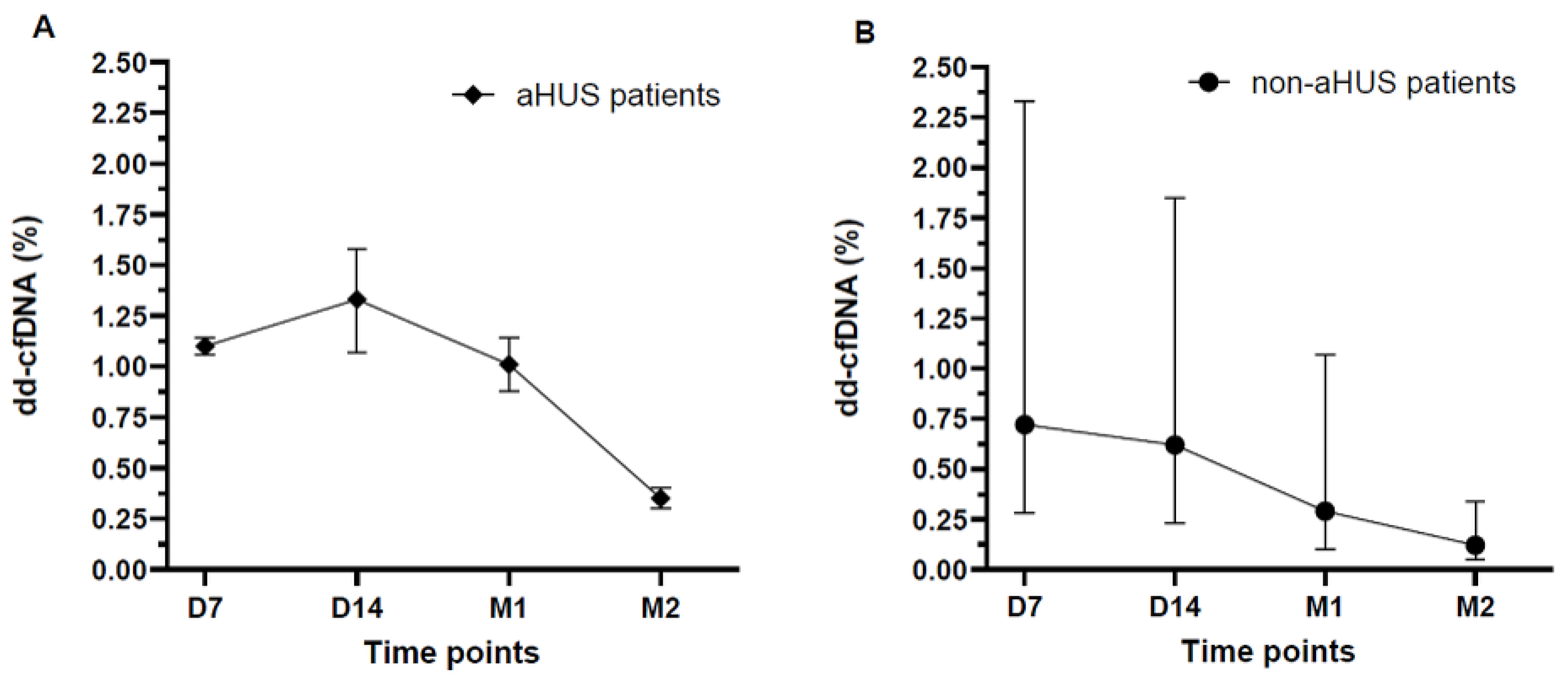
Table 1.
Individual peri-transplant characteristics of kidney transplant recipients with and without aHUS.
Table 1.
Individual peri-transplant characteristics of kidney transplant recipients with and without aHUS.
| Recipient | Group | WIT (min) | CIT (min) | Post-Tx dialysis (days) | DGF | Maintenance immunosuppression | Induction | PRA max (%) |
|---|---|---|---|---|---|---|---|---|
| Non-aHUS recipients (n=12) | ||||||||
| N1 | non-aHUS | 20 | 1320 | 0 | No | S/T/MMF | ATG | 83 |
| N2 | non-aHUS | 15 | 120 | 0 | No | S/T/MMF | Basiliximab | NA |
| N3 | non-aHUS | 30 | 860 | 0 | No | S/T/MMF | None | 0 |
| N4 | non-aHUS | 30 | 1380 | 2 | Yes | S/T/MMF | Basiliximab | 20 |
| N5 | non-aHUS | 34 | 120 | 0 | No | S/T/MMF | Basiliximab | 0 |
| N6 | non-aHUS | 20 | 1327 | 0 | No | S/T/MMF | Basiliximab | 7 |
| N7 | non-aHUS | 23 | 455 | 0 | No | S/T/MMF | Basiliximab | 3 |
| N8 | non-aHUS | 20 | 120 | 0 | No | S/T/MMF | Basiliximab | NA |
| N9 | non-aHUS | 19 | 129 | 0 | No | S/T/MMF | Basiliximab | NA |
| N10 | non-aHUS | 23 | 855 | 0 | No | S/T/MMF | Basiliximab | 0 |
| N11 | non-aHUS | 11 | 1465 | 0 | No | S/T/MMF | None | 0 |
| N12 | non-aHUS | 19 | 455 | 0 | No | S/T/MMF | Basiliximab | 30 |
| aHUS recipients (n=2) | ||||||||
| A1 | aHUS | 27 | 370 | 0 | No | S/T/MMF | Basiliximab | 8 |
| A2 | aHUS | 20 | 1680 | 11 | Yes | S/T/MMF | Basiliximab | 0 |
Abbreviations: aHUS, atypical hemolytic uremic syndrome; ATG, anti-thymocyte globulin; CIT, cold ischemia time; DGF, delayed graft function; MMF, mycophenolate mofetil; PRA, panel-reactive antibody; S, steroids; T, tacrolimus; Tx, transplantation; WIT, warm ischemia time. PRA max is shown as the maximum panel-reactive antibody value recorded for each recipient.
Disclaimer/Publisher’s Note: The statements, opinions and data contained in all publications are solely those of the individual author(s) and contributor(s) and not of MDPI and/or the editor(s). MDPI and/or the editor(s) disclaim responsibility for any injury to people or property resulting from any ideas, methods, instructions or products referred to in the content. |
© 2026 by the authors. Licensee MDPI, Basel, Switzerland. This article is an open access article distributed under the terms and conditions of the Creative Commons Attribution (CC BY) license (http://creativecommons.org/licenses/by/4.0/).
Copyright: This open access article is published under a Creative Commons CC BY 4.0 license, which permit the free download, distribution, and reuse, provided that the author and preprint are cited in any reuse.


