

Submitted:
23 July 2026
Posted:
24 July 2026
You are already at the latest version
Abstract
Background/Objectives: Cardiac troponin I (cTnI) has shown inconsistent diagnostic performance for acute cellular rejection (ACR) after heart transplantation. We evaluated whether time-normalized cTnI kinetics provide incremental diagnostic value beyond the concurrent concentration for detecting ACR grade ≥2R. Methods: This retrospective single-center study included 1237 biopsy episodes from 139 heart transplant recipients after exclusion of ambiguous measurements and episodes without a preceding confirmed cTnI value. Two generalized linear mixed-effects logistic regression models with patient-specific random intercepts were compared. M1 included concurrent log₂-transformed cTnI, whereas M2 additionally included the time-normalized change in log₂-transformed cTnI per 7 days. Discrimination was assessed using leave-one-patient-out cross-validation; 95% confidence intervals were obtained from 2000 patient-level cluster bootstrap resamples. Results: In the overall cohort, M1 showed modest discrimination (AUC 0.641, 95% CI 0.592–0.689), whereas M2 provided no improvement (AUC 0.635, 95% CI 0.584–0.685; ΔAUC −0.007, 95% CI −0.021 to 0.007). Within 90 days after transplantation, neither model was informative (M1 AUC 0.503; M2 AUC 0.494). Beyond 90 days, M1 showed moderate discrimination (AUC 0.758, 95% CI 0.664–0.838), but M2 again provided no improvement (AUC 0.746, 95% CI 0.654–0.828; ΔAUC −0.012, 95% CI −0.037 to 0.008). Conclusions: Concurrent cTnI showed no discrimination within 90 days and moderate discrimination thereafter in exploratory stratified analyses. The kinetic term provided no incremental value. cTnI cannot replace endomyocardial biopsy but may warrant further evaluation as an adjunctive late-period marker.
Keywords:
heart transplantation
; acute cellular rejection
; cardiac troponin I
; endomyocardial biopsy
; diagnostic accuracy
; biomarker kinetics
1. Introduction
In post-heart transplant patients, acute cellular rejection (ACR) poses a significant threat to patient survival. The gold standard for detecting rejection is surveillance endomyocardial biopsies (EMB), but this procedure is invasive, costly and associated with procedural risk. Additionally, the vast majority of surveillance EMBs show no significant rejection [1]. This drives a need for non-invasive, preferably inexpensive, biomarkers that could reliably exclude ACR. Cardiac troponin I (cTnI) has been investigated as a biopsy alternative. Although troponin I is highly specific to cardiomyocyte injury and could theoretically exclude rejection when low, recent studies have shown poor diagnostic accuracy [2,3]. The 2023 ISHLT guidelines note the potential of cTnI to correlate with rejection grade [4] but do not recommend routine troponin-based surveillance (Class III, LoE C); they only suggest cTnI as part of a risk stratification strategy for additional testing. Likewise the European Society for Organ Transplantation states that there is inadequate evidence to support the routine use of cardiac troponin for the diagnosis of ACR, due to conflicting data [1]. However, a systematic review by Fitzsimons et al. suggested that cTnI may have sufficient sensitivity and negative predictive value to exclude ACR and potentially reduce the number of surveillance EMBs [5]. Furthermore cTnI may potentially contribute to multimodal strategies combining laboratory biomarkers and cardiac imaging [6], alongside highly effective biomarkers such as donor-derived cell-free DNA, which may yield superior results compared to any singular biomarker. Troponin I was found to lack utility in detecting ACR especially in the immediate to early post-operative period [3], which is why the focus of this study was to include exploratory approach that includes troponin dynamics, by adding the time-normalized change in log2-transformed cTnI per 7 days as an additional predictor. Troponin concentrations are persistently elevated in the early post-heart transplantation (HTx) period due to the surgery [7], this way masking any potential events that could have been detected otherwise. Using trends rather than absolute levels might be necessary to reveal rejection-related changes. Similar approach is used in acute coronary syndromes; serial troponin measurements (and relative changes over time) are integral to diagnosis. We therefore hypothesized that relative changes in cTnI over time may better distinguish evolving rejection injury than a single snapshot. In practice, a steady decline of cTnI from the post-op period is observed, so a new rise or failure to fall might signal rejection.
Study Objective
Accordingly, we aimed to determine whether the time-normalized change in log₂-transformed cTnI per 7 days provided incremental discrimination for ACR grade ≥2R beyond the concurrent log₂-transformed cTnI concentration.
2. Methods
2.1. Study Design and Population
We conducted a retrospective, single-center diagnostic cohort study of post-HTx patients, evaluating whether troponin dynamics add to absolute troponin levels in predicting biopsy-confirmed ACR graded ≥2R according to ISHLT criteria. It was designed as a diagnostic accuracy study comparing regular cTnI testing (index test) with EMB (reference standard). The study was conducted at the Department of Heart Transplantation and Cardiac Surgery of the University Clinical Hospital in Wrocław. The pathologists assessing endomyocardial biopsies were blinded to the corresponding cTnI results. For the purposes of this study, concurrent cTnI was defined as a cTnI measurement obtained on the same calendar day as the corresponding endomyocardial biopsy. Laboratory personnel performing cTnI measurements were blinded to the biopsy results. Data from 141 recipients, comprising 1392 biopsy–troponin pairs, were initially screened. Biopsy episodes with ambiguous multiple cTnI measurements recorded for the same patient and date were excluded. For the comparative model analysis, only episodes with a concurrent cTnI measurement and a preceding confirmed cTnI measurement were retained. The final analytical dataset comprised 1237 biopsy episodes from 139 recipients.
2.2. Data Collection
Clinical, laboratory, and histopathological data were collected between February 2021 and March 2025 based on the analysis of electronic medical records and the results of laboratory and histopathological tests. Endomyocardial biopsies were assessed according to the ISHLT classification.
The analyzed data included: donor and recipient age, date of transplantation, the first TnI measurement after orthotopic heart transplantation (OHT) along with the rejection grade according to the ISHLT scale, as well as a series of cTnI measurements (up to 15 determinations) compared with the results of subsequent control heart biopsies. Cardiac troponin I was measured using a conventional (non-high-sensitivity) immunoassay.
2.3. Statistical Analysis
All analyses were performed in R using the lme4 and pROC packages. The biopsy episode was considered the unit of analysis. Because multiple biopsy episodes were available for individual recipients, generalized linear mixed-effects models with a binomial distribution, logit link, and a patient-specific random intercept were used.
Only biopsy episodes with a concurrent cTnI measurement and an available preceding cTnI measurement were included in the head-to-head comparison of the two models. Consequently, the first biopsy episode of each patient was excluded from the primary analysis. Both models were fitted and evaluated using the same complete-case analytical dataset.
Concurrent cTnI concentrations were transformed using the base-2 logarithm. Troponin kinetics were represented by the time-normalized change in log-transformed cTnI:
where and denote the current and preceding cTnI concentrations, respectively, and denotes the exact interval between the two measurements in days. The variable was expressed per 7 days to facilitate interpretation. Positive values represented an increase in cTnI, negative values represented a decrease, and a value of zero represented no change.
Two nested mixed-effects logistic regression models were evaluated. The baseline model (M1) included the concurrent log₂-transformed cTnI concentration as the only fixed-effect predictor. The extended model (M2) additionally included the time-normalized log₂ cTnI change:
Predictive discrimination was assessed using leave-one-patient-out cross-validation. At each iteration, all biopsy episodes belonging to one recipient were excluded, the model was fitted using the remaining recipients, and population-level predicted probabilities were generated for the excluded recipient. This procedure ensured that no observations from the same recipient were present simultaneously in the training and validation datasets.
Discrimination was quantified using the empirical area under the receiver-operating-characteristic curve. The primary analysis included all eligible biopsy episodes. Discrimination was also assessed exploratorily in biopsies performed within 90 days after transplantation and in biopsies performed more than 90 days after transplantation, using the same cross-validated predictions.
Ninety-five percent confidence intervals for the AUCs and for the paired difference in AUC between M2 and M1 were obtained using 2000 patient-level cluster bootstrap resamples. Patients were sampled with replacement, and all biopsy episodes belonging to each sampled patient were retained. The difference in AUC was calculated as AUC M2 – AUC M1. Full-dataset models were fitted separately to report fixed-effect estimates as log-odds coefficients, odds ratios with 95% Wald confidence intervals, and patient-level random-intercept variance. Fixed-effect p values were derived from Wald z tests. The nested full-data models were compared using a likelihood-ratio test. All reported measures of predictive discrimination were based on leave-one-patient-out cross-validated predictions.
3. Results
3.1. Baseline Comparisons
The original dataset comprised 1392 biopsy–troponin pairs from 141 heart transplant recipients. After conservative exclusion of ambiguous measurements and biopsy episodes without a preceding confirmed cTnI result, 1237 biopsy episodes from 139 recipients were included in the comparative model analysis. ACR grade ≥2R was present in 128 biopsy episodes (10.3%).
The early-period analysis included 625 biopsies from 138 recipients, of which 73 (11.7%) demonstrated ACR ≥2R. The late-period analysis included 612 biopsies from 126 recipients, of which 55 (9.0%) demonstrated ACR ≥2R.

Table 1.
Baseline characteristics of study population.
| Group | N | Age (mean ± SD) | Male, n | Female, n | p-value |
| No ACR ≥2R | 47 | 51.0 ± 12.3 | 38 | 9 | |
| ACR ≥2R | 92 | 54.1 ± 12.2 | 73 | 19 | Age: 0.161; Sex: 0.989 |
There were no significant differences in age (54 ± 12 vs 51 ± 12 years, p = 0.161) or sex (80% vs 81% male, p = 0.989) between patients who ever developed moderate/severe rejection (≥ 2R) and those who did not.
3.2. Overall Discrimination
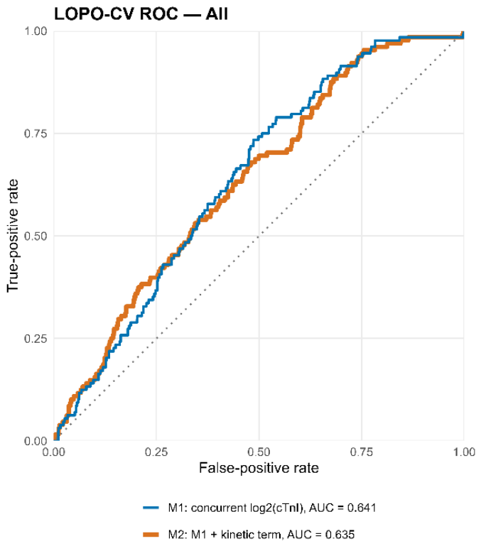
In the overall cohort, concurrent log₂-transformed cTnI demonstrated modest discrimination for ACR ≥2R. The leave-one-patient-out cross-validated AUC for M1 was 0.641 (95% CI 0.592–0.689).
Addition of the time-normalized cTnI kinetic term did not improve discrimination. The corresponding AUC for M2 was 0.635 (95% CI 0.584–0.685), with a paired ΔAUC of −0.007 (95% CI −0.021 to 0.007).
Figure 1.
Receiver Operating characteristic (ROC) curves for model M1 and M2 for overall discrimination.
Figure 1.
Receiver Operating characteristic (ROC) curves for model M1 and M2 for overall discrimination.
3.3. Early Post-Transplant Period
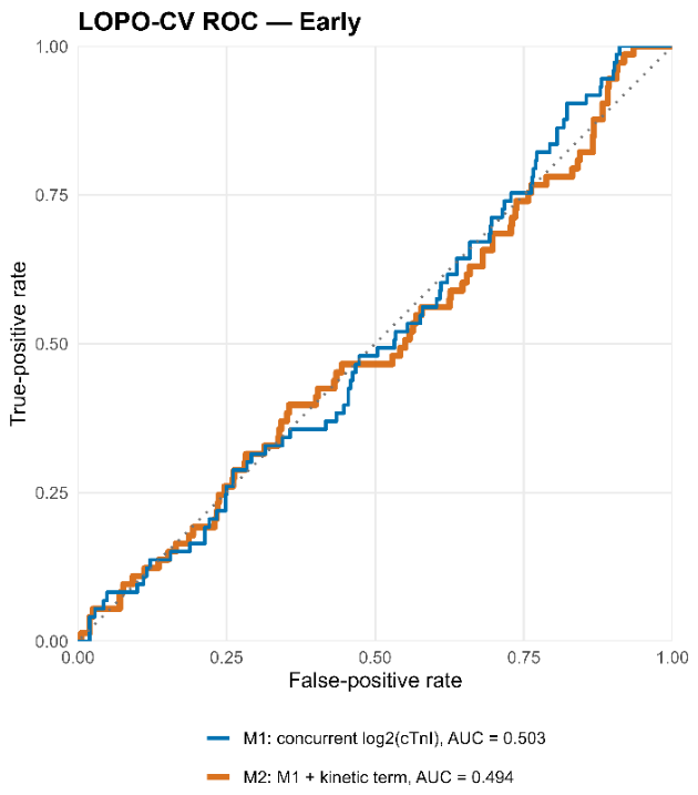
During the first 90 days after transplantation, neither model demonstrated meaningful discrimination. The AUC was 0.503 (95% CI 0.439–0.573) for M1 and 0.494 (95% CI 0.425–0.568) for M2. The paired ΔAUC was −0.009 (95% CI −0.032 to 0.013), indicating no incremental value from the kinetic term.
Figure 2.
Receiver Operating characteristic (ROC) curves for models M1 and M2 in early period (<90 days).
Figure 2.
Receiver Operating characteristic (ROC) curves for models M1 and M2 in early period (<90 days).
3.4. Late Post-Transplant Period
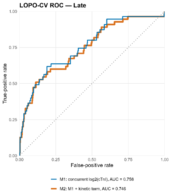
Beyond 90 days after transplantation, concurrent cTnI showed substantially greater discrimination than in the early period. The AUC for M1 was 0.758 (95% CI 0.664–0.838).
Addition of the kinetic term did not improve performance. The AUC for M2 was 0.746 (95% CI 0.654–0.828), with a paired ΔAUC of −0.012 (95% CI −0.037 to 0.008).
Figure 3.
Receiver Operating characteristic (ROC) curves for models M1 and M2 in late period.
3.5. Mixed-Effects Models and Internal Validation
Both models were fitted using patient-specific random intercepts, and neither model demonstrated a singular fit. Predictive performance was evaluated exclusively using leave-one-patient-out cross-validated probabilities. All 2000 patient-level cluster bootstrap resamples yielded valid AUC estimates.
In the full-data mixed-effects models, concurrent log₂-transformed cTnI was positively associated with ACR grade ≥2R. In M1, each two-fold increase in concurrent cTnI was associated with a 21.4% increase in the odds of ACR ≥2R (OR 1.214, 95% CI 1.127–1.309; p < 0.001). After addition of the kinetic term in M2, concurrent cTnI remained independently associated with ACR ≥2R (OR 1.234, 95% CI 1.139–1.336; p < 0.001). In contrast, the time-normalized kinetic term was not independently associated with ACR ≥2R (OR 1.099, 95% CI 0.935–1.293; p = 0.253). Addition of the kinetic term did not significantly improve full-model fit compared with M1 (likelihood-ratio χ²(1) = 1.326; p = 0.249). Patient-level random-intercept variance was 0.431 in M1 and 0.446 in M2, corresponding to standard deviations of 0.657 and 0.668, respectively.
Table 2.
AUC values for models M1 and M2.
| period | M1 | M2 (kinetics) | ΔAUC M2–M1 |
|---|---|---|---|
| All biopsies | 0.641 (0.592–0.689) | 0.635 (0.584–0.685) | −0.007 (−0.021–0.007) |
| ≤90 days | 0.503 (0.439–0.573) | 0.494 (0.425–0.568) | −0.009 (−0.032–0.013) |
| >90 days | 0.758 (0.664–0.838) | 0.746 (0.654–0.828) | −0.012 (−0.037–0.008) |
Table 3.
Full-data mixed-effects logistic regression models for ACR grade ≥2R.
| Model | Effect | β | SE | OR | 95% CI for OR | p-value |
|---|---|---|---|---|---|---|
| M1 | Intercept | −3.297 | 0.260 | 0.037 | 0.022–0.062 | <0.001 |
| M1 | Concurrent log₂(cTnI) | 0.194 | 0.038 | 1.214 | 1.127–1.309 | <0.001 |
| M2 | Intercept | −3.316 | 0.263 | 0.036 | 0.022–0.061 | <0.001 |
| M2 | Concurrent log₂(cTnI) | 0.210 | 0.041 | 1.234 | 1.139–1.336 | <0.001 |
| M2 | Time-normalized log₂(cTnI) change per 7 days | 0.095 | 0.083 | 1.099 | 0.935–1.293 | 0.253 |
β, fixed-effect coefficient on the log-odds scale; SE, standard error; OR, odds ratio; CI, confidence interval; cTnI, cardiac troponin I. For concurrent log₂(cTnI), the OR represents the change in the odds of ACR grade ≥2R associated with a two-fold increase in cTnI. For the kinetic term, a one-unit increase represents a time-normalized increase of one log₂ unit over 7 days. The patient-level random-intercept variance was 0.431 in M1 and 0.446 in M2; the corresponding standard deviations were 0.657 and 0.668.
4. Discussion
The present study yielded three principal findings. First, concurrent cTnI demonstrated modest discrimination for biopsy-confirmed ACR ≥2R in the overall cohort. Second, its performance differed markedly according to time from transplantation: cTnI was non-informative during the first 90 days but showed moderate discrimination thereafter. Third, addition of the time-normalized change in cTnI did not improve discrimination in the overall, early, or late analyses. The point estimates for M2 were slightly lower than those for M1 in all three analyses, and all confidence intervals for ΔAUC included zero. Consistently, concurrent cTnI was independently associated with ACR ≥2R in the full-data mixed-effects models, whereas the kinetic term was not, and its addition did not significantly improve model fit.
Our findings reinforce a growing consensus that cTnI alone has insufficient utility in detecting ACR after HTx. Earlier, smaller studies had been optimistic, for example Patel et al. (2014) reported that a single high-sensitivity cTnI (hs-cTnI) measurement could discriminate ACR (≥2R) with a c-statistic of ~0.82 (95% CI 0.76-0.88), and with a negative predictive value (NPV) of ~99% at cutoff of 15 ng/L [8]. A systematic review in 2018 similarly noted that hs-cTnI assays can achieve very high NPV of 97 - 100% and high sensitivity (82-100%) for ACR, suggesting potential as a “rule out” test [5]. However, more recent large analyses have shown less promising results. Liu et al. (2022) pooled data from 27 studies and found that while troponin levels were modestly elevated in patients with late (≥1 month) rejection, the overall diagnostic accuracy was poor (Bayesian AUC = 0.584) [3]. In the largest single-center series to date, Fitzsimons et al. (2022) found no meaningful association of hs-cTnI with moderate rejection: the overall AUC was ~0.509 (95% CI 0.428-0.591), essentially equivalent to chance [2]. Our results are consistent with these recent studies, showing modest diagnostic utility.
We specifically evaluated whether troponin kinetics could improve diagnostic performance. To our knowledge, this approach has not been extensively studied. One rationale is that, even if the absolute troponin is below some threshold, a rising trend might indicate evolving injury. Conversely, a very high troponin in the early period may not be indicative of ACR, provided that cTnI level is falling in a predictable pattern compared to previous measurements. Patel et al. suggested that serial hs-cTnI monitoring might provide a low-cost rule-out strategy [8], and one could analogize to the delta-troponin strategy in acute coronary syndromes. However in our analysis, the time-normalized kinetic variable provided no incremental value. This finding was consistent across all analyses. One possible explanation is that the concurrent cTnI concentration already captures much of the clinically relevant information contained in the preceding trajectory. In addition, calculation of a rate of change may amplify analytical and biological variability, particularly when measurements are separated by irregular intervals.
Troponin trajectories may also be affected by renal function, infection, hemodynamic instability, non-rejection myocardial injury, and changes in treatment. Consequently, a rise or fall in cTnI is not specific to rejection. These sources of variability may outweigh any additional signal provided by the kinetic term. Our findings therefore do not support the routine use of serial cTnI kinetics as a means of improving rejection detection beyond the concurrent concentration.
The time-dependent performance of cTnI is biologically plausible. During the early post-transplant period, cTnI concentrations are influenced by perioperative myocardial injury [7], ischemia–reperfusion injury, surgical manipulation, hemodynamic instability, and other non-rejection-related processes. These competing sources of cardiomyocyte injury may explain why concurrent cTnI was essentially non-informative during the first 90 days.
Beyond 90 days, the effect of perioperative injury is expected to diminish. In this setting, an elevated cTnI concentration may more specifically reflect new myocardial injury, including injury associated with rejection. This may explain the late-period AUC of 0.758 observed in the present cohort. However, this result should not be interpreted as evidence that cTnI can replace biopsy. The confidence interval remained relatively wide, no clinically actionable threshold was validated, and discrimination alone does not establish adequate sensitivity, specificity, calibration, or clinical utility.
Given numerous limitations, attention has turned to alternative or complementary approaches to noninvasively rule out ACR. One approach is combination biomarkers, for example combining troponin with echocardiographic strain measurements has shown promise: Clemmensen et al. (2022) demonstrated that a decline in LV global longitudinal strain combined with an increase in troponin T or NT-proBNP yielded an NPV of 98% overall and 99% in a selected post-3-month subanalysis, although sensitivity remained limited [6]. Other possibilities are genomic and molecular markers. Donor-derived cell-free DNA (dd-cfDNA) assays have shown high negative predictive value for detecting cardiac allograft rejection [9], although their performance depends on the assay, diagnostic cutoff, sampling period, and definition of rejection. Finally, machine learning and multi-dimensional biomarkers are emerging. Recent studies show that deep analysis of plasma markers can outperform traditional assays. A notable example is extracellular-vesicle (EV) profiling: Burrello et al. [10] used machine-learning to interpret plasma EV surface markers and achieved a leave-one-out AUC of 0.968 (validation AUC 0.832) for identifying ACR, with very high specificity and NPV. This EV-based model “outperformed conventional biochemical markers” and anticipated rejection before histology [10]. However, the study included only 24 transplant recipients, and the independent validation cohort comprised five recipients; therefore, the results require confirmation in larger external cohorts. Approaches that include integrating multiple biomarkers or imaging features via algorithms open up the possibility of noninvasive surveillance that troponin, or any other conventional biomarker alone cannot match.
Limitations
This study has several limitations. First, it was a retrospective, single-center analysis, which may limit generalizability. Second, we did not adjust for potential confounders such as concurrent infections, changes in immunosuppression or renal function, which may influence troponin clearance. Third, the relatively low incidence of moderate/severe rejection reduces the power to detect small diagnostic effects. Fourth, sampling intervals were not standardized across all patients, which may have affected reproducibility. Fifth, cTnI was measured using a conventional (non-high-sensitivity) assay; this lower analytical sensitivity, relative to the high-sensitivity assays used in several previous studies [2,5,8], may partly explain the limited discrimination observed, particularly during the early post-transplant period, and limits direct comparability with those results. In addition biopsy intervals varied widely and slope estimates are inherently less stable with long or irregular intervals. Finally, the early and late-period analyses were exploratory, and no formal statistical comparison of AUCs between the two periods was performed. Therefore, the observed difference should be interpreted as a descriptive time-related pattern rather than definitive evidence of effect modification. These limitations should be considered when interpreting the negative findings and motivate prospective, standardized studies to definitively address whether troponin kinetics have clinical utility. It is important to note that as long as EMBs remain the reference standard, certain biases such as long or irregular sampling intervals are inherent.
5. Conclusions
In this single-center cohort, the diagnostic performance of concurrent cTnI was strongly dependent on time from transplantation. cTnI was non-informative during the first 90 days but demonstrated moderate discrimination for ACR ≥2R thereafter. Nevertheless, addition of a time-normalized kinetic term did not improve discrimination over the concurrent concentration in any analyzed period. These findings do not support the use of cTnI kinetics for rejection surveillance. Concurrent cTnI may warrant further evaluation as an adjunctive late-period risk marker, but neither cTnI concentration nor its kinetics can currently replace endomyocardial biopsy.
Author Contributions
Conceptualization, B.B. and M.Z.; methodology, M.O., B.B. and M.Z.; software, M.O.; validation, M.O.; formal analysis, M.O.; investigation, M.O. and B.B.; resources, M.O.; data curation, M.O., B.B., M.C. and M.Z.; writing—original draft preparation, M.O. and B.B.; writing—review and editing, M.C. and M.Z.; visualization, M.O. and M.Z.; supervision, M.C. and M.Z.; project administration, M.O.; funding acquisition, M.C. All authors have read and agreed to the published version of the manuscript.
Funding
This research was funded by internal statutory funds of Wroclaw Medical University.
Institutional Review Board Statement
Ethical review and approval were not required for this study, in accordance with local legislation and institutional requirements. The study was conducted retrospectively using routinely collected clinical data that were fully anonymized prior to analysis. No intervention was performed on human subjects, and no individually identifiable information was accessed or reported.
Informed Consent Statement
Patient consent was waived due to the retrospective, non-interventional design of the study and the use of fully anonymized data.
Data Availability Statement
The data supporting the findings of this study are not publicly available due to patient confidentiality, ethical restrictions, and institutional data protection regulations. Access to appropriately anonymized data may be considered upon reasonable request to the corresponding author and subject to approval by the relevant institution and ethics committee.
Conflicts of Interest
The authors declare no conflict of interest.
References
- Nikolova A, Agbor-Enoh S, Bos S, Crespo-Leiro M, Ensminger S, Jimenez-Blanco M, Minervini A, Perch M, Segovia J, Vos R, Khush K, Potena L. European Society for Organ Transplantation (ESOT) Consensus Statement on the Use of Non-invasive Biomarkers for Cardiothoracic Transplant Rejection Surveillance. Transpl Int. 2024 Jun 11;37:12445. [CrossRef] [PubMed] [PubMed Central]
- Fitzsimons SJ, Evans JDW, Rassl DM, Lee KK, Strachan FE, Parameshwar J, Mills NL, Pettit SJ. High-sensitivity Cardiac Troponin Is Not Associated With Acute Cellular Rejection After Heart Transplantation. Transplantation. 2022 May 1;106(5):1024-1030. Epub 2021 Jun 29. [CrossRef] [PubMed]
- Liu Z, Perry LA, Penny-Dimri JC, Handscombe M, Overmars I, Plummer M, Segal R, Smith JA. Elevated Cardiac Troponin to Detect Acute Cellular Rejection After Cardiac Transplantation: A Systematic Review and Meta-Analysis. Transpl Int. 2022 Jun 8;35:10362. [CrossRef] [PubMed] [PubMed Central]
- Velleca A, Shullo MA, Dhital K, Azeka E, Colvin M, DePasquale E, Farrero M, García-Guereta L, Jamero G, Khush K, Lavee J, Pouch S, Patel J, Michaud CJ, Shullo MA, Schubert S, Angelini A, Carlos L, Mirabet S, Patel J, Pham M, Urschel S, Kim KH, Miyamoto S, Chih S, Daly K, Grossi P, Jennings DL, Kim IC, Lim HS, Miller T, Potena L, Velleca A, Eisen H, Bellumkonda L, Danziger-Isakov L, Dobbels F, Harkess M, Kim D, Lyster H, Peled Y, Reinhardt Z. The International Society for Heart and Lung Transplantation (ISHLT) guidelines for the care of heart transplant recipients. J Heart Lung Transplant. 2023 May;42(5):e1-e141. Epub 2022 Dec 20. [CrossRef] [PubMed]
- Fitzsimons S, Evans J, Parameshwar J, Pettit SJ. Utility of troponin assays for exclusion of acute cellular rejection after heart transplantation: A systematic review. J Heart Lung Transplant. 2018 May;37(5):631-638. Epub 2017 Dec 11. [CrossRef] [PubMed]
- Clemmensen TS, Firooznia N, Olawi FM, Løgstrup BB, Poulsen SH, Eiskjær H. Assessment of Acute Rejection by Global Longitudinal Strain and Cardiac Biomarkers in Heart-Transplanted Patients. Front Immunol. 2022 Mar 25;13:841849. [CrossRef] [PubMed] [PubMed Central]
- Zimmermann R, Baki S, Dengler TJ, Ring GH, Remppis A, Lange R, Hagl S, Kübler W, Katus HA. Troponin T release after heart transplantation. Br Heart J. 1993 May;69(5):395-8. [CrossRef] [PubMed] [PubMed Central]
- Patel PC, Hill DA, Ayers CR, Lavingia B, Kaiser P, Dyer AK, Barnes AP, Thibodeau JT, Mishkin JD, Mammen PP, Markham DW, Stastny P, Ring WS, de Lemos JA, Drazner MH. High-sensitivity cardiac troponin I assay to screen for acute rejection in patients with heart transplant. Circ Heart Fail. 2014 May;7(3):463-9. Epub 2014 Apr 14. [CrossRef] [PubMed]
- Kim PJ, Olymbios M, Siu A, Wever Pinzon O, Adler E, Liang N, Swenerton R, Sternberg J, Kaur N, Ahmed E, Chen YA, Fehringer G, Demko ZP, Billings PR, Stehlik J. A novel donor-derived cell-free DNA assay for the detection of acute rejection in heart transplantation. J Heart Lung Transplant. 2022 Jul;41(7):919-927. Epub 2022 Apr 10. [CrossRef] [PubMed] [PubMed Central]
- Burrello J, Panella S, Barison I, Castellani C, Burrello A, Airale L, Goi J, Dusi V, Frigerio R, Gerosa G, Tessari C, Pradegan N, Toscano G, Pedrazzini G, Corianò M, Tona F, Bolis S, Gori A, Cretich M, Fedrigo M, Angelini A, Barile L. Machine learning-assisted assessment of extracellular vesicles can monitor cellular rejection after heart transplant. Commun Med (Lond). 2025 Jul 11;5(1):288. [CrossRef] [PubMed] [PubMed Central]
Disclaimer/Publisher’s Note: The statements, opinions and data contained in all publications are solely those of the individual author(s) and contributor(s) and not of MDPI and/or the editor(s). MDPI and/or the editor(s) disclaim responsibility for any injury to people or property resulting from any ideas, methods, instructions or products referred to in the content. |
© 2026 by the authors. Licensee MDPI, Basel, Switzerland. This article is an open access article distributed under the terms and conditions of the Creative Commons Attribution (CC BY) license (http://creativecommons.org/licenses/by/4.0/).
Copyright: This open access article is published under a Creative Commons CC BY 4.0 license, which permit the free download, distribution, and reuse, provided that the author and preprint are cited in any reuse.


