

Submitted:
23 July 2026
Posted:
24 July 2026
You are already at the latest version
Abstract
Signal peptide peptidase-like 2A (SPPL2A) is an intramembrane protease identified as a binding partner of intraflagellar transport protein 27 (IFT27), suggesting a potential role in sperm flagellum development. However, the localization and trafficking of SPPL2A during spermiogenesis remain unknown. Here, we generated an endogenous SPPL2A–mCherry knock-in mouse model using CRISPR/Cas9 to visualize SPPL2A in living male germ cells. Expression of the fusion protein was confirmed by Western blotting, and fertility, spermatogenesis, sperm morphology, and motility were evaluated. Live-cell fluorescence imaging, immunofluorescence, and single-particle tracking (SPT) were used to characterize SPPL2A localization and trafficking. Homozygous knock-in mice exhibited normal fertility, spermatogenesis, and sperm parameters, indicating that the mCherry fusion did not disrupt SPPL2A function. SPPL2A–mCherry was detected as punctate structures in developing germ cells and elongating sperm tails. Its localization changed dynamically during spermiogenesis and was closely associated with developing tail microtubules. Live-cell SPT demonstrated bidirectional movement of SPPL2A-containing particles within developing sperm tails. Mean-squared displacement analysis indicated predominantly confined diffusion with a confinement length scale of 153 ± 15 nm, while particle trajectories showed a slight bias away from the cell body. Particle mobility progressively decreased during sperm maturation, with highly mobile puncta in early spermatids becoming largely immobile in mature sperm. These findings establish the first endogenous SPPL2A fluorescent knock-in mouse model and reveal that SPPL2A undergoes regulated intraflagellar trafficking during spermiogenesis. This model provides a valuable platform for investigating protein transport during sperm flagellum assembly.
Keywords:
SPPL2A
; spermiogenesis
; sperm flagellum
; intraflagellar transport
; single-particle tracking
1. Introduction
Motile cilia and flagella are specialized organelles composed of microtubules, crucial for cellular movement, fluid dynamics, and signal transduction [1]. The sperm flagellum in male germ cells is essential for sperm motility and successful fertilization. The creation relies on the meticulously coordinated assembly of axonemal structures, auxiliary cytoskeletal components, and membrane-associated proteins throughout spermiogenesis [2,3]. Furthermore, because mature sperm flagella are devoid of intrinsic protein synthesis capabilities, the accurate transfer of structural and regulatory proteins from the cytoplasm to the developing flagellum is fundamentally dependent on intraflagellar transport (IFT) [4].
IFT is facilitated by two multimeric protein complexes, IFT-A and IFT-B, which collaborate with kinesin-2 and dynein-2 motors to enable bidirectional transport along axonemal microtubules [4,5,6]. In somatic cilia, IFT is essential for ciliary assembly, maintenance, and signaling. In spermatogenesis, mounting evidence suggests that IFT is essential for the creation of the sperm flagellum, including axonemal elongation, fibrous sheath organization, and mitochondrial sheath assembly [7]. Moreover, genetic mutations in various IFT components, including IFT27, IFT20, IFT25, and IFT172, have been demonstrated to result in aberrant flagellar morphogenesis and male infertility in both humans and animal models, highlighting the essential function of IFT in spermiogenesis [8,9,10].
IFT27, a small Rab-like GTPase in the IFT-B complex, is crucial for regulating IFT train modification, BBSome recruitment, and the loading and turnover of membrane-associated cargos during cilia and flagella formation [11,12]. In male germ cells, IFT27 is crucial for sperm flagella synthesis; its deficiency results in defective axonemal elongation, abnormal accessory structures, and complete male sterility [8]. Our previous yeast two-hybrid screening identified SPPL2A as a potential binding partner of IFT27. Signal peptide peptidase-like 2A (SPPL2A) is an intramembrane aspartyl protease belonging to the signal peptide peptidase-like (SPPL) family [13]. Extensive research on immune cells has demonstrated that SPPL2A regulates the maturation of B-cells and dendritic cells through the intramembrane cleavage of type II transmembrane proteins [14,15]. Nevertheless, despite its well-defined role in the immune system, the expression profile and biological significance of SPPL2A in the male reproductive system remain largely unexplored.
The relationship between SPPL2A and IFT27 indicates an undiscovered connection among intramembrane proteolysis, vesicle trafficking, and IFT-mediated flagellar assembly. The endogenous distribution, developmental dynamics, and potential trafficking behavior of SPPL2A in spermatogenic cells are entirely unknown, creating a substantial knowledge gap that hinders our comprehension of the processing and delivery of membrane-associated cargos during sperm flagellum formation.
2. Materials and Methods
2.1. Ethics Statement
All animal procedures received approval from the Institutional Animal Care and Use Committee (IACUC) at Wayne State University and were executed in compliance with the National Institutes of Health Guide for the Care and Use of Laboratory Animals.
2.2. Generation of SPPL2A-mCherry Knock-In Mice
The SPPL2A-mCherry knock-in (KI) mouse model was generated using the CRISPR/Cas9 system at the Transgenic Animal Model Core of the University of Michigan. The mouse SPPL2A protein was fused with an mCherry tag at the C-terminus. Briefly, the mCherry coding sequence, followed by a bovine growth hormone polyadenylation (bGH PA) signal, was inserted immediately before the stop codon within exon 15 of the Sppl2a gene. The DNA donor was modified to prevent Cas9 re-cleavage after homologous recombination. To improve long single-stranded DNA synthesis efficiency, the poly-T repeat within the donor was shortened, and multiple silent wobble-base substitutions were introduced into the mCherry sequence to reduce GC content.
A validated single-guide RNA (sgRNA C328G1) targeting the terminal exon of Sppl2a was selected. Cas9 was used to introduce a double-strand break near the endogenous termination codon of Sppl2a. Homology-directed repair (HDR) of the chromosome break using a long single-stranded DNA donor placed the mCherry-bGH PA cassette immediately upstream of the termination codon. The sgRNA was tested and confirmed to be active prior to in vivo microinjection.
To identify founder mice, genomic DNA was extracted, and PCR genotyping was performed using primers spanning the knock-in region. The primer pair used for screening was: Sppl2a-mCherry span Forward: 5′-GCCTCAAATGTACATCCACATATGCAAAA-3′. Sppl2a-mCherry span Reverse: 5′-TCGCTGTAAATGTTCCCAATTTTCAACTC-3′. PCR amplification generated a 1356 bp product from the knock-in allele, whereas no corresponding band was detected in wild-type (WT) controls. The founder mice carrying the correctly targeted knock-in allele were subsequently crossed with WT mice to establish the SPPL2A-mCherry KI lines (F1 generation). These F1 generation were crossed again to the WT mice and each line was examimed by Western blot analysis to see if the fusion SPPL2A-mCherry protein was expressed.
2.3. Male Fertility Test
2-3-month-old KI male mice and age-matched WT controls were used for fertility assessment. Each male was housed with a sexually mature WT female (2–3-months-old) in a one-to-one mating scheme for at least 4 consecutive weeks. Females were examined daily for the presence of vaginal plugs, and successful pregnancy was monitored thereafter. Once pregnancy was confirmed, females were separated into individual cages. The number of pregnant females and litter size per delivery were recorded to evaluate male fertility.
2.4. Spermatozoa Counting and Motility Assay
Spermatozoa were obtained from the cauda epididymides and allowed to disperse in pre-warmed phosphate-buffered saline (PBS). For sperm counting, aliquots of sperm suspensions were fixed in 4% formaldehyde for 10 min at room temperature, washed twice with PBS, and then loaded onto a hemocytometer chamber. Sperm concentration was calculated according to standard procedures.
For sperm motility analysis, freshly isolated spermatozoa were maintained under non-capacitating conditions and examined using an inverted microscope (Nikon, Tokyo, Japan). Time-lapse images were recorded at 15 frames per second, and ten randomly selected fields were analyzed for each sample. Individual sperm tracks were analyzed to calculate curvilinear velocity (VCL), defined as the curvilinear distance (DCL) traveled by a sperm cell per second (VCL = DCL/t). In parallel, sperm motility parameters were additionally assessed using an IVOS II computer-assisted sperm analysis (CASA) system (Hamilton Thorne Inc., MA, USA).
2.5. Western Blot Analysis
Testicular tissues were lysed in ice-cold lysis buffer using an Ultra-Turrax tissue homogenizer, followed by brief sonication with an ultrasonic processor (Qsonica, CT, USA) at 70% amplitude for 10 s. The lysates were kept on ice for 30 min and subsequently centrifuged at 12,000 rpm for 20 min at 4 °C. Supernatants were collected, and protein concentrations were determined using a DC protein assay kit (Bio-Rad, CA, USA). Equal amounts of protein were denatured by heating, resolved on 12% SDS–polyacrylamide gels, and transferred onto PVDF membranes (MilliporeSigma, MA, USA). The membranes were blocked with 5% non-fat milk at room temperature for 1 h, followed by incubation at 4 °C overnight with primary antibodies (mCherry: Cat. No. 56808-1-Ig, Proteintech, 1:1000; β-actin: Proteintech, 1:10000). After extensive washing, membranes were incubated with appropriate HRP-conjugated secondary antibodies (goat anti-mouse IgG (H+L), Cat. No. 626520; goat anti-rabbit IgG (H+L), Cat. No. 656120; Invitrogen; both at 1:2000 dilution) for 1 h at room temperature. Chemiluminescent signals were visualized using a Pico Ultra ECL Western blot detection kit (Lamda Biotech Corporation, Ballwin, MO, USA; Cat. No. G075).
2.6. Fluorescence Analysis of Testicular Cells
Testes were freshly dissected from adult mice and immediately fixed in 4% paraformaldehyde in 0.1 M PBS (pH 7.4) overnight at 4 °C. For fresh germ cell isolation, testes were enzymatically digested in 0.5 mg/mL collagenase IV and 1.0 μg/mL DNase I (Sigma-Aldrich) at 32 °C for 30 min with gentle stirring to release individual germ cells. Freshly isolated germ cells were immediately diluted in PBS and observed by spinning-disk confocal microscopy (Andor Dragonfly 201 ) at room temperature. For fixed-cell imaging, a subset of cells was fixed with 4% paraformaldehyde containing 0.1 M sucrose at room temperature prior to imaging. After dehydration through a graded ethanol series, tissues were embedded in paraffin, and 5-μm sections were prepared using a Leica paraffin microtome. Sections were dewaxed in xylene and rehydrated through a series of descending ethanol concentrations. For antigen retrieval, sections were heated in either 10 mM Tris–EDTA buffer (pH 9.0) or 10 mM sodium citrate buffer (pH 6.0) for 30 min, followed by cooling to room temperature. After rinsing in PBS, sections were blocked with 10% goat serum at room temperature for 1 h and incubated overnight at 4 °C with the appropriate primary antibodies diluted in blocking buffer. After washing with PBS, sections were incubated with corresponding Alexa Fluor–conjugated secondary antibodies for 1 h at room temperature in the dark. For acrosome staining, sections were incubated with Alexa Fluor 488–conjugated peanut agglutinin (PNA-488) (1:4000 dilution) for 1 h at room temperature after serum blocking. Nuclei were counterstained with DAPI, and sections were mounted using an anti-fade mounting medium.
2.7. Fluorescence Imaging, FRAP, and Single-Particle Tracking Analyses
Fluorescence and brightfield images were captured using a spinning-disk confocal microscope (Andor Dragonfly 201) mounted on an inverted microscope (Leica DMI8) with a 63× oil immersion objective (Leica HC PL APO) and an sCMOS camera (Andor Zyla). Consistent acquisition settings were used for all color channels, and the images were uniformly displayed with the same brightness and contrast adjustments.
Fluorescence recovery after photobleaching (FRAP) and kymograph data were collected using an Olympus IX83 inverted epifluorescence microscope equipped with an Andor Zyla sCMOS camera. Illumination was provided either by a Teledyne Sola SE LED system or by a Coherent Sapphire continuous wave 561-nm laser, and the samples were viewed with oil-immersion objectives at magnifications of 60× (Olympus UPlanFlN) or 100× (Olympus UApoN). The microscope and image acquisition were controlled using Micro-Manager software, while custom Python scripts were used for image analysis [16].
For single-particle tracking analysis, cells were rinsed with HEPES and pipetted onto a 35 mm glass-bottom plate for imaging to assess SPPL2 trafficking. Brightfield and epifluorescence imaging were performed on an inverted microscope (IX83, Evident Scientific, Inc.) equipped with a 100x, 1.49 NA objective and a 2x magnification lens. Fluorescence illumination was performed with a free-space 15-mW, 561-nm laser (OBIS, Coherent Corp.), cleaned by a bandpass filter (ZET405/488/561/647X, Chroma Technology Corp.), reflected by a quad-band dichroic mirror (ZT405/488/561/647RPC, Chroma Technology Corp.), and distributed across a 2000 µm2 sample area. Emission passed through a 525/50M emission filter (Evident Scientific, Inc.) and a 4-band notch filter (ZET405/488/561/640m; Chroma Technology Corp.) before being detected by an EMCCD camera (iXon 897-Ultra, Oxford Instruments Andor) with 150 EM gain, 20 ms exposure, and 47 Hz frame rate.
ImageJ and custom Python scripts analyzed videos >15 s in duration to extract and fit kymographs. The linking and preliminary analysis of the single-particle tracking data were performed with TrackPy. Mean-squared displacement (MSD) versus delta time () curves were fit with a 1D two-regime model that accounted for localization uncertainty (), a short-time diffusion rate with minimal restrictions to motion (), a long-time diffusion with motion affected by confinement of SPPL2A–mCherry to tethering molecules or similar structures (), and a crossover time between these regimes (), according to Eq. (1):
2.8. Statistical Analyses
For this research, we utilized GraphPad Prism software to conduct the statistical analyses. The normality of the dataset was verified using the Shapiro–Wilk test, while the homogeneity of variances among groups was established through the Levene test. Provided that the quantitative data met the criteria for normal distribution and equal variances, the Student t-test was applied. A result was considered statistically significant when the P-value was below 0.05.
3. Results
3.1. Generation of SPPL2A-mCherry KI Mice
The knock-in strategy employed to generate the KI mouse model is illustrated in Figure 1A and Supplementary Figure S1. Genotyping was performed using primers flanking the C-terminal region of exon 15, yielding PCR products of 409 bp for the wild-type allele and 1356 bp for the knock-in allele (Figure 1A and Supplementary Figure S2). Westerb blot was conducted using testicular extracts from each line with an anti-mCherry antibody to examine expression of SPPL2A-mCherry fusion protein. Like the WT mice, some lines did not express fusion protein and these lienes were discarded. Some lines expressed mCherry-fusion proteins and these lines were maintained for the further experiments. It is interesting, these positive lines expressed two fusion proteins, one was about 75 kDa corresponding to the fused full-length (SPPL2A-201 in Supplemental Figure S1A) SPPL2A-mCherry protein; the 50 kDa fusion protein might correspond to the fused SPPL2A-203 (Figure 1B, Supplemental Figure S1A).
Homozygous KI mice exhibited normal development during routine observations, with some animals monitored for up to eight months (Figure 1C). Throughout the study, the gross morphology of the testes in both WT and KI mice remained comparable (Figure 1D, upper panel). Under fluorescence microscopy, strong red fluorescent signals were clearly visible in the testes of KI mice, whereas WT controls showed no fluorescence, confirming that mCherry had been successfully fused to the endogenous SPPL2A protein (Figure 1D, lower panel).
3.2. Fertility and Sperm Parameters Were Normal in the KI Mice
To evaluate the reproductive capability of the KI mice, 2-month-old homozygous KI males and age-matched WT males were each paired with 2-month-old WT females. The KI males produced litters with normal pregnancy rates and average litter sizes compared with WT controls (Table 1), indicating that mCherry insertion did not impair male fertility.
Spermatozoa collected from the cauda epididymides of WT and homozygous KI males were examined for quantitative and qualitative parameters. Morphological assessment of spermatozoa further demonstrated that sperm structure appeared normal in 2KI mice, with no detectable abnormalities in the head, midpiece, or flagellum (Figure 2A, B). Total sperm counts did not differ significantly between KI mice and WT littermates (Figure 2C). Likewise, sperm motility, assessed under non-capacitating conditions and quantified by curvilinear velocity, showed no significant reduction in KI sperm (Figure 2D, 2E, Supplemental Movies S1 and S2). Consistent with these findings, additional CASA-derived kinematic parameters, including average path velocity (VAP), straight-line velocity (VSL), amplitude of lateral head displacement (ALH), beat-cross frequency (BCF), linearity (LIN), straightness (STR), and wobble (WOB), did not differ significantly between WT and KI sperm (Table 2).
3.3. Normal Testis and Epididymis Histology in the SPPL2A-mCherry KI Mice
To further investigate the spermatogenic process in the knock-in (KI) mice, a morphological examination of the testes and epididymides was carried out. Measurements revealed that both the testis weight and the ratio of testis weight to body weight in homozygous KI mice were comparable to those observed in WT controls (Figure 1D and Figure 3A). Subsequent histological assessment using paraffin-embedded sections and hematoxylin and eosin staining demonstrated that the arrangement of germ cells in the seminiferous tubules of adult KI mice was regular and well-organized, maintaining a stratified structure indistinguishable from that of WT mice (Figure 3B). Moreover, the epididymal lumen in KI mice was densely populated with mature spermatozoa (Figure 3C).
3.4. mCherry Signal Is Detecyted in Testicular Cells in KI Mice
To determine whether the mCherry signal was detected in testicular cells, freshly isolated testicular cells from WT and KI mice were examined by fluorescence microscopy. No specific fluorescence signal was detected in the WT mice. In contrast, mCherry fluorescence was readily detectable in the KI mice under low magnification (Supplementary Figure S3). Intracellular mCherry signal was further examined under high magnification. mCherry signal appeared as punctate structures within the cytoplasm and were detected in elongating spermatids. Fluorescence was also observed along developing tails (Figure 4).
3.5. Localization of SPPL2A–mCherry During Spermatid Development
To characterize the developmental distribution of SPPL2A during spermiogenesis, isolated spermatids representing different maturation stages were analyzed by fluorescence microscopy. SPPL2A-mCherry fluorescence was detectable from early round spermatids (steps 1–3) and persisted throughout subsequent stages of spermatid differentiation (Figure 5). In early spermatids, SPPL2A-mCherry appeared as discrete puncta adjacent to the nucleus. As spermiogenesis progressed, the fluorescence became increasingly concentrated within the cytoplasmic compartment surrounding the elongating nucleus and remained detectable through the final stages of spermatid maturation, indicating sustained expression throughout spermiogenesis.
To further define the subcellular localization of SPPL2A during spermatid elongation, isolated elongating spermatids were co-stained with PNA (Figure 5A), α-tubulin (Figure 5B), and ץ-tubulin (Figurte 5C), markers of acrosome, manchette and centrioles respectively. SPPL2A–mCherry fluorescence was observed in the acrosome in early spermatids as indicated co-localization with PNA (Figure 5A). With development of spermatids, the signal seemed to migrate to the cauda region of the cells. Some signals were co-localized with α-tubulin, indicating a manchette localization (Figure 5B). Some signals were present as dots in the opposite region of the acrosome. Double stained with an anti- ץ-tubulin antibody showed co-localization of ץ-tubulin and SPPL2A-mnCherry, indicating that SPPL2A is present in the centtrioles (Figure 5C).
3.6. SPPL2A-mCherry Trafficking in the Developing Sperm Tails
The diffusion and drift analysis of SPPL showed that SPPL2 primarily behaved like a random, confined diffuser within the developing sperm trail. However, a slight and statistically significant bias in the SPPL2 motion occurred away from the cell body. A non-linear least square fitting of the MSD vs to a two-regime diffusion model, as shown Eq. 1, yielded = 0.0038 ± 0.0004 µm²/s, = 0.0018 ± 0.0002 µm²/s, = 3.1 ± 0.5 s, and = 74 ± 12 nm. This diffusion rate is significantly slower than expected for a small, free vesicle or protein cluster, and is consistent with highly confined or restricted motion. Additionally, the transition between and a confinement length scale of 153 ± 15 nm indicates a sub-diffraction-limited barrier to SPPL2 motion within the tail (Figure 6).
Analysis of the average step direction revealed a subtle but significant motion of SPPL2 away from the cell body. At short time intervals ( = 0.5 s), the displacement distribution shifted toward positive values, indicating preferential movement toward the distal region of the developing tail (p = 0.03). This directional bias became more pronounced at longer time intervals (= 7 s), with particles exhibiting significantly greater displacement away from the cell body (p = 0.0002). Representative time-lapse imaging of SPPL2A–mCherry particle movement is shown in the accompanying video, with additional examples provided (Supplementary Movies S3 and S4).
4. Discussion
In the present study, we generated a SPPL2A-mCherry knock-in mouse model to investigate the localization and trafficking behavior of SPPL2A during spermatogenesis. Homozygous knock-in mice displayed normal spermatogenesis, sperm morphology, and fertility, indicating that the mCherry tag did not disrupt the physiological function of SPPL2A. Using live-cell imaging and single-particle tracking, we observed that SPPL2A forms punctate structures within developing sperm flagella and undergoes dynamic trafficking along the elongating tail. Quantitative trajectory analysis further revealed that SPPL2A exhibits restricted diffusion with a defined confinement length scale, as well as a slight directional bias away from the cell body. These observations demonstrate that SPPL2A is not statically localized within the developing sperm flagellum but rather participates in dynamic transport processes during spermiogenesis. Since the formation of sperm flagella requires coordinated delivery of structural and regulatory proteins to the elongating axoneme, the dynamic trafficking behavior of SPPL2A suggests that it may play a role in the intracellular transport pathways involved in flagellar assembly. Motile cilia and flagella are highly specialized microtubule-based organelles that require precise spatial and temporal coordination of protein transport for their assembly and maintenance [1,4].
The assembly of cilia and flagella is critically dependent on IFT, a conserved bidirectional transport system that delivers cargo proteins along axonemal microtubules [4,5,8]. IFT is mediated by two large protein complexes, IFT-A and IFT-B, which cooperate with kinesin-2 and dynein-2 motors to transport cargo toward the distal tip and back toward the basal body, respectively [10]. In the context of spermatogenesis, several studies have demonstrated that disruption of IFT components leads to severe defects in sperm flagellar assembly and male infertility. For example, genetic ablation of IFT25, IFT27, or IFT172 results in abnormal axoneme formation and defective spermiogenesis in mice[8,9]. Notably, IFT27 functions as a small Rab-like GTPase that regulates the loading and turnover of membrane-associated cargo within the IFT complex [11]. In our previous yeast two-hybrid screening, SPPL2A was identified as a potential binding partner of IFT27, suggesting a possible link between SPPL2A and IFT-mediated trafficking. The dynamic localization of SPPL2A observed in the present study therefore supports the hypothesis that SPPL2A may participate in membrane-associated cargo transport during sperm flagellar assembly.
To investigate the localization and trafficking behavior of endogenous SPPL2A during spermatogenesis, we generated a SPPL2A–mCherry knock-in mouse model. Traditional approaches for studying protein localization in germ cells often rely on antibody-based immunostaining or small epitope tags such as HA, FLAG, or V5. While these strategies are valuable for determining static localization patterns, they do not permit direct visualization of protein dynamics in living cells [18]. In contrast, fluorescent protein knock-in models enable real-time monitoring of endogenous proteins under physiological expression levels and have become powerful tools for studying intracellular trafficking processes [19,20,21]. Fluorescently tagged mouse models have been widely used in cilia and flagella research, including ARL13B–mCherry, Centrin2–GFP, and several IFT reporter lines, providing important insights into ciliary assembly and protein transport [22,23]. Because sperm flagellar formation is a highly dynamic process involving continuous cargo movement along microtubule-based structures, direct visualization of endogenous proteins represents a significant advantage over conventional fixed-cell approaches.
Notably, homozygous SPPL2A–mCherry mice displayed normal spermatogenesis, sperm morphology, and fertility, indicating that fusion of mCherry to SPPL2A did not substantially impair its physiological function. This observation is particularly relevant because fluorescent proteins are considerably larger than conventional epitope tags and may potentially interfere with protein folding, trafficking, or enzymatic activity. The normal reproductive phenotype, therefore, supports the use of this knock-in model for studying endogenous SPPL2A behavior during spermatogenesis. Examination of isolated germ cells revealed that SPPL2A–mCherry was organized into discrete punctate structures rather than displaying diffuse cytoplasmic localization. The punctate distribution of SPPL2A is noteworthy because proteins undergoing active intracellular transport are frequently assembled into cargo-containing particles rather than remaining freely dispersed within the cytoplasm. Similar punctate structures have been described for multiple components of the IFT machinery and for cargo proteins undergoing microtubule-dependent trafficking during spermatid differentiation. Furthermore, SPPL2A became progressively enriched during the elongation phase of spermiogenesis, coinciding with the onset of manchette formation and rapid flagellar assembly [24,25]. This temporal relationship indicates that recruitment of SPPL2A is developmentally regulated and may be linked to the increased demand for protein transport that accompanies sperm tail biogenesis. Importantly, co-localization with α-tubulin demonstrated a gradual redistribution of SPPL2A from the perinuclear region toward microtubule-rich manchette and flagellar structures. Because the manchette functions as a major transport platform that mediates intramanchette transport (IMT) and coordinates delivery of proteins to the developing sperm tail [2,24,25], the observed spatial transition of SPPL2A is consistent with its incorporation into microtubule-dependent trafficking pathways.
Consistent with this interpretation, Single-particle tracking analysis revealed that SPPL2A exhibits slow diffusion and confined motion within the developing sperm tail. The measured diffusion coefficients were considerably lower than those expected for freely diffusing cytosolic proteins. In addition, mean-squared displacement analysis identified a confinement length scale of 153 ± 15 nm, indicating that SPPL2A movement is spatially restricted within nanoscale domains of the flagellum. Such confined diffusion behavior is commonly observed for proteins associated with membrane microdomains or multiprotein complexes, rather than freely diffusing molecules [26]. In ciliary membranes, many signaling and transport proteins display restricted motion due to interactions with underlying axonemal structures or transport machinery such as IFT particles [2,5]. Therefore, the diffusion properties observed for SPPL2A are consistent with a model in which the protein associates with membrane-associated transport complexes within the developing sperm flagellum.
Although SPPL2A particles displayed bidirectional movement along the developing flagellum, trajectory analysis revealed a subtle directional bias away from the cell body. In motile cilia and flagella, anterograde transport from the basal body to the distal tip is primarily driven by kinesin-2 motors, whereas retrograde transport is mediated by dynein-2 motors [27,28]. The slight directional preference observed in our analysis may therefore reflect preferential association of SPPL2A with anterograde trafficking pathways during flagellar elongation. During spermiogenesis, continuous delivery of proteins to the distal tip is required to support the rapid extension of the flagellum and the assembly of axonemal and accessory structures [2,29]. The observed trafficking behavior of SPPL2A may thus represent a component of the cargo delivery system that supplies membrane-associated proteins to the growing flagellum [27,30].
SPPL2A is a member of the signal peptide peptidase-like (SPPL) family of intramembrane aspartyl proteases, which regulate diverse cellular processes through cleavage of type II transmembrane proteins[13,31]. Beyond its established proteolytic activity, accumulating evidence indicates that SPPL2A is closely associated with membrane protein trafficking and turnover through regulation of endosomal transport pathways[31,32]. The dynamic localization and transport behavior observed in the present study therefore suggest that SPPL2A may serve functions beyond proteolytic processing during spermiogenesis. Given its localization within developing sperm flagella and its apparent association with transport-related structures, SPPL2A may contribute to the trafficking, maturation, or turnover of membrane-associated proteins required for flagellar assembly. Although the molecular substrates of SPPL2A in germ cells remain unknown, our findings raise the possibility that intramembrane proteolysis and protein transport may be functionally coordinated during sperm tail formation.
Our findings demonstrate that SPPL2A undergoes dynamic trafficking within developing sperm flagella, where it forms punctate structures and exhibits confined diffusion behavior consistent with association with membrane-associated transport complexes. These observations suggest that SPPL2A may represent a previously unrecognized component of the molecular machinery regulating protein trafficking during sperm flagellar assembly. Future studies will be required to determine whether SPPL2A directly interacts with components of the IFT machinery and to identify its potential substrates within developing sperm flagella. Elucidating these mechanisms may provide new insights into the coordination between intramembrane proteolysis and intraflagellar transport during spermiogenesis, thereby advancing our understanding of the molecular basis of sperm flagellar formation and male fertility.
Supplementary Materials
The following supporting information can be downloaded at the website of this paper posted on Preprints.org.
Funding
This research was funded by the NIH, grant numbers HD105944 and HD114311.
Author contributions
ZZ designed and supervised the study; ZC performed most of the experiments; WL, SP, AY, KB, JX, JZ, ZL, and CK performed the experiments and analyzed the data; ZC wrote the first draft of the manuscript; LZ, CK, and ZZ discussed the results and edited the manuscript. All authors agree on the order of authors.
Institutional Review Board Statement
The animal study protocol was approved by the Institutional Review Board (or Ethics Committee) of Wayne State University (protocol code 24-02-6561 and date of approval 5/31/2024).
Informed Consent Statement
Not applicable
Data Availability Statement
The original contributions presented in this study are included in the article/supplementary material. Further inquiries can be directed to the corresponding author(s).
Acknowledgments
This research was supported by Wayne State University Start-up fund, Wayne State University Research Fund, and NIH awards HD105944 and HD114311.
Conflicts of Interest
The authors declare no conflicts of interest.
References
- Satir, P.; Christensen, S.T. Overview of structure and function of mammalian cilia. Annu Rev. Physiol. 2007, 69, 377–400. [Google Scholar] [CrossRef] [PubMed]
- Lehti, M.S.; Sironen, A. Formation and function of the manchette and flagellum during spermatogenesis. Reproduction 2016, 151, R43–R54. [Google Scholar] [CrossRef] [PubMed]
- Moving to the beat: a review of mammalian sperm motility regulation - PubMed. Available online: https://pubmed.ncbi.nlm.nih.gov/16478600/ (accessed on 4 June 2026).
- Rosenbaum, J.L.; Witman, G.B. Intraflagellar transport. Nat. Rev. Mol. Cell Biol. 2002, 3, 813–825. [Google Scholar] [CrossRef] [PubMed]
- Cole, D.G.; Snell, W.J. SnapShot: Intraflagellar transport. Cell 2009, 137, 784–784.e1. [Google Scholar] [CrossRef] [PubMed]
- Scholey, J.M. Intraflagellar transport motors in cilia: Moving along the cell’s antenna. J. Cell Biol. 2008, 180, 23–29. [Google Scholar] [CrossRef] [PubMed]
- Goetz, S.C.; Anderson, K.V. The primary cilium: A signalling centre during vertebrate development. Nat. Rev. Genet 2010, 11, 331–344. [Google Scholar] [CrossRef] [PubMed]
- Liu, H.; Li, W.; Zhang, Y.; Zhang, Z.; Shang, X.; Zhang, L.; Zhang, S.; Li, Y.; Somoza, A.V.; Delpi, B.; Gerton, G.L.; Foster, J.A.; et al. IFT25, an intraflagellar transporter protein dispensable for ciliogenesis in somatic cells, is essential for sperm flagella formation†. Biol. Reprod. 2017, 96, 993–1006. [Google Scholar] [CrossRef] [PubMed]
- Zhang, Y.; Liu, H.; Li, W.; Zhang, Z.; Shang, X.; Zhang, D.; Li, Y.; Zhang, S.; Liu, J.; Hess, R.A.; Pazour, G.J.; Zhang, Z. Intraflagellar transporter protein (IFT27), an IFT25 binding partner, is essential for male fertility and spermiogenesis in mice. Dev. Biol. 2017, 432, 125–139. [Google Scholar] [CrossRef] [PubMed]
- Zhang, S.; Liu, Y.; Huang, Q.; Yuan, S.; Liu, H.; Shi, L.; Yap, Y.T.; Li, W.; Zhen, J.; Zhang, L.; Hess, R.A.; Zhang, Z. Murine germ cell-specific disruption of Ift172 causes defects in spermiogenesis and male fertility. Reproduction 2020, 159, 409–421. [Google Scholar] [CrossRef] [PubMed]
- Eguether, T.; San Agustin, J.T.; Keady, B.T.; Jonassen, J.A.; Liang, Y.; Francis, R.; Tobita, K.; Johnson, C.A.; Abdelhamed, Z.A.; Lo, C.W.; Pazour, G.J. IFT27 links the BBSome to IFT for maintenance of the ciliary signaling compartment. Dev. Cell 2014, 31, 279–290. [Google Scholar] [CrossRef] [PubMed]
- IFT cargo and motors associate sequentially with IFT trains to enter cilia of C. elegans - PubMed. Available online: https://pubmed.ncbi.nlm.nih.gov/38658528/ (accessed on 4 June 2026).
- Mentrup, T.; Schröder, B. Signal peptide peptidase-like 2 proteases: Regulatory switches or proteasome of the membrane? Biochim. Et. Biophys. Acta (BBA) -Mol. Cell Res. 2022, 1869, 119163. [Google Scholar] [CrossRef]
- Beisner, D.R.; Langerak, P.; Parker, A.E.; Dahlberg, C.; Otero, F.J.; Sutton, S.E.; Poirot, L.; Barnes, W.; Young, M.A.; Niessen, S.; Wiltshire, T.; Bodendorf, U.; et al. The intramembrane protease Sppl2a is required for B cell and DC development and survival via cleavage of the invariant chain. J. Exp. Med. 2013, 210, 23–30. [Google Scholar] [PubMed]
- Schneppenheim, J.; Dressel, R.; Hüttl, S.; Lüllmann-Rauch, R.; Engelke, M.; Dittmann, K.; Wienands, J.; Eskelinen, E.-L.; Hermans-Borgmeyer, I.; Fluhrer, R.; Saftig, P.; Schröder, B. The intramembrane protease SPPL2a promotes B cell development and controls endosomal traffic by cleavage of the invariant chain. J. Exp. Med. 2013, 210, 41–58. [Google Scholar] [PubMed]
- D. Edelstein, A.; A. Tsuchida, M.; Amodaj, N.; Pinkard, H.; D. Vale, R.; Stuurman, N. Advanced methods of microscope control using μManager software. JBM 2014, 1, 1. [Google Scholar] [CrossRef]
- Axelrod, D.; Koppel, D.E.; Schlessinger, J.; Elson, E.; Webb, W.W. Mobility measurement by analysis of fluorescence photobleaching recovery kinetics. Biophys. J. 1976, 16, 1055–1069. [Google Scholar] [CrossRef] [PubMed]
- Quanlong, L.; Chuanmao Z; Christopher J. W; Laboratory of Cellular and Developmental Signaling; Center for Cancer Research; National Cancer Institute; National Institutes of Health; Frederick; MD 21702; USA; Ministry of Education Key Laboratory of Cell Proliferation and Differentiation; College of Life Sciences; Peking University; Beijing 100871; China. Live-cell fluorescence imaging of ciliary dynamics. Biophys. Rep. 2021, 7, 101–110. [Google Scholar] [CrossRef]
- Park, J.-S.; Lee, I.-B.; Hong, S.-C.; Cho, M. Label-free interference imaging of intracellular trafficking. Acc. Chem. Res. 2024, 57, 1565–1576. [Google Scholar] [CrossRef] [PubMed]
- Li, X.; Yang, S.; Deepak, V.; Chinipardaz, Z.; Yang, S. Identification of cilia in different mouse tissues. Cells 2021, 10, 1623. [Google Scholar] [CrossRef] [PubMed]
- Bangs, F.K.; Schrode, N.; Hadjantonakis, A.-K.; Anderson, K.V. Lineage specificity of primary cilia in the mouse embryo. Nat. Cell Biol. 2015, 17, 113–122. [Google Scholar] [CrossRef] [PubMed]
- Ciliary length regulation by intraflagellar transport in zebrafish | eLife. Available online: https://elifesciences.org/articles/93168?utm_source=chatgpt.com (accessed on 4 June 2026).
- Live-cell fluorescence imaging of ciliary dynamics. Available online: https://www.biophysics-reports.org/article/doi/10.52601/bpr.2021.210005?utm_source=chatgpt.com (accessed on 4 June 2026).
- Lehti, M.S.; Zhang, F.-P.; Kotaja, N.; Sironen, A. SPEF2 functions in microtubule-mediated transport in elongating spermatids to ensure proper male germ cell differentiation. Development 2017, 144, 2683–2693. [Google Scholar] [CrossRef] [PubMed]
- Gao, T.; Liu, Y.; Li, J.; Zhang, Y.; Wu, B. Function of manchette and intra-manchette transport in spermatogenesis and male fertility. Cell Commun. Signal 2025, 23, 250. [Google Scholar] [CrossRef] [PubMed]
- Mitra, A.; Loseva, E.; Peterman, E.J.G. IFT cargo and motors associate sequentially with IFT trains to enter cilia of C. elegans. Nat. Commun. 2024, 15, 3456. [Google Scholar] [CrossRef] [PubMed]
- Liu, Y.H.; Fang, Y.; Dhikhirullahi, O.; Zhang, L.; Zhang, Z.B. Intraflagellar transport (IFT) and sperm formation. Adv. Exp. Med. Biol. 2025, 1469, 395–409. [Google Scholar] [CrossRef] [PubMed]
- Mukhopadhyay, A.G.; Toropova, K.; Daly, L.; Wells, J.N.; Vuolo, L.; Mladenov, M.; Seda, M.; Jenkins, D.; Stephens, D.J.; Roberts, A.J. Structure and tethering mechanism of dynein-2 intermediate chains in intraflagellar transport. EMBO J. 2024, 43, 1257–1272. [Google Scholar] [CrossRef] [PubMed]
- Ge, T.-T.; Yuan, L.; Xu, W.-H.; Zheng, Y. Role and mechanism of intraflagellar transport in mammalian spermiogenesis. Yi Chuan Hered. 2021, 43, 1038–1049. [Google Scholar] [CrossRef]
- Harima, R.; Hara, K.; Tanemura, K. TCTEX1D2 is essential for sperm flagellum formation in mice. Sci. Rep. 2025, 15, 2413. [Google Scholar] [CrossRef] [PubMed]
- Ballin, M.; Griep, W.; Patel, M.; Karl, M.; Mentrup, T.; Rivera-Monroy, J.; Foo, B.; Schwappach, B.; Schröder, B. The intramembrane proteases SPPL2a and SPPL2b regulate the homeostasis of selected SNARE proteins. FEBS J. 2023, 290, 2320–2337. [Google Scholar] [PubMed]
- Höppner, S.; Schröder, B.; Fluhrer, R. Structure and function of SPP/SPPL proteases: insights from biochemical evidence and predictive modeling. FEBS J. 2023, 290, 5456–5474. [Google Scholar] [CrossRef] [PubMed]
Figure 1.
Generation of a Sppl2a–mCherry KI mouse model. (A). Schematic representation of the strategy used to generate the Sppl2a–mCherry KI mouse model. The mCherry coding sequence was inserted in-frame immediately before the stop codon of exon 15 of the mouse Sppl2a gene. The positions of the genotyping primers and the expected PCR product sizes for the wild-type (509 bp) and knock-in (1356 bp) alleles are indicated.(B). A representative Western blot result of testicular lysates from different KI lines using an anti-mCherry antibody. GAPDH served as a loading control. Two fusion proteins with different sizes (75 kda and 50 kDa) were present in some KI lines (positive), and these proteins were not present in the wild type mice and other KI lines (negative). The two fusion proteins indicated that the mCherry was fused to the full length SPPL2A (Sppl2a-201) and a shorter isoform ( Sppl2a-203 in the supplemental Figure S1). (C). A representative gross image of a WT mouse and a Sppl2a–mCherry knock-in (KI) littermate. No visible difference was observed between WT and KI mice. (D). Examination of the testes. Upper: bright-field images of testes from a 3-month-old KI mouse and its WT littermate; lower: red fluorescence was specifically detected in the testes of the KI mouse under a fluorescence dissecting microscope.
Figure 1.
Generation of a Sppl2a–mCherry KI mouse model. (A). Schematic representation of the strategy used to generate the Sppl2a–mCherry KI mouse model. The mCherry coding sequence was inserted in-frame immediately before the stop codon of exon 15 of the mouse Sppl2a gene. The positions of the genotyping primers and the expected PCR product sizes for the wild-type (509 bp) and knock-in (1356 bp) alleles are indicated.(B). A representative Western blot result of testicular lysates from different KI lines using an anti-mCherry antibody. GAPDH served as a loading control. Two fusion proteins with different sizes (75 kda and 50 kDa) were present in some KI lines (positive), and these proteins were not present in the wild type mice and other KI lines (negative). The two fusion proteins indicated that the mCherry was fused to the full length SPPL2A (Sppl2a-201) and a shorter isoform ( Sppl2a-203 in the supplemental Figure S1). (C). A representative gross image of a WT mouse and a Sppl2a–mCherry knock-in (KI) littermate. No visible difference was observed between WT and KI mice. (D). Examination of the testes. Upper: bright-field images of testes from a 3-month-old KI mouse and its WT littermate; lower: red fluorescence was specifically detected in the testes of the KI mouse under a fluorescence dissecting microscope.
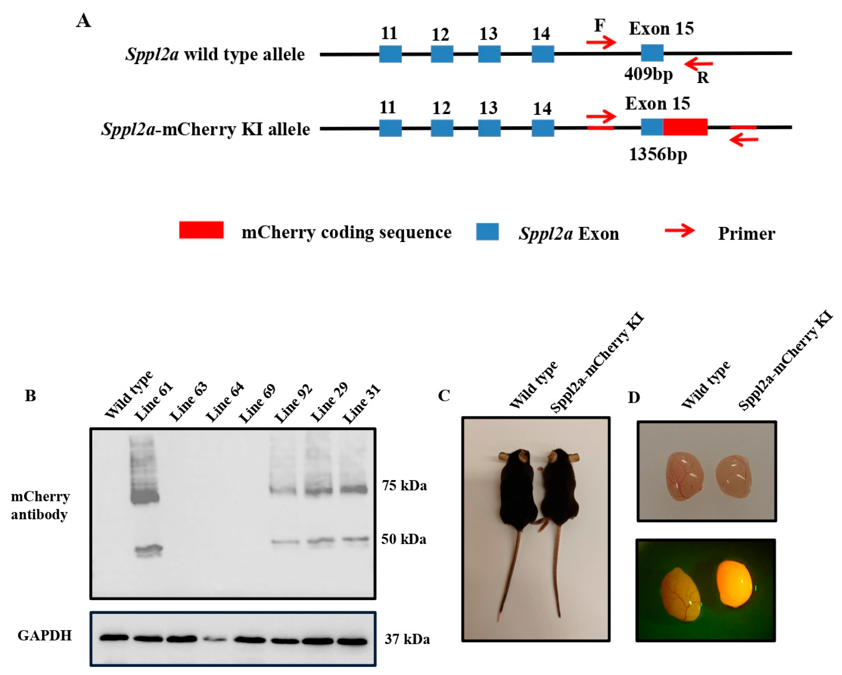
Figure 2.
Analysis of sperm parameters in WT and Sppl2a–mCherry KI mice. (A). Representative bright-field images showing normal sperm morphology from WT and KI mice; (B). Percentage of morphologically normal sperm in WT and KI mice (% , n = 3); (C). Sperm count (10^6, n = 3). (D). Percentage of motile sperm (% , n = 3); (E). Sperm motility assessed by curvilinear velocity (VCL, µm/s, n = 3). No significant difference was observed between the WT mice and the KI mice.
Figure 2.
Analysis of sperm parameters in WT and Sppl2a–mCherry KI mice. (A). Representative bright-field images showing normal sperm morphology from WT and KI mice; (B). Percentage of morphologically normal sperm in WT and KI mice (% , n = 3); (C). Sperm count (10^6, n = 3). (D). Percentage of motile sperm (% , n = 3); (E). Sperm motility assessed by curvilinear velocity (VCL, µm/s, n = 3). No significant difference was observed between the WT mice and the KI mice.
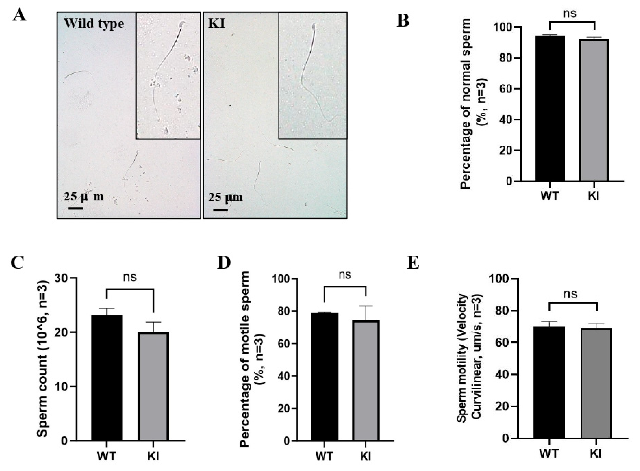
Figure 3.
Normal spermatogenesis in the Sppl2a–mCherry KI mice. (A).Testis weight is unchanged in Sppl2a–mCherry KI mice. (a) Testis weight (mg, n = 3 per genotype); (b) Testis-to-body-weight ratio (mg/g, n = 3 per genotype). (B). Representative H&E staining of testes from WT and KI mice; (C). Representative H&E staining of epididymides WT and KI mice. No difference was observed between the WT mice and the KI mice.
Figure 3.
Normal spermatogenesis in the Sppl2a–mCherry KI mice. (A).Testis weight is unchanged in Sppl2a–mCherry KI mice. (a) Testis weight (mg, n = 3 per genotype); (b) Testis-to-body-weight ratio (mg/g, n = 3 per genotype). (B). Representative H&E staining of testes from WT and KI mice; (C). Representative H&E staining of epididymides WT and KI mice. No difference was observed between the WT mice and the KI mice.
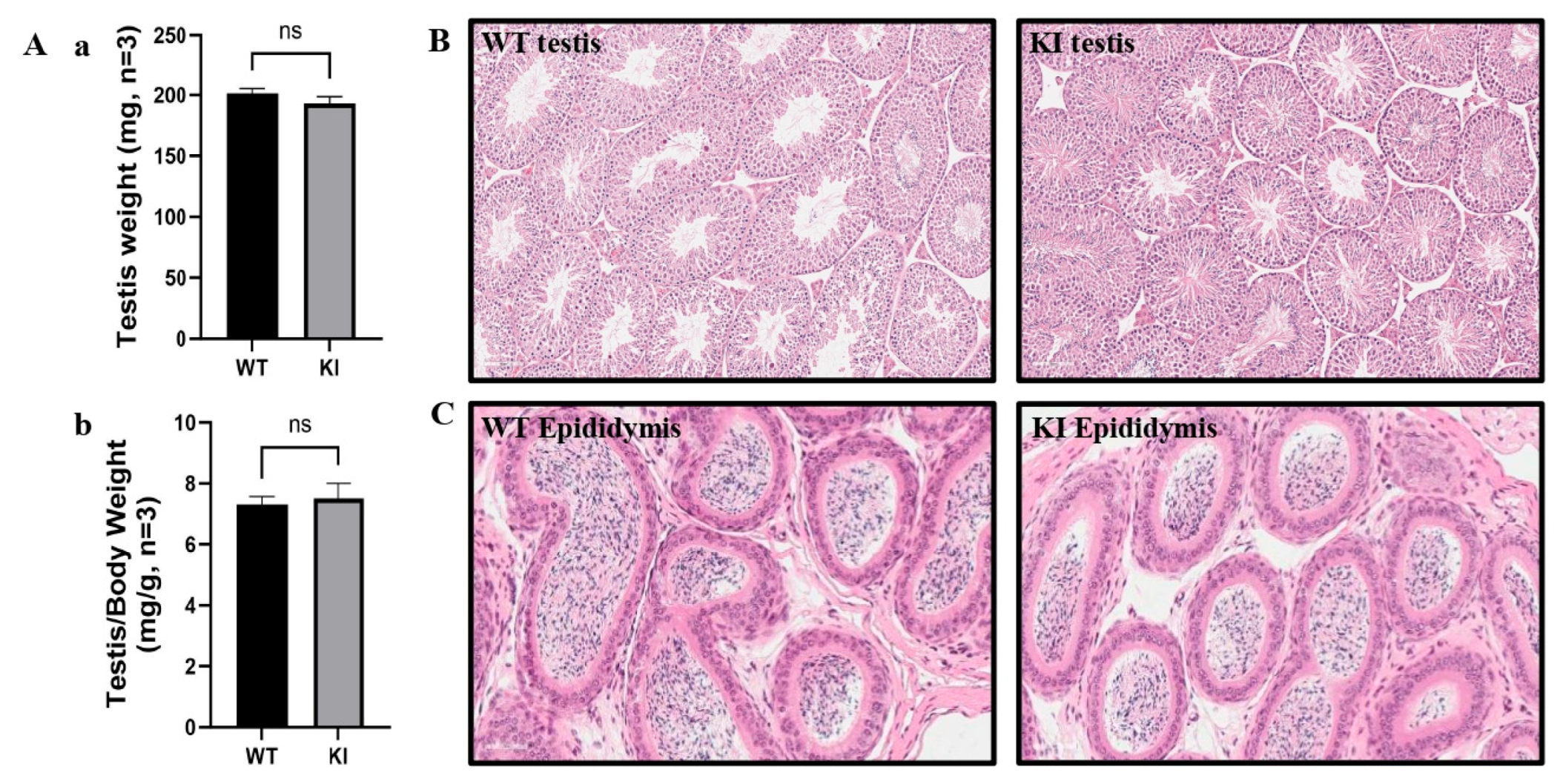
Figure 4.
Examination of mCherry signal in isolated testicular cells from the WT and the KI mice undwer high magnification. Representative bright-field and fluorescence images of germ cells isolated from WT and KI mice. No mcherry signal was present on the WT mice. However, punctated mCherry fluorescence signal was detected in cells from KI mice (arrows). The dashed arrows point to the signal in the developing tails (insert).
Figure 4.
Examination of mCherry signal in isolated testicular cells from the WT and the KI mice undwer high magnification. Representative bright-field and fluorescence images of germ cells isolated from WT and KI mice. No mcherry signal was present on the WT mice. However, punctated mCherry fluorescence signal was detected in cells from KI mice (arrows). The dashed arrows point to the signal in the developing tails (insert).
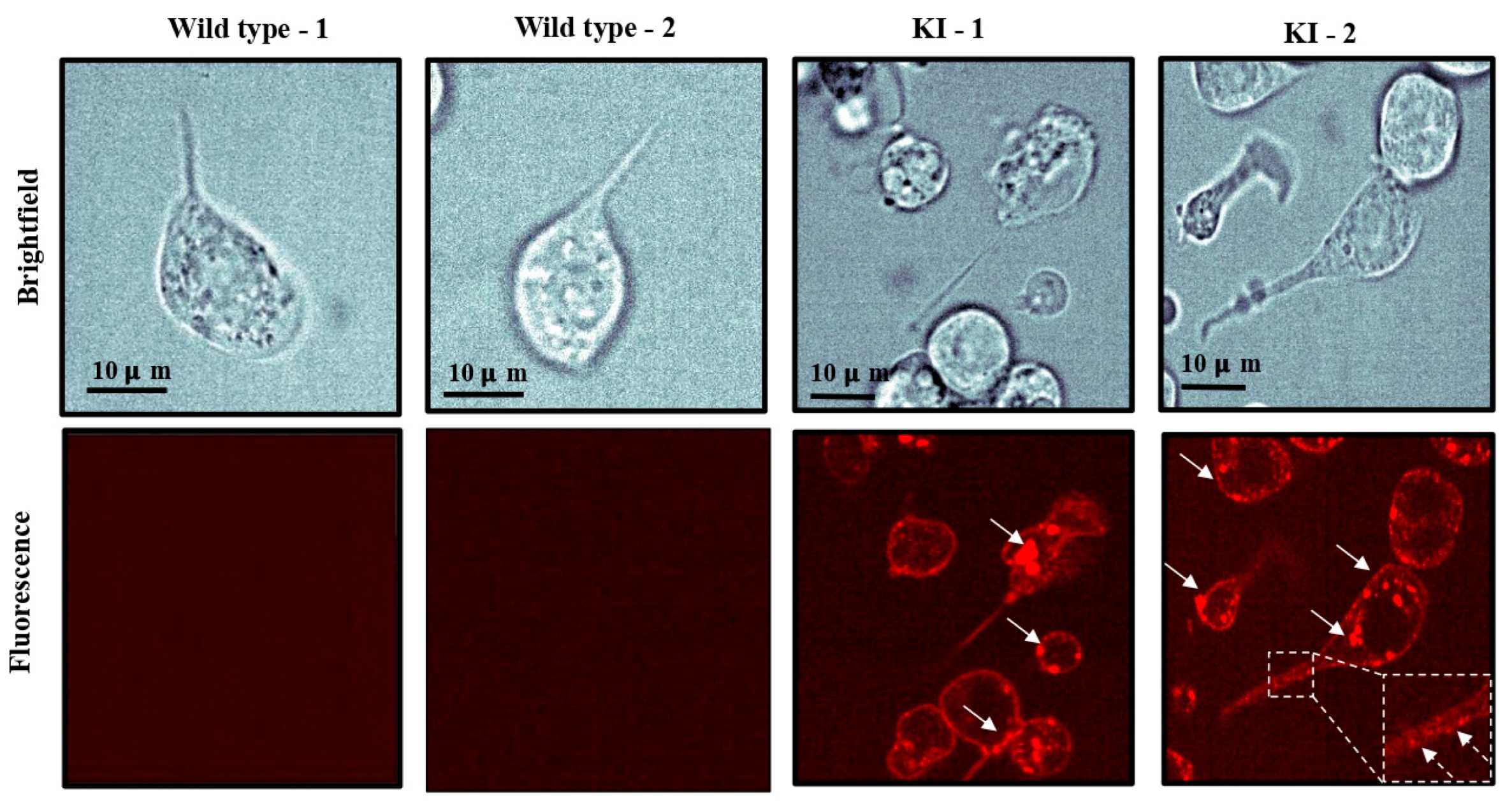
Figure 5.
Subcellular loculization of SPPL2A–mCherry fusion protein during spermiogenesis. (A). The cells were stained with PNA (green) to visualize the acrosomes, and nuclei were stained with DAPI (blue). SPPL2A–mCherry fluorescence is present in the acrosome at early step of spermartids (white arrows), and move to the cauda region at later steps. (dashed arrows). (B). Representative immunofluorescence images of isolated spermatids at steps 9-14 of spermiogenesis. α-Tubulin immunostaining (green) marks the manchette. Nuclei were counterstained with DAPI (blue). SPPL2A-mCherry fluorescence is partially co-localized in the manchette (arrows). (C). The cells were stained with an anti- ץ-tubulin antibody. SPPL2A-mCherry is co-localized with ץ-tubulin (arrow), indicating a localization in the centrioles.
Figure 5.
Subcellular loculization of SPPL2A–mCherry fusion protein during spermiogenesis. (A). The cells were stained with PNA (green) to visualize the acrosomes, and nuclei were stained with DAPI (blue). SPPL2A–mCherry fluorescence is present in the acrosome at early step of spermartids (white arrows), and move to the cauda region at later steps. (dashed arrows). (B). Representative immunofluorescence images of isolated spermatids at steps 9-14 of spermiogenesis. α-Tubulin immunostaining (green) marks the manchette. Nuclei were counterstained with DAPI (blue). SPPL2A-mCherry fluorescence is partially co-localized in the manchette (arrows). (C). The cells were stained with an anti- ץ-tubulin antibody. SPPL2A-mCherry is co-localized with ץ-tubulin (arrow), indicating a localization in the centrioles.
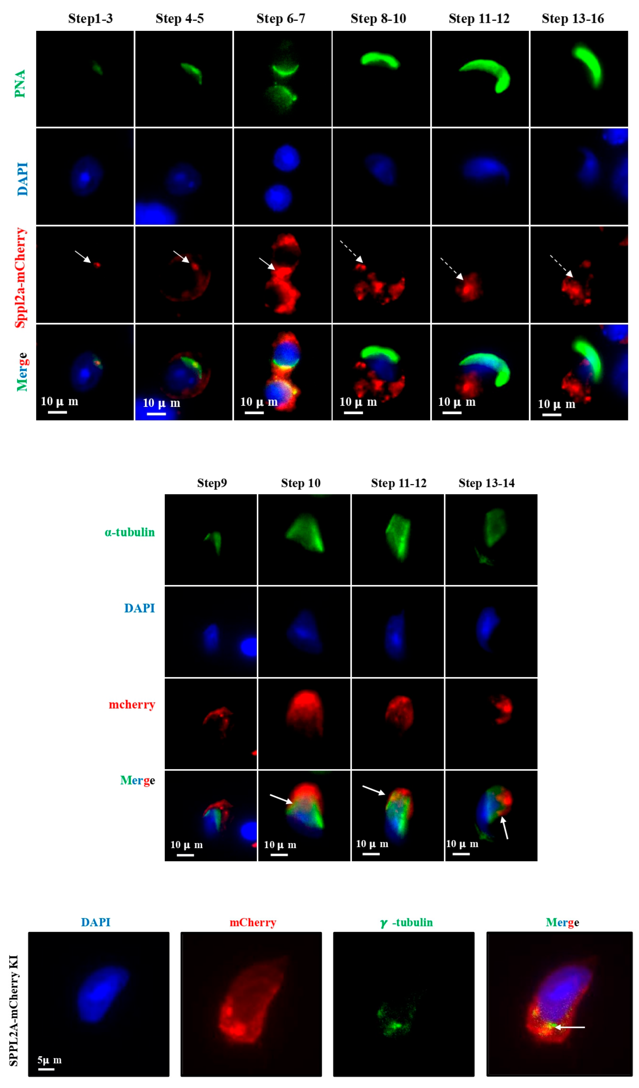
Figure 6.
SPPL2A–mCherry preferentially moves away from the cell body in developing sperm. (A). Brightfield images show the sperm cell bodies and developing tails (N = 5 cells). The tails (e.g., black dashed box) were examined with epifluorescence imaging (B) to track the dynamics of SPPL2-GFP puncta (white arrows, Supplemental Movie ##). (C) Kymographs of intensity versus time and distance along the tails revealed diffusion and trafficking of SPPL2a. Individual SPPL2a vesicles were tracked (colored lines) and analyzed. (D) Mean-squared displacement analysis demonstrated two diffusion regimes and a confinement length scale of 153 ± 15 nm, consistent with hopping diffusion and a confinement time of 3.1 ± 0.5 s. (E) At both the short times (Δt = 0.5 sec, p = 0.03) and long times (Δt = 7 sec, p = 0.0002), SPPL2a-mCherry preferentially trafficked away from the cell body. .
Figure 6.
SPPL2A–mCherry preferentially moves away from the cell body in developing sperm. (A). Brightfield images show the sperm cell bodies and developing tails (N = 5 cells). The tails (e.g., black dashed box) were examined with epifluorescence imaging (B) to track the dynamics of SPPL2-GFP puncta (white arrows, Supplemental Movie ##). (C) Kymographs of intensity versus time and distance along the tails revealed diffusion and trafficking of SPPL2a. Individual SPPL2a vesicles were tracked (colored lines) and analyzed. (D) Mean-squared displacement analysis demonstrated two diffusion regimes and a confinement length scale of 153 ± 15 nm, consistent with hopping diffusion and a confinement time of 3.1 ± 0.5 s. (E) At both the short times (Δt = 0.5 sec, p = 0.03) and long times (Δt = 7 sec, p = 0.0002), SPPL2a-mCherry preferentially trafficked away from the cell body. .
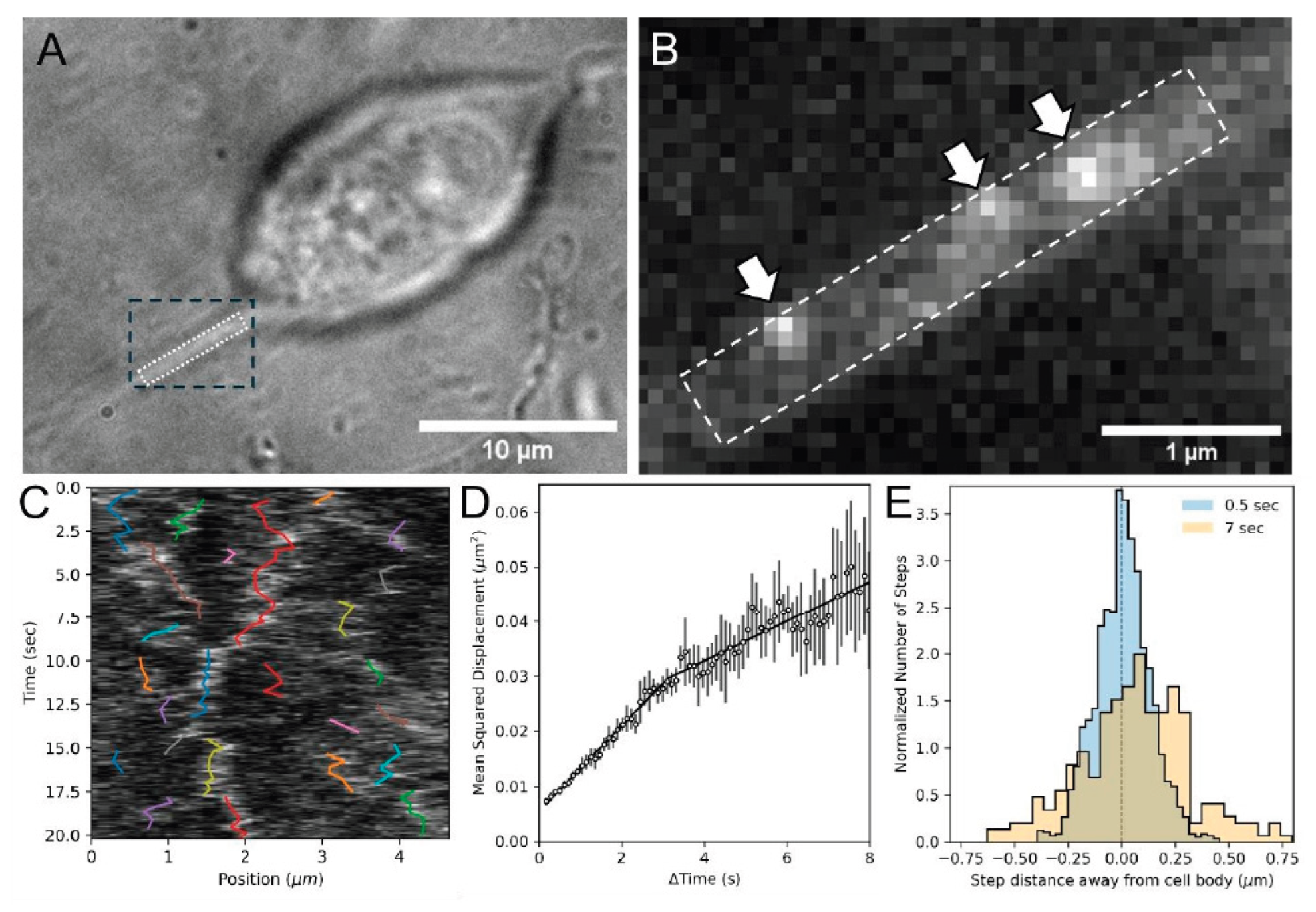

Table 1.
Fertility and fecundity of the WT and Sppl2a-mcherry KI mice.
| Genotype | Fertility | Litter size |
|---|---|---|
| WT | 4/4 | 8.25 ± 1.26 |
| Sppl2a-mcherry KI (Ho) | 4/4 | 8.00 ± 0.82 |
Table 2.
Additional CASA-derived sperm kinematic parameters in wild-type and Sppl2a-mCherry KI mice.
Table 2.
Additional CASA-derived sperm kinematic parameters in wild-type and Sppl2a-mCherry KI mice.
| Parameter | WT (n = 3) | KI (n = 3) | P value |
|---|---|---|---|
| VAP (μm/s) | 127.21 ± 0.41 | 123.71 ± 9.10 | 0.76 |
| VSL (μm/s) | 70.42 ± 1.76 | 68.22 ± 5.55 | 0.74 |
| ALH (μm) | 17.29 ± 0.03 | 16.71 ± 0.51 | 0.28 |
| BCF (Hz) | 28.46 ± 0.79 | 27.94 ± 1.74 | 0.79 |
| LIN (%) | 27.48 ± 0.58 | 26.97 ± 0.92 | 0.66 |
| STR (%) | 54.72 ± 2.45 | 53.22 ± 0.82 | 0.57 |
| WOB (%) | 49.59 ± 0.83 | 49.57 ± 2.33 | 0.99 |
Note: Data are presented as mean ± SEM (n = 3 mice per group). Statistical comparisons between WT and Sppl2a-mCherry KI mice were performed using an unpaired two-tailed Student's t-test. Abbreviations: VAP, average path velocity; VSL, straight-line velocity; ALH, amplitude of lateral head displacement; BCF, beat cross frequency; LIN, linearity; STR, straightness; WOB, wobble.
Disclaimer/Publisher’s Note: The statements, opinions and data contained in all publications are solely those of the individual author(s) and contributor(s) and not of MDPI and/or the editor(s). MDPI and/or the editor(s) disclaim responsibility for any injury to people or property resulting from any ideas, methods, instructions or products referred to in the content. |
© 2026 by the authors. Licensee MDPI, Basel, Switzerland. This article is an open access article distributed under the terms and conditions of the Creative Commons Attribution (CC BY) license (http://creativecommons.org/licenses/by/4.0/).
Copyright: This open access article is published under a Creative Commons CC BY 4.0 license, which permit the free download, distribution, and reuse, provided that the author and preprint are cited in any reuse.


