
Submitted:
21 July 2026
Posted:
23 July 2026
You are already at the latest version
Abstract
Alzheimer’s disease (AD) involves several different etiopathogenic mechanisms. A recent infectious theory proposed that pathogens, such as HSV-1, could be the root cause of AD. In this study, we investigated HSV-1 infection in an animal model to test this infectious theory. Heterozygous 5XFAD mice (Het) carrying 5 AD-linked mutations on a C57BL/6 background and wild-type littermates (WT), were infected with HSV-1 via corneal inocu-lation. Recovered mice were heat-stressed on days 30, 45, 60, and 75 post-infections. Cognitive behavior was examined in the water maze after infection and heat stress. A statistical difference in spatial learning memory was observed between infected and uninfected mice in both WT and Het mice. All Het mice showed amyloid plaque staining in the frontal cortex (FC) and dorsal subiculum (DS), regardless of HSV-1 infection or stress. However, in the hippocampal CA1 region, amyloid plaque staining was significantly higher in HSV-1-infected (HSV+) Het mice than in uninfected (HSV-) mice without heat stress. No amyloid plaque staining was observed in WT mice, regardless of HSV-1 infection or stress. Interestingly, TNF-α transcription is higher in the infected Het mice than in uninfected Hets in the hippocampal region. However, IL-1β transcription was found to be significantly higher in the hippocampus and FC region in infected WT mice than in uninfected WT mice. GFAP-positive cells were also more abundant in the hippocampus of infected WT mice than in the uninfected WT mice. Although GFAP-positive cells were abundant in both FC and CA1 regions of Het mice, no significant difference was seen be-tween infected and uninfected. Viral DNA was detected in the brains of recovered Het or WT mice at 90 dpi. HSV-1 reactivation from latency was observed in the explant of cerebrum and cerebellum at 33 dpi. These results suggest that amyloid plaque accumulation may be related to the poor learning behavior in the infected Het mice. At the same time, increased inflammatory responses may underlie the cognitive decline observed in infected WT mice.
Keywords:
HSV-1
; Heterozygous 5XFAD mice (Het)
; C57BL/6 (WT)
; cognitive behavior
; spatial learning and memory
; Amyloid beta or Amyloid plaques
; IL-1β
; TNF-α
; real-time PCR
; reactive astrocytes
; HSV-1 latency
; HSV-1 reactivation
1. Introduction
Alzheimer’s disease (AD) is the leading cause of dementia among older people worldwide [1]. Clinically, AD is characterized by progressive cognitive impairments, such as declines in memory, language, visuospatial tasks, and sometimes visual impairments [2,3]. Pathologically, the disease is recognized by the presence of amyloid plaques, neurofibrillary tangles (NFTs), neuronal and synaptic loss, dystrophic neurites, reactive astrocytes, and activated microglia. The amyloid hypothesis suggests that the processes governing the production, accumulation, or disposal of beta-amyloid are the primary cause of AD [1]. Familial and early onset AD is strongly associated with mutations in genes, such as presenilin (PSEN) and amyloid precursor protein (APP) [4], which tend to produce more amyloid in transgenic mice than in control mice [5,6]. However, only about 1% of AD is linked to genetics; the majority of the causes are unknown. An infectious hypothesis has emerged recently and proposes that pathogens are the drivers of AD [1,7]. This theory is based on evidence that some pathogens, such as genomes of herpesviruses and certain bacterial species, are often found in amyloid plaques from the brains of AD patients [8,9,10,11]. In addition, it has been reported that amyloid beta (Aβ) and tau may be the antiviral products of normal responses to infection, which intend to sequester the threat to the CNS [12,13,14]. Therefore, pathogens, such as HSV-1, could be a potential driver of AD pathologies, such as amyloid plaques.
Serology data indicate that HSV-1 infection is widespread in humans, with seropositivity at 45–98% worldwide and 57–85% in the United States [15]. Initial infection of HSV-1 is usually asymptomatic, although there may be minor local vesicular lesions on mucosal surfaces or the cornea. During primary infection, HSV-1 enters nerve endings near the infection site, then travels along nerve axons via retrograde transportation to the trigeminal ganglia (TG) or dorsal root ganglia, or the brain stem, where it establishes a life-long latent infection. HSV-1 reactivations are responsible for cold sores and recurrent corneal disease following stress or immune suppression. During HSV-1 latency, the viral genome is mostly known to persist in the TG and the brain stem [16]. The AD infectious theory comes from post-mortem studies showing that a high level of HSV-1 DNA was detected in amyloid plaques from the brain tissues of AD patients [1,17,18,19]. It has been reported that reactivation of HSV-1 increases the risk of AD [18,20,21,22]. Anti-herpetic medication can reduce the risk of dementia in patients with HSV-1 infections [23]. Growing numbers of publications demonstrate that pathological hallmarks of AD can be detected in the brain during HSV-1 infection or reactivation of HSV-1 infection [21,24,25]. Recently, Tau phosphorylation in response to HSV-1 has been linked to AD-like pathology in the mouse brain [26]. It is well established that recurrent infections are resolved following the end of a stressful stimulus, and peripheral tissues can recover from tissue damage after each reactivation episode. It is still unclear what role HSV-1 latency or reactivation plays in the pathogenesis of AD-like diseases.
In this study, the roles of HSV-1 latency and reactivation in AD-like diseases were investigated in two mouse lines: heterozygous 5xFAD on a C57BL/6J background (Het) and wild-type C57BL/6J littermates (WT). The 5xFAD mouse, an amyloid overexpression model, carries five AD-linked mutations: the Swedish (K670N/M671L), Florida (I716V), and London (V717I) mutations in APP, and the M146L and L286V mutations in PSEN1, which encodes one subunit of γ-secretase. Heterozygous 5XFAD (Het) mice are commonly used as an Alzheimer's disease model characterized by rapid, age-related amyloid plaque accumulation, robust neuroinflammation, and significant memory deficits starting at 4–6 months[27]. The effects of HSV-1 latency and reactivation on spatial reference memory, cognitive flexibility, and pathology associated with AD were investigated in this study using Het and WT mice.
2. Materials and Methods
2.1. Animals: The 5xFAD Mouse Model Used for This Research Project
B6.Cg Tg(APPSwFlLon,PSEN1*M146L*L286V)6799Vas/Mmjax, RRID:MMRRC_034848-JAX, was obtained from the Mutant Mouse Resource and Research Center (MMRRC) at The Jackson Laboratory, an NIH-funded strain repository, and was donated to the MMRRC by Robert Vassar,Ph.D., Northwestern University. The 5XFAD mice were developed by Oakley et al. [26], co-express human APP and presenilin 1 with five familial AD mutations (APP K670N/M671L + I716V + V717I and PS1 M146L + L286V). Heterozygous mice and wildtype (WT) littermates were obtained by crossing 5XFAD hemizygous males on congenic C57BL/6 genetic background with wild type C57BL/6J (WT) females (The Jackson Laboratory). Genotyping of Het was performed by PCR with total DNA from ear punch tissue. The sequences of the primers used are reported on the website: https://www.jax.org/Protocol?stockNumber=008730&protocolID=31769). Mice were housed in the Laboratory Animal Resources Center at Oregon State University and maintained in a standard 12 h light/12 h dark cycle environment. All animal procedures were approved by the Oregon State University Institutional Animal Care and Use Committee and conformed to recommendations of the American Veterinary Association Panel on Euthanasia (IACUC-2021-0194).
2.2. Cells and Viruses
Rabbit skin (RS) cells used for virus titration and Vero cells used for viral stock preparation were cultured in Dulbecco’s modified Eagle’s medium (DMEM) supplemented with 10% fetal bovine serum (FBS) (Invitrogen), penicillin (100 U/ml), streptomycin (100 g/ml) (Sigma–Aldrich, Inc.), and gentamicin (10 g/ml) (Invitrogen) at 37◦C with 5% CO2 in a humidified incubator. The GFP-HSV-1 [28] was propagated in Vero cells and used in the infection. The quantity of viruses used in the experiments was determined by standard plaque assay using RS cells as reported previously [29].
2.3. Mice Infection and Heat Stress
7-8 week-old mice were infected with 10 µL of ~4 x 10⁷ PFU/ml GFP-HSV-1 twice in each eye. Infection was monitored by eye swabbing at 1, 3, 7 and 10 days post-infection (dpi). Each cotton swab was inserted in 0.5 mL of PBS buffer and analyzed by plaque assays. Mice were stressed in a 42°C water bath cage on 30, 45, and 60 dpi for 10 min each.
2.4. Behavioral Testing
Spatial reference memory, cognitive flexibility, and associative memory (cued control task) were tested using the Morris water maze. The maze consisted of a white circular pool (122 cm in diameter and 51 cm in height) filled with water (20 ± 2°C) and made opaque by a white, nontoxic pigment. A platform (9 cm in diameter) that was not visible from the water surface was positioned 1 cm below the water surface in the NW quadrant. For the first 2 days, mice were acclimated to the water maze (swimming 1 minute, 30 seconds staying on platform), followed by 3 days of testing for spatial reference memory, 1 day of reversal training to test cognitive flexibility, and 1 day of associative memory testing (cued control task). Spatial memory and cognitive flexibility testing consisted of four place trials with an inter-trial interval of 20 min each day. The control task consisted of 6 trials with the platform visible and in different positions each trial. For all behavioral testing, the animal’s movements in the water maze were tracked and analyzed with the SMART tracking system (San Diego Instruments, San Diego, CA) as previously described [30].
2.5. Brain Sectioning
Following behavioral tests, mice were euthanized by exposure to CO2. Brains were harvested and then divided into two halves in the sagittal plane. One half was sectioned for amyloid plaque staining or immunofluorescent staining; the other half was used in DNA and RNA analysis. Chilled gelled slides (.5% gelatin solution) were used for collecting brain tissue sections. Horizontal brain sections of 10 μm thickness, from representatives of each group, were cryostat (Leitz) sectioned and placed on each slide and stored at −80°C until used.
2.6. Amyloid Plaque Staining and Imaging Analysis
Amyloid plaque staining was performed with Amylo-Glo RTD Amyloid Plaque Stain Reagent (TR-300-AG, Biosensis, Thebarton, Australia) according to the manufacturer’s instructions. Amylo-Glo stained amyloid plaques were visualized using UV epifluorescent illumination. The stained area and particle numbers were analyzed by Image J software.
2.7. DNA Extraction and PCR Genotyping
Total tissue DNA was isolated from ear notches using EZNA Tissue DNA extraction kit (Omega Bio-tec). 5XFAD PCR were run with 1.30X Kapa 2G HS buffer, 2.60 mM MgCl2, 0.26mM dNTP, 0.50 µM of common forward primer, 0.50 µM of mutant reverse primer, and wild type reverse primer, respectively, 6.50% glycerol and 0.03U/ul Kapa 2G HS taq polymerase. The reaction was run at 94°C for 2min, then 10 touchdown cycles at 94°C for 30 sec, 65°C for 30 sec with 0.5°C per cycle decrease, 68°C for 30 sec; then 30 cycles of 94°C for 30 sec, 60°C for 45sec, and 72°C for 30 sec, followed by 72°C for 5 min and holding the reaction at 20°C in the end. The PCR products were run in 1.5% agarose gel with 1X SYBR green.
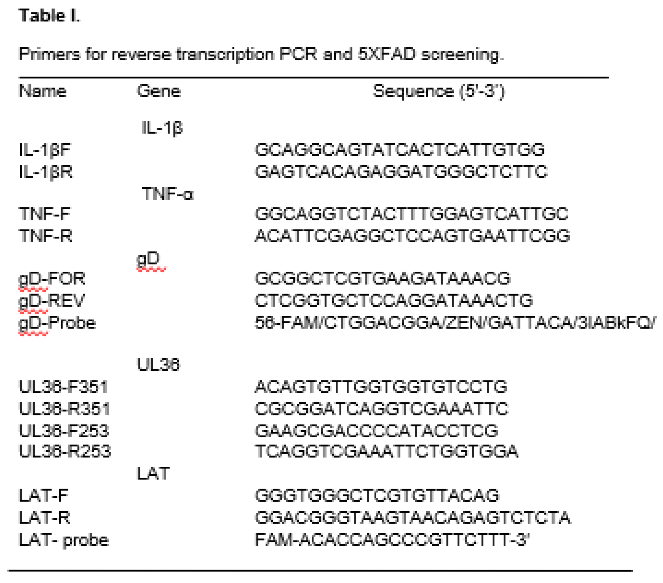
2.8. HSV-1 Real-Time PCR and PCR
Brain tissues were homogenized in 100 µL of PBS with silica beads and total tissue DNA was isolated similarly as above. Viral genome in different tissues was analyzed with Taqman real-time PCR with LAT and gD specific primers (Table 1) and Taqman probes, respectively [31]. The relative copy numbers of gD and LAT DNA were calculated using standard curves generated from plasmid containing the PCR products. UL36 nested PCR amplification was performed using a 25 μl solution consisting of 2.5 μl amplification buffer (10X PCR buffer with 15 mM MgCl2), 0.4 mM dNTP, 0.4 μM each primer, 1 U Taq (Thermo Fisher Scientific), and 20–500 ng DNA sample. The mixture was subjected to 94°C for 2 min, 35 cycles of 94 °C for 30 s, 55 °C for 30 s, and 72 °C for 30 s, followed by a 5 min elongation reaction at 72 °C after the final cycle. The nested PCR was performed using nested set primers (Table 1). A 2.5 μl aliquot of the first-run PCR product was included as a template in the second (nested) amplification under the same PCR running conditions. The PCR products were run in 1.5% agarose gel with 1X SYBR green. The 1 Kb Plus DNA Ladder (Thermo Fisher Scientific) served as size markers in gel electrophoresis.
2.9. RNA Extraction and Reverse Transcriptase-Realtime PCR
Tissue samples at ~100 mg were homogenized in 100uL of PBS with silica beads. Total RNA was extracted from mouse tissue with Trizol according to the manufacturer’s instructions (Trizol, DP424). The cDNA was synthesized with the qScript cDNA Synthesis Kit (Quantabio), which were then amplified by SYBR real-time PCR Master Mix (Thermo Fisher Scientific) with primers specific to mouse TNF-α, IL-1β, and β-actin (Table 1) as described before [32]. A standard curve was made with each gene by cloning the PCR product into a pCR2.1 TOPO vector (Invitrogen). The Ct value is converted to gene copy number per µg based on the standard curves.
2.10. Indirect Immunofluorescence Assay (IFA)
Mouse frozen sections were fixed in acetone for 10 min at -20°C and air dried for 20 min before staining. Slides were first blocked with 10% horse serum blocking buffer for 30 min at room temperature, then incubated with primary antibodies, HSV-ICP27 monoclonal antibody (H1113) (Virusys Monoclonal Antibodies) at 1:200 or Glial Fibrillary Acidic protein (GFAP) rabbit polyclonal antibody (R-1374-50, Biosensis) at 1:2000, in a humidified chamber at 4°C overnight. A secondary goat anti-rabbit IgG antibody conjugated with Tax red (Cat No. 4040-03), or a secondary goat anti-mouse antibody conjugated with FITC (cat no. 1070-02) from Southern Biotech, was applied to slides at a dilution of 1:2000 and incubated in a humidified chamber at room temperature for 1h. Slides were washed gently 3–4 times in PBS containing 0.01% Tween 20 at room temperature for 5 min before and after application of the secondary of antibody. Slides were then covered with Vectashield® mounting media containing DAPI (Vector® Laboratories) before coverslip and examined under fluorescent microscope.
2.11. Tissue Explant
To prove HSV-1 becomes latent in the mouse brain, the frontal cortex (FC), caudal cortex (Cx), hippocampus (HP), cerebellum (C), brainstems (B), and TG were each explanted at 33 dpi and cultured in 1.5 ml MEM in a 12-well plate as we described previously [33]. Infectious virus from the explant was monitored daily for 10 days in 100-µl aliquot of the explant culture, which was then used to infect RS cells in a 96-well plate. The cytopathic effect (CPE) or GPF expression from HSV-1 infection was monitored for 5 days to detect reactivated viruses in each brain tissue explant culture.
2.12. Statistics
All statistical analyses were performed using GraphPad Prism, version 9 for Windows (GraphPad Software, San Diego, California). Behavioral test results were analyzed by repeated measures ANOVA with Jamovi version 2.2.5. The difference in beta amyloid expression and gene expression was analyzed by Multiple unpaired t tests.
3. Results
3.1. HSV-1 Infection can Become Latent in Heterozygous 5xFAD (Het) Mice and Reactivate Following Heat Stress
To determine whether HSV-1 infection can become latent in heterozygous 5xFAD (Het) mice [34] and C57BL/6 mice wildtype (WT) littermates, they were infected at 7-8 weeks of age with ~2X10⁵ PFU GFP-HSV-1/ McKrae per eye [28]. The virus shedding was monitored by eye swabbing and tittered via a standard plaque assay. Both infected WT and Het mice shed between 1X10⁴ to 1X10⁵ PFU per ml during the first 5 dpi and cleared the infection by 10 dpi (Figure 1A). To prove HSV-1 becomes latent in the TG of infected Het mice, the expression of the latency-associated transcript (LAT), which is the only known viral RNA expressed during latency, was examined by reverse transcription real-time PCR. As shown in Figure 1B, amplification of LAT is only present in the reaction in the presence of reverse transcriptase, which indicates that HSV-1 had become latent in the TG of Het mice (n=3). Whether HSV-1 reactivation can be induced by heat stress in Het mice was previously unknown. To demonstrate HSV-1 reactivation in the brains of mice following heat stress, viral gene transcription, a feature of lytic infection, was examined in groups of both Het and WT mice (n=3) subjected to heat stress (HS) or not (NHS) at 30 dpi. HSV-1 LAT transcription, which is also expressed during lytic infection, was quantified by reverse-transcription real-time PCR using total RNA from the frontal lobes. As shown in Figure 2, viral gene transcription was only detectable 3 days post-heat stress in the frontal lobes of HSV-1-infected (HSV+) and stressed mice. This result demonstrated that lytic infection could occur in the brain in both Het and WT mice following heat stress.
3.2. There were Differences in Spatial Memory and Cognitive Flexibility in HSV-1 Latently Infected Mice Tested Within 3-Days Post-Heat Stress.
To understand whether HSV-1 infection alone, or stress has any effect on cognitive function, such as spatial reference memory and cognitive flexibility, both Het and WT mice were infected with HSV-1 as described in the Materials and Methods (Figure 3A). Spatial memory, cognitive flexibility, and associative memory (cued control task) were examined by the Morris water maze analysis as described previously [35]. Each infection group, with 3-4 mice per group, was tested 3 days after the heat stress on days 30 and 60 post-infection [36]. In groups of Het mice stressed on 30 dpi, the HSV-1 infected and stressed group (Het/HSV+/HS) performed significantly poorer than uninfected mice, regardless of stress (Het/HSV-/NHS: p=.0173 and Het/HSV-/HS: p=0.027; Figure 3B), during the learning phase (place trials) for long-term memory. Uninfected WT (WT/HSV-/NHS) mice also performed significantly better than WT/HSV+/HS in probe trials (p=0.04) (Fig 3C). No significant difference was observed in the probe trials for long-term memory between any treatment and stress in the Het mice (Figure 3C). No difference was observed in cued trials among the different treatment groups in either Het and WT mice stressed only at 30 dpi (Sup Fig 1A), suggesting that all groups had similar motivation, visual acuity, and physical ability. In groups of mice stressed at both 30 and 60 dpi, no difference in spatial long-term memory was observed regardless of infection, stress, or genotype (data not shown). However, there were significant differences in cognitive flexibility between Het/HSV+/HS and Het/HSV+/NHS (p=.0016) and Het/HSV-/NHS (p=0.014) and a trend for difference from Het/HSV-/HS (p=.092) (Figure 4). No significant difference in cognitive flexibility between different treatment groups in WT mice (Figure 4). No difference was observed between any different treatment groups of mice stressed on 60 dpi in cued trials (Sup Figure 1B), probe trials (Sup Figure 2) regardless of genotypes.
3.3. There was a Difference in Spatial Learning Between Uninfected Mice and HSV-1 Latently Infected Mice Tested 2 Weeks Post-Heat Stresses
Our results showed that infected and stressed mice performed worse than uninfected and unstressed Het mice (Figs 3 and 4). Since behavioral analysis was performed 3 days post-heat stress, it was possible that those mice may have still been experiencing active HSV-1 reactivation due to heat stress. To avoid active HSV-1 reactivation from heat stress before the behavior test, additional groups of infected and stressed mice were tested after a recovery period. Both Het and WT mice were infected as above, and stressed three times at 30, 45, and 60 dpi as indicated in Figure 5A. Behavior analysis was performed 15 days after the final heat stress. There was a significant difference in spatial long-term memory (Place trial) between infected mice and uninfected mice, regardless of heat stress or genotype (Figure 5B). No consistent differences in cognitive flexibility analysis were observed between different treatment groups in Het or WT mice with or without heat stress (data not shown). These results suggest that there was a difference in spatial long-term memory between latently infected mice and uninfected mice with or without heat stress.
3.4. There were Significantly more Amyloid Plaques in HSV-1 Infected Het Mice than Uninfected Het Mice in CA1 Region of Hippocampus
To determine whether the differences seen between HSV+ and HSV- Het mice in spatial learning memory were related to amyloid plaque density in the mouse brain, brain tissues were harvested 15 days post-behavior tests and sectioned via Leitz cryostat. Amyloid plaques in three mice from each treatment group were examined by Amylo-Glo staining. The positive staining appeared bright blue under UV light and white in the captured images (Figs 6A and 7A). ImageJ software was used to quantify staining area in the frontal cortex and the Cornu Ammonis 1 (CA1) region of the hippocampus. Significantly more amyloid plaque staining in CA1 region, a critical subfield of hippocampus involved in spatial memory, autobiographical memory, and memory consolidation, was seen in Het/HSV+/NHS mice than those in Het/HSV-/NHS mice (p=0.02; Figure 6B). No significant difference was seen between groups of HSV+ stressed 3 times (HSV+/HS3) and HSV+ without stress (HSV+/NHS) Het mice, which suggests heat stress had no significant effect on amyloid plaque accumulation in CA1 region. There was no significant difference in amyloid plaque staining in the frontal cortex (FC) region, the brain’s command center for cognitive flexibility, between HSV+ Het mice and HSV- Het mice regardless of stress (Fig 7). No amyloid plaque staining was observed in the brain of HSV-1 infected WT mice with or without stress (Figure 7A). This result suggests that HSV-1 infection could accelerate amyloid plaque accumulation in the hippocampus of Het mice, which may explain why Het/HSV+ mice perform poorly in the long-term memory trials. However, plaque density didn’t appear to be related to the cognitive flexibility results.
3.5. There were Increased Inflammatory Cytokines and Reactive Astrocytes in HSV-1 Infected Mice
The Amylo-Glo staining demonstrated that there was more beta amyloid in the CA1 region of the hippocampus in Het/HSV+ mice than in the Het/HSV- mice at 90 dpi, which may explain why Het/HSV+ mice had poorer learning memory than the Het/HSV- mice. However, no beta-amyloid staining was observed in WT/HSV+ mice despite also performing worse in the long-term memory trials. Chronic inflammatory response has been reported to be associated with AD-like disease [26]. The innate cGAS-STING inflammatory pathway was found to be activated in an AD-like disease in HSV-1-infected mice [26], suggesting that proinflammatory cytokine genes are activated. To determine whether inflammatory gene transcription was increased in brain tissues harvested two weeks after behavioral testing (at 90 dpi; Figure 5A), transcription of IL-1β and TNF-α in different parts of the brain was examined by reverse transcription (RT) real-time PCR with primers specific to mouse IL-1β and TNFα. The relative copy number was estimated by a standard curve made with a control plasmid containing the IL-1β and TNFα PCR sequences. In WT mice, HSV+/NHS mice had a significantly higher IL-1β gene expression in both hippocampus (Figure 8A) and frontal cortex (Figure 8B) than uninfected mice. However, no significant difference was seen in TNF-α expression in WT mice regardless of infection or stress in either hippocampus (Figure 8C) or frontal cortex (Figure 8D). In the Het mice, no difference in IL-1β expression was observed between infected and uninfected mice, regardless of stress in hippocampus (Figure 8E) or frontal cortex (Figure 8F). However, HSV+/HS3 and HSV+/NHS mice had higher TNF-α expression than HSV-/NHS and HSV-/HS3 mice in the hippocampus (Figure 8G), while only HSV+/HS3 mice had significantly higher TNF-α gene expressions than HSV-/NHS mice in the frontal cortex (Figure 8H).
Inflammatory responses to brain injury and neurodegeneration are often accompanied by reactive astrocytes (gliosis), which have increased GFAP expression. To determine if there were differences in reactive astrocytes in the brain, brain sections from all treatment groups were stained with GFAP antibodies. Although few reactive astrocytes were observed in the FC regions in the HSV-1-infected WT mice, more GFAP staining was observed in the CA1 and dentate gyrus (DG) regions in the infected WT mice (Figure 9A). The GFAP staining in the CA1 region in the WT/HSV+/NHS and WT/HSV+/HS3 mice was significantly higher than in the WT/HSV-/NHS mice (Figure 9B). On the other hand, increased GFAP staining area was seen in FC, CA, and DG regions in both infected and uninfected Het mice (Figure 9C). There was no significant difference in GFAP staining between Het/HSV+ and Het/HSV- mice in CA1 region, FC and DG region (Figure 9D). These results suggested that HSV-1 infection could induce gliosis in the hippocampus of WT mice, whereas Het mice showed high levels of gliosis regardless of HSV-1 infection. The expression of IL-1β and the presence of reactive astrocytes may explain why WT/HSV+/NHS mice performed worse than WT/HSV-/NHS mice in the place trials.
3.6. HSV-1 Genome was Detected in Many Regions of the Infected Mice at 90 dpi
To understand why inflammatory gene expression is present in the mouse brain at 90 dpi, we hypothesize that HSV-1 is not completely cleared following an acute infection, and residual viral components may trigger chronic inflammation in the brain. To determine whether HSV-1 DNA persists in the brain after exposure, HSV-1 DNA was examined directly in different parts of the brain harvested during acute phase and latent phase as shown in Figure 10A. Total DNA was isolated from frontal cortex (FC), caudal cortex (Cx), hippocampus (HP), brain stems (BS) and cerebellum (C) of 3 WT mice. HSV-1 DNA was quantitated by real-time PCR specific for LAT gene on 1, 3, 5, 15 and 30 day post-infection (dpi). As shown in Figure 10B, viral DNA was barely detectable at 1 dpi; by day 3, it was detectable in most of the brain regions examined. Viral DNA peaked around day 5 post-infections and remained detectable through 30 dpi in the tested tissues. To determine whether HSV-1 persists in the brain at 90 dpi, HSV-1 DNA was first examined in total DNA isolated from HSV infected and unstressed brain tissues by nested PCR using primers specific to UL36. Although there were few or no detections in the 1st-run PCR, HSV-1 DNA was amplified in the 2nd-run PCR reaction using the 1st-run PCR products in many brain sections, regardless of genotype (Figure 11). To determine if the HSV-1 genome increases following heat stress, HSV-1 DNA was monitored by real-time PCR. As shown in Sup Fig 3, no significant differences were observed in HSV-1-infected mice regardless of stress in either Het or WT mice. These results suggest that the viral genome persists in many regions of the brain in latently infected mice, in addition to the TG and brainstem.
3.7. HSV-1 Became Latent in Many Regions of the Brain in the Infected Mice
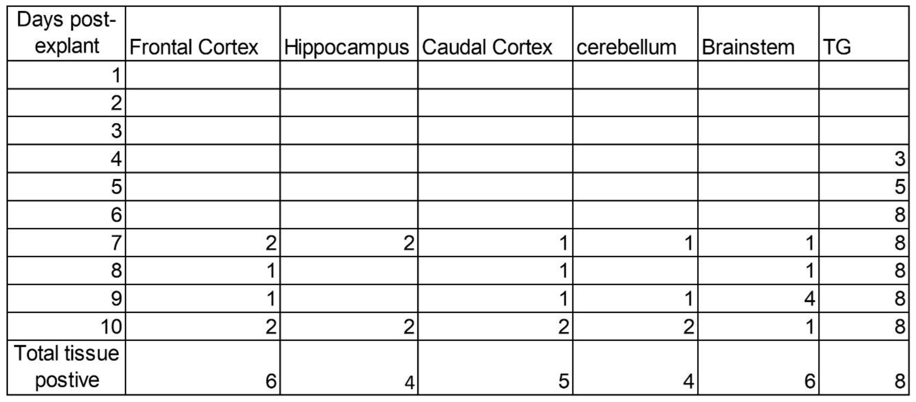
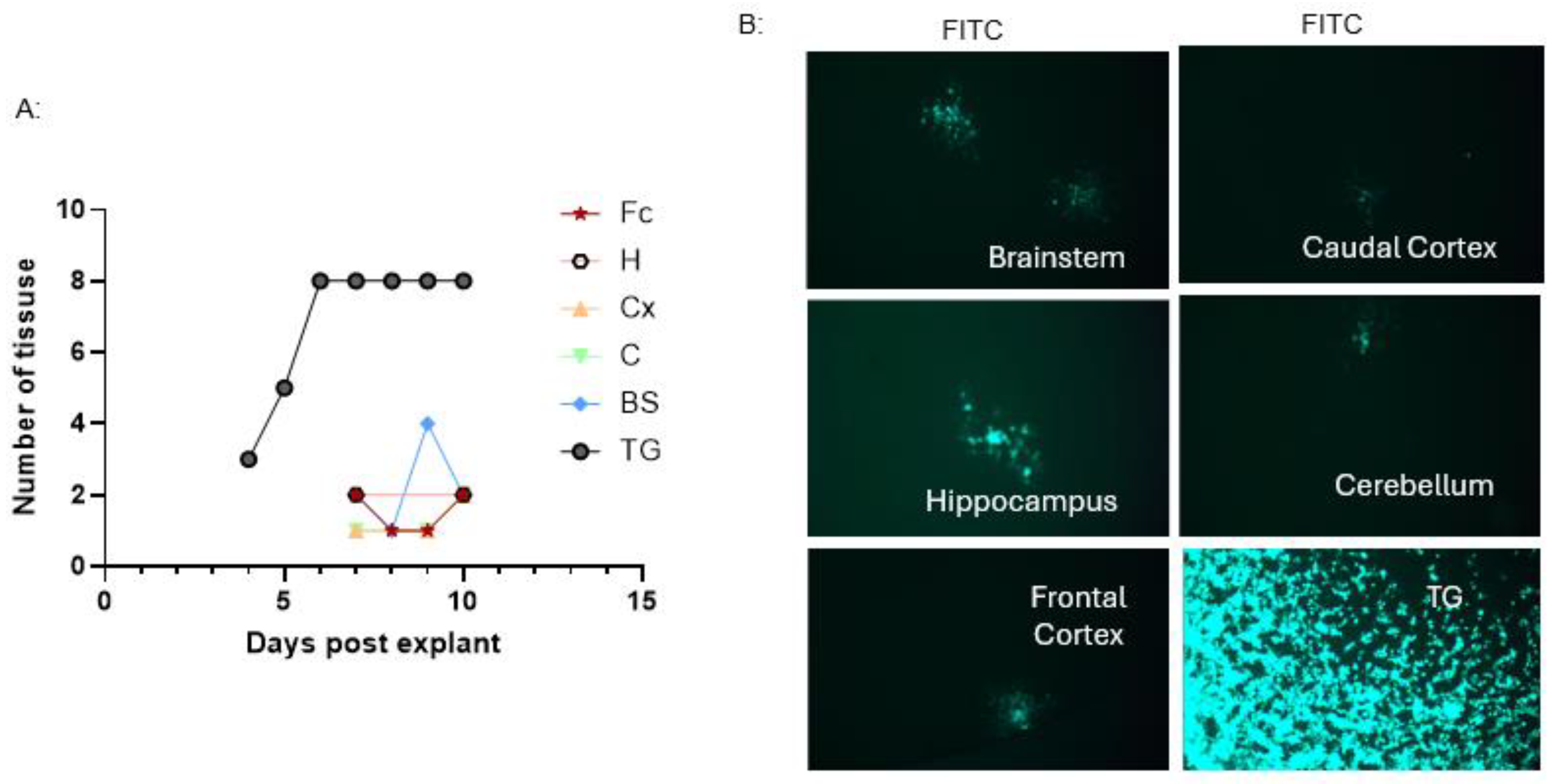
The results above demonstrated that HSV-1 DNA persists in different regions of the mouse brain after recovery from HSV-1 infection. It is possible that those viral genomes could become latent in the brain’s neurons. To determine whether HSV-1 becomes latent in the cerebrum, additional WT mice were infected with HSV-1 as described in the Materials and Methods. Frontal cortex (Fc), caudal cortex (Cx), hippocampus (H), cerebellum ( C ), and brainstem (BS) from 8 latently infected mice were explanted at 33 days post-infection and cultured in 1.5 ml DMEM in a 12-well plate as we described previously [33]. By day 4 post-tissue explant, 3 out of 8 TG explant started producing infectious virions, by day 6 explant, CPE was seen in RS cells infected with TG explant culture media from 8 out 8 mice (Figure 12A, Table 2). The CPE was verified by GFP expression in the infected RS cells (Figure 12B). By day 7 post-tissue explant, HSV-1 reactivation was also observed in Fc, H, Cx, and C explant culture media (Figure 12A, Table 2). Between day 7 and 10 post-tissue explant, HSV-1 reactivation was observed in 6 out of 8 brainstems, 6 out of 8 frontal cortexes, 5 out 8 caudal cortex, 4 out 8 hippocampus and cerebellum explants (Table 2). The amount of viruses reactivated from the brain explant was relatively low compared to the amount of viruses from the TG explant. A significantly higher level of GFP expression was observed in RS cells infected with TG explant media at 24hpi. In contrast, relatively few infection foci were observed in cells infected with brain tissue explant media (Figure 12B).
4. Discussion
Alzheimer’s disease (AD) is a disease that involves several different etiopathogenic mechanisms [37,38,39]. In this study, we investigated HSV-1 infection on cognitive function and pathologies associated with AD-like disease in heterozygous 5XFAD (Het) mice and C57BL/6J (WT). Het mice are commonly used in Alzheimer's disease model, and characterized by rapid, age-related amyloid plaque accumulation, robust neuroinflammation, and significant memory deficits starting at 4–6 months [27]. These Het mice, like WT mice, were susceptible to HSV-1 infection. Both Het and WT mice developed ocular disease following HSV-1 inoculation during acute infection phase. Only 5-10% of them died from the acute HSV-1 infection when they were infected with high infectious dose around 1X10⁵ PFU/eye; most of them survived the acute infection. HSV-1 can become latent in the TG of the Het mice, as in WT mice. HSV-1 reactivation could be induced by heat stress within 3 days post-heat stress in both Het and WT mice (Figure 2). Therefore, the effect of HSV-1 latency and reactivation on AD related disease was evaluated in Het mice in this study.
We hypothesized that HSV-1 infection or reactivation from latency in the brain could accelerate amyloid plaque accumulation and cognitive decline. We predicted that infected Het mice would have worse cognitive function. As anticipated, the spatial memory and cognitive flexibility in Het/HSV+/HS mice was significantly poorer than in Het/HSV-/NHS or Het/HSV-/HS mice stressed at 30 dpi (Figure 3B) and in Het/HSV-/NHS and Het/HSV+/NHS mice stressed at 60 dpi (Figure 4), respectively. In probe trials, WT/HSV+/HS performed significantly worse than WT/HSV-/NHS mice stressed at 30 dpi (Figure 3C). However, the difference in probe trail (long term memory) was not significant between Het/HSV+/HS and Het/HSV-/HS mice stressed twice at 30 and 60 dpi (Sup Fig 2A). The poorer performance in behavior trails observed in the HSV+/HS mice at 30 and 60 dpi could be due to active HSV-1 reactivation, since the behavioral analysis was performed 3 days after heat stress (Figs 3 and 4). This suggests HSV-1 infection could stimulate cognitive declines during active infection as reported previously [21].
The HSV-1 infection effect on long-term memory (probe trails) was not immediately obvious when those mice were tested around 4 months old after 2 heat stresses (Sup Figure 2). Two heat stresses had a limited effect on HSV+ mice, possibly due to variation in reactivation rates and sex differences in responses to stress, since we used mixed-sex mice in each group. To demonstrate that the cognitive decline observed in infected mice is not directly related to lytic HSV-1 infection, as shown in Figure 3B, the behavioral analysis was repeated at 75 dpi, 15 days post-heat stress, with more mice per group. These mice were about 4.5 months old when they were tested in the water maze. Interestingly, HSV+/NHS mice performed worse than HSV-/NHS mice when the behavior tests were run at 75 dpi (Figure 5). At 4.5 months old, no significant difference in inflammatory cytokine expression was reported between male and female Het mice [40]. These results suggest that HSV-1 infection may have a chronic effect on cognitive function, with phenotypic differences taking time to manifest. The amyloid plaque staining showed more amyloid plaque staining in the CA1 region of the hippocampus in Het/HSV+/NHS mice (Figure 6). This result suggests that HSV-1 chronic infection could stimulate amyloid plaque accumulation and contribute to memory decline in Het/HSV+ mice.
It has been shown that sex and age-dependent cytokine expression of IL-1β and TNFα occurs in Het mice at 3 and 4 months old [40]. The male mice had lower IL-1β expression between 3 and 4 months old, whereas the female mice showed higher TNF expression and more reactive astrocytes at 3 months old. The behavior for those mice with two stresses, 30 dpi and 60 dpi, was conducted when those Het mice were around 4 months old, at which time they had lower IL-1β expression. The differences in neuroinflammation between 3 and 4-month-old mice may explain why Het mice tested at 4 months old showed no significant difference between infected and uninfected mice. In addition, only three mixed-sex mice per group were used for behavioral analysis, which may not capture subtle behavioral differences associated with HSV-1 infection.
At 90 dpi, Het/HSV+/NHS mice had significantly more amyloid plaques than Het/HSV-/NHS mice in the CA1 region of the hippocampus (Figure 6). However, Het/HSV+/HS3 had relatively fewer amyloid plaques in the CA1 region compared to those in Het/HSV+/NHS mice. A similar trend was observed between Het/HSV-/NHS and Het/HSV-/HS3 mice, although the difference is not significant. It is known that heat stress can lead to a wide range of host stress responses and production of different heat shock proteins [41]. Heat shock proteins (Hsps) induced by heat stress can prevent misfolding and are capable of clearing aggregated or damaged proteins from cells [42,43]. It has been reported that induction of Hsps could reduce tau pathology and amyloid plaque formation in transgenic AD mice [44]. In a 2016 publication, Kasza et al. demonstrates that dihydropyridine derivatives (such as LA1011) as Hsp co-inducers can provide neuroprotective and disease-modifying treatment for Alzheimer's disease [44]. Another study reported in 2017 found that recombinant heat-shock protein HSP70 can reduce the elevated intracellular proteasome activity induced by isomerized amyloid-beta (Aβ42) in human SK-N-SH neuroblastoma cells [45]. Therefore, heat stress used in our study may trigger Hsps production, which may reduce amyloid peptide production in stressed Het mice. This may explain why stressed and unstressed Het mice had no significant difference in amyloid plaque staining in the infected mice (Figure 6 and Figure 7). This may also explain why some Het/HSV+/HS3 mice seem to perform better than Het/HSV+/NHS mice in behavior tests (data not shown).
Another observation is that many amyloid plaques were present in the frontal cortex of Het mice, regardless of infection or stress. There is no direct correlation between cognitive flexibility and amyloid beta staining in the frontal cortex between Het/HSV+ and Het/HSV- mice (Fig 7). Interestingly, no amyloid plaques were observed in WT mice at 90 dpi, regardless of infection or stress. This raised the question of why infected WT mice also show decreased spatial memory (Figure 5B). A 2024 study found that chronic inflammatory responses to HSV-1 infection were associated with increased tau phosphorylation [26]. To determine whether chronic inflammation plays a role in memory decline in WT mice, tissues harvested at 90 dpi were examined for IL-1β and TNF-α gene transcription. To our surprise, increased IL-1β was observed in both frontal cortex tissue and hippocampus tissues from the infected WT mice, however TNFα gene transcription were not significant different between HSV+ and HSV- WT mice (Figure 8). IL-1β is a potent, pro-inflammatory cytokine and key mediator of the innate immune response, acting as a crucial "alarm" signal during infection, injury, and stress [46,47]. The difference in IL-1β expression suggests an inflammatory response is taking place in WT/HSV+ mice. The increased inflammation in the hippocampus could explain why WT/HSV+ mice performed poorer in the probe trial than WT/HSV- mice (Figure 3C). The difference in IL-1β expression is less significant between infected and uninfected Het mice. Beta-amyloid may act as an antimicrobial peptide against HSV-1 infection; therefore, Het mice showed a lower IL-1β response. On the other hand, TNF-α mRNA transcription was significantly different in the hippocampus between HSV+ and HSV- Het mice. In Het mice, TNF-α transcription is known to be higher at 5 months of age, regardless of sex [40]. HSV-1 infection may increase TNF-α transcription more in the Het mice. TNF-α is a master pro-inflammatory cytokine that drives low-grade chronic inflammation and facilitates persistent, low-level systemic inflammation [48,49,50]. A combination of low-grade inflammation and amyloid plaque accumulation may drive the decline in spatial memory in Het/HSV+/NHS mice.
Reactive astrocytes are CNS glial cells characterized by hypertrophy and increased GFAP expression in response to injury, disease, or infection. They are key players in neuroinflammation and stroke, often releasing toxic factors or, conversely, aiding in repair [51]. Reactive astrocyte responses are driven by signals such as cytokines and can be sustained for long periods [52]. Since the IL-1β and TNFα genes were found to be upregulated, we expect to see more reactive astrocytes in the HSV+ mouse brain. Using GFAP antibody staining, increased GFAP staining was observed in the frontal cortex and hippocampus of Het mice, regardless of infection (Figure 9C and 9D). Although very little GPAP staining was observed in the frontal cortex of WT mice, there was significantly more GFAP staining in the WT/HSV+ mice than in the WT/ HSV- mice in the CA1 region (Figure 9B). These results suggest greater hippocampal neuroinflammation in WT/HSV+ mice may explain why WT/HSV+/NHS mice performed worse in place trails than WT/HSV-/NHS mice did.
To understand what is driving the inflammatory response in infected mice, we hypothesized that HSV-1 viral components, such as the viral genome and proteins, are not fully cleared from the brain. To determine whether viral proteins were present, brain sections harvested at 90 dpi were stained with antibodies specific to HSV-1 ICP27 and GFAP. No consistent detection of ICP27 was observed in the infected Het and WT mice (Sup Figs. 4 and 5). Only 1 out of 3 positive ICP27 staining was observed in DG and CA1 region in Het/HSV+ mice (Sup Figure 4, white arrow), 1 out of 3 positive staining in CA region in WT/HSV+ mice (Sup Figure 5, green arrow). The low detection frequency may suggest that only a few neurons in the brain with HSV-1 persistency. Viral DNA persistence in different regions was examined by nested PCR and real-time PCR. In agreement with our hypothesis, viral DNA was detectable in the frontal cortex, caudal cortex, hippocampus, and cerebellum, as well as in TG and the brain stem, at 90 dpi in both stressed and unstressed mice (Figure 11, Sup Figure 3). It is interesting to find that HSV-1 persists in the central nervous system. However, there was no correlation between viral genome copy number and the number of stresses. It is common for the HSV-1 latency establishment to vary between hosts. Therefore, it is not surprising to find no correlation between stressed and unstressed animals in viral genome copy numbers (Sup Figure 3). It is unknown whether HSV-1 can become latent in neurons of the cerebrum and cerebellum and reactivate from latency. Data on HSV-1 reactivation from latency in the cerebrum were contradictory so far [53,54]. Our in vitro tissue explant culture showed that infectious viruses could be reactivated from multiple regions of the brain (Figure 12 and Table 2). Another interesting finding is that HSV-1 reactivation frequency in the brain and brainstem was relatively lower than that in TG (Figure 12A and Table 2). The difference remains to be explored. The difference in reactivation efficiency between TG and the brain could lead to different outcomes in neuronal pathogenesis. Aborted or low reactivation may be implicated in chronic inflammation, neurodegeneration, and cognitive decline.
5. Conclusions
In conclusion, our study found that HSV-1 infection persisted in the brain and that some neurons can be latently infected, which can be reactivated in many brain regions. HSV-1 persistency can drive different pathologies and immune responses in different strains of mice. The accumulation of amyloid plaques and increased TNF-α expression in Het mice may contribute to cognitive decline in Het mice. At the same time, HSV-1 persistency and IL-1β inflammatory responses may increase Tau phosphorylation and drive memory decline in the WT mice.
6. Patents
N/A
Author Contributions
“Conceptualization, KRM. and L.J.; methodology, K.L.; E.G.; X.W.; I.A.; A.L.; K.R.M.; L.J.; software, KRM and LJ.; validation, KRM and LJ. formal analysis, KRM and LJ.; investigation, K.L.; E.G.; X.W..; I.A.; A.L.; K.R.M.; L.J.; resources, KRM and LJ; data curation, KRM and LJ.; writing—original draft preparation, KRM; L.J.; writing—review and editing, KRM and LJ.; visualization, L.J.; supervision, KRM, L.J.; project administration, KRM and LJ.; funding acquisition, KRM and LJ.
Funding
This research received no external funding
Institutional Review Board Statement
Not applicable
Informed Consent Statement
Not applicable
Data Availability Statement
Not applicable
Acknowledgments
We thank Rod Keller, Mr. Nelson Osorio, and Aaron Trippe at Oregon State Diagnostic Laboratory for sectioning mouse brains used in Amyloid staining and indirect immunofluorescent staining. We also thank Théotime Brehon,, MacKenzie Jantzen, Gabriel L. Oedell for animal behavior data collections.
Conflicts of Interest
The authors declare no conflict of interest. The funders had no role in the design of the study; in the collection, analysis, or interpretation of data; in the writing of the manuscript, or in the decision to publish the results
Abbreviations
AD Alzheimer’s disease
Het 5×FAD heterozygotes
HSV-1 Human Herpes Simplex type 1 virus
WT Wild-type mice
FC frontal cortex
CA1 Cornu Ammonis 1 region
GFAP Glial Fibrillary Acidic protein
ICP27 immediate-early or α protein 27
Ab amyloid beta
TG trigeminal ganglia
RS rabbit skin cells
GFP Green Fluorescent Protein
PCR Polymerase Chain Reaction
NHS No heat stress
HS Heat stress
References
- Seaks, C.E.; Wilcock, D.M. Infectious hypothesis of Alzheimer disease. PLoS Pathog. 2020, 16(11), e1008596. [Google Scholar] [CrossRef] [PubMed]
- Mirzaei, N.; Shi, H.; Oviatt, M.; Doustar, J.; Rentsendorj, A.; Fuchs, D.T.; Sheyn, J.; Black, K.L.; Koronyo, Y.; Koronyo-Hamaoui, M. Alzheimer's Retinopathy: Seeing Disease in the Eyes. Front Neurosci. 2020, 14, 921. [Google Scholar] [CrossRef] [PubMed]
- Shi, H.; Koronyo, Y.; Rentsendorj, A.; Regis, G.C.; Sheyn, J.; Fuchs, D.T.; Kramerov, A.A.; Ljubimov, A.V.; Dumitrascu, O.M.; Rodriguez, A.R. Identification of early pericyte loss and vascular amyloidosis in Alzheimer's disease retina. Acta Neuropathol. 2020, 139(5), 813–836. [Google Scholar] [CrossRef] [PubMed]
- Wu, L.; Rosa-Neto, P.; Hsiung, G.Y.; Sadovnick, A.D.; Masellis, M.; Black, S.E.; Jia, J.; Gauthier, S. Early-onset familial Alzheimer's disease (EOFAD). Can. J. Neurol. Sci. 2012, 39(4), 436–445. [Google Scholar] [CrossRef] [PubMed]
- Citron, M.; Eckman, C.B.; Diehl, T.S.; Corcoran, C.; Ostaszewski, B.L.; Xia, W.; Levesque, G.; St George Hyslop, P.; Younkin, S.G.; Selkoe, D.J. Additive effects of PS1 and APP mutations on secretion of the 42-residue amyloid beta-protein. Neurobiol. Dis. 1998, 5(2), 107–116. [Google Scholar] [CrossRef] [PubMed]
- Eckman, C.B.; Mehta, N.D.; Crook, R.; Perez-tur, J.; Prihar, G.; Pfeiffer, E.; Graff-Radford, N.; Hinder, P.; Yager, D.; Zenk, B. A new pathogenic mutation in the APP gene (I716V) increases the relative proportion of A beta 42(43). Hum. Mol. Genet 1997, 6(12), 2087–2089. [Google Scholar] [CrossRef] [PubMed]
- Sochocka, M.; Zwolinska, K.; Leszek, J. The Infectious Etiology of Alzheimer's Disease. Curr. Neuropharmacol. 2017, 15(7), 996–1009. [Google Scholar] [CrossRef] [PubMed]
- Itzhaki, R.F.; Cosby, S.L.; Wozniak, M.A. Herpes simplex virus type 1 and Alzheimer's disease: the autophagy connection. J. Neurovirol 2008, 14(1), 1–4. [Google Scholar] [CrossRef] [PubMed]
- Itzhaki, R.F. Lathe R: Herpes Viruses and Senile Dementia: First Population Evidence for a Causal Link. J. Alzheimers Dis. 2018, 64(2), 363–366. [Google Scholar] [CrossRef] [PubMed]
- Wozniak, M.A.; Mee, A.P.; Itzhaki, R.F. Herpes simplex virus type 1 DNA is located within Alzheimer's disease amyloid plaques. J. Pathol. 2009, 217(1), 131–138. [Google Scholar] [PubMed]
- Jamieson, G.A.; Maitland, N.J.; Wilcock, G.K.; Craske, J.; Itzhaki, R.F. Latent herpes simplex virus type 1 in normal and Alzheimer's disease brains. J. Med. Virol. 1991, 33(4), 224–227. [Google Scholar] [CrossRef] [PubMed]
- Kumar, D.K.; Choi, S.H.; Washicosky, K.J.; Eimer, W.A.; Tucker, S.; Ghofrani, J.; Lefkowitz, A.; McColl, G.; Goldstein, L.E.; Tanzi, R.E. Amyloid-beta peptide protects against microbial infection in mouse and worm models of Alzheimer's disease. Sci. Transl. Med. 2016, 8(340), 340ra372. [Google Scholar] [CrossRef]
- Bourgade, K.; Dupuis, G.; Frost, E.H.; Fulop, T. Anti-Viral Properties of Amyloid-beta Peptides. J. Alzheimers Dis. 2016, 54(3), 859–878. [Google Scholar] [CrossRef] [PubMed]
- Eimer, W.A.; Vijaya Kumar, D.K.; Navalpur Shanmugam, N.K.; Rodriguez, A.S.; Mitchell, T.; Washicosky, K.J.; Gyorgy, B.; Breakefield, X.O.; Tanzi, R.E.; Moir, R.D. Alzheimer's Disease-Associated beta-Amyloid Is Rapidly Seeded by Herpesviridae to Protect against Brain Infection. Neuron 2018, 100(6), 1527–1532. [Google Scholar] [CrossRef] [PubMed]
- Memish, Z.A.; Almasri, M.; Chentoufi, A.A.; Al-Tawfiq, J.A.; Al-Shangiti, A.M.; Al-Kabbani, K.M.; Otaibi, B.; Assirri, A.; Yezli, S. Seroprevalence of Herpes Simplex Virus Type 1 and Type 2 and Coinfection With HIV and Syphilis: The First National Seroprevalence Survey in Saudi Arabia. Sex. Transm. Dis. 2015, 42(9), 526–532. [Google Scholar] [CrossRef] [PubMed]
- Sivasubramanian, M.K.; Monteiro, R.; Harrison, K.S.; Plakkot, B.; Subramanian, M.; Jones, C. Herpes Simplex Virus Type 1 Preferentially Enhances Neuro-Inflammation and Senescence in Brainstem of Female Mice. J. Virol. 2022, 96(17), e0108122. [Google Scholar] [CrossRef] [PubMed]
- Itzhaki, R.F.; Tabet, N. Herpes simplex encephalitis and Alzheimer's disease: Is there a link? J. Neurol. Sci. 2017, 380, 20–21. [Google Scholar] [CrossRef] [PubMed]
- Mori, I.; Kimura, Y.; Naiki, H.; Matsubara, R.; Takeuchi, T.; Yokochi, T.; Nishiyama, Y. Reactivation of HSV-1 in the brain of patients with familial Alzheimer's disease. J. Med. Virol. 2004, 73(4), 605–611. [Google Scholar] [CrossRef] [PubMed]
- Lin, W.R.; Wozniak, M.A.; Cooper, R.J.; Wilcock, G.K.; Itzhaki, R.F. Herpesviruses in brain and Alzheimer's disease. J. Pathol. 2002, 197(3), 395–402. [Google Scholar] [CrossRef] [PubMed]
- Lovheim, H.; Gilthorpe, J.; Adolfsson, R.; Nilsson, L.G.; Elgh, F. Reactivated herpes simplex infection increases the risk of Alzheimer's disease. Alzheimers Dement 2015, 11(6), 593–599. [Google Scholar] [PubMed]
- De Chiara, G.; Piacentini, R.; Fabiani, M.; Mastrodonato, A.; Marcocci, M.E.; Limongi, D.; Napoletani, G.; Protto, V.; Coluccio, P.; Celestino, I. Recurrent herpes simplex virus-1 infection induces hallmarks of neurodegeneration and cognitive deficits in mice. PLoS Pathog. 2019, 15(3), e1007617. [Google Scholar] [CrossRef] [PubMed]
- Warren-Gash, C.; Forbes, H.J.; Williamson, E.; Breuer, J.; Hayward, A.C.; Mavrodaris, A.; Ridha, B.H.; Rossor, M.N.; Thomas, S.L.; Smeeth, L. Human herpesvirus infections and dementia or mild cognitive impairment: a systematic review and meta-analysis. Sci. Rep. 2019, 9(1), 4743. [Google Scholar] [CrossRef] [PubMed]
- Tzeng, N.S.; Chung, C.H.; Lin, F.H.; Chiang, C.P.; Yeh, C.B.; Huang, S.Y.; Lu, R.B.; Chang, H.A.; Kao, Y.C.; Yeh, H.W. Anti-herpetic Medications and Reduced Risk of Dementia in Patients with Herpes Simplex Virus Infections-a Nationwide, Population-Based Cohort Study in Taiwan. Neurotherapeutics 2018, 15(2), 417–429. [Google Scholar] [CrossRef] [PubMed]
- Cairns, D.M.; Rouleau, N.; Parker, R.N.; Walsh, K.G.; Gehrke, L.; Kaplan, D.L. A 3D human brain-like tissue model of herpes-induced Alzheimer's disease. Sci. Adv. 2020, 6(19), eaay8828. [Google Scholar] [CrossRef] [PubMed]
- Mielcarska, M.B.; Skowronska, K.; Wyzewski, Z.; Toka, F.N. Disrupting Neurons and Glial Cells Oneness in the Brain-The Possible Causal Role of Herpes Simplex Virus Type 1 (HSV-1) in Alzheimer's Disease. Int. J. Mol. Sci. 2021, 23(1). [Google Scholar] [CrossRef] [PubMed]
- Hyde, V.R.; Zhou, C.; Fernandez, J.R.; Chatterjee, K.; Ramakrishna, P.; Lin, A.; Fisher, G.W.; Celiker, O.T.; Caldwell, J.; Bender, O. Anti-herpetic tau preserves neurons via the cGAS-STING-TBK1 pathway in Alzheimer's disease. Cell Rep. 2025, 44(1), 115109. [Google Scholar] [CrossRef] [PubMed]
- Forner, S.; Kawauchi, S.; Balderrama-Gutierrez, G.; Kramar, E.A.; Matheos, D.P.; Phan, J.; Javonillo, D.I.; Tran, K.M.; Hingco, E.; da Cunha, C. Systematic phenotyping and characterization of the 5xFAD mouse model of Alzheimer's disease. Sci. Data 2021, 8(1), 270. [Google Scholar] [CrossRef] [PubMed]
- Arya, D.; Jaggi, U.; Wang, S.; Tormanen, K.; Che, M.; Mahov, S.; Jin, L.; Ghiasi, H. A novel GFP-based strategy to quantitate cellular spatial associations in HSV-1 viral pathogenesis. mBio 2024, 15(10), e0145424. [Google Scholar] [CrossRef] [PubMed]
- Jin, L.; Perng, G.C.; Mott, K.R.; Osorio, N.; Naito, J.; Brick, D.J.; Carpenter, D.; Jones, C.; Wechsler, S.L. A herpes simplex virus type 1 mutant expressing a baculovirus inhibitor of apoptosis gene in place of latency-associated transcript has a wild-type reactivation phenotype in the mouse. J. Virol. 2005, 79(19), 12286–12295. [Google Scholar] [CrossRef] [PubMed]
- Brim, B.L.; Haskell, R.; Awedikian, R.; Ellinwood, N.M.; Jin, L.; Kumar, A.; Foster, T.C.; Magnusson, K.R. Memory in aged mice is rescued by enhanced expression of the GluN2B subunit of the NMDA receptor. Behav. Brain Res. 2013, 238, 211–226. [Google Scholar] [CrossRef] [PubMed]
- Jaggi, U.; Ghiasi, H. Presence of CD80 and Absence of LAT in Modulating Cellular Infiltration and HSV-1 Latency. Viruses 2024, 16(9). [Google Scholar] [CrossRef] [PubMed]
- Monteiro, R.; Kumar Sivasubramanian, M.; Harrison, K.S.; Plakkot, B.; Sadeghi, H.; Subramanian, M.; Jones, C. Examination of neuro-inflammation and senescence in brainstem of aged mice latently infected with human alphaherpesvirus 1 (HSV-1). Virus Res. 2024, 347, 199420. [Google Scholar] [CrossRef] [PubMed]
- Perng, G.C.; Slanina, S.M.; Ghiasi, H.; Nesburn, A.B.; Wechsler, S.L. The effect of latency-associated transcript on the herpes simplex virus type 1 latency-reactivation phenotype is mouse strain-dependent. J. Gen. Virol. 2001, 82 Pt 5, 1117–1122. [Google Scholar] [CrossRef] [PubMed]
- Oakley, H.; Cole, S.L.; Logan, S.; Maus, E.; Shao, P.; Craft, J.; Guillozet-Bongaarts, A.; Ohno, M.; Disterhoft, J.; Van Eldik, L. Intraneuronal beta-amyloid aggregates, neurodegeneration, and neuron loss in transgenic mice with five familial Alzheimer's disease mutations: potential factors in amyloid plaque formation. J. Neurosci. 2006, 26(40), 10129–10140. [Google Scholar] [CrossRef] [PubMed]
- Magnusson, K.R.; Scruggs, B.; Zhao, X.; Hammersmark, R. Age-related declines in a two-day reference memory task are associated with changes in NMDA receptor subunits in mice. BMC Neurosci. 2007, 8, 43. [Google Scholar] [CrossRef] [PubMed]
- Sawtell, N.M.; Thompson, R.L. Herpes simplex virus type 1 latency-associated transcription unit promotes anatomical site-dependent establishment and reactivation from latency. J. Virol. 1992, 66(4), 2157–2169. [Google Scholar] [CrossRef] [PubMed]
- Wulfert E: [Alzheimer's disease: etiopathogenic mechanisms and development of new drugs]. J. Pharm. Belg. 1997, 52(2), 97–98. [PubMed]
- Sotiropoulos, I.; Sousa, N. Tau as the Converging Protein between Chronic Stress and Alzheimer's Disease Synaptic Pathology. Neurodegener. Dis. 2016, 16(1-2), 22–25. [Google Scholar] [PubMed]
- Iqbal, K.; Grundke-Iqbal, I. Alzheimer's disease, a multifactorial disorder seeking multitherapies. Alzheimers Dement 2010, 6(5), 420–424. [Google Scholar] [CrossRef] [PubMed]
- Manji, Z.; Rojas, A.; Wang, W.; Dingledine, R.; Varvel, N.H.; Ganesh, T. 5xFAD Mice Display Sex-Dependent Inflammatory Gene Induction During the Prodromal Stage of Alzheimer's Disease. J. Alzheimers Dis. 2019, 70(4), 1259–1274. [Google Scholar] [CrossRef] [PubMed]
- Mussa, B.M.; Srivastava, A.; Bajbouj, K.; Shafarin, J.; Rajan, R.; Hisham, A. Brain-Derived Neurotrophic Factor and Heat Shock Protein-32: Potential Targets for Microglial Response to Glucose and Oxygen Manipulation. Mol. Neurobiol. 2025, 63(1), 252. [Google Scholar] [CrossRef] [PubMed]
- Evgen'ev, M.; Bobkova, N.; Krasnov, G.; Garbuz, D.; Funikov, S.; Kudryavtseva, A.; Kulikov, A.; Samokhin, A.; Maltsev, A.; Nesterova, I. The Effect of Human HSP70 Administration on a Mouse Model of Alzheimer's Disease Strongly Depends on Transgenicity and Age. J. Alzheimers Dis. 2019, 67(4), 1391–1404. [Google Scholar] [CrossRef] [PubMed]
- Garbuz, D.G.; Sverchinsky, D.; Davletshin, A.; Margulis, B.A.; Mitkevich, V.; Kulikov, A.M.; Evgen'ev, M.B. The molecular chaperone Hsp70 from the thermotolerant Diptera species differs from the Drosophila paralog in its thermostability and higher refolding capacity at extreme temperatures. Cell Stress Chaperones 2019, 24(6), 1163–1173. [Google Scholar] [CrossRef] [PubMed]
- Kasza, A.; Hunya, A.; Frank, Z.; Fulop, F.; Torok, Z.; Balogh, G.; Santha, M.; Balind, A.; Bernath, S.; Blundell, K.L. Dihydropyridine Derivatives Modulate Heat Shock Responses and have a Neuroprotective Effect in a Transgenic Mouse Model of Alzheimer's Disease. J. Alzheimers Dis. 2016, 53(2), 557–571. [Google Scholar] [CrossRef] [PubMed]
- Morozov, A.V.; Yurinskaya, M.M.; Mitkevich, V.A.; Garbuz, D.G.; Preobrazhenskaia, O.V.; Vinokurov, M.G.; Evgen'ev, M.B.; Karpov, V.L.; Makarov, A.A. Heat-shock protein HSP70 decreases activity of proteasomes in human neuroblastoma cells treated by amyloid-beta 1-42 with isomerized Asp7. Mol. Biol. (Mosk) 2017, 51(1), 166–171. [Google Scholar] [CrossRef] [PubMed]
- Lopez-Castejon, G.; Brough, D. Understanding the mechanism of IL-1beta secretion. Cytokine Growth Factor Rev. 2011, 22(4), 189–195. [Google Scholar] [CrossRef] [PubMed]
- Boraschi, D. What Is IL-1 for? The Functions of Interleukin-1 Across Evolution. Front Immunol. 2022, 13, 872155. [Google Scholar] [CrossRef] [PubMed]
- Jang, D.I.; Lee, A.H.; Shin, H.Y.; Song, H.R.; Park, J.H.; Kang, T.B.; Lee, S.R.; Yang, S.H. The Role of Tumor Necrosis Factor Alpha (TNF-alpha) in Autoimmune Disease and Current TNF-alpha Inhibitors in Therapeutics. Int. J. Mol. Sci. 2021, 22(5). [Google Scholar] [CrossRef] [PubMed]
- Yuan, L.; Li, W.; Tian, Z.B.; Li, Y.; Sun, S. Predictive role of cytokines IL-10, IL-12 and TNF-alpha gene polymorphisms for the development of osteonecrosis of the femoral head in the Chinese Han population. Cell Mol. Biol. (Noisy-le-grand) 2017, 63(9), 144–149. [Google Scholar] [CrossRef] [PubMed]
- Huang, X.; Huang, S.; Fu, F.; Song, J.; Zhang, Y.; Yue, F. Characterization of preclinical Alzheimer's disease model: spontaneous type 2 diabetic cynomolgus monkeys with systemic pro-inflammation, positive biomarkers and developing AD-like pathology. Alzheimers Res. Ther. 2024, 16(1), 52. [Google Scholar] [PubMed]
- Abedin, M.J.; Kastanenka, K.V. The role of astrocytes in Alzheimer's disease: Pathophysiology, biomarkers, and therapeutic potential. J. Alzheimers Dis. 2026, 110(1), 26–45. [Google Scholar] [CrossRef] [PubMed]
- Herx, L.M.; Yong, V.W. Interleukin-1 beta is required for the early evolution of reactive astrogliosis following CNS lesion. J. Neuropathol. Exp. Neurol. 2001, 60(10), 961–971. [Google Scholar] [PubMed]
- Wang, Q.; Guo, J.; Jia, W. Intracerebral recombinant HSV-1 vector does not reactivate latent HSV-1. Gene Ther. 1997, 4(12), 1300–1304. [Google Scholar] [CrossRef] [PubMed]
- Starr, P.A.; Lim, F.; Grant, F.D.; Trask, L.; Lang, P.; Yu, L.; Geller, A.I. Long-term persistence of defective HSV-1 vectors in the rat brain is demonstrated by reactivation of vector gene expression. Gene Ther. 1996, 3(7), 615–623. [Google Scholar] [PubMed]
Figure 1.
Virus shedding in the tears during acute infection and detection of LAT in Het TG at 30 dpi. A: Virus titration of mouse tear swabs from HSV-1-infected Het and WT mice. Both eyes were infected with 2 × 10^5 PFU HSV-1 McKrae/eye. Tear swabs were collected on 1, 3, 5, 7, and 10 days post-infection (dpi) from 5 mice per group. WT: wildtype, Het: 5×FAD heterozygotes. B: Detection of HSV-1 latency-associated transcript (LAT) by reverse transcription real-time PCR with total RNA of TG from HSV-1-infected Het mice at 30 days post-infection. TGs were taken from three HSV-1-infected Het mice. RTase+: cDNA synthesis with reverse transcriptase, RTase-: cDNA synthesis without reverse transcriptase. N=3.
Figure 1.
Virus shedding in the tears during acute infection and detection of LAT in Het TG at 30 dpi. A: Virus titration of mouse tear swabs from HSV-1-infected Het and WT mice. Both eyes were infected with 2 × 10^5 PFU HSV-1 McKrae/eye. Tear swabs were collected on 1, 3, 5, 7, and 10 days post-infection (dpi) from 5 mice per group. WT: wildtype, Het: 5×FAD heterozygotes. B: Detection of HSV-1 latency-associated transcript (LAT) by reverse transcription real-time PCR with total RNA of TG from HSV-1-infected Het mice at 30 days post-infection. TGs were taken from three HSV-1-infected Het mice. RTase+: cDNA synthesis with reverse transcriptase, RTase-: cDNA synthesis without reverse transcriptase. N=3.
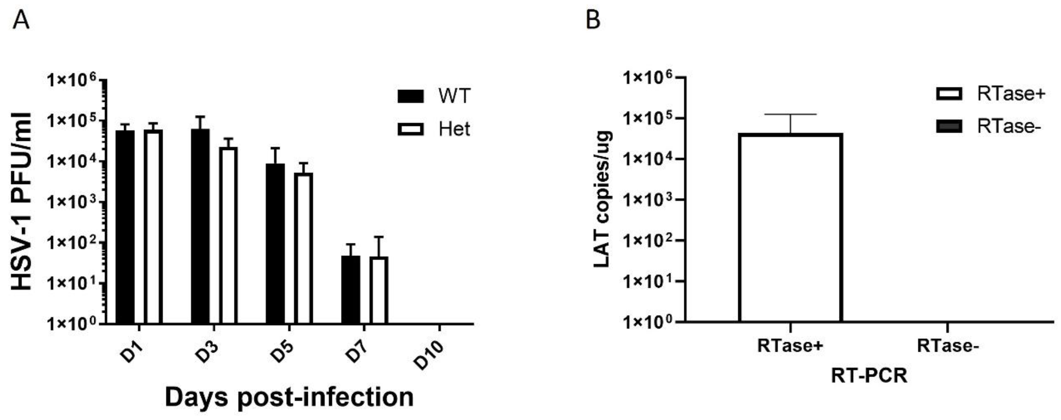
Figure 2.
Quantification of HSV-1 LAT in the mouse frontal lobe 3 days post-heat stress. The X-axis represents groups of mice ± infection with HSV-1 (1 × 10^5 PFU/eye of HSV-1 McKrae). The y-axis represents the number of LAT copies detected by reverse transcription real-time PCR using primers and a probe specific to the LAT gene. NHS: no heat stress. HS: Heat stress on days 30 post-infection. Het: 5XFAD mice. WT:C57BL/6J, wildtype littermate mice. n=2.
Figure 2.
Quantification of HSV-1 LAT in the mouse frontal lobe 3 days post-heat stress. The X-axis represents groups of mice ± infection with HSV-1 (1 × 10^5 PFU/eye of HSV-1 McKrae). The y-axis represents the number of LAT copies detected by reverse transcription real-time PCR using primers and a probe specific to the LAT gene. NHS: no heat stress. HS: Heat stress on days 30 post-infection. Het: 5XFAD mice. WT:C57BL/6J, wildtype littermate mice. n=2.
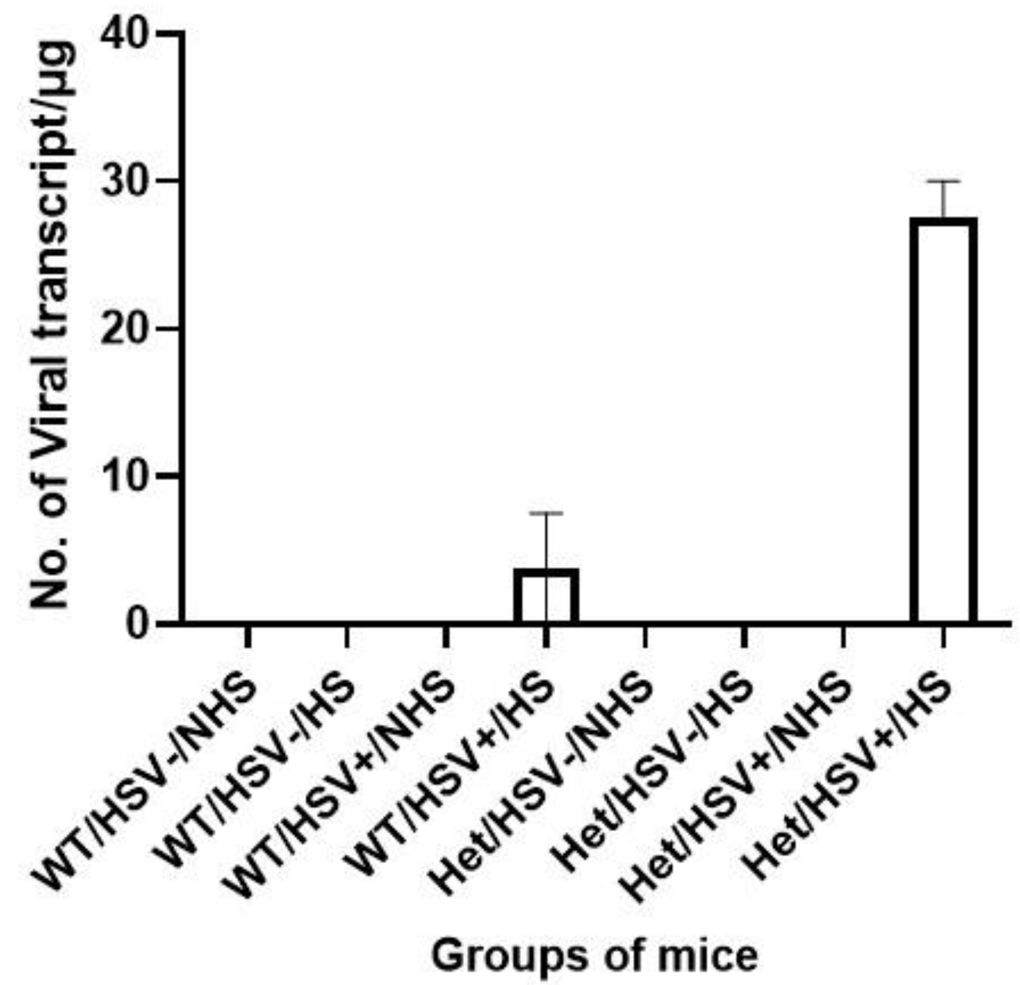
Figure 3.
Timeline for 2 heat-stress study and acute behavioral testing. A) Schematic of HSV-1 infection, heat stresses (HS), and memory testing. HS1: heat stress at 30 days post-infection, HS2: heat stress at 30 and 60 dpi, respectively. B) Spatial long-term memory (place trials) testing in Het (Left) and WT (Right) mice with (HS) or without (NHS) heat stress. C) probe trials in Het (Left) and WT (Right) mice with (HS) or without (NHS) heat stress. Mice were stressed once on day 30 post-infection and tested in a Morris water maze 3 days post-stress (42 C; 10 min). Means ± SEM. ANOVA & Fisher’s LSD post-hoc tests. n = 3.
Figure 3.
Timeline for 2 heat-stress study and acute behavioral testing. A) Schematic of HSV-1 infection, heat stresses (HS), and memory testing. HS1: heat stress at 30 days post-infection, HS2: heat stress at 30 and 60 dpi, respectively. B) Spatial long-term memory (place trials) testing in Het (Left) and WT (Right) mice with (HS) or without (NHS) heat stress. C) probe trials in Het (Left) and WT (Right) mice with (HS) or without (NHS) heat stress. Mice were stressed once on day 30 post-infection and tested in a Morris water maze 3 days post-stress (42 C; 10 min). Means ± SEM. ANOVA & Fisher’s LSD post-hoc tests. n = 3.
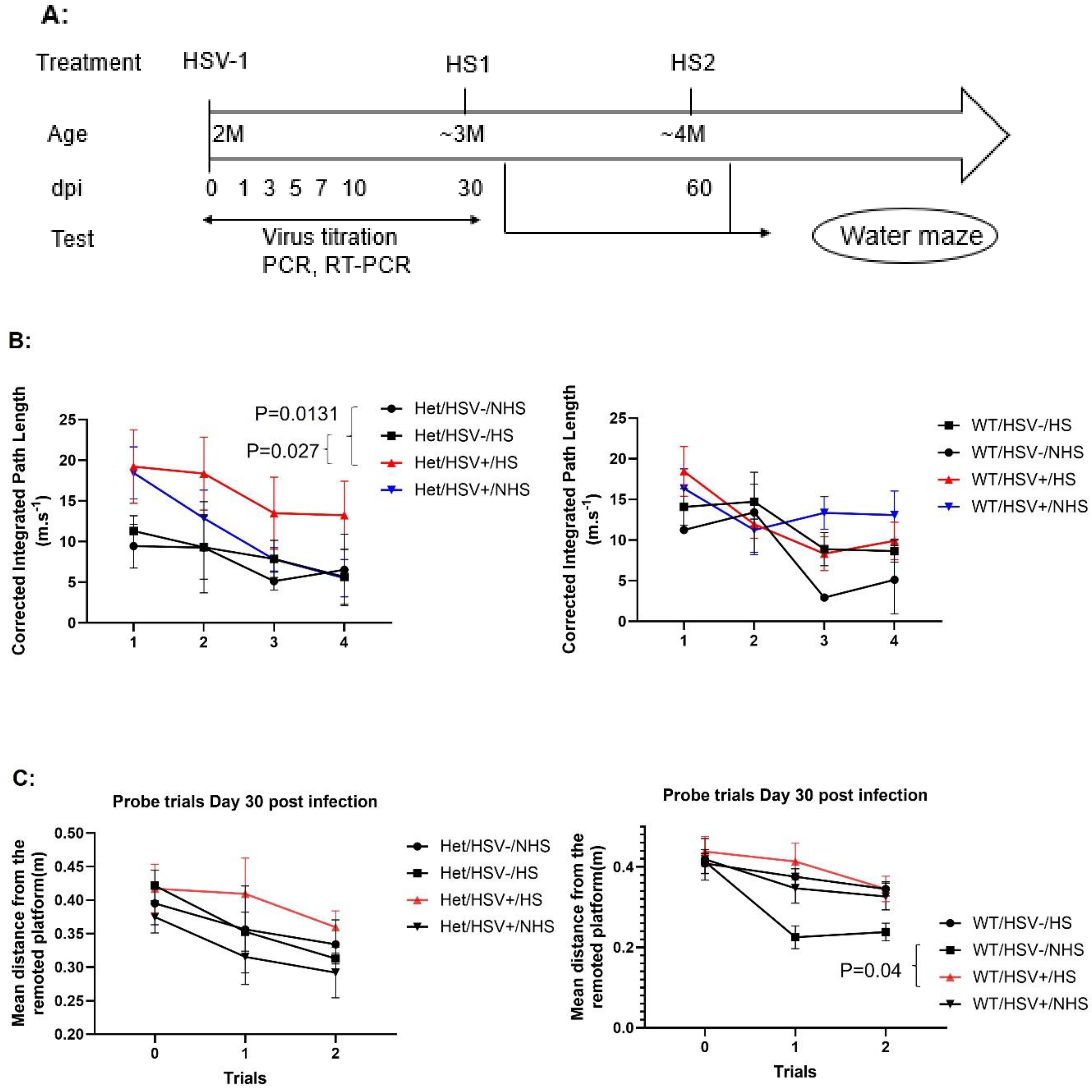
Figure 4.
Cognitive flexibility (reversal trials) analysis in Het (A) and WT (B) mice . Mice were stressed twice, on days 30 and 60 post-infection, and tested 3 days after the second heat stress at 42 C, 10 min. HSV-1-infected mice that were stressed (Het/HSV+/HS) had significantly worse outcomes than Het/HSV-/NHS (P=0.014) and Het/HSV+/NHS (P=0.016) in the reversal trials, Means ± SEM. ANOVA & Fisher’s LSD post-hoc tests. n=3.
Figure 4.
Cognitive flexibility (reversal trials) analysis in Het (A) and WT (B) mice . Mice were stressed twice, on days 30 and 60 post-infection, and tested 3 days after the second heat stress at 42 C, 10 min. HSV-1-infected mice that were stressed (Het/HSV+/HS) had significantly worse outcomes than Het/HSV-/NHS (P=0.014) and Het/HSV+/NHS (P=0.016) in the reversal trials, Means ± SEM. ANOVA & Fisher’s LSD post-hoc tests. n=3.
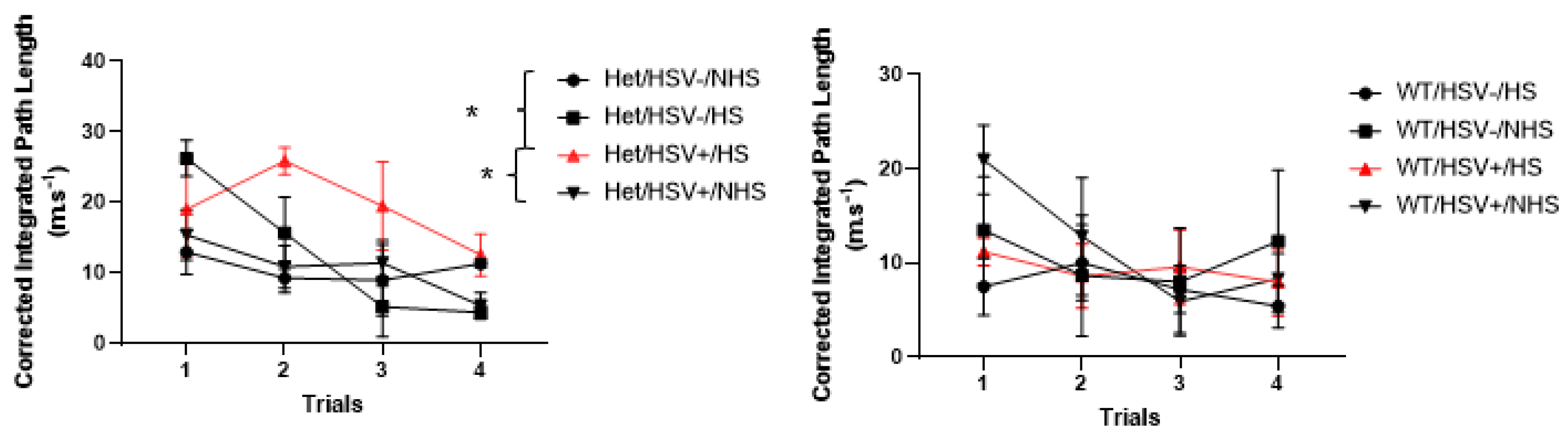
Figure 5.
Timeline for 3 heat-stress study and behavioral testing after recovery. A) Schematic of HSV-1 infection,
heat stress (HS) and behavioral tests. Het and WT mice were stressed once , twice or 3 times on 30, 45, 60 postinfection,
respectively and tested using a Morris water maze, starting 15 days post-stress (42 C; 10 min). B) Spatial
long-term memory (place trials) in HSV+ and HSV- mice. HSV+ mice regardless of stress or genotype had
significantly worse performance than HSV-mice in the place trials (p=0.027). Means ± SEM. ANOVA & Fisher’s
LSD post-hoc tests. HS1: heat stress at 30 days post-infection, HS2: heat stress at 30 and 45 dpi, HS3: heat stress
at 30, 45, and 60 dpi, respectively. n=12.
Figure 5.
Timeline for 3 heat-stress study and behavioral testing after recovery. A) Schematic of HSV-1 infection,
heat stress (HS) and behavioral tests. Het and WT mice were stressed once , twice or 3 times on 30, 45, 60 postinfection,
respectively and tested using a Morris water maze, starting 15 days post-stress (42 C; 10 min). B) Spatial
long-term memory (place trials) in HSV+ and HSV- mice. HSV+ mice regardless of stress or genotype had
significantly worse performance than HSV-mice in the place trials (p=0.027). Means ± SEM. ANOVA & Fisher’s
LSD post-hoc tests. HS1: heat stress at 30 days post-infection, HS2: heat stress at 30 and 45 dpi, HS3: heat stress
at 30, 45, and 60 dpi, respectively. n=12.
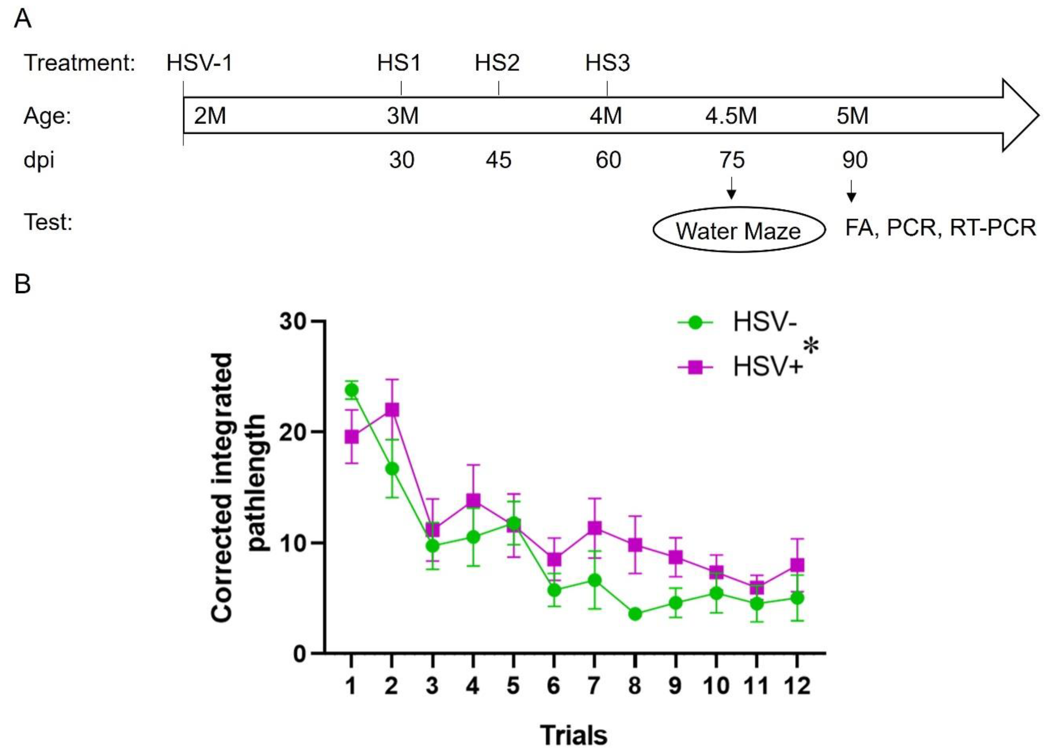
Figure 6.
Amyloid plaque staining of the hippocampus from infected (HSV+) and uninfected (HSV-) Het mice. A) Representative images of Amyloid plaque staining obtained with a fluorescence microscope under UV light. B) Amyloid plaque staining area in the CA1 region were measured by ImageJ software. The brain tissue was sectioned at 90 dpi and stained with Amylo-Glo RTD Amyloid Plaque Stain. The image was taken at 2.5X magnification. The non-stressed HSV-1-infected mice (HSV+/NHS) showed more amyloid staining than HSV-/NHS mice (p=0.02). Means ± SEM. ANOVA & Fisher’s LSD post-hoc tests. HS3: HSV-1-infected and uninfected Het mice were stressed at 30, 45, and 60 dpi, respectively. N=3.
Figure 6.
Amyloid plaque staining of the hippocampus from infected (HSV+) and uninfected (HSV-) Het mice. A) Representative images of Amyloid plaque staining obtained with a fluorescence microscope under UV light. B) Amyloid plaque staining area in the CA1 region were measured by ImageJ software. The brain tissue was sectioned at 90 dpi and stained with Amylo-Glo RTD Amyloid Plaque Stain. The image was taken at 2.5X magnification. The non-stressed HSV-1-infected mice (HSV+/NHS) showed more amyloid staining than HSV-/NHS mice (p=0.02). Means ± SEM. ANOVA & Fisher’s LSD post-hoc tests. HS3: HSV-1-infected and uninfected Het mice were stressed at 30, 45, and 60 dpi, respectively. N=3.
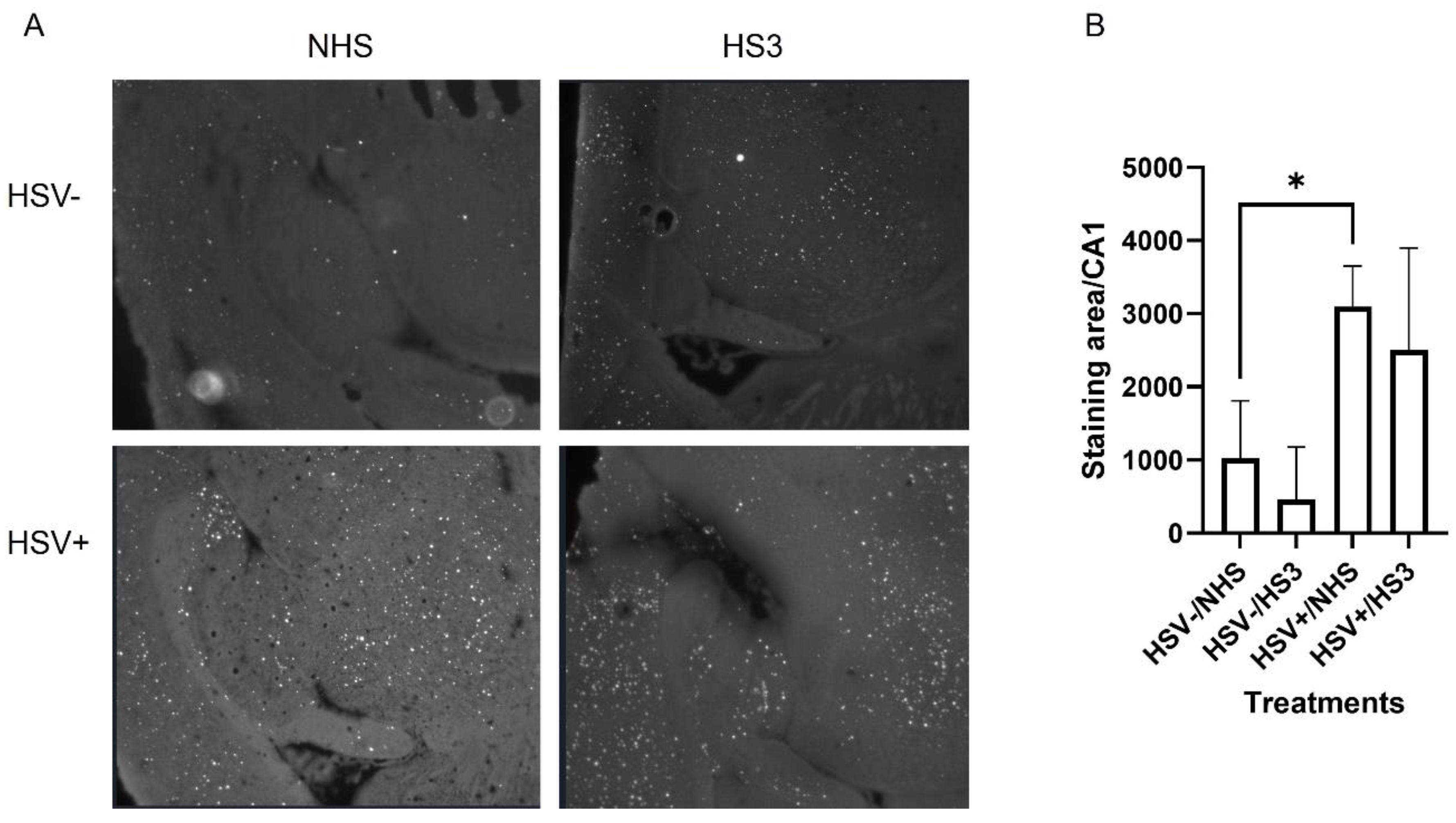
Figure 7.
Amyloid plaque staining in the frontal cortex (FC) region from infected (HSV+) and uninfected (HSV-) Het and WT mice. A) Representative images of Amyloid plaque staining obtained with a fluorescence microscope under UV light. The image was taken at 10X magnification. B) Amyloid plaque staining area in the FC region measured by ImageJ software. The brain tissue was sectioned at 90 dpi and stained with Amylo-Glo RTD Amyloid Plaque Stain. HS3: HSV-1-infected and uninfected Het mice were stressed at 30, 45, and 60 dpi, respectively. There were no differences in plaque area in the frontal cortex between treatments in the Het mice. WT mice showed no amyloid plaques in the frontal cortex. Means ± SEM. ANOVA & Fisher’s LSD post-hoc tests. n=3.
Figure 7.
Amyloid plaque staining in the frontal cortex (FC) region from infected (HSV+) and uninfected (HSV-) Het and WT mice. A) Representative images of Amyloid plaque staining obtained with a fluorescence microscope under UV light. The image was taken at 10X magnification. B) Amyloid plaque staining area in the FC region measured by ImageJ software. The brain tissue was sectioned at 90 dpi and stained with Amylo-Glo RTD Amyloid Plaque Stain. HS3: HSV-1-infected and uninfected Het mice were stressed at 30, 45, and 60 dpi, respectively. There were no differences in plaque area in the frontal cortex between treatments in the Het mice. WT mice showed no amyloid plaques in the frontal cortex. Means ± SEM. ANOVA & Fisher’s LSD post-hoc tests. n=3.
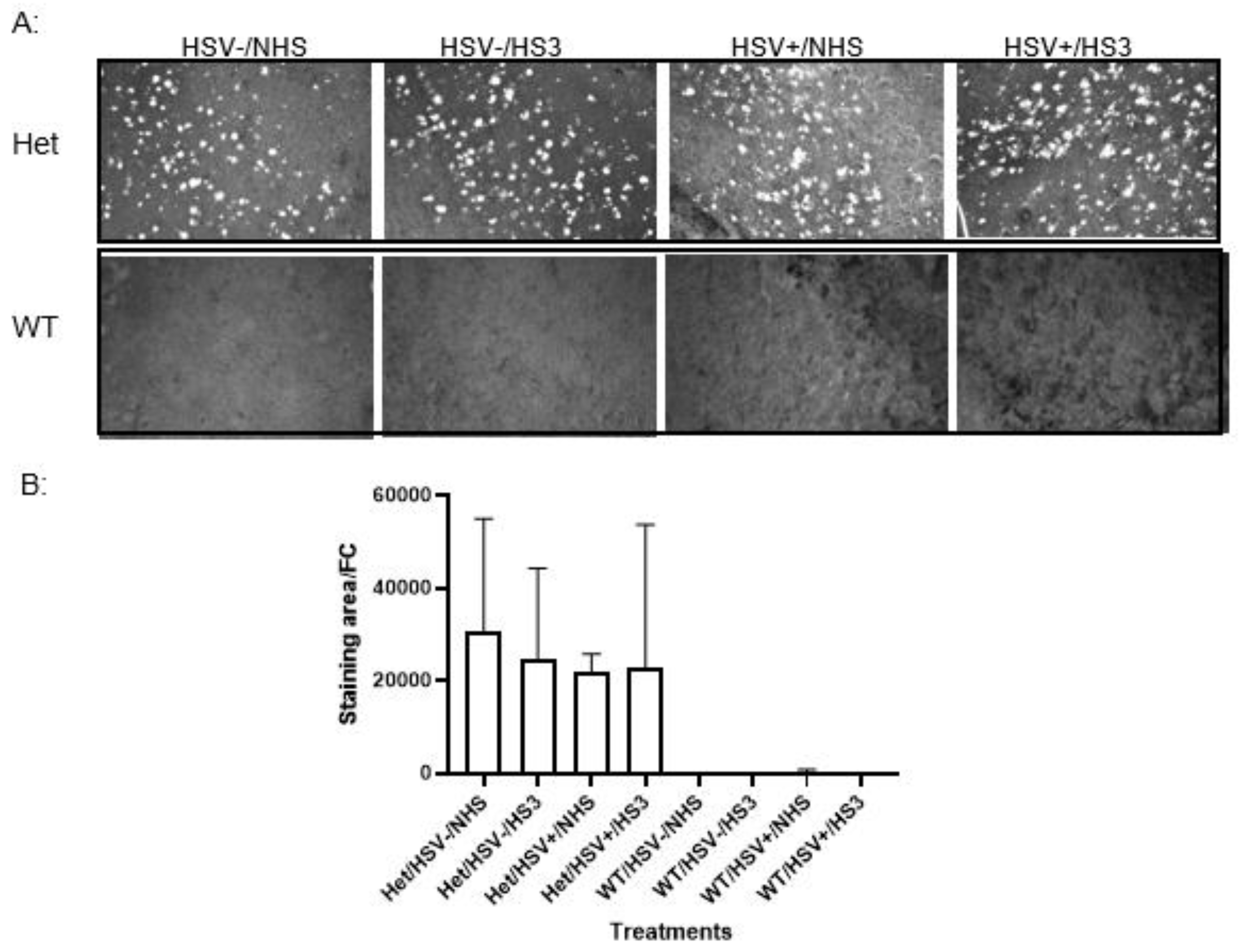
Figure 8.
Real-time PCR of IL-1β and TNFα expression in WT (A-D) and Het (E-H) hippocampus (A,C,E,G) and frontal cortex (B,D,F,H) from different treatment groups. The IL-1β and TNFα expression were measured by SYBR real-time PCR with cDNA generated from total RNA isolated from hippocampus or frontal cortex of Het and WT mice, respectively. cDNA was synthesized with 100 ng of total RNA of each tissue from different treatment groups. Transcript copy numbers were estimated by IL-1β and TNFα real-time PCR, respectively, as described in the Material and Methods. Statistical analysis was performed by using multiple unpaired t test, *: p<0.01, **: p<0.001, ***: p<0.00001. n=3.
Figure 8.
Real-time PCR of IL-1β and TNFα expression in WT (A-D) and Het (E-H) hippocampus (A,C,E,G) and frontal cortex (B,D,F,H) from different treatment groups. The IL-1β and TNFα expression were measured by SYBR real-time PCR with cDNA generated from total RNA isolated from hippocampus or frontal cortex of Het and WT mice, respectively. cDNA was synthesized with 100 ng of total RNA of each tissue from different treatment groups. Transcript copy numbers were estimated by IL-1β and TNFα real-time PCR, respectively, as described in the Material and Methods. Statistical analysis was performed by using multiple unpaired t test, *: p<0.01, **: p<0.001, ***: p<0.00001. n=3.
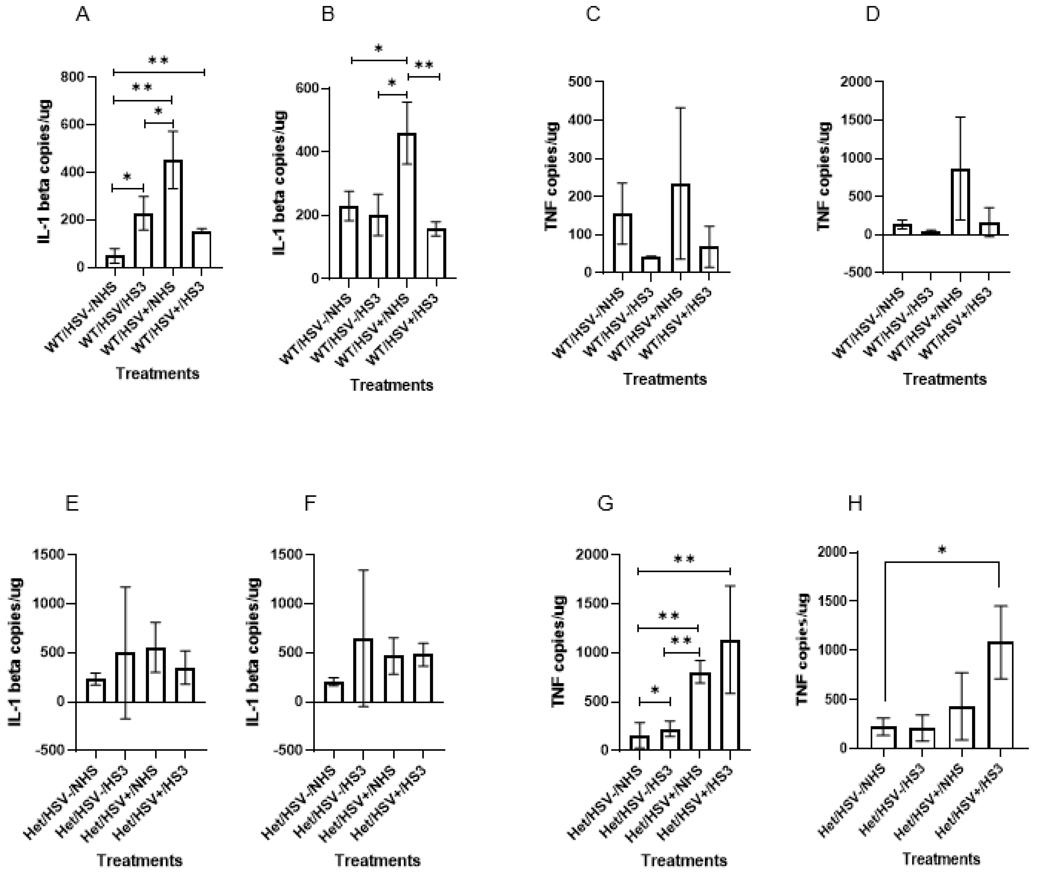
Figure 9.
Indirect immunofluorescence analyses of brain sections from WT (A,B) and Het (C,D) mice. A, C) GFAP antibody staining of brain sections from hippocampus (CA1 and DG) and frontal cortex (FC) from WT (A) and Het (C) mice. Brain sections were fixed, permeabilized in acetone first, then stained with rabbit polyclonal GFAP antibody first, then goat anti-rabbit secondary antibody. Cell nuclei were visualized with DAPI. CA1, DG, and FC correspond to the Cornu Ammonis 1 region of the hippocampus, the dentate gyrus, and the frontal cortex, respectively. The image was taken at 40X magnification. B,D) Quantification of GFAP staining area in CA1 region from WT (A) and Het (C) mice measured by ImageJ software. HS3: HSV-1-infected and uninfected Het mice were stressed at 30, 45, and 60 dpi, respectively. Statistical analysis was performed by using multiple unpaired t test, *: p<0.01, n=3.
Figure 9.
Indirect immunofluorescence analyses of brain sections from WT (A,B) and Het (C,D) mice. A, C) GFAP antibody staining of brain sections from hippocampus (CA1 and DG) and frontal cortex (FC) from WT (A) and Het (C) mice. Brain sections were fixed, permeabilized in acetone first, then stained with rabbit polyclonal GFAP antibody first, then goat anti-rabbit secondary antibody. Cell nuclei were visualized with DAPI. CA1, DG, and FC correspond to the Cornu Ammonis 1 region of the hippocampus, the dentate gyrus, and the frontal cortex, respectively. The image was taken at 40X magnification. B,D) Quantification of GFAP staining area in CA1 region from WT (A) and Het (C) mice measured by ImageJ software. HS3: HSV-1-infected and uninfected Het mice were stressed at 30, 45, and 60 dpi, respectively. Statistical analysis was performed by using multiple unpaired t test, *: p<0.01, n=3.
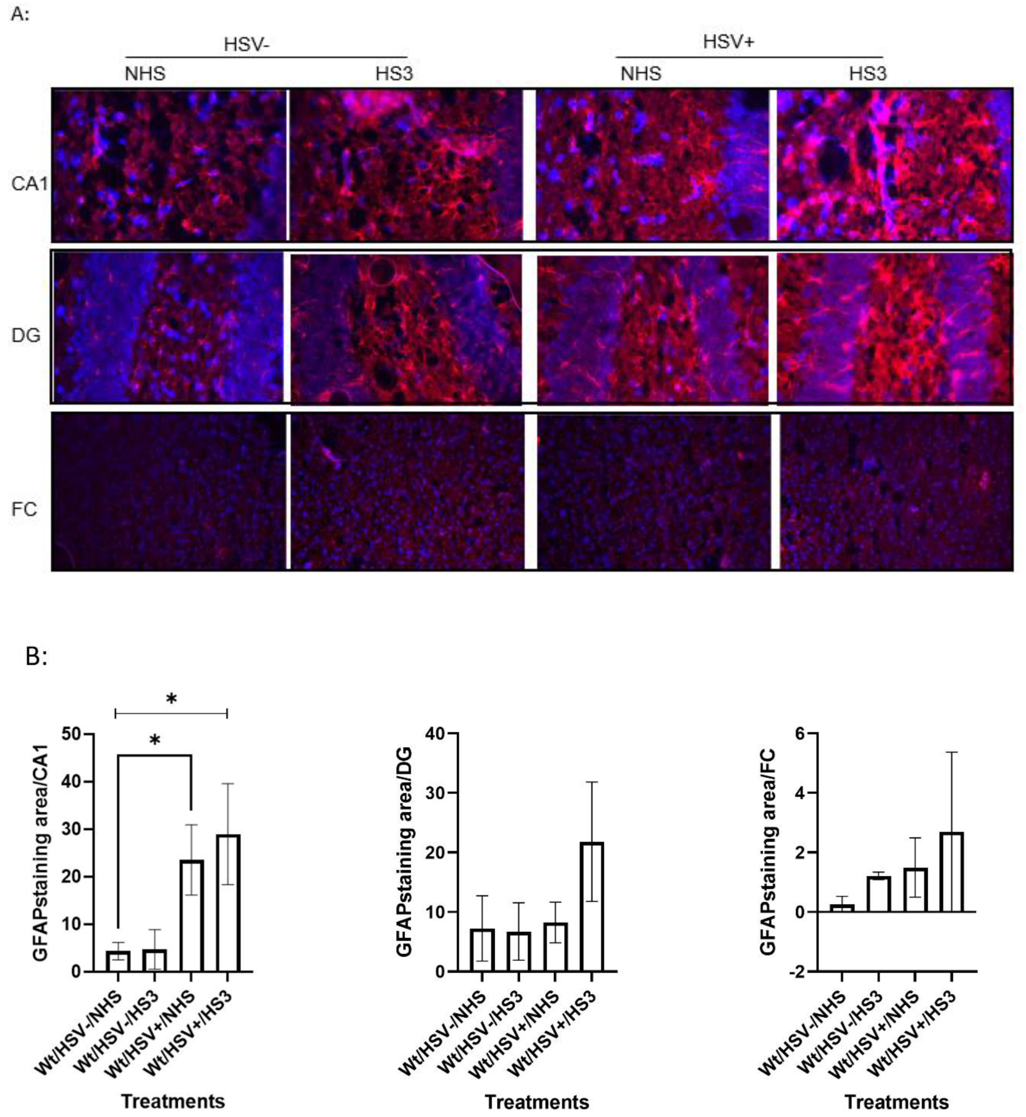
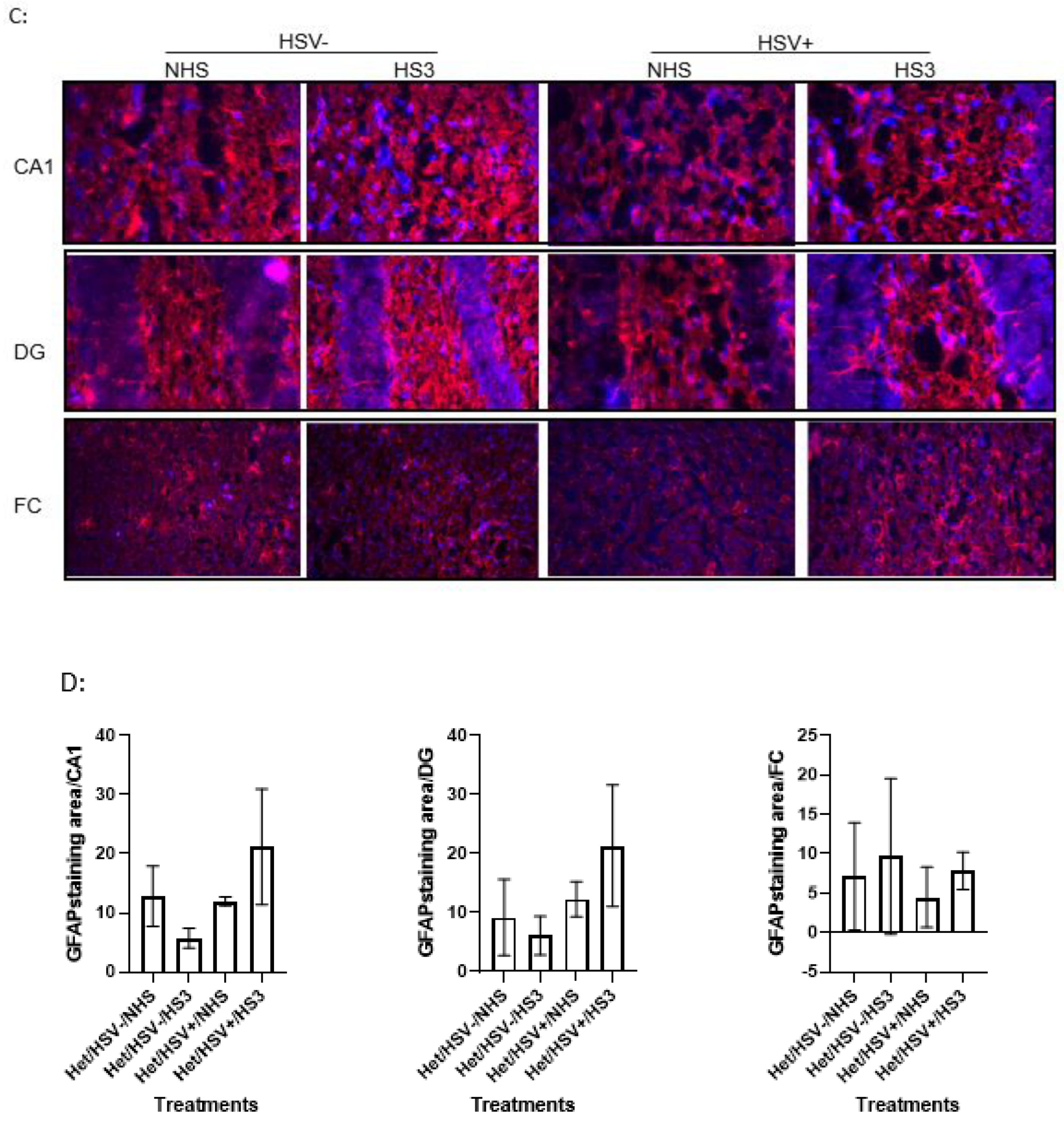
Figure 10.
Sagittal brain section and real-time PCR of HSV-1. A. Sagittal brain section of the mouse brain (made by Biorender) showing regions sampled. Labeled brain regions were collected for HSV-1 DNA or RNA analysis. FC, Cx, H, C, and BS correspond to the frontal cortex, caudal cortex, hippocampus, cerebellum, and brainstem, respectively. B. HSV-1 DNA copy per ug tissue estimated by real-time PCR with primers specific for HSV-1 LAT gene. TG: trigeminal ganglion. D1, D3, D5, D15, and D30 correspond to 1, 3, 5, 15, and 30 days post-infection. N=3.
Figure 10.
Sagittal brain section and real-time PCR of HSV-1. A. Sagittal brain section of the mouse brain (made by Biorender) showing regions sampled. Labeled brain regions were collected for HSV-1 DNA or RNA analysis. FC, Cx, H, C, and BS correspond to the frontal cortex, caudal cortex, hippocampus, cerebellum, and brainstem, respectively. B. HSV-1 DNA copy per ug tissue estimated by real-time PCR with primers specific for HSV-1 LAT gene. TG: trigeminal ganglion. D1, D3, D5, D15, and D30 correspond to 1, 3, 5, 15, and 30 days post-infection. N=3.
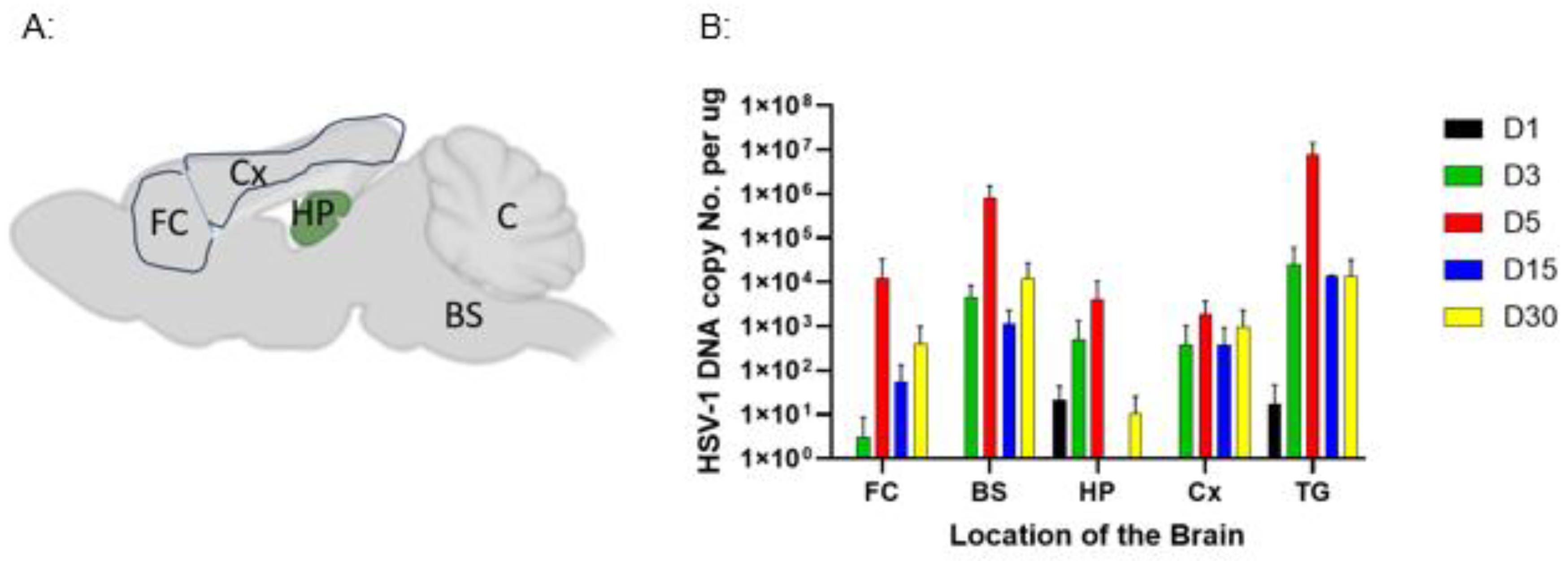
Figure 11.
Detection of HSV-1 DNA in the brain tissues by nested PCR with primers specific to UL36. The 1st run PCR product was amplified from total DNA extracted from brain tissues harvested at 90 dpi from Het (top two images) and WT (Bottom two images) mice. The 2nd-run PCR product was amplified from the 1st-run PCR product for each sample. Lanes MW, FC, Cx, HP, BS, C, P, and N correspond to 1 kb plus ladder, frontal cortex, caudal cortex, hippocampus, brainstem, cerebellum, HSV-1 genome, and uninfected tissue DNA, respectively. M1, M2, and M3 represent HSV-1 DNA detection in different mice from HSV-1+/NHS mice. n=3.
Figure 11.
Detection of HSV-1 DNA in the brain tissues by nested PCR with primers specific to UL36. The 1st run PCR product was amplified from total DNA extracted from brain tissues harvested at 90 dpi from Het (top two images) and WT (Bottom two images) mice. The 2nd-run PCR product was amplified from the 1st-run PCR product for each sample. Lanes MW, FC, Cx, HP, BS, C, P, and N correspond to 1 kb plus ladder, frontal cortex, caudal cortex, hippocampus, brainstem, cerebellum, HSV-1 genome, and uninfected tissue DNA, respectively. M1, M2, and M3 represent HSV-1 DNA detection in different mice from HSV-1+/NHS mice. n=3.
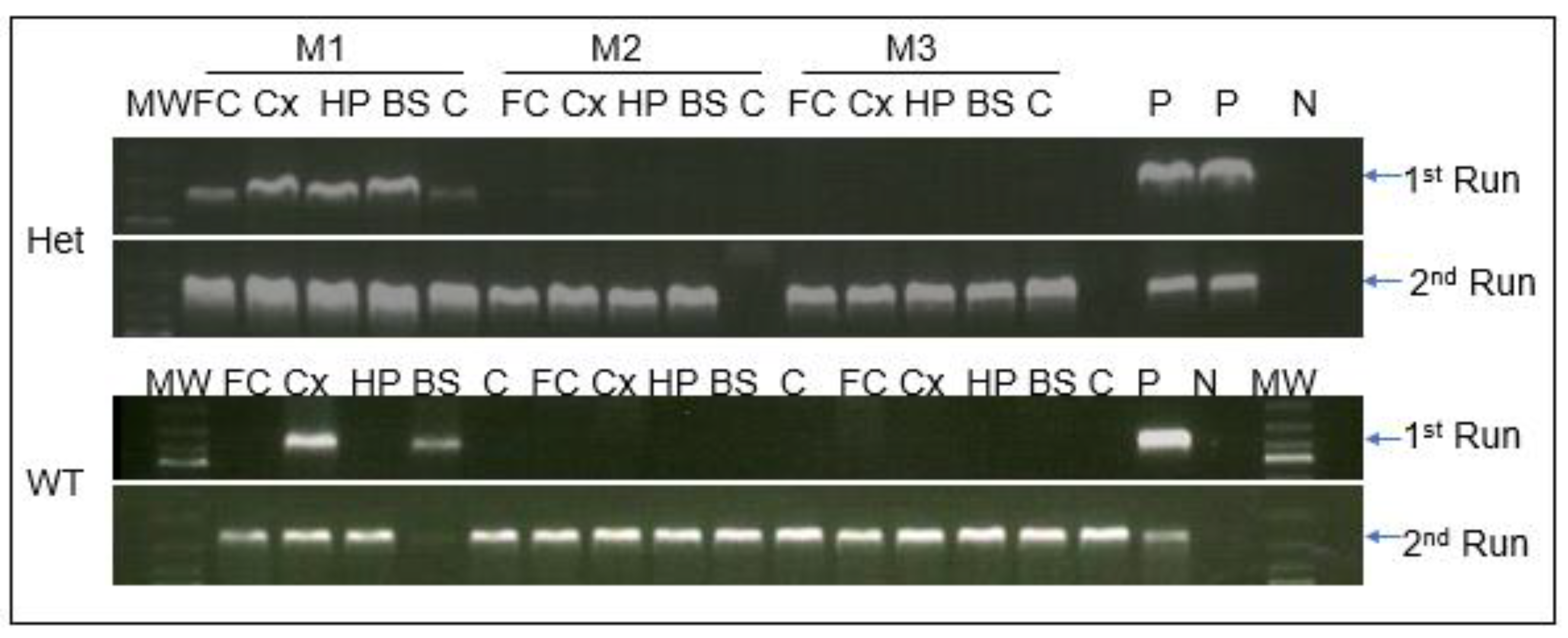
Figure 12.
HSV-1 reactivation via mouse brain tissue explant culture. A: Number of tissues with HSV-1 reactivation in tissue explant cultures. Brain tissues were harvested at 33 days post-infection and cultured in a 12-well plate in 1.5 ml DMEM with 10% FBS. Fc, Cx, H, C, BS, and TG correspond to the frontal cortex, caudal cortex, hippocampus, cerebellum, brainstem, and trigeminal ganglion, respectively. B: GFP-positive cells infected with tissue explant medium on day 7 post-explant. Images were taken at 24h post-infection with a 10x FITC fluorescent microscope. N=8.
Figure 12.
HSV-1 reactivation via mouse brain tissue explant culture. A: Number of tissues with HSV-1 reactivation in tissue explant cultures. Brain tissues were harvested at 33 days post-infection and cultured in a 12-well plate in 1.5 ml DMEM with 10% FBS. Fc, Cx, H, C, BS, and TG correspond to the frontal cortex, caudal cortex, hippocampus, cerebellum, brainstem, and trigeminal ganglion, respectively. B: GFP-positive cells infected with tissue explant medium on day 7 post-explant. Images were taken at 24h post-infection with a 10x FITC fluorescent microscope. N=8.
Disclaimer/Publisher’s Note: The statements, opinions and data contained in all publications are solely those of the individual author(s) and contributor(s) and not of MDPI and/or the editor(s). MDPI and/or the editor(s) disclaim responsibility for any injury to people or property resulting from any ideas, methods, instructions or products referred to in the content. |
© 2026 by the authors. Licensee MDPI, Basel, Switzerland. This article is an open access article distributed under the terms and conditions of the Creative Commons Attribution (CC BY) license (http://creativecommons.org/licenses/by/4.0/).
Copyright: This open access article is published under a Creative Commons CC BY 4.0 license, which permit the free download, distribution, and reuse, provided that the author and preprint are cited in any reuse.


