

Submitted:
17 October 2026
Posted:
19 October 2026
You are already at the latest version
Preprints on COVID-19 and SARS-CoV-2
Abstract
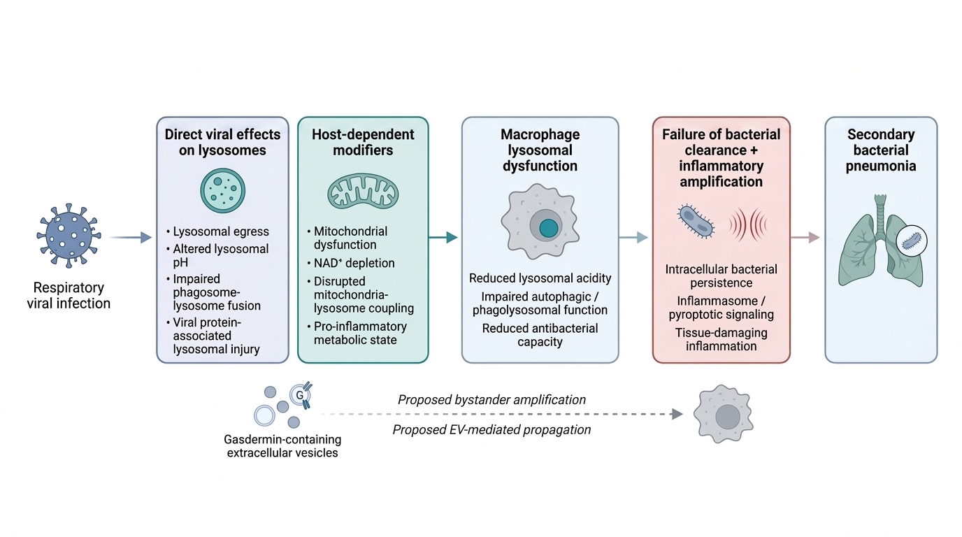
Respiratory viral infections are a major cause of global morbidity and mortality, partly because they increase susceptibility to secondary bacterial pneumonia and sepsis. Coronaviruses, influenza viruses, parainfluenza viruses, rhinoviruses, and respiratory syncytial virus have each been associated with impaired macrophage antibacterial function, although the underlying evidence differs among virus families. Direct lysosomal-pH measurements and mechanistically resolved links between infection and lysosomal de-acidification are strongest for coronaviruses. Using β-coronaviruses as the primary model for the effect of direct lysosomal disruption on macrophage antimicrobial capacity, this review examines lysosomal exploitation during viral egress, impaired phagosome-lysosome fusion, E-protein-mediated proton conductance, and ORF3a-associated lysosomal injury. It further proposes that the magnitude of these effects is shaped by host-dependent immunometabolic processes. Mitochondrial dysfunction, NAD⁺ depletion, and disrupted mitochondria-lysosome coupling may reduce the ability of macrophages to preserve lysosomal acidity, autophagic competence, and bacterial killing during viral challenge. Together, these mechanisms describe how coronavirus infection compromises macrophage antibacterial defense, and how the resulting susceptibility to secondary bacterial infection is further modulated by the metabolic and inflammatory state of the host cell.
Keywords:
coronavirus
; macrophage
; lysosomal dysfunction
; antibacterial immunity
; immunometabolism
; secondary bacterial infection
Introduction
Respiratory viral infections are a major cause of global morbidity and mortality, in part because they increase susceptibility to secondary bacterial disease. This vulnerability is well documented following coronavirus, influenza, parainfluenza, rhinovirus, and respiratory syncytial virus infection (Al Rahmoun et al., 2025; Koçak Tufan et al., 2021; Peng et al., 2022; Peng et al., 2023; Prasso & Deng, 2017; Rich et al., 2024; Shi et al., 2025; Verma et al., 2020). The same pattern is seen experimentally: bacterial challenge that is otherwise well controlled can produce severe pneumonia when preceded by respiratory viral infection (Ghoneim et al., 2013; Hraiech et al., 2015; Kalhoro et al., 2016; Small et al., 2010). Although impaired mucociliary clearance and altered receptor expression contribute to this susceptibility (Bakaletz, 2017), loss of macrophage bactericidal capacity may represent another important component.
Macrophage bacterial clearance depends on coordinated phagocytosis, intracellular killing, and lysosomal function (Hirayama et al., 2017). Lysosomal acidification activates proteases and antimicrobial peptides required for degradation of engulfed organisms, making even modest increases in lysosomal pH potentially consequential for antibacterial activity (Lie & Nixon, 2019; Prince et al., 2008; Sanman et al., 2016). De-acidification may also occur alongside impaired autophagosome-lysosome or phagosome-lysosome fusion. Where these defects coexist, bacterial killing is weakened while the loss of normal autophagic restraint can promote NLRP3 inflammasome activation and sustain inflammation (Biasizzo & Kopitar-Jerala, 2020; Blevins et al., 2022; Gupta et al., 2025).
The evidence linking respiratory viral infection directly to lysosomal dysfunction is strongest for coronaviruses, which are therefore used here as the primary mechanistic model. β-coronaviruses exploit the lysosomal pathway during egress while altering lysosomal pH and membrane integrity (Ghosh et al., 2020; Peng et al., 2023), impair fusion between lysosomes and bacteria-containing phagosomes (Peng et al., 2022), conduct protons across intracellular membranes through the envelope (E) protein, and produce ORF3a-associated lysosomal injury (Ghosh et al., 2020; Yue et al., 2018). Influenza, parainfluenza, rhinovirus, and RSV can also impair macrophage antibacterial function, but the evidence in these systems more often concerns defective phagolysosomal fusion, impaired bacterial killing, or mitochondrial injury than direct changes in lysosomal pH. These viruses are therefore considered as comparative evidence for a shared downstream defect in macrophage function, rather than as evidence that lysosomal de-acidification is universal across respiratory virus families.
The severity of this defect may also depend on the condition of the macrophage in which viral injury occurs. Mitochondrial dysfunction is associated with impaired macrophage antibacterial activity, while restoration of mitochondrial function can improve bacterial killing (Cao et al., 2020; Plataki et al., 2019; Spier et al., 2021). Alveolar macrophages also become increasingly dependent on oxidative phosphorylation as they mature (Izquierdo et al., 2018; Zhang et al., 2018), while inflammatory cytokines including TNF-α and IL-1β can suppress oxidative metabolism and promote lysosomal de-acidification (López-Armada et al., 2006; Nilsson et al., 2006; Samavati et al., 2008; Tangpong et al., 2008). Aging and chronic disease may further reduce this metabolic and lysosomal reserve (Duong et al., 2021; Kale et al., 2020; López-Vázquez et al., 2024; Watanabe et al., 2014).
This review examines how these host-dependent changes may modify the lysosomal dysfunction produced by β-coronavirus infection. Particular attention is given to mitochondrial dysfunction, NAD+ depletion, and altered mitochondria-lysosome contact sites as possible determinants of lysosomal acidification and antibacterial capacity. It also considers whether lysosomal injury can be amplified through cathepsin release, inflammasome activation, and gasdermin-mediated membrane damage, including the possibility that gasdermin-containing extracellular vesicles transmit this injury to neighboring macrophages (Du & Wu, 2025; Wright et al., 2025; Zhou et al., 2026). These latter relationships remain incompletely resolved and are treated as testable extensions of the established coronavirus-associated lysosomal defects.
Methods
This narrative review examines evidence linking respiratory viral infection, macrophage lysosomal dysfunction, and susceptibility to secondary bacterial pneumonia, with β-coronaviruses serving as the primary mechanistic focus. A narrative approach was used because the relevant evidence spans respiratory virology, macrophage biology, lysosomal physiology, immunometabolism, bacterial pathogenesis, and aging, and includes studies conducted across different cell types and experimental systems. Particular attention was given to direct evidence of coronavirus-associated lysosomal dysfunction and to findings that may explain how mitochondrial function, NAD+ metabolism, organelle communication, and inflammatory cell death modify macrophage antibacterial capacity.
Literature Search
PubMed, Scopus, and Google Scholar were searched for relevant peer-reviewed literature published through July 2026. Searches combined terms related to coronavirus and other respiratory viral infections with terms related to macrophage function, lysosomal acidification, phagolysosomal fusion, secondary bacterial pneumonia, immunometabolism, mitochondria-lysosome contact sites, NAD+, gasdermins, extracellular vesicles, and pyroptosis. Reference lists of relevant articles were also screened to identify foundational studies and additional papers not captured in the initial searches.
Inclusion and Exclusion Criteria
Studies were considered when they addressed mechanisms relevant to coronavirus-associated lysosomal dysfunction, macrophage antibacterial activity, or secondary bacterial pneumonia. Evidence from other respiratory viruses was included when it provided comparative support for macrophage dysfunction or related host-dependent mechanisms. The review includes original experimental studies, animal models, mechanistic cell-culture studies, systematic reviews, meta-analyses, and selected clinical or epidemiological studies where relevant to the proposed framework.
Only English-language articles were included. Recent studies were prioritized where possible, while older work was retained when it established foundational observations concerning lysosomal physiology, mitochondrial function, macrophage metabolism, inflammatory signaling, or viral-bacterial interactions.
Terminology
The terms M1-like and M2-like are used as conceptual reference points, as opposed to rather than fixed macrophage categories. Macrophage activation occurs along a continuum shaped by tissue origin, developmental lineage, cytokine exposure, metabolic state, and the nature and duration of the initiating stimulus. Here, M1-like refers broadly to inflammatory states commonly associated with altered mitochondrial metabolism, whereas M2-like refers to comparatively oxidative and tissue-reparative states. The mechanisms discussed in this review are not assumed to occur exclusively within either phenotype.
Study Quality and Synthesis Approach
The evidence considered here varies substantially in experimental design and in how directly it bears on the proposed mechanisms. It includes pulmonary macrophage studies, other primary and immortalized cell systems, animal infection models, clinical observations, and studies in other cell types used to establish particular organellar mechanisms. A formal risk-of-bias assessment was not performed because the review does not attempt to combine comparable outcomes across a uniform group of studies.
Instead, evidence is distinguished throughout according to whether a mechanism was demonstrated directly during respiratory viral infection, inferred from another experimental system, or proposed on the basis of convergence across separate lines of evidence. This distinction is particularly important for mitochondria-lysosome coupling, NAD+ depletion, and gasdermin-containing extracellular vesicles, since these processes have not been demonstrated together in a single coronavirus-infected pulmonary macrophage system. Evidence from separate models is therefore used to establish plausibility and generate testable predictions, not to imply that every proposed step has already been demonstrated as part of one pathway.
Immunometabolic Reprogramming and Its Convergence on Lysosomal De-Acidification
In addition to the direct viral effects described above, lysosomal function is shaped by the metabolic and inflammatory state of the macrophage. Macrophages remodel their metabolism rapidly in response to inflammatory stimuli, and these changes are closely tied to antimicrobial function (Stienstra et al., 2017). This is particularly relevant in severe respiratory disease, including COVID-19, where macrophage-driven inflammatory signaling can become a major component of tissue injury (Lee et al., 2021).
Pro-inflammatory macrophage activation is commonly associated with a shift toward aerobic glycolysis and changes in mitochondrial function (Zuo & Wan, 2019), although this pattern is not uniform across macrophage populations. Tissue-resident alveolar macrophages (TR-AMs), for example, can mount a robust inflammatory response to LPS without substantially increasing glucose metabolism (Pereverzeva et al., 2022; Woods & Mutlu, 2025). This does not necessarily make them less dependent on mitochondrial metabolism. Alveolar macrophages have comparatively low expression of glycolytic enzymes and transporters, and glycolysis cannot fully compensate when electron-transport-chain activity is impaired. In aged mice infected with Streptococcus pneumoniae, macrophages show reduced ATP production, altered respiratory-complex expression, and fewer healthy mitochondria than those from younger animals (Plataki et al., 2019; Woods et al., 2020; Woods & Mutlu, 2025). Thus, limited glycolytic reserve may leave TR-AMs particularly vulnerable when oxidative phosphorylation is compromised.
Respiratory infection can also alter the metabolic composition of the alveolar macrophage compartment itself. Loss of TR-AMs during viral infection is accompanied by recruitment of neutrophils and monocyte-derived alveolar macrophages (Mo-AMs), which are more glycolytically biased. In SARS-CoV-2 infection, depletion of TR-AMs and their replacement by recruited monocyte-derived macrophages has been described as part of a sustained inflammatory circuit in the alveolar space (Grant et al., 2021). Influenza provides additional evidence that viral infection can compromise mitochondrial function in alveolar macrophages: Gao et al. (2022) reported reduced TFAM expression, abnormal mitochondrial morphology, and diminished enrichment of oxidative-phosphorylation genes in infected human alveolar macrophages. Because TFAM-dependent mitochondrial metabolism is required for TR-AM maintenance and self-renewal, these changes provide a route through which respiratory viral infection may reduce the oxidative capacity of the resident macrophage population.
A reduction in oxidative phosphorylation is not, however, inherently pathological. During macrophage responses to live S. pneumoniae, mitochondrial fission is accompanied by a progressive decline in oxidative phosphorylation and increased mitochondrial ROS production, which can contribute directly to antibacterial activity (Choudhuri et al., 2021; Mohasin et al., 2021). The relevant distinction may therefore be between controlled metabolic remodeling that supports antimicrobial defense and mitochondrial dysfunction severe or persistent enough to reduce the energetic and organellar support required for bacterial killing.
Changes in nutrient sensing provide one route by which inflammatory metabolism may affect the lysosome more directly. mTORC1 is recruited to the lysosomal membrane through the Rag GTPase-Ragulator complex, where it phosphorylates TFEB and retains it in the cytoplasm. When mTORC1 activity falls, TFEB enters the nucleus and induces the CLEAR transcriptional network, including genes involved in lysosomal biogenesis and V-ATPase function (Peña-Llopis et al., 2011; Roczniak-Ferguson et al., 2012). Because inflammatory stimuli such as LPS can engage PI3K/Akt/mTORC1 signaling, sustained mTORC1 activity could limit TFEB-dependent lysosomal adaptation during periods of increased bactericidal demand (Weichhart et al., 2015).
This relationship is not unidirectional. LPS also induces IRG1-dependent production of itaconate, which can alkylate TFEB and prevent its inhibitory phosphorylation by mTORC1, allowing TFEB to enter the nucleus and promote lysosomal biogenesis (Zhang et al., 2022). In that study, loss of IRG1 or expression of an alkylation-resistant TFEB impaired macrophage antibacterial activity. The timing of these opposing signals may matter: TFEB alkylation was detectable after four hours of LPS stimulation but not after one hour, suggesting that the lysosome-supporting effects of itaconate emerge after the initial inflammatory response is already underway.
AMPK provides another potential counterweight. Falling ATP would ordinarily activate AMPK, inhibit mTORC1, and favor TFEB-dependent lysosomal activity. Yet sustained inflammatory signaling may weaken this compensation. LPS, TNF-α, and IL-6 have each been reported to suppress AMPK signaling through different mechanisms, raising the possibility that prolonged inflammation could interfere with the same feedback response that would otherwise restore lysosomal capacity (Fan et al., 2018).
These pathways illustrate why macrophage inflammatory state cannot be reduced simply to a glycolytic-versus-oxidative distinction. Metabolic remodeling can support antimicrobial activity in some settings while, when sufficiently prolonged or accompanied by mitochondrial dysfunction, limiting the machinery required to maintain lysosomal function. This becomes especially important because mitochondria contribute to lysosomal acidification through more than bulk ATP production. They also communicate directly with lysosomes at mitochondria-lysosome contact sites, providing a second route through which loss of mitochondrial competence may translate into impaired bacterial killing.
Figure 1.
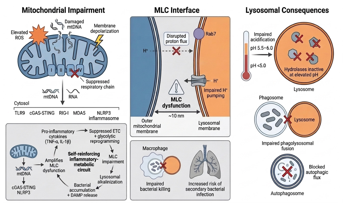
Mitochondrial impairment disrupts lysosomal acidification through mitochondria-lysosome contact site (MLC) dysfunction. Mitochondrial damage - elevated ROS, damaged mtDNA, membrane depolarization, and a suppressed respiratory chain - releases mtDNA/RNA into the cytosol, engaging cytosolic sensors (TLR9, cGAS-STING, RIG-I, MDA5, NLRP3) and establishing a self-reinforcing inflammatory-metabolic circuit. At the ~10 nm mitochondria-lysosome interface, this dysfunction disrupts Rab7-dependent proton flux, impairing H+ transfer into the lysosomal lumen. The resulting rise in lysosomal pH (5.5-6.0, versus a healthy <5.0) inactivates lysosomal hydrolases, impairs phagolysosomal fusion, and blocks autophagic flux - collectively compromising macrophage bactericidal capacity and increasing susceptibility to secondary bacterial infection. The figure depicts potential points of mechanistic convergence rather than an obligatory temporal sequence; altered lysosomal pH, impaired fusion, defective autophagic flux, and trafficking abnormalities may arise in parallel or interact in a context-dependent manner.
Figure 1.
Mitochondrial impairment disrupts lysosomal acidification through mitochondria-lysosome contact site (MLC) dysfunction. Mitochondrial damage - elevated ROS, damaged mtDNA, membrane depolarization, and a suppressed respiratory chain - releases mtDNA/RNA into the cytosol, engaging cytosolic sensors (TLR9, cGAS-STING, RIG-I, MDA5, NLRP3) and establishing a self-reinforcing inflammatory-metabolic circuit. At the ~10 nm mitochondria-lysosome interface, this dysfunction disrupts Rab7-dependent proton flux, impairing H+ transfer into the lysosomal lumen. The resulting rise in lysosomal pH (5.5-6.0, versus a healthy <5.0) inactivates lysosomal hydrolases, impairs phagolysosomal fusion, and blocks autophagic flux - collectively compromising macrophage bactericidal capacity and increasing susceptibility to secondary bacterial infection. The figure depicts potential points of mechanistic convergence rather than an obligatory temporal sequence; altered lysosomal pH, impaired fusion, defective autophagic flux, and trafficking abnormalities may arise in parallel or interact in a context-dependent manner.
Mitochondria-Lysosome Contact Sites
Mitochondrial metabolic competence is closely linked to macrophage antibacterial capacity. Aged alveolar and interstitial macrophages with impaired oxidative phosphorylation and reduced ATP production clear Streptococcus pneumoniae and influenza less effectively than macrophages from younger animals (Plataki et al., 2019; Wong et al., 2017). In S. pneumoniae-infected macrophages, this impairment is accompanied by reduced ATP production and dysregulated expression of respiratory-chain complexes (Plataki et al., 2019). The defect extends beyond phagocytic uptake to the intracellular destruction of engulfed bacteria (Wong et al., 2017). Conversely, myeloid ACE overexpression increases oxidative metabolism and ATP production together with superoxide generation, phagocytic activity, respiratory-chain protein expression, and resistance to S. pneumoniae and other invasive organisms (Cao et al., 2020). Together, these findings suggest that mitochondrial function and macrophage bactericidal capacity rise and fall together.
One mechanism that may help explain this relationship is direct communication between mitochondria and lysosomes. Mitochondria-lysosome contact sites (MLCs) are transient interfaces in which the outer mitochondrial membrane and lysosomal membrane are brought to within approximately 10 nm of one another (Tian et al., 2026; Yan et al., 2026). Recent work indicates that these contacts can support proton transfer from the mitochondrial intermembrane space into the lysosomal lumen, providing a direct means by which mitochondrial energetic state may influence lysosomal acidity (Tian et al., 2026). MLCs have been observed across multiple mammalian cell types and appear to represent a conserved form of inter-organellar communication involved in lysosomal homeostasis (Wong et al., 2018; Yan et al., 2026).
The small GTPase Rab7 regulates formation and dissolution of these contacts. In its GTP-bound state, Rab7 promotes mitochondrial-lysosomal tethering, while GTP hydrolysis releases the contact and terminates the interaction (Wong et al., 2018). If proton transfer at MLCs contributes meaningfully to lysosomal acidification, then loss of the mitochondrial proton gradient through electron-transport-chain inhibition, uncoupling, or oxidative damage would be expected to reduce the driving force for this process. Mitochondrial dysfunction could therefore impair macrophage bacterial killing through two related routes: by reducing ATP available for energy-dependent cellular functions and by weakening direct mitochondrial support of lysosomal acidification.
Evidence from other cell types supports a broader connection between mitochondrial failure and lysosomal dysfunction. Genetic or pharmacological disruption of mitochondrial function impairs lysosomal structure and activity in neuronal cells, microglia, and T cells (Baixauli et al., 2015; Demers-Lamarche et al., 2016). Mitochondrial ROS can also impair the autophagy-lysosome system in macrophages and promote lipid peroxidation at organellar membranes (Yuan et al., 2019). Whether inflammatory mitochondrial injury disrupts MLCs specifically through Rab7 dysfunction, membrane lipid damage, loss of proton motive force, or some combination of these processes has not yet been resolved.
Inflammatory signaling may further reinforce this relationship. TNF-α has been shown to suppress mitochondrial respiratory-chain activity while also increasing lysosomal pH and lysosomal membrane permeability (Beasley & Eldridge, 1994; Nilsson et al., 2006; Sánchez-Alcázar et al., 2003; Stadler et al., 1992). Mitochondrial dysfunction can in turn increase ROS production and release mitochondrial nucleic acids into the cytosol, engaging inflammatory sensors including cGAS-STING, RIG-I, MDA5, and NLRP3 (Algieri et al., 2025; López-Polo et al., 2024). These observations suggest a potential feedback loop in which inflammation weakens mitochondrial support of lysosomal function while mitochondrial injury further sustains inflammatory signaling. Whether this loop directly disrupts MLC-dependent acidification in coronavirus-exposed pulmonary macrophages remains to be tested.
The importance of MLCs to the model proposed here is therefore straightforward: a macrophage entering viral infection with reduced mitochondrial function may have less capacity to preserve lysosomal acidity when the lysosome is simultaneously being disrupted by viral trafficking or viral proteins. MLC dysfunction is not yet established as a mechanism of coronavirus-associated secondary bacterial pneumonia, but it provides a testable route through which baseline metabolic state could modify the severity of a direct viral lysosomal insult.
NAD+ Depletion
Diminution of nicotinamide adenine dinucleotide (NAD+) is common in inflammatory disease. NAD+ functions as a redox cofactor in energy metabolism, a substrate for NAD+-consuming enzymes such as CD38 and poly(ADP-ribose) polymerases (PARPs), and a regulator of mitochondrial function, autophagy, and inflammasome activity (Cantó et al., 2015; McDaniel et al., 2025; Navarro et al., 2022; Wilson et al., 2023). Its depletion can therefore affect several processes relevant to macrophage antibacterial function at once. NAD+ restoration or precursor supplementation has been associated with improved mitochondrial oxidative function, increased autophagic activity, and reduced NLRP3 inflammasome activation, while NAD+ supplementation may also limit inflammatory cell-death pathways including pyroptosis (Wang et al., 2021; Dong et al., 2026).
NAD+ metabolism is also perturbed during viral infection. SARS-CoV-2, influenza A virus, Zika virus, herpes simplex virus, and HIV have each been reported to alter NAD+ biosynthesis or consumption through pathways involving sirtuins, PARPs, CD38, or related enzymes (Jhandai et al., 2026; Saraiva et al., 2025). Similar depletion can occur during bacterial infection. The tuberculosis necrotizing toxin (TNT) produced by Mycobacterium tuberculosis, for example, acts as an NAD+ glycohydrolase and induces macrophage necrosis by depleting intracellular NAD+ (Danilchanka et al., 2014). Pajuelo et al. (2018) further showed that TNT-mediated NAD+ hydrolysis can activate RIPK3 and MLKL independently of TNF-α and RIPK1, accompanied by mitochondrial depolarization and impaired ATP synthesis. These findings establish that loss of intracellular NAD+ can itself become a driver of mitochondrial dysfunction and inflammatory cell death rather than simply a consequence of inflammation.
More recently, NAD+ depletion has also been linked to PANoptotic signaling. NLRC5 can function as an intracellular sensor of falling NAD+ and nucleate a PANoptosome capable of engaging apoptotic, pyroptotic, and necroptotic pathways. When intracellular NAD+ falls sufficiently, through mechanisms that may include enzymatic hydrolysis or increased consumption during inflammatory stress, this inhibitory interaction is relieved and NLRC5-dependent inflammatory cell death can proceed (Sundaram et al., 2024). Thus, persistent NAD+ depletion could provide another means by which metabolic stress reinforces inflammatory injury once macrophage function has begun to deteriorate.
Of particular relevance to the present model, NAD+ availability can also influence lysosomal acidification. Yagi et al. (2021) found that experimentally induced mitochondrial translation deficiency stabilized HIF1α and suppressed Nmnat3, a mitochondrial NAD+ biosynthetic enzyme. The resulting NAD+ deficit impaired localized ATP production at the lysosomal surface and was accompanied by lysosomal enlargement, defective autophagic degradation, and de-acidification. Nicotinamide mononucleotide (NMN) supplementation restored lysosomal acidity, while Nmnat3 overexpression produced a similar rescue, providing evidence that NAD+ availability can directly influence lysosomal function.
This evidence requires an important qualification. The Yagi et al. experiments were performed in cardiac muscle-derived cells rather than macrophages. HIF1α stabilization during mitochondrial stress has been observed across several cell types (Boehme et al., 2025), but whether the HIF1α–Nmnat3–NAD+ pathway similarly controls lysosomal acidification in coronavirus-exposed pulmonary macrophages has not been demonstrated. The finding therefore provides a mechanistic bridge between mitochondrial dysfunction, NAD+ depletion, and lysosomal failure, rather than direct evidence that this sequence occurs during coronavirus-associated secondary bacterial pneumonia.
Evidence from respiratory epithelial cells provides additional support for a relationship between intracellular NAD+ availability and antibacterial defense. Klabunde et al. (2023) found that NAD+ precursor supplementation increased intracellular NAD+ in immortalized and primary bronchial epithelial cells and reduced replication of Streptococcus pneumoniae. The effect depended on host-cell NAD+ metabolism: precursors did not inhibit bacterial replication in the absence of host cells, while inhibition of nicotinamide phosphoribosyltransferase (NAMPT), the rate-limiting enzyme in the NAD+ salvage pathway, increased bacterial replication and NAMPT activation reduced it. Although these experiments were not performed in macrophages, they provide independent evidence that the host cell’s capacity to maintain intracellular NAD+ can influence antibacterial outcome.
The relationship becomes more complicated when extracellular NAD+ is considered. Klabunde et al. (2023) found that NAD+ applied directly to S. pneumoniae was bacteriostatic, disrupting bacterial metabolism and reducing intracellular bacterial ATP. In contrast, extracellular NAD+ increased replication of Streptococcus agalactiae and non-typeable Haemophilus influenzae. The antibacterial consequences of NAD+ manipulation may therefore depend on both the host compartment being targeted and the bacterial species involved. This distinction is particularly important when considering NAD+ repletion therapeutically: approaches that increase intracellular NAD+ through precursor or salvage pathways may not have the same consequences as increasing extracellular NAD+ available to bacteria.
Taken together, these findings support NAD+ depletion as a plausible link between mitochondrial stress, lysosomal dysfunction, and impaired antibacterial defense. They do not establish that NAD+ loss is required for coronavirus-induced lysosomal de-acidification in pulmonary macrophages. Rather, they suggest that a macrophage entering infection with limited NAD+ availability, or losing NAD+ during sustained inflammatory stress, may have less capacity to preserve the metabolic and lysosomal functions required for bacterial killing.
Mitochondrial Dysfunction Engages MAVS
Mitochondrial dysfunction may reinforce inflammatory signaling through mitochondrial antiviral-signaling protein (MAVS), an adaptor anchored to the outer mitochondrial membrane that links cytosolic viral RNA sensing to the antiviral response. When RIG-I or MDA5 detects viral RNA, their CARD domains engage MAVS and promote its assembly into prion-like filaments along the mitochondrial membrane (Hou et al., 2011). These assemblies recruit TBK1 and IκB kinase (IKK), activating IRF3, IRF7, and NF-κB and thereby promoting type I and type III interferon production alongside other inflammatory mediators (Eren et al., 2024; Hinz & Scheidereit, 2014; Choudhury et al., 2024).
MAVS can also connect viral sensing to inflammasome activation. During infection, MAVS binds NLRP3 and recruits it to the mitochondrial surface, where assembly of the NLRP3 inflammasome leads to caspase-1 activation, maturation of IL-1β and IL-18, and cleavage of gasdermin D (Park et al., 2013; Subramanian et al., 2013; Ou et al., 2025). Mitochondrial localization may be particularly important because damaged mitochondria can release nucleic acids that provide additional inflammatory signals. Sustained MAVS activity is normally constrained by apoptotic signaling: once apoptotic caspases are activated, MAVS, cGAS, and IRF3 can themselves be cleaved, limiting continued interferon production (Yu et al., 2010; Ning et al., 2019).
Viral RNA, however, is not the only potential trigger of this pathway. RIG-I and MDA5 can also respond to mitochondrial RNA released into the cytosol during mitochondrial damage (Jiao et al., 2025; Victorelli et al., 2025). Persistent mitochondrial stress could therefore provide a means of sustaining MAVS-dependent interferon and inflammasome signaling even as the original viral stimulus declines. In metabolically or inflammatory compromised macrophages, this may contribute to the broader feedback between mitochondrial injury and inflammatory activation proposed here. Whether endogenous mitochondrial RNA sustains MAVS signaling in coronavirus-exposed pulmonary macrophages, or whether this process contributes directly to lysosomal de-acidification and impaired bacterial killing, remains to be established.
Consequences of Mitochondrial-Lysosomal Decoupling in Post-Viral Bacterial Disease
Pyroptosis and Gasdermin-Mediated Vesicular Propagation of Macrophage Dysfunction
Pyroptosis is a form of programmed necrotic cell death triggered by the cytosolic detection of dysbiotic microorganisms or endogenous danger signals. Pyroptotic cells exhibit a swollen, enlarged morphology and ultimately undergo lysis, releasing cytosolic contents - including proteins, metabolites, and nucleic acids - into the extracellular space, where these molecules function as damage-associated molecular patterns (DAMPs) and drive inflammation in neighboring cells (Broz, 2025; Wright et al., 2025). Although GSDMD is the canonical executioner of pyroptosis in the innate immune context, pore-forming capacity appears to be a conserved property of the gasdermin family more broadly - forced expression of the N-terminal domain of GSDMA, GSDMB, GSDMC, or GSDME each triggers a form of cellular necrosis that closely resembles GSDMD-driven pyroptosis (Broz, 2025). GSDME specifically is activated by caspase-3-mediated cleavage downstream of canonical apoptotic stimuli, or directly by granzyme B independent of caspase-3, while GSDMB is activated by granzyme A from cytotoxic lymphocytes (Zhou et al., 2023). The N-terminal domains of other gasdermin family members, including GSDME, GSDMA, and murine GSDMA3, exhibit similar lipid-binding properties, suggesting a common membrane-targeting mechanism across the gasdermin family (Ding et al., 2016; Liu et al., 2016).
GSDMD-NT associates with cellular membranes and preferentially targets acidic phospholipids - such as phosphoinositides - found on the inner leaflet of the plasma membrane (Devant & Kagan, 2023). Cardiolipin has additionally been identified as a strong binding partner of gasdermin N-terminal domains (Tang et al., 2024), indicating that gasdermins may also target mitochondrial and bacterial membranes - both of which are enriched in cardiolipin (Miao et al., 2023; Tang et al., 2024). Lysosomal membranes are typically cardiolipin-deficient and would therefore not be expected to be targeted through this mechanism; however, GSDME retains the capacity to permeabilize lysosomal membranes by engaging similar acidic, negatively charged phospholipids (Zhou et al., 2026). Notably, GSDME deficiency has been shown to mitigate inflammation and lung damage during influenza infection (Rosli et al., 2025). Liposome binding assays have further confirmed that upon membrane association, GSDMD-NT permeabilizes liposomal membranes, forming pore-like structures with an average inner diameter of approximately 20 nm as revealed by atomic force microscopy and electron microscopy (Aglietti et al., 2016; Ding et al., 2016; Liu et al., 2016; Sborgi et al., 2016).
Pyroptosis is initiated in macrophages through at least two main routes that are both relevant in the post-viral context. In the canonical inflammasome-dependent route, danger signals generated by lysosomal rupture, mtDAMP release, or accumulated intracellular bacterial PAMPs activate cytosolic pattern recognition receptors (Weir & Vince., 2022), leading to assembly of inflammasome complexes. These complexes promote caspase-1-dependent cleavage of pro-IL-1β and pro-IL-18, and cleavage of GSDMD after an aspartate residue within its central linker domain - D275 in humans and D276 in mice - generating a 31-kDa N-terminal fragment (GSDMD-NT) and a 22-kDa C-terminal fragment (GSDMD-CT). GSDMD-NT then inserts into the plasma membrane, driving inflammatory cell lysis. Respiratory viral infection provides multiple upstream triggers for this pathway: coronavirus-induced lysosomal swelling and rupture has been directly shown to initiate pyroptotic signaling in macrophages (Peng et al., 2022), and the sustained NLRP3 activation driven by NAD+ depletion and failed autophagic flux provides an additional convergent trigger. In the non-canonical inflammasome-independent route, direct cytosolic sensing of bacterial LPS by caspase-4/5 in humans - or caspase-11 in mice - cleaves GSDMD and triggers pore formation without inflammasome assembly (Guo et al., 2021), providing another pathway through which secondary bacterial invaders themselves can amplify pyroptotic signaling in macrophages already rendered susceptible by viral infection.
Cells undergoing pyroptotic stress also release extracellular vesicles (EVs) and membrane-derived fragments that can contain active gasdermin-NT pores together with a broad array of DAMPs (Du & Wu, 2025; Wright et al., 2025; Zhang et al., 2026). These vesicular structures have been shown to induce membrane injury in neighboring cells through pore transplantation - vesicle-membrane fusion inserts active gasdermin-NT pores into the plasma membranes of recipient cells, initiating secondary pyroptotic signaling in cells that have had no direct contact with the initiating insult (Wright et al., 2025; Zhang et al., 2026). Although this process has been characterized most extensively for GSDMD, Wright et al. (2025) found that EVs released during GSDMA- or GSDME-driven pyroptosis likewise contained the corresponding active gasdermin and induced death in naive recipient cells, indicating that vesicular transfer of gasdermin-mediated membrane injury is not restricted to GSDMD.
If the proposed endo-lysosomal extension of this pathway is correct, vesicle-mediated transfer of gasdermin activity could spread the antibacterial defect beyond the macrophages initially exposed to the initiating insult. Bystander macrophages that take up these vesicles may themselves undergo pyroptotic stress and release additional gasdermin-NT-containing EVs, creating the possibility of a self-amplifying cycle of intercellular injury. Clinical observations are consistent with the broader relevance of this pathway: elevated levels of biologically active extracellular vesicles have been reported in COVID-19, and circulating GSDMD levels have been associated with disease severity in patient cohorts (de Miguel-Pérez et al., 2024; Tahyra et al., 2022; Isik et al., 2025).
Aging may further increase susceptibility to this form of bystander propagation. Extracellular-vesicle production, cargo selection, and uptake are altered in older tissues, with many of these changes favoring pro-inflammatory activation (Manni et al., 2023). An aged macrophage population may therefore be more readily affected by vesicle-borne pyroptotic signals and less capable of containing the resulting organellar stress. In this context, EV-mediated transfer of gasdermin activity represents a possible point of convergence between inflammatory cell death, intercellular communication, and the age-associated loss of macrophage antibacterial reserve that increases vulnerability to secondary bacterial infection.
Downstream Bacterial Responses to the Injured Host Environment
Once macrophage antibacterial function has been compromised, the inflammatory and metabolic environment that follows may provide additional advantages to invading bacteria. Several respiratory and systemic pathogens can bind or respond to host cytokines, raising the possibility that bacteria use host inflammatory signals as cues about the local immune environment. TNF-α, IL-1β, and IL-6 have been shown to enhance the growth of organisms including Staphylococcus aureus, Acinetobacter spp., Pseudomonas aeruginosa, and Mycobacterium avium, while other cytokines can alter bacterial behavior or virulence-associated gene expression (Denis, 1992; Meduri et al., 1999).
The mechanisms underlying these responses vary considerably between organisms. Some bacteria bind cytokines through glycan-dependent or charge-dependent interactions, while others express outer-membrane proteins capable of directly binding or internalizing host cytokines. In Neisseria meningitidis, for example, PilQ participates in internalization of IL-8 and TNF-α, and exposure to these cytokines alters bacterial transcription, including expression of virulence-associated genes (Mahdavi et al., 2013). IFN-γ has similarly been shown to increase production of virulence factors such as pyocyanin and PA-I lectin in P. aeruginosa (Wu et al., 2005). These observations support the broader idea that sustained host inflammation can influence bacterial behavior directly rather than acting solely through immune cells.
The relevance of this process to pneumococcal disease is less direct. Cytokine sensing by Streptococcus pneumoniae has not been demonstrated to the same extent, although the organism can exploit other host-derived products released during tissue injury. S. pneumoniae binds lactate dehydrogenase A (LDH-A) through PspA and PspC, and the resulting production of lactate enhances pneumococcal virulence. In a murine pneumonia model, lactate alone was sufficient to reproduce the virulence-enhancing effect of catalytically active LDH-A, indicating that the bacterium can use host-derived lactate as a nutrient source (Park et al., 2021). This may be especially relevant in an inflamed lung, where increased glycolytic activity and cell death could increase local lactate availability.
Cell injury can also release nutrients that are normally restricted by host nutritional-immunity mechanisms. Iron, heme, zinc, manganese, and NAD+ are among the molecules whose availability can shape bacterial growth, and several respiratory pathogens possess dedicated systems for acquiring them. The dependence is particularly pronounced for Haemophilus influenzae, which cannot synthesize heme or NAD+ and must obtain both from the host (Reidl et al., 2000; Mason et al., 2011; Richard et al., 2019). Extracellular NAD+ released during host-cell injury may therefore have very different consequences depending on the bacterial species involved, consistent with the pathogen-specific effects of NAD+ discussed above.
Some bacteria may also use host-derived molecules to localize toward sites of tissue injury. P. aeruginosa possesses chemoreceptors capable of sensing several amino acids, and deletion of the relevant chemoreceptor genes reduces its ability to localize to wounded cystic-fibrosis airway epithelial cells (Gavira et al., 2020; Schwarzer et al., 2016). Comparable injury-directed chemotaxis has been described in non-respiratory pathogens, including Helicobacter pylori and Salmonella, although whether this represents a broadly conserved mechanism among respiratory bacteria remains uncertain.
These bacterial responses should be considered downstream amplification mechanisms that emerge after lysosomal dysfunction has already compromised macrophage antibacterial function. Impaired lysosomal killing allows engulfed bacteria to persist, while ongoing cytokine release, cell death, and metabolic disruption reshape the surrounding environment. Some bacterial species may then exploit these changes to enhance growth, alter virulence-gene expression, acquire limiting nutrients, or localize to damaged tissue. In this way, an initial defect in macrophage bacterial killing may generate conditions that further favor bacterial persistence and inflammatory injury.
Discussion
Coronaviruses compromise macrophage antibacterial capacity through a combination of direct viral effects and host-dependent processes, the relative contribution of which is likely shaped by the metabolic, inflammatory, and organellar state of the affected cell. The direct viral effects include several related but mechanistically distinct abnormalities of the lysosomal system. Lysosomal exploitation during viral egress and E-protein activity can alter lysosomal pH homeostasis, while ORF3a-associated dysfunction may involve ion or water conductance, lysosomal swelling, or interference with host trafficking machinery, depending on the experimental model (Michelucci et al., 2025). Coronavirus infection can also impair fusion between bacteria-containing phagosomes and lysosomes, while more severe superinfection models demonstrate lysosomal swelling, membrane permeabilization, and rupture (Peng et al., 2022). These abnormalities converge on an organelle required for intracellular bacterial killing, providing a direct route from viral infection to impaired macrophage antibacterial function.
Evidence from other respiratory viruses supports a similar downstream phenotype, although the upstream mechanisms are less clearly defined. Influenza infection can deplete alveolar macrophages and impair the function of those that remain, increasing susceptibility to secondary infection with Streptococcus pneumoniae (Ghoneim et al., 2013; Verma et al., 2020). Controlled human infection studies also suggest that host inflammatory responses to influenza vary substantially despite comparable viral exposure. Following standardized influenza A/H3N2 challenge, participants who developed symptoms showed stronger early interferon and myeloid responses than those who remained asymptomatic, while peripheral blood mononuclear cells collected before infection were already more responsive to viral stimulation (Papargyris et al., 2026). Although these findings do not establish a lysosomal mechanism, they support the broader proposition that the cellular response to viral infection depends partly on host state. Parainfluenza infection provides additional evidence for convergence at the level of macrophage function, with impaired phagolysosomal fusion, autophagy, and intracellular bacterial killing reported in experimental systems (Han et al., 2024; Hesse & Toth, 1983; Jakab et al., 1980). These observations should therefore be interpreted as comparative support for post-viral macrophage dysfunction rather than evidence that lysosomal de-acidification is universal across respiratory virus families.
The relationship between mitochondrial and lysosomal dysfunction may also be bidirectional. Impaired mitochondrial function may weaken mitochondria-lysosome contact-site-dependent proton transfer, reduce NAD+-dependent energetic support at the lysosomal membrane, and limit the capacity to maintain lysosomal acidity. Conversely, lysosomal membrane permeabilization releases cathepsins into the cytosol, where they can promote mitochondrial outer-membrane permeabilization through Bid and Bax (Appelqvist et al., 2012; de Castro et al., 2016). This provides a route through which lysosomal injury could further compromise mitochondrial function rather than remaining an isolated organellar defect.
This relationship may become still more consequential when inflammatory cell-death pathways are engaged. Cathepsin release can promote mitochondrial permeabilization and caspase activation, while caspase-3 can cleave gasdermin E (GSDME) in cells that express it, redirecting apoptotic signaling toward membrane rupture (Zhou et al., 2023). In parallel, mitochondrial dysfunction can promote RIP3-dependent injury and inflammatory signaling. RIP3 has been linked to impaired respiratory-chain function, mitochondrial fragmentation, and disruption of mitochondrial architecture, including degradation of Mitofilin and subsequent release of mitochondrial DNA capable of activating cGAS-STING signaling (Feng et al., 2022; Zhao et al., 2025). These pathways provide plausible mechanisms through which mitochondrial and lysosomal injury could reinforce one another once either process has begun.
The relevance of RIP3 to coronavirus-associated lysosomal dysfunction is particularly intriguing but remains incompletely resolved. SARS-CoV-1 3a-associated lysosomal de-acidification and cell death were enhanced in the presence of RIP3, raising the possibility that the response to a viral protein depends partly on the pre-existing signaling state of the host cell. Mitochondrial stress associated with inflammatory activation, aging, or chronic metabolic dysfunction could therefore lower the threshold for more severe organellar injury during subsequent coronavirus infection. However, the evidence for RIP3-dependent ORF3a activity is derived from SARS-CoV-1, and whether the same relationship applies to SARS-CoV-2 remains unknown.
This possibility is consistent with broader evidence that aging reduces macrophage metabolic reserve. Aged macrophages show reduced mitochondrial respiration, diminished respiratory-complex activity, lower ATP production, impaired membrane potential, and increased oxidative stress. In mice infected with S. pneumoniae, aging is also associated with reduced macrophage ATP production, dysregulated mitochondrial-complex expression, and fewer healthy mitochondria relative to younger animals (Plataki et al., 2019). These changes occur alongside reductions in NAD+ metabolism, mitophagy, and other mechanisms required to restore cellular homeostasis after stress. Aging is therefore useful here not as a separate mechanism of post-viral pneumonia, but as one example of a state in which the macrophage may begin infection with less capacity to compensate for an additional viral insult.
The resulting model is one of finite host-cell reserve. In a metabolically resilient macrophage, E-protein activity, ORF3a-associated lysosomal injury, and other virus-induced disturbances may be buffered by intact mitochondrial respiration, sufficient NAD+ availability, efficient lysosomal acidification, and effective organelle quality control. In a macrophage already affected by aging, metabolic dysfunction, chronic inflammation, or persistent inflammatory activation, the same viral insult may instead exceed that compensatory capacity. Lysosomal permeabilization, cathepsin release, mitochondrial injury, inflammatory signaling, and gasdermin-mediated membrane damage could then become mutually reinforcing rather than resolving independently.
This framework may help explain why apparently comparable coronavirus infections can produce markedly different downstream consequences. The relevant variable may not simply be how much virus is present, but how much mitochondrial and lysosomal reserve remains available when the infection occurs. Secondary bacterial susceptibility would therefore emerge from an interaction between direct viral injury and the condition of the macrophage on which that injury acts.
The model also identifies several possible points of therapeutic intervention, although these remain investigational. Strategies that preserve lysosomal acidification, intracellular NAD+ availability, mitochondrial respiration, mitophagy, or mitochondria-lysosome coupling could in principle increase the amount of viral or inflammatory stress a macrophage can tolerate before antibacterial function deteriorates. Likewise, limiting excessive RIP3-, inflammasome-, or gasdermin-dependent amplification may reduce secondary organellar injury once lysosomal dysfunction has developed. The immediate value of these pathways, however, is mechanistic rather than therapeutic: they provide experimentally testable ways to determine whether preserving host-cell reserve can prevent the transition from coronavirus-induced lysosomal disturbance to sustained failure of macrophage bacterial killing.
Limitations and Future Directions
Several limitations of the evidence base underlying this review warrant explicit acknowledgment.
Evidential Constraints on Proposed Mechanisms
The most important limitation is that the host-dependent mechanisms proposed here have not been demonstrated together in a single respiratory-virus model. Mitochondria-lysosome contact-site disruption, NAD+ depletion, altered lysosomal pH, and impaired bacterial killing are each supported by independent lines of evidence, but their relative contributions to coronavirus-associated macrophage dysfunction, and whether they act additively or synergistically, remain unresolved.
The proposed involvement of RIP3 carries a similar limitation. RIP3-dependent lysosomal injury associated with coronavirus 3a was demonstrated using SARS-CoV-1 rather than SARS-CoV-2. Mitochondrial dysfunction can independently activate RIP3, while RIP3 signaling can further damage mitochondria, making an amplifying relationship biologically plausible. Whether this interaction contributes to ORF3a-associated lysosomal dysfunction during SARS-CoV-2 infection, however, has not been directly tested.
The source and importance of NAD+ depletion during coronavirus infection also remain incompletely defined. PARPs consume NAD+ while ADP-ribosylating viral and host proteins as part of the antiviral response, while coronavirus nsp3 macrodomains counter this restriction by removing ADP-ribose modifications from viral targets. Because reversal of ADP-ribosylation does not restore the NAD+ consumed during the original reaction, sustained PARP activity provides one plausible route to NAD+ loss during infection (Jhandai et al., 2026). Whether PARP-dependent consumption is sufficient to reduce NAD+ to levels that impair lysosomal function in pulmonary macrophages remains unknown.
Scope Limitations
The evidence for direct lysosomal de-acidification is substantially stronger for coronaviruses than for influenza, parainfluenza, rhinovirus, or RSV. Evidence from these other respiratory viruses more commonly concerns impaired phagolysosomal fusion, defective bacterial killing, mitochondrial injury, or broader macrophage dysfunction. They are therefore used here as comparative evidence for convergent antibacterial defects rather than as evidence that lysosomal de-acidification is a universal consequence of respiratory viral infection. The immunometabolic framework also draws partly on experiments in non-macrophage cell types where particular mitochondrial or lysosomal mechanisms have been defined more clearly. Findings from these systems are used to establish plausibility rather than treated as direct evidence for the same pathway in coronavirus-exposed pulmonary macrophages.
The pulmonary macrophage compartment is itself heterogeneous. This review emphasizes tissue-resident alveolar macrophages because their metabolism and antibacterial function are particularly well characterized, but interstitial macrophages and recruited monocyte-derived alveolar macrophages also contribute substantially during respiratory infection. These populations differ in developmental origin, inflammatory state, and metabolic programming, and the mechanisms proposed here may therefore differ in magnitude or consequence between macrophage lineages.
Lysosomal de-acidification should likewise not be treated as interchangeable with every form of lysosomal dysfunction. Altered pH, defective phagosome-lysosome or autophagosome-lysosome fusion, trafficking abnormalities, swelling, membrane permeabilization, and rupture are related but mechanistically distinct phenotypes. They may coexist or reinforce one another, but a temporal sequence should be inferred only where it has been demonstrated experimentally. The emphasis on lysosomal pH in this review therefore does not exclude independent defects elsewhere in the phagosomal, autophagic, or lysosomal trafficking machinery.
Aging is considered here as one modifier of macrophage metabolic and lysosomal reserve rather than as a comprehensive explanation for immunosenescence. Age-associated respiratory vulnerability also reflects changes in epithelial integrity, adaptive immunity, hematopoiesis, tissue-niche signaling, senescent-cell burden, and other systemic processes outside the scope of this review. The host-reserve framework therefore addresses one component of age-associated susceptibility: the ability of pulmonary macrophages to preserve organellar function and bacterial killing during viral challenge.
Macrophage dysfunction is also unlikely to be sufficient by itself to establish lower-respiratory bacterial infection. Ciliated epithelial cells, mucus-producing cells, epithelial junctions, recruited leukocytes, and other components of the respiratory barrier contribute independently to bacterial exclusion and clearance. Whether similar metabolic-reserve constraints operate in these populations remains an open question that will require multicellular airway models capable of distinguishing macrophage-intrinsic defects from broader tissue failure.
Experimental estimates of virus-associated metabolic or lysosomal dysfunction may additionally be influenced by viral-preparation and culture conditions. Viral stocks can contain host-derived extracellular material generated during propagation, while antibiotics used during propagation or infection may alter aspects of cellular metabolism or stress signaling. Existing evidence does not establish that either factor accounts for the macrophage phenotypes discussed here, but future experiments should use appropriately matched controls and report these variables explicitly where technically feasible.
Testable Predictions and Future Directions
The central prediction arising from this review is that baseline macrophage metabolic and lysosomal reserve modifies the severity of coronavirus-induced antibacterial dysfunction. Macrophages with greater mitochondrial respiratory capacity, adequate intracellular NAD+ availability, and preserved lysosomal acidification would be expected to tolerate a larger viral or inflammatory insult before bacterial killing fails. Conversely, macrophages already affected by mitochondrial dysfunction, chronic inflammatory activation, metabolic disease, or aging may reach this threshold after a smaller additional perturbation. This possibility is consistent with impaired bacterial clearance in aged macrophages with reduced oxidative phosphorylation and with improved antibacterial function following enhancement of mitochondrial respiratory capacity (Plataki et al., 2019; Wong et al., 2017; Cao et al., 2020), but it requires direct prospective testing.
The immediate experimental priority is therefore to measure these processes together. Mitochondrial respiration, membrane potential, intracellular NAD+ availability, mitochondria-lysosome contact-site frequency, lysosomal pH, phagosome-lysosome fusion, autophagic flux, and bacterial killing should be followed within the same pulmonary macrophage population and across the same coronavirus-infection time course. Primary human alveolar macrophages would be particularly informative, complemented by murine models in which the relevant pathways can be manipulated genetically.
Causality could then be tested by manipulating individual components of the proposed host-reserve mechanism. Rab7-dependent mitochondria-lysosome tethering could be altered to determine whether preservation of contact sites maintains lysosomal acidity during viral challenge. NAMPT, CD38, PARPs, and mitochondrial NAD+ pathways could be manipulated to determine whether intracellular NAD+ availability changes the threshold for lysosomal dysfunction. Parallel manipulation of RIP3, MAVS, NLRP3, GSDMD, and GSDME could establish which inflammatory pathways amplify organellar injury once lysosomal failure has begun.
The proposed extracellular-vesicle mechanism should be tested separately because it addresses a different question: whether dysfunction can spread beyond the macrophage initially affected. Vesicles released from virus-exposed or pyroptotic macrophages could be isolated and applied to uninfected recipient macrophages while measuring lysosomal pH, membrane integrity, mitochondrial function, and bacterial killing. Gasdermin-deficient donor cells, vesicle depletion, and labeled gasdermin-transfer experiments would help determine whether any transferred phenotype depends specifically on gasdermin-containing vesicles. Demonstrating impaired bacterial killing in recipient macrophages that were never exposed to the initiating virus would provide direct evidence for a bystander-propagation mechanism.
Comparative studies with influenza, parainfluenza, rhinovirus, and RSV remain useful, but the first question should be whether these viruses reproduce the same downstream macrophage defect rather than whether they necessarily reproduce the same upstream lysosomal mechanism. Direct measurements of lysosomal pH, phagolysosomal fusion, mitochondrial function, and bacterial killing under standardized conditions could distinguish a conserved lysosomal pathway from distinct virus-specific mechanisms that converge only at the level of impaired antibacterial function.
Finally, the host-reserve model should be tested prospectively. Baseline mitochondrial respiration, NAD+ metabolism, lysosomal acidity, autophagic competence, and macrophage bacterial-killing capacity could be measured before or early during respiratory viral infection and evaluated as predictors of subsequent bacterial susceptibility independently of viral load. If these variables prove predictive, post-viral bacterial pneumonia may depend not only on the properties of the infecting virus but also on how much functional reserve remains within the pulmonary macrophage compartment when viral challenge begins.
Conclusions
Secondary bacterial pneumonia following respiratory viral infection cannot be explained fully by viral burden, epithelial injury, or impaired immune-cell recruitment alone. The evidence reviewed here identifies macrophage lysosomal dysfunction, particularly loss of lysosomal acidity, as one point at which direct viral effects and host-cell vulnerability may converge. Reduced lysosomal acidity compromises enzymatic activity and intracellular bacterial killing, while accompanying defects in phagosome-lysosome fusion, autophagic flux, intracellular trafficking, or lysosomal membrane integrity may further weaken macrophage antibacterial function.
In coronavirus-exposed macrophages, several direct viral mechanisms can disturb the lysosomal system. Lysosomal exploitation during viral egress and E-protein-mediated proton conductance can alter pH homeostasis, while coronavirus infection can also impair fusion between bacteria-containing phagosomes and lysosomes. ORF3a-associated injury may involve disrupted trafficking, ion or water conductance, lysosomal swelling, or membrane damage, although the precise mechanism remains unresolved. The central proposition developed here is that the severity of these direct effects may depend partly on the state of the host cell. Mitochondrial dysfunction, NAD+ depletion, and impaired mitochondria-lysosome communication could reduce the capacity of pulmonary macrophages to preserve lysosomal function during viral challenge.
Once lysosomal membrane injury develops, additional pathways may amplify the defect. Cathepsin release, inflammasome activation, RIP3-associated signaling, and gasdermin-mediated membrane damage provide possible routes through which lysosomal and mitochondrial dysfunction could become mutually reinforcing. Gasdermin-containing extracellular vesicles may represent a further means of extending this injury to neighboring cells. Although such vesicles can transfer membrane-damaging activity between cells, their ability to disrupt recipient macrophage lysosomes or impair bacterial killing remains untested and should be regarded as a specific hypothesis rather than an established component of post-viral disease.
The resulting model is one of finite host-cell reserve. A metabolically resilient macrophage may be able to compensate for virus-associated lysosomal stress through intact mitochondrial respiration, sufficient intracellular NAD+ availability, effective autophagy, and preserved lysosomal function. A macrophage already affected by aging, chronic inflammation, or metabolic dysfunction may have less capacity to absorb the same insult, allowing otherwise manageable disturbances in lysosomal pH, mitochondrial function, trafficking, and inflammatory signaling to become sustained defects in bacterial killing. This framework may help explain why similar respiratory viral exposures can produce markedly different susceptibility to subsequent bacterial infection.
Several parts of this model remain inferential. Mitochondria-lysosome contact-site disruption, NAD+ depletion, RIP3 activity, gasdermin-mediated injury, altered lysosomal pH, and impaired bacterial killing have not yet been demonstrated together within a single coronavirus-infected pulmonary macrophage system. Establishing which of these processes are causal, which are secondary amplifiers, and which merely occur in parallel will require integrated experiments in pulmonary macrophages. Such work may determine whether susceptibility to post-viral bacterial pneumonia depends not only on the properties of the infecting virus, but also on how much metabolic and lysosomal reserve the macrophage retains when infection begins.
Funding Declaration: There was no funding.
Ethics declaration: Ethics declaration not applicable.
Competing: Interest declaration. There are no competing interests.
Clinical trial number: not applicable.
Registry, trial registration number, and data of registration: Not applicable.
Author Contributions
Manuscript drafting and revision were conducted by Yasin Ali Muhammad.
References
- Aglietti, R. A., Estevez, A., Gupta, A., Ramirez, M. G., Liu, P. S., Kayagaki, N., Ciferri, C., Dixit, V. M., & Dueber, E. C. (2016). GsdmD p30 elicited by caspase-11 during pyroptosis forms pores in membranes. Proceedings of the National Academy of Sciences of the United States of America, 113(28), 7858–7863. [CrossRef]
- Algieri, C., Nesci, S., & Oppedisano, F. (2025). Mitochondrial dysfunction acts as a modulator of the immunometabolic route for activating the cytosolic DNA sensor pathway in triggering innate immunosurveillance. Journal of translational medicine, 23(1), 1321. [CrossRef]
- Appelqvist, H., Johansson, A. C., Linderoth, E., Johansson, U., Antonsson, B., Steinfeld, R., Kågedal, K., & Ollinger, K. (2012). Lysosome-mediated apoptosis is associated with cathepsin D-specific processing of bid at Phe24, Trp48, and Phe183. Annals of clinical and laboratory science, 42(3), 231–242.
- Al Rahmoun, M., Sabaté-Elabbadi, A., Guillemot, D., Brun-Buisson, C., & Watier, L. (2025). Impacts of the COVID-19 pandemic on sepsis incidence, etiology and hospitalization costs in France: a retrospective observational study. BMC infectious diseases, 25(1), 627. [CrossRef]
- Baixauli, F., Acín-Pérez, R., Villarroya-Beltrí, C., Mazzeo, C., Nuñez-Andrade, N., Gabandé-Rodriguez, E., Ledesma, M. D., Blázquez, A., Martin, M. A., Falcón-Pérez, J. M., Redondo, J. M., Enríquez, J. A., & Mittelbrunn, M. (2015). Mitochondrial Respiration Controls Lysosomal Function during Inflammatory T Cell Responses. Cell metabolism, 22(3), 485–498. [CrossRef]
- Bakaletz L. O. (2017). Viral-bacterial co-infections in the respiratory tract. Current opinion in microbiology, 35, 30–35. [CrossRef]
- Beasley, D., & Eldridge, M. (1994). Interleukin-1 beta and tumor necrosis factor-alpha synergistically induce NO synthase in rat vascular smooth muscle cells. The American journal of physiology, 266(4 Pt 2), R1197–R1203. [CrossRef]
- Biasizzo, M., & Kopitar-Jerala, N. (2020). Interplay Between NLRP3 Inflammasome and Autophagy. Frontiers in immunology, 11, 591803. [CrossRef]
- Blevins, H. M., Xu, Y., Biby, S., & Zhang, S. (2022). The NLRP3 Inflammasome Pathway: A Review of Mechanisms and Inhibitors for the Treatment of Inflammatory Diseases. Frontiers in aging neuroscience, 14, 879021. [CrossRef]
- Boehme, J. T., Datar, S. A., Sun, X., Gong, W., Lu, Q., Soto, J., Smith, M. A., Garcia-Flores, A. E., Raff, G. W., Wang, T., Maltepe, E., Black, S. M., & Fineman, J. R. (2025). Mechanotransductive stabilization of HIF-1α is inhibited by mitochondrial antioxidant therapy in the setting of pulmonary overcirculation. Scientific reports, 15(1), 16320. [CrossRef]
- Broz P. (2025). Pyroptosis: molecular mechanisms and roles in disease. Cell research, 35(5), 334–344. [CrossRef]
- Cantó, C., Menzies, K. J., & Auwerx, J. (2015). NAD(+) Metabolism and the Control of Energy Homeostasis: A Balancing Act between Mitochondria and the Nucleus. Cell metabolism, 22(1), 31–53. [CrossRef]
- Cao, D. Y., Spivia, W. R., Veiras, L. C., Khan, Z., Peng, Z., Jones, A. E., Bernstein, E. A., Saito, S., Okwan-Duodu, D., Parker, S. J., Giani, J. F., Divakaruni, A. S., Van Eyk, J. E., & Bernstein, K. E. (2020). ACE overexpression in myeloid cells increases oxidative metabolism and cellular ATP. The Journal of biological chemistry, 295(5), 1369–1384. [CrossRef]
- Castaño-Rodriguez, C., Honrubia, J. M., Gutiérrez-Álvarez, J., DeDiego, M. L., Nieto-Torres, J. L., Jimenez-Guardeño, J. M., Regla-Nava, J. A., Fernandez-Delgado, R., Verdia-Báguena, C., Queralt-Martín, M., Kochan, G., Perlman, S., Aguilella, V. M., Sola, I., & Enjuanes, L. (2018). Role of Severe Acute Respiratory Syndrome Coronavirus Viroporins E, 3a, and 8a in Replication and Pathogenesis. mBio, 9(3), e02325-17. [CrossRef]
- Choudhuri, S., Chowdhury, I. H., & Garg, N. J. (2021). Mitochondrial Regulation of Macrophage Response Against Pathogens. Frontiers in immunology, 11, 622602. [CrossRef]
- Choudhury, S. M., Sarkar, R., Karki, R., & Kanneganti, T. D. (2024). A comparative study of apoptosis, pyroptosis, necroptosis, and PANoptosis components in mouse and human cells. PloS one, 19(2), e0299577. [CrossRef]
- Danilchanka, O., Sun, J., Pavlenok, M., Maueröder, C., Speer, A., Siroy, A., Marrero, J., Trujillo, C., Mayhew, D. L., Doornbos, K. S., Muñoz, L. E., Herrmann, M., Ehrt, S., Berens, C., & Niederweis, M. (2014). An outer membrane channel protein of Mycobacterium tuberculosis with exotoxin activity. Proceedings of the National Academy of Sciences of the United States of America, 111(18), 6750–6755. [CrossRef]
- Demers-Lamarche, J., Guillebaud, G., Tlili, M., Todkar, K., Bélanger, N., Grondin, M., Nguyen, A. P., Michel, J., & Germain, M. (2016). Loss of Mitochondrial Function Impairs Lysosomes. The Journal of biological chemistry, 291(19), 10263–10276. [CrossRef]
- de Castro, M. A., Bunt, G., & Wouters, F. S. (2016). Cathepsin B launches an apoptotic exit effort upon cell death-associated disruption of lysosomes. Cell death discovery, 2, 16012. [CrossRef]
- de Miguel-Perez, D., Arroyo-Hernandez, M., La Salvia, S., Gunasekaran, M., Pickering, E. M., Avila, S., Gebru, E., Becerril-Vargas, E., Monraz-Perez, S., Saharia, K., Grazioli, A., McCurdy, M. T., Frieman, M., Miorin, L., Russo, A., Cardona, A. F., García-Sastre, A., Kaushal, S., Hirsch, F. R., Atanackovic, D., … Rolfo, C. (2024). Extracellular vesicles containing SARS-CoV-2 proteins are associated with multi-organ dysfunction and worse outcomes in patients with severe COVID-19. Journal of extracellular vesicles, 13(11), e70001. [CrossRef]
- Denis M. (1992). Interleukin-6 is used as a growth factor by virulent Mycobacterium avium: presence of specific receptors. Cellular immunology, 141(1), 182–188. [CrossRef]
- Devant, P., & Kagan, J. C. (2023). Molecular mechanisms of gasdermin D pore-forming activity. Nature immunology, 24(7), 1064–1075. [CrossRef]
- Dey, A. A., Mishra, S., Ashireddygari, V. R., Vemparala, B., Chandra, A., Jain, D., Dixit, N. M., Tammineni, P., & Kesavardhana, S. (2026). SARS-CoV-2 Delta variant-specific ORF3a mutations destabilize lysosomal homeostasis to trigger lysosomal damage-mediated cell death. Communications biology, 9(1), 1152. [CrossRef]
- Ding, J., Wang, K., Liu, W., She, Y., Sun, Q., Shi, J., Sun, H., Wang, D. C., & Shao, F. (2016). Pore-forming activity and structural autoinhibition of the gasdermin family. Nature, 535(7610), 111–116. [CrossRef]
- Dong, S., Liu, Y. Q., Tu, Y. J., Gao, S., Liu, Y. J., Liu, C., & Pei, Z. W. (2026). Nicotinamide adenine dinucleotide inhibits the production of reactive oxygen species and myocardial cell pyroptosis caused by hypoxia/re-oxygenation injury. World journal of cardiology, 18(1), 114108. [CrossRef]
- Du, G., & Wu, H. (2025). Gasdermin D pores hitch a ride: extracellular vesicles spread pyroptosis. Cell research, 35(9), 623–624. [CrossRef]
- Duong, L., Radley, H. G., Lee, B., Dye, D. E., Pixley, F. J., Grounds, M. D., Nelson, D. J., & Jackaman, C. (2021). Macrophage function in the elderly and impact on injury repair and cancer. Immunity & ageing : I & A, 18(1), 4. [CrossRef]
- Eren, R. O., Kaya, G. G., Schwarzer, R., & Pasparakis, M. (2024). IKKε and TBK1 prevent RIPK1 dependent and independent inflammation. Nature communications, 15(1), 130. [CrossRef]
- Fam, M. S., Sedky, C. A., Turky, N. O., Breitinger, H. G., & Breitinger, U. (2023). Channel activity of SARS-CoV-2 viroporin ORF3a inhibited by adamantanes and phenolic plant metabolites. Scientific reports, 13(1), 5328. [CrossRef]
- Fan, K., Lin, L., Ai, Q., Wan, J., Dai, J., Liu, G., Tang, L., Yang, Y., Ge, P., Jiang, R., & Zhang, L. (2018). Lipopolysaccharide-Induced Dephosphorylation of AMPK-Activated Protein Kinase Potentiates Inflammatory Injury via Repression of ULK1-Dependent Autophagy. Frontiers in immunology, 9, 1464. [CrossRef]
- Feng, Y., Imam Aliagan, A., Tombo, N., Draeger, D., & Bopassa, J. C. (2022). RIP3 Translocation into Mitochondria Promotes Mitofilin Degradation to Increase Inflammation and Kidney Injury after Renal Ischemia-Reperfusion. Cells, 11(12), 1894. [CrossRef]
- Gao, X., Zhu, B., Wu, Y., Li, C., Zhou, X., Tang, J., & Sun, J. (2022). TFAM-Dependent Mitochondrial Metabolism Is Required for Alveolar Macrophage Maintenance and Homeostasis. Journal of immunology (Baltimore, Md. : 1950), 208(6), 1456–1466. [CrossRef]
- Gavira, J. A., Gumerov, V. M., Rico-Jiménez, M., Petukh, M., Upadhyay, A. A., Ortega, A., Matilla, M. A., Zhulin, I. B., & Krell, T. (2020). How bacterial chemoreceptors evolve novel ligand specificities. mBio, 11(1), e03066–19. [CrossRef]
- Ghoneim, H. E., Thomas, P. G., & McCullers, J. A. (2013). Depletion of alveolar macrophages during influenza infection facilitates bacterial superinfections. Journal of immunology (Baltimore, Md. : 1950), 191(3), 1250–1259. [CrossRef]
- Ghosh, S., Dellibovi-Ragheb, T. A., Kerviel, A., Pak, E., Qiu, Q., Fisher, M., Takvorian, P. M., Bleck, C., Hsu, V. W., Fehr, A. R., Perlman, S., Achar, S. R., Straus, M. R., Whittaker, G. R., de Haan, C. A. M., Kehrl, J., Altan-Bonnet, G., & Altan-Bonnet, N. (2020). β-Coronaviruses Use Lysosomes for Egress Instead of the Biosynthetic Secretory Pathway. Cell, 183(6), 1520–1535.e14. [CrossRef]
- Grant, R. A., Morales-Nebreda, L., Markov, N. S., Swaminathan, S., Querrey, M., Guzman, E. R., Abbott, D. A., Donnelly, H. K., Donayre, A., Goldberg, I. A., Klug, Z. M., Borkowski, N., Lu, Z., Kihshen, H., Politanska, Y., Sichizya, L., Kang, M., Shilatifard, A., Qi, C., … Wunderink, R. G. (2021). Circuits between infected macrophages and T cells in SARS-CoV-2 pneumonia. Nature, 590(7847), 635–641. [CrossRef]
- Guo, R., Wang, H., & Cui, N. (2021). Autophagy Regulation on Pyroptosis: Mechanism and Medical Implication in Sepsis. Mediators of inflammation, 2021, 9925059. [CrossRef]
- Gupta, S., Cassel, S. L., Sutterwala, F. S., & Dagvadorj, J. (2025). Regulation of the NLRP3 inflammasome by autophagy and mitophagy. Immunological reviews, 329(1), e13410. [CrossRef]
- Han, Y., Wang, C., Bai, C., Diao, E., Yuan, B., Lu, K., Dong, X., Zhang, R., Han, B., Liu, H., Wang, J., Wang, X., Xiao, S., & Yang, Z. (2024). Bovine parainfluenza virus type 3 infections induce ER stress-mediated autophagy to facilitate virus replication. Veterinary microbiology, 292, 110051. [CrossRef]
- Hesse, R. A., & Toth, T. E. (1983). Effects of bovine parainfluenza-3 virus on phagocytosis and phagosome-lysosome fusion of cultured bovine alveolar macrophages. American journal of veterinary research, 44(10), 1901–1907.
- Hirayama, D., Iida, T., & Nakase, H. (2017). The Phagocytic Function of Macrophage-Enforcing Innate Immunity and Tissue Homeostasis. International journal of molecular sciences, 19(1), 92. [CrossRef]
- Hinz, M., & Scheidereit, C. (2014). The IκB kinase complex in NF-κB regulation and beyond. EMBO reports, 15(1), 46–61. [CrossRef]
- Hou, F., Sun, L., Zheng, H., Skaug, B., Jiang, Q. X., & Chen, Z. J. (2011). MAVS forms functional prion-like aggregates to activate and propagate antiviral innate immune response. Cell, 146(3), 448–461. [CrossRef]
- Hraiech, S., Papazian, L., Rolain, J. M., & Bregeon, F. (2015). Animal models of polymicrobial pneumonia. Drug design, development and therapy, 9, 3279–3292. [CrossRef]
- Isik, M. T., Onder, K. D., & Cekin, Y. (2025). Investigation of the effect of serum gasdermin d levels on clinical course and mortality in patients with covid-19. BMC infectious diseases, 25(1), 893. [CrossRef]
- Izquierdo, H. M., Brandi, P., Gómez, M. J., Conde-Garrosa, R., Priego, E., Enamorado, M., Martínez-Cano, S., Sánchez, I., Conejero, L., Jimenez-Carretero, D., Martín-Puig, S., Guilliams, M., & Sancho, D. (2018). Von Hippel-Lindau Protein Is Required for Optimal Alveolar Macrophage Terminal Differentiation, Self-Renewal, and Function. Cell reports, 24(7), 1738–1746. [CrossRef]
- Jakab, G. J., Warr, G. A., & Sannes, P. L. (1980). Alveolar macrophage ingestion and phagosome-lysosome fusion defect associated with virus pneumonia. Infection and immunity, 27(3), 960–968. [CrossRef]
- Jhandai, P., Vaddadi, K., & Liu, L. (2026). NAD+ metabolism at the host-virus interface. The Journal of general virology, 107(6), 002285. [CrossRef]
- Jiao, J., Fang, X., Ma, L., Shen, R., Li, D., Luo, H., Xu, A., Xia, Z., Jin, S., Shao, Y., Wang, M., & Shao, D. (2025). Mitochondrial RNA/RIG-I promotes Caspase-1/GSDMD-mediated inflammation in sepsis-associated acute kidney injury. Immunobiology, 230(4), 153085. [CrossRef]
- Jiang, Y., Deng, Y., Pang, H., Ma, T., Ye, Q., Chen, Q., Chen, H., Hu, Z., Qin, C. F., & Xu, Z. (2022). Treatment of SARS-CoV-2-induced pneumonia with NAD+ and NMN in two mouse models. Cell discovery, 8(1), 38. [CrossRef]
- Kale, A., Sharma, A., Stolzing, A., Desprez, P. Y., & Campisi, J. (2020). Role of immune cells in the removal of deleterious senescent cells. Immunity & ageing : I & A, 17, 16. [CrossRef]
- Kalhoro, D. H., Gao, S., Xie, X., Liang, S., Luo, S., Zhao, Y., & Liu, Y. (2016). Canine influenza virus coinfection with Staphylococcus pseudintermedius enhances bacterial colonization, virus load and clinical presentation in mice. BMC veterinary research, 12, 87. [CrossRef]
- Klabunde, B., Wesener, A., Bertrams, W., Beinborn, I., Paczia, N., Surmann, K., Blankenburg, S., Wilhelm, J., Serrania, J., Knoops, K., Elsayed, E. M., Laakmann, K., Jung, A. L., Kirschbaum, A., Hammerschmidt, S., Alshaar, B., Gisch, N., Abu Mraheil, M., Becker, A., Völker, U., … Schmeck, B. (2023). NAD+ metabolism is a key modulator of bacterial respiratory epithelial infections. Nature communications, 14(1), 5818. [CrossRef]
- Koçak Tufan, Z., Kayaaslan, B., & Mer, M. (2021). COVID-19 and Sepsis. Turkish journal of medical sciences, 51(SI-1), 3301–3311. [CrossRef]
- Lee, J. W., Chun, W., Lee, H. J., Min, J. H., Kim, S. M., Seo, J. Y., Ahn, K. S., & Oh, S. R. (2021). The Role of Macrophages in the Development of Acute and Chronic Inflammatory Lung Diseases. Cells, 10(4), 897. [CrossRef]
- Liu, X., Zhang, Z., Ruan, J., Pan, Y., Magupalli, V. G., Wu, H., & Lieberman, J. (2016). Inflammasome-activated gasdermin D causes pyroptosis by forming membrane pores. Nature, 535(7610), 153–158. [CrossRef]
- López-Armada, M. J., Caramés, B., Martín, M. A., Cillero-Pastor, B., Lires-Dean, M., Fuentes-Boquete, I., Arenas, J., & Blanco, F. J. (2006). Mitochondrial activity is modulated by TNFalpha and IL-1beta in normal human chondrocyte cells. Osteoarthritis and cartilage, 14(10), 1011–1022. [CrossRef]
- López-Polo, V., Maus, M., Zacharioudakis, E., Lafarga, M., Attolini, C. S., Marques, F. D. M., Kovatcheva, M., Gavathiotis, E., & Serrano, M. (2024). Release of mitochondrial dsRNA into the cytosol is a key driver of the inflammatory phenotype of senescent cells. Nature communications, 15(1), 7378. [CrossRef]
- López-Vázquez, S., Villalobos, C., & Núñez, L. (2024). SARS-CoV-2 Viroporin E Induces Ca2+ Release and Neuron Cell Death in Primary Cultures of Rat Hippocampal Cells Aged In Vitro. International journal of molecular sciences, 25(12), 6304. [CrossRef]
- Lie, P. P. Y., & Nixon, R. A. (2019). Lysosome trafficking and signaling in health and neurodegenerative diseases. Neurobiology of disease, 122, 94–105. [CrossRef]
- Lu, W., Zheng, B. J., Xu, K., Schwarz, W., Du, L., Wong, C. K., Chen, J., Duan, S., Deubel, V., & Sun, B. (2006). Severe acute respiratory syndrome-associated coronavirus 3a protein forms an ion channel and modulates virus release. Proceedings of the National Academy of Sciences of the United States of America, 103(33), 12540–12545. [CrossRef]
- Mahdavi, J., Royer, P. J., Sjölinder, H. S., Azimi, S., Self, T., Stoof, J., Wheldon, L. M., Brännström, K., Wilson, R., Moreton, J., Moir, J. W., Sihlbom, C., Borén, T., Jonsson, A. B., Soultanas, P., & Ala’Aldeen, D. A. (2013). Pro-inflammatory cytokines can act as intracellular modulators of commensal bacterial virulence. Open biology, 3(10), 130048. [CrossRef]
- Manni, G., Buratta, S., Pallotta, M. T., Chiasserini, D., Di Michele, A., Emiliani, C., Giovagnoli, S., Pascucci, L., Romani, R., Bellezza, I., Urbanelli, L., & Fallarino, F. (2023). Extracellular Vesicles in Aging: An Emerging Hallmark?. Cells, 12(4), 527. [CrossRef]
- Mason, K. M., Raffel, F. K., Ray, W. C., & Bakaletz, L. O. (2011). Heme utilization by nontypeable Haemophilus influenzae is essential and dependent on Sap transporter function. Journal of Bacteriology, 193(10), 2527–2535. [CrossRef]
- McDaniel, M., Lee, L. H., & Lin, S. J. (2025). NAD+ Homeostasis and Autophagy: Integrated Control Through Nutrient Signaling in Yeast and Mammals. Cells, 14(19), 1495. [CrossRef]
- Meduri, G. U., Kanangat, S., Stefan, J., Tolley, E., & Schaberg, D. (1999). Cytokines IL-1beta, IL-6, and TNF-alpha enhance in vitro growth of bacteria. American journal of respiratory and critical care medicine, 160(3), 961–967. [CrossRef]
- Miao, R., Jiang, C., Chang, W. Y., Zhang, H., An, J., Ho, F., Chen, P., Zhang, H., Junqueira, C., Amgalan, D., Liang, F. G., Zhang, J., Evavold, C. L., Hafner-Bratkovič, I., Zhang, Z., Fontana, P., Xia, S., Waldeck-Weiermair, M., Pan, Y., Michel, T., … Lieberman, J. (2023). Gasdermin D permeabilization of mitochondrial inner and outer membranes accelerates and enhances pyroptosis. Immunity, 56(11), 2523–2541.e8. [CrossRef]
- Michelucci, A., Sforna, L., Focaia, R. et al. SARS-CoV-2 ORF3a accessory protein is a water-permeable channel that induces lysosome swelling. Commun Biol 8, 170 (2025). [CrossRef]
- Miller, A. N., Houlihan, P. R., Matamala, E., Cabezas-Bratesco, D., Lee, G. Y., Cristofori-Armstrong, B., Dilan, T. L., Sanchez-Martinez, S., Matthies, D., Yan, R., Yu, Z., Ren, D., Brauchi, S. E., & Clapham, D. E. (2023). The SARS-CoV-2 accessory protein Orf3a is not an ion channel, but does interact with trafficking proteins. eLife, 12, e84477. [CrossRef]
- Mohasin, M., Balbirnie-Cumming, K., Fisk, E., Prestwich, E. C., Russell, C. D., Marshall, J., Pridans, C., Allen, S. P., Shaw, P. J., De Vos, K. J., Hill, C. J., Bullough, P., Condliffe, A. M., Mitchell, T. J., Marriott, H. M., & Dockrell, D. H. (2021). Macrophages utilize mitochondrial fission to enhance mROS production during responses to Streptococcus pneumoniae [Preprint]. bioRxiv. [CrossRef]
- Navarro, M. N., Gómez de Las Heras, M. M., & Mittelbrunn, M. (2022). Nicotinamide adenine dinucleotide metabolism in the immune response, autoimmunity and inflammageing. British journal of pharmacology, 179(9), 1839–1856. [CrossRef]
- Nilsson, C., Johansson, U., Johansson, A. C., Kågedal, K., & Ollinger, K. (2006). Cytosolic acidification and lysosomal alkalinization during TNF-alpha induced apoptosis in U937 cells. Apoptosis : an international journal on programmed cell death, 11(7), 1149–1159. [CrossRef]
- Ning, X., Wang, Y., Jing, M., Sha, M., Lv, M., Gao, P., Zhang, R., Huang, X., Feng, J. M., & Jiang, Z. (2019). Apoptotic Caspases Suppress Type I Interferon Production via the Cleavage of cGAS, MAVS, and IRF3. Molecular cell, 74(1), 19–31.e7. [CrossRef]
- Ou, Z., Wei, J., Tong, B., Lei, J., Liang, H., Zhu, D., Wang, H., Zhou, X., Wu, D., Xu, H., Du, Z., Du, Y., Peng, S., Zhang, X., Yin, H., Wang, K., Yang, C., & Liao, Z. (2025). Taurine can restrict MtdsRNA mediated pyroptosis by enhancing mitophagy in nucleus pulposus cells. Cell communication and signaling : CCS, 23(1), 423. [CrossRef]
- Pajuelo, D., Gonzalez-Juarbe, N., Tak, U., Sun, J., Orihuela, C. J., & Niederweis, M. (2018). NAD+ Depletion Triggers Macrophage Necroptosis, a Cell Death Pathway Exploited by Mycobacterium tuberculosis. Cell reports, 24(2), 429–440. [CrossRef]
- Papargyris, L., Xu, J., Broderick, C., Huang, A., Gore, T., Reynaldi, A., Dayananda, P., Collins, A. M., Ascough, S., Wong, N., Guy, J., Song, J., Kar, S., Bergstrom, E., Slater, L., Gardener, Z., Paterson, S., Marjaneh, M. M., Nichols, S. J., Wright, V. J., … Chiu, C. (2026). Innate immune responsiveness predicts enhanced cellular immunity and symptomatic disease after controlled human influenza infection. Nature medicine, 10.1038/s41591-026-04483-7. Advance online publication. [CrossRef]
- Park, S. S., Gonzalez-Juarbe, N., Martínez, E., Hale, J. Y., Lin, Y. H., Huffines, J. T., Kruckow, K. L., Briles, D. E., & Orihuela, C. J. (2021). Streptococcus pneumoniae Binds to Host Lactate Dehydrogenase via PspA and PspC To Enhance Virulence. mBio, 12(3), e00673-21. [CrossRef]
- Park, S., Juliana, C., Hong, S., Datta, P., Hwang, I., Fernandes-Alnemri, T., Yu, J. W., & Alnemri, E. S. (2013). The mitochondrial antiviral protein MAVS associates with NLRP3 and regulates its inflammasome activity. Journal of immunology (Baltimore, Md. : 1950), 191(8), 4358–4366. [CrossRef]
- Peña-Llopis, S., Vega-Rubin-de-Celis, S., Schwartz, J. C., Wolff, N. C., Tran, T. A., Zou, L., Xie, X. J., Corey, D. R., & Brugarolas, J. (2011). Regulation of TFEB and V-ATPases by mTORC1. The EMBO Journal, 30(16), 3242-3258. [CrossRef]
- Peng, X., Dela Cruz, C. S., & Sharma, L. (2023). Coronaviruses, Lysosomes, and Secondary Bacterial Infections: Coronaviruses Outsmart the Host. DNA and cell biology, 42(4), 189–193. [CrossRef]
- Pereverzeva, L., van Linge, C. C. A., Schuurman, A. R., Klarenbeek, A. M., Ramirez Moral, I., Otto, N. A., Peters-Sengers, H., Butler, J. M., Schomakers, B. V., van Weeghel, M., Houtkooper, R. H., Wiersinga, W. J., Bonta, P. I., Annema, J. T., de Vos, A. F., & van der Poll, T. (2022). Human alveolar macrophages do not rely on glucose metabolism upon activation by lipopolysaccharide. Biochimica et biophysica acta. Molecular basis of disease, 1868(10), 166488. [CrossRef]
- Peng, X., Kim, J., Gupta, G., Agaronyan, K., Mankowski, M. C., Korde, A., Takyar, S. S., Shin, H. J., Habet, V., Voth, S., Audia, J. P., Chang, D., Liu, X., Wang, L., Cai, Y., Tian, X., Ishibe, S., Kang, M. J., Compton, S., Wilen, C. B., … Sharma, L. (2022). Coronavirus Lung Infection Impairs Host Immunity against Secondary Bacterial Infection by Promoting Lysosomal Dysfunction. Journal of immunology (Baltimore, Md. : 1950), 209(7), 1314–1322. [CrossRef]
- Plataki, M., Cho, S.J., Harris, R.M. et al. Mitochondrial Dysfunction in Aged Macrophages and Lung during Primary Streptococcus pneumoniae Infection is Improved with Pirfenidone. Sci Rep 9, 971 (2019). [CrossRef]
- Prince, L. R., Bianchi, S. M., Vaughan, K. M., Bewley, M. A., Marriott, H. M., Walmsley, S. R., Taylor, G. W., Buttle, D. J., Sabroe, I., Dockrell, D. H., & Whyte, M. K. (2008). Subversion of a lysosomal pathway regulating neutrophil apoptosis by a major bacterial toxin, pyocyanin. Journal of immunology (Baltimore, Md. : 1950), 180(5), 3502–3511. [CrossRef]
- Pontisso, I., Ornelas-Guevara, R., Chevet, E., Combettes, L., & Dupont, G. (2024). Gradual ER calcium depletion induces a progressive and reversible UPR signaling. PNAS nexus, 3(6), pgae229. [CrossRef]
- Prasso, J. E., & Deng, J. C. (2017). Postviral Complications: Bacterial Pneumonia. Clinics in chest medicine, 38(1), 127–138. [CrossRef]
- Qin F, Yan C, Liu Z, Wang D, Zhong H, Ding Q, et al. SARS-CoV-2 enhances lysosomal exocytosis and deacidifies lysosomes to facilitate viral release. mLife. 2026; 5: 339–354. [CrossRef]
- Reidl, J., Schlör, S., Kraiss, A., Schmidt-Brauns, J., Kemmer, G., & Soleva, E. (2000). NADP and NAD utilization in Haemophilus influenzae. Molecular Microbiology, 35(6), 1573–1581. [CrossRef]
- Rich, H. E., Bhutia, S., Gonzales de Los Santos, F., Entrup, G. P., Warheit-Niemi, H. I., Gurczynski, S. J., Bame, M., Douglas, M. T., Morris, S. B., Zemans, R. L., Lukacs, N. W., & Moore, B. B. (2024). RSV enhances Staphylococcus aureus bacterial growth in the lung. Infection and immunity, 92(10), e0030424. [CrossRef]
- Richard, K. L., Kelley, B. R., & Johnson, J. G. (2019). Heme uptake and utilization by Gram-negative bacterial pathogens. Frontiers in Cellular and Infection Microbiology, 9, 81. [CrossRef]
- Roczniak-Ferguson, A., Petit, C. S., Froehlich, F., Qian, S., Ky, J., Angarola, B., Walther, T. C., & Ferguson, S. M. (2012). The transcription factor TFEB links mTORC1 signaling to transcriptional control of lysosome homeostasis. Science Signaling, 5(228), ra42. [CrossRef]
- Rosli, S., Ambrose, R. L., Harpur, C. M., Lam, M., Hodges, C., Barry, K. T., West, A. C., Mansell, A., Lawlor, K. E., & Tate, M. D. (2025). Gasdermin E deficiency limits inflammation and lung damage during influenza virus infection. Cell death & disease, 16(1), 440. [CrossRef]
- Sala, C., Ninu, A., Balducci, V., Allegro, G., Montalbano, A., Lulli, M., Boccitto, M. L., Guzzolino, E., Spinelli, V., Arcangeli, A., Sartiani, L., & Cerbai, E. (2025). Stable expression of SARS-CoV-2 envelope viroporin promotes intracellular calcium depletion in human cells: relevance for endoplasmic reticulum stress, cell proliferation, pluripotency and lineage differentiation. Cell calcium, 128, 103032. [CrossRef]
- Samavati, L., Lee, I., Mathes, I., Lottspeich, F., & Hüttemann, M. (2008). Tumor necrosis factor alpha inhibits oxidative phosphorylation through tyrosine phosphorylation at subunit I of cytochrome c oxidase. The Journal of biological chemistry, 283(30), 21134–21144. [CrossRef]
- Sánchez-Alcázar, J. A., Schneider, E., Hernández-Muñoz, I., Ruiz-Cabello, J., Siles-Rivas, E., de la Torre, P., Bornstein, B., Brea, G., Arenas, J., Garesse, R., Solís-Herruzo, J. A., Knox, A. J., & Navas, P. (2003). Reactive oxygen species mediate the down-regulation of mitochondrial transcripts and proteins by tumour necrosis factor-alpha in L929 cells. The Biochemical journal, 370(Pt 2), 609–619. [CrossRef]
- Sanman, L. E., van der Linden, W. A., Verdoes, M., & Bogyo, M. (2016). Bifunctional Probes of Cathepsin Protease Activity and pH Reveal Alterations in Endolysosomal pH during Bacterial Infection. Cell chemical biology, 23(7), 793–804. [CrossRef]
- Saraiva, G. N., Sousa, B. G., Souza, N. M. S., Vitorino, L. C., da Silva, R. C., Bacelar, T. S., Atella, M. O., Fernandes-Siqueira, L. O., Souza, I. N. O., Chini, E. N., Camacho-Pereira, J., Passos, G. F., Da Poian, A. T., & Zeidler, J. D. (2025). CD38 is a key mediator of NAD+ depletion in the brain of ZIKV-infected mice. iScience, 28(12), 114018. [CrossRef]
- Sborgi, L., Rühl, S., Mulvihill, E., Pipercevic, J., Heilig, R., Stahlberg, H., Farady, C. J., Müller, D. J., Broz, P., & Hiller, S. (2016). GSDMD membrane pore formation constitutes the mechanism of pyroptotic cell death. The EMBO journal, 35(16), 1766–1778. [CrossRef]
- Schwarzer, C., Fischer, H., & Machen, T. E. (2016). Chemotaxis and binding of Pseudomonas aeruginosa to scratch-wounded human cystic fibrosis airway epithelial cells. PLoS One, 11(3), e0150109. [CrossRef]
- Shi, J., Han, S., Zhang, Y., Lu, X., Gan, Y., Tong, N., Ma, L., Zhuan, H., Dai, Y., Ji, Y., Li, G., Chen, D., & Yang, L. (2025). Respiratory bacterial and viral pathogen spectrum among influenza-positive and influenza-negative patients. BMC infectious diseases, 25(1), 866. [CrossRef]
- Small, C. L., Shaler, C. R., McCormick, S., Jeyanathan, M., Damjanovic, D., Brown, E. G., Arck, P., Jordana, M., Kaushic, C., Ashkar, A. A., & Xing, Z. (2010). Influenza infection leads to increased susceptibility to subsequent bacterial superinfection by impairing NK cell responses in the lung. Journal of immunology (Baltimore, Md. : 1950), 184(4), 2048–2056. [CrossRef]
- Somberg, N. H., Wu, W. W., Medeiros-Silva, J., Dregni, A. J., Jo, H., DeGrado, W. F., & Hong, M. (2022). SARS-CoV-2 Envelope Protein Forms Clustered Pentamers in Lipid Bilayers. Biochemistry, 61(21), 2280–2294. [CrossRef]
- Spier, A., Connor, M. G., Steiner, T., Carvalho, F., Cossart, P., Eisenreich, W., Wai, T., & Stavru, F. (2021). Mitochondrial respiration restricts Listeria monocytogenes infection by slowing down host cell receptor recycling. Cell reports, 37(6), 109989. [CrossRef]
- Stadler, J., Bentz, B. G., Harbrecht, B. G., Di Silvio, M., Curran, R. D., Billiar, T. R., Hoffman, R. A., & Simmons, R. L. (1992). Tumor necrosis factor alpha inhibits hepatocyte mitochondrial respiration. Annals of surgery, 216(5), 539–546. [CrossRef]
- Stienstra, R., Netea-Maier, R. T., Riksen, N. P., Joosten, L. A. B., & Netea, M. G. (2017). Specific and Complex Reprogramming of Cellular Metabolism in Myeloid Cells during Innate Immune Responses. Cell metabolism, 26(1), 142–156. [CrossRef]
- Subramanian, N., Natarajan, K., Clatworthy, M. R., Wang, Z., & Germain, R. N. (2013). The adaptor MAVS promotes NLRP3 mitochondrial localization and inflammasome activation. Cell, 153(2), 348–361. [CrossRef]
- Sundaram, B., Pandian, N., Kim, H. J., Abdelaal, H. M., Mall, R., Indari, O., Sarkar, R., Tweedell, R. E., Alonzo, E. Q., Klein, J., Pruett-Miller, S. M., Vogel, P., & Kanneganti, T. D. (2024). NLRC5 senses NAD+ depletion, forming a PANoptosome and driving PANoptosis and inflammation. Cell, 187(15), 4061–4077.e17. [CrossRef]
- Tahyra, A. S. C., Calado, R. T., & Almeida, F. (2022). The Role of Extracellular Vesicles in COVID-19 Pathology. Cells, 11(16), 2496. [CrossRef]
- Tangpong, J., Sompol, P., Vore, M., St Clair, W., Butterfield, D. A., & St Clair, D. K. (2008). Tumor necrosis factor alpha-mediated nitric oxide production enhances manganese superoxide dismutase nitration and mitochondrial dysfunction in primary neurons: an insight into the role of glial cells. Neuroscience, 151(2), 622–629. [CrossRef]
- Tang, Y., Wu, J., Sun, X., Tan, S., Li, W., Yin, S., Liu, L., Chen, Y., Liu, Y., Tan, Q., Jiang, Y., Yang, W., Huang, W., Weng, C., Wu, Q., Lu, Y., Yuan, H., Xiao, Q., Chen, A. F., Xu, Q., … Cai, J. (2024). Cardiolipin oxidized by ROS from complex II acts as a target of gasdermin D to drive mitochondrial pore and heart dysfunction in endotoxemia. Cell reports, 43(5), 114237. [CrossRef]
- Tian, Z., Chen, R., Fang, G., Qiu, K., Wu, W., Shao, X., Liu, D., Que, H., Wang, X., Gao, J., Zhang, J., Kundu, B., Chen, Q., Guan, J. L., Rong, Y., Tang, B. Z., Li, K., Sun, Y., & Diao, J. (2026). Mitochondria acidify lysosomes through membrane contacts. Cell reports, 45(3), 117112. [CrossRef]
- Verma, A. K., Bansal, S., Bauer, C., Muralidharan, A., & Sun, K. (2020). Influenza Infection Induces Alveolar Macrophage Dysfunction and Thereby Enables Noninvasive Streptococcus pneumoniae to Cause Deadly Pneumonia. Journal of immunology (Baltimore, Md. : 1950), 205(6), 1601–1607. [CrossRef]
- Victorelli, S., Eppard, M., Martini, H., Woo, S. H., Everts, S. P. A., Lee, G., Pirius, N., Han, N., Liang, E. Y., Franco, A. C., Han, Y., Saul, D., Nóvoa, E., Nogueiras, R., Splinter, P. L., O’Hara, S. P., Morgenthaler, O., Valenzuela-Pérez, L., Kim Lee, H. S., Jurk, D., … Passos, J. F. (2025). Mitochondrial RNA cytosolic leakage drives the SASP. Nature communications, 16(1), 10992. [CrossRef]
- Wang, W. A., Carreras-Sureda, A., & Demaurex, N. (2023). SARS-CoV-2 infection alkalinizes the ERGIC and lysosomes through the viroporin activity of the viral envelope protein. Journal of cell science, 136(6), jcs260685. [CrossRef]
- Wang, X., Li, B., Liu, L., Zhang, L., Ma, T., & Guo, L. (2021). Nicotinamide adenine dinucleotide treatment alleviates the symptoms of experimental autoimmune encephalomyelitis by activating autophagy and inhibiting the NLRP3 inflammasome. International immunopharmacology, 90, 107092. [CrossRef]
- Wang, X., Melino, G., & Shi, Y. (2021). Actively or passively deacidified lysosomes push β-coronavirus egress. Cell death & disease, 12(3), 235. [CrossRef]
- Watanabe, H., Iqbal, M., Zheng, J., Wines-Samuelson, M., & Shen, J. (2014). Partial loss of presenilin impairs age-dependent neuronal survival in the cerebral cortex. The Journal of neuroscience : the official journal of the Society for Neuroscience, 34(48), 15912–15922. [CrossRef]
- Weichhart, T., Hengstschläger, M., & Linke, M. (2015). Regulation of innate immune cell function by mTOR. Nature reviews. Immunology, 15(10), 599–614. [CrossRef]
- Weir, A., & Vince, J. E. (2022). No longer married to inflammasome signaling: the diverse interacting pathways leading to pyroptotic cell death. The Biochemical journal, 479(10), 1083–1102. [CrossRef]
- Weng, Z. W., Farhat, D., Lee, J. Y., & Hsu, S. D. (2026). Pentameric Architecture of the SARS-CoV-2 Envelope Protein Revealed by SEC-MALS, Cryo-EM, and Molecular Dynamics. The journal of physical chemistry. B, 130(18), 4770–4779. [CrossRef]
- Wilson, N., Kataura, T., Korsgen, M. E., Sun, C., Sarkar, S., & Korolchuk, V. I. (2023). The autophagy-NAD axis in longevity and disease. Trends in cell biology, 33(9), 788–802. [CrossRef]
- Wu, L., Estrada, O., Zaborina, O., Bains, M., Shen, L., Kohler, J. E., Patel, N., Musch, M. W., Chang, E. B., Fu, Y. X., Jacobs, M. A., Nishimura, M. I., Hancock, R. E., Turner, J. R., & Alverdy, J. C. (2005). Recognition of host immune activation by Pseudomonas aeruginosa. Science, 309(5735), 774–777. [CrossRef]
- Wong, C. K., Smith, C. A., Sakamoto, K., Kaminski, N., Koff, J. L., & Goldstein, D. R. (2017). Aging Impairs Alveolar Macrophage Phagocytosis and Increases Influenza-Induced Mortality in Mice. Journal of immunology (Baltimore, Md. : 1950), 199(3), 1060–1068. [CrossRef]
- Wong, Y. C., Ysselstein, D., & Krainc, D. (2018). Mitochondria-lysosome contacts regulate mitochondrial fission via RAB7 GTP hydrolysis. Nature, 554(7692), 382–386. [CrossRef]
- Woods, P. S., Kimmig, L. M., Meliton, A. Y., Sun, K. A., Tian, Y., O’Leary, E. M., Gökalp, G. A., Hamanaka, R. B., & Mutlu, G. M. (2020). Tissue-Resident Alveolar Macrophages Do Not Rely on Glycolysis for LPS-induced Inflammation. American journal of respiratory cell and molecular biology, 62(2), 243–255. [CrossRef]
- Woods, P. S., & Mutlu, G. M. (2025). Differences in glycolytic metabolism between tissue-resident alveolar macrophages and recruited lung macrophages. Frontiers in immunology, 16, 1535796. [CrossRef]
- Wright, S. S., Kumari, P., Fraile-Ágreda, V., Wang, C., Shivcharan, S., Kappelhoff, S., Margheritis, E. G., Matz, A., Vasudevan, S. O., Rubio, I., Bauer, M., Zhou, B., Vanaja, S. K., Cosentino, K., Ruan, J., & Rathinam, V. A. (2025). Transplantation of gasdermin pores by extracellular vesicles propagates pyroptosis to bystander cells. Cell, 188(2), 280–291.e17. [CrossRef]
- Yagi, M., Toshima, T., Amamoto, R., Do, Y., Hirai, H., Setoyama, D., Kang, D., & Uchiumi, T. (2021). Mitochondrial translation deficiency impairs NAD+ -mediated lysosomal acidification. The EMBO journal, 40(8), e105268. [CrossRef]
- Yan, Y., Li, Y., & Ma, H. (2026). Mitochondria-lysosome contacts in aging: A mechanistic interface linking organelle homeostasis and cellular decline. Mechanisms of ageing and development, 231, 112191. [CrossRef]
- Yuan, Y., Chen, Y., Peng, T., Li, L., Zhu, W., Liu, F., Liu, S., An, X., Luo, R., Cheng, J., Liu, J., & Lu, Y. (2019). Mitochondrial ROS-induced lysosomal dysfunction impairs autophagic flux and contributes to M1 macrophage polarization in a diabetic condition. Clinical science (London, England : 1979), 133(15), 1759–1777. [CrossRef]
- Yu, C. Y., Chiang, R. L., Chang, T. H., Liao, C. L., & Lin, Y. L. (2010). The interferon stimulator mitochondrial antiviral signaling protein facilitates cell death by disrupting the mitochondrial membrane potential and by activating caspases. Journal of virology, 84(5), 2421–2431. [CrossRef]
- Yue, Y., Nabar, N. R., Shi, C. S., Kamenyeva, O., Xiao, X., Hwang, I. Y., Wang, M., & Kehrl, J. H. (2018). SARS-Coronavirus Open Reading Frame-3a drives multimodal necrotic cell death. Cell death & disease, 9(9), 904. [CrossRef]
- Zhang, R., Qin, H., Prasad, R., Fu, R., Zhou, H. X., & Cross, T. A. (2023). Dimeric Transmembrane Structure of the SARS-CoV-2 E Protein. Communications biology, 6(1), 1109. [CrossRef]
- Zhang, W., Li, Q., Li, D., Li, J., Aki, D., & Liu, Y. C. (2018). The E3 ligase VHL controls alveolar macrophage function via metabolic-epigenetic regulation. The Journal of experimental medicine, 215(12), 3180–3193. [CrossRef]
- Zhang, Y., Jin, S., Tian, Y., & Qi, J. (2026). “Zombie virus” like pyroptosis: Extracellular vesicles spread pyroptosis by transferring functional N-GSDMD pore. Journal of translational internal medicine, 14(1), 10–13. [CrossRef]
- Zhang, Z., Chen, C., Yang, F., Zeng, Y. X., Sun, P., Liu, P., & Li, X. (2022). Itaconate is a lysosomal inducer that promotes antibacterial innate immunity. Molecular cell, 82(15), 2844–2857.e10. [CrossRef]
- Zhao, Z., Zhu, P., Lou, Y., Hou, R., Sun, H., Du, Y., & Xu, G. (2025). Receptor-Interacting Protein Kinase 3-Mediated Modulation of Endothelial Cell Necroptosis and Mitochondrial Dysfunction through AMPK/Drp1 Signaling Pathway: Insights into the Pathophysiological Mechanisms of Lipopolysaccharide-Induced Acute Lung Injury. International journal of medical sciences, 22(1), 71–86. [CrossRef]
- Zhou, Y., Wang, D., Wang, S., Zhang, C., He, L., Kang, Y., Yuan, W., Nguyen, A., Chen, J., Zhou, N., Zhou, L., Wang, Z., Zhang, C., Yong, T., Tang, K., Zhang, H., Ma, J., Lv, J., & Huang, B. (2026). Gasdermin E-mediated lysosome-pore formation curbs pancreatic ductal adenocarcinoma via IFN-γ/IFN-β/TGF-β cocktail mRNA-LNP. Cellular & molecular immunology, 23(3), 329–343. [CrossRef]
- Zhou, Z., Wang, Y., & Shao, F. (2023). Inducing pyroptosis via gasdermin B and gasdermin E cleavage. In S. L. Fink (Ed.), Pyroptosis: Methods and protocols (Methods in Molecular Biology, Vol. 2641, pp. 147-161). Humana.
- Zuo, H., & Wan, Y. (2019). Metabolic Reprogramming in Mitochondria of Myeloid Cells. Cells, 9(1), 5. [CrossRef]
Disclaimer/Publisher’s Note: The statements, opinions and data contained in all publications are solely those of the individual author(s) and contributor(s) and not of MDPI and/or the editor(s). MDPI and/or the editor(s) disclaim responsibility for any injury to people or property resulting from any ideas, methods, instructions or products referred to in the content. |
© 2026 by the authors. Licensee MDPI, Basel, Switzerland. This article is an open access article distributed under the terms and conditions of the Creative Commons Attribution (CC BY) license (http://creativecommons.org/licenses/by/4.0/).
Copyright: This open access article is published under a Creative Commons CC BY 4.0 license, which permit the free download, distribution, and reuse, provided that the author and preprint are cited in any reuse.