

Submitted:
21 July 2026
Posted:
22 July 2026
You are already at the latest version
Preprints on COVID-19 and SARS-CoV-2
Abstract
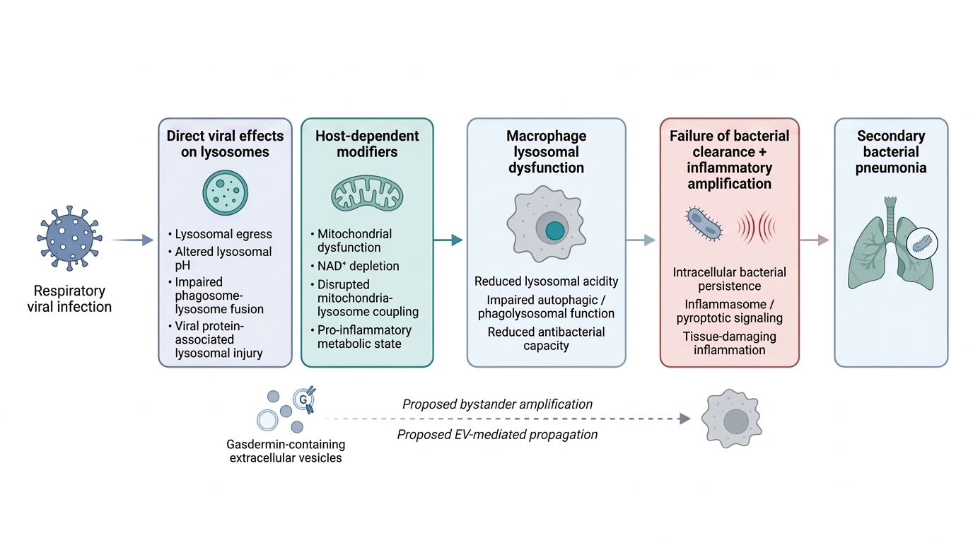
Respiratory viral infections are a major cause of global morbidity and mortality, partly because they increase susceptibility to secondary bacterial pneumonia and sepsis. Coronaviruses, influenza viruses, parainfluenza viruses, rhinoviruses, and respiratory syncytial virus have each been associated with impaired macrophage antibacterial function, although the underlying evidence differs among virus families. Direct lysosomal-pH measurements and mechanistically resolved links between infection and lysosomal de-acidification are strongest for coronaviruses. Using β-coronaviruses as the primary model for the effect of direct lysosomal disruption on macrophage antimicrobial capacity, this review examines lysosomal exploitation during viral egress, impaired phagosome-lysosome fusion, E-protein-mediated proton conductance, and ORF3a-associated lysosomal injury. It further proposes that the magnitude of these effects is shaped by host-dependent immunometabolic processes. Mitochondrial dysfunction, NAD⁺ depletion, and disrupted mitochondria-lysosome coupling may reduce the ability of macrophages to preserve lysosomal acidity, autophagic competence, and bacterial killing during viral challenge. Together, these mechanisms describe how coronavirus infection compromises macrophage antibacterial defense, and how the resulting susceptibility to secondary bacterial infection is further modulated by the metabolic and inflammatory state of the host cell.
Keywords:
coronavirus
; macrophage
; lysosomal dysfunction
; antibacterial immunity
; immunometabolism
; secondary bacterial infection
Introduction
Respiratory viral infections are a major cause of global morbidity and mortality, in part because they predispose infected individuals to secondary bacterial disease - a vulnerability that is particularly pertinent to coronavirus-related disease but is also shared across influenza, parainfluenza, rhinovirus, and coronavirus infection (Al Rahmoun et al., 2025; Koçak Tufan et al., 2021; Peng et al., 2022; Peng et al., 2023; Prasso & Deng, 2017; Rich et al., 2024; Shi et al., 2025; Verma et al., 2020). This pattern is mirrored experimentally: acute bacterial pneumonia is difficult to induce in animals using bacterial cultures alone (Ghoneim et al., 2013; Hraiech et al., 2015; Metersky & Waterer, 2020), yet prior viral infection sharply increases susceptibility to bacterial challenge (Ghoneim et al., 2013; Kalhoro et al., 2016; Small et al., 2010). This predisposition has traditionally been ascribed to impaired mucociliary clearance and altered receptor expression (Bakaletz, 2017), but it may instead reflect an underappreciated failure of macrophage bactericidal capacity.
Paradoxically, most individuals who test positive for the pathogens most often linked to pneumonia never become symptomatic - roughly 30-50% of SARS-CoV-2 infections are asymptomatic (Almadhi et al., 2021), and asymptomatic rates exceed 70% for rhinoviruses and other upper respiratory viruses (Galanti et al., 2019), with comparable figures for influenza (Furuya-Kanamori et al., 2016) and S. pneumoniae (Pahal et al., 2025). Conversely, no pathogen is identified in roughly half of community-acquired pneumonia (Arancibia et al., 2000; Brown, 2012); while much of this reflects diagnostic limitations, some likely represents sterile inflammation, in which cytosolic sensors normally engaged by microbes are instead triggered by host-derived damage-associated molecular patterns (Huang et al., 2024). Such circumstances may indicate that the host cellular environment becomes a major contributor in determining the prognosis of the exposure. Existing explanations for why severity varies among infected individuals are expansive. Defects in type I interferon signaling (Stertz & Hale, 2021; Wang et al., 2020), common risk loci such as chromosome 3p21.31 and variants in ABO, ACE2, TMPRSS2, and FURIN (Abudouleh et al., 2025; Dobrindt et al., 2021; Zeberg & Pääbo, 2020), and TLR4 polymorphisms (Asaba et al., 2024; Khanolkar et al., 2009; Sahanic et al., 2023) all modulate viral susceptibility - but they tend to pertain to their effects on viral entry, replication, interferon-mediated clearance, and pattern recognition, not on the downstream macrophage killing that governs resistance to secondary bacterial pneumonia.
Accordingly, bacterial clearance requires coordinated phagocytosis, intracellular killing, and intact lysosomal function (Hirayama et al., 2017), and acidification is what activates lysosomal proteases and antimicrobial peptides, so even modest pH shifts carry considerable consequences for post-viral microbicidal activity (Lie & Nixon, 2019; Prince et al., 2008; Sanman et al., 2016). When lysosomal pH rises, cathepsins lose activity, antimicrobial peptides fail to adopt their bactericidal conformations, and engulfed bacteria persist and replicate rather than being degraded. De-acidification also rarely acts alone: it commonly co-occurs with impaired autophagosome-lysosome fusion, which lifts the autophagic restraint that normally limits NLRP3 inflammasome activation and thereby amplifies inflammation (Biasizzo & Kopitar-Jerala, 2020; Blevins et al., 2022; Gupta et al., 2025). The two defects are mechanistically distinct but, where they coexist, convert a local failure of bacterial killing into a self-sustaining inflammatory state.
What drives de-acidification during viral infection is clearest for coronaviruses, which this review uses as its primary mechanistic model for explaining the effect of direct lysosomal disruption on macrophage antimicrobial capacity. β-coronaviruses exploit the lysosomal pathway for egress while disrupting lysosomal pH and membrane integrity (Ghosh et al., 2020; Peng et al., 2023), block fusion between lysosomes and bacteria-containing phagosomes (Peng et al., 2022), conduct protons across intracellular membranes via the envelope (E) protein, and cause ORF3a-associated lysosomal injury (Ghosh et al., 2020; Yue et al., 2018). Influenza, parainfluenza, rhinovirus, and RSV each impair macrophage antibacterial function as well, but the evidence there points more often to defective phagolysosomal fusion, impaired killing, or mitochondrial injury than to direct pH change; these families are cited as convergent, indirect support for a shared downstream phenotype rather than as evidence that de-acidification is universal. Crucially, whether viral exposure produces bactericidally significant de-acidification depends on the host state in addition to the activity of the virus involved. Mitochondrial dysfunction creates an intracellular environment permissive to bacterial growth, and restoring mitochondrial function restores macrophage antimicrobial ability (Cao et al., 2020; Plataki et al., 2019; Spier et al., 2021); alveolar macrophages shift from glycolysis toward oxidative phosphorylation as they mature (Izquierdo et al., 2018; Zhang et al., 2018); and inflammatory cytokines such as TNF-α and IL-1β suppress oxidative phosphorylation and directly induce lysosomal de-acidification (López-Armada et al., 2006; Nilsson et al., 2006; Samavati et al., 2008; Tangpong et al., 2008). This inflammatory, metabolically impaired phenotype is conserved across macrophage lineages (Cao et al., 2020; Hellén et al., 2025; Spier et al., 2021) and worsens with aging and chronic disease, when acidification, metabolism, and inflammatory control decline together (Duong et al., 2021; Kale et al., 2020; López-Vázquez et al., 2024; Watanabe et al., 2014).
This review develops the direct β-coronavirus mechanisms above and then proposes two host-dependent processes that may amplify lysosomal dysfunction: immunometabolic reprogramming, in which pro-inflammatory polarization on a background of mitochondrial impairment disfavors acidification through NAD+ depletion and disrupted mitochondria-lysosome contact sites, and pyroptotic amplification through cathepsin release, inflammasome activation, and gasdermin-mediated membrane permeabilization. It further advances a novel hypothesis - that this injury spreads to bystander macrophages via gasdermin-bearing extracellular vesicles (EVs) that induce pyroptosis or permeabilize recipient-cell lysosomes (Du & Wu, 2025; Wright et al., 2025; Zhou et al., 2026). Because these host-dependent mechanisms arise from broad metabolic and inflammatory programs rather than any single viral protein, they may operate across virus families and help explain the interindividual variability in post-viral bacterial susceptibility that current models leave unaddressed. The relevant experimental systems, the evidentiary limits of each proposed mechanism, and the review’s scope are considered in the limitations section.
Methods
The central question addressed in this review lies at the intersection of several fields that are often considered separately, including respiratory virology, macrophage biology, lysosomal physiology, immunometabolism, bacterial pathogenesis, and aging. A narrative review was therefore considered more appropriate than a quantitative or systematic synthesis of a narrowly defined group of studies. The purpose was not to estimate a pooled effect size, but to examine whether findings generated across different experimental systems converge on a biologically coherent explanation for how respiratory viral infection may weaken macrophage antibacterial capacity. Particular attention was given to evidence linking altered mitochondrial function, inflammatory signaling, NAD+ metabolism, organelle communication, and pyroptotic injury to impaired lysosomal acidification and secondary bacterial susceptibility.
Literature Search
PubMed, Scopus, and Google Scholar were searched for relevant peer-reviewed literature published through July 2026. Searches combined terms relating to respiratory viral infection - including coronavirus, influenza, parainfluenza, rhinovirus, and respiratory syncytial virus - with terms relating to macrophage function and the mechanistic pathways considered in this review. These included macrophage, alveolar macrophage, lysosomal acidification, lysosomal de-acidification, phagolysosomal fusion, secondary bacterial pneumonia, immunometabolism, mitochondria-lysosome contact sites, NAD+, gasdermin D, extracellular vesicles, and pyroptosis. Boolean operators were used to combine these terms in different arrangements according to the specific mechanism or disease context being examined. Because several of the proposed connections span research areas that do not consistently use the same terminology, the reference lists of relevant articles were also screened manually to identify foundational studies and additional sources that may not have appeared in the initial searches.
Inclusion and Exclusion Criteria
Studies were considered relevant when they addressed biological mechanisms, pathological findings, or translational implications related to macrophage lysosomal function, respiratory viral infection, or secondary bacterial pneumonia. The evidence base therefore included original experimental studies, mechanistic cell-culture investigations, animal models, systematic reviews, meta-analyses, and selected epidemiological or genetic-association studies where these informed the broader host-dependent framework. Only English-language articles were included. Recent work published within approximately the past 15 years was prioritized where possible so that the discussion would reflect the current state of the field. Older studies were retained when they established foundational observations concerning lysosomal physiology, mitochondrial function, inflammatory signaling, macrophage metabolism, or viral-bacterial interactions that remain directly relevant to the mechanisms considered here.
Terminology
The terms M1-like and M2-like are used throughout this review as conceptual reference points rather than as fixed or mutually exclusive macrophage categories. Macrophage activation exists along a functional continuum and is shaped by tissue origin, local cytokine exposure, metabolic state, developmental lineage, and the nature and duration of the initiating stimulus. In this review, M1-like states refer broadly to macrophage phenotypes associated with increased inflammatory signaling and oxidative antimicrobial activity, often accompanied by altered mitochondrial metabolism and reduced lysosomal acidification. M2-like states are used to describe the broadly opposing end of this spectrum, characterized by comparatively greater reliance on oxidative metabolism, tissue-repair functions, and lysosomal activity. The mechanisms discussed here are not presumed to occur exclusively within either category and are expected to vary across intermediate and mixed activation states.
Study Quality and Synthesis Approach
The studies incorporated into this review differ substantially in design, biological scale, and evidentiary strength. They include clinical and epidemiological observations, genetic associations, animal infection models, primary-cell studies, immortalized cell systems, and experiments in non-pulmonary or non-macrophage populations used to establish specific organellar mechanisms. A formal risk-of-bias assessment was not performed because the aim was not to combine comparable outcomes across a uniform group of studies. Instead, the strength of individual claims was qualified throughout the text according to whether the evidence was direct or inferential, whether it was obtained in pulmonary macrophages or another cellular system, and whether the relevant mechanism had been demonstrated during respiratory viral infection specifically.
This distinction is particularly important for the emerging mechanisms emphasized in this review. Mitochondria-lysosome coupling, NAD+ depletion, and extracellular-vesicle-mediated propagation of gasdermin-associated injury are supported by multiple independent lines of evidence, but not all elements of these pathways have been demonstrated together in a single respiratory viral infection model. Convergence across different experimental systems was therefore treated as support for biological plausibility rather than proof that every proposed step occurs as part of one obligatory sequence. Where the available evidence permits only a mechanistic extension or testable hypothesis, this is stated explicitly.
Immunometabolic Reprogramming and Its Convergence on Lysosomal De-acidification
In addition to the direct virion-organelle interactions described above, lysosomal de-acidification is shaped by the relationship between macrophage activation, cellular metabolism, and inter-organellar cross-talk. Macrophages are among the most metabolically plastic cells of the immune system, remodeling their bioenergetics rapidly in response to inflammation, and this immunometabolic responsiveness is tightly coupled to their antimicrobial capacity (Stienstra et al., 2017). In respiratory illnesses such as COVID-19, mortality is rarely driven by the pathogen alone but by hyperinflammation - the cytokine storm - of which macrophages are the major architects (Lee et al., 2021).
M1 polarization is often characterized by a shift toward aerobic glycolysis alongside disruptive changes in mitochondrial activity (Zuo & Wan, 2019). This glycolytic shift, while well established in many macrophage populations, is not strictly required for pro-inflammatory function in all of them, including tissue-resident alveolar macrophages (Pereverzeva et al., 2022). Human alveolar macrophages do not rely on glycolysis for pro-inflammatory processes in vitro, failing to shift toward glucose metabolism upon LPS activation despite mounting a robust inflammatory response (Pereverzeva et al., 2022; Woods & Mutlu, 2025). This independence is more fragile than it first appears: alveolar macrophages have comparatively low baseline expression of glycolytic enzymes and transporters, and glycolysis alone cannot sustain their function when the electron transport chain is inhibited - a state that arises not only under experimental manipulation but as a direct consequence of infection, as aged mice infected with Streptococcus pneumoniae show decreased macrophage ATP production, dysregulated mitochondrial respiratory-complex expression, and fewer healthy mitochondria than young controls (Plataki et al., 2019; Woods et al., 2020; Woods & Mutlu, 2025). Alveolar macrophages therefore appear to forgo glycolytic reprogramming not because they hold a glycolytic reserve they don’t need, but because that reserve is itself limited - leaving them without a compensatory ATP source when oxidative phosphorylation is disrupted, as it is during respiratory infection.
Independent of whether individual surviving tissue-resident alveolar macrophages (TR-AMs) undergo this within-cell oxidative decline, the alveolar compartment’s overall reliance on OXPHOS-dependent antibacterial function would be expected to fall over the course of infection. TR-AM depletion during respiratory viral infection (Ghoneim et al., 2013) is accompanied by recruitment of neutrophils and monocyte-derived alveolar macrophages (Mo-AMs), both constitutively glycolytic - a pattern documented directly in coronavirus infection, where SARS-CoV-2-infected TR-AMs are lost and replaced by monocyte-derived alveolar macrophages as part of a self-sustaining alveolar inflammatory circuit (Grant et al., 2021). Notably, the same vulnerability appears with influenza, which diminishes expression of mitochondrial transcription factor A (TFAM) in human alveolar macrophages and causes measurable mitochondrial damage, including accumulation of structurally abnormal mitochondria on transmission electron microscopy and reduced enrichment of oxidative phosphorylation genes on transcriptomic analysis (Gao et al., 2022). Because TFAM-dependent mitochondrial metabolism is itself required for TR-AM maintenance and self-renewal (Gao et al., 2022), this represents a route by which respiratory viral infection could directly compromise native TR-AM oxidative capacity. Notably, in macrophages responding to live S. pneumoniae, mitochondrial fission produces a progressive reduction in oxidative phosphorylation alongside enhanced mitochondrial ROS (mROS) generation (Choudhuri et al., 2021; Mohasin et al., 2021) - indicating that acute, transient OXPHOS reduction can itself serve as a non-lysosomal antibacterial mechanism rather than representing pathology outright.
Perturbations in nutrient and energy signaling may also impair lysosomal acidification directly. mTORC1 is recruited to the lysosomal membrane by the Rag GTPase-Ragulator complex, where it phosphorylates the transcription factor TFEB and retains it in the cytoplasm; when mTORC1 activity falls, TFEB is dephosphorylated, translocates to the nucleus, and drives transcription of the CLEAR gene network responsible for lysosomal biogenesis, including the V-ATPase subunits themselves (Peña-Llopis et al., 2011; Roczniak-Ferguson et al., 2012). Because M1-polarizing stimuli such as LPS engage PI3K/Akt/mTORC1 signaling as part of the broader inflammatory and glycolytic reprogramming response (Weichhart et al., 2015), M1 polarization would be expected to hold TFEB in its phosphorylated, cytoplasmically retained state, suppressing transcription of the V-ATPase machinery required to maintain lysosomal acidification under increased bactericidal demand. mTORC1 activity at the lysosome is further sustained by the glycolytic intermediate fructose-1,6-bisphosphate, sensed by aldolase to prevent assembly of an inhibitory AXIN-LKB1-AMPK complex that would otherwise inactivate mTORC1 (Li et al., 2021; Zhang et al., 2017). Given the limited glycolytic reprogramming capacity of native TR-AMs, this self-reinforcing loop would be expected to operate more directly in the glycolytically primed neutrophils and Mo-AMs that replace TR-AMs over the course of infection than in surviving TR-AMs, though it may still occur in TR-AMs under sustained inflammatory activation.
LPS also induces IRG1-dependent production of itaconate, an immunometabolite that directly alkylates TFEB at a conserved cysteine (Cys212 in humans). This modification hinders mTORC1 from phosphorylating the Ser211 residue - which holds TFEB in the cytosol - so TFEB instead translocates to the nucleus and drives lysosomal biogenesis. itaconate does not inhibit mTORC1 itself: the kinase remains active, but this one substrate is shielded from it. Disrupting the activating arm, via IRG1 knockout or an alkylation-deficient TFEB mutant, measurably impairs macrophage antibacterial capacity in vitro and in vivo (Zhang et al., 2022). These two arms are not symmetric, however. mTORC1-driven phosphorylation is the immediate default upon PI3K/Akt/mTORC1 engagement, whereas itaconate-driven counter-activation depends on IRG1 induction and itaconate accumulation to a threshold sufficient for TFEB alkylation - a process that unfolds over hours rather than minutes: in the study establishing this mechanism, TFEB alkylation was detectable at 4 hours of LPS stimulation but not at 1 hour (Zhang et al., 2022). Early in M1 activation, mTORC1-driven suppression should therefore dominate by default, with itaconate-mediated activation emerging only once IRG1 induction has had time to build.
In principle, mTORC1-driven suppression should also be self-limiting: as mitochondrial function deteriorates and ATP falls, AMPK should be activated through the same AXIN-LKB1 sensing complex, inhibiting mTORC1, releasing TFEB, and restoring V-ATPase transcription. Under sustained inflammatory activation, however, this compensatory arm may itself be compromised, as LPS and pro-inflammatory cytokines including TNF-α and IL-6 each independently suppress AMPK - LPS by downregulating LKB1 expression and phosphorylation, TNF-α via upregulation of the phosphatase PP2C, and IL-6 through SOCS3-mediated signaling (Fan et al., 2018). If this holds in M1-polarized macrophages, prolonged inflammatory exposure would not merely sustain the initial mTORC1-TFEB-V-ATPase suppression but concurrently degrade the AMPK-dependent mechanism that would otherwise reverse it.
Because lysosomal acidification depends on a carefully orchestrated set of proton pumps, counter-ions, and active mitochondrial support, these metabolic adjustments place a heavy burden on the machinery that keeps lysosomes acidic. Historically this burden was attributed mainly to diminished ATP production under tissue hypoxia impairing proton-pump activity, and that mechanism does occur. Its significance is compounded, however, by the fact that mitochondria do not merely supply the ATP that drives V-ATPase - they are physically and chemically coupled to the lysosome through mechanisms that are themselves highly sensitive to the metabolic state of the macrophage (Figure 1). The following sections address these mechanistically distinct pathways linking M1 immunometabolic reprogramming to impaired lysosomal acidification and reduced bactericidal capacity.
Mitochondria-Lysosome Contact Sites
Mitochondrial metabolic competence is a direct determinant of macrophage antibacterial capacity. Aged alveolar and interstitial macrophages, which show impaired oxidative phosphorylation and diminished ATP output, clear S. pneumoniae and influenza significantly less effectively (Plataki et al., 2019; Wong et al., 2017); in aged, S. pneumoniae-infected macrophages, this tracks with directly measured reductions in ATP production and dysregulated respiratory-complex expression (Plataki et al., 2019). The failure operates at two levels - impaired phagocytic uptake and impaired lysosomal destruction of the engulfed bacterium (Wong et al., 2017). The converse also holds: ACE overexpression in myeloid cells raises oxidative metabolism and ATP and, with it, superoxide production, phagocytic activity, respiratory-chain protein expression, and resistance to S. pneumoniae and other invasive organisms (Cao et al., 2020). Mitochondrial metabolic competence and bactericidal capacity therefore rise and fall together, pointing to a direct physical mechanism linking the two: proton transfer at mitochondria-lysosome contact sites (MLCs).
MLCs are transient physical bridges formed between the outer mitochondrial membrane and the lysosomal membrane at an intermembrane distance of ~10 nm (Tian et al., 2026; Yan et al., 2026) - active interfaces at which protons are directly transferred from the mitochondrial intermembrane space into the lysosomal lumen (Tian et al., 2026). MLCs have been observed across a wide range of mammalian cell types and likely represent a conserved means of inter-organellar communication relevant to lysosomal homeostasis (Wong et al., 2018; Yan et al., 2026). Their formation and maintenance is regulated by the small GTPase Rab7: its GTP-bound active state tethers the two organelles at the ~10 nm distance necessary for productive proton transfer, while GTP hydrolysis to the GDP-bound inactive state releases the tether, disassembles the contact site, and terminates proton flux (Wong et al., 2018). Any perturbation that reduces the mitochondrial proton gradient - inhibition of electron transport chain activity, mitochondrial uncoupling, or oxidative damage - directly reduces the driving force for proton transfer across MLCs, thereby impairing lysosomal acidification and the bactericidal activity it supports. This gives the ATP-output and phagocytic-clearance failures above a structural counterpart: a collapse of the mitochondrial proton gradient under sustained inflammation degrades both bulk ATP-dependent uptake and MLC-dependent lysosomal killing at once.
Accordingly, mitochondrial dysfunction more broadly - whether induced by genetic ablation of mitochondrial proteins or by chemical inhibition of the electron transport chain - has been shown to impair lysosomal structure and activity across neuronal cells, microglia, and T cells, with downstream consequences including neurodegeneration and dysregulated inflammatory responses (Baixauli et al., 2015; Demers-Lamarche et al., 2016). Additionally, mitochondrial ROS - generated in elevated quantities as a byproduct of remodeled and partially uncoupled electron transport chain activity during M1 activation - impairs the autophagy-lysosome system in macrophages and promotes lipid peroxidation at organellar membrane interfaces (Yuan et al., 2019). Whether the specific effect of M1-associated ROS on MLC integrity in macrophages operates through Rab7 GTPase destabilization, lipid peroxidation at contact site membranes, or some other mechanism has not yet been directly resolved and represents an important direction for future investigation.
It is likely, however, that the pro-inflammatory cytokine environment generated in these cells reinforces all of these changes simultaneously. Inflammatory cytokines - such as TNF-α - have long been known to suppress mitochondrial respiratory-chain activity and promote glycolytic gene expression across diverse cell types (Beasley & Eldridge, 1994; Sánchez-Alcázar et al., 2003; Stadler et al., 1992). TNF-α-driven signaling also directly causes lysosomal alkalinization - from pH 4.3 to 5.5 in monocytic cells - alongside increased lysosomal membrane permeability (Nilsson et al., 2006). TNF-α may additionally promote pathogenic mitochondrial ROS through reverse electron transport: in Mycobacterium-infected macrophages, excess TNF-α increased glutamine uptake and succinate accumulation, enabling complex II-driven reverse electron transport through complex I and generating mitochondrial superoxide (Roca et al., 2022). This pathway has not been demonstrated in coronavirus-exposed pulmonary macrophages, but a similar TNF-high, succinate-supported mechanism may contribute to mitochondrial ROS accumulation during viral infection or secondary bacterial challenge. TNF-α and IL-1β may further impair respiratory-chain activity through enhanced superoxide production: when superoxide generated during M1 activation acts within mitochondria - rather than being exported to the phagosome via mitochondria-derived vesicles for bactericidal use (Abuaita et al., 2018) or dismutated by superoxide dismutase (SOD) - it inhibits mitochondrial complexes I and II directly, and reacts with nitric oxide to form peroxynitrite, which inhibits complex IV, compounding the suppression of oxidative phosphorylation already driven by glycolytic reprogramming (Beasley & Eldridge, 1994; Yuan et al., 2019). Direct experimental disruption of the electron transport chain is sufficient to induce a pro-inflammatory phenotype in vitro: Ye et al. (2016) exposed BV-2 and primary microglia - the brain’s resident macrophage population - to multiple electron transport chain inhibitors, including rotenone, thenoyltrifluoroacetone, antimycin A, and sodium azide, and observed increased secretion of IL-1β, IL-6, IL-12, and TNF-α alongside activation of MAPK and NF-κB pathways. While microglia are developmentally distinct from peripheral macrophages, arising from yolk sac progenitors rather than bone marrow monocytes, their core inflammatory-metabolic coupling appears broadly conserved, and this relationship between mitochondrial dysfunction and pro-inflammatory activation is consistent with what has been observed in peripheral macrophage populations. Consistent with this, chronic low-dose complex I inhibition has been shown to sustain macrophage activation over time (Shaikh & Nicholson, 2009), suggesting that even sub-acute mitochondrial perturbation is sufficient to maintain a pro-inflammatory myeloid state. Furthermore, when mitochondrial dysfunction is sufficient to cause mitochondrial DNA/RNA release into the cytosol, innate immune sensors including TLR9, cGAS-STING, RIG-I, MDA5, and the NLRP3 inflammasome are engaged, amplifying cytokine production and further sustaining the pro-inflammatory metabolic state (Algieri et al., 2025; López-Polo et al., 2024).
NAD+ Depletion
Diminution of nicotinamide adenine dinucleotide (NAD+) is a near-ubiquitous feature of diseases in which inflammation is the governing phenotype. NAD+ serves broad functions - as a redox cofactor in energy metabolism, a substrate for NAD+-consuming enzymes including CD38 and poly(ADP-ribose) polymerase (PARP), and a regulatory signal governing mitochondrial function, autophagy, and inflammasome activity - reviewed extensively elsewhere (Cantó et al., 2015; McDaniel et al., 2025; Navarro et al., 2022; Wilson et al., 2023). Its depletion is consequential because restoration or precursor supplementation ameliorates pathology through several converging routes: replenishing the pool consumed by CD38 and PARP, supporting mitochondrial oxidative function, activating autophagy via beclin-1 and LC3 upregulation, and inhibiting NLRP3 inflammasome activation (Wang et al., 2021). This is illustrated both outside acute respiratory illness - in multiple sclerosis, where low serum NAD+ correlates with disease severity and progression (Braidy et al., 2013) and where NAD+ or precursor administration ameliorates pathogenesis (Penberthy & Tsunoda, 2009) - and within it, where NAD+ and NMN supplementation may improve acute illnesses such as SARS-CoV-2 pneumonia (Jiang et al., 2022). NAD+ supplementation may additionally limit cell death pathways including pyroptosis (Dong et al., 2026).
Consistent with this, host NAD+ metabolism has been implicated broadly across the host-virus interface: viruses from several unrelated families - SARS-CoV-2, influenza A virus, Zika virus, herpes simplex virus, and HIV - each perturb NAD+ biosynthesis or consumption in ways that favor viral persistence and replication, converging on NAD+-regulating enzymes including sirtuins, PARPs, and CD38/CD157 (Jhandai et al., 2026; Saraiva et al., 2025). NAD+ diminution is also prominent in bacterial infection. The tuberculosis necrotizing toxin (TNT), a secreted NAD+ glycohydrolase produced by Mycobacterium tuberculosis, induces necrosis in infected macrophages by actively depleting the host NAD+ pool (Danilchanka et al., 2014). Pajuelo et al. (2018) extended this, showing that TNT-mediated NAD+ hydrolysis is sufficient to activate RIPK3 and MLKL - the key mediators of necroptosis - independently of canonical initiators such as TNF-α and RIPK1, with accompanying mitochondrial depolarization and impaired ATP synthesis. This is particularly relevant to pulmonary macrophages in respiratory illnesses such as CAP and ARDS, where the resident alveolar macrophage compartment undergoes substantial remodeling during and after disease (Guillon et al., 2020).
NAD+ depletion also drives assembly of the NAD+-sensing PANoptosome nucleated by NLR family CARD domain-containing protein 5 (NLRC5), a multiprotein complex that senses intracellular NAD+ depletion as a danger signal and, once activated, simultaneously engages apoptotic, pyroptotic, and necroptotic machinery. As intracellular NAD+ falls below threshold - through TNT-mediated hydrolysis, PARP-driven consumption during inflammatory DNA-damage responses, or the metabolic depletion of M1 reprogramming - NLRC5 is released from its NAD+-bound inhibited state, oligomerizes into the PANoptosome, and drives hypersecretion of IL-1β, IL-6, and TNF-α, aggravating pulmonary fluid accumulation and tissue damage (Jiang et al., 2022; Sundaram et al., 2024). This positions NAD+ depletion not as a downstream consequence of inflammatory activation but as an active upstream trigger of amplified inflammatory cell death.
This decline in NAD+ also undermines macrophage antimicrobial activity by impairing lysosomal acidification. Yagi et al. (2021) showed that mitochondrial translation deficiency - induced experimentally with chloramphenicol - stabilizes HIF1α, which suppresses Nmnat3, a mitochondrial NAD+ biosynthetic enzyme. The resulting NAD+ shortfall impairs a localized mode of ATP production at the lysosomal surface, and this energy deficit at the lysosomal membrane produces lysosomal enlargement, impaired autophagic degradation, and de-acidification. Nicotinamide mononucleotide (NMN) rescued acidification, and Nmnat3 overexpression reproduced the rescue independently, together establishing that restoring local NAD+ is sufficient to restore lysosomal function. This work was performed in cardiac muscle-derived cells rather than macrophages; HIF1α stabilization by mitochondrial translation inhibitors is expected to be conserved, as HIF1α is ubiquitously expressed and its stabilization under mitochondrial stress is documented across diverse lineages (Boehme et al., 2025), but whether the downstream effects on Nmnat3 and lysosomal acidification recapitulate in pulmonary macrophages under after respiratory coronavirus infection remains to be tested.
Furthermore. Klabunde et al. (2023) found that NAD+ precursor supplementation raised intracellular NAD+ in both immortalized and primary bronchial epithelial cells and reduced S. pneumoniae replication during infection - an effect dependent on host NAD+ metabolism rather than direct antibacterial action, since precursors had no effect on bacterial replication in the absence of host cells. Inhibiting NAMPT, the rate-limiting salvage enzyme, increased replication, while activating it decreased replication, establishing host NAD+ biosynthetic capacity as a direct determinant of antibacterial outcome. Although performed in epithelial cells rather than macrophages, this dependence on intracellular NAD+ is consistent with the lysosomal-acidification mechanism above and supports the broader proposition that NAD+ depletion - from M1 reprogramming, PARP activation, or bacterial glycohydrolase activity - impairs the host machinery on which bacterial clearance depends.
The antibacterial effect of NAD+ is nonetheless pathogen-specific. Applied directly to S. pneumoniae, NAD+ was itself bacteriostatic - dysregulating bacterial metabolism and lowering intrabacterial ATP - which could add benefit in pneumococcal infection, where host-mediated and direct effects might operate together (Klabunde et al., 2023). By contrast, direct NAD+ increased replication of S. agalactiae and non-typeable Haemophilus influenzae by roughly 50%, indicating that elevated extracellular NAD+ is permissive rather than restrictive for these organisms (Klabunde et al., 2023). This is an important caveat for any NAD+-repletion strategy in post-viral bacterial pneumonia: the infecting organism may determine whether boosting host NAD+ is beneficial, neutral, or counterproductive. Precursor-based approaches that raise intracellular rather than extracellular NAD+ may offer a more targeted route - enhancing host lysosomal bactericidal capacity without supplying growth substrate to NAD+-utilizing pathogens.
Mitochondrial Dysfunction Engages MAVS
Mitochondrial antiviral-signaling protein (MAVS) - also known as IPS-1, VISA, or Cardif - is an adaptor protein anchored to the outer mitochondrial membrane via a C-terminal transmembrane domain, and functions as the central hub linking cytosolic viral RNA detection to the innate antiviral immune response (Figure 2). When cytosolic RIG-I-like receptors such as RIG-I and MDA5 detect intracellular viral RNA, they undergo conformational activation and dock onto MAVS through interactions between their respective caspase recruitment domains (CARDs). This docking event triggers MAVS to polymerize into prion-like fibrils along the outer mitochondrial membrane (Hou et al., 2011). The resulting MAVS aggregates recruit and activate TBK1 and IKK signaling complexes, which in turn activate the transcription factors IRF3, IRF7, and NF-κB. These factors translocate to the nucleus and drive transcription of type I and type III interferons alongside additional pro-inflammatory cytokines, establishing an antiviral state in which secreted interferons alert neighboring cells to restrict viral replication and promote apoptosis in infected cells (more so in non-immune cells) (Choudhury et al., 2024). Within the infected cell itself, MAVS activation additionally drives pyroptosis through a more indirect mechanism (Subramanian et al., 2013; Ou et al., 2025).
MAVS couples viral sensing to NLRP3 inflammasome activation. At rest, NLRP3 is distributed diffusely through the cytosol; on viral infection, MAVS binds it directly and recruits it to the mitochondrial outer membrane, concentrating it at one location and lowering the threshold for NLRP3 molecules to encounter and cooperate with one another. Because MAVS assembles into large, interconnected prion-like filaments during antiviral signaling, this scaffold also provides a structural platform on which NLRP3 rapidly oligomerizes before recruiting the adaptor ASC and pro-caspase-1 to complete assembly (Park et al., 2013; Subramanian et al., 2013). Recruitment to the mitochondrion additionally places NLRP3 beside a second activating signal - the mitochondrial DNA or RNA released during mitochondrial stress or damage in active antiviral defense (Subramanian et al., 2013). Once assembled, the inflammasome cleaves pro-caspase-1 into active caspase-1, which matures pro-IL-1β and pro-IL-18 into their secreted forms and cleaves gasdermin D to release its pore-forming N-terminal fragment. This fragment inserts into the plasma membrane, driving pyroptotic lysis of the infected cell: releasing mature cytokines as a tissue-wide alarm and destroying the intracellular niche the virus depends on for replication. MAVS signaling carries its own checkpoint. Sustained MAVS activity collapses the drives activation of apoptotic caspases (Yu et al., 2010), which then cleave MAVS - along with cGAS and IRF3 - to shut down type I interferon production (Ning et al., 2019). This checkpoint is gated on signal intensity: as MAVS output escalates toward the apoptotic threshold, the cell commits to death, and the same event that ends the cell dismantles the signaling platform, terminating the cascade before it can drive the sustained hyperinflammation associated with systemic tissue damage or autoimmune-like pathology.
This resolution mechanism assumes MAVS activation depends on continued viral RNA sensing and terminates once the virus is cleared. RIG-I and MDA5, however, are also activated by host mitochondrial RNA released into the cytosol during mitochondrial damage (Jiao et al., 2025; Victorelli et al., 2025). Because M1 polarization sustains mitochondrial stress, M1 macrophages may maintain MAVS-dependent inflammasome activation and type I interferon signaling independently of viral RNA, driven by endogenous mtRNA release.
Consequences of Mitochondrial-Lysosomal Decoupling in Post-Viral Bacterial Disease
Pyroptosis and Gasdermin-Mediated Vesicular Propagation of Macrophage Dysfunction
Pyroptosis is a form of programmed necrotic cell death triggered by the cytosolic detection of dysbiotic microorganisms or endogenous danger signals. Pyroptotic cells exhibit a swollen, enlarged morphology and ultimately undergo lysis, releasing cytosolic contents - including proteins, metabolites, and nucleic acids - into the extracellular space, where these molecules function as damage-associated molecular patterns (DAMPs) and drive inflammation in neighboring cells (Broz, 2025; Wright et al., 2025). Although GSDMD is the canonical executioner of pyroptosis in the innate immune context, pore-forming capacity appears to be a conserved property of the gasdermin family more broadly - forced expression of the N-terminal domain of GSDMA, GSDMB, GSDMC, or GSDME each triggers a form of cellular necrosis that closely resembles GSDMD-driven pyroptosis (Broz, 2025). GSDME specifically is activated by caspase-3-mediated cleavage downstream of canonical apoptotic stimuli, or directly by granzyme B independent of caspase-3, while GSDMB is activated by granzyme A from cytotoxic lymphocytes (Zhou et al., 2023). The N-terminal domains of other gasdermin family members, including GSDME, GSDMA, and murine GSDMA3, exhibit similar lipid-binding properties, suggesting a common membrane-targeting mechanism across the gasdermin family (Ding et al., 2016; Liu et al., 2016).
GSDMD-NT associates with cellular membranes and preferentially targets acidic phospholipids - such as phosphoinositides - found on the inner leaflet of the plasma membrane (Devant & Kagan, 2023). Cardiolipin has additionally been identified as a strong binding partner of gasdermin N-terminal domains (Tang et al., 2024), indicating that gasdermins may also target mitochondrial and bacterial membranes - both of which are enriched in cardiolipin (Miao et al., 2023; Tang et al., 2024). Lysosomal membranes are typically cardiolipin-deficient and would therefore not be expected to be targeted through this mechanism; however, GSDME retains the capacity to permeabilize lysosomal membranes by engaging similar acidic, negatively charged phospholipids (Zhou et al., 2026). Notably, GSDME deficiency has been shown to mitigate inflammation and lung damage during influenza infection (Rosli et al., 2025). Liposome binding assays have further confirmed that upon membrane association, GSDMD-NT permeabilizes liposomal membranes, forming pore-like structures with an average inner diameter of approximately 20 nm as revealed by atomic force microscopy and electron microscopy (Aglietti et al., 2016; Ding et al., 2016; Liu et al., 2016; Sborgi et al., 2016).
Pyroptosis is initiated in macrophages through at least two main routes that are both relevant in the post-viral context. In the canonical inflammasome-dependent route, danger signals generated by lysosomal rupture, mtDAMP release, or accumulated intracellular bacterial PAMPs activate cytosolic pattern recognition receptors (Weir & Vince., 2022), leading to assembly of inflammasome complexes. These complexes promote caspase-1-dependent cleavage of pro-IL-1β and pro-IL-18, and cleavage of GSDMD after an aspartate residue within its central linker domain - D275 in humans and D276 in mice - generating a 31-kDa N-terminal fragment (GSDMD-NT) and a 22-kDa C-terminal fragment (GSDMD-CT). GSDMD-NT then inserts into the plasma membrane, driving inflammatory cell lysis. Respiratory viral infection provides multiple upstream triggers for this pathway: coronavirus-induced lysosomal swelling and rupture has been directly shown to initiate pyroptotic signaling in macrophages (Peng et al., 2022), and the sustained NLRP3 activation driven by NAD+ depletion and failed autophagic flux described in preceding section provides an additional convergent trigger. In the non-canonical inflammasome-independent route, direct cytosolic sensing of bacterial LPS by caspase-4/5 in humans - or caspase-11 in mice - cleaves GSDMD and triggers pore formation without inflammasome assembly (Guo et al., 2021), providing another pathway through which secondary bacterial invaders themselves can amplify pyroptotic signaling in macrophages already rendered susceptible by viral infection.
Cells undergoing pyroptotic stress release extracellular vesicles (EVs) and membrane-derived fragments that can contain active gasdermin-NT pores together with a broad array of DAMPs (Du & Wu, 2025; Wright et al., 2025; Zhang et al., 2026). These vesicular structures have been shown to induce membrane injury in neighboring cells through pore transplantation - vesicle-membrane fusion inserts active gasdermin-NT pores into the plasma membranes of recipient cells (Figure 4), initiating secondary pyroptotic signaling in cells that have had no direct contact with the initiating insult (Wright et al., 2025; Zhang et al., 2026). Although this process has been characterized most extensively for GSDMD, Wright et al. (2025) found that EVs released during GSDMA- or GSDME-driven pyroptosis likewise contained the corresponding active gasdermin and induced death in naive recipient cells, indicating that vesicular transfer of gasdermin-mediated membrane injury is not restricted to GSDMD. These findings establish pore transplantation at the recipient-cell plasma membrane, but they do not demonstrate that transferred gasdermins reach or permeabilize intracellular lysosomes. GSDME is nevertheless of particular interest because activated GSDME can permeabilize lysosomal membranes, whereas GSDMD preferentially targets phospholipids associated with the plasma membrane and cardiolipin-rich membranes.
If the proposed endo-lysosomal extension of this pathway is correct, vesicle-mediated transfer of gasdermin activity could spread the antibacterial defect beyond the macrophages initially exposed to the initiating insult. Bystander macrophages that take up these vesicles may themselves undergo pyroptotic stress and release additional gasdermin-NT-containing EVs, creating the possibility of a self-amplifying cycle of intercellular injury. Clinical observations are consistent with the broader relevance of this pathway: elevated levels of biologically active extracellular vesicles have been reported in COVID-19, and circulating GSDMD levels have been associated with disease severity in patient cohorts (de Miguel-Pérez et al., 2024; Tahyra et al., 2022; Isik et al., 2025). These findings do not establish lysosomal targeting by gasdermin-containing vesicles, but they indicate that both EV signaling and gasdermin-associated injury occur on a substantial scale during respiratory viral infection in humans.
Aging may further increase susceptibility to this form of bystander propagation. Extracellular-vesicle production, cargo selection, and uptake are altered in older tissues, with many of these changes favoring pro-inflammatory activation (Manni et al., 2023). An aged macrophage population may therefore be more readily affected by vesicle-borne pyroptotic signals and less capable of containing the resulting organellar stress. In this context, EV-mediated transfer of gasdermin activity represents a possible point of convergence between inflammatory cell death, intercellular communication, and the age-associated loss of macrophage antibacterial reserve that increases vulnerability to secondary bacterial infection.
Bacteria may utilize host inflammatory cytokines to facilitate pathogenesis in secondary bacterial disease
The sustained cytokine environment generated by unresolved lysosomal failure may further benefit invading bacteria through a more direct mechanism: several human respiratory pathogens express surface proteins capable of binding host cytokines - including IL-1β and TNF-α - through diverse cytokine-receptor interactions (Figure 2; Hitzler et al., 2025), the functional and structural characteristics of which have been reviewed in detail elsewhere (Högbom & Ihalin, 2017). This capacity has been proposed to function as a virulence mechanism by which bacteria sense and respond to the host’s defensive state (Högbom & Ihalin, 2017; Wilson et al., 1998), and the evidence base supporting it is substantial. Meduri et al. (1999) demonstrated concentration-dependent growth enhancement in three clinically relevant nosocomial pathogens - Staphylococcus aureus, Acinetobacter spp., and Pseudomonas aeruginosa - when incubated with TNF-α, IL-1β, and IL-6, with blockade by neutralizing cytokine antibodies significantly inhibiting this growth enhancement. Mycobacterium avium growth has similarly been shown to be stimulated by IL-6, an effect abrogated by heat inactivation of the cytokine or anti-IL-6 antibodies (Denis, 1992). Both M. tuberculosis and M. avium cultured extracellularly can be stimulated to grow by recombinant human epidermal growth factor at concentrations of 50 ng/ml (Bermudez, 1996). Transforming growth factor β1 (TGF-β1) and TNF-α stimulate the growth of M. tuberculosis in macrophages and human monocytes, respectively (Byrd, 1997; Hirsch et al., 1994). IL-6 has been shown to increase the growth of M. avium in macrophages regardless of whether it was added before or after bacterial uptake (Denis & Gregg, 1991a), and treatment of macrophages with either CSF-1 or IL-3 enhances the growth of Listeria monocytogenes (Denis et al., 1991b). Escherichia coli, Salmonella typhimurium, L. monocytogenes, S. aureus, Streptococcus mitis, and Shigella flexneri have additionally been shown to bind TNF-α (Luo et al., 1993). Uptake of bacteria-cytokine complexes by macrophages has been shown to be considerably greater than uptake of cytokine-free bacteria, and invasion of host cells by such complexes is similarly enhanced - creating a dual-edged dynamic in which enhanced phagocytosis of bacteria-cytokine complexes may accelerate clearance when macrophage lysosomal function is intact, but facilitates intracellular survival and dissemination when lysosomal acidification is impaired and macrophages cannot effectively dispose of what they have ingested.
Notably, host cytokines may alter the gene expression of bacteria that bind them, with direct consequences for virulence. Mahdavi et al. (2013) found that Neisseria meningitidis PilQ is involved in internalization of IL-8 and TNF-α by the bacterium, leading to altered expression of approximately 20% and 45% of the bacterial genome, respectively. When internalized by N. meningitidis, TNF-α binds genomic DNA at several sites including the promoter regions of PptB transferase, adhesion and penetration protein (app), and meningococcal serine protease A (mspA), increasing their expression and enhancing virulence in animal models - N. meningitidis mutants unable to express PilQ or glycosylated PilE showed reduced mortality in infected mice (Mahdavi et al., 2013). Similarly, IFN-γ has been shown to enhance production of the virulence factor pyocyanin and PA-I lectin in P. aeruginosa - effectors likely required for full virulence in lung and gut-derived sepsis-related infections (Wu et al., 2005; Lau et al., 2004).
The extent to which these observations hold for Streptococcus pneumoniae has not been directly investigated; however, distinct cytokine profiles in the host have been linked to different stages of pneumococcal disease, suggesting that the inflammatory environment shapes infection outcome in ways that may involve bacterial sensing of host signals (Bergeron et al., 1998; Mahdi et al., 2008). Additionally, S. pneumoniae has been shown to bind host lactate dehydrogenase A (LDH-A) via a conserved 22-amino-acid motif within the proline-rich domain of the surface proteins PspA and PspC; this binding requires LDH-A’s own catalytic activity, since an enzymatically inactive LDH-A variant failed to enhance virulence, whereas preincubation of bacteria with lactate alone - the direct product of LDH-A’s conversion of pyruvate - was sufficient to reproduce the same virulence-enhancing effect in a murine pneumonia model (Park et al., 2021). This indicates that pneumococci exploit LDH-A-derived lactate specifically as a nutrient source, rather than using LDH-A binding for a purely adhesive or structural purpose. It is worth noting that the glycolytic (Warburg-like) shift characteristic of M1 macrophage polarization increases local lactate production as a direct metabolic byproduct. A cytokine-storm environment sustained by heavily glycolytic, M1-polarized macrophages would therefore be expected to elevate local lactate availability through at least two convergent routes - ongoing glycolytic output from living but polarized macrophages, and LDH-A release from dying cells - potentially amplifying the nutrient pool PspA/PspC-expressing pneumococci are positioned to exploit. It is also notable that the relationship between bacteria and host cytokines is not always unidirectional. Haemophilus influenzae has been shown to both aggravate inflammation and actively downregulate NF-κB-controlled cytokines - including IL-1β and TNF-α - in respiratory epithelial cells during persistent infection, an effect observed only with live bacteria, suggesting a temporal strategy in which early inflammatory aggravation facilitates acquisition of dying cell-derived nutrients - including iron and NAD+ - while subsequent immunomodulation reduces inflammatory pressure to promote persistence.
Collectively, these bacterial responses to the inflammatory environment are proposed as amplification mechanisms arising after macrophage antibacterial function has already been compromised. Lysosomal de-acidification permits engulfed bacteria to persist while simultaneously sustaining inflammasome activation, cytokine release, and inflammatory cell death. Several bacterial species can then sense, bind, or metabolically respond to these host-derived signals, using cytokines and products released from damaged cells to enhance growth, alter virulence-gene expression, or increase interaction with host cells. Under conditions of intact lysosomal killing, cytokine-enhanced bacterial uptake may contribute to clearance; when phagolysosomal function is impaired, however, the same process may increase intracellular bacterial burden and provide additional stimuli for pyroptosis and tissue inflammation. Bacterial exploitation of the host inflammatory environment therefore closes a positive-feedback loop in which the cellular changes that weaken antimicrobial defense also generate extracellular conditions that favor bacterial persistence and further destabilize macrophage function.
Discussion
Coronaviruses compromise macrophage antibacterial capacity through a convergence of direct viral effects and host-dependent processes, the relative contributions of which are shaped by the metabolic, inflammatory, and organellar state of the affected cell. The direct viral effects encompass several overlapping but mechanistically distinct abnormalities of the lysosomal system. Lysosomal exploitation during viral egress and E-protein activity can alter lysosomal pH homeostasis, whereas ORF3a-associated effects may involve ion or water conductance, lysosomal swelling, or interference with host trafficking machinery, depending on the experimental model (cite). Coronavirus infection can additionally impair fusion between bacteria-containing phagosomes and lysosomes, while more severe superinfection models demonstrate lysosomal swelling, membrane permeabilization, and rupture (cite).
It’s worth a brief tangent to discuss evidence for comparable effects among other respiratory viruses, which are less direct and mechanistically less resolved than it is for coronaviruses. Influenza infection has repeatedly been shown to predispose affected hosts to secondary bacterial pneumonia, but the mechanisms involved vary according to viral strain, macrophage origin, and the broader cellular context. Only a subset of influenza strains, for instance, can overcome the cellular restrictions that ordinarily prevent productive replication in macrophages (Marvin et al., 2017). Recent controlled human infection data add an important host-dependent dimension to this variability. Following standardized intranasal challenge with influenza A/H3N2, symptomatic participants exhibited earlier and more pronounced systemic interferon-driven responses, expansion and activation of circulating monocytes and dendritic cells, and subsequently greater natural killer-cell and CD8+ T-cell activation than asymptomatically infected participants (Papargyris et al., 2026). These differences were not explained by acute-phase viral load alone. Peripheral blood mononuclear cells collected before inoculation from participants who later developed symptoms were also more responsive to ex vivo viral stimulation, including greater IL-1β secretion, suggesting that baseline innate responsiveness can predispose individuals to distinct inflammatory outcomes after otherwise comparable viral exposure (Papargyris et al., 2026). Although this study did not directly examine alveolar macrophage antibacterial activity or bacterial superinfection, it demonstrates that influenza-associated myeloid responses are shaped by pre-existing host immunological states and by multicellular signaling networks that cannot be reproduced in isolated macrophage cultures. Also worth noting is that the depletion of alveolar macrophages during influenza infection has likewise been associated with increased susceptibility to bacterial superinfection in experimental systems (Ghoneim et al., 2013). Macrophages that survive the initial viral infection may remain numerically present while becoming functionally impaired, contributing to the transition of normally noninvasive Streptococcus pneumoniae into a lethal secondary pathogen (Verma et al., 2020). Coinfection studies further indicate that influenza exposure can simultaneously enhance bacterial colonization and viral burden, thereby compounding disease severity in selected models (Kalhoro et al., 2016). Evidence concerning parainfluenza virus remains more limited, relying primarily on observations of impaired phagolysosomal fusion, impaired autophagy, and defective intracellular bacterial killing in infected pulmonary macrophages (Han et al., 2024; Hesse & Toth., 1983; Jakab et al., 1980).
The broader principle that macrophage metabolic-signaling state - rather than viral identity alone - governs the magnitude of antiviral and antibacterial competence extends even beyond the respiratory virus families considered above. In porcine alveolar macrophages infected with porcine reproductive and respiratory syndrome virus (PRRSV), mTOR pathway gene expression was differentially regulated according to macrophage activation status, and inhibition of mTORC2 specifically - rather than mTORC1 - reduced PRRSV infection in part by reversing virus-mediated suppression of type I interferon production and signaling (Liu et al., 2017). This cross-family evidence should be weighted cautiously: PRRSV is evolutionarily distant from the viruses that are this review’s primary focus, and the demonstrated mechanism concerns interferon regulation rather than lysosomal acidification directly. Nonetheless, the finding lends independent support to the broader claim that host metabolic-signaling architecture, rather than any single viral protein or pathway, constitutes a general and conserved determinant of inter-individual microbial and viral competence.
Returning specifically to mitochondrial-lysosomal coupling, the relationship between mitochondrial and lysosomal dysfunction is not unidirectional. Impaired mitochondrial function may weaken mitochondrial-lysosomal contact-mediated proton transfer, reduce NAD+-dependent lysosomal ATP production, and limit the energetic support required to sustain vacuolar ATPase activity and lysosomal acidification. Conversely, lysosomal damage can feed back directly onto mitochondria through a distinct and well-characterized pathway. Lysosomal membrane permeabilization releases cathepsins, including the aspartic protease cathepsin D and the cysteine protease cathepsin B, from the lysosomal lumen into the cytosol (Boya et al., 2003). Once released, both proteases can cleave Bid, a BH3-only member of the Bcl-2 protein family that normally exists as an inactive cytosolic sensor of cellular stress, converting it into its truncated, active form (tBid). Cathepsin D cleaves Bid at three specific sites - Phe24, Trp48, and Phe183 - in a manner shown to drive Bax-dependent mitochondrial permeabilization following lysosomal membrane permeabilization (Appelqvist et al., 2012), while cathepsin B cleaves Bid near the caspase-8 cleavage site to generate tBid (de Castro et al., 2016). Released cathepsins additionally degrade the anti-apoptotic Bcl-2 family members Bcl-2, Bcl-xL, and Mcl-1, synergizing with Bid cleavage to lower the threshold for mitochondrial outer-membrane permeabilization (Cirman et al., 2004). Truncated Bid subsequently translocates to the mitochondrial outer membrane, where it promotes the activation, oligomerization, and membrane insertion of the pro-apoptotic effector Bax. Bax insertion drives mitochondrial outer-membrane permeabilization and the release of cytochrome c, which binds Apaf-1 to form the apoptosome and activate caspase-9 and, in turn, caspase-3 (Li et al., 1997; Morgan et al., 2002). Critically, in cells expressing gasdermin E (GSDME), active caspase-3 cleaves GSDME to redirect this cascade toward pyroptotic membrane rupture (Zhou et al., 2023). This cathepsin-Bid-Bax axis connects directly with the coronavirus-associated lysosomal mechanisms described earlier. The lysosomal rupture and cathepsin B release documented following coronavirus-associated phagolysosomal dysfunction would be expected not only to amplify NLRP3 inflammasome activation, but also to compromise mitochondrial membrane integrity directly through Bid cleavage (Peng et al., 2022).
A separate mitochondria-intrinsic amplification loop may further compound this cycle upstream. Mitochondrial dysfunction can increase the expression and activation of RIP3 through several convergent mechanisms, including mitochondrial reactive oxygen species-dependent NF-κB signaling, ZBP1-mediated sensing of cytosolic mitochondrial DNA, and the accumulation of damaged mitochondria caused by impaired mitophagy (Zhou et al., 2025; Lai et al., 2024). Activated RIP3, in turn, can disrupt respiratory complexes I and III and promote mitochondrial fragmentation through dynamin-related protein 1 (Zhao et al., 2025). These effects reduce respiratory efficiency, increase mitochondrial oxidant production, and generate additional damaged mitochondrial substrates that must be removed through mitophagy (Zhao et al., 2025). RIP3 also translocates into mitochondria, where it interacts with and promotes the degradation of Mitofilin. Mitofilin, encoded by the IMMT gene (Odgren et al., 1996), is a core structural component of the mitochondrial contact site and cristae organizing system complex and is essential for maintaining inner-mitochondrial-membrane architecture (Pfanner et al., 2014). Its degradation destabilizes cristae organization and the mitochondrial structural scaffold, increasing mitochondrial injury and facilitating the release of mitochondrial DNA into the cytosol (Feng et al., 2022). . Cytosolic mitochondrial DNA subsequently activates the cGAS-STING-p65 pathway, promoting transcription of inflammatory mediators including interleukin-6, tumor necrosis factor-α, and intercellular adhesion molecule 1 (Feng et al., 2022).
In addition, the persistent mitochondrial stress associated with M1-like inflammatory polarization could elevate basal RIP3 expression or activity, thereby priming macrophages for more extensive ORF3a-associated lysosomal injury upon subsequent coronavirus exposure - a possibility that, given the SARS-CoV-1-specific evidence base for RIP3-dependent ORF3a activity, remains speculative for SARS-CoV-2 specifically pending direct investigation. Nonetheless, coronavirus proteins may act on a macrophage system whose mitochondrial, lysosomal, and inflammatory thresholds have already been shifted by age, prior inflammatory exposure, metabolic disease, or chronic organellar stress. The proposition that aged cells respond worse to such stressors than youthful ones is well supported across multiple independent lines of evidence. Aged human monocyte-derived macrophages show decreased basal respiration, reduced complex I and II activity, lowered ATP production, decreased mitochondrial membrane potential, and increased oxidative stress alongside compromised antioxidant defenses (Wang et al., 2024). Mitochondrial ATP-producing capacity declines by approximately 8% per decade in humans, accompanied by reduced oxidative capacity, increased ROS generation, and inhibition of mitophagy (Chistiakov et al., 2014). Aged macrophages specifically exhibit a metabolic shift away from oxidative phosphorylation and toward glycolysis, together with reduced NAD+ synthesis via downregulated SIRT3 activity and reduced AMPK activity that impairs restoration of energy homeostasis under stress (Kominsky et al., 2010). Directly relevant to the present context, aged mice infected with Streptococcus pneumoniae show decreased macrophage ATP production, dysregulated mitochondrial complex expression, enhanced oxidative stress, and reduced numbers of healthy mitochondria relative to young controls (Plataki et al., 2019). Age-related susceptibility to E protein-mediated calcium dysregulation specifically has additional experimental support: E protein viroporin activity induces Ca2+ release and neuronal death in rat hippocampal cells aged in vitro (López-Vázquez et al., 2024).
In summary, the consequences of coronavirus infection are likely determined by a relationship between viral burden and host-cell reserve. In a metabolically resilient macrophage, E-protein viroporin activity, and ORF3a-associated lysosomal injury may be partially counterbalanced by intact mitochondrial respiration, adequate NAD+, efficient lysosomal acidification, among a myriad of other factors contributing to the baseline viability of the cell population - such that viral proteins are produced but never reach the concentration required to trigger widespread organelle failure. In a macrophage already affected by aging, metabolic dysfunction, chronic inflammation, or persistent M1-like polarization, the same viral exposure may instead cross this compensatory threshold, at which point viral protein accumulation, lysosomal permeabilization, cathepsin release, mitochondrial permeabilization, RIP3 activation, and inflammatory amplification become mutually reinforcing - reflecting not one isolated viral mechanism, but the collapse of a coupled innate-immune regulatory network. This may help explain why comparable coronavirus exposures, including SARS-CoV-2 infections, produce such markedly different clinical outcomes: many remain asymptomatic or mild (Almadhi et al., 2021; De Santis et al., 2023; Furuya-Kanamori et al., 2016; Leung et al., 2015; Ma et al., 2021; Pahal et al., 2025), while others progress to persistent organellar dysfunction and heightened susceptibility to secondary bacterial infection (Smith et al., 2020).
The resulting model also points toward potential therapeutic strategies. Preventing severe coronavirus-associated antibacterial deficiency may require more than inhibiting viral replication. Interventions that preserve selective-autophagy receptor function, support lysosomal acidification, restore NAD+ availability, stabilize mitochondrial respiration, enhance mitophagy, limit RIP3-dependent injury, preserve mitochondrial architecture, or reinforce mitochondrial-lysosomal coupling may increase the cellular threshold at which direct viral effects become severely pathological. Such approaches could potentially reduce both primary coronavirus-mediated injury and the secondary loss of macrophage antibacterial competence that permits bacterial superinfection.
Limitations and Future Directions
Several limitations of the evidence base and experimental systems underlying this review warrant explicit acknowledgment.
Evidential Constraints on Proposed Mechanisms
The most fundamental mechanistic constraint is that the host-dependent mechanisms proposed here - MLC proton flux disruption and NAD+ depletion - have not been demonstrated simultaneously in a single experimental system in the context of respiratory viral infection. Each mechanism is supported by independent lines of evidence across different cell types, disease contexts, and experimental models, but their relative contributions to lysosomal de-acidification in respiratory viral infection specifically, and the degree to which they act additively or synergistically, remain to be established directly.
The ORF3a/RIP3-dependent lysosomal damage mechanism carries its own evidentiary limitations. The RIP3-dependency of ORF3a-associated lysosomal damage was established using the original SARS-CoV-1 3a protein specifically, with generalization to SARS-CoV-2 ORF3a remaining untested. While mitochondrial dysfunction has independently been shown to activate RIP3 through several converging routes, and activated RIP3 in turn damages mitochondria (Feng et al., 2022) - raising the possibility of a convergent or amplifying loop with the MLC and NAD+ mechanisms - this potential convergence has not been directly examined.
A related evidential constraint concerns the source of NAD+ depletion assumed for coronavirus infection specifically. PARPs consume NAD+ directly as the substrate for ADP-ribosylation, and PARP-mediated ADP-ribosylation of viral components is itself an antiviral host response; coronaviruses counter this via a conserved nsp3 macrodomain that strips these ADP-ribose marks from viral targets, restoring their function, and loss of macrodomain activity accordingly sensitizes coronaviruses to PARP-dependent restriction (Jhandai et al., 2026). Critically, macrodomain activity reverses the functional consequence of ADP-ribosylation for the virus, but does not replenish the NAD+ already consumed by PARP in generating those marks. This raises the possibility that a substantial portion of the NAD+ depletion observed during coronavirus infection reflects this PARP-driven consumption.
Scope Limitations
The evidence base for lysosomal de-acidification following respiratory viral infection is substantially stronger for coronaviruses than for influenza, parainfluenza, rhinovirus, and RSV - the extension to these additional virus families rests primarily on evidence of impaired phagolysosomal fusion, defective bacterial killing, mitochondrial injury, or broader macrophage dysfunction rather than direct lysosomal pH measurements in most cases. A thorough study of these other viruses was not deployed. Nonetheless, these findings support the possibility that different respiratory viruses produce convergent antibacterial defects involving the broader lysosomal or phagolysosomal system, but they do not establish lysosomal de-acidification as a universally shared mechanism across virus families. The immunometabolic evidence base also draws substantially on non-macrophage cell types - including microglia, T cells, and neuronal cells - in which metabolic-lysosomal coupling principles may be conserved but have not always been demonstrated directly in alveolar macrophages. Evidence from these systems is therefore used to inform mechanistic hypotheses rather than treated as direct proof of the proposed pathways in coronavirus-exposed pulmonary macrophages.
This review’s mechanistic focus also centers predominantly on tissue-resident alveolar macrophages (TR-AMs), the best-characterized and most extensively studied macrophage population in the alveolar niche. However, the alveolar compartment is not populated by TR-AMs alone. Interstitial macrophages and monocyte-derived alveolar macrophages (Mo-AMs) - recruited from circulating monocytes and substantially expanded during acute inflammation - both contribute meaningfully to the pulmonary immune response and are not addressed in detail here. This omission may be significant because Mo-AMs enter the alveolar niche with metabolic and inflammatory programs that differ from those of mature TR-AMs. Mo-AMs are generally more glycolytically and pro-inflammatory primed and exhibit lower expression of OXPHOS-associated programs during viral infection, whereas mature TR-AMs rely more heavily on oxidative metabolism for maintenance and antibacterial function. The mechanisms proposed here may therefore differ in magnitude, timing, or consequence across pulmonary macrophage lineages and should not be interpreted as establishing a uniform response among all macrophage populations.
This review also focuses specifically on lysosomal pH as a central mechanistic variable linking immunometabolic dysfunction to impaired macrophage bactericidal capacity. However, lysosomal de-acidification, impaired phagosome-lysosome or autophagosome-lysosome fusion, altered intracellular trafficking, lysosomal swelling, membrane permeabilization, and membrane rupture are related but mechanistically distinct phenotypes. These abnormalities may coexist, arise in parallel, or reinforce one another, but they should not be assumed to represent obligatory stages of a single linear pathway. A causal or temporal ordering is assigned only where it has been demonstrated within the experimental system being discussed - elsewhere, the relationships among these outcomes remain context-dependent. The emphasis on lysosomal pH therefore does not exclude additional perturbations within the broader phagosomal, autophagosomal, and lysosomal trafficking network that may independently compromise macrophage antibacterial and anti-inflammatory function. For instance, the macrophage-specific V-ATPase subunit ATP6V0D2 has been shown to restrict inflammasome activation and limit bacterial infection by facilitating autophagosome-lysosome fusion (Xia et al., 2019) - a function that is distinct from, though closely intertwined with, the canonical proton-pumping role of V-ATPase in lysosomal acidification.
Aging is considered here specifically as a modifier of macrophage metabolic, lysosomal, and inflammatory reserve rather than as a comprehensive explanation for immunosenescence. Age-associated susceptibility to respiratory disease additionally involves changes in epithelial integrity, hematopoiesis, adaptive immunity, alveolar niche signaling, senescent-cell burden, extracellular-vesicle composition, and microbial ecology. A full treatment of these systemic and multicellular processes falls beyond the mechanistic scope of this review. The host-cell-reserve framework should therefore be understood as describing one component of age-associated vulnerability - specifically, the capacity of pulmonary macrophages to preserve organellar function and antibacterial activity during viral challenge - rather than as a complete account of the broader effects of aging on respiratory immunity.
Additionally, this manuscript focuses on macrophages as the primary effector population, but establishment of lower respiratory tract infection - whether by a secondary bacterial pathogen or otherwise - typically requires the failure of several cell types and physical barriers rather than macrophage dysfunction alone. Ciliated epithelial cells, which line the respiratory tract and depend on high mitochondrial ATP output to sustain continuous ciliary beating, illustrate this point with particular clarity. These cells cluster mitochondria beneath the apical membrane to supply cilia with ATP, but this arrangement - combined with the relatively high oxygen tension at the airway surface - creates conditions favorable to electron leak and oxidant production. Ciliated cells accordingly express mitochondrial uncoupling proteins, including UCP2 and UCP5, that reduce respiratory efficiency in exchange for limiting ROS generation (Jain et al., 2024). This energetic trade-off may leave less reserve available to absorb additional insults - including hypoxia, hyperglycemia, or mitochondrial dysfunction - that could impair ciliary beating, weaken mucociliary clearance, and permit aspirated organisms to descend into the lower airway. These possibilities are relevant to the broader host-cell-reserve framework but have not been integrated experimentally with the macrophage mechanisms emphasized here.
Goblet and other mucus-producing cells, which rely on a metabolically demanding biosynthetic program to generate heavily glycosylated mucins, may be similarly sensitive to metabolic perturbation. Conditions that alter glucose metabolism or endoplasmic reticulum function - as can occur during diabetes or severe cellular stress - may impair mucin folding and glycosylation, producing either a thin and poorly protective mucus layer or an abnormally viscous layer that obstructs clearance and creates a stagnant environment favorable to bacterial persistence. This vulnerability may extend upstream to goblet-cell differentiation itself. In the colonic epithelium, goblet-cell differentiation depends on a metabolic transition from glycolysis to mitochondrial oxidative phosphorylation mediated by p32/gC1qR/HABP1, and disruption of this transition - through p32 loss or genetic impairment of respiratory complex V - produces glycolytic, energy-deficient cells with impaired differentiation, as demonstrated in ulcerative colitis (Sünderhauf et al., 2021). Whether an analogous glycolysis-to-OXPHOS transition governs respiratory goblet-cell differentiation, and whether inflammatory metabolic reprogramming disrupts this process in the airway, has not been investigated. This evidence is therefore presented as a cross-tissue precedent rather than as proof of an equivalent respiratory mechanism.
Epithelial tight junctions, whose maintenance requires continuous energy-dependent protein turnover, may likewise be sensitive to metabolic and nutrient perturbations that interfere with AMPK-dependent junctional signaling, potentially increasing barrier permeability and facilitating bacterial adherence or tissue penetration (Rowart et al., 2018). However, the macrophage-specific metabolic and lysosomal mechanisms proposed throughout this review have not been directly extended to ciliated epithelial cells, goblet cells, or epithelial junctional networks. Determining whether analogous host-reserve mechanisms operate across these populations remains a distinct avenue for future investigation and will require multicellular airway models, and in vivo measurements capable of distinguishing macrophage-intrinsic dysfunction from broader failure of the respiratory barrier.
Biological Complexity of Viral Inocula and Host-Derived Factors
One important consideration in interpreting respiratory virus-induced lysosomal dysfunction is the biological complexity of viral preparations used in experimental systems. Viral propagation occurs in stressed cellular environments and generates a heterogeneous mixture of infectious particles and biologically active host-derived material, including cellular debris, oxidized lipids, nucleic acids, extracellular vesicles, and other components that can be difficult to eliminate completely using standard purification approaches (Mizenko et al., 2022; Moulin et al., 2023; Bao et al., 2024; Torabian et al., 2025). These materials may arise from virus-induced cell death, pre-existing tissue perturbation, resident microbial activity, or routine cell-culture practices (Agu et al., 2025; Ryu et al., 2017; Singh et al., 2014; Nygaard et al., 2015). Many of these constituents can independently activate pattern-recognition receptors, inflammasomes, and intracellular stress pathways in macrophages and other cell types. Host-derived material carried within viral preparations may therefore modify the magnitude or character of mitochondrial, inflammatory, or lysosomal responses attributed to viral exposure. This possibility is especially relevant when mock-infected controls consist only of phosphate-buffered saline or unconditioned medium, which do not reproduce the biological complexity of supernatants derived from stressed, injured, or dying cells.
Historical evidence illustrates the value of more closely matched control preparations. In the original work establishing measles-virus isolation in tissue culture, Enders and Peebles (1954) used fluid from uninoculated monkey-kidney cultures as a control and maintained those cultures alongside virus-inoculated cultures under comparable conditions. The authors reported that cytopathic changes resembling some of those observed in virus-exposed cultures also appeared in the uninoculated controls, although the nuclear abnormalities were specific to the measles group. This represents a documented instance in which biologically active control material produced an unexpected cellular phenotype.
A more recent example is provided by Kuksin and Norkin (2012), who produced mock-infected control medium by exposing uninfected cells to BrdU-containing medium for the same duration used for viral-stock propagation and then subjecting the control material to the same freeze-thaw and dialysis procedures. The mock cells were maintained concurrently with and processed similarly to their infected counterparts. This design demonstrates that duration- and processing-matched mock preparations are technically achievable. However, the study was designed to investigate viral disassembly rather than baseline metabolic or organellar health, and it did not report mitochondrial function, reactive oxygen species production, lysosomal physiology, or other subclinical measures of cellular stress. The absence of reported abnormalities in the mock condition should therefore not be interpreted as evidence that no such effects occurred.
An additional biological asymmetry remains even when handling and duration are matched. Viral stocks are commonly harvested after the source-cell population has undergone substantial infection-associated stress or cytopathic death, whereas mock preparations are generally conditioned by cells that remain viable. The resulting preparations therefore originate from different biological states that identical handling alone cannot fully equalize. Processing-matched controls address differences in duration, medium composition, and technical manipulation, but they do not reproduce the extracellular vesicles, damage-associated molecular patterns, oxidized molecules, and cellular debris generated specifically during viral replication and cytopathic injury.
Addressing this second dimension would require an additional control design. For example, the medium could be conditioned by cells subjected to non-viral mitochondrial impairment, lysosomal injury, or pyroptotic stress, thereby generating host-derived extracellular material without introducing infectious virions. Comparisons among purified virions, processing-matched mock preparations, infected-cell supernatants, and non-virally stress-conditioned medium could help estimate the relative contributions of direct viral activity and host-derived material. Such controls remain uncommon in the existing literature. Their absence does not invalidate the virus-specific mechanisms identified in these studies, but it leaves the quantitative contribution of co-purifying extracellular vesicles, damage-associated molecular patterns, and cellular debris incompletely resolved. These host-derived components should therefore be regarded as undercontrolled potential modifiers of virus-associated lysosomal and immunometabolic responses rather than as demonstrated alternative causes of those responses.
A second methodological consideration concerns antibiotics routinely present during viral propagation or infection experiments. Penicillin-streptomycin, sometimes supplemented with gentamicin, can affect susceptible mammalian cells independently of its antimicrobial function. Streptomycin can interact with mitochondrial ribosomes because of their evolutionary relationship to bacterial ribosomes, although effects observed during systemic exposure or prolonged culture cannot be assumed to occur at the concentrations, durations, and cellular contexts used in every virological experiment (Faille & Warren, 2022). Antibiotic exposure should therefore be considered a plausible modifier of baseline cellular state rather than a presumed confound in all viral-culture systems. Prolonged exposure of HepG2 cells to standard penicillin-streptomycin supplementation altered the expression of 209 genes and modified thousands of H3K27ac-enriched regulatory regions, including regions associated with cellular stress, xenobiotic responses, proliferation, insulin signaling, fatty-acid activation, and the mitochondrial L-carnitine shuttle (Ryu et al., 2017). This study did not directly measure mitochondrial translation or respiratory activity and could not distinguish the individual effects of penicillin and streptomycin, but it demonstrated that routine antibiotic supplementation can alter the basal transcriptional and regulatory state of cultured cells. Commonly used culture antibiotics have also been reported to suppress keratinocyte proliferation and differentiation and disrupt epidermal organization in three-dimensional culture models (Nygaard et al., 2015). More recent work examining streptomycin independently found reductions in global protein synthesis, myotube differentiation and fusion, respiratory-complex protein abundance, and mitochondrial-network organization, despite no detectable reduction in bulk mitochondrial respiration (He et al., 2025).
These findings establish that culture antibiotics are not necessarily biologically inert, but they do not demonstrate that antibiotics materially altered the outcomes of the macrophage-virus experiments considered in this review. The cited studies involved different cell types, exposure durations, and biological endpoints, and their results cannot be transferred quantitatively to pulmonary macrophages without direct testing. Their relevance is methodological - they identify antibiotic exposure as a variable that may influence transcriptional, translational, mitochondrial, differentiation, or stress-response states and whose contribution should be measured rather than assumed.
Consistent with this concern, ATCC recommends against routine prophylactic use of antibiotics and antifungal agents in virus culture, despite their customary inclusion in many cell-culture workflows (Rashid & Shifflett, n.d.). The potential contribution of antibiotics could be assessed by including antibiotic-free, antibiotic-only, and concentration-matched conditions. Such controls have rarely been incorporated into viral-culture studies. The work of Enders and Peebles (1954) provides an early example of an active antibiotic-containing control, although its experimental context differs substantially from modern macrophage-infection systems. Accordingly, Kuksin and Norkin (2012) protocol requires separate consideration. In that procedure, penicillin-streptomycin was added to the harvested viral preparation only after the final dialysis step. The antibiotics therefore appear to have functioned primarily as post-harvest preservatives rather than as constituents of the culture medium throughout propagation. This protocol cannot consequently be used as evidence either supporting or refuting antibiotic-associated modification of cellular metabolism during viral propagation.
One common counterargument is that antibiotics are present equally in infected and mock-infected conditions and should therefore be balanced across the comparison. When antibiotic concentrations and exposure durations are genuinely matched, this design controls for the principal effect of antibiotic exposure and supports attribution of the between-group difference to viral infection. It may not, however, establish how infection would affect an antibiotic-naive macrophage or exclude an interaction between antibiotic exposure and viral challenge. Because mitochondrial competence is proposed here as an upstream determinant of lysosomal acidification, a shared antibiotic-associated alteration in baseline cellular state could theoretically modify the magnitude of a subsequent viral response without independently producing that response. Whether such an interaction occurs in pulmonary macrophages, and whether it is sufficiently large to affect experimental interpretation, remain unknown.
A related issue arises when mock-infected controls consist of simple buffers, physiological solutions, or culture media rather than supernatants generated under the same antibiotic-containing, cell-stress, and culture conditions as the viral preparation. In such cases, the experimental and control groups may not be matched for either antibiotic exposure or supernatant complexity. This practice remains common and represents standard procedure in contemporary virological studies, although the specific virus-free vehicle used varies according to the experimental system. Kim et al. (2022), Huang et al. (2026), Robinot et al. (2021), Huang et al. (2020), Schwarz et al. (2022), Kishimoto-Urata et al. (2022), Bourgon et al. (2022), and Campos-Gómez et al. (2023), for example, used phosphate-buffered saline, physiological saline, DMEM, tissue-culture medium, or comparable virus-free vehicle preparations as mock controls in at least some experimental arms. These designs remain conventional and valid for isolating the effect of administered viral material relative to its vehicle, but they do not determine whether host-derived components or other culture-associated factors within the viral preparation modify the measured response.
A second counterargument is that serial dilution renders co-purifying host-derived material biologically negligible. Serial dilution reduces infectious virions and stock-derived material in parallel, but it does not selectively remove extracellular vesicles, cellular debris, oxidized lipids, nucleic acids, or damage-associated molecular patterns relative to infectious particles. Where host-derived material is present in substantial excess over infectious virions, biologically meaningful quantities may remain after dilution (McNamara et al., 2018; McNamara & Dittmer, 2020a, 2020b; Mizenko et al., 2022; Moulin et al., 2023).
Unlike non-replicating material, infectious virions can amplify within recipient cells, making direct viral activity the most plausible source of progressively expanding infection-associated effects. This distinction does not, however, establish that non-replicating host-derived components are biologically inactive during the initial exposure period. Density-gradient separation of feline calicivirus preparations produced extracellular-vesicle-enriched fractions with cytotoxic activity comparable to virus-enriched fractions (Mizenko et al., 2022). In a separate study of classical swine fever virus, antibody-mediated immunocapture depleted canonical viral markers from culture supernatants, while the resulting extracellular-vesicle-classified fraction retained measurable cytotoxic activity (Bao et al., 2024). These fractionation studies indicate that preparations enriched for host-derived extracellular material can retain biological activity. However, physical separation of extracellular vesicles from virions is rarely absolute, and residual infectivity or incomplete fraction purity must be considered when attributing these effects specifically to host-derived material. Nonetheless, these findings support the biological plausibility of an active non-virion contribution, but they do not establish that such material accounts for a substantial proportion of the effects observed in conventional viral-infection studies.
Serial dilution may also only partially address antibiotic exposure. Although dilution reduces antibiotics carried over from the viral stock, recipient cells are often maintained in medium containing newly added antibiotics throughout the infection period. Cellular exposure may therefore depend more on the concentration in the maintenance medium than on residual antibiotics introduced with the inoculum. Serial dilution alone cannot exclude the possibility that antibiotics present during culture modify mitochondrial function, protein synthesis, gene regulation, differentiation, or cellular stress responses. Direct antibiotic-free and concentration-matched comparisons are required to determine whether such effects occur in the experimental system under study.
Collectively, these considerations do not undermine the established role of respiratory viruses in producing lysosomal and immunometabolic dysfunction. Rather, they identify two incompletely characterized experimental variables - host-derived material carried within viral preparations and antibiotics present during propagation or infection - that may modify the magnitude or character of the measured cellular response. Their quantitative contributions remain unknown. Incorporating processing-matched mock preparations, stress-conditioned controls, improved fractionation, and antibiotic-free or concentration-matched conditions where technically feasible would help distinguish direct viral effects from interacting culture-associated influences and would strengthen the mechanistic conclusions of existing virological studies.
Original Hypotheses
Various hypotheses emerge from this framework that the current literature has not directly tested. First, baseline interindividual variability in mitochondrial function may serve as an important determinant of susceptibility to viral and post-viral macrophage lysosomal de-acidification and secondary bacterial infection. Under this hypothesis, individuals with higher baseline mitochondrial respiratory capacity - and therefore more robust MLC-mediated proton flux, greater NAD+ regenerative capacity, and a larger thermodynamic reserve for V-ATPase function - would be expected to maintain lysosomal acidification more effectively following respiratory viral challenge. Conversely, individuals with pre-existing mitochondrial dysfunction - whether due to aging, metabolic syndrome, chronic inflammation, or genetic variation in mitochondrial respiratory chain components - would be predicted to operate closer to the threshold at which viral challenge precipitates bactericidally significant lysosomal alkalinization. This hypothesis is consistent with the epidemiological evidence that metabolic disorders are among the strongest independent risk factors for severe post-viral bacterial infection (Ray et al., 2023; Smith et al., 2020), with the experimental demonstration that aged alveolar macrophages with impaired oxidative phosphorylation show significantly impaired bacterial clearance (Plataki et al., 2019; Wong et al., 2017), and with the converse finding that enhancing mitochondrial respiratory capacity is sufficient to restore macrophage bactericidal function (Cao et al., 2020). It further suggests that baseline mitochondrial function may be a sensitive predictor of secondary bacterial pneumonia risk following respiratory viral infection, a proposition that warrants direct prospective investigation. A related hypothesis is that transient changes in an individual’s metabolic state - including prolonged sleep deprivation, severe caloric restriction, substantial physiological stress, intercurrent illness, or possibly even transient changes in metabolic activity - may temporarily lower the threshold for post-viral lysosomal de-acidification and increase susceptibility to secondary bacterial infection, even in the absence of chronic metabolic disease. Under this model, susceptibility to post-viral pneumonia would not represent a fixed trait determined solely by baseline biology, but a dynamic state that varies with metabolic condition at the time of viral exposure.
A distantly related precedent for this type of disease-relevant metabolic transition comes from an otherwise distinct tissue and disease context. In App knock-in mouse models of Alzheimer’s disease, transcriptomic and functional analyses identified an early hippocampal hypermetabolic state, characterized by increased oxidative phosphorylation, before substantial amyloid pathology had developed. As pathology and neuroinflammation progressed over subsequent months, this early elevation was followed by declining mitochondrial function, reduced synaptic mitochondrial abundance, and impaired autophagy (Naia et al., 2023). Although the study did not establish that the initial hypermetabolic state caused the later metabolic decline, it demonstrates that cellular bioenergetic function can shift from an initially elevated state to subsequent depression as disease evolves. If a comparable transition occurs in pulmonary macrophages over the shorter timescale of acute physiological stress, a transient alteration in metabolic state before or during viral infection could plausibly reduce their capacity to maintain lysosomal acidification and antibacterial function. This represents a novel and experimentally testable proposition.
Future Directions
The framework advanced in this review generates several experimentally testable predictions. The most immediate priority is to determine whether mitochondrial dysfunction, NAD+ depletion, and mitochondria-lysosome contact-site disruption occur concurrently with lysosomal de-acidification in pulmonary macrophages following respiratory viral infection. These variables have largely been examined independently, across different cell types and disease models. Future studies should therefore quantify mitochondrial respiration, mitochondrial membrane potential, intracellular NAD+ availability, mitochondria-lysosome contact-site frequency, lysosomal pH, phagosome-lysosome fusion, autophagic flux, and bacterial killing within the same macrophage population and over the same infection time course. Such measurements should be performed preferentially in primary human alveolar macrophages and complemented by appropriate murine models, rather than relying exclusively on immortalized cell lines or macrophages derived from non-pulmonary tissues. Temporal profiling would be particularly informative, as an early adaptive increase in inflammatory or mitochondrial activity may precede the later organellar decline that produces bactericidally significant lysosomal dysfunction.
Establishing causality will require targeted perturbation and rescue experiments. Manipulation of Rab7-dependent mitochondria-lysosome tethering could determine whether preserving contact-site formation is sufficient to maintain lysosomal acidity during viral challenge. In parallel, genetic or pharmacological modulation of NAMPT, CD38, PARPs, or mitochondrial NAD+-biosynthetic pathways could establish whether NAD+ depletion lies upstream of lysosomal alkalinization in infected macrophages. These experiments should include interventions that restore NAD+ without substantially increasing extracellular NAD+ concentrations, since extracellular NAD+ may restrict some bacterial species while supporting the growth of others. Measurements of RIP3 abundance and activation should be incorporated into the same systems to determine whether mitochondrial dysfunction, NAD+ depletion, and RIP3-dependent organellar injury form an amplifying circuit. Comparing wild-type macrophages with cells deficient in RIP3, MAVS, NLRP3, GSDMD, or GSDME would further clarify where mitochondrial stress, lysosomal rupture, inflammasome activation, and pyroptotic membrane injury intersect.
The proposed extracellular-vesicle mechanism also warrants direct investigation. Extracellular vesicles released from virus-exposed or pyroptotic macrophages should be isolated, quantitatively characterized, and applied to uninfected recipient macrophages while monitoring plasma-membrane integrity, endolysosomal pH, cathepsin release, mitochondrial function, and bacterial killing. Particular attention should be given to distinguishing the effects of GSDMD-containing vesicles from those carrying GSDME. Although gasdermin-containing vesicles can transmit membrane injury to recipient cells, the specific proposition that GSDME-containing vesicles disrupt recipient-cell lysosomes has not been demonstrated. Gasdermin-deficient donor cells, neutralization or removal of extracellular vesicles, and vesicle-transfer experiments using fluorescently or genetically labeled gasdermins would allow this mechanism to be tested directly. Demonstrating that vesicle transfer impairs antibacterial function in macrophages that have never encountered the initiating virus would provide strong evidence that post-viral immune dysfunction can propagate beyond the directly infected cell population.
A second major priority is to distinguish virus-intrinsic effects from the biological activity of host-derived material present in viral inocula. Future infection studies should compare highly purified virions with host-derived fractions containing extracellular vesicles, oxidized lipids, nucleic acids, proteins, and cellular debris generated during viral propagation. Conventional phosphate-buffered-saline controls should be supplemented with duration- and processing-matched mock preparations produced from uninfected cells. An additional stress-conditioned control should be generated from cells subjected to non-viral mitochondrial dysfunction, lysosomal injury, or pyroptosis, thereby reproducing host-derived damage signals without introducing viral material. Comparing purified virions, infected-cell material, matched mock material, and non-virally stress-conditioned material would allow the relative contributions of direct viral activity and host-derived inflammatory signals to be quantified.
Experimental controls should likewise account for antibiotics present during viral propagation or macrophage infection. Antibiotic-free, antibiotic-exposed, and concentration-matched control conditions should be incorporated wherever technically feasible, with the concentrations present in both the viral inoculum and recipient-cell medium reported explicitly. These studies should measure not only overt viability but also mitochondrial translation, respiratory-complex abundance, mitochondrial morphology, NAD+ availability, lysosomal pH, autophagic flux, and antibacterial function. This is especially important when aminoglycosides are used, because sublethal changes in mitochondrial or transcriptional state could modify precisely the host-dependent pathways being attributed to respiratory viral exposure. Without these controls, the magnitude of the virus-specific contribution to macrophage metabolic and lysosomal dysfunction will remain difficult to determine.
Comparative studies across respiratory virus families are also needed. Direct lysosomal-pH measurements should be performed in alveolar macrophages exposed to coronaviruses, influenza viruses, parainfluenza viruses, rhinoviruses, and respiratory syncytial virus under standardized conditions. These experiments would establish whether lysosomal de-acidification represents a conserved mechanism of post-viral antibacterial impairment or whether different viruses disrupt macrophage killing through distinct upstream pathways that converge only at the level of bacterial persistence. Viral strain, replication competence, inoculum composition, macrophage lineage, and time after exposure should be treated as explicit experimental variables. Comparisons between tissue-resident alveolar macrophages, monocyte-derived alveolar macrophages, and interstitial macrophages would be especially informative because these populations differ substantially in developmental origin, metabolic programming, inflammatory responsiveness, and dependence on oxidative phosphorylation.
The scope should ultimately extend beyond macrophages. Coordinated studies of ciliated epithelial cells, mucus-producing cells, and epithelial tight junctions could determine whether mitochondrial and metabolic dysfunction produces a broader failure of respiratory-barrier integrity. Measurements of ciliary beating, mucin synthesis and glycosylation, epithelial permeability, bacterial adherence, and macrophage killing within multicellular airway models would better represent the sequence of failures required for lower-respiratory-tract infection. Human airway organoids, precision-cut lung slices, and alveolus-on-chip systems may be particularly useful for separating cell-autonomous macrophage defects from multicellular interactions involving epithelial, endothelial, and recruited immune-cell populations.
Finally, the host-cell-reserve model should be tested prospectively. Baseline mitochondrial respiratory capacity, NAD+ metabolism, lysosomal acidity, autophagic competence, RIP3 abundance, and macrophage bacterial-killing capacity should be compared across age groups and across individuals with differing metabolic or inflammatory states. Longitudinal studies could then determine whether these measurements predict secondary bacterial pneumonia following respiratory viral infection independently of viral load or conventional pathogen-specific immune markers. Acute modifiers of metabolic state - including sleep deprivation, caloric restriction, physiological stress, and intercurrent illness - should also be examined to determine whether susceptibility is dynamically altered rather than fixed. Such studies would help identify whether macrophage mitochondrial and lysosomal function can serve as clinically useful indicators of post-viral bacterial risk.
Translational investigations should proceed alongside this mechanistic work but remain appropriately cautious. Candidate strategies include preserving mitochondrial respiration, supporting intracellular NAD+ regeneration, enhancing mitophagy, stabilizing mitochondria-lysosome coupling, restoring lysosomal acidification, and limiting excessive RIP3-, inflammasome-, or gasdermin-dependent injury. These interventions should be evaluated for their effects on viral replication, macrophage antibacterial activity, inflammatory tissue injury, and the growth of the specific secondary bacterial pathogen. The most effective approach may not be complete suppression of inflammation, but preservation of sufficient organellar reserve to prevent an initially protective antiviral response from crossing into sustained lysosomal failure, pyroptotic amplification, and loss of antibacterial competence.
Conclusion
Secondary bacterial pneumonia following respiratory viral infection cannot be explained fully by viral burden, epithelial damage, or impaired immune-cell recruitment alone. The evidence reviewed here identifies macrophage lysosomal dysfunction - particularly loss of lysosomal acidity - as a critical point of convergence at which direct viral activity and host-cell vulnerability meet. Reduced lysosomal acidity directly compromises enzymatic activity and intracellular bacterial killing. Where de-acidification coexists with impaired phagosome-lysosome fusion, defective autophagic flux, altered trafficking, or lysosomal membrane injury, these abnormalities may interact to create an intracellular environment permissive for bacterial persistence and inflammatory amplification.
In coronavirus-exposed macrophages, several direct viral effects can disturb the broader lysosomal system. Lysosomal exploitation during viral egress and E-protein-mediated proton conductance can alter lysosomal pH homeostasis, while coronavirus infection can independently impair fusion between bacteria-containing phagosomes and lysosomes. ORF3a-associated lysosomal injury may involve altered trafficking, ion or water conductance, lysosomal swelling, or membrane damage, although the precise mechanism remains under debate. The central proposition of this review is that the biological magnitude of these direct viral effects is shaped by host-dependent processes. Mitochondrial dysfunction may weaken mitochondria-lysosome proton transfer, reduce the energetic support required for lysosomal acidification, promote mitochondrial nucleic-acid release, and sustain MAVS- and inflammasome-dependent signaling. NAD+ depletion may further impair mitochondrial and lysosomal function while lowering the threshold for inflammatory cell death.
Where lysosomal membrane permeabilization or rupture occurs, cathepsin release, RIP3 activity, inflammasome signaling, and gasdermin-mediated membrane injury may further amplify macrophage dysfunction and tissue inflammation. Gasdermin-containing extracellular vesicles can transfer membrane-damaging activity to recipient cells, but their proposed extension to lysosomal disruption, de-acidification, and impaired bacterial killing in neighboring pulmonary macrophages remains a novel and testable hypothesis. Extracellular-vesicle transfer should therefore be regarded as a potential route of bystander amplification rather than a demonstrated cause of post-viral lysosomal failure.
The resulting model is one of threshold failure rather than pathogen exposure alone. A metabolically resilient macrophage may partially compensate for viral interference through intact mitochondrial respiration, sufficient NAD+ availability, effective autophagy, and preserved lysosomal competence. In an aged, chronically inflamed, or metabolically compromised macrophage, the same viral exposure may exceed the available cellular reserve and produce interacting abnormalities in mitochondrial function, lysosomal pH, vesicular trafficking, bacterial killing, and inflammatory regulation. The relative order and severity of these abnormalities are likely to vary according to viral strain, macrophage lineage, metabolic state, and the presence of bacterial superinfection. Aging is considered here specifically as a modifier of macrophage metabolic, lysosomal, and inflammatory reserve.
This framework may help explain why comparable respiratory viral exposures produce markedly different clinical outcomes among individuals. It does not propose that every component has already been demonstrated concurrently within coronavirus-infected pulmonary macrophages. Important relationships among mitochondria-lysosome contact sites, NAD+ depletion, RIP3 activity, gasdermin-containing extracellular vesicles, lysosomal pH, membrane integrity, and bacterial killing remain inferential and require validation in integrated pulmonary-macrophage systems. Nevertheless, considering these processes together shifts the interpretation of secondary bacterial pneumonia from a simple sequential infection toward the failure of a coupled metabolic, lysosomal, and inflammatory defense network.
Preserving macrophage antibacterial immunity may consequently require more than antiviral treatment or broad suppression of inflammation. Candidate strategies include maintaining mitochondrial and lysosomal competence, protecting intracellular NAD+ availability, sustaining autophagic clearance, and limiting maladaptive pyroptotic injury. These approaches remain investigational and must be evaluated for their effects on viral replication, bacterial clearance, and inflammatory tissue damage. Defining the threshold separating controlled antiviral defense from secondary bacterial susceptibility - and determining how it changes with age, metabolic condition, inflammatory history, viral strain, macrophage lineage, and bacterial species - represents an important next step toward predicting, preventing, and treating post-viral bacterial pneumonia.
Author Contributions
Manuscript drafting and revision were conducted by Yasin Ali Muhammad.
Funding Declaration
There was no funding.
Ethics declaration
Ethics declaration not applicable.
Competing
Interest declaration. There are no competing interests.
Clinical trial number
not applicable.
Registry, trial registration number, and data of registration
Not applicable.
References
- Abuaita, B. H., Schultz, T. L., & O’Riordan, M. X. (2018). Mitochondria-Derived Vesicles Deliver Antimicrobial Reactive Oxygen Species to Control Phagosome-Localized Staphylococcus aureus. Cell host & microbe, 24(5), 625–636.e5. [CrossRef]
- Abudouleh, E., Owaidah, T., Alhamlan, F., Al-Qahtani, A. A., Al Sarar, D., Alkathiri, A., Alghannam, S., Bagasi, A., Alkhulaifi, M. M., & Al-Qahtani, A. A. (2025). Association Between ABO Blood Groups and SARS-CoV-2 RNAemia, Spike Protein Mutations, and Thrombotic Events in COVID-19 Patients. Pathogens (Basel, Switzerland), 14(8), 758. [CrossRef]
- Aglietti, R. A., Estevez, A., Gupta, A., Ramirez, M. G., Liu, P. S., Kayagaki, N., Ciferri, C., Dixit, V. M., & Dueber, E. C. (2016). GsdmD p30 elicited by caspase-11 during pyroptosis forms pores in membranes. Proceedings of the National Academy of Sciences of the United States of America, 113(28), 7858–7863. [CrossRef]
- Agu, I., & Díaz-Muñoz, S. L. (2025). Host cell state: an overlooked factor impacting the production of influenza A deletion-containing viral genomes and non-infectious particles. mBio, 16(11), e0199125. [CrossRef]
- Algieri, C., Nesci, S., & Oppedisano, F. (2025). Mitochondrial dysfunction acts as a modulator of the immunometabolic route for activating the cytosolic DNA sensor pathway in triggering innate immunosurveillance. Journal of translational medicine, 23(1), 1321. [CrossRef]
- Almadhi, M. A., Abdulrahman, A., Sharaf, S. A., AlSaad, D., Stevenson, N. J., Atkin, S. L., & AlQahtani, M. M. (2021). The high prevalence of asymptomatic SARS-CoV-2 infection reveals the silent spread of COVID-19. International journal of infectious diseases : IJID : official publication of the International Society for Infectious Diseases, 105, 656–661. [CrossRef]
- Appelqvist, H., Johansson, A. C., Linderoth, E., Johansson, U., Antonsson, B., Steinfeld, R., Kågedal, K., & Ollinger, K. (2012). Lysosome-mediated apoptosis is associated with cathepsin D-specific processing of bid at Phe24, Trp48, and Phe183. Annals of clinical and laboratory science, 42(3), 231–242.
- Al Rahmoun, M., Sabaté-Elabbadi, A., Guillemot, D., Brun-Buisson, C., & Watier, L. (2025). Impacts of the COVID-19 pandemic on sepsis incidence, etiology and hospitalization costs in France: a retrospective observational study. BMC infectious diseases, 25(1), 627. [CrossRef]
- Asaba, C. N., Ekabe, C. J., Ayuk, H. S., Gwanyama, B. N., Bitazar, R., & Bukong, T. N. (2024). Interplay of TLR4 and SARS-CoV-2: Unveiling the Complex Mechanisms of Inflammation and Severity in COVID-19 Infections. Journal of inflammation research, 17, 5077–5091. [CrossRef]
- Arancibia, F., Ewig, S., Martinez, J. A., Ruiz, M., Bauer, T., Marcos, M. A., Mensa, J., & Torres, A. (2000). Antimicrobial treatment failures in patients with community-acquired pneumonia: causes and prognostic implications. American journal of respiratory and critical care medicine, 162(1), 154–160. [CrossRef]
- Baixauli, F., Acín-Pérez, R., Villarroya-Beltrí, C., Mazzeo, C., Nuñez-Andrade, N., Gabandé-Rodriguez, E., Ledesma, M. D., Blázquez, A., Martin, M. A., Falcón-Pérez, J. M., Redondo, J. M., Enríquez, J. A., & Mittelbrunn, M. (2015). Mitochondrial Respiration Controls Lysosomal Function during Inflammatory T Cell Responses. Cell metabolism, 22(3), 485–498. [CrossRef]
- Bakaletz L. O. (2017). Viral-bacterial co-infections in the respiratory tract. Current opinion in microbiology, 35, 30–35. [CrossRef]
- Bao, X., Zhuang, T., Xu, Y., Chen, L., Feng, L., & Yao, H. (2024). Exosomes secreted by CSFV-infected cells evade neutralizing antibody to activate innate immune responses and establish productive infection in recipient cells. Veterinary microbiology, 292, 110062. [CrossRef]
- Bateman, G., Guo-Parke, H., Harvey, C., Rodgers, A., Krasnodembskaya, A., Linden, D., Tirouvanziam, R., Borthwick, L. A., Fisher, A. J., Coppinger, J., Kidney, J., & Taggart, C. C. (2026). Bronchial epithelial cell-derived extracellular vesicles drive inflammasome activation and NTHi infection in COPD. Frontiers in immunology, 16, 1713012. [CrossRef]
- Beasley, D., & Eldridge, M. (1994). Interleukin-1 beta and tumor necrosis factor-alpha synergistically induce NO synthase in rat vascular smooth muscle cells. The American journal of physiology, 266(4 Pt 2), R1197–R1203. [CrossRef]
- Bergeron, Y., Ouellet, N., Deslauriers, A. M., Simard, M., Olivier, M., & Bergeron, M. G. (1998). Cytokine kinetics and other host factors in response to pneumococcal pulmonary infection in mice. Infection and immunity, 66(3), 912–922. [CrossRef]
- Berumen Sánchez, G., Bunn, K. E., Pua, H. H., & Rafat, M. (2021). Extracellular vesicles: mediators of intercellular communication in tissue injury and disease. Cell communication and signaling : CCS, 19(1), 104. [CrossRef]
- Bermudez, L. E., Petrofsky, M., & Shelton, K. (1996). Epidermal growth factor-binding protein in Mycobacterium avium and Mycobacterium tuberculosis: a possible role in the mechanism of infection. Infection and immunity, 64(8), 2917–2922. [CrossRef]
- Biasizzo, M., & Kopitar-Jerala, N. (2020). Interplay Between NLRP3 Inflammasome and Autophagy. Frontiers in immunology, 11, 591803. [CrossRef]
- Blevins, H. M., Xu, Y., Biby, S., & Zhang, S. (2022). The NLRP3 Inflammasome Pathway: A Review of Mechanisms and Inhibitors for the Treatment of Inflammatory Diseases. Frontiers in aging neuroscience, 14, 879021. [CrossRef]
- Bourgon, C., Albin, A. S., Ando-Grard, O., Da Costa, B., Domain, R., Korkmaz, B., Klonjkowski, B., Le Poder, S., & Meunier, N. (2022). Neutrophils play a major role in the destruction of the olfactory epithelium during SARS-CoV-2 infection in hamsters. Cellular and molecular life sciences : CMLS, 79(12), 616. [CrossRef]
- Braidy, N., Lim, C. K., Grant, R., Brew, B. J., & Guillemin, G. J. (2013). Serum nicotinamide adenine dinucleotide levels through disease course in multiple sclerosis. Brain research, 1537, 267–272. [CrossRef]
- Brown J. S. (2012). Community-acquired pneumonia. Clinical medicine (London, England), 12(6), 538–543. [CrossRef]
- Boehme, J. T., Datar, S. A., Sun, X., Gong, W., Lu, Q., Soto, J., Smith, M. A., Garcia-Flores, A. E., Raff, G. W., Wang, T., Maltepe, E., Black, S. M., & Fineman, J. R. (2025). Mechanotransductive stabilization of HIF-1α is inhibited by mitochondrial antioxidant therapy in the setting of pulmonary overcirculation. Scientific reports, 15(1), 16320. [CrossRef]
- Boya, P., Andreau, K., Poncet, D., Zamzami, N., Perfettini, J. L., Metivier, D., Ojcius, D. M., Jäättelä, M., & Kroemer, G. (2003). Lysosomal membrane permeabilization induces cell death in a mitochondrion-dependent fashion. The Journal of experimental medicine, 197(10), 1323–1334. [CrossRef]
- Broz P. (2025). Pyroptosis: molecular mechanisms and roles in disease. Cell research, 35(5), 334–344. [CrossRef]
- Byrd T. F. (1997). Tumor necrosis factor alpha (TNFalpha) promotes growth of virulent Mycobacterium tuberculosis in human monocytes iron-mediated growth suppression is correlated with decreased release of TNFalpha from iron-treated infected monocytes. The Journal of clinical investigation, 99(10), 2518–2529. [CrossRef]
- Campos-Gómez, J., Fernandez Petty, C., Mazur, M., Tang, L., Solomon, G. M., Joseph, R., Li, Q., Peabody Lever, J. E., Hussain, S. S., Harrod, K. S., Onuoha, E. E., Kim, H., & Rowe, S. M. (2023). Mucociliary clearance augmenting drugs block SARS-CoV-2 replication in human airway epithelial cells. American Journal of Physiology-Lung Cellular and Molecular Physiology, 324(4), L493-L506. [CrossRef]
- Cantó, C., Menzies, K. J., & Auwerx, J. (2015). NAD(+) Metabolism and the Control of Energy Homeostasis: A Balancing Act between Mitochondria and the Nucleus. Cell metabolism, 22(1), 31–53. [CrossRef]
- Cao, D. Y., Spivia, W. R., Veiras, L. C., Khan, Z., Peng, Z., Jones, A. E., Bernstein, E. A., Saito, S., Okwan-Duodu, D., Parker, S. J., Giani, J. F., Divakaruni, A. S., Van Eyk, J. E., & Bernstein, K. E. (2020). ACE overexpression in myeloid cells increases oxidative metabolism and cellular ATP. The Journal of biological chemistry, 295(5), 1369–1384. [CrossRef]
- Castaño-Rodriguez, C., Honrubia, J. M., Gutiérrez-Álvarez, J., DeDiego, M. L., Nieto-Torres, J. L., Jimenez-Guardeño, J. M., Regla-Nava, J. A., Fernandez-Delgado, R., Verdia-Báguena, C., Queralt-Martín, M., Kochan, G., Perlman, S., Aguilella, V. M., Sola, I., & Enjuanes, L. (2018). Role of Severe Acute Respiratory Syndrome Coronavirus Viroporins E, 3a, and 8a in Replication and Pathogenesis. mBio, 9(3), e02325-17. [CrossRef]
- Chen, Q., Kou, H., Demy, D. L., Liu, W., Li, J., Wen, Z., … Xu, J. (2024). The different roles of V-ATPase a subunits in phagocytosis/endocytosis and autophagy. Autophagy, 20(10), 2297–2313. [CrossRef]
- Chistiakov, D. A., Sobenin, I. A., Revin, V. V., Orekhov, A. N., & Bobryshev, Y. V. (2014). Mitochondrial aging and age-related dysfunction of mitochondria. BioMed research international, 2014, 238463. [CrossRef]
- Choudhuri, S., Chowdhury, I. H., & Garg, N. J. (2021). Mitochondrial Regulation of Macrophage Response Against Pathogens. Frontiers in immunology, 11, 622602. [CrossRef]
- Choudhury, S. M., Sarkar, R., Karki, R., & Kanneganti, T. D. (2024). A comparative study of apoptosis, pyroptosis, necroptosis, and PANoptosis components in mouse and human cells. PloS one, 19(2), e0299577. [CrossRef]
- Cirman, T., Oresić, K., Mazovec, G. D., Turk, V., Reed, J. C., Myers, R. M., Salvesen, G. S., & Turk, B. (2004). Selective disruption of lysosomes in HeLa cells triggers apoptosis mediated by cleavage of Bid by multiple papain-like lysosomal cathepsins. Journal of Biological Chemistry, 279(5), 3578-3587.
- Clark, J. J., Penrice-Randal, R., Sharma, P., Dong, X., Pennington, S. H., Marriott, A. E., Colombo, S., Davidson, A., Kavanagh Williamson, M., Matthews, D. A., Turtle, L., Prince, T., Hughes, G. L., Patterson, E. I., Shawli, G., Mega, D. F., Subramaniam, K., Sharp, J., Turner, J. D., Biagini, G. A., … Stewart, J. P. (2024). Sequential Infection with Influenza A Virus Followed by Severe Acute Respiratory Syndrome Coronavirus 2 (SARS-CoV-2) Leads to More Severe Disease and Encephalitis in a Mouse Model of COVID-19. Viruses, 16(6), 863. [CrossRef]
- Cosson, P., de Curtis, I., Pouysségur, J., Griffiths, G., & Davoust, J. (1989). Low cytoplasmic pH inhibits endocytosis and transport from the trans-Golgi network to the cell surface. The Journal of cell biology, 108(2), 377–387. [CrossRef]
- Danilchanka, O., Sun, J., Pavlenok, M., Maueröder, C., Speer, A., Siroy, A., Marrero, J., Trujillo, C., Mayhew, D. L., Doornbos, K. S., Muñoz, L. E., Herrmann, M., Ehrt, S., Berens, C., & Niederweis, M. (2014). An outer membrane channel protein of Mycobacterium tuberculosis with exotoxin activity. Proceedings of the National Academy of Sciences of the United States of America, 111(18), 6750–6755. [CrossRef]
- Demers-Lamarche, J., Guillebaud, G., Tlili, M., Todkar, K., Bélanger, N., Grondin, M., Nguyen, A. P., Michel, J., & Germain, M. (2016). Loss of Mitochondrial Function Impairs Lysosomes. The Journal of biological chemistry, 291(19), 10263–10276. [CrossRef]
- de Castro, M. A., Bunt, G., & Wouters, F. S. (2016). Cathepsin B launches an apoptotic exit effort upon cell death-associated disruption of lysosomes. Cell death discovery, 2, 16012. [CrossRef]
- de Miguel-Perez, D., Arroyo-Hernandez, M., La Salvia, S., Gunasekaran, M., Pickering, E. M., Avila, S., Gebru, E., Becerril-Vargas, E., Monraz-Perez, S., Saharia, K., Grazioli, A., McCurdy, M. T., Frieman, M., Miorin, L., Russo, A., Cardona, A. F., García-Sastre, A., Kaushal, S., Hirsch, F. R., Atanackovic, D., … Rolfo, C. (2024). Extracellular vesicles containing SARS-CoV-2 proteins are associated with multi-organ dysfunction and worse outcomes in patients with severe COVID-19. Journal of extracellular vesicles, 13(11), e70001. [CrossRef]
- Denis, M., & Gregg, E. O. (1991a). Recombinant interleukin-6 increases the intracellular and extracellular growth of Mycobacterium avium. Canadian journal of microbiology, 37(6), 479–483. [CrossRef]
- Denis, M., & Gregg, E. O. (1991b). Identification of cytokines which enhance (CSF-1, IL-3) or restrict (IFN-gamma) growth of intramacrophage Listeria monocytogenes. Immunology letters, 27(3), 237–242. [CrossRef]
- Denis M. (1992). Interleukin-6 is used as a growth factor by virulent Mycobacterium avium: presence of specific receptors. Cellular immunology, 141(1), 182–188. [CrossRef]
- De Santis, O., Bouscaren, N., & Flahault, A. (2023). Asymptomatic dengue infection rate: A systematic literature review. Heliyon, 9(9), e20069. [CrossRef]
- Devant, P., & Kagan, J. C. (2023). Molecular mechanisms of gasdermin D pore-forming activity. Nature immunology, 24(7), 1064–1075. [CrossRef]
- Ding, J., Wang, K., Liu, W., She, Y., Sun, Q., Shi, J., Sun, H., Wang, D. C., & Shao, F. (2016). Pore-forming activity and structural autoinhibition of the gasdermin family. Nature, 535(7610), 111–116. [CrossRef]
- Dobrindt, K., Hoagland, D. A., Seah, C., Kassim, B., O’Shea, C. P., Murphy, A., Iskhakova, M., Fernando, M. B., Powell, S. K., Deans, P. J. M., Javidfar, B., Peter, C., Møller, R., Uhl, S. A., Garcia, M. F., Kimura, M., Iwasawa, K., Crary, J. F., Kotton, D. N., Takebe, T., … Brennand, K. J. (2021). Common Genetic Variation in Humans Impacts In Vitro Susceptibility to SARS-CoV-2 Infection. Stem cell reports, 16(3), 505–518. [CrossRef]
- Dongoran, R. A., Mardiana, M., Huang, C. Y., & Situmorang, J. H. (2024). Boosting NAD+ levels through fasting to aid in COVID-19 recovery. Frontiers in immunology, 15, 1319106. [CrossRef]
- Dong, S., Liu, Y. Q., Tu, Y. J., Gao, S., Liu, Y. J., Liu, C., & Pei, Z. W. (2026). Nicotinamide adenine dinucleotide inhibits the production of reactive oxygen species and myocardial cell pyroptosis caused by hypoxia/re-oxygenation injury. World journal of cardiology, 18(1), 114108. [CrossRef]
- Du, G., & Wu, H. (2025). Gasdermin D pores hitch a ride: extracellular vesicles spread pyroptosis. Cell research, 35(9), 623–624. [CrossRef]
- Duong, L., Radley, H. G., Lee, B., Dye, D. E., Pixley, F. J., Grounds, M. D., Nelson, D. J., & Jackaman, C. (2021). Macrophage function in the elderly and impact on injury repair and cancer. Immunity & ageing : I & A, 18(1), 4. [CrossRef]
- ENDERS, J. F., & PEEBLES, T. C. (1954). Propagation in tissue cultures of cytopathogenic agents from patients with measles. Proceedings of the Society for Experimental Biology and Medicine. Society for Experimental Biology and Medicine (New York, N.Y.), 86(2), 277–286. [CrossRef]
- Faille, A., & Warren, A. J. (2022). Teaching old drugs new tricks. eLife, 11, e84702. [CrossRef]
- Fam, M. S., Sedky, C. A., Turky, N. O., Breitinger, H. G., & Breitinger, U. (2023). Channel activity of SARS-CoV-2 viroporin ORF3a inhibited by adamantanes and phenolic plant metabolites. Scientific reports, 13(1), 5328. [CrossRef]
- Fan, K., Lin, L., Ai, Q., Wan, J., Dai, J., Liu, G., Tang, L., Yang, Y., Ge, P., Jiang, R., & Zhang, L. (2018). Lipopolysaccharide-Induced Dephosphorylation of AMPK-Activated Protein Kinase Potentiates Inflammatory Injury via Repression of ULK1-Dependent Autophagy. Frontiers in immunology, 9, 1464. [CrossRef]
- Farzaneh M. (2021). Concise Review; Effects of Antibiotics and Antimycotics on the Biological Properties of Human Pluripotent and Multipotent Stem Cells. Current stem cell research & therapy, 16(4), 400–405. [CrossRef]
- Feng, Y., Imam Aliagan, A., Tombo, N., Draeger, D., & Bopassa, J. C. (2022). RIP3 Translocation into Mitochondria Promotes Mitofilin Degradation to Increase Inflammation and Kidney Injury after Renal Ischemia-Reperfusion. Cells, 11(12), 1894. [CrossRef]
- Furuya-Kanamori, L., Cox, M., Milinovich, G. J., Magalhaes, R. J., Mackay, I. M., & Yakob, L. (2016). Heterogeneous and Dynamic Prevalence of Asymptomatic Influenza Virus Infections. Emerging infectious diseases, 22(6), 1052–1056. [CrossRef]
- Galanti, M., Birger, R., Ud-Dean, M., Filip, I., Morita, H., Comito, D., Anthony, S., Freyer, G. A., Ibrahim, S., Lane, B., Matienzo, N., Ligon, C., Rabadan, R., Shittu, A., Tagne, E., & Shaman, J. (2019). Rates of asymptomatic respiratory virus infection across age groups. Epidemiology and infection, 147, e176. [CrossRef]
- Gao, X., Zhu, B., Wu, Y., Li, C., Zhou, X., Tang, J., & Sun, J. (2022). TFAM-Dependent Mitochondrial Metabolism Is Required for Alveolar Macrophage Maintenance and Homeostasis. Journal of immunology (Baltimore, Md. : 1950), 208(6), 1456–1466. [CrossRef]
- Gassen, N. C., Papies, J., Bajaj, T., Emanuel, J., Dethloff, F., Chua, R. L., Trimpert, J., Heinemann, N., Niemeyer, C., Weege, F., Hönzke, K., Aschman, T., Heinz, D. E., Weckmann, K., Ebert, T., Zellner, A., Lennarz, M., Wyler, E., Schroeder, S., Richter, A., … Müller, M. A. (2021). SARS-CoV-2-mediated dysregulation of metabolism and autophagy uncovers host-targeting antivirals. Nature communications, 12(1), 3818. [CrossRef]
- Gazestani, V., Kamath, T., Nadaf, N. M., Dougalis, A., Burris, S. J., Rooney, B., Junkkari, A., Vanderburg, C., Pelkonen, A., Gomez-Budia, M., Välimäki, N. N., Rauramaa, T., Therrien, M., Koivisto, A. M., Tegtmeyer, M., Herukka, S. K., Abdulraouf, A., Marsh, S. E., Hiltunen, M., Nehme, R., … Macosko, E. Z. (2023). Early Alzheimer’s disease pathology in human cortex involves transient cell states. Cell, 186(20), 4438–4453.e23. [CrossRef]
- Ghoneim, H. E., Thomas, P. G., & McCullers, J. A. (2013). Depletion of alveolar macrophages during influenza infection facilitates bacterial superinfections. Journal of immunology (Baltimore, Md. : 1950), 191(3), 1250–1259. [CrossRef]
- Ghosh, S., Dellibovi-Ragheb, T. A., Kerviel, A., Pak, E., Qiu, Q., Fisher, M., Takvorian, P. M., Bleck, C., Hsu, V. W., Fehr, A. R., Perlman, S., Achar, S. R., Straus, M. R., Whittaker, G. R., de Haan, C. A. M., Kehrl, J., Altan-Bonnet, G., & Altan-Bonnet, N. (2020). β-Coronaviruses Use Lysosomes for Egress Instead of the Biosynthetic Secretory Pathway. Cell, 183(6), 1520–1535.e14. [CrossRef]
- Grant, R. A., Morales-Nebreda, L., Markov, N. S., Swaminathan, S., Querrey, M., Guzman, E. R., Abbott, D. A., Donnelly, H. K., Donayre, A., Goldberg, I. A., Klug, Z. M., Borkowski, N., Lu, Z., Kihshen, H., Politanska, Y., Sichizya, L., Kang, M., Shilatifard, A., Qi, C., … Wunderink, R. G. (2021). Circuits between infected macrophages and T cells in SARS-CoV-2 pneumonia. Nature, 590(7847), 635–641. [CrossRef]
- Guillon, A., Arafa, E. I., Barker, K. A., Belkina, A. C., Martin, I., Shenoy, A. T., Wooten, A. K., Lyon De Ana, C., Dai, A., Labadorf, A., Hernandez Escalante, J., Dooms, H., Blasco, H., Traber, K. E., Jones, M. R., Quinton, L. J., & Mizgerd, J. P. (2020). Pneumonia recovery reprograms the alveolar macrophage pool. JCI insight, 5(4), e133042. [CrossRef]
- Guo, R., Wang, H., & Cui, N. (2021). Autophagy Regulation on Pyroptosis: Mechanism and Medical Implication in Sepsis. Mediators of inflammation, 2021, 9925059. [CrossRef]
- Gupta, S., Cassel, S. L., Sutterwala, F. S., & Dagvadorj, J. (2025). Regulation of the NLRP3 inflammasome by autophagy and mitophagy. Immunological reviews, 329(1), e13410. [CrossRef]
- Halma, M. T. J., Marik, P. E., & Saleeby, Y. M. (2022). Exploring autophagy in treating SARS-CoV-2 spike protein–related pathology. Medical Hypotheses, 165, 110812. [CrossRef]
- Han, Y., Wang, C., Bai, C., Diao, E., Yuan, B., Lu, K., Dong, X., Zhang, R., Han, B., Liu, H., Wang, J., Wang, X., Xiao, S., & Yang, Z. (2024). Bovine parainfluenza virus type 3 infections induce ER stress-mediated autophagy to facilitate virus replication. Veterinary microbiology, 292, 110051. [CrossRef]
- He, C., Eggelbusch, M., Huijts, J. Y., Shi, A., de Wit, G. J., Offringa, C., Jaspers, R. T., & Wüst, R. C. I. (2025). The commonly used antibiotic streptomycin reduces protein synthesis and differentiation in cultured C2C12 myotubes. Physiological reports, 13(12), e70353. [CrossRef]
- Hegeman, C. V., de Jong, O. G., & Lorenowicz, M. J. (2022). A kaleidoscopic view of extracellular vesicles in lysosomal storage disorders. Extracellular vesicles and circulating nucleic acids, 3(4), 393–421. [CrossRef]
- Hellén, M., Weert, I., Müller, S. A., Räsänen, N., Kettunen, P., Lehtonen, Š., Peitz, M., Fließbach, K., Takalo, M., Koskuvi, M., Lichtenthaler, S. F., Leinonen, V., Ramirez, A., Kärkkäinen, O., Hiltunen, M., Koistinaho, J., & Rõlova, T. (2025). Inflammation-induced lysosomal dysfunction in human iPSC-derived microglia is exacerbated by APOE 4/4 genotype. Journal of neuroinflammation, 22(1), 147. [CrossRef]
- Hesse, R. A., & Toth, T. E. (1983). Effects of bovine parainfluenza-3 virus on phagocytosis and phagosome-lysosome fusion of cultured bovine alveolar macrophages. American journal of veterinary research, 44(10), 1901–1907.
- Hirayama, D., Iida, T., & Nakase, H. (2017). The Phagocytic Function of Macrophage-Enforcing Innate Immunity and Tissue Homeostasis. International journal of molecular sciences, 19(1), 92. [CrossRef]
- Hirsch, C. S., Yoneda, T., Averill, L., Ellner, J. J., & Toossi, Z. (1994). Enhancement of intracellular growth of Mycobacterium tuberculosis in human monocytes by transforming growth factor-beta 1. The Journal of infectious diseases, 170(5), 1229–1237. [CrossRef]
- Hitzler, S. U. J., Fernández-Fernández, C., Montaño, D. E., Dietschmann, A., & Gresnigt, M. S. (2025). Microbial adaptive pathogenicity strategies to the host inflammatory environment. FEMS microbiology reviews, 49, fuae032. [CrossRef]
- Högbom, M., & Ihalin, R. (2017). Functional and structural characteristics of bacterial proteins that bind host cytokines. Virulence, 8(8), 1592–1601. [CrossRef]
- Hou, F., Sun, L., Zheng, H., Skaug, B., Jiang, Q. X., & Chen, Z. J. (2011). MAVS forms functional prion-like aggregates to activate and propagate antiviral innate immune response. Cell, 146(3), 448–461. [CrossRef]
- Hraiech, S., Papazian, L., Rolain, J. M., & Bregeon, F. (2015). Animal models of polymicrobial pneumonia. Drug design, development and therapy, 9, 3279–3292. [CrossRef]
- Huang, J., Hume, A. J., Abo, K. M., Werder, R. B., Villacorta-Martin, C., Alysandratos, K. D., Beermann, M. L., Simone-Roach, C., Lindstrom-Vautrin, J., Olejnik, J., Suder, E. L., Bullitt, E., Hinds, A., Sharma, A., Bosmann, M., Wang, R., Hawkins, F., Burks, E. J., Saeed, M., Wilson, A. A., … Kotton, D. N. (2020). SARS-CoV-2 Infection of Pluripotent Stem Cell-Derived Human Lung Alveolar Type 2 Cells Elicits a Rapid Epithelial-Intrinsic Inflammatory Response. Cell stem cell, 27(6), 962–973.e7. [CrossRef]
- Huang, X., Li, Y., Deng, J., Tan, X., Yang, S., Liu, J., Wu, Z., Shi, P., Zhou, L., & Chen, Y. (2026). A biosafe mouse model for SARS-CoV-2 infection that more realistically simulates COVID-19 symptoms. Signal transduction and targeted therapy, 11(1), 97. [CrossRef]
- Huang, Y., Jiang, W., & Zhou, R. (2024). DAMP sensing and sterile inflammation: intracellular, intercellular and inter-organ pathways. Nature reviews. Immunology, 24(10), 703–719. [CrossRef]
- Isik, M. T., Onder, K. D., & Cekin, Y. (2025). Investigation of the effect of serum gasdermin d levels on clinical course and mortality in patients with covid-19. BMC infectious diseases, 25(1), 893. [CrossRef]
- Izquierdo, H. M., Brandi, P., Gómez, M. J., Conde-Garrosa, R., Priego, E., Enamorado, M., Martínez-Cano, S., Sánchez, I., Conejero, L., Jimenez-Carretero, D., Martín-Puig, S., Guilliams, M., & Sancho, D. (2018). Von Hippel-Lindau Protein Is Required for Optimal Alveolar Macrophage Terminal Differentiation, Self-Renewal, and Function. Cell reports, 24(7), 1738–1746. [CrossRef]
- Jain, A., Kim, B. R., Yu, W., Moninger, T. O., Karp, P. H., Wagner, B. A., & Welsh, M. J. (2024). Mitochondrial uncoupling proteins protect human airway epithelial ciliated cells from oxidative damage. Proceedings of the National Academy of Sciences, 121(10), e2318771121. [CrossRef]
- Jakab, G. J., Warr, G. A., & Sannes, P. L. (1980). Alveolar macrophage ingestion and phagosome-lysosome fusion defect associated with virus pneumonia. Infection and immunity, 27(3), 960–968. [CrossRef]
- Jhandai, P., Vaddadi, K., & Liu, L. (2026). NAD+ metabolism at the host-virus interface. The Journal of general virology, 107(6), 002285. [CrossRef]
- Jiang, Y., Deng, Y., Pang, H. et al. Treatment of SARS-CoV-2-induced pneumonia with NAD+ and NMN in two mouse models. Cell Discov 8, 38 (2022). [CrossRef]
- Jiao, J., Fang, X., Ma, L., Shen, R., Li, D., Luo, H., Xu, A., Xia, Z., Jin, S., Shao, Y., Wang, M., & Shao, D. (2025). Mitochondrial RNA/RIG-I promotes Caspase-1/GSDMD-mediated inflammation in sepsis-associated acute kidney injury. Immunobiology, 230(4), 153085. [CrossRef]
- Jiang, Y., Deng, Y., Pang, H., Ma, T., Ye, Q., Chen, Q., Chen, H., Hu, Z., Qin, C. F., & Xu, Z. (2022). Treatment of SARS-CoV-2-induced pneumonia with NAD+ and NMN in two mouse models. Cell discovery, 8(1), 38. [CrossRef]
- Jiao, J., Fang, X., Ma, L., Shen, R., Li, D., Luo, H., Xu, A., Xia, Z., Jin, S., Shao, Y., Wang, M., & Shao, D. (2025). Mitochondrial RNA/RIG-I promotes Caspase-1/GSDMD-mediated inflammation in sepsis-associated acute kidney injury. Immunobiology, 230(4), 153085. [CrossRef]
- John, G. B., Shang, Y., Li, L., Renken, C., Mannella, C. A., Selker, J. M., Rangell, L., Bennett, M. J., & Zha, J. (2005). The mitochondrial inner membrane protein mitofilin controls cristae morphology. Molecular biology of the cell, 16(3), 1543–1554. [CrossRef]
- Kale, A., Sharma, A., Stolzing, A., Desprez, P. Y., & Campisi, J. (2020). Role of immune cells in the removal of deleterious senescent cells. Immunity & ageing : I & A, 17, 16. [CrossRef]
- Kalhoro, D. H., Gao, S., Xie, X., Liang, S., Luo, S., Zhao, Y., & Liu, Y. (2016). Canine influenza virus coinfection with Staphylococcus pseudintermedius enhances bacterial colonization, virus load and clinical presentation in mice. BMC veterinary research, 12, 87. [CrossRef]
- Kern, D.M., Sorum, B., Mali, S.S. et al. Cryo-EM structure of SARS-CoV-2 ORF3a in lipid nanodiscs. Nat Struct Mol Biol 28, 573–582 (2021). [CrossRef]
- Khanolkar, A., Hartwig, S. M., Haag, B. A., Meyerholz, D. K., Harty, J. T., & Varga, S. M. (2009). Toll-like receptor 4 deficiency increases disease and mortality after mouse hepatitis virus type 1 infection of susceptible C3H mice. Journal of virology, 83(17), 8946–8956. [CrossRef]
- Kim, S. H., Kim, J., Jang, J. Y., Noh, H., Park, J., Jeong, H., Jeon, D., Uhm, C., Oh, H., Cho, K., Jeon, Y., On, D., Yoon, S., Lim, S. Y., Kim, S. P., Lee, Y. W., Jang, H. J., Park, I. H., Oh, J., Seo, J. S., … Seong, J. K. (2022). Mouse models of lung-specific SARS-CoV-2 infection with moderate pathological traits. Frontiers in immunology, 13, 1055811. [CrossRef]
- Kishimoto-Urata, M., Urata, S., Kagoya, R., Imamura, F., Nagayama, S., Reyna, R. A., Maruyama, J., Yamasoba, T., Kondo, K., Hasegawa-Ishii, S., & Paessler, S. (2022). Prolonged and extended impacts of SARS-CoV-2 on the olfactory neurocircuit. Scientific reports, 12(1), 5728. [CrossRef]
- Klabunde, B., Wesener, A., Bertrams, W., Beinborn, I., Paczia, N., Surmann, K., Blankenburg, S., Wilhelm, J., Serrania, J., Knoops, K., Elsayed, E. M., Laakmann, K., Jung, A. L., Kirschbaum, A., Hammerschmidt, S., Alshaar, B., Gisch, N., Abu Mraheil, M., Becker, A., Völker, U., … Schmeck, B. (2023). NAD+ metabolism is a key modulator of bacterial respiratory epithelial infections. Nature communications, 14(1), 5818. [CrossRef]
- Koçak Tufan, Z., Kayaaslan, B., & Mer, M. (2021). COVID-19 and Sepsis. Turkish journal of medical sciences, 51(SI-1), 3301–3311. [CrossRef]
- Kominsky, D. J., Campbell, E. L., & Colgan, S. P. (2010). Metabolic shifts in immunity and inflammation. Journal of immunology (Baltimore, Md. : 1950), 184(8), 4062–4068. [CrossRef]
- Kuksin, D., & Norkin, L. C. (2012). Disassembly of simian virus 40 during passage through the endoplasmic reticulum and in the cytoplasm. Journal of virology, 86(3), 1555–1562. [CrossRef]
- Lai, K., Wang, J., Lin, S., Chen, Z., Lin, G., Ye, K., Yuan, Y., Lin, Y., Zhong, C. Q., Wu, J., Ma, H., & Xu, Y. (2024). Sensing of mitochondrial DNA by ZBP1 promotes RIPK3-mediated necroptosis and ferroptosis in response to diquat poisoning. Cell death and differentiation, 31(5), 635–650. [CrossRef]
- Lau, G. W., Ran, H., Kong, F., Hassett, D. J., & Mavrodi, D. (2004). Pseudomonas aeruginosa pyocyanin is critical for lung infection in mice. Infection and immunity, 72(7), 4275–4278. [CrossRef]
- Lee, J. W., Chun, W., Lee, H. J., Min, J. H., Kim, S. M., Seo, J. Y., Ahn, K. S., & Oh, S. R. (2021). The Role of Macrophages in the Development of Acute and Chronic Inflammatory Lung Diseases. Cells, 10(4), 897. [CrossRef]
- Leist, S. R., Dinnon, K. H., 3rd, Schäfer, A., Tse, L. V., Okuda, K., Hou, Y. J., West, A., Edwards, C. E., Sanders, W., Fritch, E. J., Gully, K. L., Scobey, T., Brown, A. J., Sheahan, T. P., Moorman, N. J., Boucher, R. C., Gralinski, L. E., Montgomery, S. A., & Baric, R. S. (2020). A Mouse-Adapted SARS-CoV-2 Induces Acute Lung Injury and Mortality in Standard Laboratory Mice. Cell, 183(4), 1070–1085.e12. [CrossRef]
- Leung, N. H., Xu, C., Ip, D. K., & Cowling, B. J. (2015). Review Article: The Fraction of Influenza Virus Infections That Are Asymptomatic: A Systematic Review and Meta-analysis. Epidemiology (Cambridge, Mass.), 26(6), 862–872. [CrossRef]
- Li, M., Zhang, C. S., Feng, J. W., Wei, X., Zhang, C., Xie, C., Wu, Y., Hawley, S. A., Atrih, A., Lamont, D. J., Wang, Z., Piao, H. L., Hardie, D. G., & Lin, S. C. (2021). Aldolase is a sensor for both low and high glucose, linking to AMPK and mTORC1. Cell research, 31(4), 478–481. [CrossRef]
- Li, P., Nijhawan, D., Budihardjo, I., Srinivasula, S. M., Ahmad, M., Alnemri, E. S., & Wang, X. (1997). Cytochrome c and dATP-dependent formation of Apaf-1/caspase-9 complex initiates an apoptotic protease cascade. Cell, 91(4), 479-489.
- Liu, X., Zhang, Z., Ruan, J., Pan, Y., Magupalli, V. G., Wu, H., & Lieberman, J. (2016). Inflammasome-activated gasdermin D causes pyroptosis by forming membrane pores. Nature, 535(7610), 153–158. [CrossRef]
- López-Armada, M. J., Caramés, B., Martín, M. A., Cillero-Pastor, B., Lires-Dean, M., Fuentes-Boquete, I., Arenas, J., & Blanco, F. J. (2006). Mitochondrial activity is modulated by TNFalpha and IL-1beta in normal human chondrocyte cells. Osteoarthritis and cartilage, 14(10), 1011–1022. [CrossRef]
- López-Polo, V., Maus, M., Zacharioudakis, E., Lafarga, M., Attolini, C. S., Marques, F. D. M., Kovatcheva, M., Gavathiotis, E., & Serrano, M. (2024). Release of mitochondrial dsRNA into the cytosol is a key driver of the inflammatory phenotype of senescent cells. Nature communications, 15(1), 7378. [CrossRef]
- López-Vázquez, S., Villalobos, C., & Núñez, L. (2024). SARS-CoV-2 Viroporin E Induces Ca2+ Release and Neuron Cell Death in Primary Cultures of Rat Hippocampal Cells Aged In Vitro. International journal of molecular sciences, 25(12), 6304. [CrossRef]
- Lie, P. P. Y., & Nixon, R. A. (2019). Lysosome trafficking and signaling in health and neurodegenerative diseases. Neurobiology of disease, 122, 94–105. [CrossRef]
- Liu, Q., Miller, L. C., Blecha, F., & Sang, Y. (2017). Reduction of infection by inhibiting mTOR pathway is associated with reversed repression of type I interferon by porcine reproductive and respiratory syndrome virus. The Journal of general virology, 98(6), 1316–1328. [CrossRef]
- Luo, G., Niesel, D. W., Shaban, R. A., Grimm, E. A., & Klimpel, G. R. (1993). Tumor necrosis factor alpha binding to bacteria: evidence for a high-affinity receptor and alteration of bacterial virulence properties. Infection and immunity, 61(3), 830–835. [CrossRef]
- Lu, W., Zheng, B. J., Xu, K., Schwarz, W., Du, L., Wong, C. K., Chen, J., Duan, S., Deubel, V., & Sun, B. (2006). Severe acute respiratory syndrome-associated coronavirus 3a protein forms an ion channel and modulates virus release. Proceedings of the National Academy of Sciences of the United States of America, 103(33), 12540–12545. [CrossRef]
- Ma, Q., Liu, J., Liu, Q., Kang, L., Liu, R., Jing, W., Wu, Y., & Liu, M. (2021). Global Percentage of Asymptomatic SARS-CoV-2 Infections Among the Tested Population and Individuals With Confirmed COVID-19 Diagnosis: A Systematic Review and Meta-analysis. JAMA network open, 4(12), e2137257. [CrossRef]
- Ma, C., Chen, K., Li, L., Jiang, M., Zeng, Z., Yin, F., Yuan, J., Jia, Y., & Zhang, H. (2025). Epstein-Barr virus infection exacerbates ulcerative colitis by driving macrophage pyroptosis via the upregulation of glycolysis. Precision clinical medicine, 8(1), pbaf002. [CrossRef]
- Mahdavi, J., Royer, P. J., Sjölinder, H. S., Azimi, S., Self, T., Stoof, J., Wheldon, L. M., Brännström, K., Wilson, R., Moreton, J., Moir, J. W., Sihlbom, C., Borén, T., Jonsson, A. B., Soultanas, P., & Ala’Aldeen, D. A. (2013). Pro-inflammatory cytokines can act as intracellular modulators of commensal bacterial virulence. Open biology, 3(10), 130048. [CrossRef]
- Mahdi, L. K., Ogunniyi, A. D., LeMessurier, K. S., & Paton, J. C. (2008). Pneumococcal virulence gene expression and host cytokine profiles during pathogenesis of invasive disease. Infection and immunity, 76(2), 646–657. [CrossRef]
- Mahida, R. Y., Matsumoto, S., & Matthay, M. A. (2020). Extracellular Vesicles: A New Frontier for Research in Acute Respiratory Distress Syndrome. American journal of respiratory cell and molecular biology, 63(1), 15–24. [CrossRef]
- Manni, G., Buratta, S., Pallotta, M. T., Chiasserini, D., Di Michele, A., Emiliani, C., Giovagnoli, S., Pascucci, L., Romani, R., Bellezza, I., Urbanelli, L., & Fallarino, F. (2023). Extracellular Vesicles in Aging: An Emerging Hallmark?. Cells, 12(4), 527. [CrossRef]
- Marvin, S. A., Russier, M., Huerta, C. T., Russell, C. J., & Schultz-Cherry, S. (2017). Influenza Virus Overcomes Cellular Blocks To Productively Replicate, Impacting Macrophage Function. Journal of virology, 91(2), e01417-16. [CrossRef]
- McDaniel, M., Lee, L. H., & Lin, S. J. (2025). NAD+ Homeostasis and Autophagy: Integrated Control Through Nutrient Signaling in Yeast and Mammals. Cells, 14(19), 1495. [CrossRef]
- McNamara, R. P., & Dittmer, D. P. (2020a). Extracellular vesicles in virus infection and pathogenesis. Current opinion in virology, 44, 129–138. [CrossRef]
- McNamara, R. P., Costantini, L. M., Myers, T. A., Schouest, B., Maness, N. J., Griffith, J. D., Damania, B. A., MacLean, A. G., & Dittmer, D. P. (2018). Nef Secretion into Extracellular Vesicles or Exosomes Is Conserved across Human and Simian Immunodeficiency Viruses. mBio, 9(1), e02344-17. [CrossRef]
- McNamara, R. P., & Dittmer, D. P. (2020b). Modern Techniques for the Isolation of Extracellular Vesicles and Viruses. Journal of neuroimmune pharmacology : the official journal of the Society on NeuroImmune Pharmacology, 15(3), 459–472. [CrossRef]
- Meduri, G. U., Kanangat, S., Stefan, J., Tolley, E., & Schaberg, D. (1999). Cytokines IL-1beta, IL-6, and TNF-alpha enhance in vitro growth of bacteria. American journal of respiratory and critical care medicine, 160(3), 961–967. [CrossRef]
- Metersky, M., & Waterer, G. (2020). Can animal models really teach us anything about pneumonia? Con. The European respiratory journal, 55(1), 1901525. [CrossRef]
- Miao, R., Jiang, C., Chang, W. Y., Zhang, H., An, J., Ho, F., Chen, P., Zhang, H., Junqueira, C., Amgalan, D., Liang, F. G., Zhang, J., Evavold, C. L., Hafner-Bratkovič, I., Zhang, Z., Fontana, P., Xia, S., Waldeck-Weiermair, M., Pan, Y., Michel, T., … Lieberman, J. (2023). Gasdermin D permeabilization of mitochondrial inner and outer membranes accelerates and enhances pyroptosis. Immunity, 56(11), 2523–2541.e8. [CrossRef]
- Michelucci, A., Sforna, L., Focaia, R. et al. SARS-CoV-2 ORF3a accessory protein is a water-permeable channel that induces lysosome swelling. Commun Biol 8, 170 (2025). [CrossRef]
- Miller, A. N., Houlihan, P. R., Matamala, E., Cabezas-Bratesco, D., Lee, G. Y., Cristofori-Armstrong, B., Dilan, T. L., Sanchez-Martinez, S., Matthies, D., Yan, R., Yu, Z., Ren, D., Brauchi, S. E., & Clapham, D. E. (2023). The SARS-CoV-2 accessory protein Orf3a is not an ion channel, but does interact with trafficking proteins. eLife, 12, e84477. [CrossRef]
- Mizenko, R. R., Brostoff, T., Jackson, K., Pesavento, P. A., & Carney, R. P. (2022). Extracellular Vesicles (EVs) Are Copurified with Feline Calicivirus, yet EV-Enriched Fractions Remain Infectious. Microbiology spectrum, 10(4), e0121122. [CrossRef]
- Mohasin, M., Balbirnie-Cumming, K., Fisk, E., Prestwich, E. C., Russell, C. D., Marshall, J., Pridans, C., Allen, S. P., Shaw, P. J., De Vos, K. J., Hill, C. J., Bullough, P., Condliffe, A. M., Mitchell, T. J., Marriott, H. M., & Dockrell, D. H. (2021). Macrophages utilize mitochondrial fission to enhance mROS production during responses to Streptococcus pneumoniae [Preprint]. bioRxiv. [CrossRef]
- Morgan, D. G., Heuser, J. E., Wang, X., & Akey, C. W. (2002). Three-dimensional structure of the apoptosome: implications for assembly, procaspase-9 binding, and activation. Molecular Cell, 9(2), 423-432.
- Moulin, C., Crupi, M. J. F., Ilkow, C. S., Bell, J. C., & Boulton, S. (2023). Extracellular Vesicles and Viruses: Two Intertwined Entities. International journal of molecular sciences, 24(2), 1036. [CrossRef]
- Musher, D. M., Roig, I. L., Cazares, G., Stager, C. E., Logan, N., & Safar, H. (2013). Can an etiologic agent be identified in adults who are hospitalized for community-acquired pneumonia: results of a one-year study. The Journal of infection, 67(1), 11–18. [CrossRef]
- Na, D., Lv, Z. D., Liu, F. N., Xu, Y., Jiang, C. G., Sun, Z., Miao, Z. F., Li, F., & Xu, H. M. (2012). Gastric cancer cell supernatant causes apoptosis and fibrosis in the peritoneal tissues and results in an environment favorable to peritoneal metastases, in vitro and in vivo. BMC gastroenterology, 12, 34. [CrossRef]
- Naia, L., Shimozawa, M., Bereczki, E., Li, X., Liu, J., Jiang, R., Giraud, R., Leal, N. S., Pinho, C. M., Berger, E., Falk, V. L., Dentoni, G., Ankarcrona, M., & Nilsson, P. (2023). Mitochondrial hypermetabolism precedes impaired autophagy and synaptic disorganization in App knock-in Alzheimer mouse models. Molecular psychiatry, 28(9), 3966–3981. [CrossRef]
- Navarro, M. N., Gómez de Las Heras, M. M., & Mittelbrunn, M. (2022). Nicotinamide adenine dinucleotide metabolism in the immune response, autoimmunity and inflammageing. British journal of pharmacology, 179(9), 1839–1856. [CrossRef]
- Nilsson, C., Johansson, U., Johansson, A. C., Kågedal, K., & Ollinger, K. (2006). Cytosolic acidification and lysosomal alkalinization during TNF-alpha induced apoptosis in U937 cells. Apoptosis : an international journal on programmed cell death, 11(7), 1149–1159. [CrossRef]
- Oliva, A., Meroño, C., & Traba, J. (2022). Mitochondrial function and dysfunction in innate immunity. Current Opinion in Physiology, 28, 100571. [CrossRef]
- Ning, X., Wang, Y., Jing, M., Sha, M., Lv, M., Gao, P., Zhang, R., Huang, X., Feng, J. M., & Jiang, Z. (2019). Apoptotic Caspases Suppress Type I Interferon Production via the Cleavage of cGAS, MAVS, and IRF3. Molecular cell, 74(1), 19–31.e7. [CrossRef]
- Nygaard, U. H., Niehues, H., Rikken, G., Rodijk-Olthuis, D., Schalkwijk, J., & van den Bogaard, E. H. (2015). Antibiotics in cell culture: friend or foe? Suppression of keratinocyte growth and differentiation in monolayer cultures and 3D skin models. Experimental Dermatology, 24(12), 964–965. [CrossRef]
- Odgren, P. R., Toukatly, G., Bangs, P. L., Gilmore, R., & Fey, E. G. (1996). Molecular characterization of mitofilin (HMP), a mitochondria-associated protein with predicted coiled coil and intermembrane space targeting domains. Journal of cell science, 109 ( Pt 9), 2253–2264. [CrossRef]
- Ou, Z., Wei, J., Tong, B., Lei, J., Liang, H., Zhu, D., Wang, H., Zhou, X., Wu, D., Xu, H., Du, Z., Du, Y., Peng, S., Zhang, X., Yin, H., Wang, K., Yang, C., & Liao, Z. (2025). Taurine can restrict MtdsRNA mediated pyroptosis by enhancing mitophagy in nucleus pulposus cells. Cell communication and signaling : CCS, 23(1), 423. [CrossRef]
- Pahal P, Rajasurya V, Nguyen AD. Typical Bacterial Pneumonia. [Updated 2025 Apr 6]. In: StatPearls [Internet]. Treasure Island (FL): StatPearls Publishing; 2025 Jan-. Available from: https://www.ncbi.nlm.nih.gov/books/NBK534295/.
- Pajuelo, D., Gonzalez-Juarbe, N., Tak, U., Sun, J., Orihuela, C. J., & Niederweis, M. (2018). NAD+ Depletion Triggers Macrophage Necroptosis, a Cell Death Pathway Exploited by Mycobacterium tuberculosis. Cell reports, 24(2), 429–440. [CrossRef]
- Papargyris, L., Xu, J., Broderick, C., Huang, A., Gore, T., Reynaldi, A., Dayananda, P., Collins, A. M., Ascough, S., Wong, N., Guy, J., Song, J., Kar, S., Bergstrom, E., Slater, L., Gardener, Z., Paterson, S., Marjaneh, M. M., Nichols, S. J., Wright, V. J., … Chiu, C. (2026). Innate immune responsiveness predicts enhanced cellular immunity and symptomatic disease after controlled human influenza infection. Nature medicine, 10.1038/s41591-026-04483-7. Advance online publication. [CrossRef]
- Park, S. S., Gonzalez-Juarbe, N., Martínez, E., Hale, J. Y., Lin, Y. H., Huffines, J. T., Kruckow, K. L., Briles, D. E., & Orihuela, C. J. (2021). Streptococcus pneumoniae Binds to Host Lactate Dehydrogenase via PspA and PspC To Enhance Virulence. mBio, 12(3), e00673-21. [CrossRef]
- Park, S., Juliana, C., Hong, S., Datta, P., Hwang, I., Fernandes-Alnemri, T., Yu, J. W., & Alnemri, E. S. (2013). The mitochondrial antiviral protein MAVS associates with NLRP3 and regulates its inflammasome activity. Journal of immunology (Baltimore, Md. : 1950), 191(8), 4358–4366. [CrossRef]
- Peña-Llopis, S., Vega-Rubin-de-Celis, S., Schwartz, J. C., Wolff, N. C., Tran, T. A., Zou, L., Xie, X. J., Corey, D. R., & Brugarolas, J. (2011). Regulation of TFEB and V-ATPases by mTORC1. The EMBO Journal, 30(16), 3242-3258. [CrossRef]
- Penberthy, W. T., & Tsunoda, I. (2009). The importance of NAD in multiple sclerosis. Current pharmaceutical design, 15(1), 64–99. [CrossRef]
- Peng, X., Dela Cruz, C. S., & Sharma, L. (2023). Coronaviruses, Lysosomes, and Secondary Bacterial Infections: Coronaviruses Outsmart the Host. DNA and cell biology, 42(4), 189–193. [CrossRef]
- Pereverzeva, L., van Linge, C. C. A., Schuurman, A. R., Klarenbeek, A. M., Ramirez Moral, I., Otto, N. A., Peters-Sengers, H., Butler, J. M., Schomakers, B. V., van Weeghel, M., Houtkooper, R. H., Wiersinga, W. J., Bonta, P. I., Annema, J. T., de Vos, A. F., & van der Poll, T. (2022). Human alveolar macrophages do not rely on glucose metabolism upon activation by lipopolysaccharide. Biochimica et biophysica acta. Molecular basis of disease, 1868(10), 166488. [CrossRef]
- Peng, X., Kim, J., Gupta, G., Agaronyan, K., Mankowski, M. C., Korde, A., Takyar, S. S., Shin, H. J., Habet, V., Voth, S., Audia, J. P., Chang, D., Liu, X., Wang, L., Cai, Y., Tian, X., Ishibe, S., Kang, M. J., Compton, S., Wilen, C. B., … Sharma, L. (2022). Coronavirus Lung Infection Impairs Host Immunity against Secondary Bacterial Infection by Promoting Lysosomal Dysfunction. Journal of immunology (Baltimore, Md. : 1950), 209(7), 1314–1322. [CrossRef]
- Pfanner, N., van der Laan, M., Amati, P., Capaldi, R. A., Caudy, A. A., Chacinska, A., Darshi, M., Deckers, M., Hoppins, S., Icho, T., Jakobs, S., Ji, J., Kozjak-Pavlovic, V., Meisinger, C., Odgren, P. R., Park, S. K., Rehling, P., Reichert, A. S., Sheikh, M. S., Taylor, S. S., … Nunnari, J. (2014). Uniform nomenclature for the mitochondrial contact site and cristae organizing system. The Journal of cell biology, 204(7), 1083–1086. [CrossRef]
- Plataki, M., Cho, S.J., Harris, R.M. et al. Mitochondrial Dysfunction in Aged Macrophages and Lung during Primary Streptococcus pneumoniae Infection is Improved with Pirfenidone. Sci Rep 9, 971 (2019). [CrossRef]
- Prince, L. R., Bianchi, S. M., Vaughan, K. M., Bewley, M. A., Marriott, H. M., Walmsley, S. R., Taylor, G. W., Buttle, D. J., Sabroe, I., Dockrell, D. H., & Whyte, M. K. (2008). Subversion of a lysosomal pathway regulating neutrophil apoptosis by a major bacterial toxin, pyocyanin. Journal of immunology (Baltimore, Md. : 1950), 180(5), 3502–3511. [CrossRef]
- Pontisso, I., Ornelas-Guevara, R., Chevet, E., Combettes, L., & Dupont, G. (2024). Gradual ER calcium depletion induces a progressive and reversible UPR signaling. PNAS nexus, 3(6), pgae229. [CrossRef]
- Prasso, J. E., & Deng, J. C. (2017). Postviral Complications: Bacterial Pneumonia. Clinics in chest medicine, 38(1), 127–138. [CrossRef]
- Pu, Q., Gan, C., Li, R., Li, Y., Tan, S., Li, X., Wei, Y., Lan, L., Deng, X., Liang, H., Ma, F., & Wu, M. (2017). Atg7 Deficiency Intensifies Inflammasome Activation and Pyroptosis in Pseudomonas Sepsis. Journal of immunology (Baltimore, Md. : 1950), 198(8), 3205–3213. [CrossRef]
- Qin F, Yan C, Liu Z, Wang D, Zhong H, Ding Q, et al. SARS-CoV-2 enhances lysosomal exocytosis and deacidifies lysosomes to facilitate viral release. mLife. 2026; 5: 339–354. [CrossRef]
- Rashid, S., & Shifflett, J. (n.d.). The art and science of growing animal viruses in the laboratory: Best practices in virus culture [PowerPoint slides]. ATCC. https://www.atcc.org/-/media/resources/webinar-presentations/2017/the-art-and-science-of-growing-animal-viruses.pdf.
- Ray, A., Bonorden, M. J. L., Pandit, R., Nkhata, K. J., & Bishayee, A. (2023). Infections and immunity: associations with obesity and related metabolic disorders. Journal of pathology and translational medicine, 57(1), 28–42. [CrossRef]
- Rich, H. E., Bhutia, S., Gonzales de Los Santos, F., Entrup, G. P., Warheit-Niemi, H. I., Gurczynski, S. J., Bame, M., Douglas, M. T., Morris, S. B., Zemans, R. L., Lukacs, N. W., & Moore, B. B. (2024). RSV enhances Staphylococcus aureus bacterial growth in the lung. Infection and immunity, 92(10), e0030424. [CrossRef]
- Robinot, R., Hubert, M., de Melo, G. D., Lazarini, F., Bruel, T., Smith, N., Levallois, S., Larrous, F., Fernandes, J., Gellenoncourt, S., Rigaud, S., Gorgette, O., Thouvenot, C., Trébeau, C., Mallet, A., Duménil, G., Gobaa, S., Etournay, R., Lledo, P. M., Lecuit, M., … Chakrabarti, L. A. (2021). SARS-CoV-2 infection induces the dedifferentiation of multiciliated cells and impairs mucociliary clearance. Nature communications, 12(1), 4354. [CrossRef]
- Roca, F. J., Whitworth, L. J., Prag, H. A., Murphy, M. P., & Ramakrishnan, L. (2022). Tumor necrosis factor induces pathogenic mitochondrial ROS in tuberculosis through reverse electron transport. Science (New York, N.Y.), 376(6600), eabh2841. [CrossRef]
- Rockfield, S. M., Turnis, M. E., Rodriguez-Enriquez, R., Bathina, M., Ng, S. K., Kurtz, N., Becerra Mora, N., Pelletier, S., Robinson, C. G., Vogel, P., & Opferman, J. T. (2024). Genetic ablation of Immt induces a lethal disruption of the MICOS complex. Life science alliance, 7(6), e202302329. [CrossRef]
- Rowart, P., Wu, J., Caplan, M. J., & Jouret, F. (2018). Implications of AMPK in the Formation of Epithelial Tight Junctions. International journal of molecular sciences, 19(7), 2040. [CrossRef]
- Roczniak-Ferguson, A., Petit, C. S., Froehlich, F., Qian, S., Ky, J., Angarola, B., Walther, T. C., & Ferguson, S. M. (2012). The transcription factor TFEB links mTORC1 signaling to transcriptional control of lysosome homeostasis. Science Signaling, 5(228), ra42. [CrossRef]
- Rosli, S., Ambrose, R. L., Harpur, C. M., Lam, M., Hodges, C., Barry, K. T., West, A. C., Mansell, A., Lawlor, K. E., & Tate, M. D. (2025). Gasdermin E deficiency limits inflammation and lung damage during influenza virus infection. Cell death & disease, 16(1), 440. [CrossRef]
- Ryu, A. H., Eckalbar, W. L., Kreimer, A., Yosef, N., & Ahituv, N. (2017). Use antibiotics in cell culture with caution: genome-wide identification of antibiotic-induced changes in gene expression and regulation. Scientific reports, 7(1), 7533. [CrossRef]
- Sahanic, S., Hilbe, R., Dünser, C., Tymoszuk, P., Löffler-Ragg, J., Rieder, D., Trajanoski, Z., Krogsdam, A., Demetz, E., Yurchenko, M., Fischer, C., Schirmer, M., Theurl, M., Lener, D., Hirsch, J., Holfeld, J., Gollmann-Tepeköylü, C., Zinner, C. P., Tzankov, A., Zhang, S. Y., … Tancevski, I. (2023). SARS-CoV-2 activates the TLR4/MyD88 pathway in human macrophages: A possible correlation with strong pro-inflammatory responses in severe COVID-19. Heliyon, 9(11), e21893. [CrossRef]
- Saitoh, T., Fujita, N., Jang, M. H., Uematsu, S., Yang, B. G., Satoh, T., Omori, H., Noda, T., Yamamoto, N., Komatsu, M., Tanaka, K., Kawai, T., Tsujimura, T., Takeuchi, O., Yoshimori, T., & Akira, S. (2008). Loss of the autophagy protein Atg16L1 enhances endotoxin-induced IL-1beta production. Nature, 456(7219), 264–268. [CrossRef]
- Sala, C., Ninu, A., Balducci, V., Allegro, G., Montalbano, A., Lulli, M., Boccitto, M. L., Guzzolino, E., Spinelli, V., Arcangeli, A., Sartiani, L., & Cerbai, E. (2025). Stable expression of SARS-CoV-2 envelope viroporin promotes intracellular calcium depletion in human cells: relevance for endoplasmic reticulum stress, cell proliferation, pluripotency and lineage differentiation. Cell calcium, 128, 103032. [CrossRef]
- Samavati, L., Lee, I., Mathes, I., Lottspeich, F., & Hüttemann, M. (2008). Tumor necrosis factor alpha inhibits oxidative phosphorylation through tyrosine phosphorylation at subunit I of cytochrome c oxidase. The Journal of biological chemistry, 283(30), 21134–21144. [CrossRef]
- Sánchez-Alcázar, J. A., Schneider, E., Hernández-Muñoz, I., Ruiz-Cabello, J., Siles-Rivas, E., de la Torre, P., Bornstein, B., Brea, G., Arenas, J., Garesse, R., Solís-Herruzo, J. A., Knox, A. J., & Navas, P. (2003). Reactive oxygen species mediate the down-regulation of mitochondrial transcripts and proteins by tumour necrosis factor-alpha in L929 cells. The Biochemical journal, 370(Pt 2), 609–619. [CrossRef]
- Sanman, L. E., van der Linden, W. A., Verdoes, M., & Bogyo, M. (2016). Bifunctional Probes of Cathepsin Protease Activity and pH Reveal Alterations in Endolysosomal pH during Bacterial Infection. Cell chemical biology, 23(7), 793–804. [CrossRef]
- Saraiva, G. N., Sousa, B. G., Souza, N. M. S., Vitorino, L. C., da Silva, R. C., Bacelar, T. S., Atella, M. O., Fernandes-Siqueira, L. O., Souza, I. N. O., Chini, E. N., Camacho-Pereira, J., Passos, G. F., Da Poian, A. T., & Zeidler, J. D. (2025). CD38 is a key mediator of NAD+ depletion in the brain of ZIKV-infected mice. iScience, 28(12), 114018. [CrossRef]
- Sborgi, L., Rühl, S., Mulvihill, E., Pipercevic, J., Heilig, R., Stahlberg, H., Farady, C. J., Müller, D. J., Broz, P., & Hiller, S. (2016). GSDMD membrane pore formation constitutes the mechanism of pyroptotic cell death. The EMBO journal, 35(16), 1766–1778. [CrossRef]
- Schuster, E. M., Epple, M. W., Glaser, K. M., Mihlan, M., Lucht, K., Zimmermann, J. A., Bremser, A., Polyzou, A., Obier, N., Cabezas-Wallscheid, N., Trompouki, E., Ballabio, A., Vogel, J., Buescher, J. M., Westermann, A. J., & Rambold, A. S. (2022). TFEB induces mitochondrial itaconate synthesis to suppress bacterial growth in macrophages. Nature metabolism, 4(7), 856–866. [CrossRef]
- Schwarz, B., Roberts, L. M., Bohrnsen, E., Jessop, F., Wehrly, T. D., Shaia, C., & Bosio, C. M. (2022). Contribution of Lipid Mediators in Divergent Outcomes following Acute Bacterial and Viral Lung Infections in the Obese Host. Journal of immunology (Baltimore, Md. : 1950), 209(7), 1323–1334. [CrossRef]
- Sercel, A. J., Sturm, G., Gallagher, D., St-Onge, M. P., Kempes, C. P., Pontzer, H., Hirano, M., & Picard, M. (2024). Hypermetabolism and energetic constraints in mitochondrial disorders. Nature metabolism, 6(2), 192–195. [CrossRef]
- Shabkhizan, R., Haiaty, S., Moslehian, M. S., Bazmani, A., Sadeghsoltani, F., Saghaei Bagheri, H., Rahbarghazi, R., & Sakhinia, E. (2023). The Beneficial and Adverse Effects of Autophagic Response to Caloric Restriction and Fasting. Advances in nutrition (Bethesda, Md.), 14(5), 1211–1225. [CrossRef]
- Shaikh, S. B., & Nicholson, L. F. (2009). Effects of chronic low dose rotenone treatment on human microglial cells. Molecular neurodegeneration, 4, 55. [CrossRef]
- Shi, J., Han, S., Zhang, Y., Lu, X., Gan, Y., Tong, N., Ma, L., Zhuan, H., Dai, Y., Ji, Y., Li, G., Chen, D., & Yang, L. (2025). Respiratory bacterial and viral pathogen spectrum among influenza-positive and influenza-negative patients. BMC infectious diseases, 25(1), 866. [CrossRef]
- Singh, R., Sripada, L., & Singh, R. (2014). Side effects of antibiotics during bacterial infection: mitochondria, the main target in host cell. Mitochondrion, 16, 50–54. [CrossRef]
- Small, C. L., Shaler, C. R., McCormick, S., Jeyanathan, M., Damjanovic, D., Brown, E. G., Arck, P., Jordana, M., Kaushic, C., Ashkar, A. A., & Xing, Z. (2010). Influenza infection leads to increased susceptibility to subsequent bacterial superinfection by impairing NK cell responses in the lung. Journal of immunology (Baltimore, Md. : 1950), 184(4), 2048–2056. [CrossRef]
- Smith, M., Honce, R., & Schultz-Cherry, S. (2020). Metabolic Syndrome and Viral Pathogenesis: Lessons from Influenza and Coronaviruses. Journal of virology, 94(18), e00665-20. [CrossRef]
- Somberg, N. H., Wu, W. W., Medeiros-Silva, J., Dregni, A. J., Jo, H., DeGrado, W. F., & Hong, M. (2022). SARS-CoV-2 Envelope Protein Forms Clustered Pentamers in Lipid Bilayers. Biochemistry, 61(21), 2280–2294. [CrossRef]
- Spier, A., Connor, M. G., Steiner, T., Carvalho, F., Cossart, P., Eisenreich, W., Wai, T., & Stavru, F. (2021). Mitochondrial respiration restricts Listeria monocytogenes infection by slowing down host cell receptor recycling. Cell reports, 37(6), 109989. [CrossRef]
- Stadler, J., Bentz, B. G., Harbrecht, B. G., Di Silvio, M., Curran, R. D., Billiar, T. R., Hoffman, R. A., & Simmons, R. L. (1992). Tumor necrosis factor alpha inhibits hepatocyte mitochondrial respiration. Annals of surgery, 216(5), 539–546. [CrossRef]
- Stertz, S., & Hale, B. G. (2021). Interferon system deficiencies exacerbating severe pandemic virus infections. Trends in microbiology, 29(11), 973–982. [CrossRef]
- Stienstra, R., Netea-Maier, R. T., Riksen, N. P., Joosten, L. A. B., & Netea, M. G. (2017). Specific and Complex Reprogramming of Cellular Metabolism in Myeloid Cells during Innate Immune Responses. Cell metabolism, 26(1), 142–156. [CrossRef]
- Subramanian, N., Natarajan, K., Clatworthy, M. R., Wang, Z., & Germain, R. N. (2013). The adaptor MAVS promotes NLRP3 mitochondrial localization and inflammasome activation. Cell, 153(2), 348–361. [CrossRef]
- Sundaram, B., Pandian, N., Kim, H. J., Abdelaal, H. M., Mall, R., Indari, O., Sarkar, R., Tweedell, R. E., Alonzo, E. Q., Klein, J., Pruett-Miller, S. M., Vogel, P., & Kanneganti, T. D. (2024). NLRC5 senses NAD+ depletion, forming a PANoptosome and driving PANoptosis and inflammation. Cell, 187(15), 4061–4077.e17. [CrossRef]
- Sünderhauf, A., Hicken, M., Schlichting, H., Skibbe, K., Ragab, M., Raschdorf, A., Hirose, M., Schäffler, H., Bokemeyer, A., Bettenworth, D., Savitt, A. G., Perner, S., Ibrahim, S., Peerschke, E. I., Ghebrehiwet, B., Derer, S., & Sina, C. (2021). Loss of mucosal p32/gC1qR/HABP1 triggers energy deficiency and impairs goblet cell differentiation in ulcerative colitis. Cellular and Molecular Gastroenterology and Hepatology, 12(1), 229-250. [CrossRef]
- Tahyra, A. S. C., Calado, R. T., & Almeida, F. (2022). The Role of Extracellular Vesicles in COVID-19 Pathology. Cells, 11(16), 2496. [CrossRef]
- Tangpong, J., Sompol, P., Vore, M., St Clair, W., Butterfield, D. A., & St Clair, D. K. (2008). Tumor necrosis factor alpha-mediated nitric oxide production enhances manganese superoxide dismutase nitration and mitochondrial dysfunction in primary neurons: an insight into the role of glial cells. Neuroscience, 151(2), 622–629. [CrossRef]
- Tang, Y., Wu, J., Sun, X., Tan, S., Li, W., Yin, S., Liu, L., Chen, Y., Liu, Y., Tan, Q., Jiang, Y., Yang, W., Huang, W., Weng, C., Wu, Q., Lu, Y., Yuan, H., Xiao, Q., Chen, A. F., Xu, Q., … Cai, J. (2024). Cardiolipin oxidized by ROS from complex II acts as a target of gasdermin D to drive mitochondrial pore and heart dysfunction in endotoxemia. Cell reports, 43(5), 114237. [CrossRef]
- Tian, Z., Chen, R., Fang, G., Qiu, K., Wu, W., Shao, X., Liu, D., Que, H., Wang, X., Gao, J., Zhang, J., Kundu, B., Chen, Q., Guan, J. L., Rong, Y., Tang, B. Z., Li, K., Sun, Y., & Diao, J. (2026). Mitochondria acidify lysosomes through membrane contacts. Cell reports, 45(3), 117112. [CrossRef]
- Torabian, P., Singh, N., Crawford, J., Gonzalez, G., Burgado, N., Videva, M., Miller, A., Perdue, J., Dinu, M., Pietropaoli, A., Gaborski, T., & Michel, L. V. (2025). Effect of clinically relevant antibiotics on bacterial extracellular vesicle release from Escherichia coli. International journal of antimicrobial agents, 65(1), 107384. [CrossRef]
- van Beek, A. A., Van den Bossche, J., Mastroberardino, P. G., de Winther, M. P. J., & Leenen, P. J. M. (2019). Metabolic Alterations in Aging Macrophages: Ingredients for Inflammaging?. Trends in immunology, 40(2), 113–127. [CrossRef]
- Verma, A. K., Bansal, S., Bauer, C., Muralidharan, A., & Sun, K. (2020). Influenza Infection Induces Alveolar Macrophage Dysfunction and Thereby Enables Noninvasive Streptococcus pneumoniae to Cause Deadly Pneumonia. Journal of immunology (Baltimore, Md. : 1950), 205(6), 1601–1607. [CrossRef]
- Victorelli, S., Eppard, M., Martini, H., Woo, S. H., Everts, S. P. A., Lee, G., Pirius, N., Han, N., Liang, E. Y., Franco, A. C., Han, Y., Saul, D., Nóvoa, E., Nogueiras, R., Splinter, P. L., O’Hara, S. P., Morgenthaler, O., Valenzuela-Pérez, L., Kim Lee, H. S., Jurk, D., … Passos, J. F. (2025). Mitochondrial RNA cytosolic leakage drives the SASP. Nature communications, 16(1), 10992. [CrossRef]
- Villalpando Rodriguez, G. E., & Torriglia, A. (2013). Calpain 1 induce lysosomal permeabilization by cleavage of lysosomal associated membrane protein 2. Biochimica et biophysica acta, 1833(10), 2244–2253. [CrossRef]
- Wang, L., Hong, W., Zhu, H., He, Q., Yang, B., Wang, J., & Weng, Q. (2024). Macrophage senescence in health and diseases. Acta pharmaceutica Sinica. B, 14(4), 1508–1524. [CrossRef]
- Wang, W. A., Carreras-Sureda, A., & Demaurex, N. (2023). SARS-CoV-2 infection alkalinizes the ERGIC and lysosomes through the viroporin activity of the viral envelope protein. Journal of cell science, 136(6), jcs260685. [CrossRef]
- Wang, W. R., Yan, L., Zhao, C. Y., He, C. C., & Gao, X. H. (2026). Glycolytic reprogramming and immune responses in macrophages: a crosstalk driven by bacterial infection. Frontiers in immunology, 17, 1783266. [CrossRef]
- Wang, X., Melino, G., & Shi, Y. (2021). Actively or passively deacidified lysosomes push β-coronavirus egress. Cell death & disease, 12(3), 235. [CrossRef]
- Wang, Z., Pan, H., & Jiang, B. (2020). Type I IFN deficiency: an immunological characteristic of severe COVID-19 patients. Signal transduction and targeted therapy, 5(1), 198. [CrossRef]
- Watanabe, H., Iqbal, M., Zheng, J., Wines-Samuelson, M., & Shen, J. (2014). Partial loss of presenilin impairs age-dependent neuronal survival in the cerebral cortex. The Journal of neuroscience : the official journal of the Society for Neuroscience, 34(48), 15912–15922. [CrossRef]
- Weichhart, T., Hengstschläger, M., & Linke, M. (2015). Regulation of innate immune cell function by mTOR. Nature reviews. Immunology, 15(10), 599–614. [CrossRef]
- Weir, A., & Vince, J. E. (2022). No longer married to inflammasome signaling: the diverse interacting pathways leading to pyroptotic cell death. The Biochemical journal, 479(10), 1083–1102. [CrossRef]
- Weng, Z. W., Farhat, D., Lee, J. Y., & Hsu, S. D. (2026). Pentameric Architecture of the SARS-CoV-2 Envelope Protein Revealed by SEC-MALS, Cryo-EM, and Molecular Dynamics. The journal of physical chemistry. B, 130(18), 4770–4779. [CrossRef]
- Westman, J., & Grinstein, S. (2021). Determinants of Phagosomal pH During Host-Pathogen Interactions. Frontiers in cell and developmental biology, 8, 624958. [CrossRef]
- Wilson, M., Seymour, R., & Henderson, B. (1998). Bacterial perturbation of cytokine networks. Infection and immunity, 66(6), 2401–2409. [CrossRef]
- Wilson, N., Kataura, T., Korsgen, M. E., Sun, C., Sarkar, S., & Korolchuk, V. I. (2023). The autophagy-NAD axis in longevity and disease. Trends in cell biology, 33(9), 788–802. [CrossRef]
- Wong, C. K., Smith, C. A., Sakamoto, K., Kaminski, N., Koff, J. L., & Goldstein, D. R. (2017). Aging Impairs Alveolar Macrophage Phagocytosis and Increases Influenza-Induced Mortality in Mice. Journal of immunology (Baltimore, Md. : 1950), 199(3), 1060–1068. [CrossRef]
- Wong, Y. C., Ysselstein, D., & Krainc, D. (2018). Mitochondria-lysosome contacts regulate mitochondrial fission via RAB7 GTP hydrolysis. Nature, 554(7692), 382–386. [CrossRef]
- Woods, P. S., Kimmig, L. M., Meliton, A. Y., Sun, K. A., Tian, Y., O’Leary, E. M., Gökalp, G. A., Hamanaka, R. B., & Mutlu, G. M. (2020). Tissue-Resident Alveolar Macrophages Do Not Rely on Glycolysis for LPS-induced Inflammation. American journal of respiratory cell and molecular biology, 62(2), 243–255. [CrossRef]
- Woods, P. S., & Mutlu, G. M. (2025). Differences in glycolytic metabolism between tissue-resident alveolar macrophages and recruited lung macrophages. Frontiers in immunology, 16, 1535796. [CrossRef]
- Wright, S. S., Kumari, P., Fraile-Ágreda, V., Wang, C., Shivcharan, S., Kappelhoff, S., Margheritis, E. G., Matz, A., Vasudevan, S. O., Rubio, I., Bauer, M., Zhou, B., Vanaja, S. K., Cosentino, K., Ruan, J., & Rathinam, V. A. (2025). Transplantation of gasdermin pores by extracellular vesicles propagates pyroptosis to bystander cells. Cell, 188(2), 280–291.e17. [CrossRef]
- Wu, L., Estrada, O., Zaborina, O., Bains, M., Shen, L., Kohler, J. E., Patel, N., Musch, M. W., Chang, E. B., Fu, Y. X., Jacobs, M. A., Nishimura, M. I., Hancock, R. E., Turner, J. R., & Alverdy, J. C. (2005). Recognition of host immune activation by Pseudomonas aeruginosa. Science (New York, N.Y.), 309(5735), 774–777. [CrossRef]
- Xia, Y., Liu, N., Xie, X., Bi, G., Ba, H., Li, L., Zhang, J., Deng, X., Yao, Y., Tang, Z., Yin, B., Wang, J., Jiang, K., Li, Z., Choi, Y., Gong, F., Cheng, X., O’Shea, J. J., Chae, J. J., Laurence, A., … Yang, X. P. (2019). The macrophage-specific V-ATPase subunit ATP6V0D2 restricts inflammasome activation and bacterial infection by facilitating autophagosome-lysosome fusion. Autophagy, 15(6), 960–975. [CrossRef]
- Yagi, M., Toshima, T., Amamoto, R., Do, Y., Hirai, H., Setoyama, D., Kang, D., & Uchiumi, T. (2021). Mitochondrial translation deficiency impairs NAD+ -mediated lysosomal acidification. The EMBO journal, 40(8), e105268. [CrossRef]
- Yan, Y., Li, Y., & Ma, H. (2026). Mitochondria-lysosome contacts in aging: A mechanistic interface linking organelle homeostasis and cellular decline. Mechanisms of ageing and development, 231, 112191. [CrossRef]
- Ye, J., Jiang, Z., Chen, X., Liu, M., Li, J., & Liu, N. (2016). Electron transport chain inhibitors induce microglia activation through enhancing mitochondrial reactive oxygen species production. Experimental cell research, 340(2), 315–326. [CrossRef]
- Yuan, Y., Chen, Y., Peng, T., Li, L., Zhu, W., Liu, F., Liu, S., An, X., Luo, R., Cheng, J., Liu, J., & Lu, Y. (2019). Mitochondrial ROS-induced lysosomal dysfunction impairs autophagic flux and contributes to M1 macrophage polarization in a diabetic condition. Clinical science (London, England : 1979), 133(15), 1759–1777. [CrossRef]
- Yu, C. Y., Chiang, R. L., Chang, T. H., Liao, C. L., & Lin, Y. L. (2010). The interferon stimulator mitochondrial antiviral signaling protein facilitates cell death by disrupting the mitochondrial membrane potential and by activating caspases. Journal of virology, 84(5), 2421–2431. [CrossRef]
- Yue, Y., Nabar, N. R., Shi, C. S., Kamenyeva, O., Xiao, X., Hwang, I. Y., Wang, M., & Kehrl, J. H. (2018). SARS-Coronavirus Open Reading Frame-3a drives multimodal necrotic cell death. Cell death & disease, 9(9), 904. [CrossRef]
- Zeberg, H., & Pääbo, S. (2020). The major genetic risk factor for severe COVID-19 is inherited from Neanderthals. Nature, 587(7835), 610–612. [CrossRef]
- Zhang, C. S., Hawley, S. A., Zong, Y., Li, M., Wang, Z., Gray, A., Ma, T., Cui, J., Feng, J. W., Zhu, M., Wu, Y. Q., Li, T. Y., Ye, Z., Lin, S. Y., Yin, H., Piao, H. L., Hardie, D. G., & Lin, S. C. (2017). Fructose-1,6-bisphosphate and aldolase mediate glucose sensing by AMPK. Nature, 548(7665), 112–116. [CrossRef]
- Zhang, R., Vooijs, M. A., & Keulers, T. G. (2025). Key Mechanisms in Lysosome Stability, Degradation and Repair. Molecular and cellular biology, 45(5), 212–224. [CrossRef]
- Zhang, R., Qin, H., Prasad, R., Fu, R., Zhou, H. X., & Cross, T. A. (2023). Dimeric Transmembrane Structure of the SARS-CoV-2 E Protein. Communications biology, 6(1), 1109. [CrossRef]
- Zhang, W., Li, Q., Li, D., Li, J., Aki, D., & Liu, Y. C. (2018). The E3 ligase VHL controls alveolar macrophage function via metabolic-epigenetic regulation. The Journal of experimental medicine, 215(12), 3180–3193. [CrossRef]
- Zhang, X., Gao, Y., Zhang, S., Wang, Y., Pei, X., Chen, Y., Zhang, J., Zhang, Y., Du, Y., Hao, S., Wang, Y., & Ni, T. (2025). Mitochondrial dysfunction in the regulation of aging and aging-related diseases. Cell communication and signaling : CCS, 23(1), 290. [CrossRef]
- Zhang, Y., Jin, S., Tian, Y., & Qi, J. (2026). “Zombie virus” like pyroptosis: Extracellular vesicles spread pyroptosis by transferring functional N-GSDMD pore. Journal of translational internal medicine, 14(1), 10–13. [CrossRef]
- Zhang, Z., Chen, C., Yang, F., Zeng, Y. X., Sun, P., Liu, P., & Li, X. (2022). Itaconate is a lysosomal inducer that promotes antibacterial innate immunity. Molecular cell, 82(15), 2844–2857.e10. [CrossRef]
- Zhao, Z., Zhu, P., Lou, Y., Hou, R., Sun, H., Du, Y., & Xu, G. (2025). Receptor-Interacting Protein Kinase 3-Mediated Modulation of Endothelial Cell Necroptosis and Mitochondrial Dysfunction through AMPK/Drp1 Signaling Pathway: Insights into the Pathophysiological Mechanisms of Lipopolysaccharide-Induced Acute Lung Injury. International journal of medical sciences, 22(1), 71–86. [CrossRef]
- Zhou, M., Li, Y., Ren, Y., Li, Y., Xiang, J., Deng, F., Geng, G., Luo, J., Yu, J., Fu, Z., Ding, F., & Liu, B. (2025). Mitochondrial dysfunction triggers Zbp1-mediated necroptosis and inflammation in acute lung injury. Biomolecules & biomedicine, 26(5), 814–829. [CrossRef]
- Zhou, Y., Wang, D., Wang, S., Zhang, C., He, L., Kang, Y., Yuan, W., Nguyen, A., Chen, J., Zhou, N., Zhou, L., Wang, Z., Zhang, C., Yong, T., Tang, K., Zhang, H., Ma, J., Lv, J., & Huang, B. (2026). Gasdermin E-mediated lysosome-pore formation curbs pancreatic ductal adenocarcinoma via IFN-γ/IFN-β/TGF-β cocktail mRNA-LNP. Cellular & molecular immunology, 23(3), 329–343. [CrossRef]
- Zhou, Z., Wang, Y., & Shao, F. (2023). Inducing pyroptosis via gasdermin B and gasdermin E cleavage. In S. L. Fink (Ed.), Pyroptosis: Methods and protocols (Methods in Molecular Biology, Vol. 2641, pp. 147-161). Humana.
- Zuo, H., & Wan, Y. (2019). Metabolic Reprogramming in Mitochondria of Myeloid Cells. Cells, 9(1), 5. [CrossRef]
Figure 1.
Mitochondrial impairment disrupts lysosomal acidification through mitochondria-lysosome contact site (MLC) dysfunction. Mitochondrial damage - elevated ROS, damaged mtDNA, membrane depolarization, and a suppressed respiratory chain - releases mtDNA/RNA into the cytosol, engaging cytosolic sensors (TLR9, cGAS-STING, RIG-I, MDA5, NLRP3) and establishing a self-reinforcing inflammatory-metabolic circuit. At the ~10 nm mitochondria-lysosome interface, this dysfunction disrupts Rab7-dependent proton flux, impairing H+ transfer into the lysosomal lumen. The resulting rise in lysosomal pH (5.5-6.0, versus a healthy <5.0) inactivates lysosomal hydrolases, impairs phagolysosomal fusion, and blocks autophagic flux - collectively compromising macrophage bactericidal capacity and increasing susceptibility to secondary bacterial infection. The figure depicts potential points of mechanistic convergence rather than an obligatory temporal sequence; altered lysosomal pH, impaired fusion, defective autophagic flux, and trafficking abnormalities may arise in parallel or interact in a context-dependent manner.
Figure 1.
Mitochondrial impairment disrupts lysosomal acidification through mitochondria-lysosome contact site (MLC) dysfunction. Mitochondrial damage - elevated ROS, damaged mtDNA, membrane depolarization, and a suppressed respiratory chain - releases mtDNA/RNA into the cytosol, engaging cytosolic sensors (TLR9, cGAS-STING, RIG-I, MDA5, NLRP3) and establishing a self-reinforcing inflammatory-metabolic circuit. At the ~10 nm mitochondria-lysosome interface, this dysfunction disrupts Rab7-dependent proton flux, impairing H+ transfer into the lysosomal lumen. The resulting rise in lysosomal pH (5.5-6.0, versus a healthy <5.0) inactivates lysosomal hydrolases, impairs phagolysosomal fusion, and blocks autophagic flux - collectively compromising macrophage bactericidal capacity and increasing susceptibility to secondary bacterial infection. The figure depicts potential points of mechanistic convergence rather than an obligatory temporal sequence; altered lysosomal pH, impaired fusion, defective autophagic flux, and trafficking abnormalities may arise in parallel or interact in a context-dependent manner.
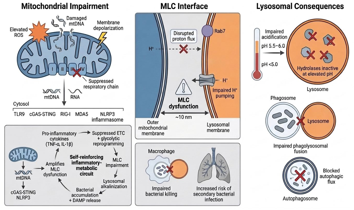
Figure 2.
Bacteria exploit the host cytokine environment through divergent strategies. Several respiratory pathogens (S. aureus, P. aeruginosa, M. avium, L. monocytogenes) exhibit cytokine-dependent growth enhancement in response to macrophage-derived TNF-α, IL-1β, and IL-6, an effect blocked by neutralizing cytokine antibodies. Others (N. meningitidis, P. aeruginosa) internalize host cytokines such as IFN-γ to directly upregulate virulence gene expression. H. influenzae instead actively suppresses host cytokine production during live, persistent infection, favoring long-term colonization over acute virulence. S. pneumoniae exploits a related but distinct host-derived resource, binding lactate dehydrogenase A (LDH-A) released from dying host cells via PspA/PspC to generate a local lactate supply that enhances virulence. These divergent strategies converge on the same outcome: sustained macrophage-driven inflammation, and the metabolic byproducts of host cell death, paradoxically facilitate bacterial growth, virulence, or persistence, depending on the organism involved.
Figure 2.
Bacteria exploit the host cytokine environment through divergent strategies. Several respiratory pathogens (S. aureus, P. aeruginosa, M. avium, L. monocytogenes) exhibit cytokine-dependent growth enhancement in response to macrophage-derived TNF-α, IL-1β, and IL-6, an effect blocked by neutralizing cytokine antibodies. Others (N. meningitidis, P. aeruginosa) internalize host cytokines such as IFN-γ to directly upregulate virulence gene expression. H. influenzae instead actively suppresses host cytokine production during live, persistent infection, favoring long-term colonization over acute virulence. S. pneumoniae exploits a related but distinct host-derived resource, binding lactate dehydrogenase A (LDH-A) released from dying host cells via PspA/PspC to generate a local lactate supply that enhances virulence. These divergent strategies converge on the same outcome: sustained macrophage-driven inflammation, and the metabolic byproducts of host cell death, paradoxically facilitate bacterial growth, virulence, or persistence, depending on the organism involved.
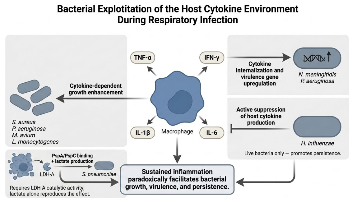
Disclaimer/Publisher’s Note: The statements, opinions and data contained in all publications are solely those of the individual author(s) and contributor(s) and not of MDPI and/or the editor(s). MDPI and/or the editor(s) disclaim responsibility for any injury to people or property resulting from any ideas, methods, instructions or products referred to in the content. |
© 2026 by the authors. Licensee MDPI, Basel, Switzerland. This article is an open access article distributed under the terms and conditions of the Creative Commons Attribution (CC BY) license (http://creativecommons.org/licenses/by/4.0/).
Copyright: This open access article is published under a Creative Commons CC BY 4.0 license, which permit the free download, distribution, and reuse, provided that the author and preprint are cited in any reuse.


