

Submitted:
21 July 2026
Posted:
22 July 2026
You are already at the latest version
Abstract
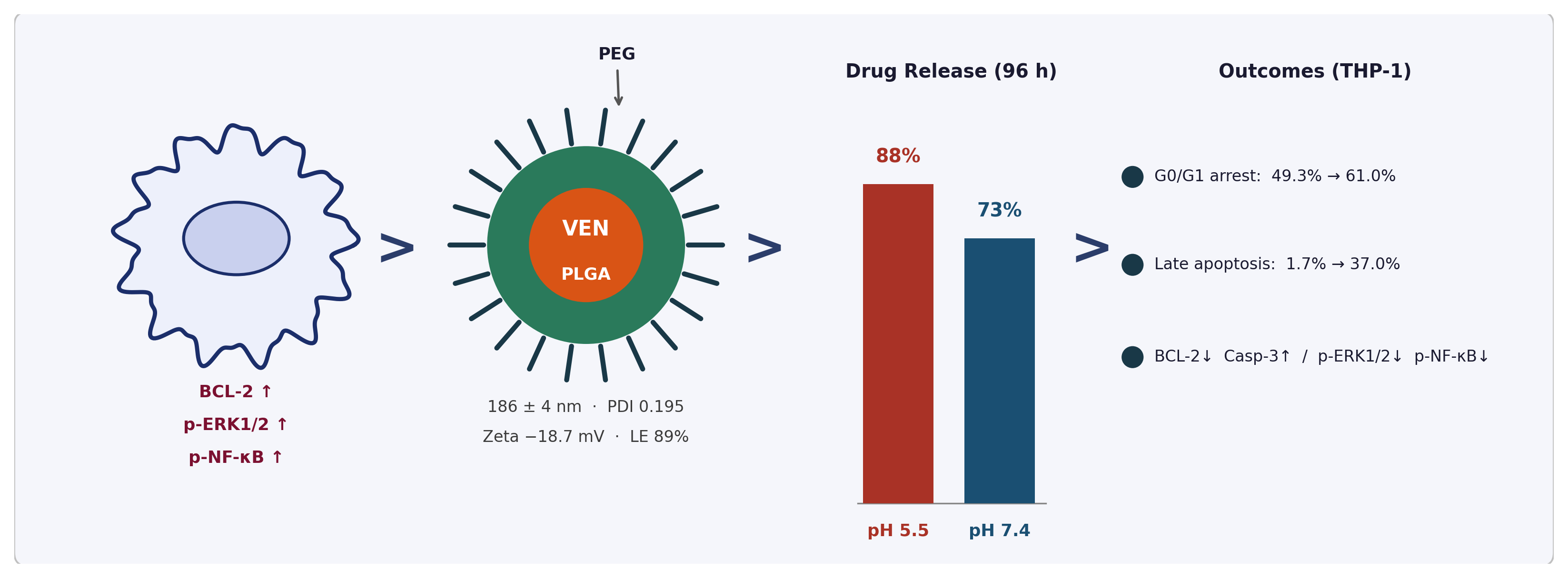
Background: Acute myeloid leukemia (AML) remains a treatment challenge due to the systemic toxicity of conventional chemotherapy. Venetoclax, a selective BCL-2 inhibitor, shows promise in the treatment of AML; however, its clinical efficacy is limited due to inadequate pharmacokinetic properties. PEG-PLGA-based polymeric nanoparticles provide a biocompatible platform for improving drug stability, controlling release behavior, and potentially enhancing intracellular drug availability. Methods: The physicochemical properties of VEN-PEG-PLGA nanoparticles were evaluated by dynamic light scattering (DLS), scanning electron microscopy (SEM), drug loading analysis, and in vitro drug release. Antileukemic activity was assessed in THP-1 cells using the CCK-8 cytotoxicity assay, propidium iodide (PI)-based cell cycle analysis, Annexin V-FITC/PI apoptosis assay, and Western blot analysis of Bcl-2, cleaved caspase-3, cleaved caspase-9, ERK1/2, phospho-ERK1/2 (p-ERK1/2), NF-κB, and phospho-NF-κB (p-NF-κB). Results: VEN-PEG-PLGA nanoparticles exhibited a mean particle size of 186 ± 4 nm with a polydispersity index of 0.195 ± 0.005, 89% loading efficiency, and 6.26% loading capacity. The nanoparticles demonstrated a pH-responsive drug release profile, reaching approximately 72–74% cumulative release at pH 7.4 and 88–89% at pH 5.5 after 96 h. In THP-1 cells, both free venetoclax and VEN-PEG-PLGA na-noparticles exhibited concentration-dependent cytotoxicity, although free venetoclax showed greater cytotoxicity, with EC₅₀ values of 5.59 × 10⁻⁷ M and 1.35 × 10⁻⁶ M, respectively. Cell cycle analysis demonstrated an increase in the G0/G1 cell population from 49.30% in the control group to 61.04% following treatment with VEN-PEG-PLGA nanoparticles, accompanied by a reduction in the G2/M phase. Annexin V-FITC/PI analysis showed that VEN-PEG-PLGA nanoparticles in-creased the proportion of late apoptotic cells to 36.99%, compared with 22.13% following free venetoclax treatment. Western blot analysis demonstrated reduced Bcl-2, ERK1/2, phos-pho-ERK1/2, and phospho-NF-κB expression, together with increased cleaved caspase-3 and cleaved caspase-9 expression in both treatment groups, with these changes being more pronounced in the VEN-PEG-PLGA nanoparticle-treated group. Conclusions: Venetoclax-loaded PEG-PLGA nanoparticles modulated apoptosis-related responses, cell cycle progression, and BCL-2/ERK1/2/NF-κB-associated signaling pathways in THP-1 cells. These findings provide preliminary in vitro evidence supporting further preclinical evaluation of VEN-PEG-PLGA na-noparticles in AML models.
Keywords:
venetoclax
; PEG-PLGA nanoparticles
; acute myeloid leukemia
; BCL-2 inhibition
; apoptosis
; ERK1/2 signaling
; NF-κB pathway
; nanomedicine
; drug delivery
1. Introduction
Acute myeloid leukemia (AML) is a heterogeneous hematologic malignancy characterized by clonal expansion of myeloid progenitor cells and impaired differentiation [1]. Despite advances in therapies, the five-year survival rate for AML remains suboptimal, particularly in elderly patients and those with high-risk cytogenetic profiles [1].
Conventional chemotherapy regimens are associated with significant systemic toxicity, limited tumor selectivity, and the emergence of drug resistance, underscoring the need for novel therapeutic strategies. Consequently, there is an urgent need for modified targeted therapies to enhance life expectancy and mitigate treatment-related toxicity [2].
The BCL-2 protein family plays a central role in the regulation of the intrinsic apoptotic pathway. BCL-2 overexpression; an anti-apoptotic event is frequently observed in AML and is associated with chemotherapy resistance and poor prognosis [3,4,5,6]. BCL-2 prevents mitochondrial outer membrane permeabilization (MOMP) by sequestering pro-apoptotic proteins such as BAX and BAK, thereby blocking cytochrome release and downstream caspase activation [7]. Venetoclax, a highly selective BCL-2 inhibitor, has demonstrated clinical efficacy in hematologic malignancies, including AML, particularly in combination with hypomethylating agents or low-dose cytarabine [8,9]. However, its clinical utility is limited by rapid systemic clearance, and the emergence of resistance mechanisms, including BCL-2 mutations (e.g., Gly101Val) and upregulation of alternative anti-apoptotic proteins such as MCL-1 [10]. When administered in combination with an HMA, venetoclax and HMA have been found to be superior to monotherapy against MCL-1 and other resistance mechanisms [9,10].
Beyond BCL-2 inhibition, emerging evidence highlights the critical role of pro-survival signaling cascades in AML pathogenesis and drug resistance. The extra-cellular signal-regulated kinase (ERK1/2) pathway—a component of the RAS/RAF/MEK/ERK cascade—is frequently activated in AML and promotes cell pro-liferation, survival, and resistance to apoptosis [11]. Concurrently, nuclear factor kappa-B (NF-κB) signaling sustains leukemic cell survival by transcriptionally upreg-ulating anti-apoptotic genes [11].
Nanoparticle (NPs) based drug delivery systems have emerged as a promising platform to overcome the pharmacokinetic limitations of hydrophobic anticancer agents. Polymeric NPs composed of poly(lactic-co-glycolic acid) (PLGA) and its PEGylated derivatives (PEG-PLGA) offer several advantages, including biocompatibility, biodegradability, controlled drug release, enhanced cellular uptake, and the ability to bypass multidrug resistance mechanisms [12,13]. PEGylation of PLGA NPs creates a hydrophilic steric barrier that reduces opsonization by serum proteins and subsequent clearance by the reticuloendothelial system (RES), thereby potentially improving systemic stability and pharmacokinetic properties of nanoparticle-based drug delivery systems[14]. Recent studies have suggested that nanoparticle-based formulations may improve the solubility, stability, and intracellular delivery characteristics of venetoclax [15,16].
The THP-1 cell line, derived from a patient with acute monocyte leukemia, is widely used in vitro model for studying monocyte/macrophage differentiation, inflammatory responses, and the efficacy of anticancer agents in AML [15]. THP-1 cells are sensitive to BCL-2 inhibition and constitute a well-established model for evaluating venetoclax-based treatment strategies in AML [17].
The objectives of this study were: (i) to develop and characterize VEN-PEG-PLGA nanoparticles with optimal physicochemical properties for AML treatment; (ii) to evaluate pH-sensitive drug release profiles relevant to acidic intracellular compartments, including endosomal/lysosomal environments; (iii) to assess antileukemic activity in THP-1 acute monocytic leukemia cells; and (iv) to elucidate the molecular mechanisms underlying the mechanistic and apoptotic activity of the nano formulation, with particular focus on BCL-2, ERK1/2, and NF-κB signaling.
2. Materials and Methods
2.1. Materials
Poly (ethylene glycol) methyl ether-block-poly(lactide-co-glycolide) (PEG average Mn 5,000, PLGA Mn 7,000) and Venetoclax (purity ≥99%) were obtained from Sigma-Aldrich (St. Louis, MO, USA). Acetone (HPLC grade), dimethyl sulfoxide (DMSO), and other analytical-grade reagents were obtained from Merck (Darmstadt, Germany). RPMI-1640 medium and fetal bovine serum (FBS) were purchased from Biowest (Nuaillé, France). The primary antibodies used for Western blotting included NF-κB p65 (clone 572, Invitrogen), ERK1/2 (Invitrogen), phospho-ERK1/2 (Thr202/Tyr204; clone MILAN8R, Invitrogen/eBioscience), phospho-NF-κB p65 (Ser337, Invitrogen), caspase-3 (ab44976, Abcam), caspase-9 (ab52298, Abcam), and β-actin (sc-47778, Santa Cruz Biotechnology).
2.2. Preparation of VEN-PEG-PLGA Nanoparticles
Venetoclax-loaded PEG-PLGA nanoparticles (VEN-PEG-PLGA NPs) were prepared using an emulsion–solvent evaporation method. Briefly, PEG-PLGA (2 mg) and venetoclax (0.15 mg) were dissolved in 500 µL of acetone to obtain the organic phase, which was mixed with 500 µL of distilled water to form an emulsion. The emulsion was incubated overnight at room temperature on an orbital shaker to allow complete acetone evaporation. The resulting nanoparticle suspension was centrifuged at 6000 rpm to precipitate the unloaded venetoclax. The supernatant containing VEN-PEG-PLGA nanoparticles was collected, while the pellet was dissolved in dimethyl sulfoxide (DMSO) for spectrophotometric determination of unloaded venetoclax using a calibration curve. Blank PEG-PLGA nanoparticles were prepared under identical conditions without venetoclax. Encapsulation efficiency (EE%) and drug loading (DL%) were determined by UV-Vis spectrometer following nanoparticle disruption in acetonitrile, as follows: EE% = (mass of drug in NPs / total drug added) × 100; DL% = (mass of drug in NPs / total NP mass) × 100.
2.3. Physicochemical Characterization
Hydrodynamic diameter and polydispersity index (PDI) were measured at 25 °C by dynamic light scattering (DLS) using a Zetasizer Nano ZS (Malvern Instruments, Malvern, UK) (n = 3). Surface morphology was examined by scanning electron microscopy (SEM; JEOL JSM-7600F, Tokyo, Japan) after gold sputter-coating.
2.4. In Vitro Drug Release Study
The in vitro release profile of venetoclax-loaded PEG-PLGA nanoparticles (VEN-PEG-PLGA NPs) was evaluated in phosphate-buffered saline (PBS, pH 7.4) and acetate buffer (pH 5.5), simulating physiological and endosomal/lysosomal pH conditions, respectively. A defined volume of the nanoparticle suspension was dispersed in 1.5 mL of each release medium and incubated at 37 °C. Samples were collected at 0, 24, 48, and 96 h, and the released venetoclax was quantified spectrophotometrically at 286 nm using a previously established calibration curve.
2.5. Cytotoxicity Assay
THP-1 human monocytic leukemia cells were cultured in RPMI-1640 medium supplemented with 10% fetal bovine serum (FBS) and 100 U/mL penicillin–streptomycin at 37 °C in a humidified atmosphere containing 5% CO₂. Cells were seeded into 96-well plates at a density of 3 × 10³ cells/well and treated with free venetoclax (VEN) or venetoclax-loaded PEG-PLGA nanoparticles (VEN-PEG-PLGA NPs) at concentrations ranging from 10⁻¹² to 10⁻³ M for 48 h. Cell viability was determined using the Cell Counting Kit-8 (CCK-8) assay by adding CCK-8 reagent (10% v/v) to each well, followed by incubation for 4 h at 37 °C. Absorbance was measured at 450 nm using a Multiskan SkyHigh microplate reader (Thermo Fisher Scientific, Waltham, MA, USA). Cell viability was expressed as a percentage relative to untreated control cells. The half-maximal effective concentration (EC₅₀) values were calculated by nonlinear regression analysis using GraphPad Prism (version 8; GraphPad Software, San Diego, CA, USA). All experiments were performed in quadruplicate (n = 3).
2.6. Cell Cycle Analysis
THP-1 cells were treated with free venetoclax (VEN) or venetoclax-loaded PEG-PLGA nanoparticles (VEN-PEG-PLGA NPs) at their EC₅₀ concentrations for 48 h. Following treatment, the cells were collected, fixed with 70% ethanol, and washed twice with phosphate-buffered saline (PBS). The fixed cells were stained with propidium iodide (PI) solution containing RNase A for 10 min in the dark. Cell cycle distribution was determined based on DNA content using a DxFLEX flow cytometer (Beckman Coulter, Brea, CA, USA.
2.7. Apoptosis Analysis
Apoptosis was evaluated using an Annexin V-FITC Apoptosis Detection Kit according to the manufacturer’s instructions. THP-1 cells (5 × 10⁴ cells/well) were treated with free venetoclax (VEN) or venetoclax-loaded PEG-PLGA nanoparticles (VEN-PEG-PLGA NPs) at their respective EC₅₀ concentrations for 48 h. Following treatment, the cells were collected by centrifugation (200 × g, 5 min), and the supernatant was discarded. The cells were stained with Annexin V-FITC and propidium iodide (PI) for 15 min at room temperature in the dark. Samples were analyzed using a DxFLEX flow cytometer (Beckman Coulter, Brea, CA, USA), and the percentages of early and late apoptotic cells were determined.
2.8. Western Blot Analysis
Total protein concentration was determined using the BCA Protein Assay with bovine serum albumin (BSA) as the standard. Proteins extracted from THP-1 cells were quantified, and equal amounts of protein (10 µg per lane) were separated by 12% SDS-PAGE and transferred onto nitrocellulose membranes. The membranes were blocked with PBS-T blocking solution for 1 h at room temperature and incubated overnight at 4 °C with primary antibodies against ERK1/2, phospho-ERK1/2 (p-ERK1/2), NF-κB, phospho-NF-κB (p-NF-κB), Bcl-2, Caspase-3, and Caspase-9. After washing, the membranes were incubated with HRP-conjugated secondary antibodies for 1 h at room temperature. Protein bands were visualized using an enhanced chemiluminescence (ECL) substrate and imaged with a Thermo Scientific iBright imaging system. β-Actin was used as the loading control.
2.8. Statistical Analysis
Data are presented as the mean ± standard deviation (SD). Statistical analyses were performed using GraphPad Prism 8.0 (GraphPad Software, San Diego, CA, USA). Comparisons among groups were performed using one-way analysis of variance (ANOVA) followed by Tukey’s multiple comparisons test. Differences were considered statistically significant at p < 0.05 and highly significant at p < 0.01.
2.9. Ethical Statement
Because a commercially available cell line (THP-1) was used in the study, no procedures were performed directly on human or animal subjects. Therefore, ethical committee approval is not required for this study.
3. Results
3.1. Physicochemical Characterization of VEN-PEG-PLGA NPs
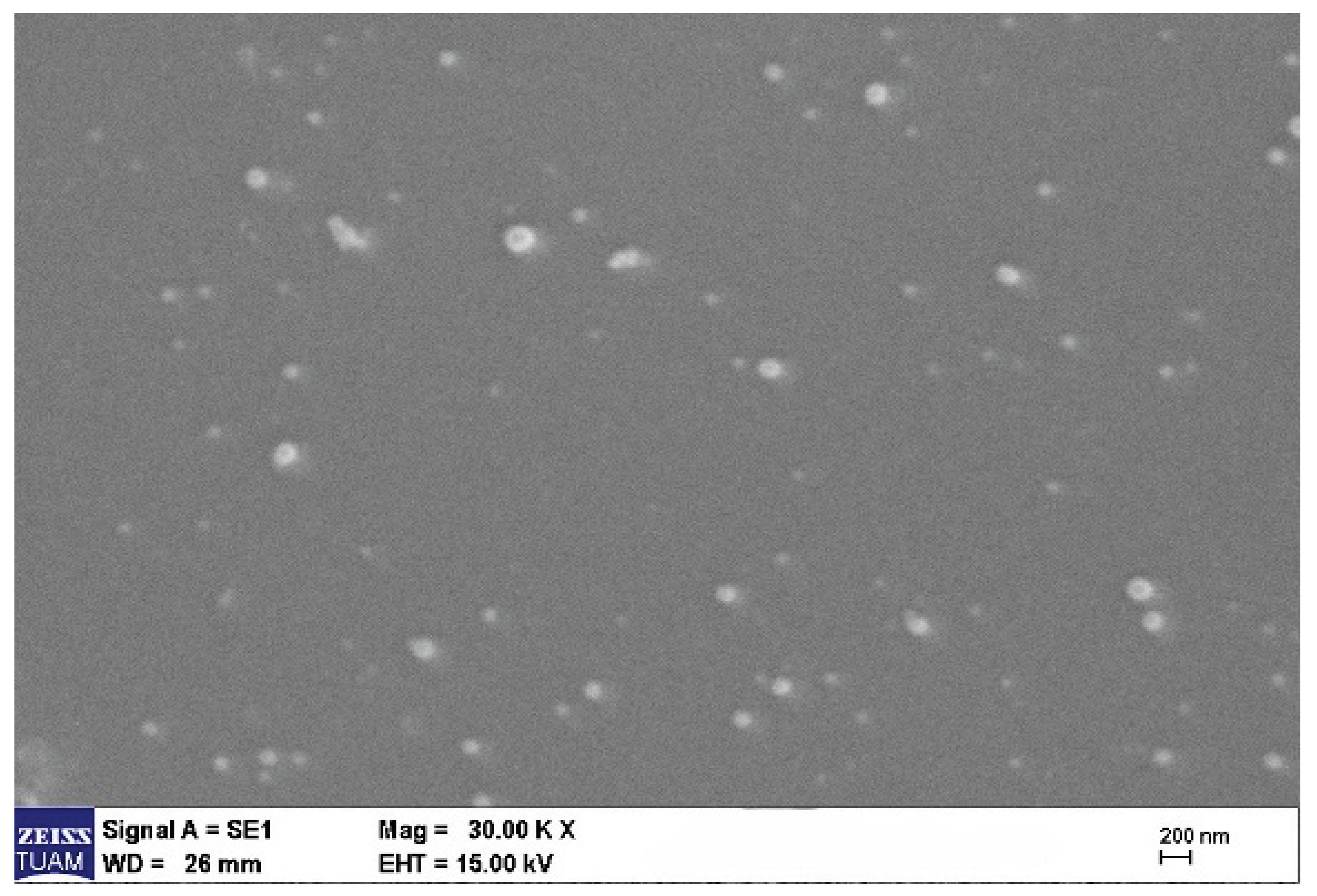
The physicochemical properties of PEG-PLGA nanoparticles were evaluated using dynamic light scattering (DLS) and scanning electron microscopy (SEM). DLS analysis revealed a mean hydrodynamic diameter of 186 ± 4 nm with a polydispersity index (PDI) of 0.195 ± 0.005, indicating a narrow and homogeneous particle size distribution (Figure 1A). SEM analysis confirmed a spherical morphology of the nanoparticles, consistent with the DLS results in terms of particle size distribution (Figure 1B).
Figure 1.
A. Particle size distribution of VEN-PEG-PLGA NPs measured by dynamic light scattering (DLS).
Figure 1.
A. Particle size distribution of VEN-PEG-PLGA NPs measured by dynamic light scattering (DLS).
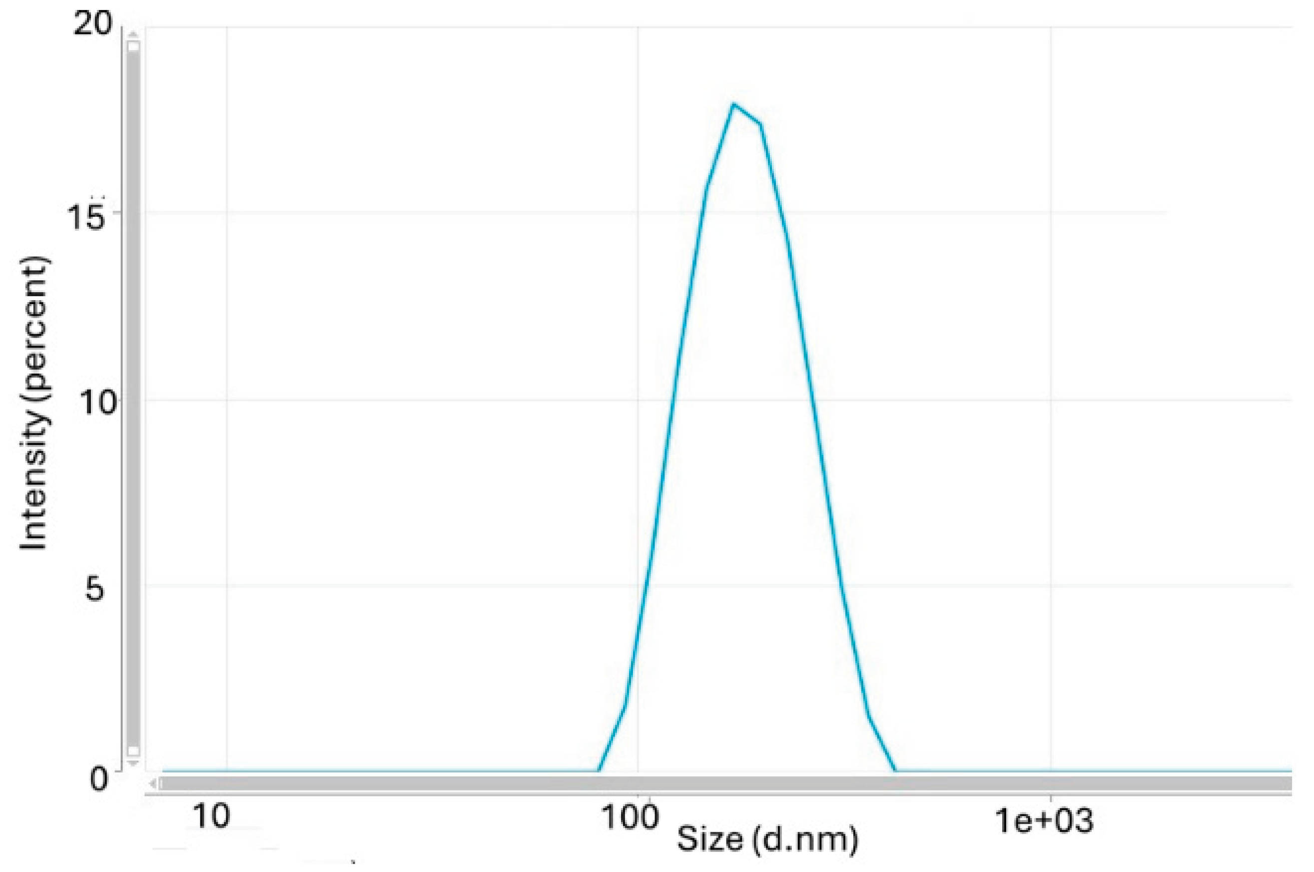
Figure 1.
B. SEM image of VEN-PEG-PLGA nanoparticles exhibiting spherical morphology and uniform size distribution. Scale bar: 200 nm.
Figure 1.
B. SEM image of VEN-PEG-PLGA nanoparticles exhibiting spherical morphology and uniform size distribution. Scale bar: 200 nm.
3.2. Drug Loading Efficiency and Capacity
The drug-loading performance of venetoclax-loaded PEG-PLGA nanoparticles was evaluated using loading efficiency (LE%) and loading capacity (LC%). The loading efficiency was determined to be 89%, indicating that approximately 0.13 mg of the initially used 0.15 mg venetoclax was successfully encapsulated within the nanoparticle structure. In addition, the loading capacity (LC%) was calculated as 6.26%.
3.3. In Vitro Drug Release
VEN-PEG-PLGA NPs exhibited a pH-responsive drug release profile (Figure 3). At physiological pH 7.4, cumulative drug release increased gradually over time, reaching approximately 72–74% after 96 h, indicating a sustained release behavior. In contrast, drug release was accelerated under acidic conditions (pH 5.5), with approximately 58–60% of the encapsulated venetoclax released within the first 48 h and a cumulative release of approximately 88–89% after 96 h.
Figure 2.
In vitro release profile of venetoclax-loaded PEG-PLGA nanoparticles (VEN-PEG-PLGA NPs) at pH 7.4 and pH 5.5 over 96 h. Drug release studies were performed at 37 °C, and cumulative drug release (%) was determined at predetermined time points. Data are presented as the mean ± SD (n = 3).
Figure 2.
In vitro release profile of venetoclax-loaded PEG-PLGA nanoparticles (VEN-PEG-PLGA NPs) at pH 7.4 and pH 5.5 over 96 h. Drug release studies were performed at 37 °C, and cumulative drug release (%) was determined at predetermined time points. Data are presented as the mean ± SD (n = 3).
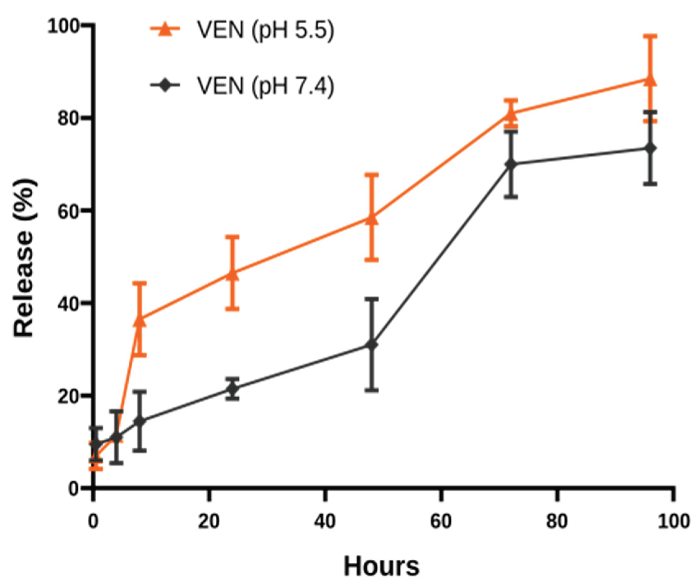
Figure 3.
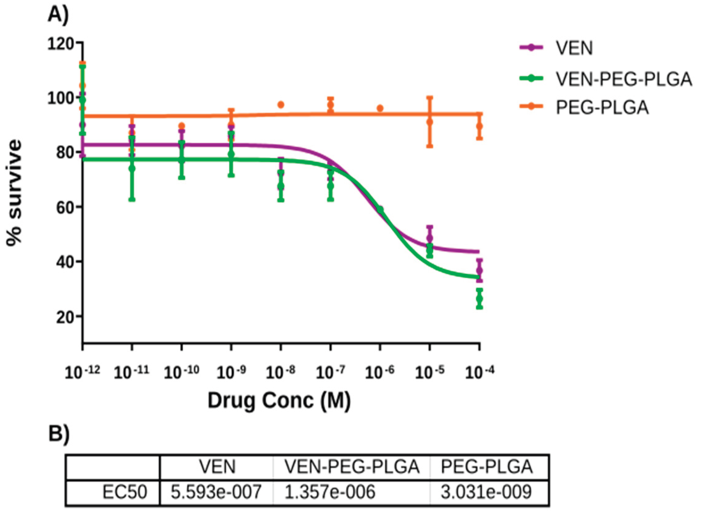
Cytotoxic effects of free venetoclax (VEN), VEN-PEG-PLGA nanoparticles (VEN-PEG-PLGA NPs), and blank PEG-PLGA nanoparticles in THP-1 cells after 48 h of treatment. Cell viability was determined using the CCK-8 assay. (A) Dose–response curves of the different treatment groups. (B) Calculated EC₅₀ values for free VEN and VEN-PEG-PLGA NPs. Data are presented as the mean ± SD (n = 3).
Figure 3.
Cytotoxic effects of free venetoclax (VEN), VEN-PEG-PLGA nanoparticles (VEN-PEG-PLGA NPs), and blank PEG-PLGA nanoparticles in THP-1 cells after 48 h of treatment. Cell viability was determined using the CCK-8 assay. (A) Dose–response curves of the different treatment groups. (B) Calculated EC₅₀ values for free VEN and VEN-PEG-PLGA NPs. Data are presented as the mean ± SD (n = 3).
3.4. Cytotoxicity Assay
The CCK-8 assay demonstrated concentration-dependent cytotoxicity for both free venetoclax (VEN) and VEN-PEG-PLGA nanoparticles (VEN-PEG-PLGA NPs) in THP-1 cells after 48 h of treatment (Figure 3A). Free VEN exhibited greater cytotoxicity than the nanoparticle formulation, with EC₅₀ values of 5.59 × 10⁻⁷ M and 1.35 × 10⁻⁶ M, respectively (Figure 3B).
3.5. Cell Cycle Analysis
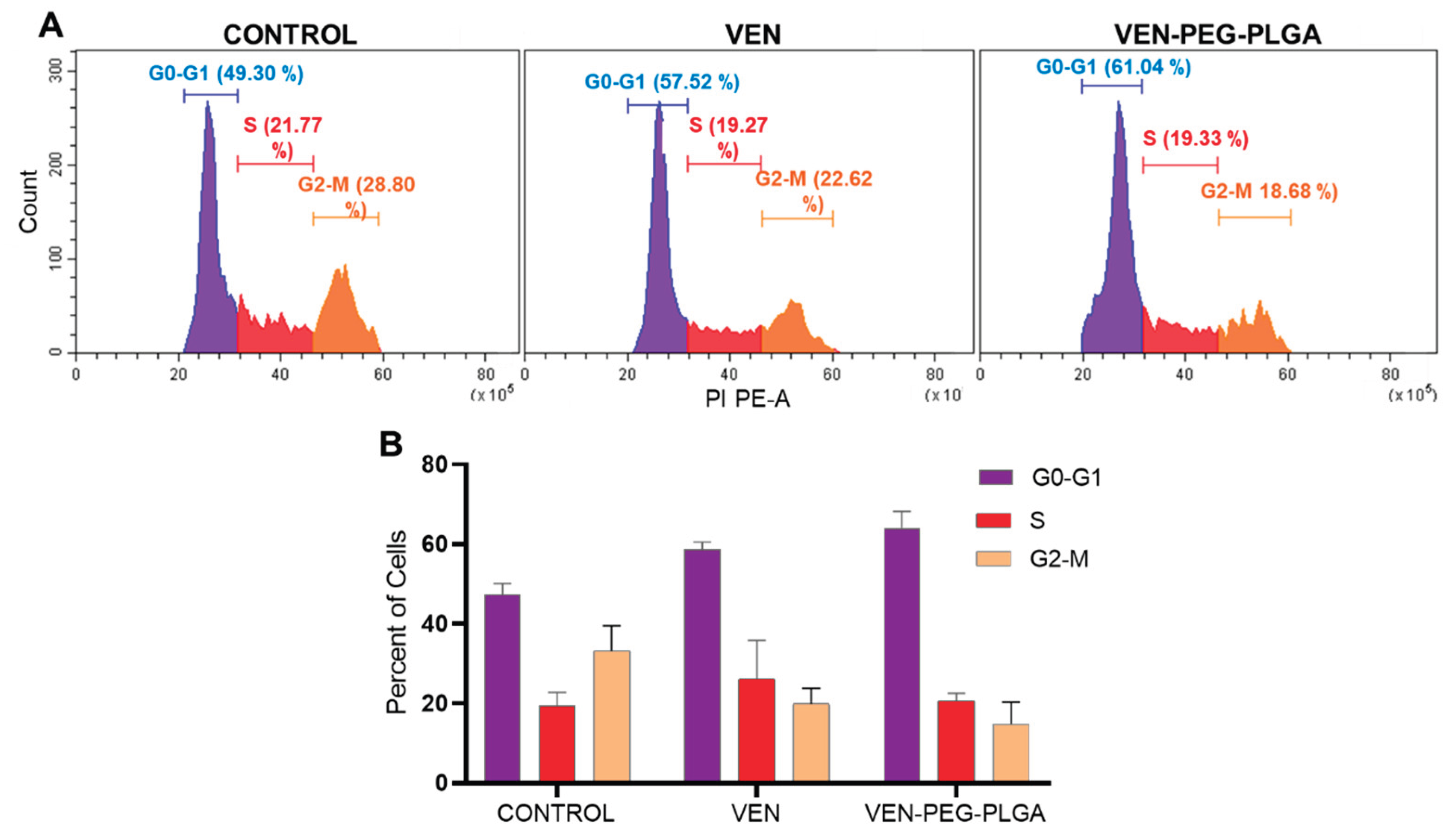
Cell cycle analysis was performed by flow cytometry to evaluate the effects of free venetoclax (VEN) and VEN-PEG-PLGA nanoparticles on cell cycle progression in THP-1 cells (Figure 4). In the control group, 49.30%, 21.77%, and 28.80% of cells were distributed in the G0/G1, S, and G2/M phases, respectively. Treatment with free VEN increased the proportion of cells in the G0/G1 phase to 57.52%, accompanied by a reduction in the G2/M phase to 22.62%. A similar trend was observed in cells treated with VEN-PEG-PLGA nanoparticles, with 61.04% of cells in the G0/G1 phase and 18.68% in the G2/M phase, while the percentage of cells in the S phase remained comparable to that of the control group (19.27% and 19.33%, respectively). Although both treatments induced a modest accumulation of cells in the G0/G1 phase with a corresponding decrease in the G2/M phase.
3.6. Apoptosis Analysis
Apoptosis induction by free venetoclax (VEN) and VEN-PEG-PLGA nanoparticles was evaluated by Annexin V-FITC/PI flow cytometry (Figure 6). In the control group, the majority of cells remained viable (96.16%). Treatment with free VEN increased the proportions of both early apoptotic (16.99%) and late apoptotic (22.13%) cells compared with the control group. Notably, treatment with VEN-PEG-PLGA nanoparticles resulted in a further increase in the late apoptotic cell population (36.99%), accompanied by a reduction in the percentage of viable cells to 51.52% (Figure 6A). Quantitative analysis confirmed that both free VEN and VEN-PEG-PLGA nanoparticles increased apoptosis relative to the untreated control, with the nanoparticle formulation producing a more pronounced increase in late apoptosis (Figure 5).
Figure 5.
Apoptosis analysis of THP-1 cells treated with free venetoclax (VEN) or VEN-PEG-PLGA nanoparticles (VEN-PEG-PLGA NPs) using Annexin V-FITC/PI flow cytometry. (A) Representative dot plots showing viable (Annexin V⁻/PI⁻), early apoptotic (Annexin V⁺/PI⁻), late apoptotic (Annexin V⁺/PI⁺), and necrotic (Annexin V⁻/PI⁺) cell populations. (B) Quantitative analysis of early and late apoptotic cell populations. Data are presented as the mean ± SD (n = 3). *p< 0.05.
Figure 5.
Apoptosis analysis of THP-1 cells treated with free venetoclax (VEN) or VEN-PEG-PLGA nanoparticles (VEN-PEG-PLGA NPs) using Annexin V-FITC/PI flow cytometry. (A) Representative dot plots showing viable (Annexin V⁻/PI⁻), early apoptotic (Annexin V⁺/PI⁻), late apoptotic (Annexin V⁺/PI⁺), and necrotic (Annexin V⁻/PI⁺) cell populations. (B) Quantitative analysis of early and late apoptotic cell populations. Data are presented as the mean ± SD (n = 3). *p< 0.05.
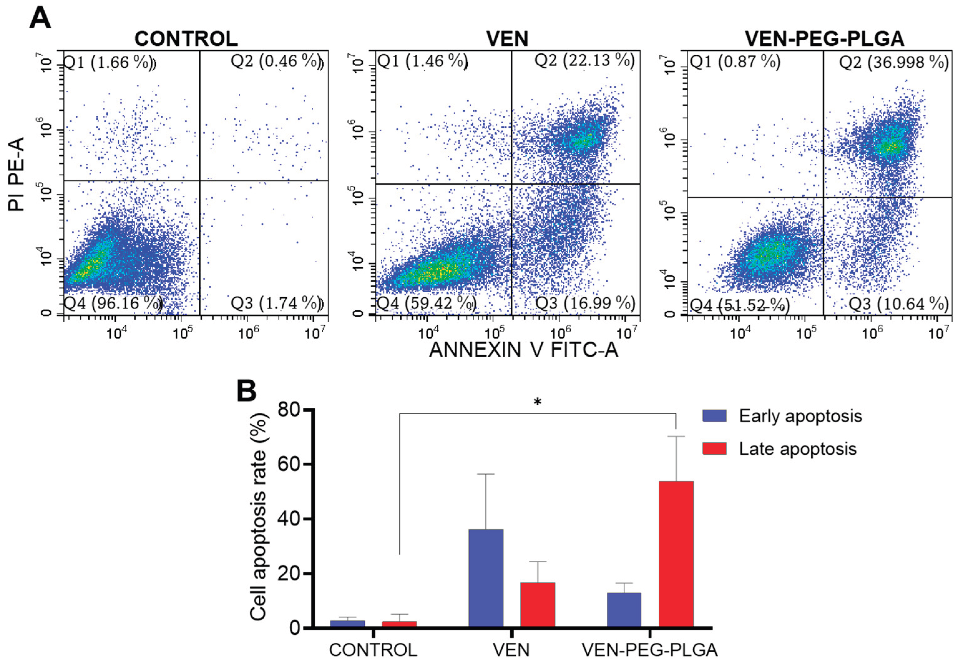
Figure 6.
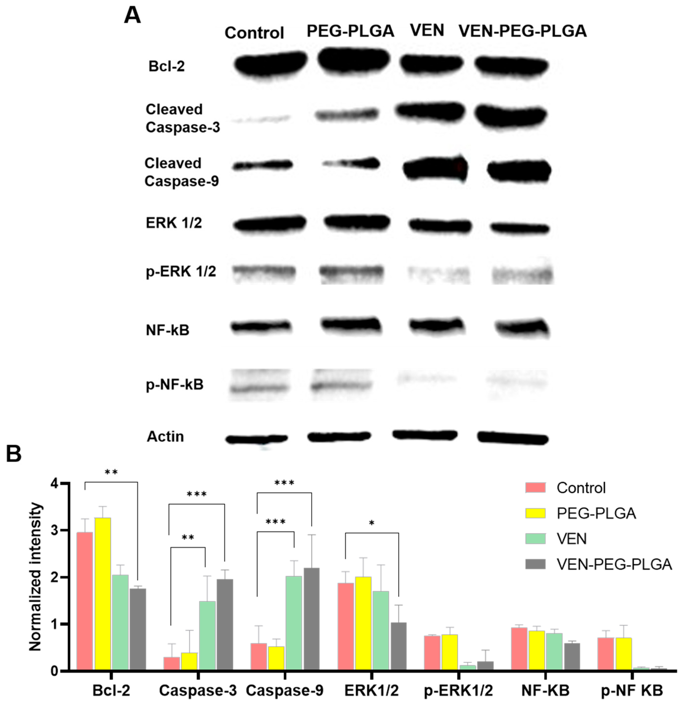
Western blot analysis of apoptosis- and survival-related proteins in THP-1 cells treated with free venetoclax (VEN), blank PEG-PLGA nanoparticles, or VEN-PEG-PLGA nanoparticles. (A) Representative immunoblots showing the expression of Bcl-2, cleaved caspase-3, cleaved caspase-9, ERK1/2, phospho-ERK1/2 (p-ERK1/2), NF-κB, phospho-NF-κB (p-NF-κB), and β-actin. (B) Densitometric analysis of protein expression normalized to β-actin. Data are presented as the mean ± SD (n = 3). Statistical significance is indicated as p < 0.05 (*), p < 0.01 (**), and p < 0.001 (***).
Figure 6.
Western blot analysis of apoptosis- and survival-related proteins in THP-1 cells treated with free venetoclax (VEN), blank PEG-PLGA nanoparticles, or VEN-PEG-PLGA nanoparticles. (A) Representative immunoblots showing the expression of Bcl-2, cleaved caspase-3, cleaved caspase-9, ERK1/2, phospho-ERK1/2 (p-ERK1/2), NF-κB, phospho-NF-κB (p-NF-κB), and β-actin. (B) Densitometric analysis of protein expression normalized to β-actin. Data are presented as the mean ± SD (n = 3). Statistical significance is indicated as p < 0.05 (*), p < 0.01 (**), and p < 0.001 (***).
3.7. Western Blot Analysis of Signaling Proteins
Western blot analysis was performed to evaluate the expression of apoptosis- and survival-related proteins in THP-1 cells following treatment with free venetoclax (VEN) or VEN-PEG-PLGA nanoparticles (Figure 7A). Quantitative analysis showed that Bcl-2 protein expression was reduced in both treatment groups compared with the control, with a more pronounced decrease observed in the VEN-PEG-PLGA group (Figure 7B). In contrast, the expression levels of cleaved caspase-3 and cleaved caspase-9 were increased following both treatments, with higher levels detected in the VEN-PEG-PLGA group than in the free VEN group. Analysis of cell survival-associated proteins revealed reduced ERK1/2 and phospho-ERK1/2 (p-ERK1/2) expression in the treatment groups compared with the control. Similarly, phospho-NF-κB (p-NF-κB) expression decreased after both treatments, whereas total NF-κB expression showed only minor changes. Overall, the observed protein expression profiles were more pronounced in the VEN-PEG-PLGA group than in the free VEN group (Figure 6B).
4. Discussion
This study presents the development and comprehensive characterization of VEN-PEG-PLGA NPs as a novel nanomedicine strategy for AML therapy. The nano formulation demonstrated distinct biological responses compared with free venetoclax, particularly regarding apoptosis induction, cell cycle regulation, and modulation of survival-associated signaling pathways.
The physicochemical properties of VEN-PEG-PLGA NPs fall within the suitable range for nanoparticle-based drug delivery applications. The particle size of 186 nm is within the nanoscale range commonly investigated for polymeric drug delivery systems and may support favorable cellular interactions and biological transport properties [23]. The negative zeta potential conferred by PEG coating reduces opsonization and extends systemic circulation time, thereby increasing the probability of tumor accumulation [16,17]. The high loading efficiency (89%) indicates efficient venetoclax incorporation into the PEG-PLGA nanoparticles, while the low PDI (0.195) reflects a narrow and homogeneous particle size distribution[19].
The pH-sensitive drug release profile is an important property of the nano formulation. Following cellular internalization, nanoparticles may encounter acidic intracellular compartments, including endosomal/lysosomal environments (pH approximately 4.5–5.5), which can influence drug release behavior [25]. The enhanced release observed under acidic conditions at pH 5.5 (88% vs. 72% at pH 7.4) suggests that VEN-PEG-PLGA NPs may provide acidic pH-responsive drug release following cellular uptake, particularly within endosomal/lysosomal compartments, thereby potentially increasing intracellular drug availability at the site of action [24].
Free venetoclax exhibited greater acute cytotoxicity than VEN-PEG-PLGA nanoparticles after 48 h of treatment, as reflected by the lower EC₅₀ value of the free drug. In contrast, VEN-PEG-PLGA nanoparticles produced more pronounced effects on apoptosis induction, cell cycle regulation, and the expression of apoptosis- and survival-related proteins. Importantly, the lower EC₅₀ value observed for free venetoclax indicates that nanoparticle encapsulation does not increase the intrinsic acute cytotoxic potency of venetoclax in this in vitro model. Instead, VEN-PEG-PLGA nanoparticles appear to modify the biological response profile by influencing apoptosis induction, cell cycle distribution, and survival-associated signaling pathways [14,15].
VEN-PEG-PLGA NPs exerted coordinated effects on multiple apoptosis- and survival-related signaling pathways, as evidenced by reduced Bcl-2, ERK1/2, phospho-ERK1/2, and phospho-NF-κB expression together with increased cleaved caspase-3 and cleaved caspase-9 expression. ERK1/2 hyperactivation is a well-established mechanism of venetoclax resistance in AML, as it transcriptionally upregulates MCL-1 and other anti-apoptotic proteins that compensate for BCL-2 inhibition [11,12]. These findings suggest that nanoparticle-mediated drug delivery may influence multiple apoptosis- and survival-associated pathways following intracellular exposure to venetoclax.
The potential mechanism linking nanoparticle delivery to enhanced ERK1/2 and NF-κB inhibition may involve sustained intracellular drug release that maintains inhibitory concentrations of venetoclax over extended periods. However, the possibility that PLGA degradation products (lactic and glycolic acid) contribute to intracellular acidification, thereby modulating signaling pathway activity, cannot be excluded and warrants further investigation.
Overall, the Nano formulation should be interpreted as enhancing mechanistic and apoptotic signaling responses rather than increasing acute cytotoxic potency compared with free venetoclax.
Several limitations of the present study should be acknowledged. First, all experiments were conducted in THP-1 cells, a monocytic AML cell line that may not represent the full spectrum of AML molecular subtypes. Validation in additional AML cell lines (e.g., HL-60, KG-1, MV4-11 with FLT3-ITD mutation) and primary patient samples are required. Second, in vivo pharmacokinetic and efficacy studies in murine AML models are necessary to confirm the translational potential of the Nano formulation. Third, the long-term stability of lyophilized VEN-PEG-PLGA NPs under storage conditions and their performance in complex biological matrices (e.g., whole blood, bone marrow) require evaluation. Fourth, the contribution of nanoparticle-mediated endocytosis to the observed signaling modulation should be mechanistically dissected using appropriate inhibitors.
From a clinical translation perspective, the use of FDA-approved PLGA biomaterials represents a significant regulatory advantage. The established safety profile of PLGA-based formulations (e.g., Lupron Depot®, Risperdal Consta®) provides a translational pathway for investigational new drug application and clinical development. The favorable physicochemical characteristics of VEN-PEG-PLGA nanoparticles support their potential for further pharmaceutical development and clinical translation.
5. Conclusions
This study demonstrates that VEN-PEG-PLGA NPs represent a promising nanomedicine strategy for AML therapy. The nanoparticles exhibited favorable physicochemical characteristics, acidic pH-responsive drug release behavior, induced apoptosis-associated changes, G0/G1 cell cycle accumulation, and modulation of apoptosis- and survival-related proteins, including Bcl-2, cleaved caspase-3, cleaved caspase-9, ERK1/2, phospho-ERK1/2, and phospho-NF-κB, compared with free venetoclax. These findings support the potential of VEN-PEG-PLGA NPs as a nanoparticle-based therapeutic approach for AML. However, further studies in additional AML models and in vivo investigations are required to confirm their therapeutic efficacy, safety, and translational potential.
Supplementary Materials
The following supporting information can be downloaded at the website of this paper posted on Preprints.org.
Author Contributions
N.K.B.: Investigation, Formal Analysis, Writing—Original Draft Preparation, Writing—Review & Editing, Project Administration. E.K.: Conceptualization, Methodology, Investigation. All authors have read and agreed to the published version of the manuscript.
Funding
This study was supported by the Scientific Research Projects Coordination Unit of Afyonkarahisar Health Sciences University under project number 26. GENEL.008.
Institutional Review Board Statement
Not applicable.
Informed Consent Statement
Not applicable.
Data Availability Statement
The data presented in this study are available upon reasonable request from the corresponding author.
Conflicts of Interest
The authors declare no conflicts of interest.
Abbreviations
The following abbreviations are used in this manuscript:
| AML | Acute Myeloid Leukemia |
| NPs | Nanoparticles |
| BCL-2 | B-cell Lymphoma-2 Protein |
| DLS | Dynamic Light Scattering |
| DL% | Drug Loading Percentage |
| EPR | Enhanced Permeability and Retention Effect |
| ERK1/2 | Extracellular Signal-Regulated Kinase 1/2 |
| GMP | Good Manufacturing Practice |
| EC50 | Half-Maximal Inhibitory Concentration |
| MAPK | Mitogen-Activated Protein Kinase |
| MOMP | Mitochondrial Outer Membrane Permeabilization |
| MTT | 3-(4,5-Dimethylthiazol-2-yl)-2,5-Diphenyltetrazolium Bromide |
| NF-κB | Nuclear Factor Kappa-B |
| PDI | Polydispersity Index |
| PEG-PLGA | Polyethylene Glycol-Poly(lactic-co-glycolic acid) |
| PI | Propidium Iodide |
| PLGA | Poly(lactic-co-glycolic acid) |
| SEM | Scanning Electron Microscopy |
| VEN | Venetoclax |
| VEN-PEG-PLGA NPs | Venetoclax-Loaded PEG-PLGA Nanoparticles |
References
- DiNardo, C.D.; Erba, H.P.; Freeman, S.D.; Wei, A.H. Acute myeloid leukaemia. Lancet 2023, 401(10393), 2073. [Google Scholar] [CrossRef]
- Bishoyi, A.K.; Nouri, S.; Hussen, A.; Bayani, A.; Khaksari, M.N.; Soleimani Samarkhazan, H. Nanotechnology in leukemia therapy: revolutionizing targeted drug delivery and immune modulation. Clin. Exp. Med. 2025, 25(1), 166. [Google Scholar] [CrossRef] [PubMed]
- Zhang, Q.; Riley-Gillis, B.; Han, L.; Jia, Y.; Lodi, A.; Zhang, H.; et al. Activation of RAS MAPK pathway confers MCL-1 mediated acquired resistance to BCL-2 inhibitor venetoclax in acute myeloid leukemia. Signal Transduct. Target Ther. 2022, 7(1), 51. [Google Scholar] [CrossRef] [PubMed]
- Adams, J.M.; Cory, S. The BCL-2 arbiters of apoptosis and their growing role as cancer targets. Cell Death Differ. 2018, 25, 27–36. [Google Scholar] [PubMed]
- Kaloni, D.; Diepstraten, S.T.; Strasser, A.; Kelly, G.L. BCL-2 protein family: attractive targets for cancer therapy. Apoptosis 2023, 28(1–2), 20–38. [Google Scholar] [CrossRef] [PubMed]
- Roberts, A.W.; Davids, M.S.; Pagel, J.M.; Kahl, B.S.; Puvvada, S.D.; Gerecitano, J.F.; etal. Targeting BCL2 with venetoclax in relapsed chronic lymphocytic leukemia. N. Engl. J. Med. 2016, 374, 311–322. [Google Scholar] [CrossRef] [PubMed]
- DiNardo, C.D.; Jonas, E.A.; Pullarkat, V.; Thirman, M.J.; Garcia, J.S.; Wei, A.H.; et al. Azacitidine and venetoclax in previously untreated acute myeloid leukemia. N Engl. J. Med. 2020, 383, 617–629. [Google Scholar] [CrossRef] [PubMed]
- Yuan, F.; Li, D.; Li, G.; Cheng, C.; Wei, X. Synergistic efficacy of homoharringtonine and venetoclax on acute myeloid leukemia cells and the underlying mechanisms. Ann. Transl. Med. 2022, 10(8), 490. [Google Scholar] [CrossRef] [PubMed]
- Salem, A.H.; Menon, R.M. Clinical pharmacokinetics and pharmacodynamics of venetoclax, a selective B-cell lymphoma-2 inhibitor. Clin. Transl. Sci. 2024, 17(5), e13807. [Google Scholar] [CrossRef] [PubMed]
- Qi, Zhang; Riley-Gillis, Bridget; Han, Lina; Jia, Yannan; Lod, Alessia; Zhang, Haijiao; et al. Activation of RAS/MAPK pathway confers MCL-1 mediated acquired resistance to BCL-2 inhibitor venetoclax in acute myeloid leukemia. Signal Transduct. Target. Ther. 2022, 7, 51. [Google Scholar] [CrossRef] [PubMed]
- Salaroglio, I.; Mungo, E.; Gazzano, E.; Kopecka, J. ERK is a Pivotal Player of Chemo-Immune-Resistance in Cancer. Int. J. Mol. Sci. 2019, 20, 2505. [Google Scholar] [CrossRef] [PubMed]
- Zhang, D.; Liu, L.; Wang, J.; Zhang, H.; Zhang, Z.; Xing, G.; Wang, X.; Liu, M. Drug-loaded PEG-PLGA nanoparticles for cancer treatment. Front Pharmacol. 2022, 13, 990505. [Google Scholar] [CrossRef] [PubMed]
- Handa, M.; Singh, A.; Flora, S.J.S.; Shukla, R. Stimuli-responsive polymeric nanosystems for therapeutic applications. Curr. Pharm. Des. 2022, 28(11), 910–921. [Google Scholar] [CrossRef] [PubMed]
- Yang, H.; Sung, J.; Alghoul, Z.; Merlin, D. Preparation, characterization, and cell uptake of PLGA/PLA-PEG-FA nanoparticles. Bio Protoc. 2022, 12(7), e4373. [Google Scholar] [CrossRef] [PubMed]
- Thing Mortensen, B.; Østrup Jensen, P.; Helledie, N.; Iversen, P.O.; Ralfkiær, E.; Knud Larsen, J.; et al. Changing bone marrow micro-environment during development of acute myeloid leukaemia in rats. Br. J. Haematol. 1998, 102(2), 458–464. [Google Scholar] [CrossRef] [PubMed]
- Gu, H.; Weng, L.; Chen, C.; et al. Nonionotropic action of an acid-sensing ion channel inhibits leukemogenesis in the acidic bone marrow niche. J. Clin. Invest. 2025, 135(24), e189051. [Google Scholar] [CrossRef] [PubMed]
- Kanamala, M.; Wilson, W.R.; Yang, M.; Palmer, B.D.; Wu, Z. Mechanisms and biomaterials in pH-responsive tumour targeted drug delivery: A review. Biomaterials 2016, 85, 152–167. [Google Scholar] [CrossRef] [PubMed]
- Fredenberg, S.; Wahlgren, M.; Reslow, M.; Axelsson, A. The mechanisms of drug release in poly(lactic-co-glycolic acid)-based drug delivery systems—A review. Int. J. Pharm. 2011, 415, 34–52. [Google Scholar] [CrossRef] [PubMed]
- Ando, H.; Murakami, Y.; Eshima, K.; Ishida, T. A novel polyethylene glycol (PEG)-drug conjugate of venetoclax, a Bcl-2 inhibitor, for treatment of acute myeloid leukemia (AML). Cancer Rep. 2022, 5(3), e1485. [Google Scholar]
- Patel, D.; Bhardwaj, S.; Yadav, V.; Kuche, K.; Dhake, P.; Jain, S. Exploring the co-delivery of sorafenib and venetoclax to induce ferroptosis and apoptosis mediated cancer cell death using surface modified PLGA nanoparticles. AAPS PharmSciTech 2025, 26(1), 58. [Google Scholar] [CrossRef] [PubMed]
- Panagiotou, E.; Gomatou, G.; Trontzas, I.P.; Syrigos, N.; Kotteas, E. Cyclin-dependent kinase (CDK) inhibitors in solid tumors: a review of clinical trials. Clin. Transl. Oncol. 2022, 24(2), 161–192. [Google Scholar] [CrossRef] [PubMed]
- Ramedani, F.; Jafari, S.M.; Saghaeian Jazi, M. Anti-cancer effect of entacapone on esophageal cancer cells via apoptosis induction and Annexin V/PI flowcytometry. Cancer Rep. 2023, 6(3), e1759. [Google Scholar] [CrossRef] [PubMed]
- Vringer, E.; Tait, S.W.G. Mitochondria and cell death-associated inflammation. Cell Death Differ. 2023, 30(2), 304–312. [Google Scholar] [CrossRef] [PubMed]
- Ramedani, F.; Jafari, S. M.; Saghaeian Jazi, M. Anti-cancer effect of entacapone on esophageal cancer cells via apoptosis induction and Annexin V/PI flow cytometry. *Cancer Rep.* 2023, vol. 6(no. 3), e1759. [Google Scholar] [CrossRef] [PubMed]
- Ye, F.; Zhang, W.; Fan, C.; Dong, J.; Peng, M. Antileukemic effect of venetoclax and hypomethylating agents via caspase-3/GSDME-mediated pyroptosis. J. Transl. Med. 2023;21 Transl Med. 2023, 21. [Google Scholar] [CrossRef] [PubMed]
- Han; Zhang, Q.; Dail, M.; Shi, C.; Cavazos, A. Concomitant targeting of BCL2 with venetoclax and MAPK signaling with cobimetinib in acute myeloid leukemia models. *Haematologica* vol. 105, 697–707. [CrossRef] [PubMed]
- Itoh, M.; Tohda, S. Effects of ERK1/2 Inhibitors on the Growth of Acute Leukemia Cells. *Anticancer Res.* 2024, vol. 44(no. 12), 5263–5270. [Google Scholar] [CrossRef] [PubMed]
- Yan, M.; Chen, X.; Ye, Q.; Li, H.; Wu, J. IL-33-dependent NF-κB activation inhibits apoptosis and drives chemoresistance in acute myeloid leukemia. *Cytokine* 2024, vol. 180, 156672. [Google Scholar] [CrossRef] [PubMed]
- Manni, S.; Pesavento, M.; Spinello, Z.; Saggin, L.; et al. Protein Kinase CK2 represents a new target to boost Ibrutinib and Venetoclax induced apoptosis in NF-κB-dependent B cell malignancies. *Front. Cell Dev. Biol.* 2022, vol. 10, 935023. [Google Scholar] [CrossRef]
- DiNardo, C. D.; Pratz, K.; Pullarkat, V.; Jonas, B. A.; Arellano, M.; Becker, P. S.; et al. Venetoclax combined with decitabine or azacitidine in treatment-naive, elderly patients with acute myeloid leukemia. *Blood* 2019, vol. 133, 7–17. [Google Scholar] [CrossRef]
- Konopleva, M.; Pollyea, D. A.; Potluri, J.; Chyla, B.; Hogdal, L.; Busman, T.; et al. Efficacy and biological correlates of response in a phase II study of venetoclax monotherapy in patients with acute myelogenous leukemia. *Cancer Discov. 2016, vol. 6, 1106 1117. [Google Scholar]
- Wang, X.; Mak, P. Y.; Mu, H.; Tao, W. Combinatorial inhibition of focal adhesion kinase and BCL-2 enhances antileukemia activity of venetoclax in acute myeloid leukemia. *Mol. Cancer Ther.* 2020, vol. 19, 1636–1648. [Google Scholar] [CrossRef] [PubMed]
Figure 4.
Effects of free venetoclax (VEN) and VEN-PEG-PLGA nanoparticles (VEN-PEG-PLGA NPs) on cell cycle distribution in THP-1 cells. (A) Representative flow cytometric histograms showing the distribution of cells in the G0/G1, S, and G2/M phases. (B) Quantitative analysis of cell cycle phase distribution. Data are presented as the mean ± SD (n = 3).
Figure 4.
Effects of free venetoclax (VEN) and VEN-PEG-PLGA nanoparticles (VEN-PEG-PLGA NPs) on cell cycle distribution in THP-1 cells. (A) Representative flow cytometric histograms showing the distribution of cells in the G0/G1, S, and G2/M phases. (B) Quantitative analysis of cell cycle phase distribution. Data are presented as the mean ± SD (n = 3).
Disclaimer/Publisher’s Note: The statements, opinions and data contained in all publications are solely those of the individual author(s) and contributor(s) and not of MDPI and/or the editor(s). MDPI and/or the editor(s) disclaim responsibility for any injury to people or property resulting from any ideas, methods, instructions or products referred to in the content. |
© 2026 by the authors. Licensee MDPI, Basel, Switzerland. This article is an open access article distributed under the terms and conditions of the Creative Commons Attribution (CC BY) license (http://creativecommons.org/licenses/by/4.0/).
Copyright: This open access article is published under a Creative Commons CC BY 4.0 license, which permit the free download, distribution, and reuse, provided that the author and preprint are cited in any reuse.


