

Submitted:
21 July 2026
Posted:
21 July 2026
You are already at the latest version
Abstract
The treatment of Ce(OH)4 in thionyl chloride and dimethoxyethane (DME) resulted in the formation of a highly unstable red Ce(IV) complex with the composition [CeCl4(DME)2] (1). From the reaction of 1 with the tripodal Kläui ligand η5-cyclopentadienyltris(dimethylphosphonato)cobaltate(III), [Co(η5-C5H5){P(O)(OMe)2}3]– (LOMe), the two new cerium(III) complexes [Ce(LOMe)2(H2O)2]Cl·2.5H2O (2), 1D-[Ce(μ-Cl)(LOMe)(H2O)3]Cl (3) and the cerium(IV) complex [CeCl2(LOMe)2]·acetone (4) were obtained and structurally characterized. The complexes 3 and 4 are the first cerium complexes featuring the LOMe ligand together with chlorido ligands. Due to the potential exchange of the chloride counterions and ligands in 2 - 4, these complexes could serve as precursors in molecular cerium coordination chemistry. The assigned cerium oxidation states are supported by the bond valence sum (BVS) method. The 31P-NMR spectra with d(31P) = 160.20 ppm (2) and 159.43 ppm (3) are diagnostic of cerium(III).
Keywords:
cerium
; crystal structure
; tripod ligands
; Kläui ligands
; shape calculation
; bond valence sum
; metal-organic chemistry
1. Introduction
Cerium with its +III and +IV oxidation states has a wide range of established and emerging applications, spanning the water-gas shift reaction [1], catalytic oxidation processes [2], organic synthesis [3], and more recent developments in solar energy to fuel conversion [4]. In addition, cerium-based materials attract increasing attention in biomedical contexts, where they can contribute to advanced chemo- and immunotherapeutic strategies [5].
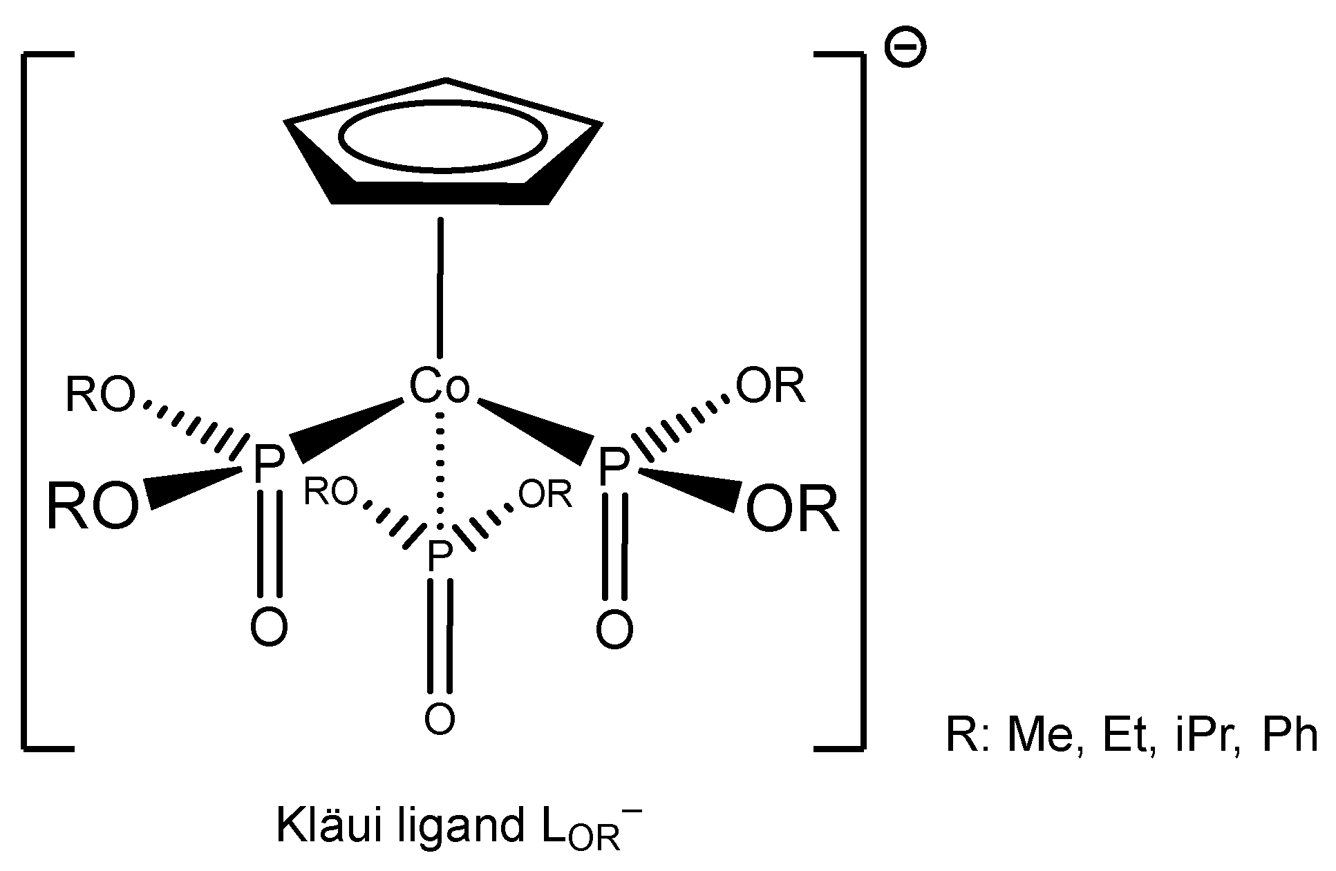
Monoanionic tripodal ligands of the type [Co(η5-C5H5){P(O)(OR)2}3]– (abbreviated as LOR with R = Me, Et, iPr, Ph) are commonly known as Kläui ligands (Scheme 1). These six-electron, oxygen-donor ligands can to some extent be regarded as analogues of the cyclopentadienyl ligand C5H5– (Cp–) [6]. On the other hand, the ligand is a relatively weak-field and π-donating donor with bonding characteristics comparable to water or dimethyl sulfoxide yet exhibit enhanced stability [6]. Owing to these properties, they effectively stabilize metals in high oxidation states across the periodic table and have been widely employed as models for metal-oxygen environments in catalysis [7]. At the same time, their good solubility and hydrolytic stability make them useful mimics of facially coordinated aqua ligands in organometallic systems [8,9,10].
Due to the intriguing properties of cerium and the stabilizing effect of Kläui ligands, their combination is of interest. This is evidenced by previously reported applications in catalysis. On the one hand, such systems have been employed in the aerobic oxidation of cumene to 2-phenyl-2-propanol and acetophenone [11], and on the other hand in the oxidation of substituted thioanisoles [12]. Both Ce(IV) and Ce(III) complexes have been shown to be active in these transformations.
In particular, the Kläui ligand-cerium(IV)-chloride complexes [CeCl3(LOEt)] [13] and [CeCl2(LOEt)2] [14] (LOEt = η5-cyclopentadienyltris(diethylphosphonato)cobaltate(III), [Co(η5-C5H5){P(O)(OEt)2}3]–) are of significant importance, as they serve as suitable precursors for the synthesis of Ce-oxo clusters [13] and enable the incorporation of addition metals such as Mo, Re, V [12] Mn [11] Ru, Os, and V [15]. Furthermore, the chloride ligands can be substituted by anions such as IO4–, S2O8–, and IO3– [16]. Replacement of chloride by alkoxide ligands can promote oxidative C–C coupling reactions, especially when the alkoxide corresponds to 2,4-di-tert-butylphenol [17] Moreover, when the aryloxide ligand is 2,4,6-tri-tert-butylphenol, oxidative transformations of these ligands can also occur [18].
Although the Kläui ligand-cerium-chloride complexes represent highly versatile precursors, only the two complexes [CeCl3(LOEt)] [13] and [CeCl2(LOEt)2] [14], containing exclusively chloride and Kläui ligands, have been reported in the Cambridge Structure Databank (CSD) [19]. Due to the diverse potential applications of cerium coordination complexes, there is significant interest in the synthesis of analogous cerium-chloride complexes with other Kläui ligands having for example the methoxy substituent on phosphorus, [Co(η5-C5H5){P(O)(OMe)2}3]– (LOMe). Consequently, this work addressed the synthesis of novel, Kläui ligand-cerium-chloride complexes with LOMe.
2. Results and Discussion
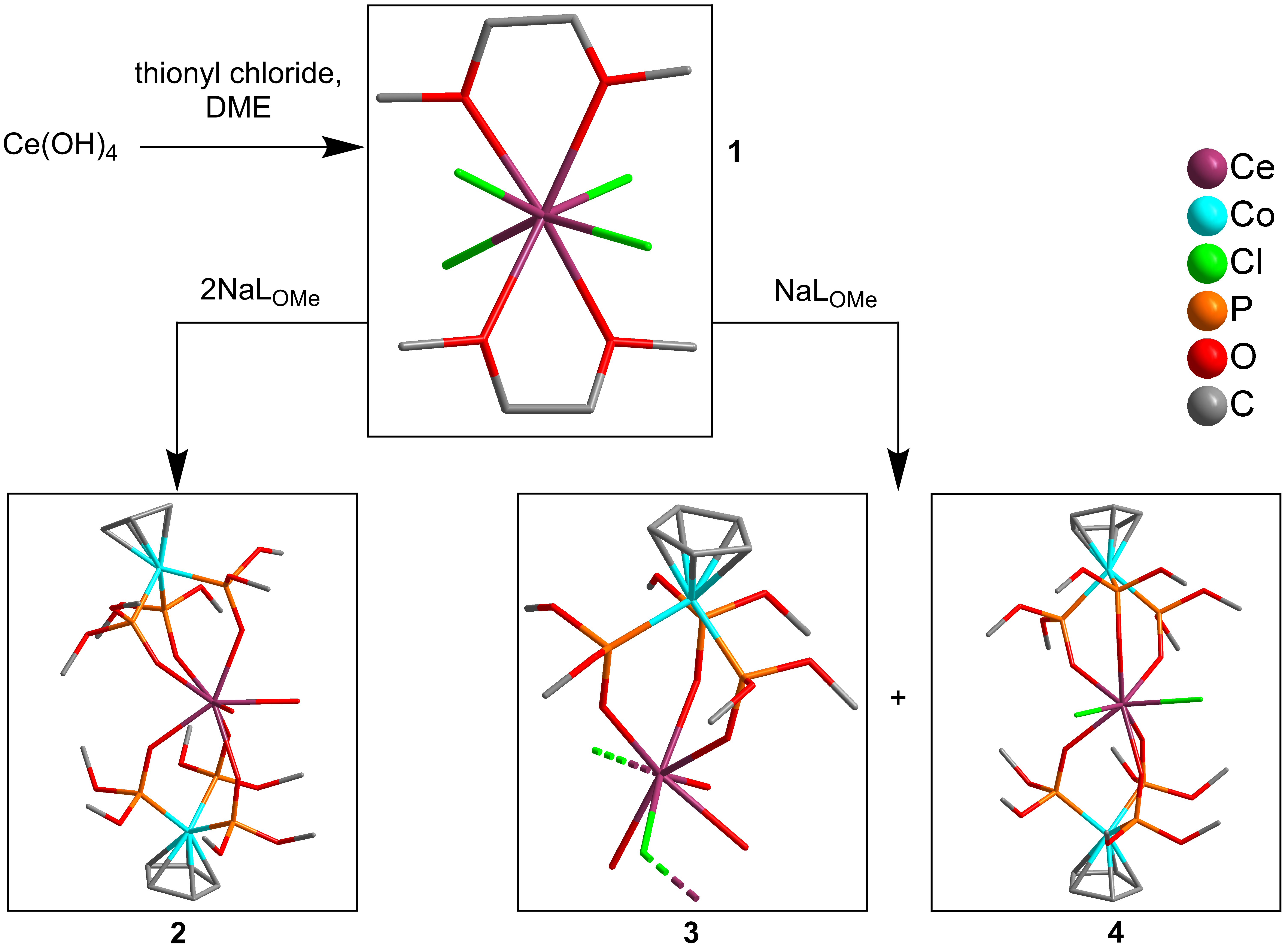
The treatment of Ce(OH)4 with thionyl chloride in dimethoxyethane (DME) yielded a dark red solid of formula [CeCl4(DME)2] (1) (Scheme 2, Figure S1), which was isolated and handled under inert conditions. The intense red color indicates a Ce(IV) complex. However, this complex is not stable under air due to its strongly hygroscopic nature.
2.1. Synthesis of [CeCl4(DME)2] (1)
It is observed that, within minutes upon exposure to the atmosphere, the red color of [CeCl4(DME)2] disappears and a white solid forms within the resulting pasty material which was identified as cerium(III) chloride heptahydrate (CeCl3·7H2O) by powder X-ray diffraction (Figure S2).
Even crystals of [CeCl4(DME)2] coated with perfluorinated oil and maintained under a nitrogen stream at 150 K degraded within two hours, which was clearly evident during the single-crystal X-ray diffraction (SCXRD) measurement. Nevertheless, the structure of 1 could be successfully determined. Based on these data, the bond valence sum (BVS) analysis (see below) supports the assignment as tetravalent cerium.
In the structure of 1 the asymmetric unit comprises one central cerium atom, two dimethoxyethane (DME) molecules and four chloride ligands (Figure 1). This gives a coordination number of eight for the Ce atom.
The Ce–Cl bond lengths range from 2.5795 to 2.6107 Å, while the Ce–O distances lie between 2.5047 and 2.603 Å (Table 1). Accordingly, the Ce–Cl distances are slightly shorter than those reported for the also eight-coordinate cerium(III) compound [Ce2Cl6(DME)4] [20] (Ce–Cl: 2.7280–2.9269 Å; Ce–O: 2.5084–2.666 Å) as well as for the cerium(IV) compound (N(CH3)4)2[CeCl4(NO3)2] [21] (Ce–Cl: 2.6496 Å; Ce–O: 2.5013 Å), whereas the Ce–O distances in 1 are longer compared to those in (N(CH3)4)2[CeCl4(NO3)2].
Furthermore, the cis-Cl–Ce–Cl bond angles in 1 range from 84.44° to 105.79°, while the O–Ce–O angles within the respective DME ligands are 63.82° and 65.44° (Table S6). The cis-Cl–Ce–Cl angles differ significantly from those observed in the dinuclear [Ce2Cl6(DME)4] structure (72.46° and 85.82°) [20] In contrast, the O–Ce–O angles of the DME ligands remain largely unchanged due to the structural constraints of the DME backbone, with values of approximately 63°. The cis-Cl–Ce–Cl angles in the Ce(IV) compound (N(CH3)4)2[CeCl4(NO3)2] are exactly 90° by symmetry.
A Continuous Shape Measure (CShM) analysis of the coordination sphere of the Ce atoms was performed with the SHAPE V2.1 software [22] In this approach, an ideal polyhedron has a shape measure of S = 0, while increasing S values indicate increasing distortion from the ideal geometry. Complex 1 shows a preference for a square antiprismatic shape (SAPR), based on: S(SAPR) = 1.37, S(triangular dodecahedron (TDD)) = 1.98, and S(biaugmented trigonal prism (BTPR)) = 1.71. If the S values are not close to zero the structure can lie between two ideal geometries. This can be determined by the interconversion analysis with a generalized coordinate and the deviation from the path between these two ideal geometries. The interconversion analysis reveals a significant generalized coordinate for TDD→SAPR = 83% and for SAPR→TDD = 69%, and large deviations from the ideal paths (SAPR, TDD) = 52% and (SAPR, BTPR) = 64%. Since there are large deviations from the indicated paths the structure is not exactly on the direct path between two ideal shapes. The coordination geometry is best described as a distorted square antiprism, due to the lowest shape Measure parameter (S) and the higher interconversion coordinate in direction to the SAPR-shape, with contributions from triangular dodecahedral (TDD) and biaugmented trigonal prismatic (BTPR) geometries. In comparison, the polyhedron of [Ce2Cl6(DME)4] (Figure S17) can be described as a distorted triangular dodecahedron (S(TDD) = 0.91) and the geometry of the Ce in (N(CH3)4)2[CeCl4(NO3)2] (Figure S18) is best described as a gyrobifastigium, which is distorted by the two nitrate anions, even if the CShM is high (S(Johnsons gyrobifastigium J26) = 4.24).
2.2. Synthesis of [Ce(LOme)2(H2O)2]Cl·2.5H2O (2)
The reaction of 1 with two equivalents of NaLOme in acetone resulted in the formation of intense green crystals (Scheme S2, Figure S1) upon slow solvent evaporation over several days. The crystals were analyzed as [Ce(LOme)2(H2O)2]Cl·2.5H2O with Ce(III) by single-crystal X-ray diffraction. The 31P-NMR also indicates the presence of a trivalent cerium with a single peak at 160.20 ppm (Figure S7), since the 31P resonances at 115-125 ppm are diagnostic for tetravalent cerium in Ce-LOet complexes and resonances at 150-160 ppm indicate trivalent cerium in Ce-LOet complexes [18]. The presence of a trivalent cerium is also underlined by the BVS method calculated by the results of the SCXRD measurement (see below). The asymmetric unit in the structure of [Ce(LOme)2(H2O)2]Cl·2.5H2O corresponds to the formula unit and contains an eight-coordinated central cerium atom, which binds two Kläui ligands and two aqua ligands (Figure 2). In addition, there is a disordered chloride counter anion and two and a half crystal water molecules. The aqua ligands build hydrogen bonds to the disordered chloride anion (Figure S12, Figure S13).
The structure of 2 can be compared best with the structures of the two Ce(IV) complexes [Ce(LOet)2(H2O)2](Otf)2·H2O (Otf = CF3SO3–) [23] and [Ce(LOet)2(H2O)2](BF4)2 [18] as well as to the Ce(III) complex in [Ce(LOet)2(H2O)2](LOet) [23], since all of these compounds feature two Kläui and two aqua ligands, giving rise to eight-coordinate cerium centers surrounded exclusively by oxygen atoms.
The Ce–O distances of the aqua ligands in 2 range from 2.546 to 2.583 Å, whereas the Ce–O distances to the Kläui ligands lie between 2.395 and 2.484 Å. The distances in the above Ce(IV) from the literature are shorter reflecting the higher charge and smaller size of the Ce(IV) centers. In Ce(LOet)2(H2O)2](Otf)2·H2O the Ce–O(aqua) distances are 2.475 and 2.466 Å, the Ce–O(LOet) distances span 2.226–2.310 Å [23]. In [Ce(LOet)2(H2O)2](BF4)2 the Ce–O(aqua) distances are between 2.505 and 2.506 Å, Ce–O(LOet) 2.265–2.293 Å [18].
Comparison with the Ce(III) complex [Ce(LOet)2(H2O)2](LOet) shows only minor variations, with Ce–O(aqua) distances of 2.531 and 2.579 Å and Ce–O(LOet) distances of 2.404–2.516 Å. The most noticeable difference concerns the O–Ce–O angles between the aqua ligands: in 2, this angle is 80.8°, compared to 73.8° in Ce(LOet)2(H2O)2](Otf)2·H2O [23] 77.0° in [Ce(LOet)2(H2O)2](BF4)2 [18], and the Ce(III) complex [Ce(LOet)2(H2O)2][LOet] with 71.3° [23].
The coordination polyhedron of Ce in 2 fits best as a square antiprism, since S(SAPR) = 0.65 is clearly lower than S(TDD) = 1.93 and S(BTPR) = 1.45. This is supported by the generalized coordinates for the interconversions, TDD→SAPR = 82% and SAPR→TDD = 48%, plus a moderate deviation from the path (SAPR, TDD) = 30% and also by the interconversion from BTPR→SAPR with 80% and SAPR→BTPR with 54% and the deviation from the path with 33%. Thus, the SAPR polyhedron in 2 is slightly distorted towards the BTPR and TDD geometry.
In comparison the Ce(IV) literature complexes feature an even more ideal SAPR, with S(SAPR) = 0.24 for [Ce(LOet)2(H2O)2](Otf)2·H2O (Figure S19) and S(SAPR) = 0.24 for [Ce(LOet)2(H2O)2](BF4)2 (Figure S20). [Ce(LOet)2(H2O)2][LOet] (Figure S21) shows a better fit with a biaugmented trigonal prism geometry for Ce(III) (S(BTPR) = 1.46).
The experimental PXRD pattern of 2 agrees well with the simulated pattern with respect to peak positions, confirming phase purity (Figure S3). Differences in relative intensities, namely the attenuation of the (110) (5.97° 2θ) and (020) (8.47° 2θ) reflections and the increased intensity of the (101) (9.52° 2θ) reflection, are attributed to preferred orientation on the flat sample holder with Bragg-Brentano geometry. Simulations employing a March-Dollase correction along [101] reproduce these features, supporting this assignment (Figure S3). No additional reflections indicative of crystalline impurities are observed.
2.3. Synthesis of 1D-[Ce(μ-Cl)(LOme)(H2O)3]Cl (3) and [CeCl2(LOme)2]·Acetone (4)
Treatment of 1 with only one equivalent NaLOme in acetone yielded slightly yellowish crystals (Figure S1), which analyzed as 1D-[Ce(μ-Cl)(LOme)(H2O)3]Cl (3) as the main product, alongside trace amounts of intensely red crystals of [CeCl2(LOme)2]·acetone (4). The intense red color of 4 (Figure S1) indicates the presence of a tetravalent cerium atom, but due to the low yield it could be only investigated by single-crystal X-ray diffraction (see below). At the same time compound 3 could be obtained in relatively pure form by washing with ice-cold water which removed the intense red color of the side product 4. This was also proven by the very good between the measured PXRD pattern (Figure S4) and the simulated pattern from the SCXRD measurement. The pale yellowish color of 3 indicates a trivalent cerium atom. The 31P-NMR of 3 with a chemical shift at 159.43 ppm (Figure S8) also supports the trivalent state of the cerium. The oxidation states of the cerium atoms in 3 and 4 could also be derived by the BVS analysis (see below).
The asymmetric unit in the structure of 1D-[Ce(μ-Cl)(LOme)(H2O)3]Cl (3) corresponds to the formula unit which includes a cerium atom, coordinated with one Kläui ligand LOme, a bridging hloride ligand and three aqua ligands (Figure 3). An additional, non-coordinated chloride counter-anion (Cl2) is hydrogen-bonded to the water molecules (Figure S15). The cerium atoms are connected through the coordinated Cl1 atom, which results in the formation of a 42-helical chain structure along the c axis (Figure 4 and Figure 5b).
The Ce(III) structure of 3 can be compared with the structures of two Ce(IV) complexes [CeCl(LOet)2(OC6H3Cl2-2,4)] [18] and [CeCl(LOet)2(OC6H3Cl3-2,4,6)] [18], as these feature eight-coordinate cerium centers with both chloride and oxygen donors in the coordination sphere, albeit with a different, namely Ce(IV) oxidation state. To the best of our knowledge, no Ce(III) complex with a non-coordinating chloride and a Kläui ligand is reported in the literature. The complex [Ce2Cl6(DME)4] can also be compared with 3, because as it also exhibits Ce(III) centers bridged by μ-chlorido ligands.
In 3, the Ce–Cl bond lengths are 2.9137 and 2.9552 Å, the Ce–O(LOme) distances range from 2.370 to 2.479 Å, the Ce–O(H2O) distances lie between 2.510 and 2.592 Å and are therefore only marginally longer than those observed in 2. In contrast, the Ce–Cl bond lengths in the cerium(III) complex 3 are expectedly longer than those reported for [CeCl(LOet)2(OC6H3Cl3-2,4,6)] (2.737 Å) and [CeCl(LOet)2(OC6H3Cl2-2,4)] (2.7544 Å) with their Ce(IV) oxidation state [18] Likewise, the Ce–O bond lengths in [CeCl(LOet)2(OC6H3Cl2-2,4)] and [CeCl(LOet)2(OC6H3Cl3-2,4,6)], which range from 2.327 to 2.349 Å and from 2.320 to 2.370 Å, respectively, are slightly shorter than in 3.
Notably, the Ce–Cl–Ce bond angle in 3 is 135.82°, which is significantly larger than the corresponding bridging-chloride angle of 107.54° observed in [Ce2Cl6(DME)4] [20].
For 3, the lowest CShM value is for SAPR (S(SAPR) = 1.10), while S(TDD) = 2.87 and S(BTPR) = 1.87 show more distortion. The generalized coordinate for the interconversion TDD→SAPR yields 100% and SAPR→TDD 62%, but with a significant deviation from the path (SAPR, TDD) of 62%, which indicates that the structure is quite far from the ideal SAPR→TDD path. The path between SAPR and BTPR also gives a deviation with 60%, but also a high interconversion coordinate BTPR→SAPR with 91% and a significant lower coordinate for SAPR→BTPR of 70%. Together, this suggests 3 is a distorted eight-coordinate structure with SAPR character and distortion to BTPR and TDD geometry. In comparison the polyhedron of cerium in [CeCl(LOet)2(OC6H3Cl2-2,4)] could be described as triangular dodecahedron (Figure S22) (S(TDD) = 1.11) and [CeCl(LOet)2(OC6H3Cl3-2,4,6)] as a less distorted square antiprism (Figure S23) (S(SAPR) = 0.95).
It is interesting to note that the packing in the structure of 3 with space group P42bc is similar to the structure of 2 in the lower symmetry space group P21212 along the crystallographic c axis (Figure 5). The hydrophobic C5H5 ligands are oriented towards each other, while the hydrophilic aqua ligands and the methoxy groups surround the channels with the chloride counter-anions which in the structure of 2 also contain water solvent molecules.
The trace-amount side product 4, which forms together with 3 (Scheme 1) analyzed as [CeCl2(LOme)2]·acetone with two coordinated Kläui ligands and two coordinated chlorido ligands at the eight-coordinate Ce(IV) center (Figure 6). In addition, there is a disordered acetone molecule with two positions around a C2 axis which fills the space between the complex molecules in the crystal packing (Figure 7). There is only half of the molecule in the asymmetric unit with a C2 axis bisecting the L-Ce-L and Cl-Ce-Cl angles.

Table 2.
Bond lengths around cerium in 2. a.
| Bond length [Å] | |
| Ce1—O1 | 2.546 (11) |
| Ce1—O2 | 2.583 (9) |
| Ce1—O3 | 2.479 (7) |
| Ce1—O4 | 2.447 (8) |
| Ce1—O5 | 2.419 (8) |
| Ce1—O12 | 2.395 (8) |
| Ce1—O13 | 2.414 (7) |
| Ce1—O14 | 2.484 (7) |
a Bond angles around Ce are given in Table S10.
Table 3.
Bond lengths around cerium in 3. a.
| Bond length [Å] | |
| Ce1—Cl1 | 2.9137 (16) |
| Ce1—Cl1i | 2.9552 (18) |
| Ce1—O1 | 2.513 (5) |
| Ce1—O2 | 2.592 (5) |
| Ce1—O3 | 2.510 (5) |
| Ce1—O4 | 2.479 (5) |
| Ce1—O5 | 2.370 (4) |
| Ce1—O6 | 2.369 (4) |
a Bond angles around Ce are given in Table S13. Symmetry code: (i) y+1/2, x−1/2, z−1/2.
Table 4.
Bond lengths around cerium in 4. a.
| Bond length [Å] | |
| Ce1—Cl1i | 2.7128 (18) |
| Ce1—Cl1 | 2.7128 (18) |
| Ce1—O1i | 2.329 (5) |
| Ce1—O1 | 2.329 (5) |
| Ce1—O2 | 2.336 (5) |
| Ce1—O2i | 2.336 (5) |
| Ce1—O3 | 2.350 (5) |
| Ce1—O3i | 2.350 (5) |
a Bond angles around Ce are given in Table S15. Symmetry code: (i) −x+1, −y+1, z.
Complex 4 is best compared to the coordination environment of [CeCl2(LOEt)2] [14], as the two Ce(IV) compounds differ only in the side groups in the Kläui ligand (methoxy vs. ethoxy). The Ce–Cl bond lengths in 4 (2.7128 Å) are a bit longer than those reported for the LOEt literature compound (2.7090 and 2.7118 Å). The Ce–O bond distances in 4 from 2.329 to 2.350 Å, show less variation compared to the LOEt reference compound (2.3171 to 2.3780 Å). The Cl–Ce–Cl bond angle in 4 is 97.33°, which is close to the 99.55° angle reported in [CeCl2(LOEt)2] [14].
The coordination environment around Ce in 4 is best described as a trigonal dodecahedron, with the lowest value S(TDD) = 0.67, while S(SAPR) = 2.77 and S(BTPR) = 2.92 are less suitable. The generalized coordinate for the interconversion SAPR→TDD is 99% and for TDD→SAPR is 48%, along with 47% deviation to the path (SAPR, TDD), confirm that the TDD shape dominates, though there is a noticeable distortion. The test on the interconversion BTPR→TDD shows a generalized coordinate of 104% while TDD→BTPR gives the generalized coordinate of 49% with a deviation of the path of 53%. This shows the structure deviates from the ideal path between shapes of TDD, BTPR and SAPR arrangement with a strong dominance of the triangular dodecahedral geometry. [CeCl2(LOEt)2] also shows TDD character with S(TDD) = 0.62 (Figure S24). More remarkable is that [CeCl2(LOEt)2] crystallizes in the monolinic centrosymmetric space group P21/c, in contrast to 4, which crystallizes in the orthorhombic acentric space group P21212.
The occurrence of the acentric space groups P42bc (for 3) and P21212 (for 2 and 4) is consistent with Kitaigorodski’s [24,25] description of molecular crystals where packings of irregularly shaped entities favor acentric over centrosymmetric arrangements for efficient space filling.
A bond valence sum (BVS) analysis based on Pauling’s model using the Ce–O/Cl bond lengths [26], supports the assigned oxidation states (Table 5) of the cerium atoms in compounds 1–4, from the single-crystal X-ray refinements. Irrespective if the bond length parameters for Ce(III) and Ce(IV) were used, the BVS analysis gives oxidation states of IV for 1 and 4, and oxidation states of III for 2 and 3 (Table 5). The BVS analysis was carried out with Eq. (1) using R0(Ce(III)–O) = 2.112 Å [27], R0(Ce(III)–Cl) = 2.504 Å [28], R0 (Ce(IV)–O) = 2.076 Å [28], R0(Ce(IV)–Cl) = 2.46 Å [13], Rij = bond length between atoms i and j, B = 0.37 Å [28].
Oxidation State = Σ exp((R0 – Rij)/B)
Since the dataset of eight coordinated tetravalent cerium complexes with chloride ligands is quite low a recalculation of R0 using the distances of the coordination sphere of 1 would result in an R0(Ce(IV)–Cl) value of 2.489 Å to yield a valence sum of 4.000, with the B value of 0.37.
3. Materials and Methods
All chemicals were purchased from commercial suppliers and used as received unless otherwise stated (see SI for further information Table S1). The Kläui ligands were synthesized according to the literature [29].
NMR spectra were recorded on a Bruker Avance NEO evo – 600 spectrometer operating at 600 and 243 MHz for 1H and 31P, respectively. Chemical shifts (∆, ppm) were referenced to the residual proton solvent signal of CHCl3 (∆ = 7.26 ppm) for 1H and to neat H3PO4 for 31P. Elemental analyses were conducted on a Perkin Elmer 2400 series II elemental analyzer. Powder X-ray diffractograms (PXRD) were collected at room temperature on a Rigaku Mini-Flex 600 diffractometer (600 W, 40 kV, 15 mA) using Cu Kα radiation (λ = 1.54182 Å). The experiments were performed with 0.01° steps in the range of 5-50° 2θ. Simulated PXRD patterns were obtained by MERCURY 2020.3.0.
3.1 Single-Crystal X-Ray Crystallography
Suitable single crystals were selected under a polarized-light microscope, covered in protective oil and mounted on a cryo-loop. A Rigaku XtaLAB Synergy S four circle diffractometer with a hybrid pixel array detector and a PhotonJet X-ray source for CuKα radiation (λ = 1.54184 Å) and MoKα radiation (λ = 0.71073 Å) with a multilayer mirror monochromator was used to collect the single-crystal diffraction data. Data were optained at 150.0 ± 0.1 K using ω-scans. Data reduction and absorption correction were performed via CrysAlisPro 1.171.41.90a [30]. The structures were solved by direct methods with SHELXT-2015), Full-matrix least-squares refinements on F2 were carried out using the SHELXL-2017/1 program package in Olex 2.1.5 [31,32,33]. The hydrogen atoms on C were positioned geometrically (with C–H = 0.95 Å for aromatic and aliphatic CH, C–H = 0.99 Å for CH2 and C–H = 0.98 Å for CH3) and refined using riding models (AFIX 43, AFIX 23 and AFIX 137) with Uiso(H) = 1.2 Ueq (CH and CH2) and 1.5 Ueq (CH3). In 2 and 3 the hydrogen atoms (O1 and O2 in 2 and O1, O2 and O3 in 3) on the directly to the cerium coordinated water molecules could have been located in the electron density map and have been refined with free Uiso(H). The lattice water molecules in 2 (O21, O21A, O22, O22A, O23, O23A) and the carbon atoms (C12 and C14) in the aceton solvent molecule in 4 have been refined without hydrogen atoms, due to their disorder and the resulting larger atomic displacement parameters. The crystal data and details on the structure refinement are given in Table 6. Graphics were drawn with DIAMOND [34]. The CCDC numbers are 2573145–2573147 for 1-3, respectively and 2573144 for 4 and contain supplementary crystallographic data reported in this paper. These data can be obtained free of charge from the Cambridge Crystallographic Data Centre (CCDC) via www. ccdc.cam.ac.uk/data_request/cif.
3.2 Synthesis of Tetrachlorido-bis(dimethoxyethane)cerium(IV), [CeCl4(DME)2] (1)
To a solution of 25 mL of thionylchloride in 50 mL DME, 5 g of finely ground Ce(OH)4 (24 mmol) is slowly added within 5 minutes. After stirring the dark red solution for 3 h, the red solid was isolated by vacuum filtration and washed with ice-cold acetone (5 x 3 mL). The solid was stored under inert atmosphere in a flask. Intensely red crystals (Figure S1) crystallized from the acetone washing solution over the next three hours upon evaporation from an open vessel. (Yield: 8.93 g, 80% based on Ce(OH)4).
3.3 Synthesis of Diaqua-bis{[η5-cyclopentadienyltris(dimethylphosphonato-κP,κO)cobaltate(III)]}cerium(III) Chloride 2.5 Hydrate,[Ce(LOMe)2(H2O)2]Cl·2.5H2O(2)
To a solution of 153.1 mg of 1 (0.736 mmol) in 70 mL of acetone 169.5 mg of solid NaLOMe (0.358 mmol) was added, and the resulting mixture was stirred for 4 h. After 4 days of evaporation from an open vial most of the solvent afforded intense green crystals (Figure S1). The crystals were washed three times with 3 mL of ice-cold water. (Yield: 134 mg, 79% based on L). 1H NMR (CDCl3). δ 3.82 (s, 36H, CH3), 5.13 (s, 10H, Cp) (Figure S5). 31P-{1H} NMR (CDCl3): δ 160.20 (Figure S7). Anal. calc. excluding lattice water, C38H100Ce2Cl2Co4O40P12 (3 – 5H2O): C, 21.17; H, 4.68. Found: C, 22.80; H, 4.47. Fully dehydrated, C38H92Ce2Cl2Co4O36P12 (3 – 9H2O): C, 21.90; H, 4.45. Found: C, 22.80; H, 4.47.
3.4. Synthesis of Triaqua-Chlorido-[η5-cyclopentadienyltris(dimethylphosphonato-κP,κO)cobaltate(III)]cerium(III) chloride, [CeCl(LOMe)(H2O)3]Cl (3) and Dichlorido-bis{[η5-cyclopentadienyltris(dimethylphosphonato-κP,κO)cobaltate(III)]}cerium(IV) Acetone Solvate, [CeCl2(LOMe)2]·Acetone (4)
To a solution of 81.26 mg of 1 (0.390 mmol) in 40 mL of acetone, 168.71 mg of solid NaLOMe (0.356 mmol) were added and stirred for 4 h. After 2 days, once most of the solvent had evaporated from an open vial, and slightly yellowish crystals were obtained, together with trace amounts of tiny intense red crystals of 4. The crystals were washed three times with 3 mL each of ice-cold water and only the slightly yellowish crystals remained (Figure S1). (Yield of 3: 185.23 mg, 73% based on L. 1H NMR (CDCl3). δ 3.84 (s, 18H, CH3), 5.12 (s, 5H, Cp) (Figure S6). 31P-{1H} NMR (CDCl3): δ 159.43 (Figure S8). Anal. Calc. for C11H29CeCl2CoO12P3: C, 18.45; H, 4.08. Found: C, 18.58; H, 3.98.
4 Conclusion
Compound 1 was shown to be a useful starting material for cerium complexes, exemplified here through the synthesis of Ce complexes with the Kläui ligand η5-cyclopentadienyltris(dimethylphosphonato)cobaltate(III), [Co(η5-C5H5){P(O)(OMe)2}3]– (LOMe). Among the three obtained complexes [Ce(LOMe)2(H2O)2]Cl·2.5H2O (2) and [CeCl(LOMe)(H2O)3]Cl (3), and [CeCl2(LOMe)2]·C3H6O (4) complexes 3 and 4 also contain chlorido ligands which could be substituted further. The structures of 1-3 show distorted square antiprism shapes while 4 represents a distorted triangular dodecahedron for the cerium atoms with coordination number eight and a different number of O and Cl donor atoms. The polymeric structure of 3 also differs in to the molecular structures of 2 and 3, because of the 1-dimensional chain, which resulted throw the bridging action of chlorine between the cerium atoms. This results also in the space group P42bc while 2 and 3 share the space group P21212. Noteworthy, all three space groups are acentric. Thereby, the complexes 2-4 complement the series of compounds containing the LOEt Kläui ligand.
Supplementary Materials
The following supporting information can be downloaded at the website of this paper posted on Preprints.org, Section S1: List of chemicals; Section S2: Crystal images and powder X-ray diffraction; Section S3: 1H-, and 31P-NMR spectra of [Ce(LOMe)2(H2O)2]Cl·2.5H2O (2) and 1D-[Ce(μ-Cl)(LOMe)(H2O)3]Cl (3); Section S4: Continuous shape measure calculations for complexes [CeCl4(DME)2] (1), [Ce(LOMe)2(H2O)2]Cl·2.5H2O (2), 1D-[Ce(μ-Cl)(LOMe)(H2O)3]Cl (3) and [CeCl2(LOMe)2]·acetone (4) and the compared literature structures, with use of SHAPE V2.1 software; Section S5: X-ray structure of monoclinic [CeCl4(DME)2] (1); Section S6: X-ray structure of orthorhomic [Ce(LOMe)2(H2O)2]Cl·2.5H2O (2); Section S7: X-ray structure of tetragonal 1D-[Ce(μ-Cl)(LOMe)(H2O)3]Cl (3); Section S8 X-ray structure of orthorhombic [CeCl2(LOMe)2]·acetone (4); Section S9: References. Reference [35] is cited in the Supplementary Materials
Author Contributions
Conceptualization, C.J. and P.F.; methodology, P.F.; software, P.F.; validation, C.J. and P.F.; formal analysis, P.F.; investigation, P.F.; resources, C.J.; data curation, P.F.; writing—original draft preparation, C.J. and P.F.; writing—review and editing, C.J. and P.F.; visualization, P.F.; supervision, C.J.; project administration, C.J.; funding acquisition, C.J. All authors have read and agreed to the published version of the manuscript.
Funding
Funding was provided by Deutsche Forschungsgemeinschaft (DFG), under grant 440366605 (for the Rigaku diffractometer). Institutional Review Board Statement: Not applicable.
Informed Consent Statement
Not applicable.
Data Availability Statement
The original contributions presented in this study are included in the article/Supplementary Material. Further inquiries can be directed to the corresponding author(s). Crystallographic data for compounds 1-4 have been deposited at the CCDC under CCDC-2573145, CCDC-2573146, CCDC-2573147, and CCDC-2573144, respectively. These data can be obtained free of charge from the Cambridge Crystallographic Data Center via www.ccdc.cam.ac.uk/data_request/cif (accessed on 20 July 2026).
Acknowledgments
We thank the Center for Molecular and Structural Analytics at Heinrich Heine University (CeMSA@HHU) for recording the mass spectrometric and NMR-spectrometric data.
Conflicts of Interest
The authors declare no conflicts of interest.
References
- Shan, W.J.; Shen, W.J.; Li, C. Structural Characteristics and Redox Behaviors of Ce1-xCuxOy Solid Solutions. Chem. Mater. 2003, 15, 4761–4767. [Google Scholar] [CrossRef]
- Aneggi, E.; Leitenburg, C.; Dolcetti, G.; Trovarelli, A. Promotional effect of rare earths and transition metals in the combustion of diesel soot over CeO2 and CeO2–ZrO2. Catal. Today 2006, 114, 40–47. [Google Scholar] [CrossRef]
- Tamura, M.; Tonomura, T.; Shimizu, K.-I.; Satsuma, A. CeO2-catalysed one-pot selective synthesis of esters from nitriles and alcohols. Green Chem. 2012, 14, 984–991. [Google Scholar] [CrossRef]
- Li, X.; Zhang, X.; Huang, J.; Liu, Y.; Xu, X.; Kong, H.; Liu, B.; Yang, J. Porous CeO2 Ceramics Integrated Multiscale Pores with Enhanced Heat Transfer Capacity for Solar-Driven Thermochemical CO2 Splitting. Adv. Funct. Mater. 2026, 36, e18145. [Google Scholar] [CrossRef]
- Wu, Z.; Zeng, J.; Feng, L.; Zhu, X.; Guo, R.; Feng, W.; Deng, W. Tumor-homing cerium nanozyme platform enables coordinated drug release and STING activation for bladder cancer treatment. J. Nanobiotechnol. 2026, 24, 138. [Google Scholar] [CrossRef] [PubMed]
- Kläui, W. The Coordination Chemistry and Organometallic Chemistry of Tridentate Oxygen Ligands with π-Donor Properties. Int. Ed. Engl. 1990, 29, 627–637. [Google Scholar] [CrossRef]
- Leung, W.-H.; Zhang, Q.-F.; Yi, X.-Y. Recent developments in the coordination and organometallic chemistry of Kläui oxygen tripodal ligands. Coord. Chem. Rev. 2007, 251, 2266–2279. [Google Scholar] [CrossRef]
- Tam, T.C.; Chan, E.Y.; Mak, W.L.; Lo, S. M.; Williams, I.D.; Wong, W.T.; Leung, W.-H. Half-Sandwich Titanium(IV) Complexes with Kläui's Tripod Ligand. Inorg. Chem. 2003, 42, 1842–1847. [Google Scholar] [CrossRef] [PubMed]
- Zhang, Q.F.; Lam, T.C.; Chan, E.Y.; Lo, S.M.; Williams, I.D.; Leung, W.-H. A water-soluble tetranuclear ZrIV compound supported by the Kläui tripodal ligand: a model of ZrIV in aqueous media. Angew. Chem. Int. Ed. 2004, 43, 1715–1718. [Google Scholar] [CrossRef] [PubMed]
- Zhang, Q.F.; Lam, T.C.; Yi, X.Y.; Chan, E.Y.; Wong, W.-Y.; Sung, H.H.; Williams, I.D.; Leung, W.-H. Titanium(IV) and Zirconium(IV) Sulfato Complexes Containing the Kläui Tripodal Ligand: Molecular Models of Sulfated Metal Oxide Surfaces. Chem. Eur. J. 2004, 11, 101–111. [Google Scholar] [CrossRef] [PubMed]
- Yi, X.-Y.; Wang, G.-C.; Ip, H.-F.; Wong, W.-Y.; Chen, L.; Sung, H.H.-Y.; Williams, I.D.; Leung, W.-H. Oxidation of Alkylbenzenes with Cerium Complexes Containing a Tripodal Oxygen Ligand. Eur. J. Inorg. Chem. 2014, 2014, 6097–6103. [Google Scholar] [CrossRef]
- Wong, K.-L.; So, Y.-M.; Wang, G.-C.; Sung, H.H.-Y.; Williams, I.D.; Leung, W.-H. Heterobimetallic cerium(IV) oxo clusters supported by a tripodal oxygen ligand. Dalton Trans. 2016, 45, 8770–8776. [Google Scholar] [CrossRef] [PubMed]
- Wang, G.-C.; So, Y.-M.; Wong, K.-L.; Au-Yeung, K.-C.; Sung, H.H.-Y.; Williams, I.D.; Leung, W.-H. Synthesis, Structure, and Reactivity of a Tetranuclear Cerium(IV) Oxo Cluster Supported by the Kläui Tripodal Ligand [Co(η5-C5H5){P(O)(OEt)2}3]−. Chem. Eur. J. 2015, 21, 16126–16135. [Google Scholar] [CrossRef] [PubMed]
- So, Y.-M.; Wang, G.-C.; Li, Y.; Sung, H.H.-Y.; Williams, I.D.; Lin, Z.; Leung, W.-H. A Tetravalent Cerium Complex Containing a Ce=O Bond. Angew. Chem. Int. Ed. 2014, 53, 1626–1629. [Google Scholar] [CrossRef] [PubMed]
- Cheung, W.-M.; Au-Yeung, K.-C.; Wong, K.-H.; So, Y.-M.; Sung, H.H.-Y.; Williams, I.D.; Leung, W.-H. Reactions of cerium complexes with transition metal nitrides: synthesis and structure of heterometallic cerium complexes containing bridging catecholate ligands. Dalton Trans. 2019, 48, 13458–13465. [Google Scholar] [CrossRef] [PubMed]
- Wong, W.-H.; Wong, K.-H.; Au-Yeung, K.-C.; So, Y.-M.; Cheung, W.-M.; Sung, H.H.-Y.; Williams, I.D.; Leung, W.-H. Synthesis, structure, and reactivity of tetravalent cerium complexes containing oxidizing oxyanion ligands. J. Organomet. Chem. 2019, 899, 120902. [Google Scholar] [CrossRef]
- Wong, K.-H.; Cheung, W.-M.; Pham, H.-L.; So, Y.-M.; Sung, H.H.-Y.; Williams, I.D.; Leung, W.-H. Oxidizing Cerium(IV) Alkoxide Complexes Supported by the Kläui Ligand [Co(η5-C5H5){P(O)(OEt)2}3]−: Synthesis, Structure, and Redox Reactivity. Inorg. Chem. 2021, 60, 2261–2270. [Google Scholar] [CrossRef] [PubMed]
- Pham, H.-L.; Rajeshkumar, T.; Wang, L.U.; Ng, Y.H.; Wong, K.-H.; So, Y.-M.; Sung, H.H.-Y.; Lortz, R.; Williams, I.D.; Maron, L.; Leung, W.-H. Synthesis and Reactivity of Redox-Active Cerium(IV) Aryloxide Complexes. Organometallics 2023, 42, 1094–1105. [Google Scholar] [CrossRef]
- Cambridge Crystallographic Data Centre (CCDC). Available online: https://www.ccdc.cam.ac.uk/ (accessed on 03.07.2026).
- Baisch, U.; Belli Dell’Amico, D.; Calderazzo, F.; Conti, R.; Labella, L.; Marchetti, F.; Quadrelli, E.A. The mononuclear and dinuclear dimethoxyethane adducts of lanthanide trichlorides [LnCl3(DME)2]n, n=1 or 2, fundamental starting materials in lanthanide chemistry: preparation and structures. Inorg. Chim. Acta 2004, 357, 1538–1548. [Google Scholar] [CrossRef]
- Brandt, A.; Kiselev, Yu. M.; Martynenko, L.I. Synthesis and structure of tetramethyl-ammonium tetrachlorodinitratocerate (4). Zh. Neorg. Khim. 1981, 26, 2950. [Google Scholar]
- Llunell, M.; Casanova, D.; Cirera, J.; Alemany, P.; Alvarez, S. SHAPE V2.1. Program for the Stereochemical Analysis of Moelcular Fragments by Means of Continuous Shape Measures and Associated Tools; University of Barcelona, Spain, 2013. [Google Scholar]
- So, Y.-M.; Li, Y.; Au-Yeung, K.-C.; Wang, G.-C.; Wong, K.-L.; Sung, H.H.Y.; Arnold, P.L.; Williams, I.D.; Lin, Z.; Leung, W.-H. Probing the Reactivity of the Ce═O Multiple Bond in a Cerium(IV) Oxo Complex. Inorg. Chem. 2016, 55, 10003–10012. [Google Scholar] [CrossRef] [PubMed]
- Kitaigorodski, A.I.; Neels, H.; Kühn, G. Molekülkristalle; De Gruyter: Berlin, Boston, 1979. [Google Scholar]
- Kitaigorodsky, A.I. Molecular Crystals and Molecules; Academic Press: New York, USA; New York, 1973. [Google Scholar]
- Pauling, L. The principles determining the structure of complex ionic crystals. J. Am. Chem. Soc. 1929, 51, 1010–1026. [Google Scholar] [CrossRef]
- Roulhac, P.L.; Palenik, G.J. Bond Valence Sums in Coordination Chemistry. The Calculation of the Oxidation State of Cerium in Complexes Containing Cerium Bonded Only to Oxygen. J. Am. Chem. Soc. 2003, 42, 118–121. [Google Scholar] [CrossRef] [PubMed]
- Trzesowska, A.; Kruszynski, R.; Bartczak, T.J. Bond-valence parameters of lanthanides. Acta Crystallogr. Sect. B 2006, 62, 745–753. [Google Scholar] [CrossRef] [PubMed]
- Kläui, W.; Otto, H.; Eberspach, W.; Buchholz, E. Die Michaelis-Arbusov-Reaktion als Zugang zu neuen Sauerstoff-Tripodliganden des Typs [C5R5M{P(O)(OCH3)2}3]−. Ligandfeldspektroskopische und koordinationschemische Charakterisierung der Liganden. Chem. Ber. 1982, 115, 1922–1933. [Google Scholar] [CrossRef]
- CrysAlisPro, Agilent. Rigaku Oxford Diffraction; Release 1.171.40.103a; Agilent Technologies Ltd.: Yarnton, UK, 2014; Available online: https://scholar.google.com/scholar_lookup?title=Rigaku+Oxford+Diffraction&author=CrysAlisPro&publication_year=2014.
- Dolomanov, O.V.; Bourhis, L.J.; Gildea, R.J.; Howard, J.A.K.; Puschmann, H.J. OLEX2: a complete structure solution, refinement and analysis program. Appl. Crystallogr. 2009, 42, 339–341. [Google Scholar] [CrossRef]
- Sheldrick, G.M. SHELXT – Integrated space-group and crystal-structure determinatiuon. Acta Crystallogr. Sect. A Found. Adv. 2015, 71, 3–8. [Google Scholar] [CrossRef]
- Sheldrick, G.M. Crystal structure refinement with SHELXL. Acta Crystallogr. Sect. C Struct. Chem. 2015, 71, 3–8. [Google Scholar] [CrossRef]
- Brandenburg, K. Diamond (Version 4.5), Crystal and Molecular Structure Visualization, Crystal Impact GbR.; Brandenburg & H. Putz Gbr: Bonn, Germany, 2017. [Google Scholar]
- Miessler, G.L.; Fischer, P.J.; Tarr, D.A. Inorganic Chemistry, 5th ed.; Pearson, NJ, USA, 2014; p. 52. [Google Scholar]
Scheme 1.
Schematic presentation for the monoanionic tripodal Kläui ligands [Co(η5-C5H5){P(O)(OR)2}3]– (abbreviated as LOR with R = Me, Et, iPr, Ph).
Scheme 1.
Schematic presentation for the monoanionic tripodal Kläui ligands [Co(η5-C5H5){P(O)(OR)2}3]– (abbreviated as LOR with R = Me, Et, iPr, Ph).
Scheme 2.
Formation of the complex [CeCl4(DME)2] (1) as starting material for the Kläui ligand complexes 2-4 (LOMe = [Co(η5-C5H5){P(O)(OMe)2}3]–, DME = dimethoxyethane).
Scheme 2.
Formation of the complex [CeCl4(DME)2] (1) as starting material for the Kläui ligand complexes 2-4 (LOMe = [Co(η5-C5H5){P(O)(OMe)2}3]–, DME = dimethoxyethane).
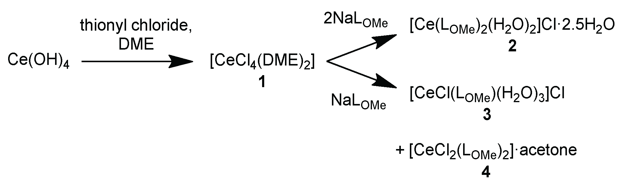
Figure 1.
(a) Asymmetric unit in the structure of [CeCl4(DME)2] (1) (50% thermal ellipsoids, H atoms with arbitrary radii); (b) coordination polyhedron of cerium. Bond lengths around Ce are given in Table 1, bond angles in Table S6 (see Figure S9 for the packing diagram).
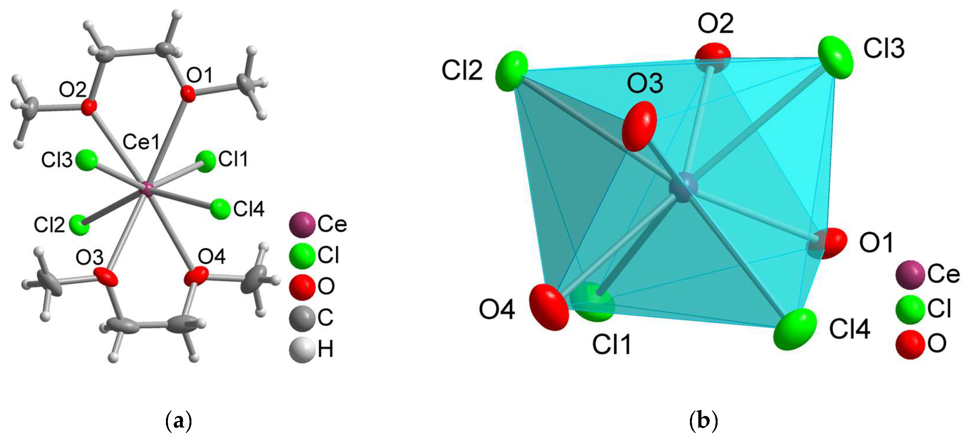
Figure 2.
(a) Asymmetric unit in the structure of [Ce(LOme)2(H2O)2]Cl·2.5H2O (2) (50% thermal ellipsoids, H atoms with arbitrary radii); (b) coordination polyhedron of cerium. A disorder of three of the phosphonate groups is not shown here for clarity (see Figure S11). Bond lengths around Ce are given in Table 2, bond angles in Table S10.
Figure 2.
(a) Asymmetric unit in the structure of [Ce(LOme)2(H2O)2]Cl·2.5H2O (2) (50% thermal ellipsoids, H atoms with arbitrary radii); (b) coordination polyhedron of cerium. A disorder of three of the phosphonate groups is not shown here for clarity (see Figure S11). Bond lengths around Ce are given in Table 2, bond angles in Table S10.
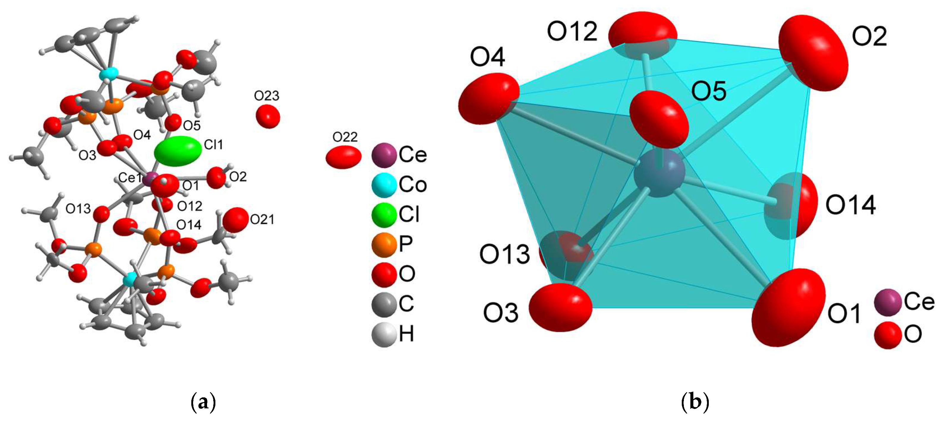
Figure 3.
(a) Asymmetric unit in the structure of 1D-[Ce(µ-Cl)(LOme)(H2O)3]Cl (3) (50% thermal ellipsoids, H atoms with arbitrary radii), (b) coordination polyhedron of cerium. Symmetry code: (i) y+1/2, x−1/2, z−1/2. Bond lengths around Ce are given in Table 3, bond angles in Table S13.
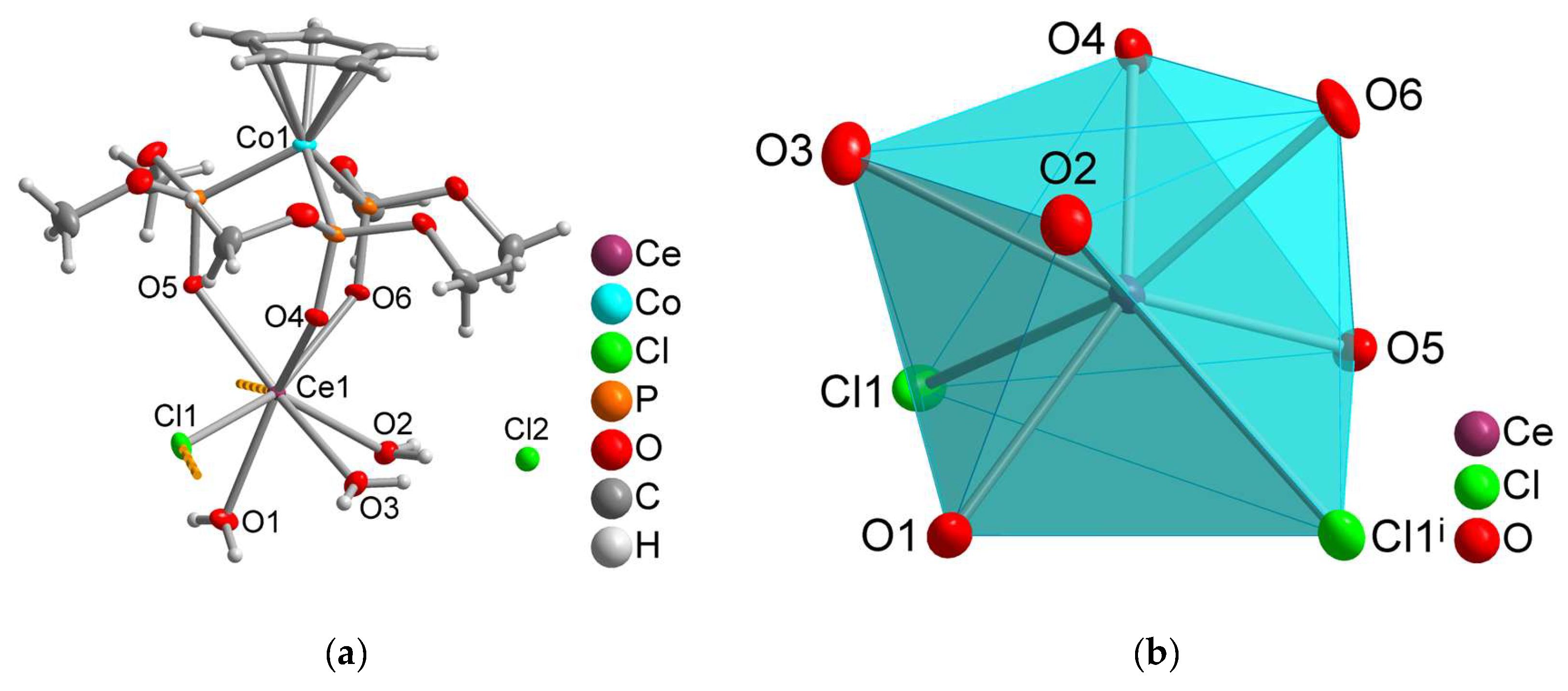
Figure 4.
1D-chain structure in [CeCl(LOme)(H2O)3]·Cl (3), with the Kläui ligand as a wireframe model (H atoms are omitted for clarity).
Figure 4.
1D-chain structure in [CeCl(LOme)(H2O)3]·Cl (3), with the Kläui ligand as a wireframe model (H atoms are omitted for clarity).
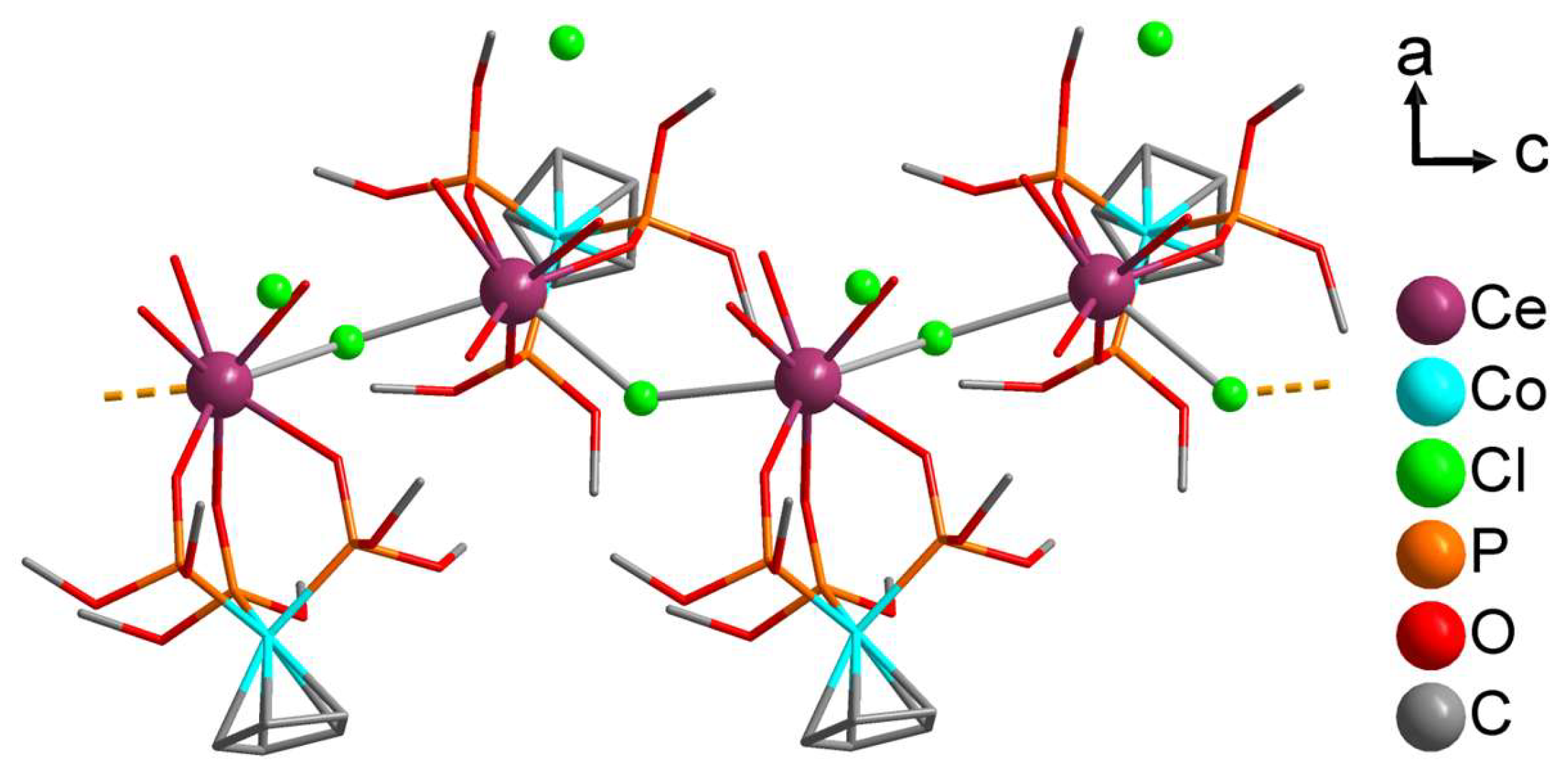
Figure 5.
Packing diagrams with two unit cells of (a) [Ce(LOme)2(H2O)2]Cl·2.5H2O (2); (b) [CeCl(LOme)(H2O)3]·Cl (3), both viewed along the c-axis to show the similar orientation of hydrophobic and hydrophilic regions (Kläui ligand as a wireframe model, H atoms are omitted for clarity; see Figure S10 and S14 for ball-and-stick reprentations).
Figure 5.
Packing diagrams with two unit cells of (a) [Ce(LOme)2(H2O)2]Cl·2.5H2O (2); (b) [CeCl(LOme)(H2O)3]·Cl (3), both viewed along the c-axis to show the similar orientation of hydrophobic and hydrophilic regions (Kläui ligand as a wireframe model, H atoms are omitted for clarity; see Figure S10 and S14 for ball-and-stick reprentations).
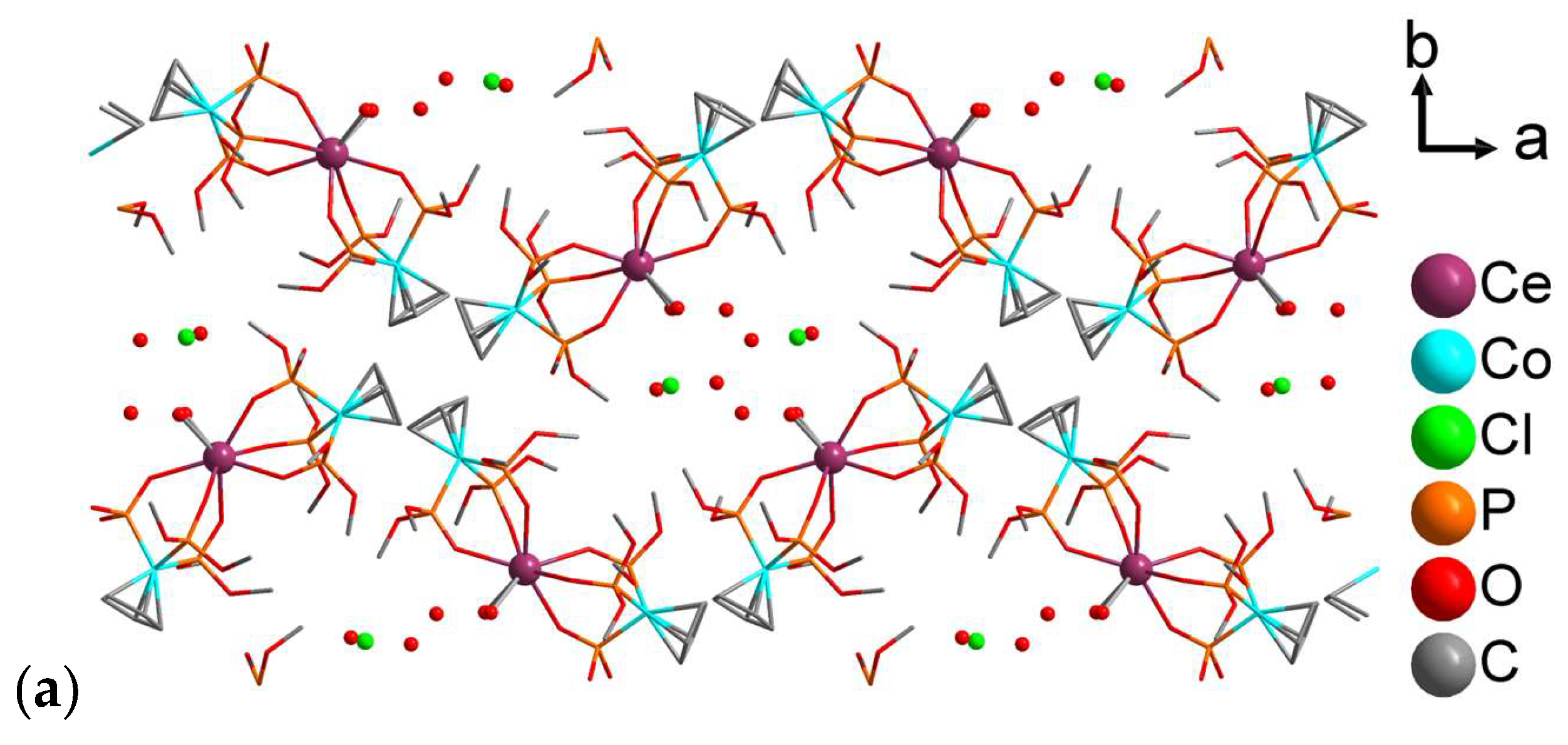
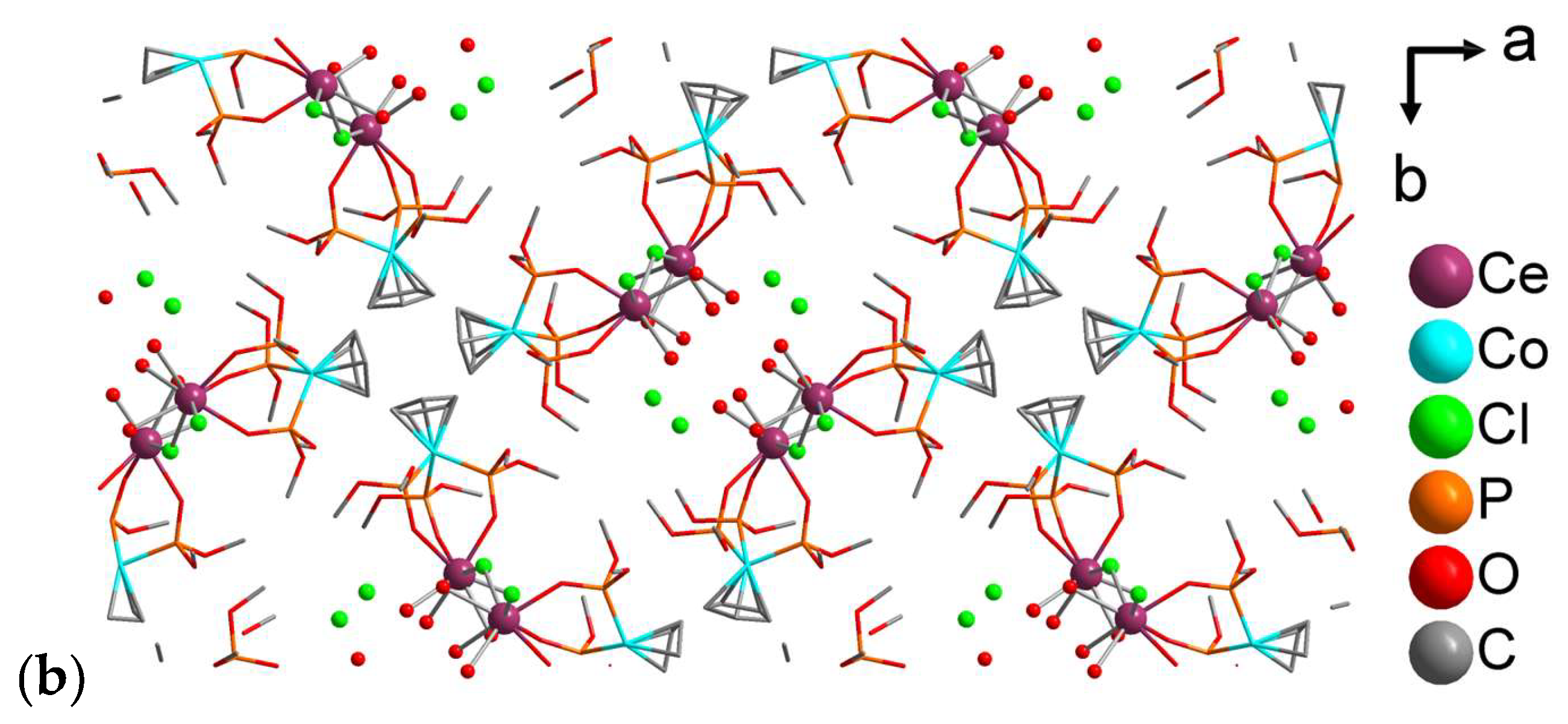
Figure 6.
(a) Extended asymmetric unit in the structure of [CeCl2(LOMe)2]·acetone (4) (50% thermal ellipsoids, H atoms with arbitrary radii and a disordered acetone molecule); (b) coordination polyhedron of cerium. Symmetry code: (i) −x+1, −y+1, z. Bond lengths around Ce are given in Table 4, bond angles in Table S15.
Figure 6.
(a) Extended asymmetric unit in the structure of [CeCl2(LOMe)2]·acetone (4) (50% thermal ellipsoids, H atoms with arbitrary radii and a disordered acetone molecule); (b) coordination polyhedron of cerium. Symmetry code: (i) −x+1, −y+1, z. Bond lengths around Ce are given in Table 4, bond angles in Table S15.
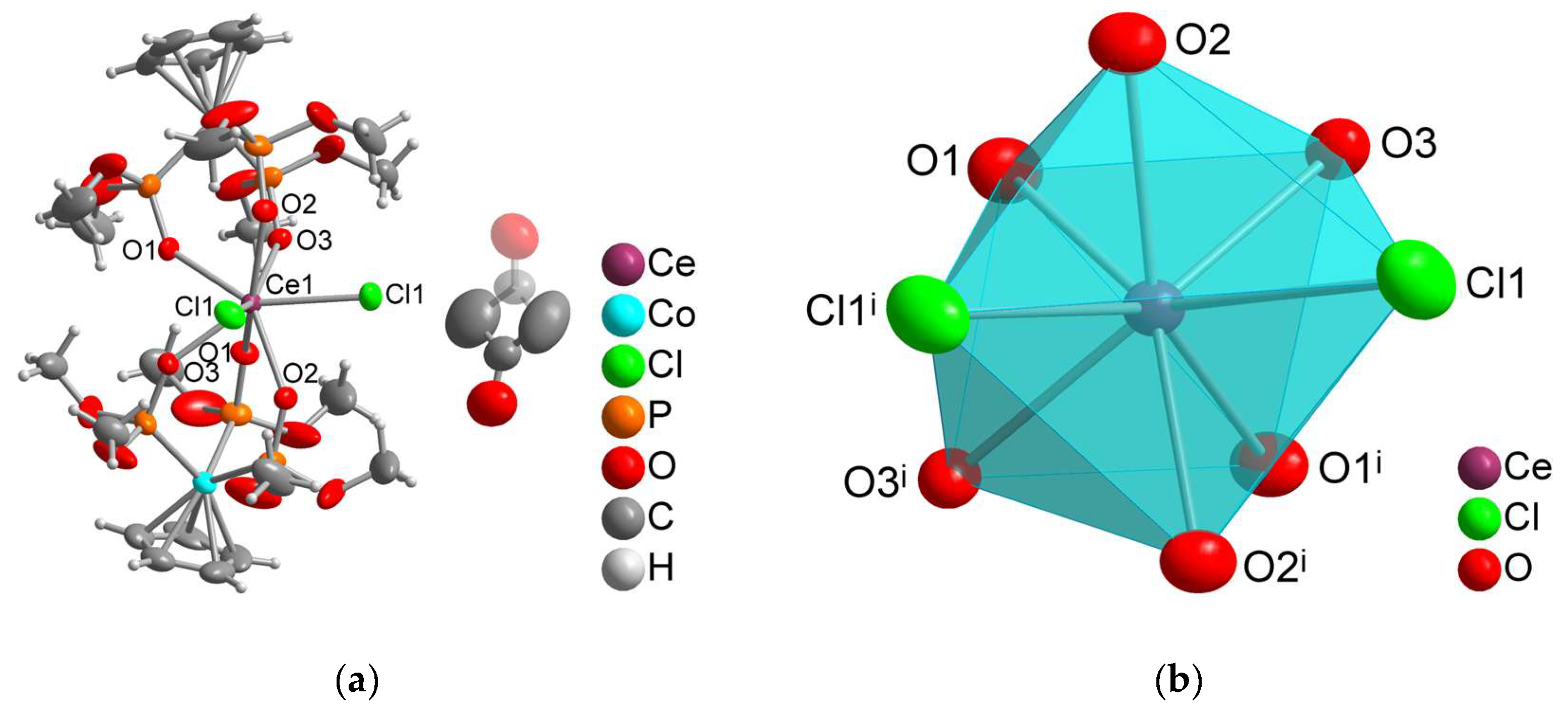
Figure 7.
Packing diagram with four unit cells of [CeCl2(LOMe)2]·acetone (4) with view along the c-axis (Ce, Co, O, Cl in ball and stick model and ligand in capped stick model, and H atoms are omitted for clarity; see Figure S16 for ball-and-stick reprentation).
Figure 7.
Packing diagram with four unit cells of [CeCl2(LOMe)2]·acetone (4) with view along the c-axis (Ce, Co, O, Cl in ball and stick model and ligand in capped stick model, and H atoms are omitted for clarity; see Figure S16 for ball-and-stick reprentation).
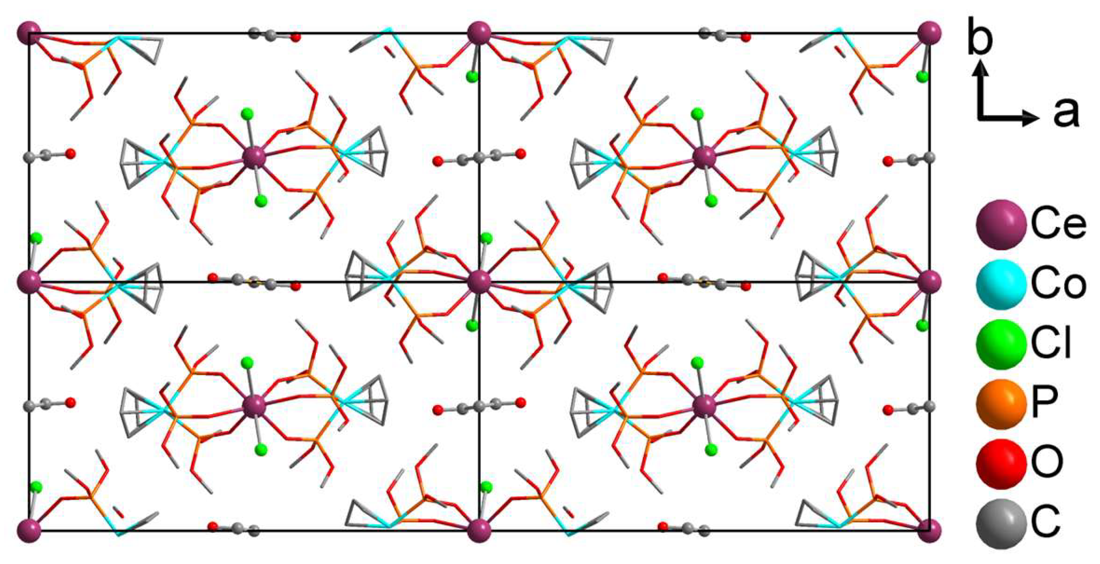
Table 1.
Bond lengths around cerium in 1. a.
| Bond length [Å] | |
| Ce1–Cl1 | 2.5795(8) |
| Ce1–Cl2 | 2.6107(8) |
| Ce1–Cl3 | 2.6008(8) |
| Ce1–Cl4 | 2.6038(8) |
| Ce1–O1 | 2.603(2) |
| Ce1–O2 | 2.584(2) |
| Ce1–O3 | 2.547(2) |
| Ce1–O4 | 2.586(3) |
a Bond angles around Ce are given in Table S6.
Table 5.
Oxidation states of Ce by the bond valence sum analysis (BVS) for 1-4. a.
| Complex | BVS for Ce with R0(Ce(III)–O) and R0(Ce(III)–Cl) a | BVS for Ce with R0(Ce(IV)–O) and R0(Ce(IV)–Cl) a |
| [CeCl4(DME)2] (1) | +4.23 | +3.78 |
| [Ce(LOMe)2(H2O)2]Cl·2.5H2O (2) | +3.05 | +2.77 |
| [CeCl(LOMe)(H2O)3]Cl (3) | +2.94 | +2.66 |
| [CeCl2(LOMe)2]·C3H6O (4) | +4.39 | +3.96 |
a R0(Ce(III)–O) = 2.112 Å [27], R0(Ce(III)–Cl) = 2.504 Å [28], R0 (Ce(IV)–O) = 2.076 Å [28], R0(Ce(IV)–Cl) = 2.46 Å [13]. The reported R0(Ce(IV)–Cl) values for the coordination number of eight represent approximations, as relevant parameters are not fully available in existing databases and the literature. The Rij values are listed in Table 1, Table 2, Table 3 and Table 4 for 1-4, respectively.
Table 6.
Crystal and structure refinement data for 1 – 4.
| 1 | 2 | 3 | 4 | |
| Empirical formula | C8H20CeClO4 | 2[C19H50CeCo2O20P6] ·2Cl·5H2O |
C11H29CeClCoO12P3·Cl | C22H46CeCl2Co2O18P6·C3H6O |
| M/g mol-1 | 462.16 | 2245.67 | 716.20 | 1165.32 |
| T/K | 150 | 150 | 150 | 150 |
| Wavelength/Å a | 0.701073 | 1.54184 | 1.54184 | 1.54184 |
| Crystal system | Monoclinic | Orthorhombic | Tetragonal | Orthorhombic |
| Space group | P21/c | P21212 | P42bc | P21212 |
| a/Å | 16.5744(6) | 20.9616(2) | 21.89770(10) | 20.8164(2) |
| b/Å | 7.26589(16) | 20.8611(2) | 21.89770(10) | 11.51080(10) |
| c/Å | 14.8768(5) | 10.34780(10) | 9.95180(10) | 9.1672010) |
| β/deg | 115.735(4) | 90 | 90 | 90 |
| V/Å3 | 1613.87(8) | 4524.91(8) | 4771.98(6) | 2196.58(4) |
| Z | 4 | 2 | 8 | 2 |
| Calcd. Density/g cm-3 | 1.902 | 1.641 | 1.994 | 1.762 |
| μ/mm-1 | 3.48 | 16.50 | 24.46 | 17.51 |
| F(000)/e | 904 | 2252 | 2840 | 1168 |
| Crystal size/mm3 | 0.28 × 0.1 × 0.09 | 0.46 × 0.29 × 0.25 | 0.96 × 0.11 × 0.07 | 0.16 × 0.1 × 0.07 |
| θ Range/deg | 2.7–36.5 | 3.0–79.2 | 2.8–78.0 | 4.2–76.6 |
| Reflections collected | 12133 | 40891 | 16828 | 8979 |
| Independent reflections; Rint | 8235; 0.076 | 9252; 0.086 | 4420; 0.043 | 4071; 0.032 |
| Completeness to θmax/% | 1.000 | 0.999 | 1.000 | 0.998 |
| R1; wR2 (I > 2 σ(I)) 1 | 0.0509; 0.1185 | 0.0692; 0.1883 | 0.0302; 0.0812 | 0.0363; 0.0964 |
| R1; wR2 (all data) 2 | 0.0692; 0.1277 | 0.0696; 0.1890 | 0.0310; 0.0817 | 0.0371; 0.0970 |
| Goodness-of-fit 3 | 0.97 | 1.07 | 1.09 | 1.07 |
| ∆ρfin (max; min)/e Å-3 4 | 2.34; -1.56 | 1.30; -1.32 | 0.98; -1.36 | 0.83; -1.05 |
| Flack parameter | - | 0.003(6) | -0.010(4) | -0.011(3) |
| CCDC no. | 2573145 | 2573146 | 2573147 | 2573144 |
1 R1 = ∑ | |Fo| - |Fc| | /∑|Fo|. 2 wR2 [∑w(Fo2- Fc2)2/∑[w(Fo2)2]1/2, w = [σ2(Fo2) + (aP)2 + bP]-1, where P = (Fo2 + 2Fc2)/3, a and b are constants adjusted by the program. 3 GoF = S = [∑w(Fo2- Fc2)2 / (nobs-nparam)]1/2, where nobs is the number of data and nparam the number of refined parameters. 4 Largest peak and hole in final difference map.
Disclaimer/Publisher’s Note: The statements, opinions and data contained in all publications are solely those of the individual author(s) and contributor(s) and not of MDPI and/or the editor(s). MDPI and/or the editor(s) disclaim responsibility for any injury to people or property resulting from any ideas, methods, instructions or products referred to in the content. |
© 2026 by the authors. Licensee MDPI, Basel, Switzerland. This article is an open access article distributed under the terms and conditions of the Creative Commons Attribution (CC BY) license.
Copyright: This open access article is published under a Creative Commons CC BY 4.0 license, which permit the free download, distribution, and reuse, provided that the author and preprint are cited in any reuse.


