Preprint
Review

This version is not peer-reviewed.

The Impact of Physical Exercise on Erythrocyte Deformability: A Comprehensive Review of Mechanisms and Physiological Adaptations

Submitted:

20 July 2026

Posted:

21 July 2026

You are already at the latest version

Abstract

The aim of the present review was to summarize the effects of physical exercise of varying intensity, duration, and frequency on the key biological and biomechanical factors that determine erythrocyte deformability. Erythrocyte deformability is a multifactorial property influenced by mechanical, biochemical, metabolic, and hemodynamic mechanisms activated during exercise, including membrane rheology, osmotic balance, hemoglobin concentration, intracellular Ca²⁺ homeostasis, ATP availability and release, nitric oxide (NO) signaling, oxidative stress, erythrocyte age, and temperature. Current evidence indicates that the effect of physical exercise on erythrocyte deformability is bidirectional. Moderate and well-adapted exercise generally improves membrane properties, microcirculatory blood flow, and oxygen delivery, whereas exhaustive or prolonged exercise may induce oxidative and structural damage and transiently reduce deformability. NO-dependent vasodilation and ATP release from erythrocytes appear to play central regulatory roles in optimizing microvascular perfusion during exercise. Osmotic changes, lactate accumulation, intracellular Ca²⁺ concentration, dehydration, and temperature are critical physiological modulators of erythrocyte mechanics. Because each athlete may exhibit an individual erythrocyte deformation profile shaped by biological, hemorheological, physiological, and training-related factors, individualized training loads and regular monitoring of hemorheological indicators are recommended. Such an approach may contribute to the development of hemorheological fitness and improved exercise performance.

Keywords: 
;  ;  ;  ;  ;  ;  ;  

1. Introduction

One of the primary functions of the cardiovascular system during physical exercise is to ensure the adequate supply of oxygen, nutrients, and other essential substances to the actively working muscles, organs, and tissues. An important task of red blood cells (RBCs) is to transport oxygen (O₂) from the lungs to the tissues and to deliver metabolically produced carbon dioxide (CO₂) back to the lungs for exhalation. The quantity and quality of hemoglobin within erythrocytes also contribute to maintaining the stable functional capacity of the blood. The release of adenosine triphosphate (ATP) and nitric oxide (NO) from red blood cells induces vasodilation, thereby ensuring maximal and sufficient blood flow to the working muscles and organs. Optimal erythrocyte function in the vascular system is maintained by RBCs with favorable micromechanical, rheological, morphological, physiological, and biochemical properties Mairbäurl, 2013 [1]; Ivanov, 2022 [2]; Nemkov, 2021 [3]; Caimi, 2023 [4].
It is well established that blood viscosity is one of the main factors determining blood flow behavior in the macrocirculatory system (arteries and veins) Huang et al., 2024 [5]; Sloop et al., 2024 [6]. On the other hand, the micromechanical properties of blood cells—erythrocytes, leukocytes, and platelets—play a critical role in governing blood flow through the microcirculatory system (arterioles, capillaries, venules). In the microcirculation, blood cells often must pass through vessels with diameters smaller than those of erythrocytes and leukocytes. Thus, the mechanical deformability of erythrocytes becomes crucial Smith et al., 1999 [7]. It is widely accepted that erythrocytes are the main determinant of blood flow behavior, as they are the most numerous blood cells—about 4.7–6.1 × 10¹²/L, depending on sex, age, and various other factors Alexy et al., 2022 [8]; Zhang et al., 2024 [9].
The movement of erythrocytes in the microcirculation determines flow resistance and the distribution of all blood cell types within the microvascular network. Erythrocyte deformability is therefore a key hemorheological factor requiring careful and in-depth investigation Fedosov et al., 2014 [10]; Brun et al., 2021 [11]; Grau et al., 2022 [12]. Erythrocyte deformability refers to the ability of red blood cells to reversibly change their shape during flow and under external mechanical forces. This property allows them to pass through narrow microvessels while continuing to transport oxygen (O₂) and carbon dioxide (CO₂) to tissues and cells [13].
Normal erythrocytes in healthy individuals exhibit several modes of deformation Kuhn et al., 2017 [14]:
A. Elongation of the discoid shape under shear stress along the longitudinal axis.
B. Tank-treading rotation of the erythrocyte membrane around the cytoplasm, with no major shape changes. This motion forces the erythrocyte to align along the vessel axis, thereby improving blood flow.
C. Swinging, a combination of membrane rotation with oscillations relative to the major axis of the ellipsoid erythrocyte shape.
D. Tumbling, which is facilitated by turbulent blood flow under high shear rates, observed mainly in larger blood vessels Kuhn et al., 2017 [14].
Regular physical activity of varying duration, frequency, and intensity is an important factor shaping the rheological status of blood as a fluid and influencing all its elements and internal interactions. In this section, we will focus on erythrocyte deformability and the effects of different types, durations, intensities, and frequencies of physical exercise on this property.
When exposed to external stresses, human erythrocytes possess the unique ability to undergo large deformations, enabling them to pass through capillaries and venules narrower than their resting diameter. Red blood cells are among the most deformable cells, and their deformation can occur in vivo under physiological blood flow conditions. This cellular phenomenon may occur without changes in the erythrocyte membrane, but it can also involve alterations in erythrocyte shape and/or increases in membrane surface area Kim et al., 2015 [15].
Erythrocyte deformability plays a crucial role in gas transport (O₂ and CO₂) via circulation to tissues and cells. Even a slight reduction in red blood cell deformability can significantly increase resistance in microvascular blood flow and elevate blood viscosity, as illustrated in Figure 1.
Well-trained athletes, particularly those engaged in endurance and strength sports, often present with lower hematocrit values—a condition sometimes inaccurately referred to as “sports anemia.” This is not anemia in the clinical sense, since these athletes typically demonstrate both an increased number of red blood cells and elevated hemoglobin levels compared with sedentary individuals. The slight decrease in hematocrit observed immediately after exercise is most likely due to an increase in plasma volume Sawka & Coyle, 1999 [16]. The pathophysiological mechanisms and factors that drive the overall increase in red blood cell count during regular physical training remain incompletely understood. Despite enhanced erythropoiesis in active athletes, exercise can, in some cases, reduce total erythrocyte number through intravascular hemolysis. This may be caused by mechanical stress as RBCs pass through capillaries within contracting skeletal muscles, or through direct compression of erythrocytes (e.g., in the feet during running, or in the hands and fingers of weightlifters handling heavy barbells). Taken together, these effects reduce the “average age” and volume of circulating erythrocytes in trained athletes. Importantly, “younger” erythrocytes are characterized by improved deformability and more efficient oxygen delivery to tissues during exercise Chatzinikolaou et al., 2024 [17].
Healthy red blood cells deform readily in response to shear stress within the vascular lumen under normal circulation. This property—excellent deformability—facilitates their effective passage through the microvasculature McMahon, 2019 [18]; Barshtein et al., 2024 [19]; Gunina et al., 2021 [20].
Erythrocytes also synthesize and release vasoactive mediators during deformation and in response to various physiological and pathological stimuli. Hemoglobin deoxygenation in red blood cells triggers the release of vasodilators, anti-adhesive S-nitrosothiols (SNOs), and adenosine triphosphate (ATP). The combined effects of shear stress–mediated signaling and oxygen transport during respiration optimize oxygen delivery (O₂) to tissues and cells. In certain diseases (e.g., sickle cell anemia), these adaptive functions may be impaired due to limited deformability, altered synthesis, or dysfunctional export of mediators McMahon, 2019 [18].
Recent findings clearly demonstrate that erythrocyte deformability is a unique property on which essential physiological processes in both humans and animals depend. Each deformation event of an erythrocyte passing through narrow arterioles and venules initiates two key biological reactions: (1) the expression of vasoactive mediators across the erythrocyte membrane, and (2) the synthesis and release of ATP.
It is also known that during intense exercise or prolonged physical activity, exercise-induced hypoxemia may alter lactate uptake in red blood cells. Elevated lactate influx into erythrocytes is directly linked to hypoxemic states, although some authors suggest that this does not significantly affect red blood cell deformability. Further studies are required to elucidate the cellular and molecular mechanisms underlying these physiological changes in erythrocyte deformability Connes et al., 2004c [21].
The aim of the present study was to summarise the effects of physical exercise of varying intensity, duration, and frequency on the key biological and biomechanical factors determining erythrocyte deformability.

2. Topics and Results

2.1. Factors Determining Erythrocyte Deformability Under the Influence of Physical Exercise

The main biological and hemorheological factors influenced by physical activity that determine erythrocyte deformability Kim et al., 2015 [15] are:
• geometric shape;
• rheological properties of the erythrocyte membrane;
• biochemical and structural changes in membrane proteins and lipids;
• hemoglobin (Hb) concentration;
• calcium ion (Ca²⁺) concentration;
• intracellular diffusion and osmosis (osmotic concentration);
• nitric oxide (NO);
• adenosine triphosphate (ATP);
• erythrocyte age and degree of cell differentiation;
• body temperature and erythrocyte temperature.
Physical exercise of defined intensity, frequency, and duration has a significant impact on these factors.

2.1.1. Geometric Shape of Erythrocytes

Erythrocyte morphology changes under different conditions within the blood flow. During intense physical activity, varying levels of blood flow are achieved across different vascular regions. This implies that shear rates also vary, and individual erythrocytes are exposed to different mechanical forces while passing through vessels of various diameters—especially within the narrower arterioles and venules of the microcirculation, and conversely through larger vessels.
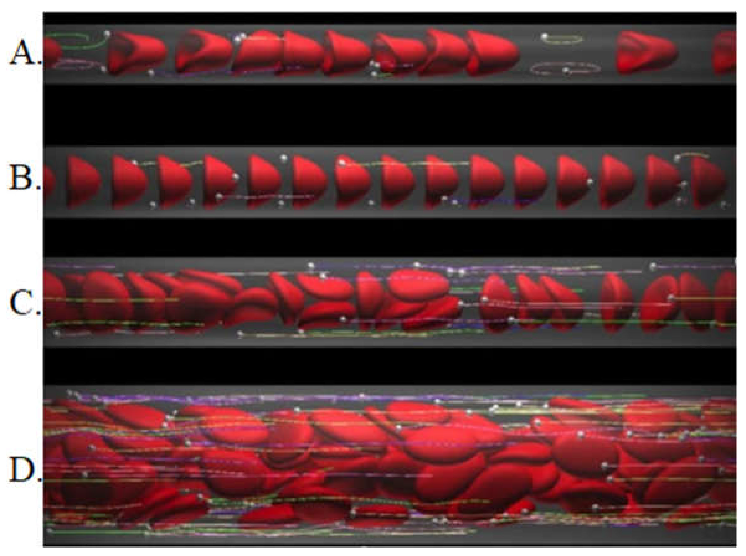
Another important factor associated with erythrocyte shape changes is the vessel diameter. Figure 2 demonstrates how the morphology of erythrocytes varies depending on vascular lumen diameter under constant shear rate and hematocrit.
These shape transformations are accompanied by additional alterations, such as bending and deformation of the erythrocyte membrane, as well as intracellular “movement” of the cytoplasm.
The resting shape of human erythrocytes—a biconcave disc in the absence of external mechanical forces—is defined by a specific but precise surface-to-volume (S/V) ratio. This ratio enables substantial reversible elastic transformations into different shapes, thereby allowing significant erythrocyte deformations Safeukui et al., 2012 [22].
The typical surface-to-volume (S/V) ratio of erythrocytes is approximately 1.5, but this may change under altered osmotic pressure. The S/V ratio of normal red blood cells enables significant deformations. Conversely, any reduction in the S/V ratio leads to diminished deformability and correlates with pathological conditions such as spherocytosis, hemolytic anemia, malaria, and others Kim et al., 2015 [15].
Intensive physical exercise alters blood flow conditions, thereby affecting the S/V ratio Baskurt et al., 2024 [24]. Smith et al. (1999) reported increased erythrocyte deformability in elite Australian cyclists (national team members, n = 9) [7]. Two weeks prior to blood sampling, the athletes trained for 25–29 hours per week, covering 510–631 miles weekly. The authors found a statistically significant increase in the hematological parameter MCV (mean corpuscular volume, fL) compared with a non-training control group. This increase reflects a change in the surface-to-volume ratio of individual erythrocytes, directly linked to deformability.
Similarly, Romagnoli et al. (2014) reported elevated MCV values and improved erythrocyte deformability in a group of ten young athletes aged 12–16 years. The exercise protocol consisted of a one-hour morning submaximal session on a cycle ergometer at 70% of HRmax (the maximal heart rate). HRmax values for all participants were determined one week before the submaximal test [25].
In a similar study, Alis et al. (2015) estimated erythrocyte deformability (ED) in 10 sedentary and in 16 trained subjects, both before and after a maximal incremental test, and after recovery, along with mean corpuscular volume (MCV) and red blood cell lactate concentrations [26]. Exercise-induced arterial hypoxemia was found in 6 trained subjects. Sedentary and non-EIAH trained subjects showed reduced ED after exercise, while no effect on ED was found in EIAH trained subjects [26].
On the other hand, Brinkmann et al. (2015) estimated the effects of endurance training on red blood cells (RBCs) in seventeen men with non-insulin-dependent type 2 diabetes from the perspective of in vivo RBC aging [27]. They reported that the proportion of “young” RBCs was significantly higher after training. Overall RBC deformability remained unchanged after training; however, deformability decreased with RBC aging both before and after training. Training significantly increased deformability in young RBCs but reduced deformability in older RBCs [27].

2.1.2. Rheological Properties of the Erythrocyte Membrane

Membrane deformability is a crucial component of overall erythrocyte deformability. It can be quantitatively assessed through three deformation modes, each described by an elastic modulus, and by one viscosity index Hochmuth & Waugh, 1987 [28]:
• Shear modulus (L, N/m): describes elastic energy storage during uniaxial stretching or shear deformation of the membrane.
• Area expansion modulus (K, mN/m): measurable via micropipette aspiration of the membrane.
• Bending modulus (B, Nm): characterizes bending resistance of the lipid bilayer and its resting shape.
• Membrane viscosity (ηm, μNs/m): measures the rate of membrane deformation under mechanical stress.
Tsuda et al. (2003) reported that aerobic physical exercise (performed twice weekly for 6 months at the aerobic threshold) enhanced erythrocyte membrane deformability and improved membrane viscosity in hypertensive patients. The authors attributed these benefits to the favorable effects of exercise on the rheological properties of RBCs [29].
Yunus (2023) concluded that eight weeks of moderate-intensity aerobic exercise significantly increased erythrocyte membrane resistance and erythrocyte count [30].
Nemkov et al. (2021) concluded that RBCs demonstrated decreased deformability and increased generation of microparticles after the 30-min submaximal cycling. In association with these properties, metabolites involved in oxidative stress response and membrane remodeling and repair emerged as top correlates, thus indicating a metabolic response in RBC to damage resulting from increased circulation and oxygen delivery during exercise [3].

2.1.3. Biochemical and Structural Changes in Erythrocyte Membrane Lipids and Proteins

Jordan et al. (1998) [31], using SEM analysis, demonstrated disrupted erythrocyte membrane structure after marathon running, along with loss of membrane material compared with pre-race samples. Erythrocyte membrane surface area increased by 30% (p < 0.01), and hemolysis was confirmed by a 57% decrease in plasma haptoglobin levels (p < 0.001). These findings indicate significant structural changes in the cytoskeletal architecture of erythrocyte membranes after endurance exercise. Notably, such alterations could be detected by SEM but not by TEM. These results suggest increased susceptibility of RBCs to chemical and mechanical stress and hemolysis after intense endurance activity [31].
Membrane lipids, forming the bilayer of all cells, consist of phospholipids, glycolipids, and cholesterol. An elevated cholesterol-to-phospholipid ratio (C/PL) from 1.28 to 2.0 reduces erythrocyte filterability and consequently their deformability Kim et al., 2015 [15]; Corrons et al., 2023 [32]. However, deformability is determined not only by lipids themselves but also by lipid–protein interactions Chien, 1987 [33].
Brzeszczynska et al. (2008) reported that oxidative stress (the accumulation of free radicals capable of damaging cells) during exercise in untrained subjects (22 ± 2 years; 187 ± 7.84 cm; 87 ± 15.6 kg; mean BMI 25 ± 4.5 kg/m²) consuming a balanced diet caused erythrocyte alterations including [34]:
• protein aggregation;
• disrupted membrane organization and lipid rigidification due to lipid peroxidation.
Similarly, Berzosa et al. (2011) found that in untrained healthy men, erythrocyte membranes became less deformable after maximal or submaximal workloads (incremental ergometer test at 60 rpm) [35]. This biomechanical phenomenon was explained by dynamic structural changes in erythrocyte membranes caused by oxidative stress (overproduction of free radicals during exercise).

2.1.4. Hemoglobin (Hb) Concentration

The hematological parameter MCHC (mean cell hemoglobin concentration) reflects cytoplasmic viscosity, which affects erythrocyte deformability. Increased MCHC is associated with reduced deformability. Elevated MCHC values may result from both exercise and pathological conditions Clark et al., 1978 [36], Ivanov, 2022 RBC [37]. Reduced deformability is also observed with erythrocyte “aging,” which is likewise associated with increased intracellular Hb concentration. Furthermore, RBCs lose water in hypertonic environments during dehydration, leading to increased MCHC and decreased deformability Bareford et al., 1985 [38]. Thus, water loss contributes to elevated cytoplasmic viscosity, further impairing erythrocyte deformability.
Radomski et al. (1980) conducted a study on hematological changes in soldiers (marching 35 km/day for 6 consecutive days, at 35% of their own VO₂max), which showed a decrease in the number of erythrocytes (RBC) and reduced hematocrit (Hct) values [39]. After the 6th day of training, the authors also reported decreases in hemoglobin (Hb), mean corpuscular hemoglobin (MCH), mean corpuscular hemoglobin concentration (MCHC), and mean corpuscular volume (MCV).
The results indicate that maximal physical exertion affects key erythrocyte characteristics – RBC, Hct, erythrocyte deformability, among others.

2.1.5. Calcium Ion Concentration (Ca²⁺)

The deformability of red blood cells and the elasticity of their membranes are highly dependent on the intracellular concentration of calcium ions (Ca²⁺). An increase in intracellular Ca²⁺ concentration leads to reduced red blood cell deformability Chien, 1987 [31]. Accumulation of intracellular Ca²⁺ results in changes in cell shape and volume, increased rigidity, and even the potential development of hemolysis Romero & Romero, 1997 [40]. Intracellular calcium is subject to "metabolic control" through an adenosine triphosphate (ATP)-dependent "calcium pump" mechanism. It has been shown that in vivo older (“aging”) erythrocytes contain more calcium ions (almost four times more compared to “young” cells) Romero & Romero, 1997 [40]. This is likely the result of reduced capacity for Ca²⁺ efflux in older erythrocytes, even though the rate of Ca²⁺ influx is almost identical in both “old” and “young” red blood cells.
The literature also contains contradictory results, ranging from higher to lower intracellular Ca²⁺ concentrations, including reports of no significant differences between active athletes and non-athletic individuals.
Nikolaidis et al. (2003) reported interesting findings regarding the relationship between erythrocyte deformability and calcium ion concentration in erythrocytes of adolescents compared to adults [41]. Their research group observed significantly higher calcium concentrations in adolescents compared to adults. With regard to the influence of physical activity, however, all athletes included in the study (579 athletes compared with 241 physically inactive participants) exhibited significantly higher calcium concentrations in erythrocytes compared to their respective non-athletic control groups. This finding is difficult to interpret, given the calcium homeostasis in the cytoplasm, and suggests the importance of investigating this effect in greater depth. Future studies should more precisely evaluate all biological and biomechanical factors affecting Ca²⁺ homeostasis in the cytoplasm of red blood cells.

2.1.6. Intracellular Diffusion and Osmosis (Osmotic Concentration)

Osmosis is the process of water movement and its entry into cells through a semipermeable cell membrane. In this process, water molecules move from the less concentrated to the more concentrated intracellular environment. Thus, osmosis serves to balance both sides of the semipermeable cell membrane, diluting the solute-rich environment with the solvent, which is water.
Osmosis is considered a form of passive transport, since no energy is expended when passing through the membrane. In the process of osmosis, water, as the solvent, passes through the semipermeable membrane in order to balance and stabilize the solution’s concentration. This continues until osmotic pressure stabilizes.
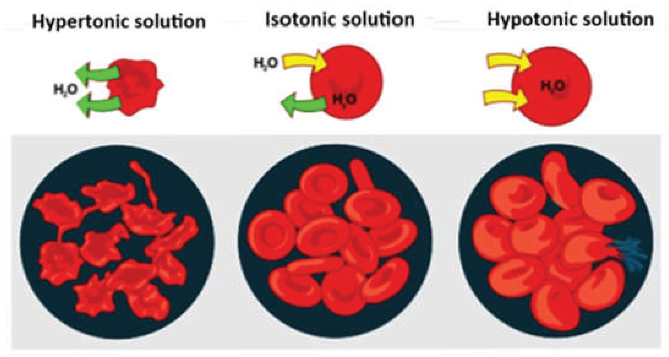
The goal of osmosis is to equalize solute concentrations until equilibrium is reached. There are the following types of solutions (Figure 3):
a) Hypertonic solution – has higher osmotic pressure and solute concentration;
b) Hypotonic solution – has lower osmotic pressure and solute concentration;
c) Isotonic solution – solute concentration and osmotic pressure are equalized, thus achieving equilibrium.
Osmolarity testing is an assessment that evaluates the concentration of all chemical substances present in the liquid phase (plasma) of the blood. Osmolarity can also be measured in urine. The shape and deformability of red blood cells can be significantly altered by deviations from the normal physiological osmolarity range (i.e., 275–295 mOsm/kg). In a hypotonic environment, the biconcave shape of erythrocytes is transformed into a spherical one (for echinocytes, this occurs in a hypertonic environment). Since changes in osmolarity affect the shape of red blood cells, their deformability is also altered. Osmotic deformability of erythrocytes provides information about their viability, water content, surface area, particularly in certain pathological conditions, and more Cluitmans et al., 2012 [42].
A study by Heo et al. (2015) found that red blood cell deformation at low shear stress (1–3 Pa) is maximal under hypotonic conditions (225–250 mOsm/kg H₂O), which is slightly below the normal plasma osmolarity range (275–295 mOsm/kg) [43]. It has also been shown that the relationship between erythrocyte deformability and medium osmolarity depends on the applied shear stress. This conclusion is significant for microcirculatory blood flow, such as in the Fahraeus–Lindqvist effect Fung, 1990 [44].
French researchers Connes et al., 2004b [45] investigated the influence of lactate anions on the rheological properties of blood and blood cells. They observed a decrease in erythrocyte deformability when lactate concentration was artificially increased (via lactate anion addition to blood samples) or naturally elevated (e.g., during exercise). A reduction in erythrocyte deformability during physical exercise has been reported in individuals with low physical fitness Yalcin et al., 2003 [46]; Bouix et al., 1998 [47]. Research groups including Lipovac et al. (1985) and Reinhart et al. (2000) explained that in vitro lactate addition or in vivo lactate accumulation in the blood leads to an increase in extra-erythrocytic osmolarity [48,49]. As a result of hyperosmolar conditions, red blood cells progressively lose their water content, which increases their intracellular viscosity and reduces their deformability.
Hardeman et al. (1995) and Connes et al. (2004a) reported a paradoxical increase in erythrocyte deformability in athletes undergoing specialized endurance training programs [50,51]. In another study, the same authors found no change in erythrocyte deformation in trained athletes performing submaximal exercise, even as plasma lactate concentration increased. The existing discrepancies in data regarding the effect of lactate on erythrocyte deformability require further investigation and analysis.

2.1.7. Nitric Oxide and Erythrocytic Nitric Oxide Synthase

Endothelial cells synthesize numerous paracrine factors, including nitric oxide (NO), which strengthen and regulate vascular wall functions Green et al., 2004 [52].
Nitric oxide (NO) is synthesized naturally and/or after pharmacological stimulation in endothelial cells from the precursor amino acid L-arginine, with the participation of the enzyme endothelial nitric oxide synthase (eNOS) (53 Palmer et al., 1988). A major physiological stimulus for natural endothelial NO synthesis is increased blood flow through the vessel lumen Pohl et al., 1986 [54]; Rubanyi et al., 1986 [55]. Released NO induces vasodilation, which normalizes shear stress Dimmeler & Zeiher, 2003 [56]. At the same time, NO can contribute to the development of hyperemia during physical exertion and sports activity, which are always associated with elevated blood pressure and increased heart rate. Repeated but moderate shear stress, resulting from moderate exercise training, may improve the bioavailability of NO in the body Green et al., 2004 [52].
In the bloodstream, endothelial nitric oxide (NO) also influences platelet biological activity, particularly in those located close to the vascular wall surface. In addition to interacting with certain proteins, NO can be oxidized to nitrites and nitrates. For example, nitrite has been shown to represent an endocrine bioavailable reservoir for NO storage, which can be bioactivated under hypoxic conditions Grau et al., 2013 [57]; Bryan et al., 2005 [58]; Gladwin, 2005 [59]. This reaction, called “hypoxic vasodilation,” ensures an increase in local blood flow. In this way, endogenous oxygen delivery to metabolically active tissues and cells is improved, for example under pathological hypoxic vasoconstriction. In vascular smooth muscle cells, nitrite is converted into NO through interaction with cellular myoglobin Totzeck et al., 2012 [60].
Besides endothelial cells, nitric oxide is also synthesized by monocytes/macrophages and erythrocytes. It is assumed that erythrocyte-derived NO can contribute to hypoxic vasodilation, thereby supporting the role of red blood cells in the regulation of local blood flow and vascular tone. In this regard, NO is an extremely important compound for cardiovascular regulation, due to its effects on smooth muscle cells in vascular walls. NO also plays an essential regulatory role in red blood cell deformability under both normal and pathological conditions Bor-Kucukatay et al., 2003 [61]. Different NO concentrations may be relevant for monitoring erythrocyte deformability and aggregability Starzyk et al., 1997 [62]. It is known that within certain concentration ranges, NO can improve erythrocyte deformability, but the effect may also be opposite (at very low or very high concentrations) Korbut et al., 1993 [63]. Reduction of NO levels (e.g., through the use of nitric oxide synthase inhibitors) often leads to red blood cell damage Grau et al., 2013 [57].
Erythrocyte-derived NO exhibits the same important biological functions, including regulation of red blood cell deformability Grau et al., 2013 [57]. Grau and colleagues demonstrated that activated RBC-NOS is the enzyme responsible for nitric oxide production in red blood cells. RBC-NOS enzymatic activity contributes to improved physical condition of the individual through enhanced erythrocyte deformability [57]. These scientific and applied findings are of great importance, as they suggest that patients suffering from diseases that reduce red blood cell deformability may benefit from treatment with specific RBC-NOS-activating pharmaceutical agents. Additionally, such patients may benefit from specialized sports and rehabilitation programs involving physical exercise, supporting the restoration of erythrocyte deformability and thus alleviating disease symptoms.
The results of a study by Bor-Kucukatay et al. (2003) also confirmed the regulatory role of NO, which can significantly influence and maintain normal erythrocyte deformability – a factor crucial for the control and regulation of blood flow in the body [61].
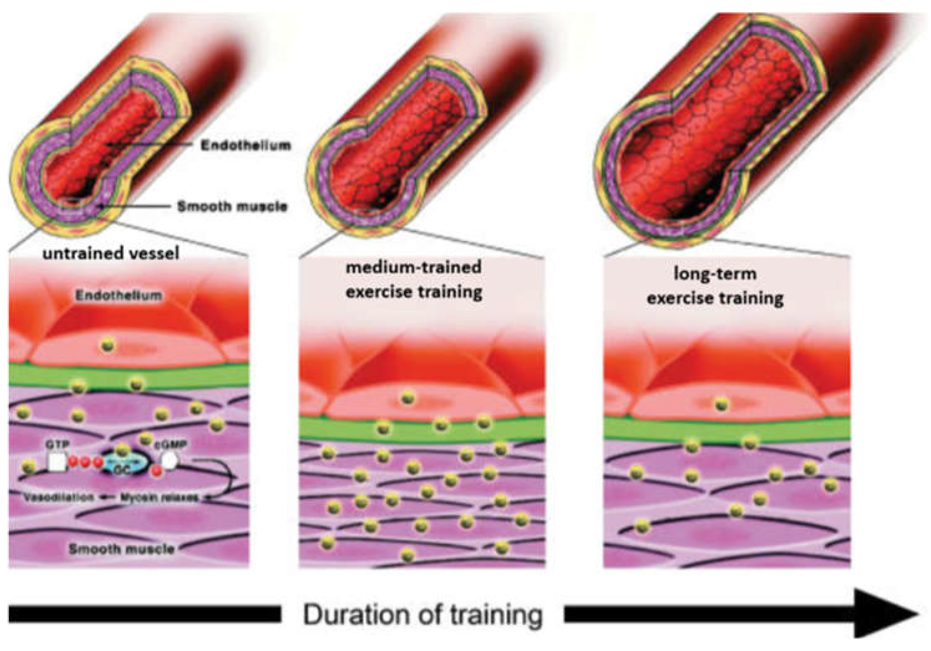
Figure 4 graphically illustrates the “hypothetical response” of arteries to increased blood flow and elevated shear stress as a result of performing exercise training sessions of different duration and intensity Green et al., 2004 [52].
In an untrained blood vessel (left panel), NO expression induces vasodilation through relaxation of smooth muscle cells, acting homeostatically to regulate shear stress on the vessel wall.
In response to moderate-duration training (middle panel), the sudden increase in shear stress, associated with repeated exposure to elevated blood flow during exercise repetitions, stimulates enhanced endothelial NO synthesis and subsequent vasodilation (expansion of the vessel lumen). Positive regulation of vasodilation mechanisms via NO serves as a buffer against increased shear stress.
During prolonged training (right panel), structural adaptation likely occurs due to NO-mediated remodeling, resulting in chronic enlargement of the vessel lumen and “structural normalization” of shear stress on the vessel wall.
The exact impact of NO on the mechanical properties of erythrocytes remains incompletely understood at the cellular and molecular level. Bor-Kucukatay et al. (2003) hypothesize that this effect is likely due to altered phosphorylation of the erythrocyte cytoskeleton [61]. Further studies are needed to clarify the primary aspects of nitric oxide’s effect on erythrocyte deformability and mechanical behavior in blood flow.

2.1.8. Erythrocyte ATP (Adenosine Triphosphate)

Maintenance of the geometric shape and deformability of red blood cells depends on the generation of erythrocyte ATP. It is known that significantly reduced ATP levels contribute to decreased erythrocyte deformability. The primary mechanism of ATP production in erythrocytes and its relationship to erythrocyte deformability are described very well by McMahon (2019) [18].
Research has shown that hemoglobin (Hb) can bind and transport vasoactive mediators from erythrocytes, which further regulate and optimize oxygen delivery to working muscles and all body organs Jia et al., 1996 [64]; McMahon et al., 2002 [65]; Sonveaux et al., 2007 [66]. McMahon (2019) describes this phenomenon in the context of red blood cell deformation in flow. Adhesive interactions of erythrocytes with endothelial cells in vessel walls may also facilitate mediator transport [18].
According to McMahon (2019), during deformation, erythrocytes release ATP and the vasodilator S-nitrosothiol (SNO), pre-formed on the hemoglobin molecule by nitric oxide (NO). This process occurs simultaneously with oxygen release, allowing erythrocytes to preferentially target areas with the highest nutrient demand. Released ATP and SNO molecules regulate microvascular tone and prevent certain intercellular adhesion processes [18].
This physiological/pathophysiological mechanism facilitates adaptive and unobstructed erythrocyte flow, optimizing oxygen (O₂) delivery and carbon dioxide (CO₂) removal [18]. The responsible transporter channels for ATP release are Panx1 (pannexin 1) and for SNO – LAT1 (L-type amino acid transporter 1).
ATP is a high-energy compound, composed of three primary elements: the adenine nitrogenous base, ribose, and three phosphate residues designated alpha (α), beta (β), and gamma (γ). Its two high-energy phosphate bonds (α–β and β–γ) provide energy for cellular processes.
Like other cells, erythrocytes depend on ATP. They can release ATP in response to hypoxia or mechanical stress 67 Kirby et al., 2015 [18]. ATP release occurs via transmembrane transport, though the cellular mechanisms are not fully elucidated. Erythrocytes cannot form or extrude vesicles in a regulated manner for extracellular ATP export, although rare formation of micro- and nanoparticles from erythrocytes has been studied Bennett-Guerrero et al., 2007 [68].
Experimental evidence suggests that membrane shear stress, erythrocyte deformation, and ATP release are closely linked. Deformation creates cytoskeletal defects, releasing actin molecules, which aggregate on the fibrous transmembrane conductance regulator (FTRP), activating ATP release Zhang et al., 2018 [69]. FTRP is thought to indirectly influence ATP release by regulating the Panx1 hemichannel, which is highly sensitive to mechanical stress, as well as intracellular oxygen and calcium levels. For example, during intense prolonged exercise, both mechanical stress and intracellular oxygen and calcium levels are significantly affected.
Several studies suggest Piezo1, a mechanosensitive cation channel, responds to membrane shear stress, allowing calcium influx [69]. Increased intracellular calcium facilitates Panx1 activation and ATP release.
Elevated ATP in plasma further stimulates endothelial NO production [1 Mairbäurl, 2013]. Red blood cell-derived ATP induces NO-dependent increases in blood flow to cells, tissues, and muscles, essential for athletic performance Zhang et al., 2018 [69]. ATP release from erythrocytes occurs both in vitro and in vivo, with increased ATP levels observed in venous outflow during forearm exercise in early exercise studies Forrester, 1972 [70]; Ellsworth et al., 1995 [71]. The effect is amplified under hypoxic conditions Gonzalez-Alonso, 2002 [72].
Enhanced erythrocyte deformability is crucial for oxygen delivery to working muscles. In athletes, highly deformable erythrocytes ensure optimal tissue perfusion, particularly in skeletal muscles [1,73] Tomschi et al., 2018.

2.1.9. Erythrocyte Age, Maturation, and Differentiation

Advanced mean erythrocyte age is a biological factor contributing to reduced deformability Teległów et al., 2024 [74]; Pospieszna et al., 2021 [75]. Mohanty et al. (2014) demonstrated that oxidative stress plays a key role in erythrocyte membrane damage, impairing deformability. Erythrocytes are chronically exposed to oxidative stress and other damaging factors, despite antioxidant defenses including catalase, glutathione peroxidase, peroxiredoxin-2, and others [76].
Recent studies show a linear relationship between erythrocyte deformability and oxidative stress, measured via blood degradation products Barodka et al., 2014 [77]. Other processes not directly linked to oxidative stress, such as calcium-induced erythrocyte contraction, also alter cellular deformability and increase cell density Winder et al., 2000 [78].
Endurance training can induce “sports anemia.” Although red blood cells normally have a 120-day lifespan, cell aging can accelerate due to intense and prolonged exercise Smith, 1995 [79]. The erythrocyte deficit is rare in athletes; sports anemia is often attributed to other factors.
Markers of erythrocyte damage, such as antioxidant depletion or protein/lipid injury, are considered useful indicators of exercise-induced erythrocyte stress Smith, 1995 [79]. Oxidative damage may impair ion homeostasis and promote cell dehydration, reducing deformability and hindering passage through capillaries and venules. This can lead to hypoxia in working muscles and accelerate erythrocyte turnover. Increased erythropoiesis produces younger, more effective oxygen-carrying cells.

2.1.10. Body and Erythrocyte Temperature

During intense physical exercise, heat production may exceed 1000 W Gleeson, 1998 [80]. Some heat is stored, raising core body temperature by several degrees. Central and skin thermoreceptors detect this, and the hypothalamus triggers appropriate effector responses. Sweating and increased cutaneous blood flow are effective heat-dissipation mechanisms, but dehydration, hot and humid environments, or inappropriate clothing can impair heat loss. Training improves heat tolerance by increasing the sensitivity of the sweat/temperature relationship, lowering the sweating threshold, and expanding blood volume Gleeson, 1998 [80].
Lowering temperature from 37 °C to 4 °C significantly increases erythrocyte membrane shear modulus and alters viscosity Kim et al., 2015 [15]. These membrane changes substantially reduce erythrocyte deformability. No significant changes are observed between 25 °C and 37 °C, but below 25 °C, deformability decreases significantly. Temperature-dependent deformability is observed at all applied shear stresses. Increased temperature (2–24 °C) raises the kinetic energy of membrane molecular components, weakening intermolecular bonds until shear stress causes breakage (Singh & Stoltz, 2002). Thermal treatment above 45 °C reduces the elongation index (EI) of erythrocytes, a measure of deformability.

3. Conclusions

Erythrocyte deformability is a key determinant of microcirculatory blood flow and oxygen delivery during physical activity. The present review shows that this property is regulated by a complex interaction of mechanical, biochemical, metabolic, and hemodynamic factors, including membrane rheology, osmotic balance, intracellular calcium concentration, hemoglobin content, nitric oxide (NO), adenosine triphosphate (ATP), oxidative stress, erythrocyte age, and temperature.
The influence of exercise on erythrocyte deformability is bidirectional. Moderate, regular, and appropriately prescribed physical activity generally improves erythrocyte mechanical properties, enhances NO- and ATP-mediated vascular regulation, and promotes efficient oxygen transport. In contrast, exhaustive or prolonged exercise may induce oxidative stress, membrane damage, dehydration, and metabolic disturbances, leading to a transient reduction in erythrocyte deformability.
Current evidence suggests that trained athletes develop specific hemorheological adaptations characterized by improved erythrocyte function, enhanced microcirculatory blood flow, and optimized oxygen delivery. However, these responses remain highly individual and depend on training status, exercise modality, intensity, duration, recovery, environmental conditions, and individual physiological characteristics.
Each athlete possesses a unique erythrocyte deformation profile formed by the interaction of biological, hemorheological, physiological, and psychological factors [81,82,83,84,85,86,87,88,89,90,91,92]. Therefore, individualized training strategies and regular monitoring of hemorheological parameters may provide valuable information for optimizing performance and preventing excessive physiological stress.
The concept of hemorheological fitness appears to be an important component of athletic preparedness. Its main characteristics include optimal blood and plasma viscosity, balanced hematocrit, reduced erythrocyte aggregation, and increased erythrocyte deformability. These indicators may support more precise assessment of adaptation, fatigue, recovery, and readiness for training or competition.
Future studies should further investigate the molecular mechanisms linking exercise-induced changes in NO signaling, ATP release, calcium homeostasis, oxidative stress, membrane lipid remodeling, and erythrocyte deformability. Particular attention should be paid to sport-specific responses, sex- and age-related differences, recovery kinetics, and the potential clinical application of exercise programs aimed at improving RBC mechanical function.

Conflicts of Interest

The authors declare no conflicts of interest.

References

  1. Mairbäurl, H., (2013). Red blood cells in sports: effects of exercise and training on oxygen supply by red blood cells. Frontiers in physiology, 4, p.332. [CrossRef]
  2. Ivanov, I. (2022). Hemorheological Alterations and Physical Activity. Applied Sciences, 12(20), 10374. [CrossRef]
  3. Nemkov, T., Skinner, S. C., Nader, E., Stefanoni, D., Robert, M., Cendali, F., .. & D’Alessandro, A. (2021). Acute cycling exercise induces changes in red blood cell deformability and membrane lipid remodeling. International journal of molecular sciences, 22(2), 896.
  4. Caimi, G., Carlisi, M., & Presti, R. L. (2023). Red Blood Cell Distribution Width, Erythrocyte Indices, and Elongation Index at Baseline in a Group of Trained Subjects. Journal of Clinical Medicine, 13(1), 151. [CrossRef]
  5. Huang, Y. C., Chen, Y. C., Lin, Y. T., Hsu, C. C., Fu, T. C., & Wang, J. S. (2024). Effects of concentric and eccentric cycling training on muscular hemodynamic and erythrocyte rheological responses to exercise in sedentary males. European Journal of Sport Science, 24(2), 266-276. [CrossRef]
  6. Sloop, G. D., Pop, G., Weidman, J. J., & Cyr, J. A. S. (2024). Hormonal Control of Blood Viscosity. Cureus, 16(2). [CrossRef]
  7. Smith, J. A., Martin, D. T., Telford, R. D. & Ballas, S.K., (1999). Greater erythrocyte deformability in world-class endurance athletes. American Journal of Physiology-Heart and Circulatory Physiology, 276(6), pp.H2188-H2193. [CrossRef]
  8. Alexy, T., Detterich, J., Connes, P., Toth, K., Nader, E., Kenyeres, P., .. & Simmonds, M. J. (2022). Physical properties of blood and their relationship to clinical conditions. Frontiers in Physiology, 13, 906768.
  9. Zhang, X., Lin, Y., Xin, J., Zhang, Y., Yang, K., Luo, Y., & Wang, B. (2024). Red blood cells in biology and translational medicine: natural vehicle inspires new biomedical applications. Theranostics, 14(1), 220. [CrossRef]
  10. Fedosov, D.A., Peltomäki, M. and Gompper, G., (2014). Deformation and dynamics of red blood cells in flow through cylindrical microchannels. Soft matter, 10(24), pp.4258-4267. [CrossRef]
  11. Brun, J. F., Varlet-Marie, E., Myzia, J., Raynaud de Mauverger, E., & Pretorius, E. (2021). Metabolic influences modulating erythrocyte deformability and eryptosis. Metabolites, 12(1), 4. [CrossRef]
  12. Grau, M., Zollmann, E., Bros, J., Seeger, B., Dietz, T., Noriega Ureña, J. A., .. & Schumann, M. (2022). Autologous blood doping induced changes in red blood cell rheologic parameters, RBC age distribution, and performance. Biology, 11(5), 647.
  13. Braslavskaya, K., Reformato, V., & Pantin, E. (2025). Blood Physiology and Oxygen Transport. In Clinical Perfusion for Cardiac Surgery: A Step-by-Step Guide to the Fundamentals (pp. 27-38). Cham: Springer Nature Switzerland.
  14. Kuhn, V., Diederich, L., Keller IV, T. S., Kramer, C. M., Lückstädt, W., Panknin, C., .. & Cortese-Krott, M. M. (2017). Red blood cell function and dysfunction: redox regulation, nitric oxide metabolism, anemia. Antioxidants & redox signaling, 26(13), 718-742.
  15. Kim, J., Lee, H., & Shin, S. (2015). Advances in the measurement of red blood cell deformability: A brief review. Journal of Cellular Biotechnology, 1(1), 63-79. [CrossRef]
  16. Sawka, M. N., & Coyle, E. F. (1999). Influence of body water and blood volume on thermoregulation and exercise performance in the heat. Exercise and sport sciences reviews, 27, 167-218.
  17. Chatzinikolaou, P. N., Margaritelis, N. V., Paschalis, V., Theodorou, A. A., Vrabas, I. S., Kyparos, A., .. & Nikolaidis, M. G. (2024). Erythrocyte metabolism. Acta Physiologica, 240(3), e14081.
  18. McMahon, T.J., (2019). Red blood cell deformability, vasoactive mediators, and adhesion. Frontiers in physiology, 10, p.1417. [CrossRef]
  19. Barshtein, G., Livshits, L., Gural, A., Arbell, D., Barkan, R., Pajic-Lijakovic, I., & Yedgar, S. (2024). Hemoglobin Binding to the Red Blood Cell (RBC) Membrane Is Associated with Decreased Cell Deformability. International Journal of Molecular Sciences, 25(11), 5814. [CrossRef]
  20. Gunina, L. M., Rybina, I. L., Ataman, Y. O., & Voitenko, V. L. (2021). Oxidative stress as a factor in the deterioration of oxygen transfer during exercise. [CrossRef]
  21. Connes, P., Bouix, D., Py, G., Caillaud, C., Kippelen, P., Brun, J. F., .. & Mercier, J. (2004c). Does exercise-induced hypoxemia modify lactate influx into erythrocytes and hemorheological parameters in athletes?. Journal of Applied Physiology, 97(3), 1053-1058.
  22. Safeukui, I., Buffet, P. A., Deplaine, G., Perrot, S., Brousse, V., Ndour, A., .. & Mohandas, N. (2012). Quantitative assessment of sensing and sequestration of spherocytic erythrocytes by the human spleen. Blood, The Journal of the American Society of Hematology, 120(2), 424-430.
  23. Takeishi, N. & Imai, Y., (2017). Capture of microparticles by bolus flow of red blood cells in capillaries. Scientific reports, 7(1), pp.1-8. [CrossRef]
  24. Baskurt, O. K., & Meiselman, H. J. (2024, September). Blood rheology and hemodynamics. In Seminars in Thrombosis and Hemostasis (Vol. 50, No. 06, pp. 902-915). Thieme Medical Publishers, Inc.
  25. Romagnoli, M., Alis, R., Martinez-Bello, V., Sanchis-Gomar, F., Aranda, R., & Gómez-Cabrera, M. C. (2014). Blood rheology effect of submaximal exercise on young subjects. Clinical hemorheology and microcirculation, 56(2), pp. 111-117. [CrossRef]
  26. Alis, R., Sanchis-Gomar, F., Ferioli, D., Torre, A. L., Blesa, J. R., & Romagnoli, M. (2015). Exercise effects on erythrocyte deformability in exercise-induced arterial hypoxemia. International Journal of Sports Medicine, 36(04), 286-291. [CrossRef]
  27. Brinkmann C, Bizjak DA, Bischof S, et al. Endurance training alters enzymatic and rheological properties of red blood cells (RBC) in type 2 diabetic men during in vivo RBC aging. Clinical Hemorheology and Microcirculation. 2015;63(3):173-184. [CrossRef]
  28. Hochmuth, R. M., & Waugh, R. E. (1987). Erythrocyte membrane elasticity and viscosity. Annual review of physiology, 49, 209-219. [CrossRef]
  29. Tsuda, K., Yoshikawa, A., Kimura, K. & Nishio, I., (2003). Effects of mild aerobic physical exercise on membrane fluidity of erythrocytes in essential hypertension. Clinical and experimental pharmacology and physiology, 30(5-6), pp.382-386. [CrossRef]
  30. Yunus, M. (2023). Effect Of 8 Weeks Of Moderate-Intensity Aerobic Exercise On Increasing Erytocyte Cell Membrane Endurance And Erytrocyte Cell Count. Asian Journal of Healthy and Science, 2(7), 314-325. [CrossRef]
  31. Jordan, J., Kiernan, W., Merker, H. J., Wenzel, M., & Beneke, R. (1998). Red cell membrane skeletal changes in marathon runners. International journal of sports medicine, 19(01), 16-19. [CrossRef]
  32. Corrons, J. L. V., & Krishnevskaya, E. (2023). Congenital Defects with Impaired Red Blood Cell Deformability–The Role of Next-Generation Ektacytometry. In The Erythrocyte-A Unique Cell. IntechOpen.
  33. Chien S., (1987). Red cell deformability and its relevance to blood flow. Annual Review of Physiology; 49:177-92.
  34. Brzeszczynska, J., Pieniazek, A., Gwozdz¬inski, L., Gwozdzinski, K., & Jegier, A. (2008). Structural alterations of erythrocyte membrane components induced by exhaustive exercise. Applied Physiology, Nutrition, and Metabolism, 33(6), pp. 1223-1231. [CrossRef]
  35. Berzosa, C., Gómez–Trullén, E. M., Pie¬drafita, E., Cebrián, I., Martínez–Ballarín, E., Miana–Mena, F. J., .. & García, J. J. (2011). Erythrocyte membrane fluidity and indices of plasmatic oxidative damage after acute physical exercise in humans. European journal of applied physiology, 111(6), pp. 1127-1133.
  36. Clark, M. R., Mohandas, N., Caggiano, V., & Shohet, S. B. (1978). Effects of abnor¬mal cation transport on deformability of des¬iccytes. Journal of Supramolecular Struc-ture, 8(4), pp. 521-532.
  37. Ivanov, I. (2022). Red blood cell deformability and physical activity. Journal of Applied Sports Sciences, 6, 106-118. [CrossRef]
  38. Bareford, D., Stone, P. C. W., Caldwell, N. M., Meiselman, H. J., & Stuart, J. (1985). Comparison of instruments for measurement of erythrocyte deformability. Clinical Hemorheology and Microcirculation, 5(4), 311-322. [CrossRef]
  39. Radomski, M. W., Sabiston, B. H., & Isoard, P. (1980). Development of „sports ane-mia“ in physically fit men after daily sustained submaximal exercise. Aviation, Space, and Environmental Medicine, 51(1), pp. 41-45.
  40. Romero, P.J., & Romero, E.A., (1997). Differences in Ca2+ pumping activity between sub-populations of human red cells. Cell calcium, 21(5), pp.353-358. [CrossRef]
  41. Nikolaidis, M.G., Protosygellou, M.D., Petridou, A., Tsalis, G., Tsigilis, N. and Mougios, V., (2003). Hematologic and biochemical profile of juvenile and adult athletes of both sexes: implications for clinical evaluation. International journal of sports medicine, 24(07), pp.506-511.
  42. Cluitmans, J. C., Hardeman, M. R., Dinkla, S., Brock, R., & Bosman, G. J. (2012). Red blood cell deformability during storage: towards functional proteomics and metabolomics in the Blood Bank. Blood Transfusion, 10(Suppl 2), s12.
  43. Heo, Y., Jung, H., & Shin, S. (2015). Osmotic deformability of erythrocytes at various shear stresses. Clinical Hemorheology and Microcirculation, 59(3), 211-218.
  44. Fung, Y. C., (1990). Biomechanics, Springer. (book).
  45. Connes, P., Bouix, D., Py, G., Prefaut, C., Mercier, J., Brun, J. F., & Caillaud, C. (2004b). Opposite effects of in vitro lactate on erythrocyte deformability in athletes and untrained subjects. Clinical hemorheology and microcirculation, 31(4), 311-318.
  46. Yalcin, O., Erman, A., Muratli, S., Bor-Kucukatay, M., & Baskurt, O. K. (2003). Time course of hemorheological alterations after heavy anaerobic exercise in untrained human subjects. Journal of Applied Physiology, 94(3), 997-1002.
  47. Bouix, D., Peyreigne, C., Raynaud, E., Monnier, J. F., Micallef, J. P., & Brun, J. F. (1998). Relationships among body composition, hemorheology and exercise performance in rugbymen. Clinical Hemorheology and Microcirculation, 19(3), 245-254.
  48. Lipovac, V., Gavella, M., Turk, Z., & Škrabalo, Z. (1985). Influence of lactate on the insulin action on red blood cell filterability. Clinical Hemorheology and Microcirculation, 5(5), 421-428.
  49. Reinhart, W. H., Gaudenz, R., & Walter, R. (2000). Lactate and pyruvate increase blood viscosity. JOURNAL DES MALADIES VASCULAIRES, 25, 171-171.
  50. Hardeman, M. R., Peters, H. P. F., & Goedhart, P. T. (1995). Low hematocrit and plasma fibrinogen in trained athletes increase hemorheological tolerance for physical stress. Clinical Hemorheology, 3(15), 506.
  51. Connes, P., Bouix, D., Durand, F., Kippelen, P., Mercier, J., Prefaut, C., .. & Caillaud, C. (2004a). Is hemoglobin desaturation related to blood viscosity in athletes during exercise?. International journal of sports medicine, 25(08), 569-574.
  52. Green, D. J., Maiorana, A., O'Driscoll, G., & Taylor, R. (2004). Effect of exercise training on endothelium-derived nitric oxide function in humans. The Journal of physiology, 561(1), 1-25.
  53. Palmer, R. M., Rees, D. D., Ashton, D. S., & Moncada, S. (1988). L-arginine is the physiological precursor for the formation of nitric oxide in endothelium-dependent relaxation. Biochemical and biophysical research communications, 153(3), 1251-1256.
  54. Pohl, U., Holtz, J., Busse, R., & Bassenge, E. (1986). Crucial role of endothelium in the vasodilator response to increased flow in vivo. Hypertension, 8(1), 37-44.
  55. Rubanyi, G. M., Romero, J. C., & Vanhoutte, P. M. (1986). Flow-induced release of endothelium-derived relaxing factor. American Journal of Physiology-Heart and Circulatory Physiology, 250(6), H1145-H1149.
  56. Dimmeler S & Zeiher AM (2003). Exercise and cardiovascular health. Get active to AKTivate your endothelial nitric oxide synthase. Circulation 107, 3118–3120.
  57. Grau, M., Pauly, S., Ali, J., Walpurgis, K., Thevis, M., Bloch, W., & Suhr, F. (2013). RBC-NOS-dependent S-nitrosylation of cytoskeletal proteins improves RBC deformability. PloS one, 8(2), e56759.
  58. Bryan, N. S., Fernandez, B. O., Bauer, S. M., Garcia-Saura, M. F., Milsom, A. B., Rassaf, T., .. & Feelisch, M. (2005). Nitrite is a signaling molecule and regulator of gene expression in mammalian tissues. Nature chemical biology, 1(5), 290-297.
  59. Gladwin, M. T. (2005). Nitrite as an intrinsic signaling molecule. Nature Chemical Biology, 1(5), 245-246.
  60. Totzeck, M., Hendgen-Cotta, U. B., Luedike, P., Berenbrink, M., Klare, J. P., Steinhoff, H. J., .. & Rassaf, T. (2012). Nitrite regulates hypoxic vasodilation via myoglobin-dependent nitric oxide generation. Circulation, 126(3), 325-334.
  61. Bor-Kucukatay, M., Wenby, R. B., Meiselman, H. J., & Baskurt, O. K. (2003). Effects of nitric oxide on red blood cell deformability. American Journal of Physiology-Heart and Circulatory Physiology, 284(5), H1577-H1584.
  62. Starzyk, D., Korbut, R., & Gryglewski, R. J. (1997). The role of nitric oxide in regulation of deformability of red blood cells in acute phase of endotoxaemia in rats. Journal of physiology and pharmacology, 48(4).
  63. Korbut, R., & Gryglewski, R. J. (1993). Nitric oxide from polymorphonuclear leukocytes mo¬du¬lates red blood cell deformability in vitro. European journal of pharmacology, 234(1), 17-22.
  64. Jia, L., Bonaventura, C., Bonaventura, J., and Stamler, J. S. (1996). S-nitrosohaemoglobin: a dynamic activity of blood involved in vascular control. Nature 380, 221–226.
  65. McMahon, T. J., Moon, R. E., Luchsinger, B. P., Carraway, M. S., Stone, A. E., Stolp, B. W., et al. (2002). Nitric oxide in the human respiratory cycle. Nat. Med. 8, 711–717.
  66. Sonveaux, P., Lobysheva, I. I., Feron, O., and McMahon, T. J. (2007). Transport and peripheral bioactivities of nitrogen oxides carried by red blood cell hemoglobin: role in oxygen delivery. Physiology 22, 97–112.
  67. Kirby, B. S., Schwarzbaum, P. J., Lazarowski, E. R., Dinenno, F. A., and McMahon, T. J. (2015). Liberation of ATP secondary to hemolysis is not mutually exclusive of regulated export. Blood 125, 1844–1845.
  68. Bennett-Guerrero, E., Veldman, T. H., Doctor, A., Telen, M. J., Ortel, T. L., Reid, T. S., et al. (2007). Evolution of adverse changes in stored RBCs. Proc. Natl. Acad. Sci. U.S.A. 104, 17063–17068.
  69. Zhang, H., Shen, Z., Hogan, B., Barakat, A.I. and Misbah, C., (2018). ATP release by red blood cells under flow: model and simulations. Biophysical journal, 115(11), pp.2218-2229.
  70. Forrester, T., (1972). An estimate of adenosine triphosphate release into the venous effluent from exercising human forearm muscle. The Journal of Physiology, 224(3), pp.611-628.
  71. Ellsworth, M. L., Forrester, T. H. O. M. A. S., Ellis, C. G., & Dietrich, H. H. (1995). The erythrocyte as a regulator of vascular tone. American Journal of Physiology-Heart and Circulatory Physiology, 269(6), H2155-H2161.
  72. González-Alonso, J., Olsen, D. B., & Saltin, B. (2002). Erythrocyte and the regulation of human skeletal muscle blood flow and oxygen delivery: role of circulating ATP. Circulation research, 91(11), 1046-1055.
  73. Tomschi, F., Bloch, W., & Grau, M. (2018). Impact of type of sport, gender and age on red blood cell deformability of elite athletes. International journal of sports medicine, 40(01), 12-20.
  74. Teległów, A., Konieczny, K., Dobija, I., Kuśmierczyk, J., Tota, Ł., Rembiasz, K., & Maciejczyk, M. (2024). Effect of regular winter swimming on blood morphological, rheological, and biochemical indicators and activity of antioxidant enzymes in males. BMC Sports Science, Medicine and Rehabilitation, 16(1), 138.
  75. Pospieszna, B., Kusy, K., Slominska, E. M., & Zieliński, J. (2021). Life-long sports engagement enhances adult erythrocyte adenylate energetics. Scientific Reports, 11(1), 1-9.
  76. Mohanty, J. G., Nagababu, E., & Rifkind, J. M. (2014). Red blood cell oxidative stress impairs oxygen delivery and induces red blood cell aging. Frontiers in physiology, 5, 84.
  77. Barodka, V. M., Nagababu, E., Mohanty, J. G., Nyhan, D., Berkowitz, D. E., Rifkind, J. M., & Strouse, J. J. (2014). New insights provided by a comparison of impaired deformability with erythrocyte oxidative stress for sickle cell disease. Blood Cells, Molecules, and Diseases, 52(4), 230-235.
  78. Winder, W. W., Holmes, B. F., Rubink, D. S., Jensen, E. B., Chen, M., & Holloszy, J. O. (2000). Activation of AMP-activated protein kinase increases mitochondrial enzymes in skeletal muscle. Journal of applied physiology, 88(6), 2219-2226.
  79. Smith, J.A., (1995). Exercise, training and red blood cell turnover. Sports medicine, 19(1), pp.9-31.
  80. Gleeson, M., (1998). Temperature regulation during exercise. International Journal of Sports Medicine, 19(S 2), pp.S96-S99.
  81. Teległów, A., Marchewka, J., Tota, Ł., Ptaszek, B., Pilch, W., Pałka, T., .. & Marchewka, A. (2020). Changes in the morphological, rheological, and biochemical blood indicators in triathletes. Folia Biologica (Kraków), 68(3), 107-120.
  82. Bizjak, D. A., Tomschi, F., Bales, G., Nader, E., Romana, M., Connes, P., .. & Grau, M. (2020). Does endurance training improve red blood cell aging and hemorheology in moderate-trained healthy individuals?. Journal of sport and health science, 9(6), 595-603.
  83. Mardyła, M., Teległów, A., Ptaszek, B., Jekiełek, M., Mańko, G., & Marchewka, J. (2023). Effects of rowing on rheological properties of blood. International Journal of Environmental Research and Public Health, 20(6), 5159.
  84. Maruyama, T., Hieda, M., Mawatari, S., & Fujino, T. (2022). Rheological abnormalities in human erythrocytes subjected to oxidative inflammation. Frontiers in Physiology, 13, 837926.
  85. Teległów, A., Mardyła, M., Myszka, M., Pałka, T., Maciejczyk, M., Bujas, P., .. & Marchewka, J. (2022). Effect of intermittent hypoxic training on selected biochemical indicators, blood rheological properties, and metabolic activity of erythrocytes in rowers. Biology, 11(10), 1513.
  86. Huang, Y. C., Chen, Y. C., Lin, Y. T., Hsu, C. C., Fu, T. C., & Wang, J. S. (2024). Effects of concentric and eccentric cycling training on muscular hemodynamic and erythrocyte rheological responses to exercise in sedentary males. European Journal of Sport Science, 24(2), 266-276.
  87. Freitag, N., Böttrich, T., Weber, P. D., Manferdelli, G., Bizjak, D. A., Grau, M., .. & Schumann, M. (2020). Acute low-dose hyperoxia during a single bout of high-intensity interval exercise does not affect red blood cell deformability and muscle oxygenation in trained men—A randomized crossover study. Sports, 8(1), 4.
  88. Carin, R., Deglicourt, G., Rezigue, H., Martin, M., Nougier, C., Boisson, C., .. & Nader, E. (2023). Effects of a maximal exercise followed by a submaximal exercise performed in normobaric hypoxia (2500 m), on blood rheology, red blood cell senescence, and coagulation in well-trained cyclists. Metabolites, 13(2), 179.
  89. Ciekot-Sołtysiak, M., Kusy, K., Podgórski, T., Pospieszna, B., & Zieliński, J. (2024). Changes in red blood cell parameters during incremental exercise in highly trained athletes of different sport specializations. PeerJ, 12, e17040.
  90. Barshtein, G., Livshits, L., Gural, A., Arbell, D., Barkan, R., Pajic-Lijakovic, I., & Yedgar, S. (2024). Hemoglobin Binding to the Red Blood Cell (RBC) Membrane Is Associated with Decreased Cell Deformability. International Journal of Molecular Sciences, 25(11), 5814.
  91. Antonova, N., Khristov, K., Alexandrova, A., Muravyov, A., & Velcheva, I. (2023). Development of experimental microfluidic device and methodology for assessing microrheological properties of blood. Clinical Hemorheology and Microcirculation, 83(3), 231-245.
  92. Antonova, N., & Khristov, K. (2025). Microrheological and microfluidic approaches for evaluation of the mechanical properties of blood cells. Applied Sciences, 15(15), 8291. [CrossRef]
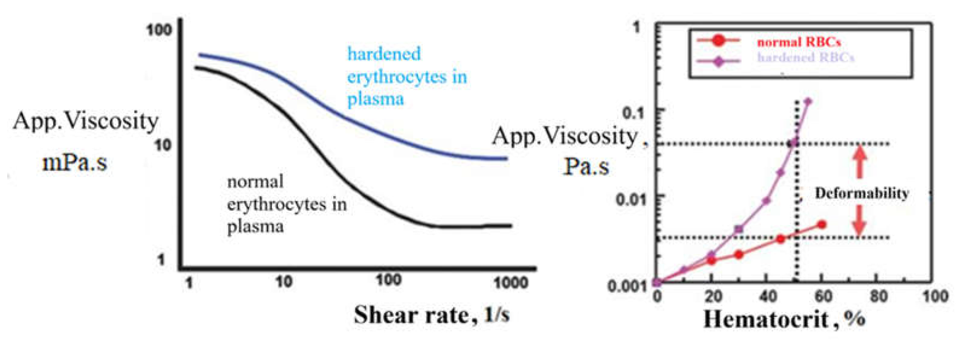
Figure 1. A. Comparison of blood viscosity in the presence of normal and “stiffened” (reduced deformability) erythrocytes suspended in plasma at different shear rates; B. Effect of erythrocyte deformability on blood viscosity at high shear rates and under different hematocrit levels (figures adapted and translated from [15] Kim et al., 2015).
Preprints 224123 g001
Figure 2. Changes in the geometric shape of erythrocytes flowing through vessels of different diameters under constant conditions (Hct = 0.2, shear rate = 167 s−1). Vessel diameter: (A) D = 8 μm, (B) 10 μm, (C) 12 μm and (D) 22 μm [adapted from [23] Takeishi & Imai, 2017].
Preprints 224123 g002
Figure 3. Erythrocytes in solutions with different solute concentrations and osmotic pressures.
Preprints 224123 g003
Figure 4. “Hypothetical Response” of Arteries to Increased Blood Flow and Elevated Shear Stress During Exercise of Varying Duration [figure adapted from [52] Green et al., 2004].
Preprints 224123 g004
Disclaimer/Publisher’s Note: The statements, opinions and data contained in all publications are solely those of the individual author(s) and contributor(s) and not of MDPI and/or the editor(s). MDPI and/or the editor(s) disclaim responsibility for any injury to people or property resulting from any ideas, methods, instructions or products referred to in the content.
Copyright: This open access article is published under a Creative Commons CC BY 4.0 license, which permit the free download, distribution, and reuse, provided that the author and preprint are cited in any reuse.
Prerpints.org logo

Preprints.org is a free preprint server supported by MDPI in Basel, Switzerland.

Subscribe

© 2026 MDPI (Basel, Switzerland) unless otherwise stated

Accessibility

Disclaimer

Terms of Use

Privacy Policy

Privacy Settings