

Submitted:
20 July 2026
Posted:
21 July 2026
You are already at the latest version
Abstract
Background/Objectives: Although dual HER2-targeted neoadjuvant therapy improves pathological complete response (pCR) rates in HER2-positive early breast cancer (EBC), a substantial proportion of patients have residual disease. We evaluated the association between PIK3CA mutations, inflammation–nutritional biomarkers, and pCR. Methods: This retrospective, single-center cohort study included 50 patients with HER2-positive EBC who underwent surgery after neoadjuvant therapy with trastuzumab and pertuzumab. PIK3CA mutations were assessed in diagnostic tumor samples using real-time PCR. Pretreatment HALP, CALLY, CRII, and other inflammation-based indices were calculated. Logistic regression and receiver operating characteristic (ROC) analyses were used to identify determinants of pCR, defined as ypT0/is ypN0. Results: pCR was observed in 32 patients (64.0%), and 15 patients (30.0%) had PIK3CA-mutant tumors. The pCR rate was lower in PIK3CA-mutant than in wild-type tumors (40.0% vs. 74.3%; Pearson’s chi-square p = 0.021; Fisher’s exact p = 0.028). Response did not differ significantly among treatment regimens. The highest pCR rate occurred in hormone receptor (HR)-negative/PIK3CA wild-type tumors (81.3%), whereas the lowest occurred in HR-positive/PIK3CA-mutant tumors (30.0%). CALLY was independently associated with pCR (AUC = 0.755, p = 0.003). LCR (AUC = 0.776, p = 0.001) and CAR (AUC = 0.763, p = 0.002) also showed discriminatory ability, whereas HALP was not associated with response. Conclusions: PIK3CA mutations were associated with lower pCR rates, while inflammation–nutritional indices provided complementary predictive information. This combined molecular and host-related biomarker assessment may offer a more comprehensive approach to baseline response stratification. These findings require confirmation in larger prospective multicenter studies before clinical implementation.
Keywords:
HER2-positive breast cancer
; PIK3CA mutation
; pathological complete response
; neoadjuvant therapy
; dual HER2 blockade
; CALLY score
; CRII
; inflammatory–nutritional biomarkers
; real-world study
1. Introduction
Breast cancer remains the most frequently diagnosed malignancy among women and one of the leading causes of cancer-related mortality. Rather than representing a single disease entity, breast cancer comprises a heterogeneous group of molecular and histopathological subtypes with distinct prognoses and treatment responses. HER2-positive breast cancer accounts for approximately 15–20% of this heterogeneous disease group. Before the introduction of trastuzumab, HER2-positive disease was characterized by aggressive tumor biology, higher recurrence rates, and poorer survival outcomes. The subsequent development of HER2-targeted therapies has substantially altered the natural history of this disease, leading to marked improvements in both pathological response and long-term survival [1,2,3,4].
For patients with stage II–III HER2-positive breast cancer, neoadjuvant therapy incorporating dual HER2 blockade has become a standard treatment approach. Achieving a pathological complete response (pCR) is a key therapeutic objective in the neoadjuvant setting, as pCR is a clinically meaningful indicator associated with improved long-term outcomes, including event-free and overall survival [5].
The clinical benefit of dual HER2 blockade was first demonstrated in the NeoSphere trial. In that study, the addition of pertuzumab to trastuzumab and docetaxel resulted in a marked increase in the pCR rate compared with trastuzumab-containing therapy without pertuzumab [6].
The TRYPHAENA trial subsequently confirmed both the efficacy and cardiac safety of dual HER2 blockade administered with anthracycline-containing or anthracycline-free chemotherapy, reporting pCR rates exceeding 60% [7]. More recent studies, including KRISTINE, have supported the prognostic relevance of pCR and further strengthened the central role of dual HER2 blockade in the neoadjuvant treatment of HER2-positive breast cancer [8].
Despite these advances, approximately one-third of patients receiving contemporary dual HER2-targeted therapy do not achieve a pathological complete response. This finding further supports the biological heterogeneity of HER2-positive breast cancer and has increased interest in predictive biomarkers that may enable more accurate treatment selection and response stratification. Among the molecular mechanisms involved in resistance to HER2-targeted therapies, activation of the phosphatidylinositol 3-kinase (PI3K) signaling pathway has been highlighted in the literature as one of the most important.
PIK3CA is one of the most frequently mutated oncogenes in human malignancies and represents the most common genomic alteration in HER2-positive breast cancer [9]. The gene encodes the catalytic p110α subunit of phosphoinositide 3-kinase alpha (PI3Kα), which forms a functional complex with the regulatory p85α subunit. Activation of the PI3K/AKT/mTOR signaling pathway regulates cell growth, proliferation, metabolism, and survival following stimulation of the receptor tyrosine kinases. Activating PIK3CA mutations, predominantly occurring in hotspot regions within exon 9 (codons 542 and 545) and exon 20 (codon 1047), result in constitutive pathway activation and contribute to oncogenesis across multiple tumor types, including breast cancer [10,11]. Consequently, PIK3CA has emerged as a therapeutic target and potential predictive biomarker.
Accumulating evidence suggests that activation of the PI3K pathway contributes to resistance to anti-HER2 therapy [12,13]. Several neoadjuvant studies have demonstrated lower pCR rates in patients with PIK3CA-mutant tumors, particularly in those with hormone receptor-positive disease [14,15,16]. Furthermore, some studies have linked PIK3CA mutations to shorter disease-free survival and inferior clinical outcomes [17].
In addition to their role in treatment resistance, the prognostic significance of PIK3CA mutations remains controversial. Several studies have reported reduced treatment sensitivity in PIK3CA-mutant tumors, whereas others have found no significant association with clinical outcomes. A potential favorable prognostic effect has even been suggested in selected patient populations [13,14,17,18,19,20,21,22,23].
The strongest evidence supporting the predictive role of PIK3CA mutations has been derived from pooled analyses of neoadjuvant HER2-targeted therapy trials. In a pooled analysis of 967 patients receiving neoadjuvant anti-HER2 therapy, Loibl et al. demonstrated significantly lower pCR rates in PIK3CA-mutant tumors, with this association being particularly evident in hormone receptor-positive disease [16].
These observations were subsequently strongly supported by a large meta-analysis involving more than 11,000 patients across 43 studies. The meta-analysis demonstrated that patients with PIK3CA-mutant HER2-positive tumors had a significantly lower probability of achieving pCR [24].
Translational analyses of major neoadjuvant trials have provided further evidence supporting this association. Biomarker analyses from the NeoSphere trial reported lower pCR rates in patients with PIK3CA-mutant tumors. Similarly, genomic analyses from CALGB 40601 demonstrated that alterations in the PI3K signaling pathway were associated with reduced treatment sensitivity and less favorable clinical outcomes [25,27]. Taken together, these findings support a biologically meaningful role for PI3K pathway activation in resistance to HER2-targeted therapies.
Although a substantial proportion of recent research has focused on tumor genomic characteristics, accumulating evidence indicates that patient-related factors also have a considerable influence on treatment response. Cancer-related systemic inflammation contributes to tumor progression, angiogenesis, immune evasion, and treatment resistance.
Accordingly, several inflammation-based biomarkers, including the neutrophil-to-lymphocyte ratio (NLR), platelet-to-lymphocyte ratio (PLR), systemic immune-inflammation index (SII), and prognostic nutritional index (PNI), have previously been investigated as potential predictors of treatment response and survival in breast cancer. However, the predictive performance of these conventional biomarkers has not been consistent across studies or molecular subtypes [26].
More recently, there has been growing interest in composite biomarkers that simultaneously evaluate inflammatory, immune, and nutritional parameters. The C-reactive protein–albumin–lymphocyte (CALLY) score integrates systemic inflammation, nutritional status, and immune competence into a single index.
Similarly, the Comprehensive Repair–Inflammation Index (CRII) combines neutrophil, monocyte, platelet, lymphocyte, hemoglobin, and albumin measurements, thereby providing a broader assessment of the patient’s biological status. Although these indices have shown promising prognostic and predictive value in several malignancies, data supporting their use in HER2-positive breast cancer—particularly for predicting response to neoadjuvant therapy—remain limited [26].
Most of the available evidence regarding the impact of PIK3CA mutations has been derived from subgroup analyses of prospective clinical trial populations. In addition, real-world data examining the association between these mutations and pCR in patients treated with contemporary dual HER2 blockade remain underrepresented in the literature.
To the best of our current knowledge, no previously published real-world study has comprehensively evaluated PIK3CA status together with emerging inflammation–nutritional biomarkers, such as CALLY and CRII, in patients with HER2-positive early breast cancer receiving neoadjuvant dual HER2-targeted therapy.
In light of these considerations, the primary objective of this study was to investigate the association between PIK3CA mutations and pathological complete response in a real-world cohort of patients with HER2-positive early breast cancer who received neoadjuvant dual HER2-targeted therapy.
The secondary objective was to evaluate the predictive performance of the HALP, CALLY, and CRII scores in this population and to determine whether the combined assessment of tumor genomic characteristics and patient-related inflammation–nutritional biomarkers provides additional predictive information for pathological response beyond conventional clinicopathological variables and the selected treatment regimen.
We hypothesized that the combined assessment of PIK3CA mutation status and patient-related inflammation–nutritional biomarkers would provide a more comprehensive and clinically relevant approach to predicting response to neoadjuvant dual HER2 blockade in HER2-positive early breast cancer.
2. Materials and Methods
2.1. Study Design and Patient Selection
This retrospective single-center cohort study included patients diagnosed with HER2-positive early-stage breast cancer (EBC) who received dual HER2-targeted neoadjuvant therapy at the Medical Oncology Department of Çukurova University Hospital between January 2022 and November 2025.
This study was conducted in accordance with the principles of the Declaration of Helsinki and was approved by the local institutional ethics committee on 9 January 2026 (Approval No. 162/55).
Patient Inclusion Criteria:
- ≥18 years of age,
- Histologically confirmed invasive breast carcinoma, HER2-positive disease,
- Current pretreatment clinicopathological data,
- Completion of neoadjuvant dual HER2-targeted therapy after definitive surgery
Patients with metastatic disease at diagnosis, lack of pathological response assessment, or lack of essential laboratory parameters required for biomarker calculation were excluded from the study.
Fifty patients met the eligibility criteria and were included in the final analysis.
2.2. Clinicopathological Variables
Demographic, clinical, pathological, laboratory, treatment, and outcome data were retrospectively reviewed from the institution’s electronic medical records.
Age and menopausal status were categorized as premenopausal or postmenopausal. Histological subtype, tumor grade, Ki-67 proliferation index, estrogen receptor (ER) status, progesterone receptor (PR) status, tumor size, lymph node status, and skin involvement were recorded.
Hormone receptor (HR) positivity was defined as ER or PR positivity. For subgroup analyses, tumors with low ER and PR positivity were grouped as positive tumors.
2.3. HER2 Assessment
HER2 positivity was determined according to the current international guidelines. Tumors showing a score of 3+ on immunohistochemistry (IHC) or tumors in which HER2 amplification was confirmed by fluorescent or chromogenic in situ hybridization (FISH/CISH) were considered HER2 positive. All patients showed a score of 3+ on IHC.
2.4. PIK3CA Mutation Analysis
PIK3CA mutation status was assessed using tumor tissue obtained at the time of diagnosis. FFPE DNA isolation was performed. PIK3CA status was assessed using real-time PCR. Patients were classified as PIK3CA wild-type or PIK3CA mutants.
The mutation subtypes were as follows.
- E545X
- H1047X
- C420R
- N345X
For the primary analyses, the mutation subtypes were grouped as PIK3CA mutants. Additional exploratory analyses were performed according to the mutation subtypes.
2.5. Neoadjuvant Therapy
All patients received dual HER2-targeted neoadjuvant therapy consisting of trastuzumab and pertuzumab in combination with chemotherapy.
Neoadjuvant chemotherapy regimens consisted of three different options:
- AC-THP
- TCHP
- THP
For exploratory analyses, treatment regimens were grouped into anthracycline-containing and anthracycline-free strategies.
2.6. Pathological Response Assessment
Surgical specimens were reviewed by specialist breast pathologists at our institution.
The primary endpoint was pathological complete response (pCR), defined as the absence of residual invasive carcinoma in the breast and axillary lymph nodes following the completion of neoadjuvant therapy (ypT0/is ypN0).
Patients with residual invasive disease were classified as non-pCR.
2.7. Inflammation and Nutritional Biomarkers
Pre-treatment laboratory values obtained before the initiation of neoadjuvant therapy were used for the biomarker calculations.
The following indices were evaluated:
HALP Score
HALP = Hemoglobin × Albumin × Lymphocyte count / Platelet count
CALLY Score
CALLY = Albumin × Lymphocyte count / C-reactive protein
Comprehensive Repair-Inflammation Index (CRII)
CRII = (Platelet × Neutrophil × Monocyte) / (Lymphocyte × Hemoglobin × Albumin)
In addition, the following experimental inflammation indices were evaluated.
- Neutrophil-Lymphocyte Ratio (NLR)
- Platelet-Lymphocyte Ratio (PLR)
- Lymphocyte-Monocyte Ratio (LMR)
- Systemic Immuno-Inflammation Index (SII)
- Systemic Inflammatory Response Index (SIRI)
- Prognostic Nutritional Index (PNI)
- C-reactive Protein-Albumin Ratio (CAR)
- Lymphocyte-C-reactive Protein Ratio (LCR)
2.8. Follow-Up Period
The follow-up period was calculated from the date of diagnosis to the date of the last clinical evaluation.
No relapses, disease progression, or death were observed during the study period. Therefore, event-free survival (EFS) and overall survival (OS) analyses were not performed.
2.9. Statistical Analysis
Statistical analyses were performed using IBM SPSS Statistics version 29.0 (IBM Corp., Armonk, NY, USA) and MedCalc version 23.2.1 (MedCalc Software, Ostend, Belgium).
Continuous variables were tested for normality using the Shapiro–Wilk test. Normally distributed variables are expressed as mean ± standard deviation, while non-normally distributed variables are expressed as median and interquartile range.
Comparisons between the pCR and non-pCR groups were performed using Student’s t-test or the Mann–Whitney U test for continuous variables and Pearson’s chi-square test or Fisher’s exact test for categorical variables.
Receiver operating characteristic (ROC) curve analyses were performed to evaluate the predictive performance of the inflammatory–nutritional biomarkers, and the areas under the curve (AUCs) were calculated with 95% confidence intervals.
Variables with p < 0.10 in the univariable analyses, together with clinically relevant covariates, were included in the multivariable logistic regression models.
The relationships between ER status, PR status, PIK3CA status, inflammatory markers, and pCR were examined using multivariable analyses.
3. Results
In the final analysis, 50 patients with HER2-positive early-stage breast cancer (stages II–III) treated with dual HER2-targeted therapy in the neoadjuvant phase were included.
All patients were women (n = 50). The median age at diagnosis was 50.3 years (interquartile range, 43.4–58.5 years), and the mean age was 50.5 ± 10.5. The youngest patient was 28.7 years old, and the oldest was 75.3 years old.
Twenty-one (42.0%) patients were premenopausal, and 29 (58.0%) were postmenopausal.
The neoadjuvant treatment regimens comprised TCHP in 24 patients (48.0%), AC→THP in 22 patients (44.0%), and THP in four patients (8.0%). (Basic characteristics are presented in Table 1)
Thirty-two patients (64.0%) achieved a complete pathological response (pCR), whereas 18 (36.0%) had residual disease or were classified as non-pCR.
The pCR rates did not differ significantly between the neoadjuvant treatment regimens (TCHP, 66.7%; AC→THP, 63.6%; THP, 50.0%; Pearson’s chi-square p = 0.812). When TCHP and AC→THP were directly compared, no significant difference was observed in pCR (66.7% vs. 63.6%; Fisher’s exact test, p = 1.000) (Figure 2)
Fifteen patients (30.0%) had PIK3CA mutations, whereas 35 (70.0%) had wild-type tumors.
PIK3CA mutation status was significantly associated with pCR. Patients with PIK3CA-mutant tumors had a lower pCR rate than those with PIK3CA wild-type tumors (40.0% vs. 74.3%; Pearson’s chi-square p = 0.021; Fisher’s exact p = 0.028) (Table 3). The E545X and H1047X subgroups had identical pCR rates (3/6, 50.0% each) (Figure 1).
In the integrated HR-PIK3CA analysis, the lowest pCR rate was observed in HR-positive/PIK3CA-mutated tumors (30.0%, 3/10), whereas the highest response was observed in HR-negative/PIK3CA wild-type tumors (81.3%, 13/16). (Table 4) The four-level HR-PIK3CA classification showed a trend towards association with pCR in the current dataset (Pearson’s chi-square p = 0.063). (Figure 3)
Among the primary inflammation–nutritional indices evaluated, CALLY showed the strongest association with pCR. CALLY scores were significantly higher in patients who achieved pCR than in those who did not (median, 3.4 vs. 1.6; p = 0.003). CRII was significantly lower in patients who achieved pCR (median, 5.9 vs. 8.4; p = 0.027), whereas HALP was not associated with treatment response (p = 0.524). Among the additional exploratory biomarkers, LCR was significantly higher in patients who achieved pCR (median, 0.8 vs. 0.4; p = 0.001), whereas CAR was significantly lower (median, 0.7 vs. 1.1; p = 0.002) (Table 2 and Table 5). Conventional inflammatory indices, including NLR and PLR, were not significantly associated with pathological response, whereas SII showed a weak association (p = 0.049).
ROC analysis confirmed that CALLY had the best discriminatory performance among the primary biomarker indices (AUC = 0.755). Interestingly, LCR (AUC = 0.776) and CAR (AUC = 0.763) demonstrated comparable or slightly superior discriminatory abilities. CRII showed moderate discrimination (AUC = 0.691), whereas HALP had a limited predictive value (AUC = 0.556) (Figure 4).
In the univariate logistic regression analysis, PIK3CA mutations were associated with a decreased probability of pCR (OR 0.23, 95% CI 0.06–0.83; p = 0.025) (Figure 5).
In multivariable analyses, CALLY remained independently associated with pCR, whereas the PIK3CA mutation demonstrated borderline significance after adjustment.(Table 6)
No relapse, progression, or death was observed during the follow-up period. Therefore, Kaplan–Meier survival analysis and Cox proportional hazards modeling were not performed in this study.
The AUC is presented after direction correction for indices inversely associated with pCR.
4. Discussion
In this real-world study of patients with HER2-positive early breast cancer treated with neoadjuvant dual HER2-targeted therapy, we demonstrated that PIK3CA mutations were associated with a significantly lower pCR rate, consistent with findings from prospective clinical trials.
In addition, patient-related inflammation–nutritional biomarkers provided clinically relevant predictive information. Among the biomarkers evaluated in our study, the CALLY score showed the strongest association with treatment response.
Furthermore, the combined assessment of hormone receptor status and PIK3CA mutations enabled the identification of distinct biological response phenotypes. This finding emphasizes the importance of considering tumor genomic characteristics together with patient-related clinical, pathological, and laboratory features when evaluating treatment sensitivity.
The negative impact of PIK3CA mutations on the pathological response observed in our study is consistent with that reported in previous studies. Only 40.0% of patients with PIK3CA mutant tumors achieved a complete pathological response, compared with 74.3% in patients with wild-type disease.
This finding is consistent with the results of a pooled analysis by Loibl et al., involving 967 patients receiving neoadjuvant anti-HER2 therapy, which showed significantly lower pCR rates, particularly in patients with PIK3CA-mutated tumors in HR-positive disease [16].
A recent large meta-analysis further confirmed the adverse association between PIK3CA mutations and response to anti-HER2 therapy. Taken together, these findings support the hypothesis that constitutive activation of the PI3K pathway is a clinically relevant mechanism underlying primary resistance to anti-HER2 therapy [24].
Biomarker analyses from the NeoSphere study reported lower pCR rates among patients harboring PIK3CA-mutated tumors, whereas genomic analyses from CALGB 40601 demonstrated that PI3K pathway alterations were associated with reduced treatment sensitivity and less favorable clinical outcomes. Together, these findings further support the biological role of PI3K pathway activation in mediating resistance to HER2-directed therapy [25,27].
Although PIK3CA was not the primary focus of the TRYPHAENA and KRISTINE trials, these studies provided evidence supporting the efficacy of dual HER2 blockade and reinforced the importance of identifying biological factors that determine treatment response in HER2-positive disease [7,8].
A closer examination of our results showed that the highest pCR rate occurred in hormone receptor-negative/PIK3CA wild-type tumors, whereas hormone receptor-positive/PIK3CA-mutant tumors constituted the group with the lowest pCR rate.
These findings are biologically plausible because activation of both estrogen receptor signaling and the PI3K pathway may reduce tumor dependence on HER2-mediated oncogenic signaling. Previous studies have also suggested that the unfavorable effect of PIK3CA mutations may be more pronounced in hormone receptor-positive/HER2-positive breast cancer.
Therefore, our results support the view that the combined assessment of biological characteristics may provide more informative predictive data than the evaluation of a single biomarker and may be relevant for predicting pCR [15,22].
Although tumor genomics has been extensively investigated, patient-related biological factors have received comparatively less attention.
Systemic inflammation has increasingly been recognized as an important mechanism contributing to tumor progression, immune evasion, and treatment resistance. Accordingly, various inflammatory biomarkers have been investigated as potential predictors of treatment response in breast cancer.
In this context, the findings obtained with conventional markers such as NLR, PLR, and SII have been inconsistent across studies and molecular subtypes. Similarly, none of these conventional inflammatory indices showed significant predictive value for pCR in our cohort [26].
In contrast, the CALLY score showed the strongest association with treatment response among the inflammation–nutritional biomarkers evaluated in our study. CALLY scores were significantly higher in patients who achieved pCR, and ROC analyses showed that CALLY had greater discriminatory performance than HALP, CRII, and the conventional inflammatory markers.
When the components of the CALLY score are considered, this index combines three biologically relevant domains—systemic inflammation, nutritional status, and immune capacity—within a single composite measure. It is therefore reasonable to suggest that CALLY may reflect the complex interaction between the patient and the tumor more comprehensively than individual inflammatory parameters.
According to current evidence, data regarding the role of CALLY in HER2-positive breast cancer remain limited. Our study is among the first real-world investigations to evaluate the CALLY score in patients with HER2-positive early breast cancer treated uniformly with dual HER2 blockade. Thus, our findings provide additional evidence suggesting that CALLY may represent a practical and clinically applicable biomarker for predicting treatment response in this population.
In addition, other inflammation-based biomarkers showed predictive value. Higher LCR and lower CAR values were significantly associated with pCR and demonstrated good discriminatory performance in ROC analyses, with AUC values of 0.776 for LCR and 0.763 for CAR.
Collectively, these findings support the hypothesis that systemic inflammatory and nutritional status may influence sensitivity to HER2-targeted therapy.
4.1. Strengths and Limitations
This study had several limitations.
First, this was a retrospective, single-center study. This design increases the possibility of selection bias and limits the generalizability of the findings to other centers. Although all patients were treated according to contemporary clinical practice, the retrospective nature of the study inherently restricted our ability to control for potential confounding factors.
Second, the sample size was relatively small, particularly for the molecular subgroup analyses. Because PIK3CA mutations were detected in only 15 patients, the mutation subtype analyses involving the E545X, H1047X, C420R, and N345X variants should be interpreted with caution.
Third, the median follow-up duration was relatively short, and no recurrence, progression, or death events occurred during the follow-up. Consequently, event-free, disease-free, and overall survival analyses could not be performed. Therefore, the long-term prognostic significance of PIK3CA mutations and inflammatory–nutritional biomarkers remains uncertain in the present cohort.
Fourth, although multivariable analyses were performed, the limited sample size restricted the number of variables that could be reliably included in the regression models. Therefore, the possibility of residual confounding cannot be completely excluded.
Fifth, the inflammation–nutritional biomarkers were calculated using laboratory measurements obtained at a single time point before treatment. Dynamic changes in these biomarkers during neoadjuvant therapy were not evaluated. Such changes during treatment may provide additional predictive or prognostic information regarding treatment response.
Finally, no external validation was performed in this study. Therefore, the predictive performance of the CALLY score, CRII, and combined biomarker assessments incorporating PIK3CA status should be confirmed in larger, multicenter cohorts before their implementation in routine clinical practice.
Despite these limitations, our study has several important strengths. All patients received contemporary neoadjuvant therapy incorporating dual HER2 blockade, and pathological response was evaluated using a uniform definition. In addition, molecular and patient-related biomarkers were assessed simultaneously.
Real-world evidence evaluating PIK3CA mutation status together with emerging inflammation–nutritional biomarkers remains limited, which highlights the potential clinical relevance of our findings.
4.2. Future Perspectives
The increasing availability of molecular profiling in routine oncology practice offers new opportunities for individualized treatment strategies in HER2-positive early breast cancer. While PIK3CA mutations provide important information regarding tumor biology and mechanisms of treatment resistance, our findings indicate that patient-related inflammation–nutritional biomarkers may offer complementary predictive information that cannot be obtained through genomic profiling alone.
Our study supports the concept that treatment response is determined by the interaction between the intrinsic biological characteristics of the tumor and systemic patient-related factors. In particular, the combined assessment of hormone receptor status, PIK3CA mutation status, and the CALLY score enabled the identification of distinct biological response phenotypes within our cohort.
One promising direction for future research may be the development of integrated risk-stratification approaches that combine PIK3CA status, hormone receptor expression, and inflammation–nutritional biomarkers such as CALLY.
Such approaches may help identify patients with a low probability of achieving pCR who could potentially benefit from treatment-escalation strategies. Conversely, identifying patients with favorable biological characteristics and a high probability of pCR may contribute to the evaluation of treatment-de-escalation approaches.
Larger prospective multicenter studies are required to externally validate our findings and determine the optimal cutoff values for these biomarkers. In addition, longer follow-up is needed to establish whether the predictive value of PIK3CA mutations and inflammation–nutritional biomarkers extends beyond pathological response to clinical outcomes such as event-free survival, disease-free survival, and overall survival.
During the ongoing development of more effective HER2-targeted agents and PI3K pathway inhibitors, the combined assessment of genomic and patient-related biomarkers may contribute to the development of more biologically informed treatment algorithms.
Larger and more comprehensive studies evaluating different combinations of agents targeting these distinct pathways are needed. Such an approach may represent an important step toward precision medicine in HER2-positive early breast cancer.
5. Conclusions
In our real-world cohort of patients with HER2-positive early breast cancer treated with neoadjuvant dual HER2-targeted therapy, PIK3CA mutations were associated with significantly lower pCR rates. Our results support the existing evidence that PI3K pathway activation contributes to resistance to HER2-targeted therapies.
Among the inflammation–nutritional biomarkers evaluated, CALLY showed the strongest overall association with pathological response. In addition, LCR and CAR demonstrated clinically relevant predictive performance. These findings suggest that the combined inflammatory and nutritional status of the patient may contribute to treatment sensitivity in HER2-positive early breast cancer.
The combined assessment of hormone receptor status and PIK3CA mutations enabled the identification of distinct biological response phenotypes. As biologically expected, hormone receptor-positive/PIK3CA-mutant tumors constituted the group with the lowest probability of achieving pCR.
Overall, our findings support the hypothesis that evaluating PIK3CA mutation status together with inflammation–nutritional biomarkers, including CALLY, LCR, and CAR, may improve response stratification in HER2-positive early breast cancer. Larger prospective multicenter studies with longer follow-up are required to validate these findings and clarify the potential role of these biomarkers in treatment individualization.
Author Contributions
Conceptualization, Ş.A.İ. and İ.O.K.; methodology, Ş.A.İ., İ.K. and İ.O.K.; formal analysis, Ş.A.İ.; investigation, Ş.A.İ., Ş.Y. and İ.K.; resources, İ.K. and İ.O.K.; data curation, Ş.A.İ. and Ş.Y.; writing—original draft preparation, Ş.A.İ.; writing—review and editing, Ş.Y., İ.K. and İ.O.K.; supervision, İ.O.K.; project administration, Ş.A.İ. and İ.O.K. All authors have read and agreed to the published version of the manuscript.
Funding
This research received no external funding.
Institutional Review Board Statement
The study was conducted in accordance with the Declaration of Helsinki and approved by the Ethics Committee of Çukurova University Faculty of Medicine (protocol code 162/55; approval date 9 January 2026).
Informed Consent Statement
Informed consent was obtained from all subjects involved in the study.
Data Availability Statement
The data supporting the findings of this study are available from the corresponding author upon reasonable request.
Acknowledgments
Not applicable.
Conflicts of Interest
The authors declare no conflict of interest.
References
- Carvalho, E.; Canberk, S.; Schmitt, F.; Vale, N. Molecular subtypes and mechanisms of breast cancer: precision medicine approaches for targeted therapies. Cancers 2025, 17, 1102. [Google Scholar] [CrossRef] [PubMed]
- Loibl, S.; Gianni, L. HER2-positive breast cancer. Lancet 2017, 389, 2415–2429. [Google Scholar] [CrossRef] [PubMed]
- Pusztai, L.; Földi, J.; Dhawan, A.; DiGiovanna, M.P.; Mamounas, E.P. Changing frameworks in treatment sequencing of triple-negative and HER2-positive early-stage breast cancers. Lancet Oncol. 2019, 20, e390–e396. [Google Scholar] [CrossRef] [PubMed]
- Davey, M.G.; Browne, F.; Miller, N.; Lowery, A.J.; Kerin, M.J. Pathological complete response as a surrogate to improved survival in human epidermal growth factor receptor-2-positive breast cancer: systematic review and meta-analysis. BJS Open. 2022, 6, zrac028. [Google Scholar] [CrossRef] [PubMed]
- Gianni, L.; Eiermann, W.; Semiglazov, V.; et al. Neoadjuvant chemotherapy with trastuzumab followed by adjuvant trastuzumab versus neoadjuvant chemotherapy alone in patients with HER2-positive locally advanced breast cancer (NOAH): a randomized controlled superiority trial with a parallel HER2-negative cohort. Lancet 2010, 375, 377–384. [Google Scholar] [CrossRef] [PubMed]
- Gianni, L.; Pienkowski, T.; Im, Y.H.; Roman, L.; Tseng, L.M.; Liu, M.C.; et al. Efficacy and safety of neoadjuvant pertuzumab and trastuzumab in women with locally advanced, inflammatory, or early HER2-positive breast cancer (NeoSphere): a randomized multicenter, open-label, phase 2 trial. Lancet Oncol. 2012, 13, 25–32. [Google Scholar] [CrossRef] [PubMed]
- Schneeweiss, A.; Chia, S.; Hickish, T.; Harvey, V.; Eniu, A.; Hegg, R.; et al. Pertuzumab plus trastuzumab in combination with standard neoadjuvant anthracycline-containing and anthracycline-free chemotherapy regimens in patients with HER2-positive early breast cancer (TRYPHAENA): a randomized phase II cardiac safety study. Ann. Oncol. 2013, 24, 2278–2284. [Google Scholar] [CrossRef] [PubMed]
- Hurvitz, S.A.; Martin, M.; Jung, K.H.; Huang, C.S.; Harbeck, N.; Valero, V.; et al. Neoadjuvant trastuzumab emtansine and pertuzumab in HER2-positive breast cancer: three-year outcomes from the phase III KRISTINE study. J. Clin. Oncol. 2019, 37, 2206–2216. [Google Scholar] [CrossRef] [PubMed]
- Bailey, M.H.; Tokheim, C.; Porta-Pardo, E.; Sengupta, S.; Bertrand, D.; Weerasinghe, A.; et al. Comprehensive characterization of cancer driver genes and mutations. Cell. 2018, 174, 1034–1035. [Google Scholar] [CrossRef] [PubMed]
- Yuan, T.L.; Cantley, L.C. PI3K pathway alterations in cancer: variations on a theme. Oncogene 2008, 27, 5497–5510. [Google Scholar] [CrossRef] [PubMed]
- André, F.; Ciruelos, E.; Rubovszky, G.; Campone, M.; Loibl, S.; Rugo, H.S.; et al. Alpelisib for PIK3CA-mutated hormone receptor-positive advanced breast cancer. N Engl. J. Med. 2019, 380, 1929–1940. [Google Scholar] [CrossRef] [PubMed]
- Berns, K.; Horlings, H.M.; Hennessy, B.T.; Madiredjo, M.; Hijmans, E.M.; Beelen, K.; et al. A functional genetic approach identifies the PI3K pathway as a major determinant of trastuzumab resistance in breast cancer. Cancer Cell. 2007, 12, 395–402. [Google Scholar] [CrossRef] [PubMed]
- Zhao, F.; Huo, X.; Wang, M.; Liu, Z.; Zhao, Y.; Ren, D.; et al. Comparing biomarkers for predicting pathological responses to neoadjuvant therapy in HER2-positive breast cancer: a systematic review and meta-analysis. Front Oncol. 2021, 11, 731148. [Google Scholar] [CrossRef] [PubMed]
- Seo, Y.; Park, Y.H.; Ahn, J.S.; Im, Y.H.; Nam, S.J.; Cho, S.Y.; et al. PIK3CA mutations and neoadjuvant therapy outcome in patients with HER2-positive breast cancer: a sequential analysis. J. Breast Cancer 2018, 21, 382–390. [Google Scholar] [CrossRef] [PubMed]
- Majewski, I.J.; Nuciforo, P.; Mittempergher, L.; Bosma, A.J.; Eidtmann, H.; Holmes, E.; et al. PIK3CA mutations are associated with decreased benefit to neoadjuvant HER2-targeted therapies in breast cancer. J. Clin. Oncol. 2015, 33, 1334–1339. [Google Scholar] [CrossRef] [PubMed]
- Loibl, S.; Majewski, I.J.; Guarneri, V.; Nekljudova, V.; Holmes, E.; Bria, E.; et al. PIK3CA mutations are associated with reduced pathological complete response rates in primary HER2-positive breast cancer: pooled analysis of 967 patients from five prospective trials investigating lapatinib and trastuzumab. Ann. Oncol. 2016, 27, 1519–1525. [Google Scholar] [CrossRef] [PubMed]
- Kalinsky, K.; Jacks, L.M.; Heguy, A.; Patil, S.; Drobnjak, M.; Bhanot, U.K.; et al. PIK3CA mutation associates with improved outcome in breast cancer. Clin. Cancer Res. 2009, 15, 5049–5059. [Google Scholar] [CrossRef] [PubMed]
- Engels, C.C.; Kiderlen, M.; Bastiaannet, E.; van Eijk, R.; Mooyaart, A.; Smit, V.T.H.B.M.; et al. The clinical value of HER2 overexpression and PIK3CA mutations in the older breast cancer population: a FOCUS study analysis. Breast Cancer Res. Treat. 2016, 156, 361–370. [Google Scholar] [CrossRef] [PubMed]
- Toomey, S.; Eustace, A.J.; Fay, J.; Sheehan, K.M.; Carr, A.; Milewska, M.; et al. Impact of somatic PI3K pathway and ERBB family mutations on pathological complete response in HER2-positive breast cancer patients receiving neoadjuvant HER2-targeted therapies. Breast Cancer Res. 2017, 19, 87. [Google Scholar] [CrossRef] [PubMed]
- Loibl, S.; de la Peña, L.; Nekljudova, V.; Zardavas, D.; Michiels, S.; Denkert, C.; et al. Neoadjuvant buparlisib plus trastuzumab and paclitaxel for women with HER2-positive primary breast cancer (NeoPHOEBE). Eur. J. Cancer 2017, 85, 133–145. [Google Scholar] [CrossRef] [PubMed]
- Pogue-Geile, K.L.; Song, N.; Jeong, J.H.; Gavin, P.G.; Kim, S.R.; Blackmon, N.L.; et al. Intrinsic subtypes, PIK3CA mutation, and benefit from adjuvant trastuzumab in NSABP B-31. J. Clin. Oncol. 2015, 33, 1340–1347. [Google Scholar] [CrossRef] [PubMed]
- Goel, S.; Krop, I.E. PIK3CA mutations in HER2-positive breast cancer: an ongoing conundrum. Ann. Oncol. 2016, 27, 1368–1372. [Google Scholar] [CrossRef] [PubMed]
- Wang, Y.; Liu, Y.; Du, Y.; Yin, W.; Lu, J. The predictive role of PTEN loss, PIK3CA mutation, and PI3K pathway activation in sensitivity to trastuzumab in HER2-positive breast cancer: a meta-analysis. Curr. Med. Res. Opin. 2013, 29, 633–642. [Google Scholar] [CrossRef] [PubMed]
- Chen, H.; Wang, Y.; Zhang, X.; et al. Association of PIK3CA mutation with outcomes in HER2-positive breast cancer treated with anti-HER2 therapy: a meta-analysis and bioinformatic analysis of TCGA-BRCA data. Transl. Oncol. 2023, 34, 101738. [Google Scholar] [CrossRef] [PubMed]
- Fernandez-Martinez, A.; Krop, I.E.; Hillman, D.W.; Polley, M.Y.; Parker, J.S.; Hoadley, K.A.; et al. Survival, pathologic response, and genomics in CALGB 40601 (Alliance), a neoadjuvant phase III trial of paclitaxel-trastuzumab with or without lapatinib in HER2-positive breast cancer. J. Clin. Oncol. 2020, 38, 4184–4193. [Google Scholar] [CrossRef] [PubMed]
- Birsin, Z.; Nazlı, I.; Alkan, O.; Odabaşı Bükün, H.; Günaltılı, M.; Çerme, E.; et al. Inflammatory and nutritional markers predicting pathological complete response to neoadjuvant therapy in HER2-positive breast cancer: a multicenter real-world study. J. Clin. Med. 2025, 14, 7271. [Google Scholar] [CrossRef] [PubMed]
- Bianchini, G.; Kiermaier, A.; Bianchi, G.V.; et al. Biomarker analysis of the NeoSphere study: pertuzumab, trastuzumab and docetaxel in HER2-positive breast cancer. Ann. Oncol. 2017, 28, 3081–3088. [Google Scholar] [CrossRef]
Figure 1.
Pathological complete response according to PIK3CA status.
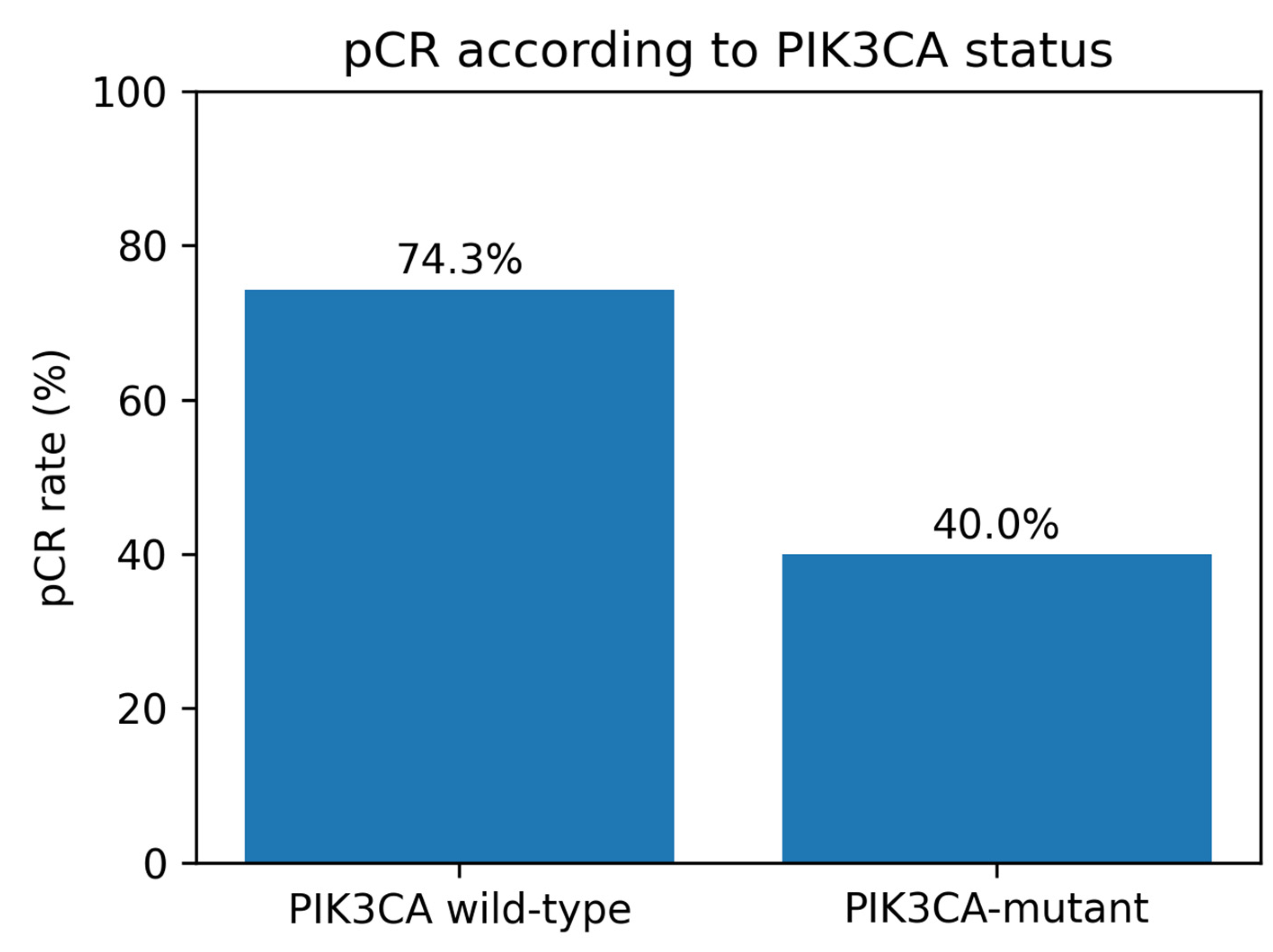
Figure 2.
Pathological complete response according to neoadjuvant treatment regimen.
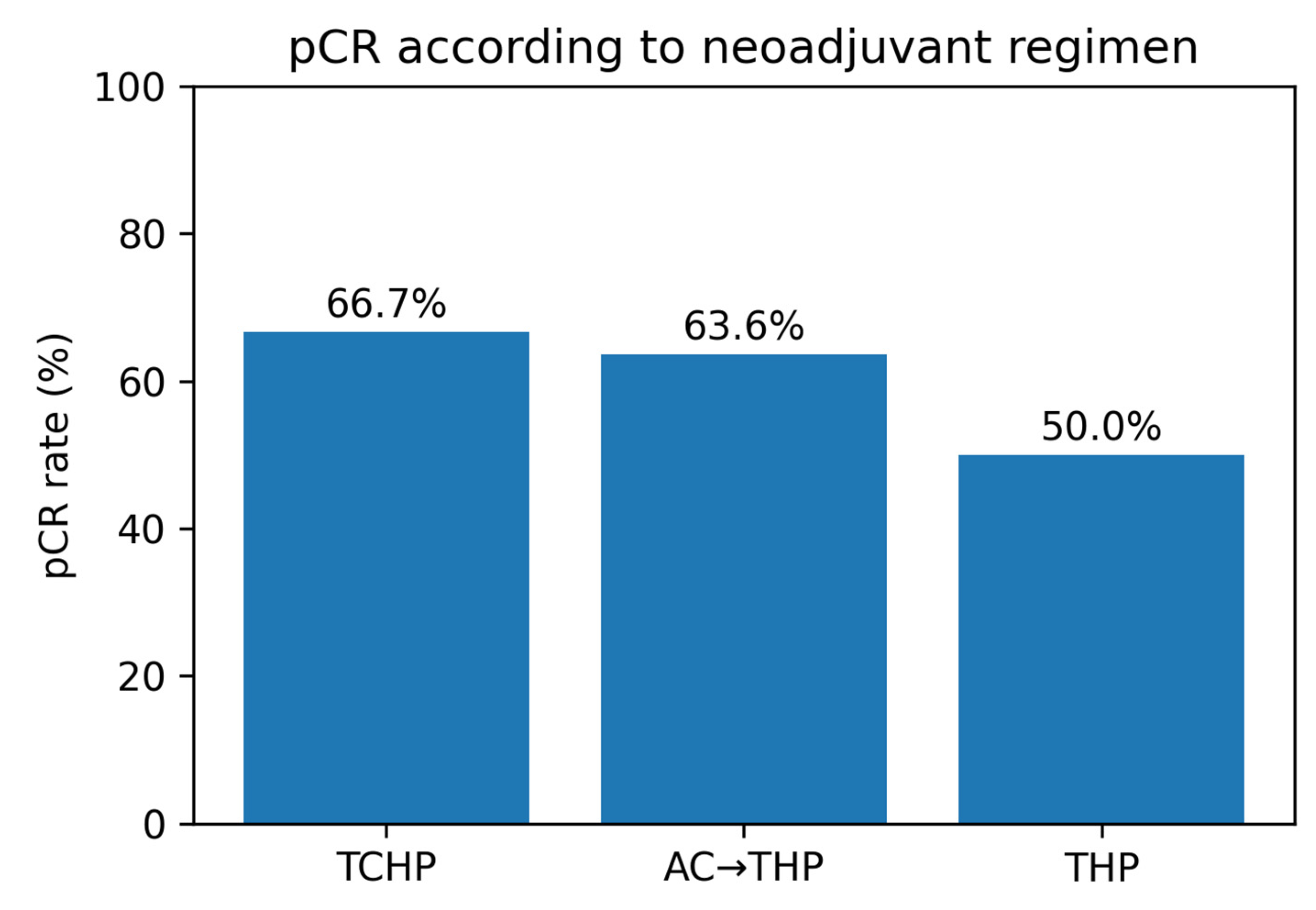
Figure 3.
pCR according to combined HR–PIK3CA status.
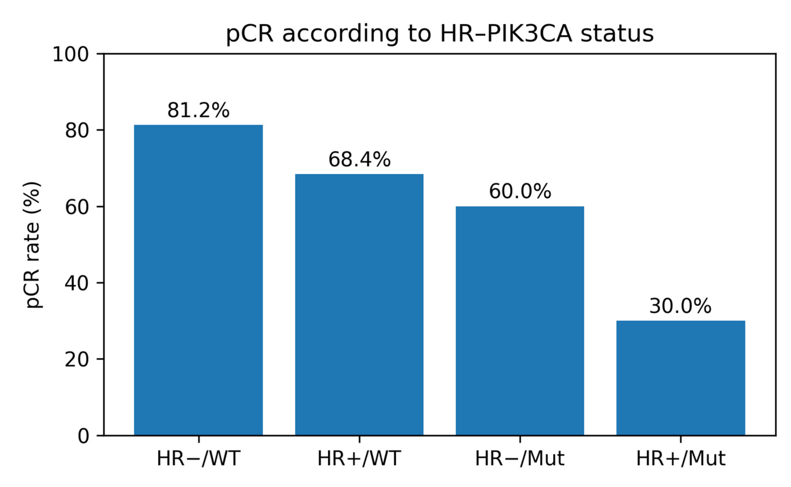
Figure 4.
Receiver operating characteristic (ROC) analysis of inflammatory–nutritional biomarkers for the prediction of pathological complete response.
Figure 4.
Receiver operating characteristic (ROC) analysis of inflammatory–nutritional biomarkers for the prediction of pathological complete response.
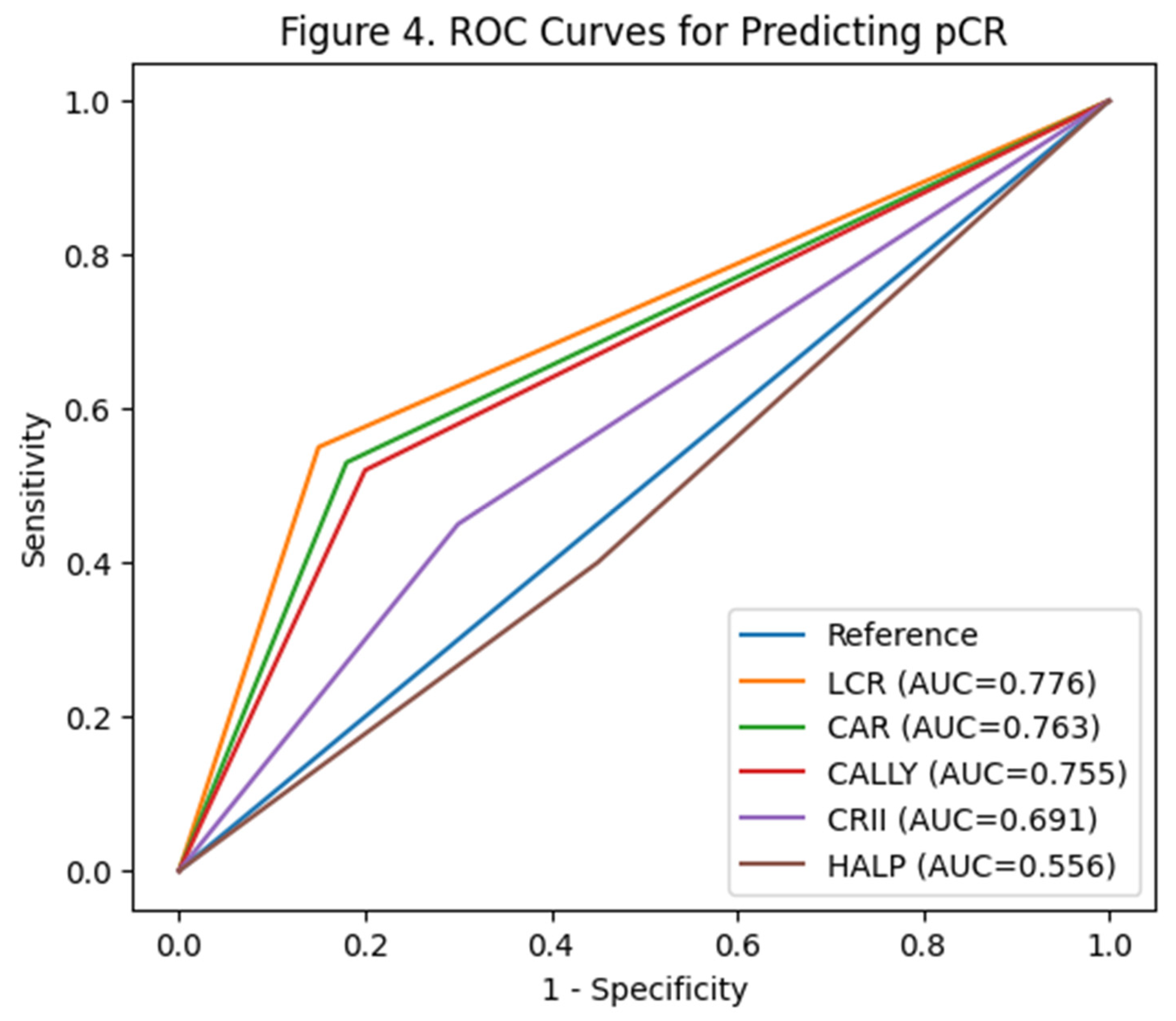
Figure 5.
Forest plot of multivariable logistic regression for pCR.
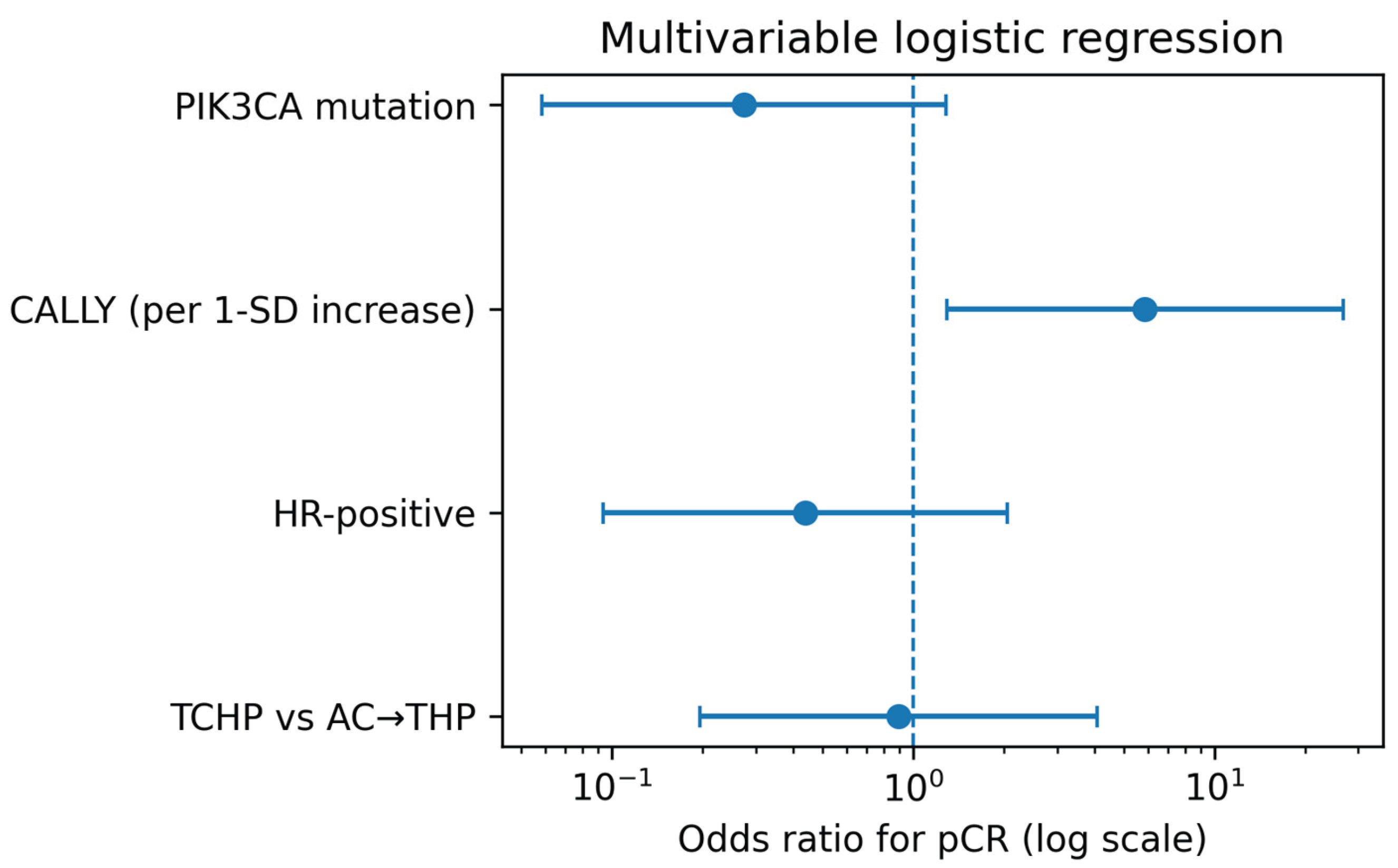

Table 1.
Baseline clinicopathological characteristics of the study population.
|
Characteristic |
Overall cohort |
| Patients, n |
50 |
| Age at diagnosis, years* |
50.3 ( 43.4–58.5) |
| Premenopausal |
21 (42.0) |
| Postmenopausal |
29 (58.0) |
| Invasive ductal carcinoma, NOS |
44 (88.0) |
| Grade 2 |
9 (19.1) |
| Grade 3 |
38 (80.9) |
| Ki-67, % |
30.0 (25.0–52.5) |
| Tumor size, mm |
29.0 (20.0–35.0) |
| ER-positive/low-positive |
29/49 (59.2) |
| PR-positive/low-positive |
18/49 (36.7) |
| HR-positive |
29/49 (59.2) |
| Clinically/pathologically node-positive |
35 (70.0) |
| Skin involvement |
8/49 (16.3) |
| PIK3CA-mutant |
15 (30.0) |
| TCHP |
24 (48.0) |
| AC→THP |
22 (44.0) |
| THP |
4 (8.0) |
Table 2.
Comparison of patients according to pathological complete response.
|
Variable |
pCR (n=32) |
Non-pCR (n=18) |
p value |
| Age at diagnosis, years* |
48.7 (44.6–59.1) |
50.8 (38.2–56.6) |
0.395 |
| Ki-67, % |
30.0 (25.0–57.5) |
30.0 (21.2–40.0) |
0.387 |
| Tumor size, mm |
31.0 (20.0–35.0) |
28.0 (22.5–34.8) |
0.700 |
| ER-positive/low-positive |
16/31 (51.6) |
13/18 (72.2) |
0.230 |
| PR-positive/low-positive |
8/31 (25.8) |
10/18 (55.6) |
0.064 |
| HR-positive |
16/31 (51.6) |
13/18 (72.2) |
0.230 |
| Node-positive |
21/32 (65.6) |
14/18 (77.8) |
0.523 |
| Skin involvement |
2/31 (6.5) |
6/18 (33.3) |
0.039 |
| PIK3CA-mutant |
6/32 (18.8) |
9/18 (50.0) |
0.028 |
| Treatment: TCHP |
16/32 (50.0) |
8/18 (44.4) |
0.812 |
| Treatment: AC→THP |
14/32 (43.8) |
8/18 (44.4) |
|
| Treatment: THP |
2/32 (6.2) |
2/18 (11.1) |
|
| HALP |
0.4 (0.3–0.5) |
0.3 (0.2–0.4) |
0.524 |
| CALLY |
3.4 (2.0–7.4) |
1.6 (1.1–2.6) |
0.003 |
| CRII |
5.9 (4.6–9.0) |
8.4 (6.2–13.7) |
0.027 |
| NLR |
2.1 (1.7–2.8) |
2.2 (1.8–4.3) |
0.347 |
| PLR |
148.4 (100.3–186.9) |
159.9 (132.7–240.9) |
0.207 |
| SII |
657.8 (472.9–821.4) |
771.7 (606.9–1207.6) |
0.049 |
| LCR |
0.8 (0.5–1.7) |
0.4 (0.3–0.6) |
0.001 |
| CAR |
0.7 (0.2–0.8) |
1.1 (0.8–1.7) |
0.002 |
Continuous variables are shown as median (IQR). Categorical variables are shown as n/N (%). P values were calculated using the Mann–Whitney U, Fisher’s exact, or Pearson chi-square tests as appropriate.
Table 3.
PIK3CA mutation status and pathological complete response.
|
PIK3CA status |
n |
pCR |
non-pCR |
pCR (%) |
| PIK3CA wild-type |
35 |
26 |
9 |
74.3 |
| PIK3CA-mutant |
15 |
6 |
9 |
40.0 |
Pearson’s χ² p = 0.021; Fisher’s exact p = 0.028.
Table 4.
Combined HR–PIK3CA analysis.
|
Group |
n |
pCR |
non-pCR |
pCR (%) |
| HR− / PIK3CA wild-type |
16 |
13 |
3 |
81.3 |
| HR+ / PIK3CA wild-type |
19 |
13 |
6 |
68.4 |
| HR− / PIK3CA-mutant |
5 |
3 |
2 |
60.0 |
| HR+ / PIK3CA-mutant |
10 |
3 |
7 |
30.0 |
Table 5.
Biomarker performance for predicting pCR.
|
Index |
AUC |
Group-comparison p |
Direction |
| HALP |
0.556 |
0.524 |
Higher values: not predictive |
| CALLY |
0.755 |
0.003 |
Higher values favored pCR |
| CRII |
0.691 |
0.027 |
Lower values favored pCR |
| NLR |
0.582 |
0.347 |
Not significant |
| PLR |
0.609 |
0.207 |
Not significant |
| SII |
0.670 |
0.049 |
Lower values favored pCR; borderline |
| LCR |
0.776 |
0.001 |
Higher values favored pCR |
| CAR |
0.763 |
0.002 |
Lower values favored pCR |
Table 6.
Logistic regression analysis for pCR.
|
Model |
Variable |
OR |
95% CI |
p value |
| Univariable |
PIK3CA mutation |
0.23 |
0.06–0.83 |
0.025 |
| Univariable |
CALLY per 1-SD increase |
5.49 |
1.36–22.20 |
0.017 |
| Multivariable |
PIK3CA mutation |
0.27 |
0.06–1.28 |
0.100 |
| Multivariable |
CALLY (per 1-SD increase) |
5.87 |
1.29–26.67 |
0.022 |
| Multivariable |
HR-positive |
0.44 |
0.09–2.05 |
0.295 |
| Multivariable |
TCHP vs AC→THP |
0.89 |
0.20–4.07 |
0.885 |
The multivariable model included PIK3CA mutation, CALLY, HR status, and TCHP versus AC→THP. An OR >1 indicates higher odds of pCR.
Disclaimer/Publisher’s Note: The statements, opinions and data contained in all publications are solely those of the individual author(s) and contributor(s) and not of MDPI and/or the editor(s). MDPI and/or the editor(s) disclaim responsibility for any injury to people or property resulting from any ideas, methods, instructions or products referred to in the content. |
© 2026 by the authors. Licensee MDPI, Basel, Switzerland. This article is an open access article distributed under the terms and conditions of the Creative Commons Attribution (CC BY) license.
Copyright: This open access article is published under a Creative Commons CC BY 4.0 license, which permit the free download, distribution, and reuse, provided that the author and preprint are cited in any reuse.


