
Submitted:
20 July 2026
Posted:
21 July 2026
You are already at the latest version
Abstract
Cancer cachexia is a multifactorial systemic syndrome characterized by progressive muscle loss, with or without adipose tissue depletion, that cannot be reversed by conventional nutritional support. It affects cancer patients and is associated with reduced treatment tolerance, impaired physical function, poor quality of life, and increased mortality. The understanding of cachexia has evolved recently, from the perception of a simple nutritional disorder to a complex immune-metabolic syndrome, based on tumor-host interactions, systemic inflammation, metabolic dysregulation, and multi-organ dysfunction. This review summarizes the progression of cachexia research, highlighting key findings involving inflammatory cytokines, proteolytic pathways, mitochondrial dysfunction, and immune dysregulation. The development of therapeutic strategies is examined, from early nutritional and appetite-stimulating interventions to contemporary targeted therapies, including ghrelin receptor agonists, cytokine inhibitors, and anabolic agents. Despite advances in mechanistic understanding, numerous trials targeting single pathways have failed to produce meaningful functional or survival benefits, underscoring the limitations of reductionist approaches. Emerging evidence supports a paradigm shift toward multimodal, biomarker-guided, and patient-centered interventions that address the interconnected biological mechanisms underlying cachexia. Particular emphasis is given to novel immunomodulatory strategies, including agents such as R-ketorolac, which may restore immune homeostasis and target the root causes of cachexia. It is hypothesized that future therapeutic success will likely depend on integrated approaches combining immunological, metabolic, nutritional, and rehabilitative interventions.
Keywords:
cancer cachexia
; immune dysregulation
; systemic inflammation
; multimodal therapy
; immunomodulation
; R-ketorolac
1. Introduction
Cancer cachexia is caused by the presence of certain cancers. It is a widely recognized multifactorial syndrome affecting the whole body. It is characterized by progressive weight loss, skeletal muscle wasting, and metabolic disturbances. It cannot be entirely reversed through standard nutritional support but is reversed if the patient has a complete anti-tumour response. The syndrome presents with debilitating symptoms such as unintentional weight loss, muscle atrophy, fatigue, anorexia, and generalized weakness, which collectively diminish patient function and overall well-being. These manifestations result from a sustained negative energy balance driven by reduced food intake and heightened metabolic demands related to tumour progression and systemic inflammation [1]. This condition also affects multiple organ systems, altering muscle metabolism, immune responses, and neuroendocrine signalling pathways, thereby contributing to disease progression. As the syndrome advances, patients encounter severe fatigue, apathy, and reduced physical performance, markedly impairing quality of life and complicating clinical management [1,2].
The high prevalence and profound effect on disease outcomes among cancer patients underscore the clinical significance of cancer cachexia. Approximately 50% of individuals with cancer develop cachexia; however, its incidence can vary considerably depending on tumour type and stage, with reported risks ranging from 20% to 80% in cases of advanced malignancies. This syndrome is closely linked to progressive functional decline and decreased survival rates, thereby representing one of the most critical complications associated with the progression of cancer [1,2]. Cachexia has been frequently observed in patients with gastrointestinal and advanced cancers, often manifesting as severe weight loss and muscle wasting prior to diagnosis. These metabolic changes expedite disease progression and exacerbate clinical outcomes by diminishing treatment tolerance and heightening susceptibility to complications. Consequently, the presence of cachexia serves as a significant prognostic indicator in oncological care and underscores the importance of early detection and intervention [1,2].
Furthermore, cancer cachexia substantially contributes to organ dysfunction and physiological decline, chiefly through progressive skeletal muscle depletion and systemic inflammation. These phenomena impair functional capacity, weaken respiratory and cardiac musculature, and disrupt metabolic homeostasis, collectively elevating the risk of organ failure and mortality among cancer patients. Additionally, cachexia often diminishes patients’ tolerance for chemotherapy, radiotherapy, surgery and other anticancer interventions, leading to dose reductions, treatment delays, or cessation of therapy. Clinical implications, including altered treatment outcomes and overall survival rates, underscore the importance of cancer cachexia as a critical therapeutic target in oncology research and clinical practice [2,3]. This review assesses why drug development based on specific pathways in cancer cachexia have been unsuccessful in the past and explores the potential of immunomodulatory therapy for improving treatment tolerance, physical function and patient survival.
A literature search was conducted using PubMed and Google Scholar to identify relevant publications on cancer cachexia and its treatment. The search was restricted to articles published in English and no lower date limit was applied, so as to include all historically significant research alongside recent advances. Search terms comprised of keywords such as “cancer cachexia”, “immunomodulation”, “multimodal therapy”, “drug development”, “immune dysregulation”, “immunomodulatory agents” and reference list of the important articles was screened for additional sources. Publications were selected on the basis of relevance to the pathophysiology, therapeutic evolution, and emerging immunomodulatory approaches for cancer cachexia.
2. Historical Overview of Cancer Cachexia Research
Cancer cachexia is frequently observed in patients with advanced malignancies and contributes significantly to morbidity, decreased tolerance to anticancer therapies, reduced quality of life, and increased mortality. Over the past several decades, the scientific understanding of cancer cachexia has evolved from early clinical descriptions of disease-related wasting to a complex mechanistic framework involving metabolic dysregulation, systemic inflammation, and multi-organ interactions. Current research on the syndrome reports advances in experimental biology, immunology, and metabolism, providing clarity on the mechanisms underlying cancer-associated wasting [4,5,6].
2.1. Early Recognition of Disease-Associated Wasting
Ancient medical texts mention cancer cachexia, with Hippocratic writings from the fourth century BC describing progressive wasting and weakness in patients with chronic diseases, including cancers. These patients often experienced severe weight loss, poor clinical outcomes, and the condition was hard to reverse despite efforts to provide adequate nutrition. Although the mechanisms behind these observations were not understood then, they signalled early recognition that chronic illness could cause systemic wasting separate from starvation [7].
Throughout the nineteenth and early twentieth centuries, clinicians observed cancer-related wasting. They noted that patients with advanced cancers suffered from significant weight loss, muscle wasting, fatigue, and functional decline. Initially, many attributed this wasting mainly to anorexia and decreased calorie intake. However, experimental tumour models developed in the mid-twentieth century challenged this hypothesis [8].
2.2. Experimental Evidence and Tumour Host Interactions
During the 1950s and early 1960s, experimental studies utilizing tumour-bearing animals provided crucial insights into the systemic nature of cancer cachexia. These studies reported that animals with implanted tumours exhibited weight loss and muscle wasting despite maintained food intake. Such findings implied that tumour growth could induce metabolic alterations promoting tissue catabolism independent of nutritional deficiency. Further experimental research demonstrated that the injection of tumour cells into experimental animals resulted in a wasting syndrome akin to clinical cachexia. These observations suggested that tumours release circulating factors capable of modifying host metabolism and inducing systemic catabolic responses. The understanding of tumour-host interactions became fundamental to elucidating cachexia pathophysiology, thereby transforming the perspective regarding nutritional deficiencies [9,10,11,12]
2.3. Evolution of Understanding: Metabolic Dysfunction and Energy Imbalance
Across the period from the 1970s to the 1990s, studies showed that cachexia involves intricate metabolic changes rather than straightforward malnutrition. Clinical and experimental evidence indicated that cancer patients suffering from cachexia often had increased resting energy expenditure, disrupted carbohydrate metabolism, and heightened lipid mobilization. These metabolic disruptions led to a negative energy balance, resulting in tissue loss. Additionally, efforts to combat cachexia with intensive nutritional supplementation frequently failed to restore muscle mass or prevent weight loss. These observations underscored the importance of underlying metabolic dysregulation in the development of cachexia [9].
Subsequent research has demonstrated that the loss of skeletal muscle in cachexia is principally caused by the activation of proteolytic pathways, including the ubiquitin-proteasome system and autophagy-related degradation mechanisms. These pathways expedite the degradation of myofibrillar proteins and result in progressive muscle atrophy [13].
Moreover, cachexia results in remarkable alterations in adipose tissue metabolism, manifested as accelerated lipolysis and impaired lipid storage, leading to rapid depletion of fat mass, contributing to systemic energy imbalance and metabolic dysfunction [14,15]. These metabolic alterations established cancer cachexia as a systemic syndrome comprising changes in multiple metabolic pathways rather than a localized effect of tumour growth.
2.4. Identification of Inflammatory Mediators
Parabolic research studies suggested that cancer cachexia was associated with circulating stable molecules. Further studies indicated that these factors were cytokines [16]. Identifying inflammatory cytokines that drive tissue wasting is crucial for understanding cancer cachexia [17]. Tumour necrosis factor alpha (TNF alpha) was recognized as a cytokine that can trigger metabolic shifts associated with cachexia. Due to its role in causing weight loss and muscle wasting in experimental models, TNF-alpha was initially referred to as cachectin [9]. Cleverly, cachectin/tumour necrosis was noted to be two sides of an experimental coin [18]. TNF-alpha activates the inflammatory signalling pathways, including nuclear factor kappa B, promoting proteolysis in skeletal muscle while suppressing protein synthesis [19]. These mechanisms play a direct role in causing muscle wasting and disrupting metabolic balance. Furthermore, cytokines like interleukin 1 (IL-1), interleukin 6 (IL-6), and leukaemia inhibitory factor (LIF) are involved in the development of cachexia. High levels of these inflammatory mediators trigger catabolic signalling pathways and interfere with metabolic stability. Notably, IL-6 has been identified as a crucial factor in cancer cachexia, activating JAK-STAT signalling pathways that lead to muscle protein breakdown and suppress anabolic processes [20] (Figure 1). Chronic systemic inflammation induced by these cytokines also affects adipose tissue metabolism by stimulating lipolysis and reducing lipid storage capacity. These alterations further contribute to the progressive loss of body mass observed in cachectic patients [14] (Table 1).
2.5. Recognition of Multi-Organ Interactions
Studies have demonstrated that tumour-derived factors and inflammatory mediators influence skeletal muscle, adipose tissue, liver, and the central nervous system. Skeletal muscle is the primary site of tissue loss in cachexia. The activation of the ubiquitin-proteasome system and other proteolytic pathways results in the progressive degradation of muscle proteins and a decline in muscle function [38]. Adipose tissue experiences notable metabolic changes, with heightened lipolysis and decreased adipogenesis leading to fat store depletion and shifts in systemic energy metabolism [15]. Furthermore, the liver contributes to the progression of cachexia by increasing the production of acute-phase proteins in response to systemic inflammation. This hepatic response elevates metabolic demand and enhances inflammatory signalling throughout the body [39]. Anorexia is another clinical sign of cancer cachexia [40]. Furthermore, inflammatory cytokines impact hypothalamic pathways responsible for regulating appetite and energy homeostasis. These alterations frequently lead to anorexia and decreased caloric intake, thereby contributing to weight loss [41].
2.6. Immune Dysregulation and Inflammatory Signalling
Immune dysregulation plays a key role in cancer cachexia. Tumour cells interact with immune cells to produce inflammatory cytokines and signalling molecules that disrupt metabolic homeostasis throughout the body. Persistent activation of inflammatory pathways leads to chronic catabolism and progressive tissue wasting. These immune-mediated mechanisms are crucial for cachexia pathogenesis [6,42,43]. The loss of skeletal muscle during cachexia may be attributed to mitochondrial dysfunction and compromised muscle regeneration. Mitochondrial changes diminish energy production and elevate oxidative stress within muscle cells, thereby fostering muscle degradation [44]. Moreover, signalling pathways involving myostatin and activin have been implicated in the regulation of muscle mass during cancer cachexia. Activation of these pathways inhibits muscle growth and promotes muscle atrophy [45] (Figure 2).
2.7. Systems Biology and Emerging Research Directions
Advances in systems biology have facilitated a deeper understanding of cancer cachexia as an intricate network of interactions among multiple organs and signalling pathways. Communication between skeletal muscle, adipose tissue, liver, immune cells, and the central nervous system plays a role in the development and maintenance of the cachectic state [47]. Further, the gut microbiome modulates inflammation and metabolic dysfunction in cancer cachexia. Alterations in microbial composition may influence immune responses and metabolic regulation, thereby contributing to the progression of the syndrome [48]. Contemporary molecular methodologies have empowered researchers to detect alterations in gene expression, metabolic profiles, and signalling pathways associated with the progression of cachexia. These insights facilitate the identification of potential biomarkers and therapeutic targets for this condition [49].
2.8. Recent Advances and Therapeutic Strategies
Recent clinical research has emphasized the criticality of early diagnosis and multimodal treatment approaches for cancer cachexia. Anti-inflammatory agents, anabolic therapies, appetite stimulants, and metabolic modulators targeting specific molecular pathways involved in muscle wasting are current areas of research [50]. Advances in biomarker discovery and metabolic profiling may allow earlier detection of cachexia during cancer progression. Early identification of patients at risk may improve treatment outcomes and quality of life [51]. Therapeutic strategies targeting inflammatory pathways, mitochondrial dysfunction, and muscle regeneration mechanisms involved in cachexia are now emerging [21,52].
Further research continues to explore the role of immune-mediated mechanisms and systemic inflammation in cancer-associated wasting [43]. In addition, studies investigating sarcopenic obesity have revealed complex interactions between adipose tissue metabolism and muscle loss in cancer patients [47]. Therefore, integrating immunology, molecular biology, metabolic science, and clinical research to develop effective therapeutic strategies for cachexia is essential [6,53].
2.9. Contemporary Perspective
Today, cancer cachexia is acknowledged as a complex systemic syndrome characterized by metabolic disturbances, ongoing inflammation, immune system disorder, and communication across multiple organs. Over the years, research has shifted the view of this condition from a basic nutritional problem to a complex metabolic disease. Despite substantial progress in understanding its mechanisms, effective treatments are still scarce. Ongoing investigation into the molecular pathways responsible for muscle loss and metabolic issues is crucial for creating targeted therapies that could improve patient outcomes [53].
3. Drug Development in Cancer Cachexia
Early therapeutic strategies for cancer cachexia primarily concentrated on nutritional supplementation and appetite stimulation, adhering to the belief that cachexia was predominantly a result of decreased caloric intake and anorexia in patients with advanced malignancies [44]. Nutritional interventions were thus extensively used to restore body weight and energy balance in cachectic patients [54]. Megestrol acetate, a synthetic progesterone derivative, was one of the most extensively researched drugs for stimulating appetite in cancer cachexia. This drug has been found to stimulate appetite and increase caloric intake by modulating hypothalamic appetite-regulating pathways and suppressing pro-inflammatory cytokines. A study suggests that megestrol acetate will cause weight gain in patients with advanced cancer [55]. Nevertheless, the augmentation in body weight associated with megestrol acetate therapy predominantly resulted from an increase in adipose tissue and fluid retention, rather than the restoration of lean skeletal muscle mass [44].
Corticosteroids were also frequently used to stimulate appetite and improve a sense of well-being in cancer patients experiencing cachexia [54]. Corticosteroids may provide short-term improvements in appetite and fatigue; however, their long-term use is limited by adverse effects such as muscle weakness, immunosuppression, and metabolic complications [54]. Cannabinoid-based therapies such as dronabinol were evaluated for appetite stimulation in cancer patients with cachexia. However, clinical trials evaluating cannabinoids demonstrated only modest improvements in appetite and limited benefits in terms of weight gain and muscle mass recovery [56]. These early pharmacological interventions highlighted the limitations of therapies on appetite stimulation and indicated that cancer cachexia involves complex metabolic disturbances that cannot be reversed by nutritional or appetite-based therapies alone [44].
As research advanced, an enhanced understanding of the biological mechanisms underlying cancer cachexia facilitated the development of therapies targeting specific molecular pathways involved in the syndrome. Cancer cachexia involves systemic inflammation driven by cytokines such as tumour necrosis factor-α, interleukin-1, and interleukin-6, which contribute to metabolic dysregulation and muscle wasting. Therefore, anti-inflammatory agents capable of suppressing cytokine signalling were explored as potential treatments for cachexia [57]. Thalidomide demonstrates the ability to inhibit tumour necrosis factor-α production and reduce inflammation in cancer patients with cachexia [54]. Clinical investigations suggested that thalidomide could improve appetite and body weight in certain cancer populations [54]. Monoclonal antibodies targeting the interleukin-6 receptor, including tocilizumab, have been investigated as potential treatments for cachexia-related inflammation [58]. Another IL-6 pathway inhibitor, ALD518, demonstrated promising results in early clinical trials, improving lean body mass and reducing fatigue in patients with advanced cancers [1]. These targeted approaches represented an important shift from purely symptomatic treatments toward therapies designed to interrupt the inflammatory mechanisms driving cancer cachexia [58].
More recent drug development efforts have increasingly focused on hormonal and metabolic regulators of appetite and muscle metabolism. One important therapeutic target is ghrelin, a peptide hormone that stimulates appetite and promotes anabolic signalling through growth hormone pathways. Ghrelin receptor agonists have therefore been investigated as treatments for cancer cachexia because of their ability to increase appetite and improve energy balance [2]. The orally active ghrelin receptor agonist anamorelin has been reported to increase appetite, body weight, and lean body mass in patients with cancer cachexia. Anamorelin has also been reported to improve symptoms such as fatigue and reduced food intake in cancer patients with advanced disease [59]. It has been approved in Japan for the treatment of cancer cachexia in certain malignancies, including lung cancer [2].
In addition to appetite-regulating therapies, drug development has also targeted pathways involved in skeletal muscle loss, particularly the myostatin and activin signalling pathways, which regulate muscle growth and degradation. Experimental therapies designed to inhibit myostatin or activin receptors have shown potential for increasing muscle mass in patients with cancer-related wasting [20]. Moreover, molecular targets such as growth differentiation factor-15 (GDF-15) are also being investigated for their potential role in modulating appetite and metabolic regulation in cancer cachexia [60]. Despite these advances, many investigators emphasize that cancer cachexia is a multifactorial syndrome and an effective treatment should comprise multimodal therapeutic strategies combining pharmacological agents, nutritional interventions, and physical rehabilitation [49].
Recent cancer cachexia drug development research shows a shift toward therapies that target the syndrome’s underlying biological mechanisms, though challenges in clinical trial design and regulatory approval persist. Studies now focus more on agents that influence metabolic and inflammatory pathways causing muscle wasting and systemic metabolic issues, rather than just addressing appetite or weight loss. For example, Espindolol, a non-selective β-blocker, and monoclonal antibodies targeting growth differentiation factor-15 (GDF-15) are being explored. These interventions aim to directly target the molecular drivers of cachexia instead of merely alleviating its symptoms [61].
Ghrelin receptor agonists, such as anamorelin, have undergone comprehensive investigation in randomized controlled trials, including the phase III ROMANA 1 and ROMANA 2 studies. These trials demonstrated enhancements in lean body mass, body weight, and symptoms associated with anorexia in patients diagnosed with non-small cell lung cancer and cachexia [62]. Nonetheless, even with these positive metabolic results, gains in functional measures like hand-grip strength did not reach statistical significance, highlighting a major challenge in assessing clinical benefits in cachexia [61]. The selection of appropriate clinical endpoints continues to present challenges, as improvements in body composition do not invariably correspond to measurable enhancements in physical function or survival. This discrepancy complicates the regulatory evaluation of novel therapies [2].
Obtaining regulatory approval has been challenging for many investigational drugs, even when initial clinical results seem promising. For instance, the ghrelin agonist anamorelin has been approved in Japan for treating cancer cachexia but has not yet gained approval in Europe or North America. This delay is partly due to discrepancies between gains in lean body mass and the functional improvements that regulators require [2]. Likewise, other targeted therapies such as myostatin inhibitors, cytokine-targeting antibodies, and selective androgen receptor modulators have shown potential benefits in early-phase studies but have not yet achieved consistent success in late-phase trials. These challenges underscore the complexity of cancer cachexia and have led researchers to increasingly advocate for multimodal treatment strategies combining pharmacological therapies with nutritional and exercise interventions, while continuing to refine clinical trial endpoints and regulatory frameworks for this multifactorial syndrome [2].
4. Lessons from the Failure of Multiple Clinical Trials Targeting Single Pathways
The international consensus definition of cachexia as a “multifactorial syndrome characterized by ongoing loss of skeletal muscle mass (with or without fat loss) that cannot be fully reversed by conventional nutritional support and leads to progressive functional impairment” [4] explicitly highlights metabolic dysregulation, systemic inflammation, and tumour–host interactions as central drivers. However, many therapeutic strategies have focused on single mechanistic axes such as appetite stimulation (ghrelin agonists), anabolic signalling (androgens, selective androgen receptor modulators), or isolated cytokine blockade (TNF-α, IL-6, IL-1) [17]. Cachexia involves complex cross-talk between tumour-derived factors, host inflammatory mediators, neuroendocrine changes, and altered muscle protein turnover, suggesting that reductionist approaches are unlikely to provide durable clinical benefit [47]. Similarly, the activation of proteolytic systems, such as the ubiquitin - proteasome pathway and autophagy, alongside anorexia and metabolic inefficiency, affects the systemic pathophysiology [9].
High failure rates in phase II/III trials may be due to the differences in drug mechanism and syndrome complexity. Agents targeting a single pathway may influence body composition but fail to meaningfully alter overall function, survival, food intake or quality of life because compensatory mechanisms and ongoing tumour-driven inflammation persist [47]. These findings highlight that cachexia is not merely a nutritional deficit or isolated endocrine abnormality but a multi-system metabolic challenge requiring integrated therapeutic strategies.
4.1. Case Studies
In the ROMANA 1 and 2 Phase III trials evaluating anamorelin, in patients with advanced non–small cell lung cancer (NSCLC) and cachexia, anamorelin markedly increased lean body mass compared to placebo. However, handgrip strength, an essential endpoint, did not show a significant improvement. Consequently, although appetite and certain patient-reported outcomes demonstrated enhancement, the lack of functional gains impeded regulatory approval. These findings thus reveal a dissociation between body composition and clinically meaningful function [63].
Similarly, anti-inflammatory strategies targeting individual cytokines have largely proven ineffective. Initial trials employing TNF-α inhibitors, such as etanercept, in cases of advanced cancer did not exhibit substantial enhancements in weight or survival [64]. Trials targeting IL-6 (e.g., clazakizumab or ALD518) showed modest improvements in anaemia or fatigue but inconsistent effects on lean body mass or overall outcomes. IL-1 blockade (e.g., MABp1 targeting IL-1α) demonstrated some symptom improvement in colorectal cancer but failed to impact survival [65]. Therefore, these case studies demonstrate that targeting a single inflammatory mediator may be ineffective due to redundancy and compensatory signalling within the cytokine network.
4.2. Common Pitfalls
Methodological limitations may result in therapeutic failures. Inadequate patient stratification has affected observable effects. Cachexia occurs in a spectrum ranging from pre-cachexia to refractory cachexia, yet many trials enrolled heterogeneous populations without biomarker-driven selection [4]. Further, endpoint selection has often focused on weight gain or lean body mass rather than physical performance, functional capacity, or survival. The ROMANA trials highlighted the misalignment between increased lean mass and unchanged strength [63]. Moreover, Prado et al. (2018) emphasized that muscle quantity does not necessarily equate to muscle quality or performance. Nonetheless, progressive malignancy itself influences outcomes [65]. Tumour burden, chemotherapy toxicity, and systemic inflammation independently drive muscle catabolism, making it difficult for anabolic agents to overcome ongoing catabolic stimuli [50]. Further, cancer trajectory and concurrent treatments may also affect the interpretability of results.
4.3. Broader Implications
These failures collectively highlight cancer cachexia as a multi-organ failure syndrome that affects skeletal muscle, adipose tissue, liver (through the acute-phase response), the brain (regulating appetite), and the immune system. Central to this process is tumour–host interaction, which drives metabolic reprogramming and systemic inflammation. The continued presence of cachexia despite nutritional support and single-drug treatments indicates that addressing the root causes, such as systemic inflammation, tumour-derived factors, and immune system disorder, may be crucial [5,6]. The multimodal therapy combining pharmacologic, nutritional, and exercise interventions appears to be essential for addressing cancer cachexia [47]. Thus, past failures highlight that simply targeting one pathway is inadequate because the syndrome is characterized by multiple factors and involves systemic dysregulation. Therefore, emerging strategies should focus on integrated immune-metabolic modulation rather than isolated appetite or anabolic stimulation (Table 2).
5. The Need for a Fundamental Change in Reversing the Syndrome
5.1. Critique of Current Paradigms
Cancer cachexia is characterized by ongoing skeletal muscle loss that cannot be fully reversed by conventional nutritional support and leads to progressive functional impairment. Cachexia is not only the result of reduced caloric intake but also of complex metabolic abnormalities and systemic inflammation [4]. Subsequent mechanistic analyses have demonstrated that tumour-host interactions activate a coordinated network involving skeletal muscle proteolysis, adipose tissue lipolysis, hepatic acute-phase response, neuroendocrine alterations, and immune dysregulation [5,6,47].
Systemic inflammation is a key factor, with pro-inflammatory cytokines IL-6, TNF-α, and IL-1β promoting muscle breakdown, loss of appetite, insulin resistance, and alterations in hepatic metabolism [9,20]. Further, cytokine signalling operates within redundant and compensatory immune networks, limiting the efficacy of single-cytokine blockade strategies [9]. Cachexia is therefore increasingly conceptualized as an immune-metabolic disorder, where immune disorder and metabolic reprogramming are tightly linked [6,50,63].
Clinical trial failures targeting isolated pathways such as appetite stimulation or muscle anabolism highlight differences between disease complexity and therapeutic design. Improvements in lean body mass frequently fail to translate into enhanced muscle strength, physical function, or survival [50,63]. Observations demonstrate that these approaches do not sufficiently address the interconnected inflammatory, metabolic, neuroendocrine, and immune disturbances underlying the syndrome.
5.2. Paradigm Shift
Given this complexity, most experts recommend a multimodal approach that targets the root causes for managing cachexia [4,47]. International consensus recommendations highlight the importance of integrating pharmacologic agents with nutritional intervention, exercise therapy, and anti-inflammatory strategies, rather than using monotherapy alone [66]. Biomarker-guided patient stratification is increasingly viewed as essential. Inflammatory markers (e.g., CRP, IL-6), body composition analysis, and metabolic signatures may help identify subgroups most likely to benefit from specific interventions [47]. A systematic review of endpoints in cachexia trials highlighted substantial heterogeneity and emphasized the need for validated biomarker-based outcomes to enhance trial outcomes and interpretability [67].
The integration of cachexia management into routine oncology practice has also been recommended. Early screening and intervention, rather than late-stage reactive treatment, may enhance functional outcomes and treatment tolerance [68]. By aligning cachexia management with standard cancer care protocols, clinicians may mitigate treatment toxicity, enhance physical performance, and potentially influence overall prognosis [47,68]. Thus, the emerging paradigm supports combination therapy, biomarker-based personalization, and early integrated care, targeting the underlying inflammatory and metabolic drivers rather than isolated downstream manifestations.
5.3. Regulatory and Methodological Reforms
Reforming clinical trial design is essential for progressing therapeutic development. Previous studies focused on endpoints including body weight or lean body mass, but these measures do not reliably correlate with functional improvements [63,67]. Regulatory requirements now focus on patient-centered endpoints such as handgrip strength, physical performance tests, and quality-of-life measures [63]. Adaptive and biomarker-stratified trial designs may improve sensitivity to treatment effects in heterogeneous populations. Incorporating composite endpoints that combine body composition, functional performance, and inflammatory biomarkers may provide a clinically meaningful benefit. Furthermore, cachexia is comparable with aging-related sarcopenia and chronic inflammatory diseases [50,65]. Collaborative, interdisciplinary frameworks involving oncology, geriatrics, immunology, and nutrition science are therefore essential to address shared pathophysiology and enhance generalizability. Recognizing cachexia as a systemic comorbidity rather than a secondary symptom of advanced cancer may facilitate more appropriate regulatory evaluation and therapeutic prioritization [68]. Therefore, these changes indicate a need to shift toward integrated, biologically informed, and patient-centered research frameworks aligned with the systemic nature of cancer cachexia.
6. Emerging and Promising Immunomodulatory Approaches
Cancer cachexia is driven primarily by systemic immune dysregulation rather than nutritional deficiency, making immunomodulation a rational therapeutic strategy. Chronic inflammation mediated by cytokines such as IL-6, TNF-α, and IL-1β induces muscle wasting and metabolic dysfunction via pathways like JAK/STAT and NF-κB [6,69]. The tumour microenvironment further amplifies this process through dynamic interactions between tumour and immune cells, sustaining cytokine signalling and immune suppression [70]. Tumour-induced immune disorder, characterized by T-cell dysfunction, lymphopenia, and expansion of MDSCs, creates a catabolic state linked to poor outcomes [6]. Similar cytokine-driven immune imbalance is observed in chronic inflammatory conditions, reinforcing the centrality of immune dysregulation in disease progression [71]. While early approaches targeting single mediators (e.g., GDF-15 or individual cytokines) show partial benefit, they are limited by pathway redundancy, compensatory mechanisms, and patient heterogeneity [6]. Thus, broader immunomodulatory strategies that restore immune homeostasis are essential for meaningful reversal of cachexia.
Emerging therapies increasingly target the tumour–immune–host axis in an integrated manner. Cytokines regulate tumour sensitivity and immune responses, influencing apoptosis, immune activation, and therapeutic susceptibility [70]. Advances in cytokine engineering, biologics, and JAK inhibitors highlight the importance of modulating multiple inflammatory pathways simultaneously [71]. Chronic inflammation within the tumour microenvironment promotes immune loss and treatment resistance, necessitating systemic immune reprogramming rather than isolated cytokine blockade [69]. Spatial and metabolic heterogeneity further complicates targeted approaches, reinforcing the need for multi-pathway interventions [70]. Consequently, modern strategies focus on restoring immune balance while enhancing anti-tumour immunity. This represents a shift from single-target inhibition to systems-level immunomodulation.
R-ketorolac is a promising next-generation immunomodulator targeting tumour-induced immune disorders. Preclinical studies demonstrate reversal of cachexia in tumour-bearing models, including reduced weight loss and muscle wasting independent of tumour size or food intake. Treatment restores immune balance by improving T-cell populations and reducing inflammatory cytokines such as IL-6. These effects translate into improved survival and near-complete reversal of cachexia phenotypes in animal models. Its mechanism suggests root-cause targeting of immune dysfunction rather than symptomatic relief. Given the multifactorial nature of cachexia, combination strategies with immunotherapy or chemotherapy may further enhance outcomes. Therefore, R-ketorolac is a strong candidate within multimodal, immune-centric treatment paradigms [6].
7. Conclusion
Cancer cachexia is now clearly identified as a complex immune-metabolic syndrome in which tumour-host interactions, systemic inflammation, and multi-organ dysfunction converge to drive progressive decline, rendering traditional single-pathway and symptom-focused interventions inadequate. The recurrent lack of success in such approaches has elucidated a vital insight: substantial clinical benefit will not be achieved by targeting isolated mechanisms, but rather by addressing the integrated biological network that underpins the syndrome. Emerging evidence highlights immunomodulation, especially approaches that restore systemic immune balance, as a promising, mechanism-based direction. Early results with agents like R-ketorolac suggest the possibility of addressing cachexia at its core. Future efforts should adopt a framework that combines multimodal, biomarker-guided, and patient-centred strategies, integrating immunotherapy, metabolic regulation, nutrition, and physical rehabilitation into routine cancer care. Advances in systems biology, clinical trial design, and translational science offer a real and promising chance to reframe cancer cachexia from an unavoidable outcome of advanced disease into a treatable condition, ultimately enhancing treatment tolerance, functional ability, and survival for cancer patients.
Author Contributions
Conceptualization, writing—original draft preparation and editing, L.Z.; writing—review, revision and editing, J.A.N. All authors have read and agreed to the published version of the manuscript.
Funding
This research received no external funding.
Acknowledgments
Editorial assistance for the development of this manuscript was supported by Servier.
Conflicts of Interest
The authors declare no conflict of interest.
References
- Law, M.L. Cancer cachexia: Pathophysiology and association with cancer-related pain. Front. Pain Res. 2022, 3, 971295. [Google Scholar] [CrossRef]
- Nishikawa, H.; Goto, M.; Fukunishi, S.; Asai, A.; Nishiguchi, S.; Higuchi, K. Cancer cachexia: its mechanism and clinical significance. Int. J. Mol. Sci. 2021, 22, 8491. [Google Scholar] [CrossRef] [PubMed]
- Argilés, J.M.; Busquets, S.; López-Soriano, F.J. Anti-inflammatory therapies in cancer cachexia. Eur. J. Pharmacol. 2011, 668, S81–S86. [Google Scholar] [CrossRef] [PubMed]
- Fearon, K.; Strasser, F.; Anker, S.D.; Bosaeus, I.; Bruera, E.; Fainsinger, R.L.; Jatoi, A.; Loprinzi, C.; MacDonald, N.; Mantovani, G. Definition and classification of cancer cachexia: an international consensus. Lancet Oncol. 2011, 12, 489–495. [Google Scholar] [CrossRef] [PubMed]
- Argilés, J.M.; Busquets, S.; Stemmler, B.; López-Soriano, F.J. Cancer cachexia: understanding the molecular basis. Nat. Rev. Cancer 2014, 14, 754–762. [Google Scholar] [CrossRef] [PubMed]
- Zhang, L.; Bonomi, P.D. Immune system disorder and cancer-associated cachexia. Cancers 2024, 16, 1709. [Google Scholar] [CrossRef] [PubMed]
- Ni, J.; Zhang, L. Cancer cachexia: definition, staging, and emerging treatments. Cancer Manag. Res. 2020, 5597–5605. [Google Scholar] [CrossRef] [PubMed]
- Aversa, Z.; Costelli, P.; Muscaritoli, M. Cancer-induced muscle wasting: latest findings in prevention and treatment. Ther. Adv. Med. Oncol. 2017, 9, 369–382. [Google Scholar] [CrossRef] [PubMed]
- Tisdale, M.J. Mechanisms of cancer cachexia. Physiol. Rev. 2009. [Google Scholar] [CrossRef] [PubMed]
- Stovroff, M.C.; Fraker, D.L.; Norton, J.A. Cachectin activity in the serum of cachectic, tumor-bearing rats. Arch. Surg. 1989, 124, 94–99. [Google Scholar] [CrossRef] [PubMed]
- Stovroff, M.C.; Fraker, D.L.; Swedenborg, J.A.; Norton, J.A. Cachectin/tumor necrosis factor: a possible mediator of cancer anorexia in the rat. Cancer Res. 1988, 48, 4567–4572. [Google Scholar] [PubMed]
- Stovroff, M.C.; Fraker, D.L.; Travis, W.D.; Norton, J.A. Altered macrophage activity and tumor necrosis factor: Tumor necrosis and host cachexia. J. Surg. Res. 1989, 46, 462–469. [Google Scholar] [CrossRef] [PubMed]
- Talbert, E.E.; Cuitiño, M.C.; Ladner, K.J.; Rajasekerea, P.V.; Siebert, M.; Shakya, R.; Leone, G.W.; Ostrowski, M.C.; Paleo, B.; Weisleder, N. Modeling human cancer-induced cachexia. Cell Rep. 2019, 28, 1612–1622. e1614. [Google Scholar] [CrossRef] [PubMed]
- Das, S.K.; Hoefler, G. The role of triglyceride lipases in cancer associated cachexia. Trends Mol. Med. 2013, 19, 292–301. [Google Scholar] [CrossRef] [PubMed]
- Ebadi, M.; Mazurak, V.C. Evidence and mechanisms of fat depletion in cancer. nutrients 2014, 6, 5280–5297. [Google Scholar] [CrossRef] [PubMed]
- Norton, J.A.; Moley, J.F.; Green, M.V.; Carson, R.E.; Morrison, S.D. Parabiotic transfer of cancer anorexia/cachexia in male rats. Cancer Res. 1985, 45, 5547–5552. [Google Scholar] [PubMed]
- Mcnamara, M.J.; Alexander, H.R.; Norton, J.A. Cytokines and their role in the pathophysiology of cancer cachexia. J. Parenter. Enter. Nutr. 1992, 16, 50S–55S. [Google Scholar] [CrossRef]
- Beutler, B.; Cerami, A. Cachectin and tumour necrosis factor as two sides of the same biological coin. Nature 1986, 320, 584–588. [Google Scholar] [CrossRef] [PubMed]
- Langstein, H.N.; Norton, J.A. Mechanisms of cancer cachexia. Hematol. Clin. N. Am. 1991, 5, 103–123. [Google Scholar] [CrossRef]
- Argilés, J.M.; López-Soriano, F.J.; Busquets, S. Mediators of cachexia in cancer patients. Nutrition 2019, 66, 11–15. [Google Scholar] [CrossRef] [PubMed]
- Setiawan, T.; Sari, I.N.; Wijaya, Y.T.; Julianto, N.M.; Muhammad, J.A.; Lee, H.; Chae, J.H.; Kwon, H.Y. Cancer cachexia: molecular mechanisms and treatment strategies. J. Hematol. Oncol. 2023, 16, 54. [Google Scholar] [CrossRef] [PubMed]
- McDonald, J.J.; McMillan, D.C.; Laird, B.J.A. Targeting IL-1alpha in cancer cachexia: a narrative review. Curr. Opin. Support Palliat. Care 2018, 12, 453–459. [Google Scholar] [CrossRef] [PubMed]
- Miller, J.; Laird, B.J.; Skipworth, R.J. The immunological regulation of cancer cachexia and its therapeutic implications. J. Cancer Metastasis Treat. 2019, 5, N/A–N/A. [Google Scholar]
- Rounis, K.; Makrakis, D.; Gioulbasanis, I.; Ekman, S.; De Petris, L.; Mavroudis, D.; Agelaki, S. Cancer cachexia and antitumor immunity: common mediators and potential targets for new therapies. Life 2022, 12, 880. [Google Scholar] [CrossRef] [PubMed]
- Zhang, D.; Zheng, H.; Zhou, Y.; Tang, X.; Yu, B.; Li, J. Association of IL-1beta gene polymorphism with cachexia from locally advanced gastric cancer. BMC Cancer 2007, 7, 45. [Google Scholar] [CrossRef] [PubMed]
- Scheede-Bergdahl, C.; Watt, H.L.; Trutschnigg, B.; Kilgour, R.D.; Haggarty, A.; Lucar, E.; Vigano, A. Is IL-6 the best pro-inflammatory biomarker of clinical outcomes of cancer cachexia? Clin. Nutr. 2012, 31, 85–88. [Google Scholar] [CrossRef] [PubMed]
- Rébé, C.; Ghiringhelli, F. Interleukin-1β and cancer. Cancers 2020, 12, 1791. [Google Scholar] [CrossRef] [PubMed]
- Braun, T.P.; Zhu, X.; Szumowski, M.; Scott, G.D.; Grossberg, A.J.; Levasseur, P.R.; Graham, K.; Khan, S.; Damaraju, S.; Colmers, W.F. Central nervous system inflammation induces muscle atrophy via activation of the hypothalamic–pituitary–adrenal axis. J. Exp. Med. 2011, 208, 2449–2463. [Google Scholar] [CrossRef] [PubMed]
- Peyta, L.; Jarnouen, K.; Pinault, M.; Coulouarn, C.; Guimaraes, C.; Goupille, C.; de Barros, J.-P.P.; Chevalier, S.; Dumas, J.-F.; Maillot, F. Regulation of hepatic cardiolipin metabolism by TNFα: Implication in cancer cachexia. Biochim. Et. Biophys. Acta (BBA) -Mol. Cell Biol. Lipids 2015, 1851, 1490–1500. [Google Scholar] [CrossRef]
- Siddiqui, J.A.; Pothuraju, R.; Jain, M.; Batra, S.K.; Nasser, M.W. Advances in cancer cachexia: Intersection between affected organs, mediators, and pharmacological interventions. Biochim. Et. Biophys. Acta (BBA) -Rev. Cancer 2020, 1873, 188359. [Google Scholar] [CrossRef]
- Cao, Z.; Scott, A.M.; Hoogenraad, N.J.; Osellame, L.D. Mediators and clinical treatment for cancer cachexia: A systematic review. JCSM Rapid Commun. 2021, 4, 166–186. [Google Scholar] [CrossRef]
- Ling, T.; Zhang, J.; Ding, F.; Ma, L. Role of growth differentiation factor 15 in cancer cachexia. Oncol. Lett. 2023, 26, 462. [Google Scholar] [CrossRef] [PubMed]
- Mah, A.Y.; Cooper, M.A. Metabolic regulation of natural killer cell IFN-γ production. Crit. Rev. Immunol. 2016, 36. [Google Scholar] [CrossRef] [PubMed]
- Jorgovanovic, D.; Song, M.; Wang, L.; Zhang, Y. Roles of IFN-γ in tumor progression and regression: a review. Biomark. Res. 2020, 8, 49. [Google Scholar] [CrossRef] [PubMed]
- Kandarian, S.C.; Nosacka, R.L.; Delitto, A.E.; Judge, A.R.; Judge, S.M.; Ganey, J.D.; Moreira, J.D.; Jackman, R.W. Tumour-derived leukaemia inhibitory factor is a major driver of cancer cachexia and morbidity in C26 tumour-bearing mice. J. Cachexia Sarcopenia Muscle 2018, 9, 1109–1120. [Google Scholar] [CrossRef] [PubMed]
- Zeng, R.; Tong, C.; Xiong, X. The Molecular Basis and Therapeutic Potential of Leukemia Inhibitory Factor in Cancer Cachexia. Cancers 2022, 14. [Google Scholar] [CrossRef] [PubMed]
- Seto, D.N.; Kandarian, S.C.; Jackman, R.W. A key role for leukemia inhibitory factor in C26 cancer cachexia. J. Biol. Chem. 2015, 290, 19976–19986. [Google Scholar] [CrossRef] [PubMed]
- Talbert, E.E.; Guttridge, D.C. Emerging signaling mediators in the anorexia–cachexia syndrome of cancer. Trends Cancer 2022, 8, 397–403. [Google Scholar] [CrossRef] [PubMed]
- Petruzzelli, M.; Wagner, E.F. Mechanisms of metabolic dysfunction in cancer-associated cachexia. Genes Dev. 2016, 30, 489–501. [Google Scholar] [CrossRef] [PubMed]
- Ezeoke, C.C.; Morley, J.E. Pathophysiology of anorexia in the cancer cachexia syndrome. J. Cachexia Sarcopenia Muscle 2015, 6, 287–302. [Google Scholar] [CrossRef] [PubMed]
- Plata-Salamán, C.R. Central nervous system mechanisms contributing to the cachexia–anorexia syndrome. Nutrition 2000, 16, 1009–1012. [Google Scholar] [CrossRef] [PubMed]
- Aoyagi, T.; Terracina, K.P.; Raza, A.; Matsubara, H.; Takabe, K. Cancer cachexia, mechanism and treatment. World J. Gastrointest. Oncol. 2015, 7, 17. [Google Scholar] [CrossRef] [PubMed]
- Baazim, H.; Antonio-Herrera, L.; Bergthaler, A. The interplay of immunology and cachexia in infection and cancer. Nat. Rev. Immunol. 2022, 22, 309–321. [Google Scholar] [PubMed]
- Van de Worp, W.R.; Schols, A.M.; Theys, J.; Van Helvoort, A.; Langen, R.C. Nutritional interventions in cancer cachexia: evidence and perspectives from experimental models. Front. Nutr. 2020, 7, 601329. [Google Scholar] [CrossRef] [PubMed]
- Sartori, R.; Romanello, V.; Sandri, M. Mechanisms of muscle atrophy and hypertrophy: implications in health and disease. Nat. Commun. 2021, 12, 330. [Google Scholar] [CrossRef] [PubMed]
- Soria Rivas, A.; Escobar Alvarez, Y.; Blasco Cordellat, A.; Majem Tarruella, M.; Molina Mata, K.; Motilla de la Camara, M.; Del Mar Munoz Sanchez, M.; Zafra Poves, M.; Beato Zambrano, C.; Cabezon Gutierrez, L. SEOM clinical guidelines for cancer anorexia-cachexia syndrome (2023). Clin. Transl. Oncol. 2024, 26, 2866–2876. [Google Scholar] [CrossRef] [PubMed]
- Baracos, V.E.; Martin, L.; Korc, M.; Guttridge, D.C.; Fearon, K.C. Cancer-associated cachexia. Nat. Rev. Dis. Prim. 2018, 4, 17105. [Google Scholar] [CrossRef] [PubMed]
- Lefevre, C.; Bindels, L.B. Role of the gut microbiome in skeletal muscle physiology and pathophysiology. Curr. Osteoporos. Rep. 2022, 20, 422–432. [Google Scholar] [CrossRef] [PubMed]
- Biswas, A.K.; Acharyya, S. Cancer-associated cachexia: a systemic consequence of cancer progression. Annu. Rev. Cancer Biol. 2020, 4, 391–411. [Google Scholar] [CrossRef]
- Baracos, V.; Arribas, L. Sarcopenic obesity: hidden muscle wasting and its impact for survival and complications of cancer therapy. Ann. Oncol. 2018, 29, ii1–ii9. [Google Scholar] [CrossRef] [PubMed]
- Permuth, J.B.; Park, M.A.; Chen, D.-T.; Basinski, T.; Powers, B.D.; Gwede, C.K.; Dezsi, K.B.; Gomez, M.; Vyas, S.L.; Biachi, T. Leveraging real-world data to predict cancer cachexia stage, quality of life, and survival in a racially and ethnically diverse multi-institutional cohort of treatment-naïve patients with pancreatic ductal adenocarcinoma. Front. Oncol. 2024, 14, 1362244. [Google Scholar] [CrossRef] [PubMed]
- Dev, R.; Amano, K.; Naito, T.; Del Fabbro, E. Anamorelin for the treatment of cancer anorexia-cachexia syndrome. Curr. Oncol. Rep. 2024, 26, 762–772. [Google Scholar] [CrossRef] [PubMed]
- Argilés, J.M.; López-Soriano, F.J.; Stemmler, B.; Busquets, S. Cancer-associated cachexia—understanding the tumour macroenvironment and microenvironment to improve management. Nat. Rev. Clin. Oncol. 2023, 20, 250–264. [Google Scholar] [CrossRef] [PubMed]
- Clemente-Suárez, V.J.; Redondo-Flórez, L.; Rubio-Zarapuz, A.; Martínez-Guardado, I.; Navarro-Jiménez, E.; Tornero-Aguilera, J.F. Nutritional and exercise interventions in cancer-related cachexia: an extensive narrative review. Int. J. Environ. Res. Public Health 2022, 19, 4604. [Google Scholar] [CrossRef] [PubMed]
- Zhan, P.; Wang, Q.; Qian, Q.; Yu, L.-K. Megestrol acetate in cancer patients with anorexia-cachexia syndrome: a meta-analysis. Transl. Cancer Res. 2013, 2. [Google Scholar]
- Vimal, D.; D’Souza, L.C.; Rai, V.; Lal, S.; Sharma, A.; Gupta, S.C. Efficacy of cannabis and its constituents in disease management: Insights from clinical studies. Curr. Med. Chem. 2023, 30, 178–202. [Google Scholar] [CrossRef] [PubMed]
- Chen, G.; Chen, B.; Wu, Y.; Nie, H.; Zhong, Z.; Yang, S.; Qin, R.; Kang, W.; Zhang, C.; Wang, N. Bridging the tumor microenvironment: the pivotal role of cancer-associated fibroblasts in tumor cachexia development. Mol. Cancer 2025, 24, 194. [Google Scholar] [CrossRef] [PubMed]
- Weidle, U.H.; Klostermann, S.; Eggle, D.; Krüger, A. Interleukin 6/interleukin 6 receptor interaction and its role as a therapeutic target for treatment of cachexia and cancer. Cancer Genom. Proteom. 2010, 7, 287–302. [Google Scholar]
- Garcia, J.M.; Boccia, R.V.; Graham, C.D.; Yan, Y.; Duus, E.M.; Allen, S.; Friend, J. Anamorelin for patients with cancer cachexia: an integrated analysis of two phase 2, randomised, placebo-controlled, double-blind trials. Lancet Oncol. 2015, 16, 108–116. [Google Scholar] [CrossRef] [PubMed]
- Hüllwegen, M.; Kleinert, M.; von Haehling, S.; Fischer, A. GDF15: from biomarker to target in cancer cachexia. Trends Cancer 2025. [Google Scholar] [CrossRef] [PubMed]
- Pandey, S.; Bradley, L.; Del Fabbro, E. Updates in cancer cachexia: clinical management and pharmacologic interventions. Cancers 2024, 16, 1696. [Google Scholar] [CrossRef] [PubMed]
- Fonseca, G.W.P.D.; von Haehling, S. An overview of anamorelin as a treatment option for cancer-associated anorexia and cachexia. Expert Opin. Pharmacother. 2021, 22, 889–895. [Google Scholar] [CrossRef] [PubMed]
- Temel, J.S.; Abernethy, A.P.; Currow, D.C.; Friend, J.; Duus, E.M.; Yan, Y.; Fearon, K.C. Anamorelin in patients with non-small-cell lung cancer and cachexia (ROMANA 1 and ROMANA 2): results from two randomised, double-blind, phase 3 trials. Lancet Oncol. 2016, 17, 519–531. [Google Scholar] [CrossRef] [PubMed]
- Jatoi, A.; Dakhil, S.R.; Nguyen, P.L.; Sloan, J.A.; Kugler, J.W.; Rowland, K.M., Jr.; Soori, G.S.; Wender, D.B.; Fitch, T.R.; Novotny, P.J. A placebo-controlled double blind trial of etanercept for the cancer anorexia/weight loss syndrome: results from N00C1 from the North Central Cancer Treatment Group. Cancer 2007, 110, 1396–1403. [Google Scholar] [CrossRef] [PubMed]
- Prado, B.L.; Qian, Y. Anti-cytokines in the treatment of cancer cachexia. Ann. Palliat. Med. 2019, 8, 679–679. [Google Scholar] [CrossRef]
- Solheim, T.S.; Laird, B.J.; Balstad, T.R.; Bye, A.; Stene, G.; Baracos, V.; Strasser, F.; Griffiths, G.; Maddocks, M.; Fallon, M. Cancer cachexia: rationale for the MENAC (Multimodal—Exercise, Nutrition and Anti-inflammatory medication for Cachexia) trial. BMJ Support. Palliat. Care 2018, 8, 258–265. [Google Scholar] [CrossRef] [PubMed]
- Advani, S.M.; Advani, P.G.; VonVille, H.M.; Jafri, S.H. Pharmacological management of cachexia in adult cancer patients: a systematic review of clinical trials. BMC Cancer 2018, 18, 1174. [Google Scholar] [CrossRef] [PubMed]
- Fearon, K.; Arends, J.; Baracos, V. Understanding the mechanisms and treatment options in cancer cachexia. Nat. Rev. Clin. Oncol. 2013, 10, 90–99. [Google Scholar] [PubMed]
- Wen, Y.; Zhu, Y.; Zhang, C.; Yang, X.; Gao, Y.; Li, M.; Yang, H.; Liu, T.; Tang, H. Chronic inflammation, cancer development and immunotherapy. Front. Pharmacol. 2022, 13, 1040163. [Google Scholar] [CrossRef] [PubMed]
- Aghaei, M.; Bahreiny, S.S.; Mahdizade, A.H.; Abbasi, A.; Magharati, A.; Yousefi-Avarvand, A. Cytokine-Mediated tumor sensitization: mechanistic frameworks and therapeutic opportunities in cancer Immunotherapy. Clin. Exp. Med. 2026. [Google Scholar] [CrossRef] [PubMed]
- Obeagu, E.I. Immunomodulatory strategies for managing cytokine storms in chronic COVID: mechanisms, therapeutic targets, and clinical advances. Ann. Med. Surg. 2025, 88, 653. [Google Scholar] [CrossRef]
Figure 1.
Inflammatory mediators and multi-organ involvement in Cancer cachexia (adapted from Setiawan et al. 2023) [21]. Abbreviations: IL-1: Interleukin 1; IL-6: Interleukin 6; IL-8: Interleukin 8; IL-10: Interleukin 10; NF-κB: Nuclear Factor kappa B; PTHrP: Parathyroid Hormone-related Protein; TNF-α: Tumour Necrosis Factor alpha.
Figure 1.
Inflammatory mediators and multi-organ involvement in Cancer cachexia (adapted from Setiawan et al. 2023) [21]. Abbreviations: IL-1: Interleukin 1; IL-6: Interleukin 6; IL-8: Interleukin 8; IL-10: Interleukin 10; NF-κB: Nuclear Factor kappa B; PTHrP: Parathyroid Hormone-related Protein; TNF-α: Tumour Necrosis Factor alpha.
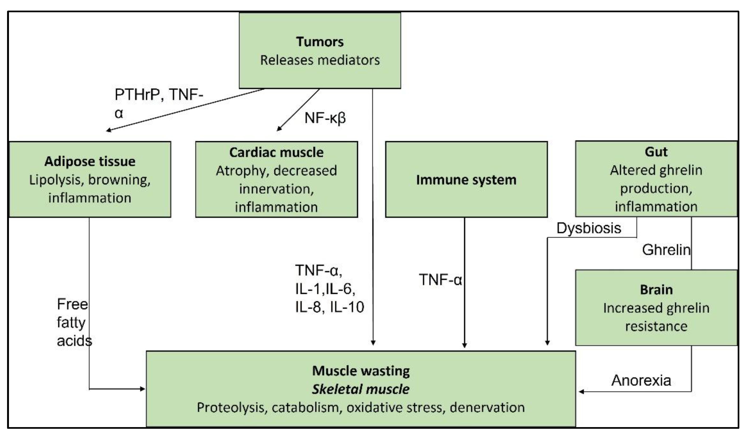
Figure 2.
Causes and outcomes of cancer cachexia [46].
Figure 2.
Causes and outcomes of cancer cachexia [46].
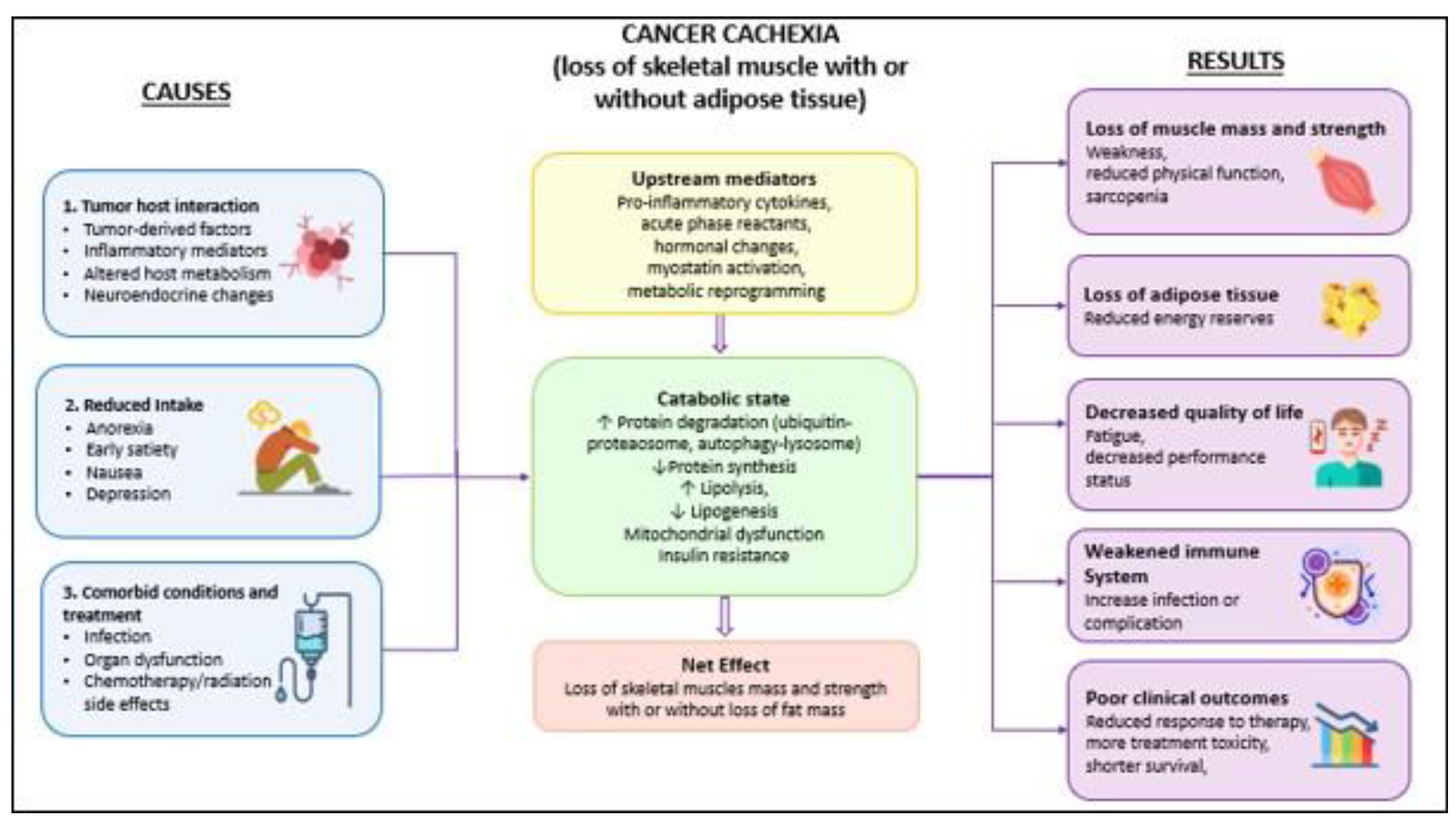

Table 1.
Cytokines and their role in cachexia.
| Cytokine | Source cells | Cancer-affected organs | Experimental model | Effects observed in cachexia | References |
| IL-1α | Macrophages and endothelial cells | Pancreas, breast, colorectal regions | Rodents (mice and rats) | Enhanced lipid breakdown, reduced appetite, body weight decline, decreased hunger signals | [22,23,24] |
| IL-1β | Macrophages | Stomach, breast, lung, and multiple other sites | Mice and humans | Appetite loss, reduction in body mass, sarcopenia, fatigue, disrupted mitochondrial function | [25,26,27] |
| IL6 | Activated macrophages | Colorectal region, pancreas, and additional organs | Mice and humans | Fat tissue depletion, skeletal muscle degradation, impact on gut and liver, mitochondrial dysfunction | [23,24,27] |
| TNFα | Activated macrophages, CD4+ cells, neutrophils, mast cells, eosinophils, and neurons | Pancreas, lungs, colon, and other organs | Mice and humans | Appetite suppression, loss of muscle and fat mass, insulin resistance, elevated energy usage, mitochondrial impairment | [28,29,30] |
| GDF15 | Adipocytes, macrophages, endothelial cells, vascular smooth muscle cells, cardiomyocytes, and trophoblastic cells | Lungs, pancreas, colorectal region, prostate, and others | Mice | Altered energy regulation, appetite suppression, muscle wasting, fat depletion, reduced bone density, anemia | [24,31,32] |
| IFNγ | Activated T cells and NK cells | Pancreas, lung, colon, prostate | Rodents (mice and rats) | Decrease in body weight, lowered appetite, fat tissue atrophy | [24,33,34] |
| LIF | Melanoma cells, Neuroepithelioma cells, Gastric cancer cells, Pancreatic cancer cells, | epididymal fat, spleen, and different muscle types |
Mice | loss of body fat, myotube atrophy | [35,36,37] |
Abbreviations: CD4+ cells: Cluster of Differentiation 4 positive T cells; GDF15: Growth Differentiation Factor 15; IFNγ: Interferon gamma; IL-1α: Interleukin 1 alpha; IL-1β : Interleukin 1 beta; IL-6 : Interleukin 6; LIF: Leukaemia Inhibitory Factor; NK cells: Natural Killer cells; TNFα :Tumour Necrosis Factor alpha.
Table 2.
Overview of pharmacotherapies targeting cancer cachexia pathways.
| Drug | Class | Mechanism of action | References |
| Anamorelin | Ghrelin receptor agonist (orally active) | Stimulates appetite and anabolic signalling via growth hormone pathway; increases appetite, body weight, lean body mass | [59,62,63] |
| Clazakizumab (formerly ALD518) | IL-6 pathway inhibitor / monoclonal antibody | Blocks IL-6 signalling; reported improved lean body mass, reduced fatigue; modest effect on anaemia/fatigue, inconsistent effect on lean mass across trials | [65] |
| Corticosteroids | - | Stimulates appetite and sense of well-being (short-term; limited by muscle weakness, immunosuppression) | [54] |
| Dronabinol | Cannabinoid | Appetite stimulation (modest effect on appetite/weight/muscle mass) | [56] |
| Espindolol | Non-selective β-blocker | Targets molecular/metabolic drivers of cachexia (beyond symptomatic relief) | [61] |
| Etanercept | TNF-α inhibitor | Blocks TNF-α signalling (no substantial improvement in weight/survival) | [64] |
| GDF-15-targeting monoclonal antibodies | Monoclonal antibody | Modulates GDF-15 signalling to affect appetite and metabolic regulation | [60,61] |
| MABp1 | IL-1α-blocking antibody | Blocks IL-1α signalling; some symptom improvement, no survival benefit | [65] |
| Megestrol acetate | Synthetic progesterone derivative | Modulates hypothalamic appetite-regulating pathways; suppresses pro-inflammatory cytokines (weight gain mainly from fat/fluid, not lean mass) | [44,55] |
| Myostatin/activin receptor inhibitors | Anabolic pathway inhibitor | Blocks myostatin/activin signalling to increase muscle mass | [20] |
| R-ketorolac | Next-generation immunomodulator | Restores immune balance (improves T-cell populations, reduces IL-6); reverses cachexia independent of tumour size or food intake | [6] |
| Thalidomide | Anti-inflammatory / immunomodulatory | Inhibits TNF-α production, reduces inflammation | [54] |
| Tocilizumab | Monoclonal antibody | Targets IL-6 receptor, suppressing IL-6-mediated inflammation | [58] |
Abbreviations: GDF-15: Growth Differentiation Factor 15; IL-1α: Interleukin 1 alpha; IL-6: Interleukin 6; MABp1: Monoclonal Antibody p1; TNFα: Tumour Necrosis Factor alpha.
Disclaimer/Publisher’s Note: The statements, opinions and data contained in all publications are solely those of the individual author(s) and contributor(s) and not of MDPI and/or the editor(s). MDPI and/or the editor(s) disclaim responsibility for any injury to people or property resulting from any ideas, methods, instructions or products referred to in the content. |
© 2026 by the authors. Licensee MDPI, Basel, Switzerland. This article is an open access article distributed under the terms and conditions of the Creative Commons Attribution (CC BY) license (http://creativecommons.org/licenses/by/4.0/).
Copyright: This open access article is published under a Creative Commons CC BY 4.0 license, which permit the free download, distribution, and reuse, provided that the author and preprint are cited in any reuse.


