
Submitted:
17 July 2026
Posted:
20 July 2026
You are already at the latest version
Abstract
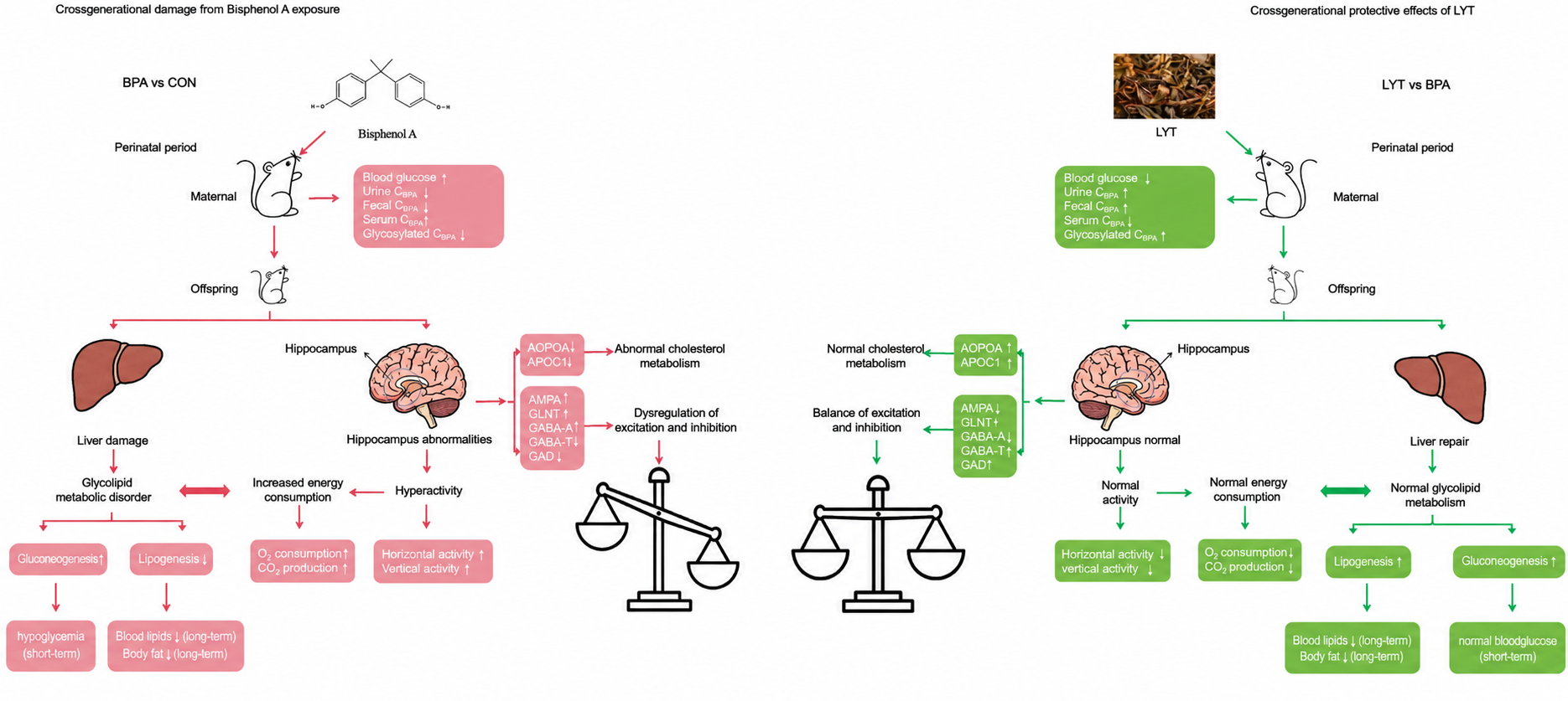
Background: Large yellow tea (LYT), a distinctive variety made from mature leaves, has recently gained attention for its remarkable health benefits. However, whether these benefits can be transmitted from mother to offspring remains unexplored. Purpose: This study investigated whether maternal LYT consumption confers cross-generational protection against metabolic and behavioral disorders induced by perinatal bisphenol A (BPA) exposure. Methods: A mouse model of perinatal BPA exposure (0.03% in diet) was established with or without maternal LYT supplementation (2.5% in diet). Metabolic parameters were assessed through biochemical assays and gene expression analysis (RT-PCR). Energy expenditure and spontaneous activity were monitored using a Comprehensive Lab Animal Monitoring System (CLAMS). Hippocampal proteomic profiling was performed using label-free quantitative proteomics. Results: LYT significantly reduced BPA absorption and enhanced its glucuronidation and excretion. Notably, LYT exhibited bidirectional metabolic regulation, alleviating gestational hyperglycemia in dams while restoring hypoglycemia in offspring, and normalizing underweight and hypolipidemia without disturbing normal physiology. Mechanistically, LYT modulated the SIRT6 (sirtuin 6)/FOXO1 and SIRT6/SREBP1 pathways to enhance gluconeogenesis and lipogenesis. Concurrently, LYT rectified BPA-induced hyperactivity and reduced energy expenditure. Proteomic analysis revealed that LYT partially restores BPA-induced dysregulation of cholesterol metabolism and glutamatergic/GABAergic synaptic pathways, rebalancing excitation-inhibition homeostasis. Conclusions: These findings establish LYT as a promising dietary intervention for counteracting environmental health risks across generations, acting through toxin clearance, bidirectional metabolic regulation, behavioral normalization, and neural excitation-inhibition balance restoration.
Keywords:
large yellow tea
; Bisphenol A
; cross-generational protection
; bidirectional metabolic regulation
; hyperactivity
; excitation-inhibition balance
1. Introduction
Large yellow tea (LYT) is a traditional Chinese tea made from mature leaves (“one bud with three to six leaves”) [1,2]. Its unique yellowing process modifies the chemical profile, contributing to bioactivities distinct from those of green teas. Previous studies from our laboratory have demonstrated that LYT exerts significant anti-hyperglycemic effects in high-fat diet-fed mice [3] and ameliorates metabolic syndrome in db/db mice [4]. However, these investigations were confined to adult animals, leaving a critical gap: whether the health benefits of LYT can be transmitted from mother to offspring.
This question is particularly relevant to the Developmental Origins of Health and Disease (DOHaD) paradigm, which posits that early-life exposures profoundly influence long-term health outcomes [5,6]. The perinatal period represents a window of heightened susceptibility, with toxicant exposure linked to metabolic and neurodevelopmental disorders later in life [7,8].
Bisphenol A (BPA), a ubiquitous endocrine-disrupting chemical used in polycarbonate plastics and epoxy resins, exemplifies such environmental threats [9]. Perinatal BPA exposure is implicated in metabolic disorders—including obesity and diabetes [10,11,12,13]—as well as hyperactivity [14], with divergent phenotypes (hyper/hypoglycemia, obesity/leanness) reported depending on exposure conditions [15]. Human exposure to BPA is virtually unavoidable through contaminated food, water, and air, leading to widespread public health concerns regarding its toxic effects.
Given the widespread exposure to BPA and its documented health impacts, identifying dietary interventions that could mitigate its toxicity represents a significant public health priority. Tea and its constituents, particularly epigallocatechin gallate (EGCG), have shown promise in counteracting BPA-induced toxicity in cellular and animal models [15,16,17]. However, whether whole LYT—with its complex mixture of catechins, theanine, caffeine, and polysaccharides—could confer cross-generational protection against BPA has not been investigated.
Here, we established a mouse model of perinatal BPA exposure and tested whether maternal dietary LYT supplementation confers cross-generational protection against BPA-induced metabolic and behavioral disorders. Our findings reveal that LYT acts as a natural BPA antagonist through multiple mechanisms: enhancing toxin clearance, bidirectionally regulating glucose and lipid metabolism, correcting hyperactivity, and restoring excitation-inhibition balance in the hippocampus.
Large yellow tea (LYT) is a traditional Chinese tea made from mature leaves (“one bud with three to six leaves”) [1,2]. Its unique yellowing process modifies the chemical profile, contributing to bioactivities distinct from those of green teas. Previous studies from our laboratory have demonstrated that LYT exerts significant anti-hyperglycemic effects in high-fat diet-fed mice [3] and ameliorates metabolic syndrome in db/db mice [4]. However, these investigations were confined to adult animals, leaving a critical gap: whether the health benefits of LYT can be transmitted from mother to offspring.
This question is particularly relevant to the Developmental Origins of Health and Disease (DOHaD) paradigm, which posits that early-life exposures profoundly influence long-term health outcomes [5,6]. The perinatal period represents a window of heightened susceptibility, with toxicant exposure linked to metabolic and neurodevelopmental disorders later in life [7,8]. Bisphenol A (BPA), a ubiquitous endocrine-disrupting chemical used in polycarbonate plastics and epoxy resins, exemplifies such environmental threats [9]. Perinatal BPA exposure is implicated in metabolic disorders—including obesity and diabetes [10,11,12,13]—as well as hyperactivity [14], with divergent phenotypes (hyper/hypoglycemia, obesity/leanness) reported depending on exposure conditions [15]. Human exposure to BPA is virtually unavoidable through contaminated food, water, and air, leading to widespread public health concerns regarding its toxic effects. Given the widespread exposure to BPA and its documented health impacts, identifying dietary interventions that could mitigate its toxicity represents a significant public health priority. Tea and its constituents, particularly epigallocatechin gallate (EGCG), have shown promise in counteracting BPA-induced toxicity in cellular and animal models [15,16,17]. However, whether whole LYT—with its complex mixture of catechins, theanine, caffeine, and polysaccharides—could confer cross-generational protection against BPA has not been investigated.
Here, we established a mouse model of perinatal BPA exposure and tested whether maternal dietary LYT supplementation confers cross-generational protection against BPA-induced metabolic and behavioral disorders. Our findings reveal that LYT acts as a natural BPA antagonist through multiple mechanisms: enhancing toxin clearance, bidirectionally regulating glucose and lipid metabolism, correcting hyperactivity, and restoring excitation-inhibition balance in the hippocampus.
2. Materials and Methods
2.1. Preparation and Chemical Analysis of Large Yellow Tea Extract
LYT samples were provided by Bao Er Zhong Xiu Tea Industry Co., Ltd. (Lu’an, China). Water-soluble components were prepared as previously described [4]. Briefly, tea powder (50 g) was boiled in ultra-pure water under stirring at 85 °C for 30 min, followed by ultrasonic extraction at 75 °C for 30 min (100 W average power). The extract was filtered, concentrated, and lyophilized.
Catechin and caffeine contents were analyzed using a Waters Ultra Performance Liquid Chromatography (UPLC) system equipped with a 600 controller and 2489 UV/Visible detector, following our established protocol [18]. Amino acid composition was determined using a HITACHI L-8900 automatic amino acid analyzer. Polysaccharide content was measured by the phenol-sulfuric acid method.
2.2. Animals and Experimental Design
CD-1 mice (female, n=18; male, n=9) were purchased from the National Resource Center of Model Mice (NRCMM, Nanjing, China). Mice were housed under specific pathogen-free (SPF) conditions at the Laboratory Animal Center of Anhui Agricultural University, with constant temperature (22 ± 1 °C), humidity (50 ± 5%), and a 12:12 h light-dark cycle (lights on at 8:00 a.m.). All animals had free access to water and diet.
After one week of acclimatization, female mice were randomly assigned to three dietary groups (n=6 per group):
Control group: modified AIN93 diet with 7% corn oil replacing soybean oil
BPA group: modified AIN93 diet supplemented with 0.03% BPA (≥99.0% purity, MACKLIN, Shanghai, China)
BPA+LYT group: modified AIN93 diet supplemented with 0.03% BPA and 2.5% LYT
Male mice were maintained on the control diet. All diets were prepared by Trophic Animal Feed High-Tech Co., Ltd. (Nantong, China). After five weeks of dietary intervention, females were mated with males (female:male = 2:1). Females continued on their respective diets throughout gestation and lactation. After weaning, offspring were maintained on the control diet. One female and one male pup from each litter were randomly selected for subsequent experiments.
Food and water intake were monitored daily. Body weight and fasting blood glucose (measured from tail vein using Nova StatStrip Xpress Glucose Meter) were recorded weekly. At 24 weeks of age, offspring mice were fasted for 12 h and euthanized by spinal dislocation. Liver tissues were collected for gene expression analysis, with portions preserved in RNAlater or flash-frozen in liquid nitrogen for subsequent assays.
2.3. BPA Quantification
Urine and feces were collected from maternal mice housed in metabolic cages for 48 h. Free BPA in serum, urine, and feces was extracted with acetonitrile and analyzed using a Waters ACQUITY UPLC H-Class System with fluorescence detection (excitation 276 nm, emission 333 nm). Separation was performed on a CORTECS UPLC C18+ column (2.1 × 100 mm, 1.6 μm) at 30 °C with acetonitrile:water (50:50, v/v) mobile phase at 0.4 mL/min. Total BPA (free plus conjugated) was measured following enzymatic hydrolysis with β-glucuronidase (Shanghai Yuanye Bio-Technology Co., Ltd.) in ammonium acetate buffer (pH 5) overnight at 37 °C. Conjugated BPA was calculated as total BPA minus free BPA.
2.4. Serum Biochemical Parameters
Serum was collected by centrifugation at 3000 rpm for 5 min at 4 °C. Triglyceride (TG), total cholesterol (TC), and low-density lipoprotein cholesterol (LDL-C) were measured using commercial kits (Nanjing Jiancheng Bioengineering Institute, Nanjing, China). Glucagon, insulin, glucagon-like peptide-1 (GLP-1), and leptin were quantified by enzyme-linked immunosorbent assay (ELISA) kits (Shanghai Jianglai Biotech, Shanghai, China) according to the manufacturer’s instructions. Hepatic glycogen content was measured as previously reported with minor modifications [19].
2.5. Metabolic and Behavioral Analyses
At 20 weeks of age, offspring mice were evaluated using a Comprehensive Lab Animal Monitoring System (CLAMS; Columbus Instruments, Columbus, OH, USA). Oxygen consumption (VO2), carbon dioxide production (VCO2), energy expenditure, spontaneous activity (ambulatory, total horizontal, and vertical movements), and food intake were monitored continuously for 72 h. Data were collected at 20-min intervals. Respiratory exchange ratio (RER = VCO2/VO2) was calculated for metabolic substrate analysis.
Body composition (fat mass, lean mass, and fluid mass) was assessed by nuclear magnetic resonance (NMR; Minispec LF90II; Bruker Optics, Billerica, MA, USA) in conscious mice following CLAMS measurements.
2.6. Quantitative Real-Time PCR Analysis
Total RNA was extracted from liver tissues using RNA isolator (Vazyme Biotech, Nanjing, China). Reverse transcription was performed with HiScript® II 1st Strand cDNA Synthesis Kit (Vazyme). Real-time PCR was conducted using AceQ qPCR SYBR Green Master Mix (Vazyme) on a Bio-Rad CFX System. Primer sequences are provided in Supplementary Table 1. Relative gene expression was calculated by the 2−ΔΔCt method with β-actin as the internal control.
2.7. Label-Free Proteomic Analysis of Hippocampus
Hippocampal tissues from female offspring mice were subjected to label-free quantitative proteomics. Protein extraction, trypsin digestion, and peptide purification were performed following standard protocols. LC-MS/MS analysis was conducted on an EASY-nLC 1200 system coupled to an Orbitrap Exploris 480 mass spectrometer. Data-independent acquisition (DIA) data were processed using DIA-NN against the Mus musculus UniProt database.
Bioinformatic analyses included Gene Ontology (GO) annotation, Kyoto Encyclopedia of Genes and Genomes (KEGG) pathway enrichment, and protein-protein interaction network construction using the STRING database (confidence score > 0.7). Differentially expressed proteins were identified with fold change > 1.5 and p < 0.05. Functional enrichment was assessed by Fisher’s exact test.
2.8. Statistical Analysis
Data are presented as mean ± SEM. Comparisons between two groups were performed using unpaired two-tailed Student’s t-test. Multiple group comparisons were analyzed by one-way ANOVA followed by Tukey’s post hoc test. Statistical significance was set at p < 0.05.
3. Results
3.1. Chemical Composition of LYT
Quantitative analysis revealed that LYT water extract contains substantial amounts of catechins (10.46 ± 0.48%), caffeine (3.71 ± 0.02%), and polysaccharides (6.27 ± 0.14%) (Table 1). Among catechins, EGCG (4.26 ± 0.27%) and GCG (2.11 ± 0.03%) were the most abundant. Theanine, the signature amino acid of tea, accounted for 0.69 ± 0.01% of the extract. These data provide the chemical basis for subsequent mechanistic investigations.
3.2. LYT Promotes BPA Detoxification via Enhanced Glucuronidation and Excretion
To determine whether LYT influences BPA toxicokinetics, we measured free and conjugated BPA in maternal serum, urine, and feces. Perinatal BPA exposure significantly elevated free BPA levels in all three compartments (Figure 1A–C). LYT supplementation markedly reduced serum free BPA while increasing conjugated (glucuronidated) BPA (Figure 1A), indicating enhanced phase II metabolism. In urine, LYT increased both free and conjugated BPA excretion (Figure 1B), suggesting facilitated elimination. Fecal free BPA was also elevated in the LYT group (Figure 1C), implying reduced intestinal absorption. Collectively, these results demonstrate that LYT acts at multiple levels to reduce BPA body burden: limiting absorption, promoting glucuronidation, and enhancing urinary and fecal excretion.
3.3. LYT Exerts Bidirectional Regulation of Glucose and Lipid Homeostasis Disrupted by Perinatal BPA Exposure
3.3.1. Opposite Effects on Maternal and Offspring Glycemia
Perinatal BPA exposure exerted diametrically opposite effects on glucose homeostasis in mothers and offspring. In maternal mice, BPA induced significant hyperglycemia during gestation, with fasting blood glucose levels (6.48 ± 0.71 mmol/L) exceeding the diagnostic threshold for gestational diabetes (5.1 mmol/L) (Figure 2A). LYT supplementation completely normalized maternal blood glucose (5.08 ± 0.39 mmol/L).
Remarkably, the same BPA exposure that caused maternal hyperglycemia induced profound hypoglycemia in offspring (Figure 2A). F1 female and male mice from the BPA group exhibited significantly lower fasting glucose compared to controls. LYT intervention restored offspring glucose to normal levels, demonstrating that LYT can both lower hyperglycemia and raise hypoglycemia—a unique bidirectional regulatory capacity.
3.3.2. LYT Normalizes Underweight and Hypolipidemia in Offspring
BPA-exposed offspring showed significant growth retardation, with reduced body weight in both sexes (Figure 2B). Body composition analysis revealed decreased fat mass and increased lean mass ratio (Figure 2C), indicating a shift toward leanness. Consistent with reduced adiposity, serum TG, TC, and LDL-C were markedly decreased in BPA-exposed offspring (Figure 2D–F). LYT supplementation fully restored body weight, body composition, and serum lipid profiles to control levels.
Notably, LYT did not affect body weight or serum lipids in control animals (data not shown), indicating that its effects are homeostatic rather than unidirectional—it corrects metabolic deviations in either direction without disturbing normal physiological function. To explore the molecular basis of this bidirectional regulation, we examined mRNA expression of key metabolic genes in offspring liver. BPA exposure significantly downregulated gluconeogenic enzyme PCK1 and its transcriptional regulator FOXO1, while upregulating SIRT6, a negative regulator of FOXO1. In lipogenic pathways, BPA suppressed lipogenic enzyme FASN and its master regulator SREBP1, again with concurrent SIRT6 overexpression (Figure 3).
LYT intervention reversed all BPA-induced gene expression abnormalities, restoring transcriptional levels of key gluconeogenic and lipogenic genes. These results indicate that LYT modulates the SIRT6/FOXO1 and SIRT6/SREBP1 pathways to exert bidirectional metabolic regulation.
3.4. LYT Rectifies Hyperactivity and Reduces Energy Expenditure in Offspring Mice
3.4.1. BPA-Induced Hyperactivity Is Normalized by LYT
Perinatal BPA exposure induced pronounced hyperactivity in offspring. Both female and male BPA-exposed mice showed significantly increased ambulatory, horizontal, and vertical activity during the light cycle (Figure 4). LYT supplementation completely normalized all activity parameters, restoring behavior to control levels.
3.4.2. Hyperactivity Drives Increased Energy Expenditure
Consistent with their hyperactive phenotype, BPA-exposed offspring exhibited elevated oxygen consumption (VO2) and energy expenditure during both light and dark cycles (Figure 5). Carbon dioxide production (VCO2) was similarly increased (Figure 5). No significant differences in respiratory exchange ratio were observed among groups (data not shown), indicating that the increased energy expenditure was not caused by metabolic fuel substrate switching.
LYT supplementation normalized all metabolic parameters, reducing VO2, VCO2, and energy expenditure to control levels. These data suggest that LYT’s metabolic benefits are partially mediated by behavioral normalization: alleviating hyperactivity reduces excessive energy demand, thereby facilitating the recovery of glucose and lipid homeostasis.
3.5. LYT Partially Restores BPA-Induced Dysregulation of Excitation-Inhibition Balance in Hippocampus
Given the prominent hyperactive behavioral phenotype observed in female offspring, we performed label-free quantitative proteomic analysis on hippocampal tissues from female F1 mice to explore the underlying neural molecular mechanism (Figure 6).
Functional annotation of differentially expressed proteins induced by BPA confirmed that the core dysregulated signaling pathways were glutamatergic synapse and GABAergic synapse, which were systematically summarized in the self-organized pathway mechanism diagram (Figure 7).
BPA exposure triggered extensive dysregulation of glutamatergic and GABAergic synaptic signaling pathways. In the glutamatergic synapse pathway, the glutamine transporter GLNT was downregulated, limiting glutamate precursor supply, while postsynaptic AMPA/NMDA receptors and scaffolding proteins (PSD95, GKAP, SHANK) showed compensatory upregulation. Downstream signaling molecules were also dysregulated, with increased PLC and decreased PKC/PLD levels. In the GABAergic synapse pathway, glutamate decarboxylase (GAD), the key enzyme for GABA synthesis, was downregulated, reducing inhibitory neurotransmitter production. Concurrently, postsynaptic GABA-A and GABA-B receptors showed compensatory upregulation, while the GABA-degrading enzyme GABA-T was suppressed. LYT supplementation effectively corrected most abnormal protein expression in both synaptic pathways.
The simultaneous disruption of excitatory glutamatergic and inhibitory GABAergic synaptic systems destabilizes the neural excitation-inhibition (E/I) balance. The net effect of BPA-induced molecular dysregulation favors excitatory neurotransmission, which is the core pathological mechanism of offspring hyperactivity. LYT-mediated partial restoration of synaptic protein expression and E/I homeostasis contributes substantially to the normalization of BPA-induced abnormal behaviors.
4. Discussion
This study provides the first evidence that maternal consumption of large yellow tea confers cross-generational protection against metabolic and behavioral disorders induced by perinatal BPA exposure. Our findings reveal that LYT acts through multiple, interconnected mechanisms: (1) reducing BPA body burden by enhancing detoxification and excretion; (2) bidirectionally regulating glucose and lipid homeostasis via modulation of the SIRT6/FOXO1 and SIRT6/SREBP1 pathways; (3) normalizing hyperactivity and associated excessive energy expenditure; and (4) partially restoring excitation-inhibition balance in the hippocampus. Together, these mechanisms position LYT as a natural, multi-targeted antagonist of BPA toxicity with transgenerational protective effects.
4.1. LYT as a Natural Antagonist of BPA: Enhanced Detoxification as the Upstream Mechanism
The first line of defense against xenobiotic toxicity is efficient detoxification and elimination. Our results demonstrate that LYT reduces BPA body burden through multiple regulatory pathways: inhibiting intestinal BPA absorption (increased fecal free BPA), enhancing hepatic BPA glucuronidation (elevated serum conjugated BPA), and promoting urinary excretion of BPA metabolites (increased urinary total and conjugated BPA). This pattern suggests that LYT may induce UDP-glucuronosyltransferase (UGT) activity, enhancing BPA glucuronidation—the primary detoxification pathway in mammals [20,21,22].
Previous studies have confirmed that tea polyphenols represented by EGCG can modulate the activity of phase II detoxification enzymes [23]. The detoxification effect of LYT observed in this study may be attributed to its single active component or the synergistic effect of multiple ingredients including catechins, theanine, caffeine, and polysaccharides, which requires further verification. Notably, the LYT dosage adopted in this study (2.5% in diet, equivalent to approximately 10 g daily tea consumption in humans [24]) is half the effective dosage used in previous metabolic syndrome intervention models [4], suggesting that moderate LYT intake confers significant protective effects.
4.2. Bidirectional Metabolic Regulation via SIRT6-Centered Mechanisms
The most innovative finding of this study is the unique bidirectional homeostatic regulatory effect of LYT on BPA-disrupted glucose and lipid metabolism. LYT alleviated gestational hyperglycemia in maternal mice and rescued BPA-induced hypoglycemia, underweight and hypolipidemia in offspring, without interfering with normal metabolic physiology of control mice. This homeostatic regulatory characteristic distinguishes LYT from single-target pharmacological drugs, which often exert unidirectional effects and carry potential adverse side effects.
Mechanistically, SIRT6 acts as the core regulatory node of LYT’s bidirectional metabolic effects. As an NAD+-dependent deacetylase primarily localized to chromatin, SIRT6 exerts its metabolic regulatory functions predominantly at the transcriptional level, which is the well-recognized primary working mode of SIRT6 in modulating hepatic glucose and lipid metabolism [38,39,40]. It inhibits hepatic gluconeogenesis by promoting FOXO1 nuclear exclusion, thereby suppressing the transcriptional activation of gluconeogenic genes such as PCK1 [25,26]. In parallel, SIRT6 represses lipogenesis through histone deacetylation-mediated transcriptional inhibition of SREBP1 and its downstream lipogenic target genes includingFASN [27,28]. Multiple studies have verified that SIRT6 governs the expression of SREBP1/2 and FOXO1 signaling cascades mainly via transcriptional repression and chromatin remodeling, rather than post-translational protein modification [38,39]. Consistently, the upstream regulatory role of SIRT6 in metabolic pathways is primarily manifested by altering the transcriptional abundance of downstream core genes, providing a solid theoretical basis for evaluating pathway activity through mRNA expression detection.
In the present study, perinatal BPA exposure triggered abnormal SIRT6 overexpression at the mRNA level, which persistently inhibited FOXO1 and SREBP1 transcriptional signaling, leading to suppressed gluconeogenesis and lipogenesis, and ultimately causing hypoglycemia and growth retardation in offspring. LYT intervention normalized SIRT6 transcriptional levels, relieved the inhibitory effect of SIRT6 on downstream pathways, and restored the dynamic balance of hepatic glucose and lipid metabolism. This SIRT6-dependent transcriptional regulatory mechanism explains the unique homeostatic characteristics of LYT, which repairs metabolic disorders rather than imposes fixed metabolic changes.
It should be acknowledged that the current mechanistic investigation has certain limitations: we only verified the transcriptional expression changes of core regulatory genes via RT-PCR, and did not further confirm the protein expression levels and post-translational modification of SIRT6, FOXO1, and SREBP1 by Western blotting. Nevertheless, existing authoritative studies have fully confirmed that the regulatory effects of SIRT6 on hepatic gluconeogenesis and lipogenesis pathways are mainly achieved through transcriptional-level gene regulation [38,39,40]. Therefore, the detected mRNA expression alterations of key pathway genes in this study can reliably reflect the activation status of the SIRT6-centered metabolic regulatory network. Future studies will supplement protein-level validation and post-translational modification analysis to further refine and consolidate the mechanistic conclusions of this study.
4.3. Behavioral-Metabolic Interconnection: LYT Breaks the Hyperactivity-Energy Depletion Cycle
This study clarifies the critical correlation between BPA-induced behavioral abnormalities and metabolic disorders. Perinatal BPA exposure induces persistent hyperactivity in offspring mice, which significantly increases daily energy consumption. In the absence of compensatory increased food intake, excessive energy expenditure leads to reduced fat accumulation, insufficient energy supply, and subsequent hypoglycemia and lean phenotype, forming a vicious cycle of hyperactivity-energy depletion-metabolic disorder. LYT fundamentally breaks this pathological cycle by normalizing offspring hyperactivity, reducing unnecessary energy consumption, and creating favorable conditions for the recovery of glucose and lipid homeostasis.
This behavioral-metabolic linkage has important clinical translational significance. Clinical studies have confirmed that children with attention-deficit/hyperactivity disorder (ADHD) often present abnormal growth and metabolic parameters [29,30], but the underlying mechanism remains unclear. Our animal model findings provide a mechanistic basis for the comorbidity of hyperbehavior and metabolic disorders, suggesting that targeted intervention on abnormal behaviors can effectively improve metabolic health, and vice versa.
4.4. Restoring Excitation-Inhibition Balance: A Neural Mechanism Underlying Behavioral Improvement
Hippocampal proteomic analysis revealed the neural molecular mechanism of BPA-induced hyperactivity and LYT-mediated behavioral correction. BPA exposure causes consistent adaptive dysregulation in both excitatory glutamatergic and inhibitory GABAergic synaptic systems: reducing the synthesis of core neurotransmitters while inducing compensatory upregulation of postsynaptic receptors. This molecular mismatch disrupts the dynamic balance of neural excitation and inhibition, and the net enhancement of glutamatergic excitatory transmission ultimately drives hyperactive behaviors, consistent with the core pathophysiology of ADHD [31,32]. Clinical studies have also confirmed reduced GABA concentration and abnormal glutamate signaling in ADHD patients [33,34], further verifying the translational value of our findings.
Notably, LYT only partially restored the BPA-disrupted synaptic protein expression profile, but achieved complete normalization of behavioral phenotypes. This partial restoration phenomenon can be explained by multiple factors: first, LYT intervention only lasts throughout gestation and lactation, and the post-weaning protective effect is indirect, showing certain time and dose dependence; second, hippocampus is only one of the key brain regions regulating behavioral activity, and LYT may exert more comprehensive regulatory effects on other ADHD-related brain regions such as the prefrontal cortex; third, partial correction of molecular pathway disorders is sufficient to reverse macroscopic functional and behavioral abnormalities. This also reflects the multi-target collaborative regulatory advantage of natural dietary interventions.
4.5. Potential Mechanisms of Cross-Generational Effects and Future Directions
The persistent protective effect of maternal LYT supplementation on offspring after weaning indicates genuine cross-generational protection rather than direct drug intervention in offspring. The cross-generational protective mechanism of LYT may involve three aspects: first, LYT reduces maternal BPA accumulation and transmission during gestation and lactation, reducing toxic stimulation of fetal and neonatal developing organs during critical developmental windows; second, BPA exposure induces heritable epigenetic modifications including DNA methylation and histone modification [35,36], and LYT may alleviate BPA-induced epigenetic aberrations to maintain long-term metabolic and neural homeostasis; third, maternal metabolic health regulated by LYT improves placental function and milk composition, providing a healthy developmental environment for offspring [37].
Future research will focus on five core directions to further improve the research system: (1) Investigate the persistence of LYT’s protective effects in F2/F3 generations and the underlying epigenetic mechanisms; (2) Clarify the dose-response relationship of LYT intervention and identify its core active protective components; (3) Analyze the molecular mechanism of sex-specific differences in BPA toxicity and LYT protection; (4) Expand brain region research to explore the regulatory effect of LYT on prefrontal cortical neural function; (5) Verify whether LYT exerts protective effects against common BPA substitutes (BPS, BPF).
5. Conclusion
This study demonstrates that maternal LYT consumption confers cross-generational protection against BPA-induced metabolic and behavioral disorders through enhanced toxin clearance, bidirectional SIRT6-mediated glucose and lipid metabolic regulation, hyperactivity correction, and restoration of hippocampal excitation-inhibition balance. These findings establish LYT as a promising dietary strategy for mitigating environmental health risks across generations, providing new insights and theoretical basis for the development of natural functional foods against environmental toxicant damage.
Supplementary Materials
The following supporting information can be downloaded at the website of this paper posted on Preprints.org, Figure S1: Biochemical index of maternal mice; Figure S2. Biochemical index of offspring mice; Figure S3. The mRNA expression of genes involved in lipolysis and gluconeogenesis in the liver F1 mice. Table S1. Primer sequences used for RT-PCR gene expression experiment.
Author Contributions
Zhongwen Xie designed the experiments, analyzed the results, and drafted the manuscript. Erkang Jiang designed the methods, performed the experiments, analyzed the results, and drafted the manuscript. Hongyu Wang performed the experiments, analyzed the results. Meiyun Li performed the experiments, analyzed the results. Xi Wang analyzed the results and drafted the manuscript of proteomics. Guohuo Wu analyzed main chemical compounds of Large Yellow Tea. Shoujun Huang gave advice in writing the manuscript. Huijun Cheng performed the experiments. Zhuang Li provided help in the experiment of mice. All authors read and approved the final manuscript.
Funding
This work was supported by a Key Project of Natural Science Foundation from Department of Education, Anhui Provincial (Grant number 03087060 to ZX); a Key Natural Science Foundation from Department of Education, Anhui Provincial (Grant Number 2023AH051029 to EJ). Funding for open access charge: Key Project of Natural Science Foundation of the Anhui Provincial Education Department.
Institutional Review Board Statement
All animal procedures were approved by the Institutional Animal Care and Use Committee of Anhui Agricultural University (Approval No. AHAU2020027).
Informed Consent Statement
Not applicable.
Data Availability Statement
Data Availability Statements are available in section “MDPI Research Data Policies” at https://www.mdpi.com/ethics.
Acknowledgments
All authors appreciate the editors and reviewers to improve the manuscript.
Conflicts of Interest
The authors declare no conflicts of interest.
Abbreviations
The following abbreviations are used in this manuscript:
| ADHD | Attention-deficit/hyperactivity disorder |
| AMPA | α-Amino-3-hydroxy-5-methyl-4-isoxazolepropionic acid receptor |
| BPA | Bisphenol A |
| C | (+)-Catechin |
| DOHaD | Developmental origins of health and disease |
| EC | (−)-Epicatechin |
| EDC | Endocrine-disrupting chemical |
| EGCG | (−)-Epigallocatechin gallate |
| FASN | Fatty acid synthase |
| FOX01 | Forkhead box protein O1 |
| GABA | γ-Aminobutyric acid |
| GABA-T | GABA transaminase |
| GAD | Glutamate decarboxylase |
| GC | (+)-Gallocatechin |
| GCG | (−)-Gallocatechin gallate |
| GLP-1 | Glucagon-like peptide-1 |
| GLNT | Glutamine transporter |
| LDL-C | Low-density lipoprotein cholesterol |
| LYT | Large yellow tea |
| NMDAR | N-Methyl-D-aspartate receptor |
| PCK1 | Phosphoenolpyruvate carboxykinase 1 |
| SIRT6 | Sirtuin 6 |
| SREBP-1 | Sterol regulatory element-binding protein 1 |
| TC | Total cholesterol |
| TG | Triglyceride |
References
- Guo, X.; Ho, C.T.; Schwab, W.; Song, C.; Wan, X. Aroma compositions of large-leaf yellow tea and potential effect of theanine on volatile formation in tea. Food Chem. 2019, 280, 73–82. [Google Scholar] [CrossRef] [PubMed]
- Guo, X.; Ho, C.T.; Schwab, W.; Wan, X. Effect of the roasting degree on flavor quality of large-leaf yellow tea. Food Chem. 2021, 347, 129016. [Google Scholar] [CrossRef] [PubMed]
- Zhou, J.; Zhang, L.; Meng, Q.; et al. Roasting improves the hypoglycemic effects of a large-leaf yellow tea infusion by enhancing the levels of epimerized catechins that inhibit α-glucosidase. Food Funct. 2018, 9(10), 5162–5168. [Google Scholar] [CrossRef] [PubMed]
- Teng, Y.; Li, D.; Guruvaiah, P.; Xu, N.; Xie, Z. Dietary Supplement of Large Yellow Tea Ameliorates Metabolic Syndrome and Attenuates Hepatic Steatosis in db/db Mice. Nutrients 2018, 10(1), 75. [Google Scholar] [CrossRef] [PubMed]
- Carpinello, O.J.; DeCherney, A.H.; Hill, M.J. Developmental Origins of Health and Disease: The History of the Barker Hypothesis and Assisted Reproductive Technology. Semin Reprod. Med. 2018, 36(3-04), 177–182. [Google Scholar] [CrossRef] [PubMed]
- Morley, R.; Dwyer, T. Fetal origins of adult disease? Clin. Exp. Pharmacol. Physiol. 2001, 28(11), 962–966. [Google Scholar] [CrossRef] [PubMed]
- Nobile, S.; Di Sipio Morgia, C.; Vento, G. Perinatal Origins of Adult Disease and Opportunities for Health Promotion: A Narrative Review. J. Pers. Med. 2022, 12(2), 157. [Google Scholar] [CrossRef] [PubMed]
- Hoffman, D.J.; Powell, T.L.; Barrett, E.S.; Hardy, D.B. Developmental origins of metabolic diseases. Physiol. Rev. 2021, 101(3), 739–795. [Google Scholar] [CrossRef] [PubMed]
- Ma, Y.; Liu, H.; Wu, J.; et al. The adverse health effects of bisphenol A and related toxicity mechanisms. Env. Res. 2019, 176, 108575. [Google Scholar] [CrossRef]
- Ma, Y.; Liu, H.; Wu, J.; et al. Bisphenol A and the risk of obesity a systematic review with meta-analysis of the epidemiological evidence. Dose Response 2020, 18(2), 1559325820916949. [Google Scholar] [CrossRef]
- Xia, B.T.; He, Y.; Guo, Y.; et al. Multi- and transgenerational biochemical effects of low-dose exposure to bisphenol A and 4-nonylphenol on testicular interstitial (Leydig) cells. Env. Toxicol. 2022, 37(5), 1032–1046. [Google Scholar] [CrossRef]
- Naomi, R.; Yazid, M.D.; Bahari, H.; et al. Bisphenol A (BPA) Leading to Obesity and Cardiovascular Complications: A Compilation of Current In Vivo Study. Int. J. Mol. Sci. 2022, 23(6), 2969. [Google Scholar] [CrossRef] [PubMed]
- Anderson, O.S.; Peterson, K.E.; Sanchez, B.N.; Zhang, Z.; Mancuso, P.; Dolinoy, D.C. Perinatal bisphenol A exposure promotes hyperactivity, lean body composition, and hormonal responses across the murine life course. FASEB J. 2013, 27(4), 1784–1792. [Google Scholar] [CrossRef] [PubMed]
- Ji, H.; Song, N.; Ren, J.; et al. Metabonomics reveals bisphenol A affects fatty acid and glucose metabolism through activation of LXR in the liver of male mice. Sci. Total Environ. 2020, 703, 134681. [Google Scholar] [CrossRef] [PubMed]
- Wang, C.; He, J.; Xu, T.; et al. Bisphenol A(BPA), BPS and BPB-induced oxidative stress and apoptosis mediated by mitochondria in human neuroblastoma cell lines. Ecotoxicol. Env. Saf. 2021, 207, 111299. [Google Scholar] [CrossRef]
- Mohsenzadeh, M.S.; Razavi, B.M.; Imenshahidi, M.; et al. Evaluation of green tea extract and epigallocatechin gallate effects on bisphenol A-induced vascular toxicity in isolated rat aorta and cytotoxicity in human umbilical vein endothelial cells. Phytother Res. 2021, 35(2), 996–1009. [Google Scholar] [PubMed]
- Mohsenzadeh, M.S.; Razavi, B.M.; Imenshahidi, M.; et al. Potential role of green tea extract and epigallocatechin gallate in preventing bisphenol A-induced metabolic disorders in rats: Biochemical and molecular evidence. Phytomedicine 2021, 92, 153754. [Google Scholar] [CrossRef] [PubMed]
- Wu, G.; Sun, X.; Cheng, H.; Xu, S.; Li, D.; Xie, Z. Large Yellow Tea Extract Ameliorates Metabolic Syndrome by Suppressing Lipogenesis through SIRT6/SREBP1 Pathway and Modulating Microbiota in Leptin Receptor Knockout Rats. Foods 2022, 11(11), 1638. [Google Scholar] [CrossRef] [PubMed]
- Seifter, S.; Dayton, S.; Novic, B.; Muntwyler, E. The estimation of glycogen with the anthrone reagent. Arch. Biochem. 1950, 25(1), 191–200. [Google Scholar] [PubMed]
- Zalko, D.; Soto, A.M.; Dolo, L.; et al. Biotransformations of bisphenol A in a mammalian model: answers and new questions raised by low-dose metabolic fate studies in pregnant CD1 mice. Env. Health Perspect. 2003, 111(3), 309–319. [Google Scholar] [CrossRef]
- Arakawa, C.; Fujimaki, K.; Yoshinaga, J. Daily urinary excretion of bisphenol A. Env. Health Prev. Med. 2004, 9(1), 22–26. [Google Scholar] [CrossRef]
- Lombó, M.; González-Rojo, S.; Fernández-Díez, C.; Herráez, M.P. Cardiogenesis impairment promoted by bisphenol A exposure is successfully counteracted by epigallocatechin gallate. Env. Pollut. 2019, 246, 1008–1019. [Google Scholar] [CrossRef]
- Han, M.; Zhao, G.; Wang, Y.; et al. Safety and anti-hyperglycemic efficacy of various tea types in mice. Sci. Rep. 2016, 6, 31703. [Google Scholar] [CrossRef] [PubMed]
- Bian, C.; Zhang, R.; Wang, Y.; et al. Sirtuin 6 affects glucose reabsorption and gluconeogenesis in type 1 diabetes via FoxO1. Mol. Cell Endocrinol. 2022, 547, 111597. [Google Scholar] [CrossRef] [PubMed]
- Han, Y.; Liang, C.; Yu, Y.; Zhang, J.; Wang, J.; Cao, J. Gluconeogenesis Alteration and p53-SIRT6-Fox01 Signaling Adaptive Regulation in Sheep from Different Grazing Periods. Comput Math. Methods Med. 2022, 2022, 4614665. [Google Scholar] [CrossRef] [PubMed]
- Yu, S.; Meng, S.; Xiang, M.; Ma, H. Phosphoenolpyruvate carboxykinase in cell metabolism: Roles and mechanisms beyond gluconeogenesis. Mol. Metab. 2021, 53, 101257. [Google Scholar] [CrossRef] [PubMed]
- Zhu, C.; Huang, M.; Kim, H.G.; et al. SIRT6 controls hepatic lipogenesis by suppressing LXR, ChREBP, and SREBP1. Biochim Biophys. Acta Mol. Basis Dis. 2021, 1867(12), 166249. [Google Scholar] [CrossRef] [PubMed]
- Sanders, F.W.; Griffin, J.L. De novo lipogenesis in the liver in health and disease: more than just a shunting yard for glucose. Biol. Rev. Camb. Philos. Soc. 2016, 91(2), 452–468. [Google Scholar] [PubMed]
- Carrey, N.J.; MacMaster, F.P.; Gaudet, L.; Schmidt, M.H. Striatal creatine and glutamate/glutamine in attention-deficit/hyperactivity disorder. J. Child Adolesc. Psychopharmacol. 2007, 17(1), 11–17. [Google Scholar] [CrossRef] [PubMed]
- Zheng, J.; Chen, Y.H. Research advances in pathogenesis of attention deficit hyperactivity disorder. Chin. J. Contemp. Pediatr. 2018, 20(9), 775–780. [Google Scholar]
- Kennedy, M.B. The Protein Biochemistry of the Postsynaptic Density in Glutamatergic Synapses Mediates Learning in Neural Networks. Biochemistry 2018, 57(27), 4005–4009. [Google Scholar] [CrossRef] [PubMed]
- Edden, R.A.; Crocetti, D.; Zhu, H.; Gilbert, D.L.; Mostofsky, S.H. Reduced GABA concentration in attention-deficit/hyperactivity disorder. Arch. Gen. Psychiatry 2012, 69(7), 750–753. [Google Scholar] [CrossRef] [PubMed]
- Bai, W.J.; Luo, X.G.; Jin, B.H.; et al. Deficiency of transmembrane AMPA receptor regulatory protein γ-8 leads to attention-deficit hyperactivity disorder-like behavior in mice. Zool. Res. 2022, 43(5), 851–870. [Google Scholar] [CrossRef] [PubMed]
- Cimmino, I.; Fiory, F.; Perruolo, G.; et al. Potential Mechanisms of Bisphenol A (BPA) Contributing to Human Disease. Int. J. Mol. Sci. 2020, 21(16), 5761. [Google Scholar] [CrossRef] [PubMed]
- Alonso-Magdalena, P.; Rivera, F.J.; Guerrero-Bosagna, C. Bisphenol-A and metabolic diseases: epigenetic, developmental and transgenerational basis. Env. Epigenet. 2016, 2(3), dvw022. [Google Scholar] [CrossRef]
- Rahman, M.S.; Pang, W.K.; Ryu, D.Y.; Park, Y.J.; Pang, M.G. Multigenerational and transgenerational impact of paternal bisphenol A exposure on male fertility in a mouse model. Hum. Reprod. 2020, 35(8), 1740–1748. [Google Scholar] [CrossRef] [PubMed]
- Khadrawy, Y.A.; Noor, N.A.; Mourad, I.M.; Ezz, H.S. Neurochemical impact of bisphenol A in the hippocampus and cortex of adult male albino rats. Toxicol. Ind. Health 2016, 32(9), 1711–1719. [Google Scholar] [CrossRef] [PubMed]
- Elhanati, S.; Kanfi, Y.; Varvak, A.; et al. Multiple Regulatory Layers of SREBP1/2 by SIRT6. Cell Rep. 2013, 4(3), 678–688. [Google Scholar] [CrossRef]
- Tao, R.; Xiong, X.; DePinho, R.A.; Deng, C.X.; Dong, X.C. Hepatic SREBP-2 and cholesterol biosynthesis are regulated by FoxO3 and Sirt6. J. Lipid Res. 2013, 54(10), 2745–2753. [Google Scholar] [CrossRef] [PubMed]
- Masri, S.; Rigor, P.; Cervantes, M.; et al. Partitioning Circadian Transcription by SIRT6 Leads to Segregated Control of Cellular Metabolism. Cell. 2014, 159(5), 1104–1117. [Google Scholar]
Figure 1.
LYT promotes BPA detoxification and excretion. (A) Serum free and conjugated BPA concentrations. (B) Urinary free and conjugated BPA excretion. (C) Fecal free BPA content. Data are mean ± SEM. *p < 0.05, **p < 0.01 vs. Control; #p < 0.05, ##p < 0.01 vs. BPA group.
Figure 1.
LYT promotes BPA detoxification and excretion. (A) Serum free and conjugated BPA concentrations. (B) Urinary free and conjugated BPA excretion. (C) Fecal free BPA content. Data are mean ± SEM. *p < 0.05, **p < 0.01 vs. Control; #p < 0.05, ##p < 0.01 vs. BPA group.
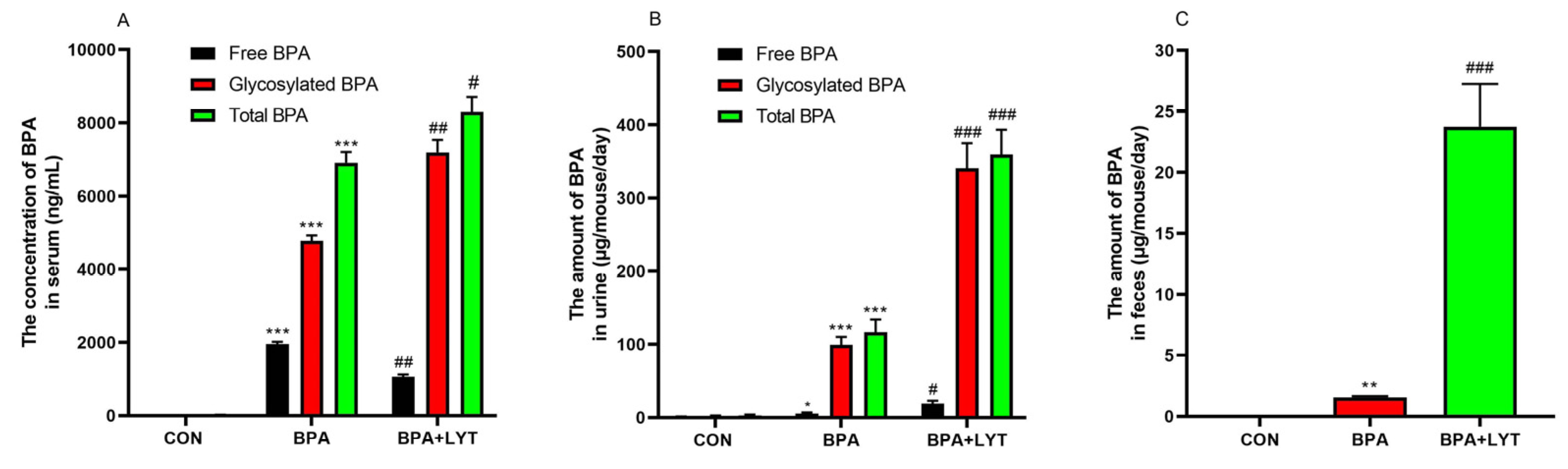
Figure 2.
LYT bidirectionally regulates glucose and lipid homeostasis. (A) Fasting blood glucose in maternal mice (during gestation) and offspring (F1, 12 weeks). (B) Body weight of F1 mice at 24 weeks. (C) Body composition (fat mass and lean mass ratio) in F1 mice. (D–F) Serum TG, TC, and LDL-C levels in F1 mice. Data are mean ± SEM. *p < 0.05, **p < 0.01 vs. Control; #p < 0.05, ##p < 0.01 vs. BPA group.
Figure 2.
LYT bidirectionally regulates glucose and lipid homeostasis. (A) Fasting blood glucose in maternal mice (during gestation) and offspring (F1, 12 weeks). (B) Body weight of F1 mice at 24 weeks. (C) Body composition (fat mass and lean mass ratio) in F1 mice. (D–F) Serum TG, TC, and LDL-C levels in F1 mice. Data are mean ± SEM. *p < 0.05, **p < 0.01 vs. Control; #p < 0.05, ##p < 0.01 vs. BPA group.
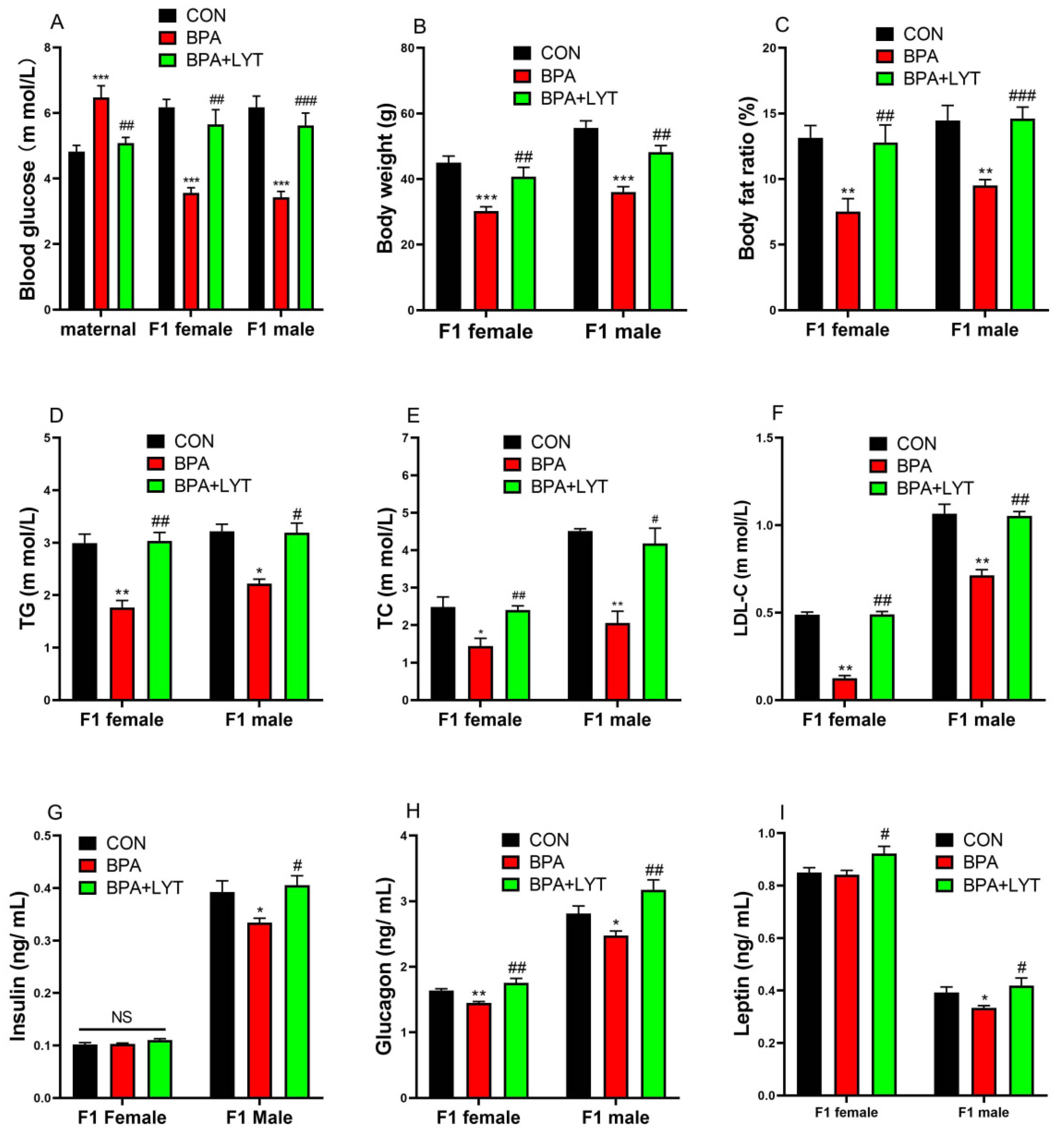
Figure 3.
LYT modulates hepatic gene expression. mRNA expression of (A) gluconeogenic genes (PCK1, FOXO1), (B) lipogenic genes (FASN, SREBP1), and (C) SIRT6 in liver tissues of F1 mice. Data are mean ± SEM. *p < 0.05, **p < 0.01 vs. Control; #p < 0.05, ##p < 0.01 vs. BPA group.
Figure 3.
LYT modulates hepatic gene expression. mRNA expression of (A) gluconeogenic genes (PCK1, FOXO1), (B) lipogenic genes (FASN, SREBP1), and (C) SIRT6 in liver tissues of F1 mice. Data are mean ± SEM. *p < 0.05, **p < 0.01 vs. Control; #p < 0.05, ##p < 0.01 vs. BPA group.

Figure 4.
LYT rectifies BPA-induced hyperactivity. Spontaneous activity in F1 female (A,B) and male (C,D) mice measured by CLAMS. (A,C) Ambulatory and total horizontal activity. (B,D) Vertical activity. Data are mean ± SEM. *p < 0.05, **p < 0.01 vs. Control; #p < 0.05, ##p < 0.01 vs. BPA group.
Figure 4.
LYT rectifies BPA-induced hyperactivity. Spontaneous activity in F1 female (A,B) and male (C,D) mice measured by CLAMS. (A,C) Ambulatory and total horizontal activity. (B,D) Vertical activity. Data are mean ± SEM. *p < 0.05, **p < 0.01 vs. Control; #p < 0.05, ##p < 0.01 vs. BPA group.
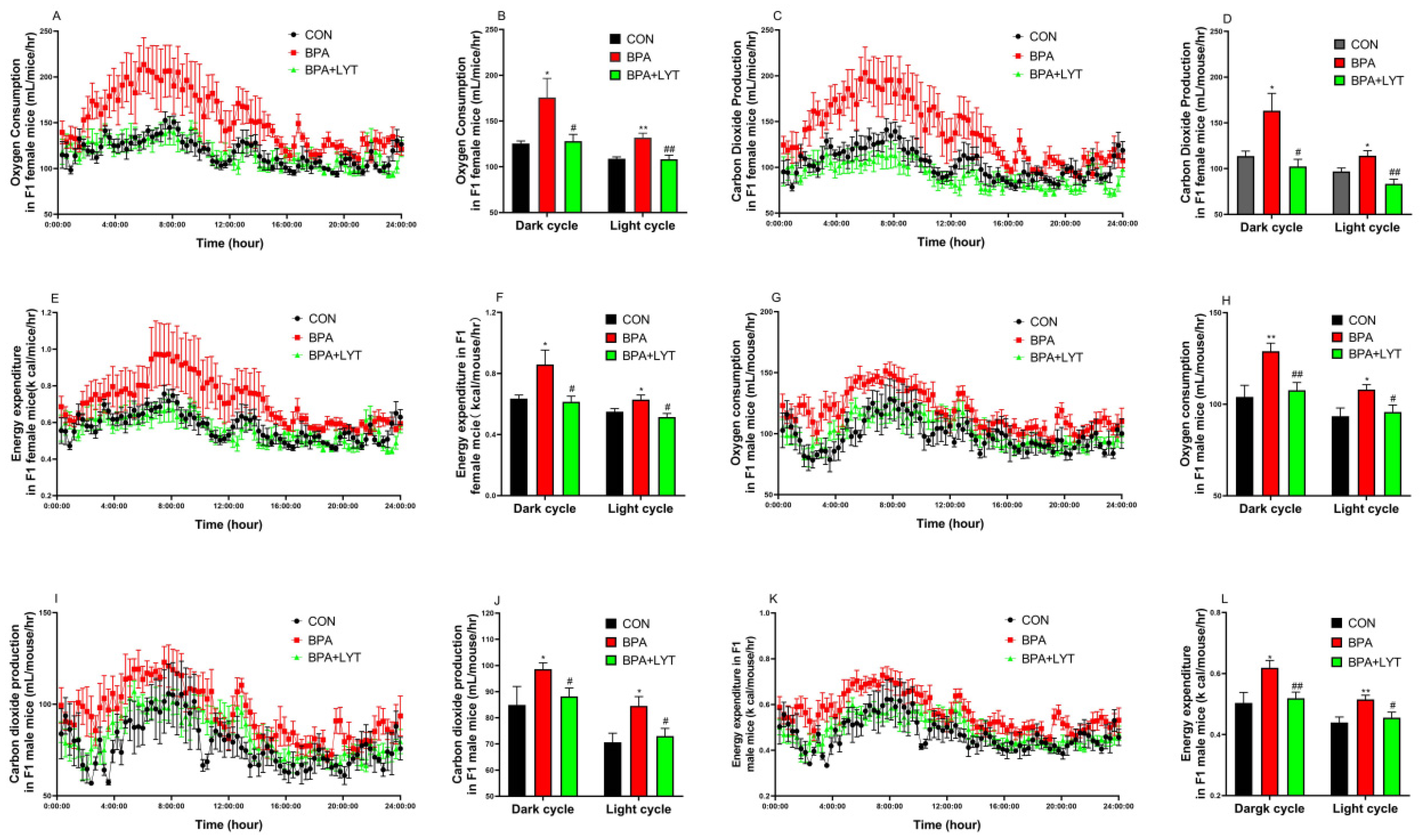
Figure 5.
LYT normalizes energy expenditure. (A) Oxygen consumption (VO2), (B) energy expenditure, and (C) carbon dioxide production (VCO2) in F1 mice measured by CLAMS. Data are mean ± SEM. *p < 0.05, **p < 0.01 vs. Control; #p < 0.05, ##p < 0.01 vs. BPA group.
Figure 5.
LYT normalizes energy expenditure. (A) Oxygen consumption (VO2), (B) energy expenditure, and (C) carbon dioxide production (VCO2) in F1 mice measured by CLAMS. Data are mean ± SEM. *p < 0.05, **p < 0.01 vs. Control; #p < 0.05, ##p < 0.01 vs. BPA group.
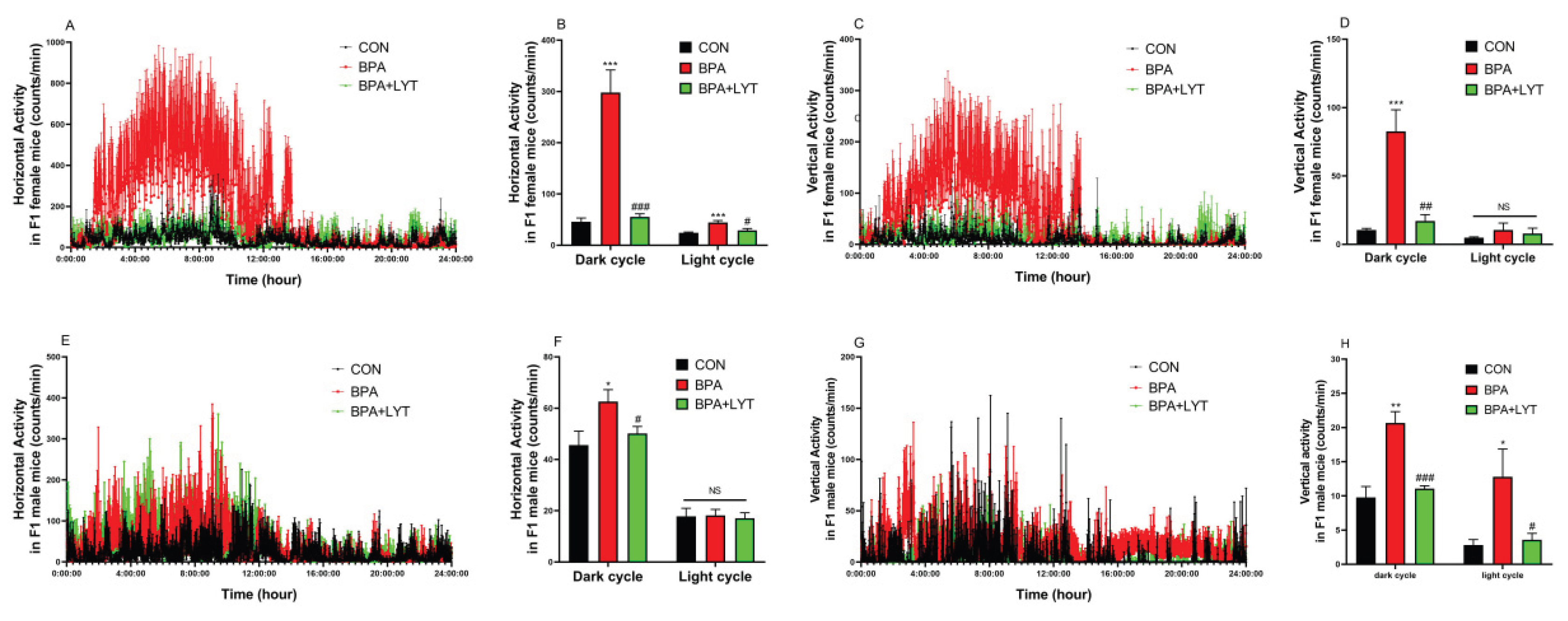
Figure 6.
Sample correlation analysis and differential protein expression analysis. The three groups in the figure are C (control group), P (BPA-induced group), and T (LYT intervention group). A. Principal component analysis (PCA) plot; B. Venn diagram of differential proteins; C. Statistical bar chart of differential protein numbers; D. Volcano plot of differential proteins in the C/P group; E. Volcano plot of differential proteins in the T/P group; F. Heatmap of differential protein expression.
Figure 6.
Sample correlation analysis and differential protein expression analysis. The three groups in the figure are C (control group), P (BPA-induced group), and T (LYT intervention group). A. Principal component analysis (PCA) plot; B. Venn diagram of differential proteins; C. Statistical bar chart of differential protein numbers; D. Volcano plot of differential proteins in the C/P group; E. Volcano plot of differential proteins in the T/P group; F. Heatmap of differential protein expression.
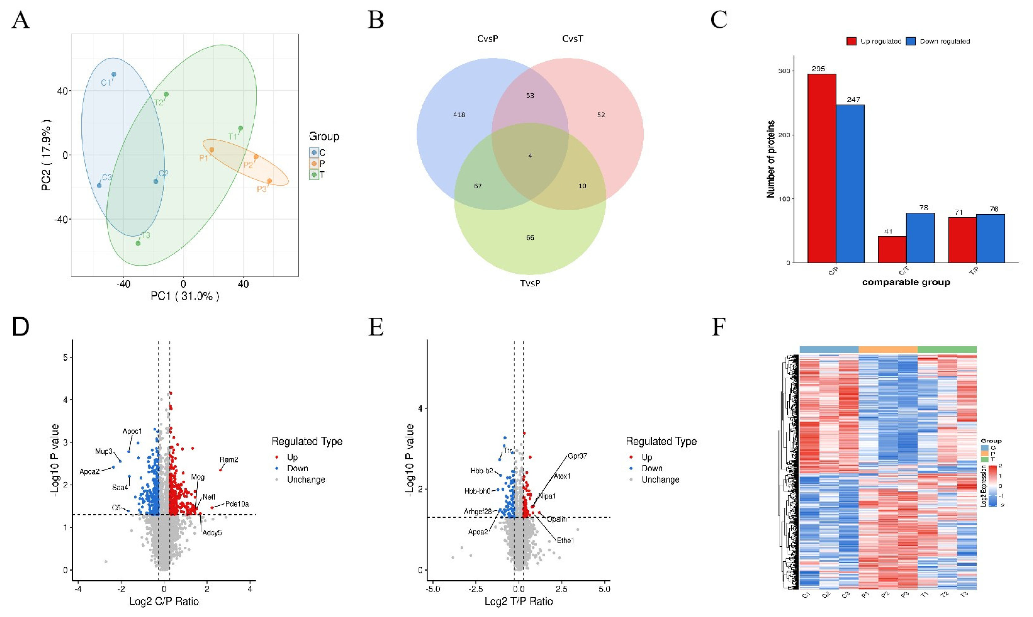
Figure 7.
Potential mechanism underlying BPA-induced hyperactive behavior and the intervention effect of yellow large-leaf tea. P↑/P↓ indicates upregulated/downregulated protein expression after BPA treatment; T↑/T↓ indicates upregulated/downregulated protein expression after yellow large-leaf tea intervention. Solid arrows indicate statistically significant differences, while dashed arrows indicate a recovery trend without statistical significance.
Figure 7.
Potential mechanism underlying BPA-induced hyperactive behavior and the intervention effect of yellow large-leaf tea. P↑/P↓ indicates upregulated/downregulated protein expression after BPA treatment; T↑/T↓ indicates upregulated/downregulated protein expression after yellow large-leaf tea intervention. Solid arrows indicate statistically significant differences, while dashed arrows indicate a recovery trend without statistical significance.
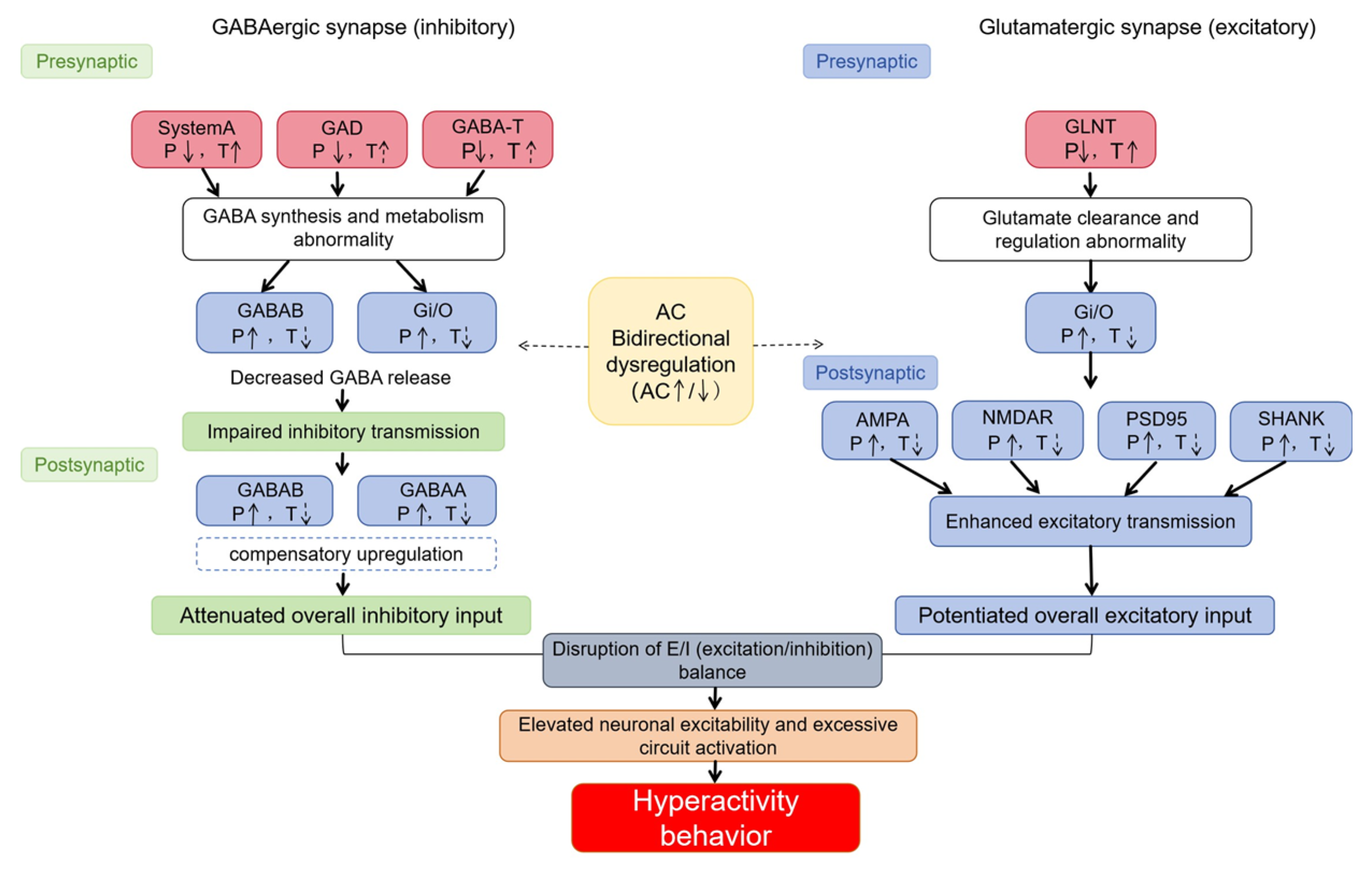

Table 1.
The contents of catechins, caffeine, amino acid and polysaccharide in Water-soluble component of LYT by UPLC analysis.
Table 1.
The contents of catechins, caffeine, amino acid and polysaccharide in Water-soluble component of LYT by UPLC analysis.
| Compound | LYT (%) |
| EGCG | 4.26 ± 0.27 |
| GCG | 2.11 ± 0.03 |
| EC | 1.68 ± 0.19 |
| GC | 1.38 ± 0.02 |
| C | 0.92 ± 0.01 |
| Total catechins | 10.46 ± 0.48 |
| Theanine | 0.69 ± 0.01 |
| Total amino acid | 1.68 ± 0.01 |
| Caffeine | 3.71 ± 0.02 |
| Polysaccharide | 6.27 ± 0.14 |
Abbreviations: (−)-epigallocatechin gallate (EGCG), (−)-gallocatechin gallate (GCG), (−)-epicatechin (EC), (+)-gallocatechin (GC), (+)-catechin (C). Total catechins are sum amount of EGCG, EC, GCG, GC, and C; total amino acids are amount of theanine plus other free amino acids. LYT, large yellow tea.
Disclaimer/Publisher’s Note: The statements, opinions and data contained in all publications are solely those of the individual author(s) and contributor(s) and not of MDPI and/or the editor(s). MDPI and/or the editor(s) disclaim responsibility for any injury to people or property resulting from any ideas, methods, instructions or products referred to in the content. |
© 2026 by the authors. Licensee MDPI, Basel, Switzerland. This article is an open access article distributed under the terms and conditions of the Creative Commons Attribution (CC BY) license (http://creativecommons.org/licenses/by/4.0/).
Copyright: This open access article is published under a Creative Commons CC BY 4.0 license, which permit the free download, distribution, and reuse, provided that the author and preprint are cited in any reuse.


