

Submitted:
16 July 2026
Posted:
20 July 2026
You are already at the latest version
Abstract
Antimicrobial resistance (AMR) is a growing worldwide challenge, treatment options for bacterial infections are increasingly limited. Non-typhoidal Salmonella (NTS), particularly Salmonella enterica serovar Typhimurium, poses a serious threat to public health due to rising antimicrobial resistance. It can cause invasive bloodstream infections that have a high death rate. This study aimed to identify and isolate exclusively lytic bacteriophages that target multidrug-resistant Salmonella Typhimurium (ATCC 13311) from river water and poultry farm environments in Addis Ababa, Ethiopia and assessed their potential as an alternative treatment. A total of 38 environmental samples were collected from poultry farms and rivers. From these samples, 22 different bacteriophages were successfully isolated using S. Typhimurium ATCC 13311 as the host strain. The majority (63.6%) were isolated from river water samples. Spot assays were used to isolate phages, and the double-layer agar method (DLA) was used for purification. Characterization of phages of phages performed by PCR-based identification and classification, latent period determination, plating efficiency, burst size, stability testing, and assessment of in vitro bactericidal activity. Host range analysis showed that all isolates were active against the primary host, and four isolates showed cross-genus efficacy against specific pathotypes of Escherichia coli. The latent periods of the isolated phages ranged from 10 to 20 minutes, indicating a wide range of infection kinetics. Some of the most productive isolates attained Peak titers as high as 10¹³ PFU/mL, indicating significant replication capacity. Additionally, the ideal multiplicity of infection differed significantly amongst isolates, ranging from 0.001 to 100. Regarding environmental stability, all phages were active in a pH range of 5 to 9 and withstood temperatures ranging from 4°C to 37°C. Molecular analysis classified most isolates into the T4-like and T7-like lineages, two distinct phage groups with known lytic characteristics. This study demonstrates that locally isolated lytic bacteriophages can effectively inhibit multidrug-resistant Salmonella Typhimurium ATCC 13311. The findings support their potential as safe and effective biocontrol agents for combating MDR S. Typhimurium infection. However, clinical trials, in vivo research, and genome sequencing are necessary to confirm these phages' effectiveness and safety.
Keywords:
S. Typhimurium
; AMR
; MDR
; phage isolation
; lytic phages
; phage therapy
; T4-like and T7-like
1. Introduction
AMR has become one of the most urgent global health crises of the 21st century. This emergency is caused by the rapid spread of resistant bacterial pathogens alongside declining development of new antibiotics [1,2]. Salmonella enterica is one of multidrug-resistant (MDR) bacterial pathogens which causes bacterial infections with high morbidity and mortality due to the ineffectiveness of commonly used antibiotics. Specifically, non-typhoidal Salmonella (NTS) serovars, such as S. Typhimurium, cause significant public health concerns. These strains often infiltrate normally sterile locations to cause severe invasive non-typhoidal salmonellosis (iNTS) bloodstream infections, which carry high case fatality rates among vulnerable populations, despite the fact that NTS ordinarily causes self-limiting gastroenteritis worldwide. The world Health Organization (WHO) has been designated fluoroquinolone-resistant NTS as a high-priority pathogen, underscoring the pressing need for investigation and the creation of substitute antimicrobial approaches [3,4,5,6,7].
Overuse and misuse of antibiotics in clinical settings, agriculture, veterinary medicine, and food production systems are the main contributors for the emergence and spread of AMR S. enterica strains [8]. In poultry farming, farmers use antibiotics for growth promotion besides its therapeutic use which expose the environment for intense selection of MDR strains. This burden is especially high in sub-Saharan Africa, including Ethiopia, where invasive non-typhoidal salmonellosis (iNTS), mainly caused by S. Typhimurium leads to high morbidity and mortality rates. This is mainly due to the declining effectiveness of available and affordable antimicrobial therapies [9,10,11,12]. In Ethiopia, the rising prevalence of AMR S. enterica presents a significant challenge for effective infection management. The emergence of these resistant strains is largely because of agricultural practices, particularly in poultry farming, where intensive antibiotic promotes the selection of MDR phenotypes. Consequently, these resistant pathogens contaminate the surrounding environment, including adjacent river networks and creating complex eco-epidemiological reservoirs. Given the increasing threat of AMR and the reduced effectiveness of the traditional antibiotic pipeline, there is growing interest in alternative therapeutic approaches for controlling bacterial infections [13,14,15].
Bacteriophages are naturally occurring viruses that specifically infect and lyse bacterial cells. Their high host specificity, ability to self-replicate at the site of infection, co-evolution with bacterial hosts, and widespread presence in the environment make them better candidates for alternative antimicrobial applications. Studies on isolating lytic phages from diverse environmental sources in developing countries remain critically lacking. Poultry farm environments and their connected aquatic systems, which are recognized as important sites for Salmonella transmission, have not been adequately explored as potential reservoirs of phages that target virulent multidrug-resistant S. enterica strains [10,16,17]. This shortage of locally sourced and well-characterised viral evidence limits the development of context-specific phage-based interventions to address the regional AMR crisis. Therefore, this study aimed to address this gap by isolating and characterizing lytic bacteriophages from poultry farms (including chicken feces, farm soil, and sewage) and river water in Addis Ababa, Ethiopia. Using the well-characterized reference strain S. Typhimurium ATCC 13311 as the primary bacterial host.
2. Materials and Methods
2.1. Description of the Study Area
The study was carried out in Addis Ababa, Ethiopia. The city is 2355 meters above sea level and is located at latitude 9.0192° N and longitude 38.7525° E. The selection of sampling sites was based on the possibility of environmental and poultry-associated bacterial contamination. The main sources of the samples were river water sources and poultry farms in the sub-cities of Yeka, Nifas Silk Lafto, and Arada. A Global Positioning System (GPS) was used to record the geo-referenced coordinates of a total of nine unique sampling locations, and the sampling site map was generated using AGM software, as shown in Table 1 and Figure 1.
2.2. Study Design
This study was carried out using a laboratory-based experimental design. S. Typhimurium ATCC 13311 was the target of lytic bacteriophages that were isolated and identified. In Addis Ababa, Ethiopia, chicken farms and adjacent river locations were chosen using a purposive sample technique because to their potential as bacteriophage reservoirs.
2.3. Host Bacterial Strain and Culture Media
The host strain used in this study, Salmonella enterica serovar Typhimurium ATCC 13311, was taken from the Ethiopian Public Health Institute (EPHI). It is a whole-genome sequenced isolate originally recovered from a case of food poisoning. It is widely used as a model organism in bioinformatics and enteric disease research (Terabayashi et al., 2014). To confirm the purity of the obtained stock culture, it was first streaked onto Xylose Lysine Deoxycholate (XLD) agar. After 24 hours of growth, single colonies were inoculated into Tryptic Soy Broth (TSB) supplemented with 15% (v/v) glycerol and stored at 4°C until needed.
2.4. Sample Collection
A total of 38 environmental samples were collected from November 13, 2024, to March 25, 2025 from the four poultry farms and four river sites. A variety of environmental samples, such as farm soil, fluid sewage, and chicken fecal droppings, were collected from the poultry farms. Additionally, environmental boot sock samples were collected by wearing sterile boot socks and walking in the poultry houses and surrounding areas along a defined route. These samples were then put in sterile plastic bags with saline-magnesium buffer (SM) buffer to facilitate phage release. In order to isolate phages from different water layers, river water samples were also collected. Both superficial and stirred (mixed) water samples were collected from the rivers. All samples were collected aseptically to ensure integrity. Solid poultry samples were collected using sterile fecal cups. Boot samples were handled using sterile covers and plastic bags. Water samples were collected directly into sterile 50 ml Falcon tubes. All samples were placed in an icebox as soon as they were collected and transported to the Health Biotechnology Laboratory, Biotechnology Research Center, Addis Ababa University. The samples were stored at 4°C and processed within 24 hours after arrival.
2.5. Sample Processing
Standard microbiological procedures adapted from Esmael et al., (2021) and Sada & Tessema; (2024) were used to process all collected samples for phage isolation. To sediment debris and large particles from the liquid samples including poultry farm sewage and river water were centrifuged at 6,000 rpm for 10 minutes. The resultant supernatants were carefully decanted and filtered through sterile 0.22 μm pore-size membrane filters in order to remove bacterial cells. Solid samples (chicken feces and soil) were suspended in sterile TSB and then homogenized to break down aggregates. The suspension was then placed on a shaker overnight at room temperature to maximize the release of viral particles and facilitate initial amplification. After incubation, the suspension was subjected to the same centrifugation and filtration procedures described for liquid samples. The resulting bacteria-free filtrates were immediately used for subsequent isolation steps.
2.6. Enrichment of Bacteriophages
Following sample preparation, enrichment of bacteriophages was performed using S. Typhimurium ATCC 13311 as the host bacterium. The host strain was grown overnight in TSB at 37°C and used as the propagation host for enrichment experiments. For the enrichment process, 500 µL of each filtered environmental sample supernatant was mixed with 100 µL of the overnight S. Typhimurium ATCC 13311 culture. This mixture was added to 5 mL of double-strength LB broth supplemented with 100 μl of 10 mM calcium chloride (CaCl2) to facilitate phage adsorption. The cultures were incubated at 37°C for 18 hours to allow for bacteriophage replication. After incubation, the enrichment cultures were centrifuged at 6,000 × g for 10 minutes to remove bacterial cells and debris. The resulting supernatants were filtered through sterile 0.22 µm pore-size syringe filters to obtain Bacterium-Free Phage Lysates (BFP). These lysates were subsequently screened for the presence of lytic bacteriophages using plaque assay methods (Joe et al., 2022; Sada & Tessema, 2024; Yousefi et al., 2023).
2.7. Isolation of Bacteriophages
Bacteriophages were isolated using the double-layer agar (DLA) plaque assay, adapted from the protocols described by Sada & Tessema, (2024) and Yousefi et al., (2023) with minor modifications. Each filtered BFP was tested against the host strain S. Typhimurium ATCC 13311. For the DLA assay, 100 μL of an overnight S. Typhimurium ATCC 13311 culture and 100 μL of the BFP were mixed and incubated at room temperature for 10 minutes to allow for phage adsorption to the host cells. Subsequently, the mixture was added to 3 mL of molten 0.6% Tryptic Soy Agar (TSA) and gently mixed. After that, the suspension was transferred onto 1.5% TSA foundation plates that had already solidified, and it was incubated upright at 37°C for a whole day to create a homogenous top layer. The plates were inspected for distinct plaques (clear zones of lysis) on the bacterial lawn following incubation. Plaques were thought to be a sign that lytic bacteriophages had been successfully isolated from the samples.
2.8. Purification of Isolated Bacteriophages
Phage purification was performed according to previously established methods [17,18] with little modification. Phage plaques were selected and picked using sterile micropipette tips based on distinct plaque morphology (size, clarity, and edge appearance). The selected plaques were resuspended in 500 μL of sterile saline–magnesium (SM) buffer (100 mM NaCl, 25 mM Tris-HCl, 8 mM MgSO₄, pH 7.5, 0.01% gelatin). To enable phages to diffuse from the agar matrix, the suspension was gently vortexed and incubated at 4°C for an hour. After being incubated to sediment agar pieces and cellular debris, the suspension was centrifuged at 10,000 × g for 10 minutes at 4°C. To get a purified monophage solution, the resultant supernatant containing the eluted phage particles was carefully transferred to a new sterile tube and filtered using a syringe filter with a pore size of 0.22 μm. The monophage suspension performed three rounds of single-plaque isolation and propagation using the DLA method in order to guarantee a genetically homogeneous population. For temporary storage, the final purified bacteriophage isolates were kept at 4°C. These isolates were then utilized to extract genomic DNA and assess lytic activity against the host bacterium in vitro.
2.9. Phage Titer Determination
The DLA method was used to calculate the bacteriophage titer. The isolated BFP lysates were serially diluted ten times in sterile SM buffer (100 mM NaCl, 25 mM Tris-HCl, 8 mM MgSO4, pH 7.5, 0.01% gelatin). An 18-hour log-phase culture of S. Typhimurium ATCC 13311 (100 μL) was combined with 50 μL of the phage suspension from each dilution. This mixture was quickly poured onto the surface of TSA plates that had already set after being added to 5 mL of molten soft agar and lightly stirred to achieve homogeneity. The plates were incubated for 24-hour aerobic incubation at 37°C. In order to reduce counting mistakes, plates with between 30 and 300 plaques were chosen for precise titer determination after the plaques were counted. Triplicates of each titration were carried out [19]. The following formula was used to compute and express phage titers as plaque-forming units per milliliter (PFU/mL):
2.10. Characterization of Isolated Phages
2.10.1. Host Range Determination
By measuring the isolated bacteriophages' lytic activity against several bacterial strains using the spot test method, their host range was determined. The panel included Escherichia coli pathotypes (Enteropathogenic Escherichia coli (EPEC 46A), Enterotoxigenic Escherichia coli (ETEC 73A), Enteroinvasive Escherichia coli (EIEC C24), Enteroaggregative Escherichia coli (EAEC 02A), Shiga toxin-producing Escherichia coli (STEC C12), clinical isolates of Klebsiella pneumoniae (TA-SP-17, TA-SP-54, TA-SP-04, TA-SP-28), Pseudomonas aeruginosa, and Staphylococcus aureus. Every test strain was acquired from Addis Ababa University's Biotechnology Research Center's culture collection. The DLA overlay method was used to prepare a lawn for every bacterial strain. To create a homogenous bacterial lawn, 100 μL of bacterial culture and 3 mL of molten soft agar were combined for each isolate and placed on top of a TSA foundation plate that had already solidified. Using sterile micropipette tips, 10 μL of pure phage solution was applied to the surface of each bacterial lawn once the overlay had solidified. As a negative control, SM buffer was added. After allowing the plates to dry at room temperature, they were aerobically incubated for 24 hours at 37°C. Plates were checked for lysis at the indicated sites after incubation. The bacterial strain's susceptibility to the tested phage was shown by clear zones, which were recorded as complete lysis, and murky zones, which were reported as partial lysis.
2.10.2. Efficiency of Plating (EOP) Determination
The ability of particular bacteriophages to form plaques on various susceptible bacterial hosts in comparison to their original host was evaluated using the efficiency of plating (EOP). Following previously outlined procedures, EOP testing was carried out for phages that demonstrated lytic activity in the spot assay screening on host range determination [19]. Every strain of bacteria was cultivated at 37°C to produce a culture that was actively developing. For every phage stock, ten-fold serial dilutions were made in SM buffer. The matching bacterial culture (100 μL) and 100 μL of the diluted phage suspension were combined for each dilution, and the mixture was briefly incubated to promote adsorption. The mixture was then combined with molten soft agar and poured onto pre-solidified TSA base plates. Plates were incubated aerobically at 37°C for 24 hours, then plaques were counted, and PFU/mL values were determined. All assays were performed in triplicate. EOP was calculated as the ratio of the mean phage titer (PFU/mL) obtained on the test strain to the mean phage titer obtained on the host:
Based on EOP values, phage infectivity was classified according to Khan Mirzaei and Nilsson (2015), High efficiency (EOP ≥0.5), Medium efficiency (0.1≤ EOP <0.5), Low efficiency (0.001≤ EOP <0.1), and Inefficient (EOP <0.001).
2.10.3. Optimal Multiplicity of Infection (MOI)
The optimal Multiplicity of Infection (MOI) was determined to identify the phage-to-host ratio that yielded highest progeny. An exponentially growing culture of S. Typhimurium ATCC 13311 was infected with bacteriophages at five different MOI ratios: 100, 10, 1, 0.1, 0.01, and 0.001. The infected cultures were incubated at 37°C for 6 hours for multiple rounds of phage replication. Following incubation, the suspensions were centrifuged at 6,000 rpm for 10 minutes to pellet bacterial cells and debris. The supernatant containing the progeny phages was carefully recovered and filtered through a 0.22 µm pore-size syringe filter to ensure the complete removal of bacterial cells [20]. Bacteriophage titers were subsequently determined using the DLA assay. All MOI experiments were performed in triplicate. The ratio of infectious virions to host cells that results in the highest peak titer was identified as the optimal MOI of the isolated phage.
2.10.4. One-Step Growth Experiment
A one-step growth experiment was performed to characterize the replication dynamics of the isolated bacteriophages, Previously used method described [21]with minor modifications. S. Typhimurium ATCC 13311 was cultured in TSB at 37°C overnight and sub-cultured into 5 mL of fresh TSB until mid-log phase (OD₆₀₀ ≈ 0.5). The bacterial culture was infected with bacteriophage at MOI of 0.01 to support a single-cycle infection analysis. The phage–host mixture was incubated at room temperature for 2 minutes to allow adsorption. Following adsorption, the mixture was centrifuged at 10,000 × g for 2 minutes at 4°C to pellet infected bacterial cells and remove un adsorbed phage particles. The supernatant was discarded, and the pellet was gently resuspended in 5 mL of pre-warmed TSB. The suspension was incubated at 37°C with shaking for 60 minutes. Aliquots of (500 μL) were collected at 10-minute intervals. The cell-free phage lysate was obtained by centrifuging each aliquot at 10,000 × g for 30 seconds and filtering the supernatant through a 0.22 μm membrane filter. Using the DLA assay, phage titers (PFU/mL) were calculated at each time point. Every experiment was carried out in triplicates.
2.11. Determination of Phage Stability
2.11.1. Thermal Stability
By incubating phage suspensions at various temperatures, the thermal stability of the isolated bacteriophages was assessed. Phage stocks were incubated for one hour at 4°C (control), 25°C, 37°C, 45°C, 50°C, and 60°C after being diluted tenfold in SM buffer. Following incubation, phage titers (PFU/mL) were measured using the previously published DLA assay. Plaques were counted after plates were incubated for eighteen hours at 37°C. Every experiment was carried out three times. Using the following formula, phage viability was calculated as the proportion of infectious particles that survived in comparison to the 4°C control:
2.11.2. pH Stability
The pH stability of the bacteriophages was assessed by exposing phage suspensions to SM buffer adjusted to pH 3, 5, 7 (control), 9, and 12. Buffer pH was adjusted using 1 M HCl or 1 M NaOH prior to phage addition. Phage suspensions were diluted tenfold ratio into the pH-adjusted buffers and incubated at 37°C for 1 hour. A control suspension was incubated in SM buffer at pH 7.0. Following incubation, phage titers were determined using the double-layer agar DLA assay. All experiments were performed in triplicates. Phage viability was expressed as a percentage relative to the pH 7.0 control as follows:
2.12. Molecular Characterization of Isolated Phages
2.12.1. Phage DNA Extraction
The DNA extraction was done following the previously established method [22] with minor modifications. The DNeasy Blood & Tissue Kit (Qiagen) was used to extract genomic DNA from the purified bacteriophage lysates, with modifications to guarantee the elimination of host bacterial nucleic acids. In order to break down any remaining bacterial DNA and RNA, 50 µL of DNase I buffer (10×), 1 µL of DNase I (1 U/µL), and 1 µL of RNase A (10 mg/mL) were added to 450 µL of the filter-sterilized phage lysate. The mixture was incubated without shaking for 1.5 hours at 37°C. To inactivate the nucleases, 20 µL of 0.5 M EDTA (final concentration 20 mM) was added after the incubation period.20 µL of Proteinase K (20 mg/mL) and 200 µL of Buffer AL (lysis buffer) were added to the capsid proteins to digest them and liberate the phage genomic DNA. After thoroughly vortexing the mixture, it was incubated for 1.5 hours at 56°C. Following lysis, 200 µL of 99.9% ethanol was added, and the mixture was vigorously vortexed. A 2 mL collection tube containing a DNeasy Mini spin column was filled with the mixture. For one minute, the column was centrifuged at 6,000 × g. Until the full sample was loaded, this process was repeated. Buffer AW1(500 µL) (centrifuged at 6,000 x g for one minute) and 500 µL of Buffer AW2 (centrifuged at 20,000 × g for three minutes) were used to wash the column. The column was centrifuged once more at 20,000 × g for one minute in a new collecting tube to guarantee the total elimination of any remaining ethanol. After the DNeasy column was put in a sterile 1.5 mL microcentrifuge tube, the membrane was promptly treated with 30 µL of Buffer AE. Centrifugation at 6,000 × g for one minute was used to elute the DNA after it had been incubated for one minute at room temperature. The eluate was re-applied to the column and centrifuged once more in order to optimize DNA yield. A NanoDrop spectrophotometer was used to measure the isolated phage DNA's concentration and purity. The DNA was then kept at -20°C for use in further processes.
2.12.2. PCR-Based Characterization
The conventional PCR assay was carried out by using specific PCR primers (Table 2). Each PCR assay was performed in 25 µl final reaction volume containing 2.5 µL PCR buffer, 2.5 µL of MgSO4, 0.5 µL of dNTP, 0.5 µL of each forward and reverse primer, 1.5 µL of Taq DNA polymerase (8U), 14 µL of nuclease-free water and 3 µL of DNA template. The same reaction mixture without template DNA was used as a negative control. Amplification was carried out with an initial denaturation temperature of 94 °C for 5 min, followed by 35 cycles, each consisting of 40 s of denaturation, 40 s of annealing, and 1 min of extension. The last amplification cycle included an additional step of final extension at 72 °C for 10min. All amplifications were carried out in a Prima 96 plus thermal cycler (Himedia India). The PCR product was subjected to 1.5% agarose gel electrophoresis to analyze DNA fragments at 100 V for 1hr in 1X TAE buffer. A 100-bp DNA molecular weight marker was used to estimate the product size. The resulting DNA bands were visualized and documented using a UV gel documentation system.
2.13. Statistical Analysis and Data Visualization
All statistical analyses and graphical data visualizations were conducted using R programming language (R version 4.5.2) (2025-10-31). R programs were used to organize and handle the data, and graphical representations were produced to make it easier to interpret and compare the phage features of different isolates. Tables and figures were prepared to clearly illustrate the distribution of host range results, and quantitative measurements obtained during the study.
3. Results
3.1. Isolation of Lytic Bacteriophage
A total of 38 environmental samples were collected from poultry farms including feces, soil, boot sock samples, manure, and farm sewage where available and river sites including superficial and stirred/mixed water in Addis Ababa, Ethiopia. All samples were screened for the presence of bacteriophages using S. Typhimurium ATCC 13311 as the isolation host. From the 38 samples processed, 22 distinct bacteriophages were successfully isolated. Across the different environmental sources, variation in isolation yield was observed. The majority of the isolates (14/22; 63.6%) were from river water samples. Phages from kebena and China rivers samples dominated among the isolate from the river samples. The remaining isolates (8/22; 36.4%) were from poultry farm samples. Ferensay poultry farm yielded the highest number of phages among poultry farm sites (6 isolates). The bar graph illustrated on Figure 2 shows the number of Salmonella lytic bacteriophages isolated from each location.
3.2. Isolation of Salmonella Typhimurium bacteriophages
The isolated phages produced distinct plaques on S. Typhimurium ATCC 13311 lawns. The purified isolates have different plaque morphologies, clear plaques, both small and large, with well-defined edges being the most prevalent ones. Additionally, haloed plaques, which had a surrounding zone of reduced turbidity beyond the main plaque boundary, were observed. Some isolates also produced bull’s-eye plaques with a distinct central clearing and a peripheral ring. Figure 3 Shows representative plaque morphologies.
3.3. Determination of Bacteriophage Titer
Using the DLA assay, viral titers were determined as plaque-forming units per milliliter (PFU/mL). The results showed significant variability in phage productivity among the isolates. Titers ranging from 1.43*10⁶ PFU/mL to 9.4*1012 PFU/mL were recorded. The lowest productivity was observed in an isolate recovered from a superficial water sample at Kebena River Site 2 (KS2B) (1.43*10⁶ PFU/mL). In contrast, the highest titer was recorded for the CS1 phage isolate, recovered from a superficial water sample from the China River, which reached a concentration of 9.40*1012 PFU/mL, overall titers and morphological characteristics are placed in Table 3.
3.4. Determination of Host Range
The host range of the isolated phages were evaluated by spot assay against a panel of non-target bacterial strains, including five Escherichia coli pathotypes (EPEC, ETEC, EIEC, EAEC, and STEC), four clinical strains of Klebsiella pneumoniae (TA-SP-17, TA-SP-04, TA-SP-54, and TA-SP-28), Staphylococcus aureus and Pseudomonas aeruginosa. All phage isolates exhibited lytic activity against the primary isolation host, S. Typhimurium ATCC 13311, producing discrete plaques on the host lawn. In contrast, no lytic activity was observed against P. aeruginosa or any of the K. pneumoniae strains under the tested conditions, indicating a restricted host range with respect to these genera. Phages KM3, KM3B, KM1, and Ff1B demonstrated cross-genus activity against E. coli, producing lysis on at least one E. coli pathotype. Phage KM3 and Ff1B displayed lysis on ETEC and STEC. Phages KM3B and KM1 also produced lysis on ETEC only as shown on Figure 4 host range heat map. While plaques on the host were typically clear, the lysis observed on E. coli lawns were turbid, suggesting reduced infection efficiency and/or incomplete lysis on the non-target host compared with the primary host bacterium. Representative Spot assay for Host range characterization is shown in Figure 5.
3.5. Efficiency of Plating (EOP)
To quantify productive infection of selected phages on non-target hosts, the EOP was calculated as the ratio of phage titer (PFU/mL) on a test strain to the titer on the primary host S. Typhimurium ATCC 13311. EOP analysis was performed for the phages Ff1B, KM3, KM3B and KM1 against the E. coli pathotypes for which lytic activity had been observed in spot assays. All tested phages to E. coli combinations demonstrated very low plating efficiency (EOP < 0.001). EOP values ranged from 7.65×10⁻⁷ (KM1 on ETEC) to 3.28×10⁻⁹ (KM3 on STEC), indicating reduced productive infection on E. coli relative to the Salmonella host. These low EOP values, together with the predominantly turbid plaques observed on E. coli lawns, suggest that although these phages can initiate infection on selected E. coli pathotypes, successful replication and/or plaque formation is strongly limited compared with the primary host under the tested conditions, overall efficiency of Plating (EOP) shown in Table 4.
3.6. Determination of Optimal Multiplicity of Infection (MOI)
The isolates displayed a wide range of optimum MOIs from 0.001 to 100, with peak titers ranging from 106 to 1010 PFU/ml. Phages CM1(1) and CS1 showed significant infectivity. These isolates attained maximal titers of 4.9 * 1010 and 4.4 * 109 PFU/ml, respectively, at a relatively low MOI of 0.001. To get maximal yields, isolates Ff1B and KM3 needed high MOIs of 100. Their peak titers, 1.9 * 108 and 2.6 * 109 PFU/ml, were nevertheless lower than those of the low-MOI group despite the large dosage. This need for high starting concentrations could indicate ineffective adsorption or the Lysis from Without phenomena, in which high phage density kills host cells before substantial replication can take place. Figure 6 displays the isolated phages' overall MOI result.
3.7. One-Step Growth Curve and Infection Kinetics
The latent periods of the isolated phages ranged from 10 to 20 minutes, and they showed different initial kinetic characteristics. The outcome demonstrated quick adsorption and intracellular phage replication. Quick initial infection cycles were shown by isolates Ff1T2, F2T2, and CM1(2), with a latent duration of just 10 minutes. The latent phase for KM1, MHF, and Ff1B2.1 was a typical 20 minutes. The cultures experienced rapid multi-step amplification after this initial latency, and most phages reached a stable maximum plateau between 40 and 50 minutes after infection (Figure 7).
3.8. Thermal Stability
The isolated phages' thermal stability was evaluated during an hour-long incubation session at temperatures ranging from 4°C to 60°C.The results shown in Figure 8 reveal a greater degree of stability at lower temperatures and a clear divergence in thermal tolerance as temperatures exceeded 45°C.All of the isolates, 100% (n=22) showed complete stability between 4°C and 37°C. Titers across this range remained within 0.2 to 0.5 log units of the initial control 4°C.This indicates the integrity of the phages is maintained in typical environmental and physiological host circumstances. At 50°C, the first thermal deterioration threshold was found. All of the isolates100% (n=22) kept viable titers over 108 PFU/ml at 45°C. They started to diverge at 50°C; 36.4% (n=8) showed a decrease in titer greater than 1.5 log units, whereas 63.6% (n=14) of the isolates retained stability. For the isolates, exposure to 60°C constituted the greatest selective pressure. With titers below the 105 PFU/ml visualization baseline, the majority of the isolated phages, 77.3% (n=17), demonstrated a total or nearly total loss of vitality. Titers exceeding 105 PFU/ml were maintained by a resilient minority, accounting for 22.7% (n=5) of the isolates. Isolates Ff1T2, Ff1B2.1, KM1S, CS1, and KM3 in particular showed exceptional thermal resilience by enduring the high-temperature stress
3.9. pH Stability
The pH stability of the isolated phages was evaluated throughout a broad pH range of 3 to 12 as shown in Figure 9. At pH 7.0, all isolates demonstrated an optimal stability with high viability and structural integrity. A broad stability plateau was observed between pH 5.0 and 9.0. However, extreme environmental conditions revealed critical survival thresholds. significant titer reductions were observed at pH 3. Nevertheless, isolates CS1 and CM2 displayed remarkable acid-tolerance. Furthermore, almost all isolates were lethal at pH 12 except isolate MHF. It maintained a surviving titer of 2.4 * 108 PFU/ml.
3.10. Molecular Characterization of Isolated Phages
To characterize the genetic diversity and taxonomy of the 22 bacteriophage isolates, PCR-based screening was performed. Four primer sets targeting major phage lineages T4-like, FO1-like, T7-like, and T5-like were used. Half of the isolated phages (11 isolates; 50%) produced positive amplification with the Myoviridae (T4-like) primer, indicating T4 lineage. The remaining 10 isolates (45.5%) were successfully amplified using Podoviridae (T7-like) primer, confirming their classification as T7-like phages. No amplification was observed when screened using the Myoviridae (FO1-like) primer sets. A single isolate (4.5%) demonstrated a negative result across all four primer sets. Overall, molecular classification indicates that the isolated phages are dominated by T4-like and T7-like bacteriophages as illustrated on Figure 10 and the PCR product shown in Figure 11 & Figure 12.
4. Discussions
The successful isolation of 22 phages from 38 environmental samples, yielding a total of 57.9%, indicating how phages with ubiquitous nature are common in environments where their host bacteria are present. River water samples, particularly those from Kebena and China rivers had the highest isolation yield (63.6%). This result is consistent with earlier research that have identified rivers, natural water bodies, and sewage systems as the main reservoirs of enteric pathogens and their specific phages [25,26]. Furthermore, phages were successfully recovered from sources related to poultry farms (36.4%), especially the Ferensay farm, which is directly linked to Salmonella's natural colonization within the chicken food chain [27].
The diversity in plaque morphology observed from the isolated phages indicates the potential for the variety of biological properties to be exhibited by these phages. The presence of clear plaques with well-defined margins strongly suggests that these isolates are strictly lytic phages. Rapid and complete lysis of the host cell, demonstrated by clear plaque formation, is important for selecting phages for therapeutic or biocontrol applications, as it diminishes the risk of lysogeny and horizontal gene transfer [27,28]. Moreover, the heterogeneity of plaque size is indicative of a diverse viral population. In general, smaller phages diffuse faster through semi-solid agar, producing resulting in larger plaques. Unlikely, larger plaques are often formed by phages with larger physical dimensions or slower replication kinetics [29]. Furthermore, a well-known indicator of phage-encoded soluble enzymes, particularly exopolysaccharide depolymerases, is the development of a semi-transparent halo represented by a zone of decreased turbidity outside the primary margin of cell lysis [27,28,30,31].
Phages with depolymerase activity have great advantage in food processing as Salmonella can easily form biofilms that are highly resistant to traditional disinfectants. These isolates in particular held potential for advanced surface disinfecting agents. That is due to their ability to enzymatically remove the protective polysaccharide layers and effectively penetrate and kill Salmonella biofilms [16,27]. The viral titer measurement is a direct and reliable method to determine the number of viable, infective virions. The phage productivity of the isolates in this study was highly variable, with titers ranging from 1.43*10⁶ PFU/mL to 9.4*1012 PFU/mL. This variation highlights the intrinsic biological diversity within environmental phage populations. The wide spectrum of viral yield reflects basic differences between isolates in infection kinetics like variations in their latent periods, adsorption rates, and burst sizes [32,33].
Phage CS1 demonstrated a significant productivity reaching a concentration of 9.4*1012 PFU/mL. This rapid amplification and high titration is an indication of a highly virulent lytic phage with strong replication machinery. Even when given at a low MOI, it ensures that the phage can beat bacterial replication by effectively clearing the pathogen [34]. Furthermore, these isolates are an essential precondition for downstream characterization due to their successful production of high-titer stocks ( >109 PFU/mL). High viral concentrations are important in vitro time-kill assays for environmental stability assessment, and the extraction of high-quality genomic DNA for whole-genome sequencing. However, KS2B showed a lower titer (1.43*10⁶ PFU/mL) which indicates a smaller burst size, less ideal laboratory culture conditions, or a slower replication cycle. This emphasizes the need for thorough screening to identify the most effective candidates for formulation into therapeutic phage cocktails [34].
In comparison to other distantly related pathogens, the host range of the isolated phages showed a very limited host range. Moreover, all the isolated phages showed no lytic activity against Pseudomonas aeruginosa, Staphylococcus aureus or Klebsiella pneumoniae. This high degree of specificity is a highly desirable characteristic of phages used for therapy; their specificity indicates that the phages can function as precision antimicrobials. In addition, it reduces the risk of dysbiosis, which is commonly associated with broad-spectrum antibiotic treatments [35,36]. Phages KM3, KM3B, KM1, and Ff1B exhibited cross-genus activity against E. coli pathotypes. This cross-genus activity shows the close evolutionary relationship between Salmonella and Escherichia [37].
The EOP assay is a crucial metric to distinguish between bactericidal effects and true viral propagation. It quantitatively measures the titer of viable phage progeny produced on a target strain relative to the primary host [38]. Extremely low plating efficiencies (EOP < 0.001, ranging from 7.65×10⁻⁷ to 3.28×10⁻⁹) were obtained from the EOP of Ff1B, KM3, KM3B and KM1 on the susceptible E. coli pathotypes. These results confirm that although these phages are capable of successfully initiating an interaction with E. coli surface receptors, their capacity to cause a productive infection is very limited.
Furthermore, the determination of MOI is a critical parameter for assessing a phage's lytic efficiency and developing cost-effective, large-scale phage production protocols. A lower optimal MOI indicates that fewer phages are required to successfully dominate and lyse a target bacterial population [39,40]. Phages like CS1 and CM1(1) exhibit exceptional infective efficiency, with high successful adsorption rates and massive burst sizes. They achieved maximum peak titers of 109 to 1010 PFU/mL at an extremely low MOI of 0.001, highlighting their high potency at such low dosages. These phage's natural "auto-dosing" pharmacokinetics to self-amplify at the infection site, thereby reducing the initial viral dose required for treatment and significantly improving the economic efficiency of industrial-scale production [39,41]. Moreover, most of the isolated phages such as Ff2T2, optimized at moderate MOIs between 0.01 and 1, representing balanced kinetics that require multiple rounds of infection to reach saturation. In contrast, isolates Ff1B and KM3 required an exceptionally high MOI of 100 to reach peak yields, yet their final titers remained comparatively stunted (108 to 109 PFU/mL). This high dosage requirement coupled with a reduced viral yield strongly suggests the occurrence of lysis from without. Lysis from without happens when the bacterial outer membrane is substantially destabilized by the simultaneous adsorption of an excessive number of phage particles. This results early host lysis without real productive viral multiplication [25,38].
The isolated phages also displayed an efficient kinetic profile. They have stringent latent periods of 10 to 20 minutes. This suggests that all of the isolates have highly efficient intracellular replication processes and quick adsorption. With a remarkable short latent period of only 10 minutes, isolates Ff1T2, F2T2, and CM1(2) showed accelerated infection cycles. On the other hand, most phages had a typical latent phase of 20 minutes. The length of the latent period is a crucial evolutionary strategy in phage ecology. A shorter latent period allows the phage to swiftly go through several replication cycles before host bacterial populations may become resistant or enter stationary phase [40,42]. Therefore, these phages’ short latent periods support their utility for time-sensitive biocontrol applications. According to established ecological models, there is often a recognized evolutionary trade-off between the latent period and progeny yield. Longer latent periods give more time for intracellular viral assembly, which leads to higher yields. While phages with very short latent periods typically produce fewer progeny [33,43]. However, this study’s quantitative analysis identifies a subset of hyper-productive candidates that effectively overcome this limitation. Because within a synchronized 40- to 50-minute post-infection window, isolates CS1, CM1(1), and CM2 rapidly self-amplified to huge peak plateau titers between 1011 and 1013 PFU/mL. Achieving such extremely high exponential titers highlights successful, multi-step population growth, where newly released virions immediately and effectively infect remaining susceptible hosts [33].
The stability range observed in this study (4°C to 37°C and pH 5 to 9) is particularly important because it closely matches the environments where these phages would need to function. In both humans and poultry, the gastrointestinal tract generally maintains temperatures between 37°C and 42°C, with pH levels ranging from about 5.5 in the stomach to around 7.4 in the intestines, which are the main sites of Salmonella colonization and infection [44]. The fact that all 22 isolates remained stable at 37°C and within a pH range of 5 to 9 suggests that these phages could potentially survive and remain active under normal gastrointestinal conditions without the need for chemical modification or protective encapsulation. This is an important requirement for phages intended for oral or enteric therapy because phages that are sensitive to acidic conditions or unstable at body temperature may be inactivated before they reach the site of infection [45,46]. Moreover, the targeted PCR screening revealed population evenly dominated by T4-like (50%) and T7-like (45.5%) lineages. This provides a crucial foundational understanding of the viral diversity of the isolated phages. The predominance of these specific groups is a promising outcome for downstream applications. Both lineages are well known for lacking integrases and having strictly lytic lifecycles [15,47].
Beyond taxonomic classification, these molecular findings also provide insight into how the phages interact with S. Typhimurium ATCC 13311. T4-like phages are known to attach to conserved structures on the surface of Gram-negative bacteria, particularly outer membrane proteins and lipopolysaccharides (LPS), which are abundantly expressed by Salmonella species (Leiman et al., 2003). Likewise, T7-like phages commonly recognize components of the LPS layer, including the O-antigen and core polysaccharides that are present on the surface of S. Typhimurium [48]. This compatibility between phage receptors and bacterial surface structures likely explains the consistent lytic activity observed against the primary host across all 22 isolates. Furthermore, the incapacity to categorize isolate KS2B using conventional primers emphasizes the enormous need for whole-genome sequencing (WGS) as a conclusive next step to determine the evolutionary origins of unclassified isolates.
5. Conclusions
The strong antibacterial activity of the bacteriophages identified in this work against antimicrobial-resistant S. Typhimurium shows considerable promise as alternative treatments for treating infections brought on by MDR S. Typhimurium. These phages' remarkable biocontrol capacity was demonstrated by phenotypic and kinetic profiling. With peak offspring yields of up to 10¹³ PFU/mL and fast latent periods of 10 to 20 minutes, the phages demonstrated aggressive infection dynamics. This study lays the groundwork for converting these isolates into useful biocontrol agents to fight the localized AMR epidemic by offering crucial empirical proof of their target selectivity and strong lytic kinetics. Furthermore, to assess the isolated bacteriophages' safety and therapeutic efficacy against S. Typhimurium it is recommended to conduct further whole genome sequencing of isolated phages and in vivo research.
Funding
The authors received no specific funding for this work.
Institutional Review Board Statement
Not applicable.
Informed Consent Statement
Not applicable.
Data Availability Statement
All data generated or analysed during this study are included in this published article.
Conflicts of Interest
The authors declare no conflict of interest.
References
- Ohia, C.M.D.; Falodun, O.I.; Adebudo, L.I.; Bakarey, A.S. A one health perspective on multidrug-resistant bacterial infections: integrated approaches for surveillance, policy and innovation. Front. Cell. Infect. Microbiol. 2025, 15, 1614232. [Google Scholar] [CrossRef] [PubMed]
- Coque, T.M.; Cantón, R.; Pérez-Cobas, A.E.; Fernández-De-Bobadilla, M.D.; Baquero, F. Antimicrobial Resistance in the Global Health Network: Known Unknowns and Challenges for Efficient Responses in the 21st Century. Microorganisms 2023, 11, 1050. [Google Scholar] [CrossRef] [PubMed]
- Patra, S.D.; Mohakud, N.K.; Panda, R.K.; Sahu, B.R.; Suar, M. Prevalence and multidrug resistance in Salmonella enterica Typhimurium: an overview in South East Asia. World J. Microbiol. Biotechnol. 2021, 37, 1–17. [Google Scholar] [CrossRef] [PubMed]
- Almuzaini, A.M. Phytochemicals: potential alternative strategy to fight Salmonella enterica serovar Typhimurium. Front. Veter-Sci. 2023, 10. [Google Scholar] [CrossRef] [PubMed]
- Cuypers, W.L.; Jacobs, J.; Wong, V.; Klemm, E.J.; Deborggraeve, S.; Van Puyvelde, S. Fluoroquinolone resistance in Salmonella: insights by whole-genome sequencing. Microb. Genom. 2018, 4, e000195. [Google Scholar] [CrossRef] [PubMed]
- Buddhasiri, S.; Sukjoi, C.; Tantibhadrasapa, A.; Mongkolkarvin, P.; Boonpan, P.; Pattanadecha, T.; Onton, N.; Laisiriroengrai, T.; Coratat, S.; Khantawa, B.; et al. Clinical Characteristics, Antimicrobial Resistance, Virulence Genes and Multi-Locus Sequence Typing of Non-Typhoidal Salmonella Serovar Typhimurium and Enteritidis Strains Isolated from Patients in Chiang Mai, Thailand. Microorganisms 2023, 11, 2425. [Google Scholar] [CrossRef] [PubMed]
- Marchello, C.S.; Birkhold, M.; A Crump, J.; Martin, L.B.; Ansah, M.O.; Breghi, G.; Canals, R.; Fiorino, F.; Gordon, M.A.; Kim, J.-H.; et al. Complications and mortality of non-typhoidal salmonella invasive disease: a global systematic review and meta-analysis. Lancet Infect. Dis. 2022, 22, 692–705. [Google Scholar] [CrossRef] [PubMed]
- Jajere, S.M. A review of Salmonella enterica with particular focus on the pathogenicity and virulence factors, host specificity and antimicrobial resistance including multidrug resistance. Vet. World 2019, 12, 504–521. [Google Scholar] [CrossRef] [PubMed]
- Samtiya, M.; Matthews, K.R.; Dhewa, T.; Puniya, A.K. Antimicrobial Resistance in the Food Chain: Trends, Mechanisms, Pathways, and Possible Regulation Strategies. Foods 2022, 11, 2966. [Google Scholar] [CrossRef] [PubMed]
- Abdelsattar, A.S.; Eita, M.A.; Hammouda, Z.K.; Gouda, S.M.; Hakim, T.A.; Yakoup, A.Y.; Safwat, A.; El-Shibiny, A. The Lytic Activity of Bacteriophage ZCSE9 against Salmonella enterica and Its Synergistic Effects with Kanamycin. Viruses 2023, 15, 912. [Google Scholar] [CrossRef] [PubMed]
- Salam, A.; Al-Amin, Y.; Salam, M.T.; Pawar, J.S.; Akhter, N.; Rabaan, A.A.; Alqumber, M.A.A. Antimicrobial Resistance: A Growing Serious Threat for Global Public Health. Healthcare 2023, 11, 1946. [Google Scholar] [CrossRef] [PubMed]
- Girma, G. Prevalence, Antibiogram and Growth Potential of Salmonella and Shigella in Ethiopia: Implications for Public Health: A Review. Res. J. Microbiol. 2015, 10, 288–307. [Google Scholar] [CrossRef]
- Ayichew, S.; Zewdu, A.; Megerrsa, B.; Sori, T.; Gutema, F.D. Occurrence and antimicrobial susceptibility of Salmonella enterica in milk along the supply chain, humans, and the environment in Woliata Sodo, Ethiopia. BMC Microbiol. 2024, 24, 1–12. [Google Scholar] [CrossRef] [PubMed]
- Singha, B.; Singh, V.; Soni, V. Alternative therapeutics to control antimicrobial resistance: a general perspective. Front. Drug Discov. 2024, 4, 1385460. [Google Scholar] [CrossRef]
- Li, X.; He, Y.; Wang, Z.; Wei, J.; Hu, T.; Si, J.; Tao, G.; Zhang, L.; Xie, L.; Abdalla, A.E.; et al. A combination therapy of Phages and Antibiotics: Two is better than one. Int. J. Biol. Sci. 2021, 17, 3573–3582. [Google Scholar] [CrossRef] [PubMed]
- Torkashvand, N.; Kamyab, H.; Aarabi, P.; Shahverdi, A.R.; Torshizi, M.A.K.; Khoshayand, M.R.; Sepehrizadeh, Z. Evaluating the effectiveness and safety of a novel phage cocktail as a biocontrol of Salmonella in biofilm, food products, and broiler chicken. Front. Microbiol. 2024, 15, 1505805. [Google Scholar] [CrossRef] [PubMed]
- Mirmiran, S.D.; Li, X.; Li, X.; Qian, P. Isolation and characterization of Salmonella phages for controlling bacterial infections. Front. Veter-Sci. 2025, 12, 1695255. [Google Scholar] [CrossRef] [PubMed]
- Sun, Z.; Mandlaa; Wen, H.; Ma, L.; Chen, Z. Isolation, characterization and application of bacteriophage PSDA-2 against Salmonella Typhimurium in chilled mutton. PLoS ONE 2022, 17, e0262946. [Google Scholar] [CrossRef] [PubMed]
- Sada, T.S.; Tessema, T.S. Isolation and characterization of lytic bacteriophages from various sources in Addis Ababa against antimicrobial-resistant diarrheagenic Escherichia coli strains and evaluation of their therapeutic potential. BMC Infect. Dis. 2024, 24, 1–21. [Google Scholar] [CrossRef] [PubMed]
- Baqer, A.A.; Fang, K.; Mohd-Assaad, N.; Adnan, S.N.A.; Nor, N.S.M. In Vitro Activity, Stability and Molecular Characterization of Eight Potent Bacteriophages Infecting Carbapenem-Resistant Klebsiella pneumoniae. Viruses 2022, 15, 117. [Google Scholar] [CrossRef] [PubMed]
- Liao, Y.-T.; Salvador, A.; Harden, L.A.; Liu, F.; Lavenburg, V.M.; Li, R.W.; Wu, V.C.H. Characterization of a Lytic Bacteriophage as an Antimicrobial Agent for Biocontrol of Shiga Toxin-Producing Escherichia coli O145 Strains. Antibiotics 2019, 8, 74. [Google Scholar] [CrossRef] [PubMed]
- Jakočiūnė, D.; Moodley, A. A Rapid Bacteriophage DNA Extraction Method. Methods Protoc. 2018, 1, 27. [Google Scholar] [CrossRef] [PubMed]
- Duc, H.M.; Son, H.M.; Honjoh, K.-I.; Miyamoto, T. Isolation and application of bacteriophages to reduce Salmonella contamination in raw chicken meat. LWT 2018, 91, 353–360. [Google Scholar] [CrossRef]
- Aprea, G.; D’aNgelo, A.R.; Prencipe, V.A.; Migliorati, G. Bacteriophage Morphological Characterization by Using Transmission Electron Microscopy. J. Life Sci. 2015, 10. [Google Scholar] [CrossRef]
- Ahamed, S.K.T.; Rai, S.; Guin, C.; Jameela, R.M.; Dam, S.; Sethuvel, D.P.M.; Balaji, V.; Giri, N. Characterizations of novel broad-spectrum lytic bacteriophages Sfin-2 and Sfin-6 infecting MDR Shigella spp. with their application on raw chicken to reduce the Shigella load. Front. Microbiol. 2023, 14, 1240570. [Google Scholar] [CrossRef] [PubMed]
- Alexyuk, P.; Bogoyavlenskiy, A.; Alexyuk, M.; Akanova, K.; Moldakhanov, Y.; Berezin, V. Isolation and Characterization of Lytic Bacteriophages Active against Clinical Strains of E. coli and Development of a Phage Antimicrobial Cocktail. Viruses 2022, 14, 2381. [Google Scholar] [CrossRef] [PubMed]
- Torres-Boncompte, J.; Gómez-Cano, I.S.; Garcia-Llorens, J.; Soriano, J.M.; Catalá-Gregori, P.; Sevilla-Navarro, S. Characterization and therapeutic potential of newly isolated bacteriophages targeting the most common Salmonella serovars in Europe. Sci. Rep. 2025, 15, 1–17. [Google Scholar] [CrossRef] [PubMed]
- Liu, J.; Gao, S.; Dong, Y.; Lu, C.; Liu, Y. Isolation and characterization of bacteriophages against virulent Aeromonas hydrophila. BMC Microbiol. 2020, 20, 1811. [Google Scholar] [CrossRef] [PubMed]
- Hyman, P. Phages for Phage Therapy: Isolation, Characterization, and Host Range Breadth. Pharmaceuticals 2019, 12, 35. [Google Scholar] [CrossRef] [PubMed]
- Ullah, R.; Khan, J.; Ali, M.; Abbasi, F.; Waqar, A.; Kashaf, D.E.; Sajjad, W.; Ullah, F.; Farooq, S.; Ullah, Z. Isolation and Characterization of Bacteriophage Targeting E. Coli: A Study on Host Interaction. Indus J. Biosci. Res. 2025, 3, 511–517. [Google Scholar] [CrossRef]
- Amarillas, L.; Padilla-Lafarga, F.; Chan, R.G.L.; Padilla, J.; Lugo-Melchor, Y.; Avendaño, J.E.L.; Lightbourn-Rojas, L.; Estrada-Acosta, M. Isolation and Characterization of a Bacteriophage with Potential for the Control of Multidrug-Resistant Salmonella Strains Encoding Virulence Factors Associated with the Promotion of Precancerous Lesions. Viruses 2024, 16, 1711. [Google Scholar] [CrossRef] [PubMed]
- Santos, S.B.; Carvalho, C.M.; Sillankorva, S.; Nicolau, A.; Ferreira, E.C.; Azeredo, J. The use of antibiotics to improve phage detection and enumeration by the double-layer agar technique. BMC Microbiol. 2009, 9, 148–148. [Google Scholar] [CrossRef] [PubMed]
- Abedon, S. Dos and don’ts of bacteriophage one-step growth. Preprint 2025. [Google Scholar] [CrossRef]
- Almashtoub, S.A.; Fares, G.H.; Ahmad, T.A.A.; Barada, S.; Turk, A.; Shoukair, D.; Matar, G.M.; Saba, E.S. Isolation and Characterization of a Novel Thermostable Bacteriophage Targeting Multi-Drug-Resistant Salmonella Enteritidis. Viruses 2025, 17, 1518. [Google Scholar] [CrossRef] [PubMed]
- Cheng, P.; Li, Z.; Liu, L.; Li, R.; Zhou, J.; Luo, X.; Mu, X.; Sun, J.; Ma, J.; A, X. Characterization of the novel cross-genus phage vB_SmaS_QH3 and evaluation of its antibacterial efficacy against Stenotrophomonas maltophilia. Front. Microbiol. 2025, 16, 1570665. [Google Scholar] [CrossRef] [PubMed]
- Ross, A.; Ward, S.; Hyman, P. More Is Better: Selecting for Broad Host Range Bacteriophages. Front. Microbiol. 2016, 7, 1352. [Google Scholar] [CrossRef] [PubMed]
- Kumar, P.; Meghvansi, M.K.; Kamboj, D.V. Isolation, phenotypic characterization and comparative genomic analysis of 2019SD1, a polyvalent enterobacteria phage. Sci. Rep. 2021, 11, 1–15. [Google Scholar] [CrossRef] [PubMed]
- Mirzaei, M.K.; Nilsson, A.S. Isolation of Phages for Phage Therapy: A Comparison of Spot Tests and Efficiency of Plating Analyses for Determination of Host Range and Efficacy. PLoS ONE 2015, 10, e0118557. [Google Scholar] [CrossRef] [PubMed]
- Cheng, P.; Li, Z.; Liu, L.; Li, R.; Zhou, J.; Luo, X.; Mu, X.; Sun, J.; Ma, J.; A, X. Characterization of the novel cross-genus phage vB_SmaS_QH3 and evaluation of its antibacterial efficacy against Stenotrophomonas maltophilia. Front. Microbiol. 2025, 16, 1570665. [Google Scholar] [CrossRef] [PubMed]
- Pang, S.; Zhang, H.; Liu, X.; Wang, J.; Pan, S.; Kong, X.; Song, J.; Gao, D. Characterization of broad-host-range Salmonella phage GSP006 and its efficacy in controlling Salmonella Pullorum contamination in poultry feed and drinking water. BMC Biotechnol. 2026, 26. [Google Scholar] [CrossRef] [PubMed]
- Kim, M.K.; Suh, G.A.; Cullen, G.D.; Rodriguez, S.P.; Dharmaraj, T.; Chang, T.H.W.; Li, Z.; Chen, Q.; Green, S.I.; Lavigne, R.; et al. Bacteriophage therapy for multidrug-resistant infections: current technologies and therapeutic approaches. J. Clin. Investig. 2024, 135. [Google Scholar] [CrossRef] [PubMed]
- Nabergoj, D.; Modic, P.; Podgornik, A. Effect of bacterial growth rate on bacteriophage population growth rate. MicrobiologyOpen 2018, 7, e00558. [Google Scholar] [CrossRef] [PubMed]
- De Oliveira, D.M.; et al. Antimicrobial resistance in ESKAPE pathogens. Clin. Microbiol. Rev. 2020, 33(3). [Google Scholar] [CrossRef]
- Koziolek, M.; Grimm, M.; Becker, D.; Iordanov, V.; Zou, H.; Shimizu, J.; Wanke, C.; Garbacz, G.; Weitschies, W. Investigation of pH and Temperature Profiles in the GI Tract of Fasted Human Subjects Using the Intellicap® System. J. Pharm. Sci. 2015, 104, 2855–2863. [Google Scholar] [CrossRef] [PubMed]
- Ly-Chatain, M.H. The factors affecting effectiveness of treatment in phages therapy. Front. Microbiol. 2014, 5, 51. [Google Scholar] [CrossRef] [PubMed]
- Siafakas, E.; Duong, H.T.; Iredell, J.R. Formulating and stabilising therapeutic phages: A review. Int. J. Antimicrob. Agents 2026, 67, 107759. [Google Scholar] [CrossRef] [PubMed]
- Makky, S.; Dawoud, A.; Safwat, A.; Abdelsattar, A.S.; Rezk, N.; El-Shibiny, A. The bacteriophage decides own tracks: When they are with or against the bacteria. Curr. Res. Microb. Sci. 2021, 2, 100050. [Google Scholar] [CrossRef] [PubMed]
- A González-García, V.; Bocanegra, R.; Pulido-Cid, M.; Martín-Benito, J.; Cuervo, A.; Carrascosa, J.L. Characterization of the initial steps in the T7 DNA ejection process. Bacteriophage 2015, 5, e1056904–e1056904. [Google Scholar] [CrossRef] [PubMed]
Figure 1.
The map shows the precise locations of river sampling sites and chicken farms where environmental samples were gathered in Addis Ababa, Ethiopia.
Figure 1.
The map shows the precise locations of river sampling sites and chicken farms where environmental samples were gathered in Addis Ababa, Ethiopia.
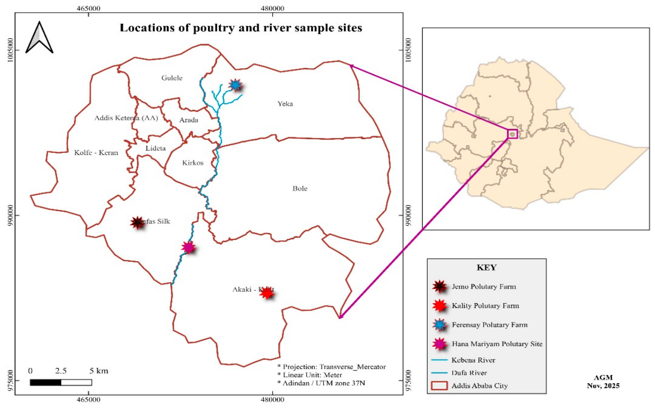
Figure 2.
Distribution of isolated phages across different sample sites.
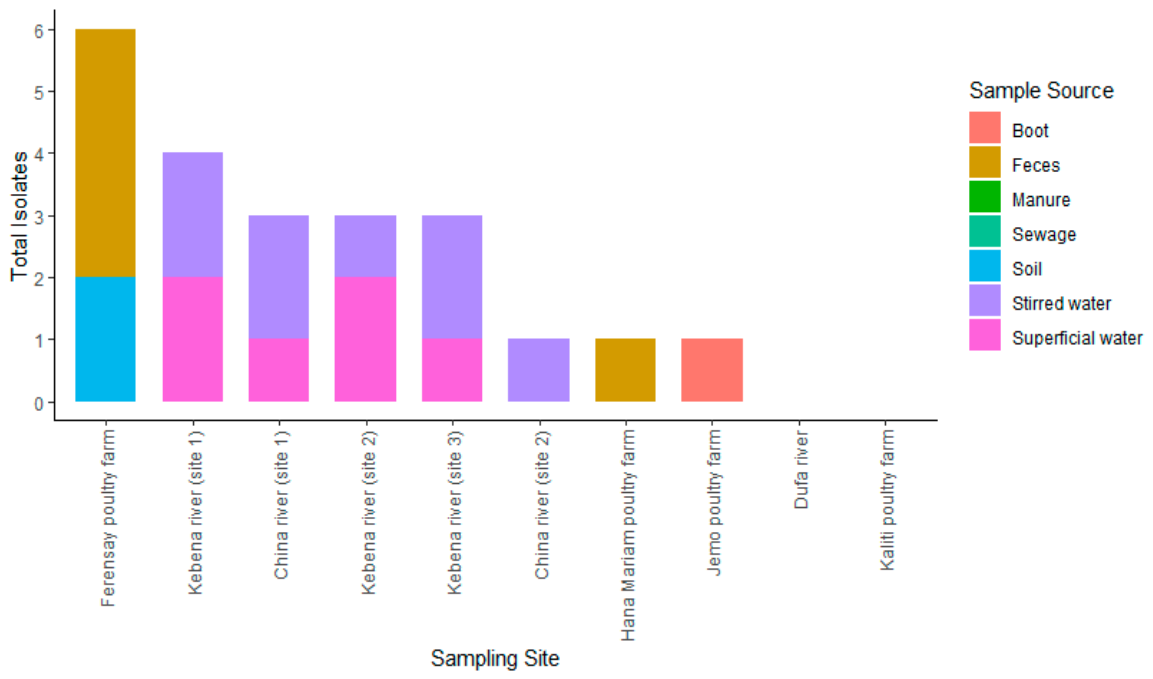
Figure 3.
Representative plaque morphologies of isolated bacteriophages. A. middle, well-defined plaques surrounded by concentric rings. B. Large plaques with halo zones. C. Large plaques with halo zones, D. High-density, small clear plaques, E: Medium-sized, clear plaques. F. High-density, small, clear plaques.
Figure 3.
Representative plaque morphologies of isolated bacteriophages. A. middle, well-defined plaques surrounded by concentric rings. B. Large plaques with halo zones. C. Large plaques with halo zones, D. High-density, small clear plaques, E: Medium-sized, clear plaques. F. High-density, small, clear plaques.
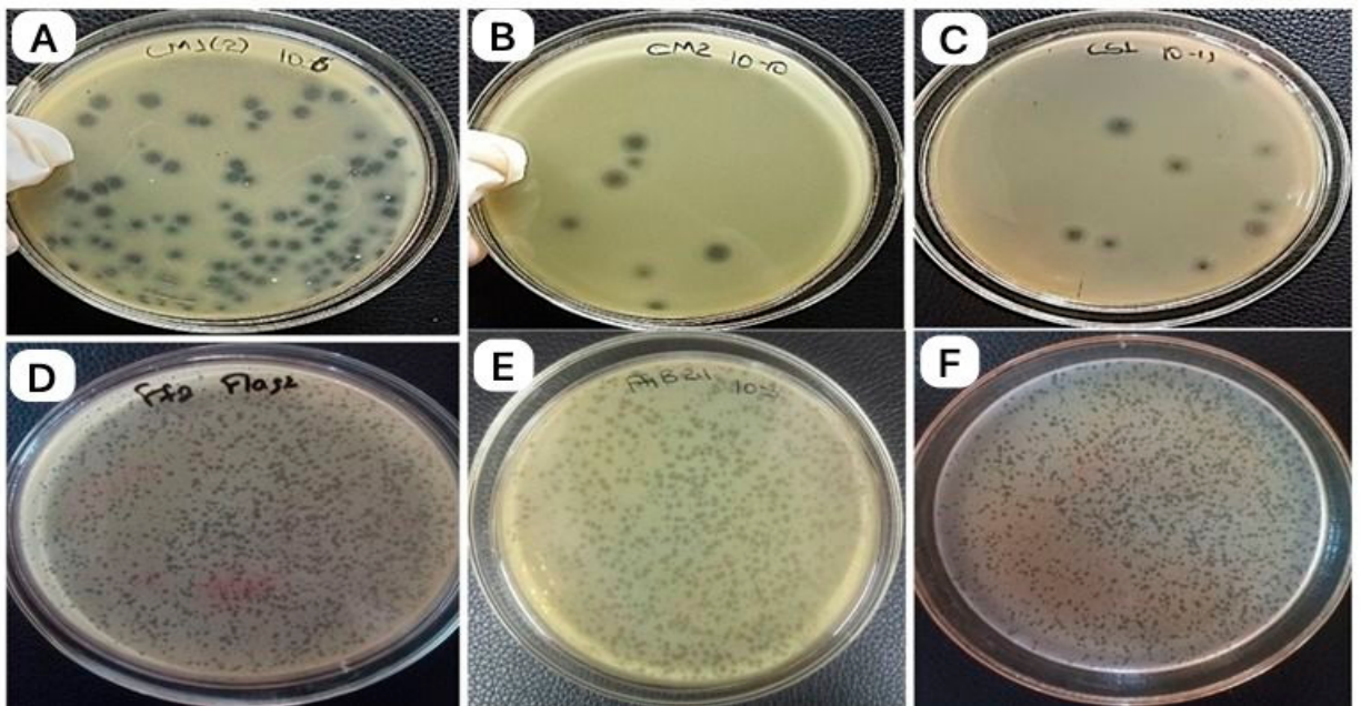
Figure 4.
Host range activity of isolated phages.
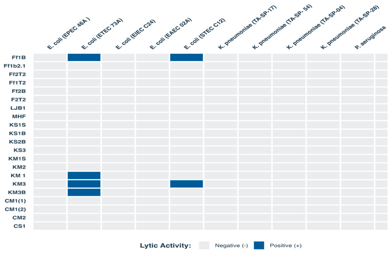
Figure 5.
Representative spot assay results for host range characterization of isolated bacteriophages. (A) Phage Ff1B produces turbid lysis zones on ETEC 73A lawn, indicating limited cross-genus activity. (B) Phage Ff1B producing turbid lysis zones on STEC C12 lawn. (C) Phage KM3 produces turbid clearing on STEC C12 lawn. (D) Phages Ff1B, KM3, KM3B, and KM1 producing turbid clearing on ETEC 73A lawn. (E) All tested phages produced clear, well-defined lysis zones on S. Typhimurium ATCC 13311 (primary host) lawn, confirming strong lytic activity against the target host. Turbid lysis on non-target E. coli strains (panels A–D) contrasts with clear lysis on the primary host (panel E), suggesting reduced infection efficiency on non-target hosts.
Figure 5.
Representative spot assay results for host range characterization of isolated bacteriophages. (A) Phage Ff1B produces turbid lysis zones on ETEC 73A lawn, indicating limited cross-genus activity. (B) Phage Ff1B producing turbid lysis zones on STEC C12 lawn. (C) Phage KM3 produces turbid clearing on STEC C12 lawn. (D) Phages Ff1B, KM3, KM3B, and KM1 producing turbid clearing on ETEC 73A lawn. (E) All tested phages produced clear, well-defined lysis zones on S. Typhimurium ATCC 13311 (primary host) lawn, confirming strong lytic activity against the target host. Turbid lysis on non-target E. coli strains (panels A–D) contrasts with clear lysis on the primary host (panel E), suggesting reduced infection efficiency on non-target hosts.
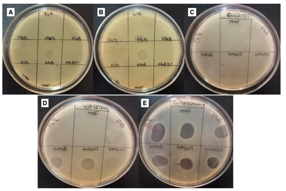
Figure 6.
MOI of the isolated phages.
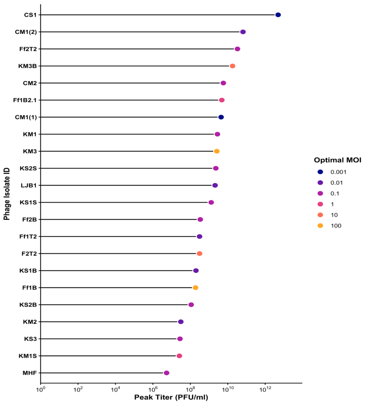
Figure 7.
One step growth curve.
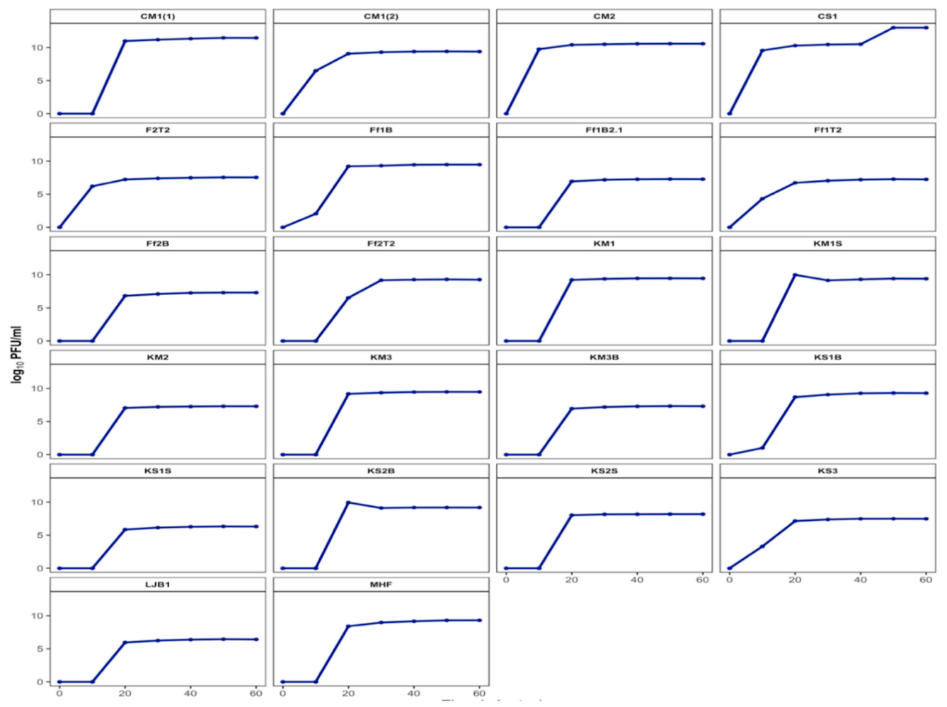
Figure 8.
The isolated phages' thermal stability.
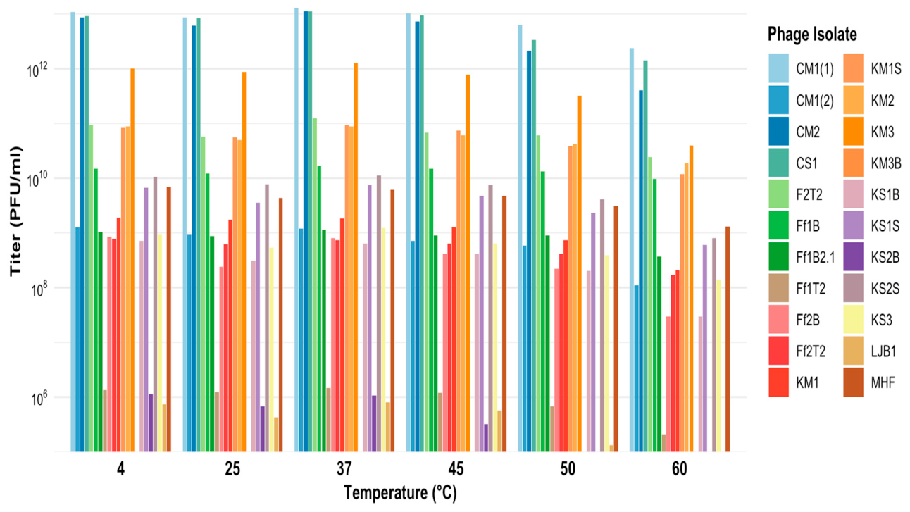
Figure 9.
pH stability of the isolated phages.
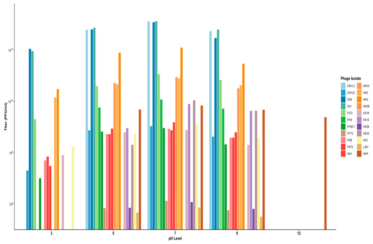
Figure 10.
Molecular characterization of isolated phages.
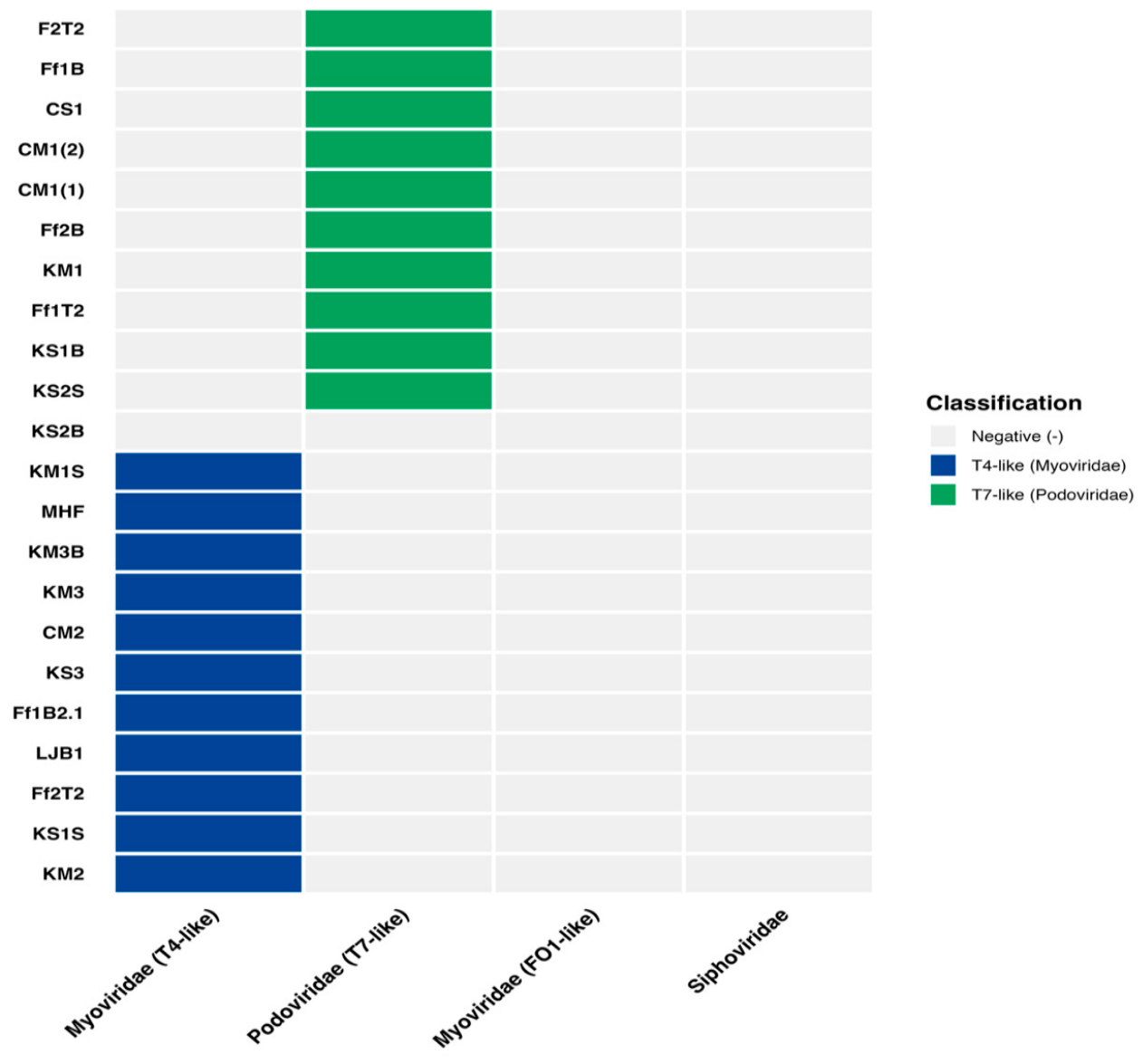
Figure 11.
Distribution of isolated bacteriophages categorized as Podoviridae (T7-like): lane M: DNA ladder (100+), PCR amplification product size of ten T7 isolates was 461 bp. lane 1: F2T2, lane2: Ff1B, lane3:CS1, Lane 4: CM1(1), lane 5: CM1(2), lane 6: Ff2b, lane 7: KM1, Lane 8: Ff1T2, Lane 9: KS1B, lane 10: KS2S.
Figure 11.
Distribution of isolated bacteriophages categorized as Podoviridae (T7-like): lane M: DNA ladder (100+), PCR amplification product size of ten T7 isolates was 461 bp. lane 1: F2T2, lane2: Ff1B, lane3:CS1, Lane 4: CM1(1), lane 5: CM1(2), lane 6: Ff2b, lane 7: KM1, Lane 8: Ff1T2, Lane 9: KS1B, lane 10: KS2S.
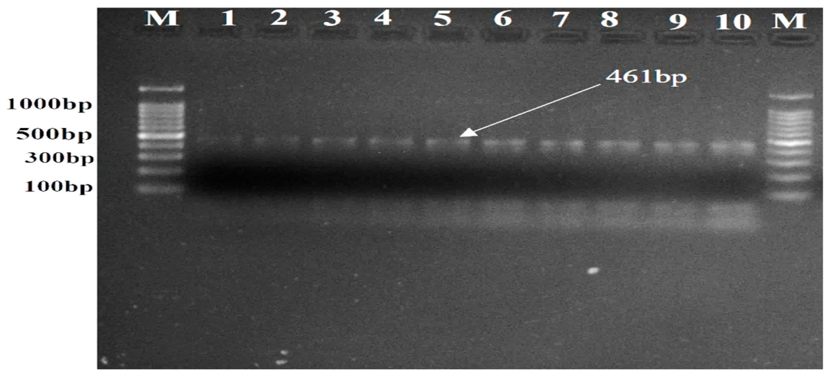
Figure 12.
Distribution of isolated bacteriophages classified as Myoviridae (T4-like): PCR amplification product of 11 T4 isolates product size was 240 bp; lane M: DNA ladder (100plus), lane 1:KM1S, lane2: MHF, lane3: KM3B, lane 4:KM3, lane 5: CM2, lane 6: KS3, Lane 7: Ff1B2.1 , lane8: LJB1 , lane 9:Ff2T2 , lane 10:KS1S ,Lane11: KM2,Lane12:NC.
Figure 12.
Distribution of isolated bacteriophages classified as Myoviridae (T4-like): PCR amplification product of 11 T4 isolates product size was 240 bp; lane M: DNA ladder (100plus), lane 1:KM1S, lane2: MHF, lane3: KM3B, lane 4:KM3, lane 5: CM2, lane 6: KS3, Lane 7: Ff1B2.1 , lane8: LJB1 , lane 9:Ff2T2 , lane 10:KS1S ,Lane11: KM2,Lane12:NC.
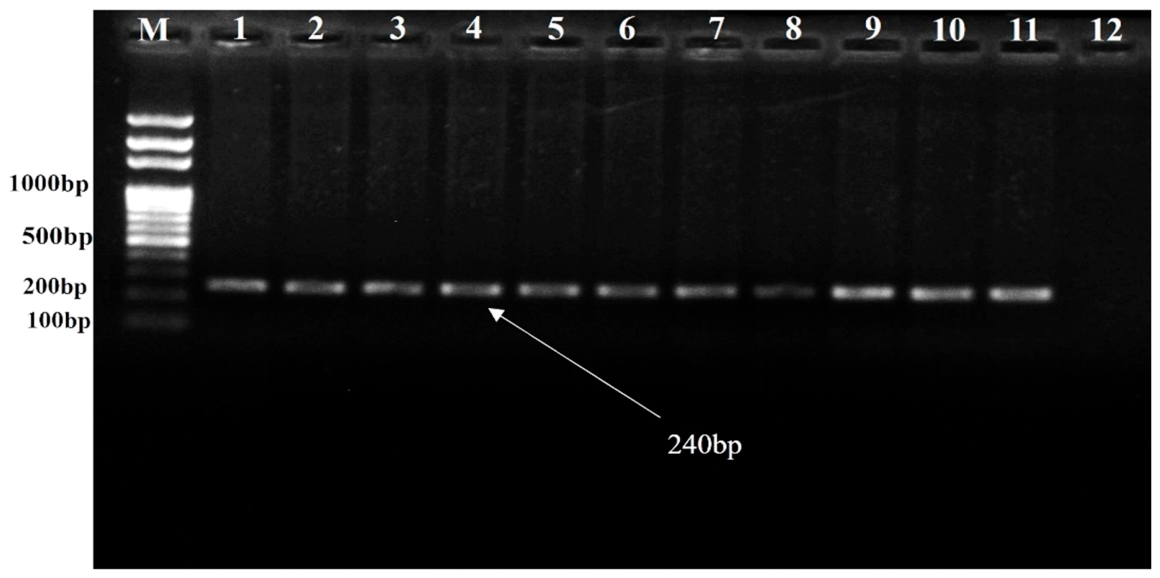

Table 1.
Geographic coordinates of sampling sites in Addis Ababa.
| Sub-City | Sampling Site | Latitude (N) | Longitude (E) |
| Yeka | Ferensay Poultry Farm | 9.065403 | 38.790343 |
| Kaliti | Poultry Farm | 8.894888 | 38.773144 |
| Nifas Silk Lafto | Jemo Poultry Farm | 8.959745 | 38.713310 |
| Nifas Silk Lafto | China River | 8.965904 | 38.711764 |
| Nifas Silk Lafto | Hana Mariam Poultry Farm | 8.931617 | 38.756997 |
| Nifas Silk Lafto | Dufa River | 8.932642 | 38.755721 |
| Arada | Kebena River (Site 1) | 9.035782 | 38.778762 |
| Arada | Kebena River (Site 2&3) | 9.037622 | 38.779024 |
Table 2.
Primer used for characterization of phages.
| Gene | Family | Sub-family | Genus |
Sequence (5’→3’) |
Annealing Temp. | Product Size (Bp) | Ref. |
| MCP | Myoviridae | Tevenvirinae | T4-like | fw: CCC TGC TGT TCC AGA TCG ANA ARG ARG C | 52 | 240 Bp | [23,24] |
| rev: CTG CCT GGC GTA CTG GTC DAT RWA NAC | |||||||
| Ounavirinae | FO1-like | fw: CGC CAT TGA AGA ACT GCG TRW RCA YAT GGA | 52 | 519Bp | |||
| rev: GGC ATC ATA TAG GAA TGC GCY TCR AAR TC | |||||||
| MCP | Podoviridae | Autographivirinae | T7-like | fw: GAC AAG CGG AAG GAC ATC AAN CAY ACN GAR A | 52 | 461 Bp | |
| rev: CGC GTA GTT GGC GGC RTT NGG CAT NA | |||||||
| McoP | Siphoviridae | - | T5-like | MCF-2F: GCG TGA TGG TTG GGA TGG TA | 56 | 200Bp | |
| MCF-2R: GAC GCT CAA TCT GAC GAC CA |
Table 3.
Titers and morphological characteristics of isolated Salmonella lytic bacteriophages.
| Phage ID | Titer (PFU/ml) | Plaque Size | Edge/Margin |
| Ff1B2.1 | 3.10 × 10¹⁰ | Small | Haloed |
| LJB1 | 3.10 × 10¹⁰ | Small | Sharp |
| Ff1B | 5.20 × 10¹⁰ | Medium | Sharp |
| Ff2T2 | 3.20 × 10⁹ | Small | Sharp |
| F2T2 | 9.10 × 10¹¹ | Small | Sharp |
| MHF | 3.80 × 10⁹ | Medium | Sharp |
| Ff1T2 | 3.20 × 10⁸ | Small | Bull's eye |
| Ff2B | 3.50 × 10⁹ | Small | Sharp |
| KM1 | 2.08 × 10⁹ | Small | Sharp |
| KS3 | 1.20 × 10⁹ | Medium | Sharp |
| KM3 | 1.22 × 10¹⁰ | Small | Sharp |
| KS2S | 4.30 × 10¹⁰ | Small | Sharp |
| KS1S | 3.10 × 10⁹ | Medium | Sharp |
| KS1B | 5.20 × 10⁸ | Medium | Haloed |
| KM3B | 4.60 × 10¹² | Small | Haloed |
| KS2B | 1.43 × 10⁶ | Small | Sharp |
| KM2 | 5.40 × 10¹⁰ | Small | Sharp |
| KM1S | 7.10 × 10¹⁰ | Small | Sharp |
| CM1(1) | 9.00 × 10¹² | Large | Haloed |
| CM1(2) | 1.37 × 10⁹ | Medium | Bull's eye |
| CM2 | 4.50 × 10¹⁰ | Large | Haloed |
| CS1 | 9.40 × 10¹² | Large | Bull's eye |
Table 4.
Efficiency of Plating (EOP) of selected phages on E. coli lawn.
| Test Strain | Reference host | Test strain | PFU_host mean | PFU_test mean | EOP |
| Ff1B | STy_ATCC13311 | ETEC_73A | 1.52 × 10¹⁰ | 1.00 × 10⁴ | 6.58 × 10⁻⁷ |
| Ff1B | STy_ATCC13311 | STEC_C12 | 1.52 × 10¹⁰ | 3.00 × 10³ | 1.97 × 10⁻⁷ |
| KM3 | STy_ATCC13311 | ETEC_73A | 1.22 × 10¹² | 8.00 × 10³ | 6.56 × 10⁻⁹ |
| KM3 | STy_ATCC13311 | STEC_C12 | 1.22 × 10¹² | 4.00 × 10³ | 3.28 × 10⁻⁹ |
| KM1 | STy_ATCC13311 | ETEC_73A | 1.96 × 10⁹ | 1.50 × 10³ | 7.65 × 10⁻⁷ |
| KM1 | STy_ATCC13311 | EIEC_C24 | 1.96 × 10⁹ | 7.00 × 10² | 3.57 × 10⁻⁷ |
| KM3B | STy_ATCC13312 | ETEC_73A | 1.84 × 10⁹ | 6.00 × 10² | 3.30 × 10⁻⁷ |
Disclaimer/Publisher’s Note: The statements, opinions and data contained in all publications are solely those of the individual author(s) and contributor(s) and not of MDPI and/or the editor(s). MDPI and/or the editor(s) disclaim responsibility for any injury to people or property resulting from any ideas, methods, instructions or products referred to in the content. |
© 2026 by the authors. Licensee MDPI, Basel, Switzerland. This article is an open access article distributed under the terms and conditions of the Creative Commons Attribution (CC BY) license.
Copyright: This open access article is published under a Creative Commons CC BY 4.0 license, which permit the free download, distribution, and reuse, provided that the author and preprint are cited in any reuse.


