

Submitted:
15 July 2026
Posted:
17 July 2026
You are already at the latest version
Abstract
Fowl adenovirus serotype 11 (FAdV-11) is an important viral pathogen affecting both broiler and layer chickens and is the primary causative agent of inclusion body hepatitis (IBH), resulting in substantial economic losses through reduced meat and egg production worldwide. Despite its economic significance, no commercial vaccine is currently available specifically against FAdV-11. In the present study, to the best of our knowledge, the first immunoinformatics-based multi-epitope vaccine candidate against FAdV-11 was designed using conserved regions of the penton and fiber proteins. Conserved B-cell, cytotoxic T-lymphocyte (CTL), and helper T-lymphocyte (HTL) epitopes were identified, screened for antigenicity, allergenicity, toxicity, and immunogenicity, and assembled into a multi-epitope vaccine construct using appropriate linker peptides and a β-defensin adjuvant. The designed vaccine exhibited favorable physicochemical properties and was predicted to be highly antigenic, non-allergenic, and non-toxic. Secondary and tertiary structure prediction, refinement, and validation confirmed the structural stability and reliability of the vaccine construct. Molecular docking and molecular dynamics simulation analysis demonstrated stable interactions with immune-related Toll-like receptors (TLRs), while immune simulation predicted robust humoral and cellular immune responses with the establishment of long-term immunological memory. Codon optimization yielded a Codon Adaptation Index (CAI) of 0.982, and in silico cloning into the pET-28a(+) expression vector suggested efficient recombinant expression in Escherichia coli. Collectively, these findings indicate that the proposed vaccine represents a promising candidate for the prevention of FAdV-11-induced IBH in poultry. Nevertheless, comprehensive in vitro and in vivo studies are required to validate its immunogenicity, safety, and protective efficacy.
Keywords:
fowl adenoviruses
; inclusion body hepatitis
; pathogenicity
; vaccine
1. Introduction
The poultry industry is one of the fastest-growing sectors of global agriculture and serves as a major source of affordable, high-quality animal protein for the rapidly increasing human population. Chicken meat and eggs contribute substantially to global food security owing to their high nutritional value, short production cycle, and relatively low production costs (1). Worldwide poultry meat production exceeds 105 million metric tons annually and continues to increase in response to the growing demand for animal-derived protein (2). Despite remarkable advances in breeding, nutrition, and intensive farming practices, infectious diseases remain one of the greatest challenges limiting sustainable poultry production. Viral infections, in particular, continue to cause substantial economic losses through increased mortality, impaired growth performance, reduced feed efficiency, decreased egg production, and increased costs associated with disease control and biosecurity measures (3).
Among the economically important viral diseases affecting commercial poultry, inclusion body hepatitis (IBH) has emerged as a major cause of production losses, especially in young broiler chickens. The disease is characterized by acute hepatitis, hepatocellular degeneration, multifocal hepatic necrosis, and the presence of distinctive basophilic intranuclear inclusion bodies within infected hepatocytes (4). Clinical manifestations may include depression, anorexia, poor weight gain, anemia, and sudden death. Depending on the virulence of the infecting strain, age of the birds, immune status of the flock, and the occurrence of concurrent infections, mortality rates may vary considerably, ranging from approximately 2% to more than 40% (5). Although IBH has been reported from poultry-producing regions across Asia, Europe, North and South America, Africa, and Australia, its true prevalence is likely underestimated because clinical signs frequently overlap with those of other poultry diseases, making laboratory confirmation essential for accurate diagnosis (6).
The principal etiological agent responsible for IBH is Fowl adenovirus serotype 11 (FAdV-11), a member of the genus Aviadenovirus within the family Adenoviridae. Fowl adenoviruses are non-enveloped viruses possessing an icosahedral capsid enclosing a linear double-stranded DNA genome approximately 43–45 kb in length (7). Based on molecular and serological characteristics, twelve recognized serotypes (FAdV-1 to FAdV-8a, FAdV-8b, FAdV-9, FAdV-10, and FAdV-11) have been classified into five species (FAdV-A to FAdV-E) (8). Among these, serotypes 2, 8a, 8b, and 11 are most frequently associated with IBH outbreaks worldwide (9). Reports from numerous countries have demonstrated the widespread distribution of FAdV-11, emphasizing its growing veterinary importance and the need for effective disease prevention strategies (10).
The FAdV-11 genome exhibits a highly organized architecture that supports efficient viral replication and adaptation. It comprises approximately 35–37 open reading frames (ORFs) with an overall GC content ranging from 53% to 55%. Similar to other aviadenoviruses, the genome contains a highly conserved central region flanked by comparatively variable left and right terminal regions. The conserved core encodes proteins essential for viral DNA replication, transcription, and virion assembly, whereas the terminal regions harbor genes involved in host adaptation, immune modulation, and viral pathogenicity. Inverted terminal repeat (ITR) sequences located at both ends of the genome function as origins of viral DNA replication and are indispensable for successful genome amplification during infection (11).
Despite belonging to the same serotype, FAdV-11 isolates exhibit considerable variation in their pathogenic potential. While some strains cause severe clinical disease accompanied by high mortality, others establish subclinical infections with little or no apparent clinical signs. These differences are largely attributed to genomic variations, particularly within regions associated with host adaptation, immune modulation, and viral virulence (12). Although the central region of the genome remains highly conserved because of its essential role in viral replication, greater genetic diversity occurs within the terminal regions, where several genes associated with pathogenicity are located. In addition to the conserved coding sequences, non-coding regions also contribute to the genetic diversity observed among FAdV-11 isolates, resulting in differences in biological behavior, tissue tropism, and disease severity (13).
The FAdV-11 genome encodes numerous proteins that contribute directly or indirectly to viral pathogenicity. Among these are the terminal repeat region sequences, GAM-1 protein, 100-kDa protein, 52-kDa protein, 22-kDa protein, TSH protein, and several other viral factors that regulate replication, cellular interactions, and host immune modulation. Although many of these proteins participate in intracellular viral replication and assembly, the structural proteins of the viral capsid remain the primary determinants of viral infectivity and host immune recognition (14). Consequently, they have become the principal targets for molecular characterization and vaccine development (15).
Three major structural proteins constitute the outer capsid of FAdV-11: the hexon, penton base, and fiber proteins. Each performs a distinct biological function during viral infection. The hexon protein forms the major component of the viral capsid and contains hypervariable regions responsible for serotype specificity and antigenicity. These exposed regions interact extensively with the host immune system and have therefore been widely employed in molecular typing and phylogenetic analyses of fowl adenoviruses (16). The penton base protein is positioned at each of the twelve vertices of the viral capsid and mediates viral internalization through interactions with cellular integrin receptors. This interaction facilitates virus entry into susceptible host cells and represents an essential step in establishing infection (17). Extending outward from the penton base, the fiber protein mediates the initial attachment of the virus to specific cellular receptors and largely determines viral tissue tropism, host specificity, and pathogenic potential (16). Owing to their direct involvement in viral attachment, entry, and immune recognition, the penton and fiber proteins are considered highly attractive targets for epitope-based vaccine design (18).
Following infection, FAdV-11 primarily targets hepatocytes, where viral replication induces marked cytopathic effects characterized by hepatocellular necrosis and the formation of prominent basophilic intranuclear inclusion bodies. Besides direct cellular damage, infection also disrupts the host immune response through alterations in cytokine production. Excessive production of pro-inflammatory cytokines contributes to hepatic inflammation and tissue injury, thereby aggravating disease progression (19). Experimental infection studies in specific pathogen-free (SPF) chickens have demonstrated a rapid increase in both B- and T-lymphocyte populations within three to four days after infection, accompanied by lymphocytic infiltration and the development of necrotic lesions within affected tissues. Additional pathological findings include petechial and ecchymotic hemorrhages involving the liver, heart, and kidneys. Collectively, these pathological changes compromise the immune competence of infected birds, rendering them more susceptible to secondary infections caused by other important poultry pathogens, including Fowl adenovirus serotype 4 (FAdV-4), infectious bursal disease virus (IBDV), and Newcastle disease virus (NDV) (20).
Because of its economic importance, effective control of FAdV-11 has become a major priority for the poultry industry. Biosecurity remains the first line of defense and includes strict farm sanitation, isolation of infected flocks, prevention of fecal contamination, provision of clean feed and drinking water, and maintenance of adequate ventilation and environmental hygiene (21). Since fecal shedding constitutes one of the principal routes of viral transmission, minimizing direct and indirect contact between infected and healthy birds is essential for reducing disease spread. Although these preventive measures contribute significantly to outbreak control, they cannot completely eliminate the risk of infection under intensive poultry production systems (22).
Vaccination therefore represents the most reliable strategy for controlling FAdV infections by inducing protective immune responses before natural viral exposure. Various vaccine platforms, including inactivated, live-attenuated, recombinant, viral vector, and subunit vaccines, have been investigated against different FAdV serotypes (23). Nevertheless, vaccine development specifically targeting FAdV-11 remains in its infancy (24). To date, only limited progress has been achieved toward the development of commercially available vaccine candidates. One of the earliest recombinant subunit vaccine candidates, designated cre-Fib4/11, was developed by combining immunogenic fiber epitopes derived from different FAdV-11 isolates. This recombinant construct elicited significantly enhanced antibody responses while providing simultaneous protection against inclusion body hepatitis and hepatitis-hydropericardium syndrome (15). Furthermore, previous studies targeting the hexon, penton, and fiber proteins of other FAdV serotypes have demonstrated the strong immunogenic potential of these structural proteins, suggesting that similar antigenic targets could be exploited for the rational development of effective vaccines against FAdV-11 (25-28).
In light of these challenges, this study aims to illustrates the molecular characteristics and pathogenic mechanisms of FAdV-11, contributing to an enhanced understanding that may facilitate effective control strategies against IBH in commercial poultry production (29, 30).
Despite the considerable progress achieved in understanding the molecular biology and epidemiology of FAdV-11, the development of effective vaccines against this economically important pathogen remains limited (31). Conventional vaccine development is often expensive, labor-intensive, and time-consuming, requiring extensive laboratory experimentation, animal trials, and stringent biosafety conditions before a vaccine can be considered for commercial production. Furthermore, even when effective vaccines become available, their accessibility to poultry producers, particularly in developing countries, is frequently restricted because of production costs and limited distribution. These limitations emphasize the need for alternative vaccine development strategies that are rapid, cost-effective, and capable of providing broad protective immunity against genetically diverse viral strains (32).
Recent advances in computational biology have transformed vaccine development through the emergence of immunoinformatics and reverse vaccinology. Unlike conventional approaches, reverse vaccinology begins with genomic and proteomic information to identify potential vaccine targets using computational analyses. This strategy enables the systematic screening of conserved antigenic regions capable of stimulating both humoral and cell-mediated immune responses while simultaneously evaluating important characteristics such as antigenicity, allergenicity, toxicity, structural stability, and population coverage. Consequently, immunoinformatics substantially reduces the time and resources required for vaccine discovery and increases the likelihood of identifying promising vaccine candidates before experimental validation (33).
Among the various computational vaccine platforms, multi-epitope vaccines have gained considerable attention because they combine immunodominant B-cell and T-cell epitopes into a single recombinant construct. Such vaccines are designed to activate multiple arms of the immune system by simultaneously stimulating cytotoxic T lymphocytes, helper T lymphocytes, and B lymphocytes, thereby promoting both cellular and humoral immunity. The incorporation of suitable adjuvants and linker peptides further improves antigen presentation, structural stability, and immunogenicity while reducing undesirable immune responses. In addition, selecting highly conserved epitopes from genetically diverse viral strains increases the potential for broad-spectrum protection against circulating variants (34).
Considering the essential roles of the penton and fiber proteins in viral attachment, cellular entry, tissue tropism, and host immune recognition, these proteins represent promising targets for epitope-based vaccine development against FAdV-11 (25). The availability of complete genome sequences from geographically distinct isolates provides an opportunity to identify conserved immunogenic regions that may induce cross-protective immune responses against multiple circulating strains. Such an approach is particularly valuable for FAdV-11 because of the limited availability of commercial vaccines and the continuous circulation of genetically diverse isolates in poultry populations.
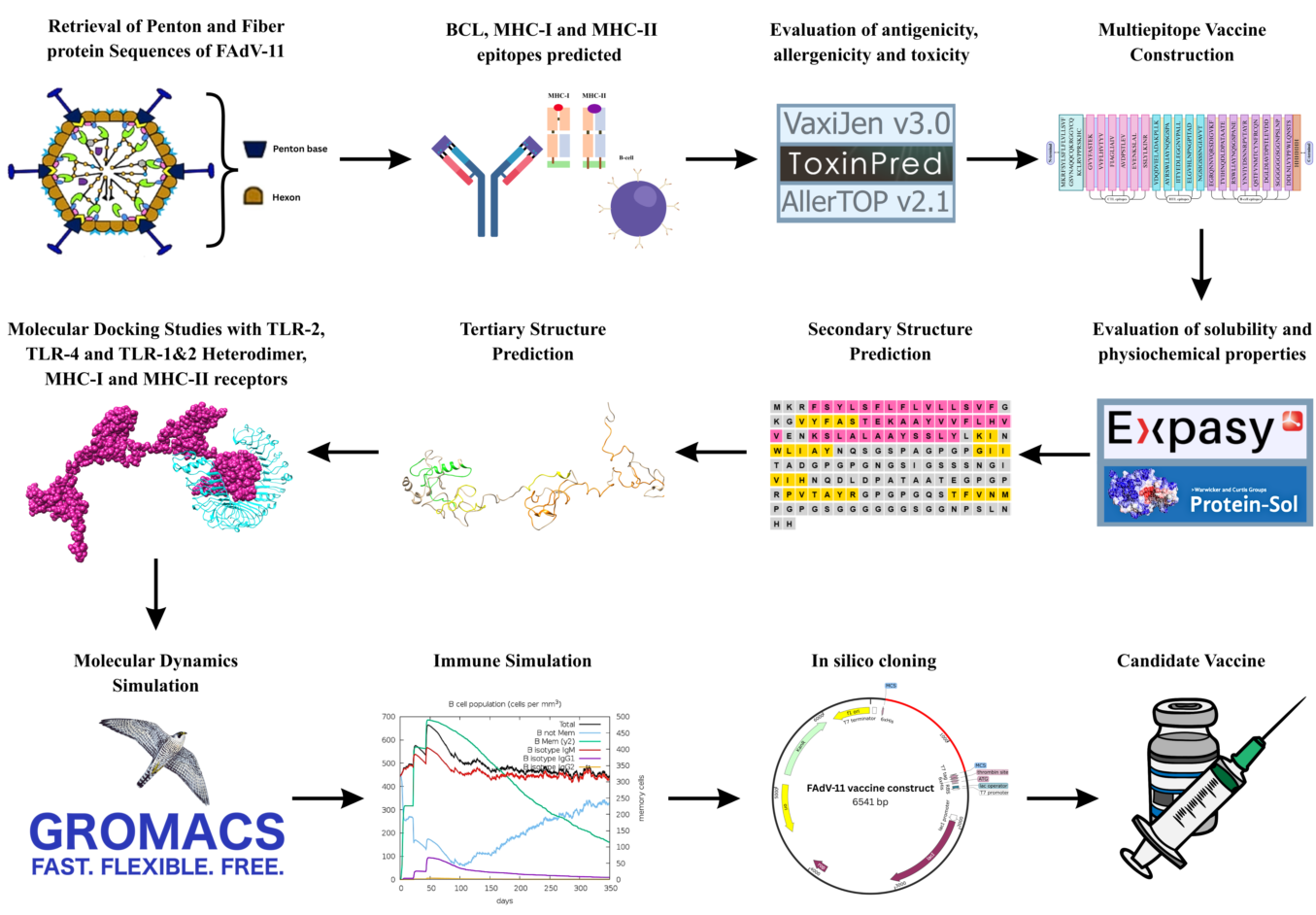
Therefore, the present study employed an immunoinformatics-based reverse vaccinology approach to design a novel multi-epitope vaccine candidate against FAdV-11. Conserved regions of the penton and fiber proteins from genetically distinct pathogenic and mildly pathogenic isolates were identified and screened for cytotoxic T-lymphocyte, helper T-lymphocyte, and B-cell epitopes (16, 17). Selected epitopes were further evaluated for antigenicity, allergenicity, toxicity, and sequence conservation before being assembled into a multi-epitope vaccine construct. The designed construct was subsequently subjected to comprehensive physicochemical characterization, secondary and tertiary structural prediction, molecular docking with Toll-like receptors, immune simulation, codon optimization, and in silico cloning to evaluate its potential as a safe, stable, and immunogenic vaccine candidate for the prevention of inclusion body hepatitis in poultry.
2. Materials and Methods
2.1. Protein Sequence Retrieval
Amino acid sequences of the hexon, penton, and fiber proteins from three genetically distinct FAdV-11 strains, including the Pakistani pathogenic strain (NCBI accession no. MN428137)(35), the Mexican mildly pathogenic strain (NCBI accession no. KU746335)(11), and the Canadian pathogenic strain (NCBI accession no. KU310942)(36), were retrieved from the NCBI (National Center for Biotechnology Information) database. Multiple sequence alignment (MSA) was performed using Clustal Omega to identify conserved regions within each protein (37). Based on the alignment results, only the penton and fiber proteins were selected for subsequent analyses, whereas the hexon protein was excluded because it exhibited a high degree of sequence conservation among all three strains without any variability (38).
2.2. Epitope Prediction Using Immunoinformatic Tools
To identify potential antigenic regions, B-cell, C-cell and T-cell epitopes were predicted using several immunoinformatic platforms.
2.2.1. Linear B-Cell Epitopes
B-cell epitopes were predicted using ABCpred, a web-based server that employs an artificial neural network (ANN) algorithm for the prediction of linear B-cell epitopes (39). The prediction performance of ABCpred is evaluated based on four parameters: precision, sensitivity, positive predictive value, and specificity, with an overall reported prediction accuracy of approximately 65.93% (40). B-cell epitopes were predicted using the consensus sequence with a threshold score of >0.5 and a peptide length of 16 amino acids. Additionally, linear B-cell epitope scores across the consensus sequence were predicted using BepiPred (41).
2.2.2. Cytotoxic T Lymphocyte (CTL)
CTL epitopes were predicted using the IEDB MHC-I Binding Prediction tool based on the NetMHCpan EL algorithm (42, 43). The consensus sequence was used as the input for epitope prediction with a peptide length of 9 amino acids (9-mer) across five different HLA class I alleles (44). This surrogate strategy has been reportedly previously based on similar immune responses in avian species keeping in mind the availability of avian specific MHC datasets.
2.2.3. Helper T Lymphocyte (HTL)
HTL epitopes were predicted using the IEDB MHC-II Binding Prediction tool. This server employs artificial neural network (ANN)-based models trained on more than 200,000 randomly selected peptides and supports the prediction of peptide binding to a wide range of HLA class II alleles (45, 46). The consensus sequence was used as the input for prediction with a peptide length of 15 amino acids and a peptide shift of 5.
2.2.4. Epitope Screening and Conservancy Analysis
The antigenicity of the selected epitopes was evaluated using the VaxiJen v2.0 server (47). Epitopes predicted to be non-antigenic were excluded, whereas antigenic epitopes were retained for further analysis. To ensure broad-spectrum vaccine coverage, the selected antigenic epitopes were also assessed for sequence conservation across different viral strains, and highly conserved epitopes were prioritized (48).
The allergenicity of the selected epitopes was evaluated using the AllerTOP v2.0 server. Epitopes predicted to be allergenic were excluded, while non-allergenic epitopes were retained for subsequent analyses (49).
The toxicity of the selected epitopes was also assessed using the ToxinPred server (50). Epitopes predicted to be toxic were discarded, whereas non-toxic epitopes were selected for further evaluation. Consequently, only epitopes that satisfied all three selection criteria: high antigenicity, non-allergenicity, and non-toxicity were retained for downstream analyses. Epitopes that failed to meet any of these criteria were excluded from further investigation (18).
2.3. Multi-Epitope Vaccine Construct Design
The multi-epitope vaccine construct was designed by assembling the filtered cytotoxic T lymphocyte (CTL), helper T lymphocyte (HTL), and B-cell epitopes into a single chimeric protein (51). A total of six CTL epitopes, five HTL epitopes, and eight B-cell epitopes were incorporated into the final construct. The selected epitopes were arranged sequentially, with epitopes derived from the penton protein placed before those derived from the fiber protein.
To ensure proper processing and presentation of the epitopes, suitable linker peptides were employed during vaccine construction. CTL epitopes were joined using AAY linkers, whereas HTL and B-cell epitopes were linked using GPGPG linkers (52). A β-defensin adjuvant was incorporated at the N-terminus of the vaccine construct to enhance its immunogenic potential and was connected to the first epitope using an EAAAK linker (53). EAAAK linkers were also used where appropriate to separate different functional regions of the construct and minimize structural interference. A methionine residue was manually added at the N-terminus to facilitate protein expression, while a 6×His tag was appended to the C-terminus to enable downstream purification and detection of the recombinant protein (54). The antigenicity and allergenicity of the complete construct were subsequently evaluated using the VaxiJen v2.0 and AllerTOP v2.1 servers, respectively.
2.4. Physicochemical Properties Analysis
The physicochemical properties of the final vaccine construct were predicted using the ExPASy ProtParam server. Parameters including molecular formula, molecular weight, theoretical isoelectric point (pI), estimated half-life, instability index (II), aliphatic index, and grand average of hydropathicity (GRAVY) were calculated to evaluate the physicochemical characteristics and stability of the vaccine construct (55).
In addition, the solubility of the vaccine construct upon recombinant expression was predicted using the SolPro server to assess its likelihood of soluble expression in a host expression system (51).
2.5. Secondary Structure Prediction
The secondary structure of the vaccine construct was predicted using the Self-Optimized Prediction Method with Alignment (SOPMA) server with the default online alignment parameters (56). The proportions of major secondary structural elements, including α-helices, β-sheets, β-turns, and random coils, were estimated. To further validate the predicted secondary structure, PSIPRED was also employed to provide additional information on the distribution of secondary structural elements and residue-specific structural assignments (57).
2.6. Three-Dimensional Structure Prediction, Refinement and Validation
The three-dimensional (3D) structure of the FAdV-11 multi-epitope vaccine construct was predicted using the I-TASSER server. The server employs a hierarchical protein structure prediction approach based on multiple threading alignments and iterative structural assembly simulations to generate reliable tertiary structure models (58). The predicted models were evaluated using confidence (C-) scores, structural templates obtained from the Protein Data Bank (PDB), and other model quality parameters provided by the server.
The initial 3D model was subsequently refined using the GalaxyRefine server to improve its structural quality (59). Refined models were assessed based on several quality parameters, including the Global Distance Test-High Accuracy (GDT-HA) score, root mean square deviation (RMSD), MolProbity score, clash score, percentage of poor rotamers, and the proportion of residues in favored regions of the Ramachandran plot (60). The refined model exhibiting the best overall structural quality was selected for subsequent analyses.
2.7. Molecular Docking with TLRs
The three-dimensional crystal structures of Toll-like receptor 2 (TLR-2) and Toll-like receptor 3 (TLR-3) were retrieved from the Protein Data Bank (PDB) using accession IDs 4G8A and 2Z7X, respectively (61). The downloaded structures were preprocessed using UCSF Chimera by removing unnecessary chains and heteroatoms, adding hydrogen atoms, and optimizing the receptor structures to prepare them for molecular docking (62).
Molecular docking was performed using the ClusPro online server to investigate the binding interactions between the multi-epitope vaccine construct and the TLR receptors (63). ClusPro generated multiple docked complexes based on different clustering and energy parameters. The docked model with the lowest energy score and the largest cluster size was selected for further analysis, as these parameters indicate the most stable and reliable binding conformation (64).
The selected docked complexes were further analyzed using PDBsum to characterize the molecular interactions between the vaccine construct and the TLR receptors. PDBsum provided detailed information regarding hydrogen bonds, salt bridges, interface residues, and other intermolecular contacts, allowing assessment of the stability and quality of the docked complexes (65). The final docked complexes were visualized using UCSF Chimera.
2.8. Molecular Dynamics Simulation
To further evaluate the structural stability of the docked vaccine–receptor complexes under dynamic physiological conditions, molecular dynamics (MD) simulations were performed for the vaccine complexes with TLR-2, the TLR-1/TLR-2 heterodimer, and TLR-4. The docked complexes obtained from molecular docking were used as the initial structures for the simulations. All simulations were carried out using the GROMACS 2022 package with the CHARMM36 force field and the TIP3P water model. Each complex was placed in a dodecahedral simulation box with a minimum distance of 1.2 nm between the protein and the box boundaries, and was subsequently solvated with explicit water molecules. Counterions (Na+/Cl−) were added to neutralize the system while maintaining a physiological salt concentration of 0.15 M. Energy minimization was performed using the steepest descent algorithm until the maximum force converged to below 1000 kJ mol−1 nm−1. The minimized systems were subsequently equilibrated under constant volume and temperature (NVT) conditions for 100 ps, followed by constant pressure and temperature (NPT) equilibration for an additional 100 ps at 300 K and 1 bar, using the modified Berendsen thermostat and the Parrinello–Rahman barostat. Long-range electrostatic interactions were calculated using the Particle Mesh Ewald (PME) method, with a cutoff distance of 1.0 nm applied to both electrostatic and van der Waals interactions. Following equilibration, each system was subjected to a 100 ns production MD simulation under periodic boundary conditions.
2.9. In-Silico Codon Optimization and Cloning
Codon optimization of the final vaccine construct was performed using the JCat (Java Codon Adaptation Tool) server (66). The amino acid sequence of the vaccine construct was submitted to the server, and Escherichia coli K-12 was selected as the preferred expression host (67). JCat generated an optimized DNA sequence along with the Codon Adaptation Index (CAI) and GC content %. A CAI value greater than 0.5 was considered indicative of suitable expression in the selected host.
The optimized nucleotide sequence was subsequently processed using the Bio-Web Sequence Cleaner to remove unwanted or redundant sequence information. To facilitate cloning, restriction enzyme recognition sites were incorporated into both ends of the optimized sequence. The EcoRI recognition site (GAATTC) was added to the 5′ end, whereas the XhoI recognition site (CTCGAG) was added to the 3′of the sequence (68).
The pET-28a(+) expression vector was retrieved from the SnapGene plasmid database and imported into SnapGene software. The optimized vaccine sequence was inserted into the vector through restriction cloning using the EcoRI and XhoI restriction sites at the 5′ and 3′ ends, respectively. Where necessary, the insert orientation was manually adjusted within SnapGene to ensure correct directional cloning (69).
This workflow enabled the successful in silico codon optimization and cloning of the vaccine construct into the pET-28a(+) expression vector, providing a suitable construct for subsequent experimental expression studies.
2.10. Host-Immune System Simulation
The immune response elicited by the vaccine construct was evaluated using the C-ImmSim server, an in silico immune simulation platform that predicts both humoral and cellular immune responses using position-specific scoring matrices (PSSMs) and machine learning-based algorithms. A three-dose vaccination regimen was simulated at time steps 1, 64, and 128, corresponding to approximately three-week intervals between immunizations. The simulation was carried out for a total of 1,050 time steps to evaluate both the primary and long-term immune responses. The host HLA profile included HLA-A01:01, HLA-A02:01, HLA-B07:02, HLA-B08:02, HLA-DRB101:01, and HLA-DRB103:01. All remaining simulation parameters were maintained at their default settings (70).
3. Results
3.1. Proteins Sequence Retrieval
Amino acid sequences of the core structural proteins from the selected FAdV-11 isolates were retrieved from the NCBI database. Multiple sequence alignment revealed that the hexon protein was highly conserved among the selected isolates and was therefore excluded from subsequent immunoinformatics analyses. In contrast, the penton and fiber proteins exhibited conserved regions comprising 545 and 572 amino acid residues, respectively. These conserved sequences were subsequently used for epitope prediction and downstream analyses.
3.2. B-Cell Epitope Prediction
The conserved sequences of the penton and fiber proteins were analyzed using the ABCpred, BCEPRED, and BepiPred servers to identify linear B-cell epitopes. A total of 56 linear B-cell epitopes were predicted from the penton protein, whereas 60 epitopes were identified from the fiber protein.
3.3. Cytotoxic T-Lymphocyte (CTL) Epitope Prediction
MHC class I-restricted CTL epitopes were predicted using the IEDB MHC-I Binding Prediction tool. Based on the conserved sequences of the fiber and penton proteins, a total of 2,820 and 6,325 potential CTL epitopes were predicted, respectively. To identify high-confidence candidates, a prediction score threshold of ≥0.5 was applied, reducing the number of shortlisted epitopes to 23 for the fiber protein and 57 for the penton protein.
3.4. Helper T-Lymphocyte (HTL) Epitope Prediction
MHC class II-restricted HTL epitopes were predicted using the IEDB MHC-II Binding Prediction tool. A total of 784 epitopes were predicted for the fiber protein, whereas 636 epitopes were identified for the penton protein. After applying score thresholds of ≥0.2 for the fiber protein and ≥0.5 for the penton protein, the number of shortlisted epitopes was reduced to 22 and 45, respectively.
3.5. Selection of Epitopes for Vaccine Construction
The shortlisted B-cell, CTL, and HTL epitopes were further evaluated for antigenicity, allergenicity, and toxicity using the VaxiJen v2.0, AllerTOP v2.1, and ToxinPred servers, respectively. Only epitopes predicted to be antigenic, non-allergenic, and non-toxic were selected for inclusion in the multi-epitope vaccine construct.

Table 1.
Description of NCBI reported FAdV-11 whole genome sequence isolates.
| NCBI Accession no. | Nature | Origin/ Country |
Genome Size (bp) |
Host Organism | NCBI Accession Link |
|---|---|---|---|---|---|
| KU746335 | Least Pathogenic | Mexico | 44,326 | Chicken | https://www.ncbi.nlm.nih.gov/nuccore/KU746335 |
| KU310942 | Pathogenic | Canada | 44,377 | Avian | https://www.ncbi.nlm.nih.gov/nuccore/KU310942 |
| MN428137 | Pathogenic | Pakistan | 43,840 | Chicken | https://www.ncbi.nlm.nih.gov/nuccore/MN428137 |
This table shows the details of FAdV-11 isolates used in the present study. The first column lists the accession number of isolates whereas the second column describes its nature. The third column specifies the origin of the isolate. The size of the sequence is mentioned in column four with the host organism mentioned in the following column. The last column contains the links from NCBI to retrieve the enlisted data.
Among the B-cell epitopes, six fiber-derived and ten penton-derived epitopes satisfied all selection criteria. Similarly, five fiber-derived and nine penton-derived CTL epitopes, along with four fiber-derived and nine penton-derived HTL epitopes, were identified as suitable candidates for vaccine construction. The selected B-cell, CTL and HTL epitopes for penton and fiber proteins are given in Table 2, Table 3 and Table 4 respectively.
3.6. Construction of the Multi Epitope Vaccine Candidate
The final multi-epitope vaccine construct was assembled by integrating six CTL, five HTL, and eight B-cell epitopes using appropriate linker peptides. The selected epitopes were arranged sequentially, with penton-derived epitopes preceding those derived from the fiber protein. A methionine residue was added at the N-terminus, followed by a β-defensin adjuvant, whereas a 6×His tag was incorporated at the C-terminus to facilitate downstream purification and detection. The final vaccine construct comprised 403 amino acid residues. Antigenicity analysis using VaxiJen v2.0 predicted the construct to be antigenic, with a score of 0.5352 (threshold = 0.4 for viruses), while AllerTOP v2.1 classified the construct as non-allergenic. The primary structure/one letter code for the assembled vaccine construct along with its other properties is illustrated in Figure 1. Whereas the list of tools utilized in the construction of the vaccine candidate in the current study provided in the Table S1.
3.7. Physicochemical Properties of the Vaccine Construct
The physicochemical properties of the vaccine construct were analyzed using the ExPASy ProtParam server. The predicted molecular weight was 41.52 kDa, and the theoretical isoelectric point (pI) was 5.79, indicating that the construct is slightly acidic. The vaccine construct contained 33 negatively charged residues (Asp + Glu) and 26 positively charged residues (Arg + Lys). The instability index was calculated to be 31.36, classifying the protein as stable, whereas the aliphatic index of 75.52 suggested moderate thermostability. In addition, the grand average of hydropathicity (GRAVY) score of −0.243 indicated the hydrophilic nature of the construct. The solubility of the engineered vaccine construct was evaluated using the Protein-Sol and SolPro servers. Protein-Sol predicted a scaled solubility value of 0.443 for the vaccine construct, compared with the population average solubility threshold of 0.45 for Escherichia coli proteins. To further validate this prediction, the construct was analyzed using the SolPro server, which predicted a solubility probability of 0.856842, exceeding the threshold value of 0.5 and indicating that the vaccine construct is likely to be soluble upon recombinant expression. Additional details of the physicochemical analysis for the FAdV-11 vaccine construct are provided in the Table S2.
3.8. Secondary Structure Prediction
The secondary structure of the vaccine construct was predicted using the SOPMA server. The analysis indicated that the construct consisted of 15.17% α-helices, 8.46% extended β-strands, and 76.37% random coils. The predominance of random coils suggested a relatively flexible protein structure, which may facilitate epitope accessibility and immune recognition. Detailed explanations for the secondary structure elements are provided in the Table S3.
3.9. Tertiary Structure Prediction, Refinement, and Validation
The tertiary structure of the vaccine construct was predicted using the I-TASSER server, which generated five candidate models. Among these, the model with the highest confidence (C-) score of −2.31 was selected for further refinement. Structural refinement using the GalaxyRefine server produced five refined models. Based on the refinement quality parameters, including RMSD, clash score, and the percentage of poor rotamers, the best-performing model (Model 2) was selected for subsequent analyses. The results of tertiary structure refinement are provided in the Table S4.
The refined structure was validated using PROCHECK, PDBsum, and ProSA-web. Ramachandran plot analysis showed that 81.2% of residues were located in the most favored regions with only 0.7% poor rotamers. Also, the RMSD and clash score values were 0.8700 and 0.617 respectively. Furthermore, ProSA-web generated a Z-score of −2.04, supporting the overall structural quality of the vaccine construct. Collectively, these results demonstrated that the refined model possessed acceptable structural characteristics for subsequent molecular docking and immunological analyses.
3.10. Molecular Docking Analysis
Molecular docking was performed to evaluate the interaction between the multi-epitope vaccine construct and the selected Toll-like receptors (TLRs). The docking results indicated that the vaccine construct exhibited the strongest binding affinity toward TLR-2 compared with TLR-3 and the TLR-1/TLR-2 heterodimer. The docked complexes were visualized using ChimeraX.
Interaction analysis using PDBsum revealed that the vaccine–TLR-2 complex formed one salt bridge, 18 hydrogen bonds, and 348 non-bonded contacts. In comparison, the vaccine–TLR-3 complex exhibited four salt bridges, 10 hydrogen bonds, and 171 non-bonded contacts, whereas the vaccine–TLR-1/TLR-2 complex formed four salt bridges, 24 hydrogen bonds, and 248 non-bonded contacts. No disulfide bonds were observed in any of the docked complexes. The three-dimensional structures and interaction maps of the docked complexes are presented in Figure 2A–C. Furthermore, the comparison table for PROGIDY analysis carried out for the docked structures is provided in the Table S5 and Table S6 respectively.
The structural quality of the selected docked complexes was further assessed using PROCHECK. Ramachandran plot analysis and G-factor evaluation indicated acceptable stereochemical quality of the docked models. Overall, the docking results demonstrated stable interactions between the vaccine construct and the selected immune receptors, supporting the potential of the vaccine candidate to stimulate innate immune responses.
3.11. Molecular Dynamics Simulation
The RMSD trajectories showed distinct equilibration behavior across the three complexes over the 100 ns simulation (Figure 3A-3C). The TLR-1/TLR-2–vaccine complex reached a relatively stable baseline within the first ~5–10 ns, with the receptor RMSD fluctuating predominantly between 2.6 and 3.5 Å (with transient excursions to ~4.0 Å near 5, 45, and 65 ns) and the vaccine RMSD ranging from approximately 1.8 to 3.1 Å (Figure 3A). In contrast, the vaccine–TLR-2 complex displayed a more gradual conformational adjustment: the receptor RMSD remained between 1.8 and 2.5 Å for approximately the first 40–50 ns before rising to a peak of 3.0–3.5 Å between 60 and 90 ns, subsequently declining slightly to approximately 2.5–2.8 Å by the end of the simulation; the vaccine RMSD fluctuated mainly between 1.5 and 2.5 Å, punctuated by brief transient spikes near 5, 32, and 95 ns (Figure 3B). The TLR-4 complex exhibited the lowest overall RMSD values but showed the most pronounced upward drift, with the receptor RMSD increasing steadily from approximately 1.2 Å at the start of the simulation to 2.5–2.9 Å by 80–100 ns, and the vaccine RMSD rising correspondingly from approximately 1.0 to 2.0–2.5 Å over the same period; a quasi-plateau was reached only in the final 15–20 ns of the trajectory (Figure 3C). These trends indicate that, while all three systems remained bound without dissociation, the TLR-2 and TLR-4 complexes required considerably longer than the TLR-1/TLR-2 complex to approach a stable conformational state (Figure 3A-3C).
Residue flexibility was subsequently investigated using RMSF analysis (Figure 3D). Most residues in all complexes exhibited fluctuations below 2.0 Å, demonstrating limited backbone flexibility throughout the simulation, with higher fluctuations restricted largely to loop and terminal regions. The vaccine–TLR-2 complex showed two localized flexibility peaks near residues 210–220 and 530–540, reaching approximately 6.5 Å, whereas the remaining residues fluctuated predominantly between 0.8 and 1.6 Å. The TLR-1/TLR-2 complex displayed moderate flexibility, with the highest RMSF values reaching approximately 4.8 Å near the C-terminal region. Similarly, the TLR-4 complex exhibited localized peaks around residues 80, 315, and 545, with the most prominent peak (residue ~315) reaching approximately 6.0 Å, while the overall residue fluctuations elsewhere remained below 2.0 Å, suggesting that the receptor–vaccine interfaces remained structurally stable during the simulation despite the differing RMSD equilibration timelines (Figure 3D).
The structural compactness of the complexes was evaluated using the radius of gyration (Rg). Throughout the 100 ns simulation, all complexes maintained nearly constant Rg values with only minor fluctuations (Figure 3E). The TLR-2 complex exhibited an average Rg of approximately 27.8 ± 0.2 Å, whereas the TLR-4 and TLR-1/TLR-2 complexes maintained average Rg values of 22.0 ± 0.2 Å and 18.0 ± 0.2 Å, respectively. No significant increase or decrease in Rg was observed during the simulation, indicating preservation of the overall structural compactness of each complex. Similarly, SASA analysis demonstrated that solvent exposure remained nearly constant throughout the simulation (Figure 3F). The average SASA values were approximately 31,000 ± 250 Å2 for the TLR-2 complex, 17,900 ± 250 Å2 for the TLR-4 complex, and 12,000 ± 250 Å2 for the TLR-1/TLR-2 complex. Only small oscillations were observed, with no abrupt increases or decreases in solvent-accessible surface area, suggesting that all three complexes retained their folded conformations and structural integrity over the entire 100 ns trajectory. Further details of MD simulation are provided in Table S7.
3.12. Immune Simulation
Immune simulation demonstrated a strong and sustained immune response following the three-dose vaccination regimen. A pronounced primary immune response was observed after the first immunization, which was further enhanced following the booster doses, indicating the establishment of effective immunological memory. IgM antibodies peaked rapidly during the early stages of the immune response and were subsequently followed by elevated IgG, IgG1, and IgG2 antibody titers, demonstrating efficient immunoglobulin class switching and long-term humoral immunity. The simulation also revealed a marked increase in B-cell populations together with a progressive accumulation of memory B cells after each vaccine dose. Similarly, helper T lymphocytes (Th) and cytotoxic T lymphocytes (Tc) exhibited robust activation and expansion, accompanied by the generation of memory T-cell populations, suggesting the induction of durable cellular immunity. Elevated populations of macrophages, dendritic cells, and natural killer (NK) cells further indicated efficient activation of innate immune responses and enhanced antigen presentation. In addition, increased cytokine production, particularly interferon-γ (IFN-γ) together with interleukin-2 (IL-2), reflected a strong cell-mediated immune response following vaccination. Collectively, these findings indicate that the designed multi-epitope vaccine is capable of eliciting balanced humoral and cellular immune responses while promoting long-term immunological memory. The immune simulation results are presented in Figure 4.
3.13. Codon Optimization and In-Silico Cloning
Codon optimization was performed using the JCat codon optimization server. The optimized codon showed GC content of 55.32% and a CAI value of 0.982, indicative of the potential to be highly expressed in E. coli. The optimized sequence was successfully cloned into the pET28a (+) vector using SnapGene. The length of the generated vaccine construct was 6541 base pairs. The final construct is shown in Figure 5.
4. Discussion
Inclusion body hepatitis (IBH), caused by Fowl adenovirus serotype 11 (FAdV-11), is one of the major viral diseases affecting both broiler and layer chickens, resulting in significant economic losses to the global poultry industry. Although FAdV-11 generally causes lower mortality than other highly pathogenic fowl adenovirus serotypes, it remains a major concern because infection compromises the immune status of affected birds, increasing their susceptibility to secondary viral and bacterial infections. Consequently, FAdV-11 not only reduces poultry productivity but also poses a substantial threat to global poultry production and food security. Despite its considerable economic impact, no specific antiviral treatment or commercial vaccine is currently available for FAdV-11-induced inclusion body hepatitis, making disease prevention the primary strategy for controlling infection.
Vaccination remains the most effective approach for preventing viral diseases by priming the host immune system to recognize and eliminate invading pathogens upon subsequent exposure. However, the development of conventional vaccines is often labor-intensive, time-consuming, and expensive, requiring extensive laboratory experimentation, animal trials, and rigorous quality control before commercialization. In contrast, reverse vaccinology has emerged as a powerful alternative strategy that exploits advances in genomics, proteomics, bioinformatics, and computational biology to rapidly identify promising vaccine antigens. By systematically screening pathogen genomes and proteins for conserved immunogenic epitopes, immunoinformatics-based approaches significantly accelerate vaccine discovery while reducing both the cost and time associated with traditional vaccine development. Additionally, reverse vaccinology facilitates the development of vaccine constructs that are highly antigenic while minimizing allergenicity and toxicity through comprehensive computational screening. Nevertheless, despite the considerable advantages of immunoinformatics-based vaccine design, experimental validation remains indispensable for confirming the safety, immunogenicity, and protective efficacy of computationally predicted vaccine candidates.
In the present study, an immunoinformatics-driven reverse vaccinology approach was employed to design a novel multi-epitope vaccine candidate against FAdV-11-induced inclusion body hepatitis (IBH) in chickens. To date, only a limited number of studies have reported the development of vaccine candidates specifically targeting FAdV-11, highlighting the need for alternative strategies to accelerate vaccine discovery against this economically important pathogen.
The selected epitopes were identified from conserved regions of the penton and fiber proteins, which play essential roles in viral attachment to host cells and the progression of the viral life cycle. B-cell, cytotoxic T-lymphocyte (CTL), and helper T-lymphocyte (HTL) epitopes were predicted using available MHC alleles as surrogate markers because comprehensive chicken MHC allele datasets are currently limited. The use of surrogate MHC alleles for epitope prediction has been adopted in several previous immunoinformatics studies for avian vaccine development. As a result, a multi-epitope vaccine construct was designed by integrating eight B-cell, six CTL, and five HTL epitopes using appropriate linker peptides, resulting in a construct comprising 402 amino acid residues. A β-defensin adjuvant was incorporated at the N-terminus to enhance the immunogenicity of the vaccine, as its immune-stimulatory properties have been demonstrated in previous studies. Furthermore, a 6×His tag was appended at the C-terminus to facilitate the purification and detection of the recombinant vaccine construct. The vaccine construct demonstrated favorable properties including high antigenicity, non-toxicity, non-allergenicity, and high solubility.
Secondary structure analysis revealed that the vaccine construct comprised 15.17% α-helices, 8.46% extended β-strands, and 76.37% random coils, indicating a predominantly flexible structure that may facilitate epitope accessibility. The tertiary structure of the vaccine construct was predicted using the I-TASSER server, and the model with the highest confidence score was selected for further refinement using the GalaxyRefine server. Among the refined models, Model 2 exhibited the most favorable structural characteristics, with 81.2% of residues located in the favored regions of the Ramachandran plot and only 0.7% poor rotamers, indicating improved stereochemical quality. The refined model was further validated using the ProSA-web server, which generated a Z-score of −2.04, confirming that the predicted structure falls within the range of experimentally determined protein structures and is suitable for subsequent structural analyses.
Toll-like receptors (TLRs) are among the first pattern recognition receptors (PRRs) involved in initiating innate immune responses by recognizing pathogen-associated molecular patterns (PAMPs) and activating downstream signaling pathways that bridge innate and adaptive immunity. Therefore, evaluating the interaction between the vaccine construct and TLRs provides an indication of its potential to stimulate early immune activation. In the present study, molecular docking demonstrated that the multi-epitope vaccine construct exhibited the strongest binding affinity toward TLR-2 compared with TLR-3 and the TLR-1/TLR-2 heterodimer. Human TLR structures were employed because experimentally resolved three-dimensional structures of chicken TLRs are currently limited, and the use of human TLRs as surrogate receptors has been widely adopted in immunoinformatics-based vaccine studies for preliminary assessment of receptor–ligand interactions. . Interaction analysis using PDBsum further demonstrated that the vaccine–TLR-2 complex formed one salt bridge, 18 hydrogen bonds, and 348 non-bonded contacts, compared with four salt bridges, 10 hydrogen bonds, and 171 non-bonded contacts for the vaccine–TLR-3 complex, and four salt bridges, 24 hydrogen bonds, and 248 non-bonded contacts for the vaccine–TLR-1/TLR-2 complex. No disulfide bonds were observed in any of the docked complexes. The stronger interaction observed with TLR-2 may be attributed to the favorable structural complementarity between the vaccine construct and the receptor-binding interface, resulting in a more extensive network of stabilizing non-bonded interactions. As TLR-2 recognizes a broad spectrum of microbial ligands and plays a central role in initiating inflammatory cytokine production and antigen-presenting cell activation, the observed interaction suggests that the designed vaccine has the potential to effectively stimulate innate immune signaling. Nevertheless, these findings are based on computational predictions and require experimental validation to confirm receptor activation and downstream immune responses.
To further validate the docking results, molecular dynamics simulations were performed to examine the structural stability of the vaccine–TLR complexes under dynamic physiological conditions. While molecular docking predicts a favorable binding pose, MD simulations provide additional insight into the temporal stability and flexibility of protein–protein interactions by incorporating solvent effects and atomic motion. The RMSD trajectories indicated that all three complexes remained bound throughout the 100 ns simulation, though they differed in the timescale over which conformational equilibrium was reached. The TLR-1/TLR-2–vaccine complex stabilized rapidly, reaching a relatively constant RMSD within the first 5–10 ns and maintaining this baseline for the remainder of the simulation, aside from brief transient excursions. In contrast, the vaccine–TLR-2 and vaccine–TLR-4 complexes displayed a more gradual conformational adjustment, with RMSD values increasing progressively over the course of the simulation before approaching a quasi-plateau only in the final 15–25 ns. This pattern suggests that, while all three complexes ultimately adopted a stable bound conformation, the TLR-2 and TLR-4 complexes underwent more extensive conformational settling before reaching this state, whereas the TLR-1/TLR-2 complex attained a stable configuration more rapidly. The RMSF analysis further supported the overall stability of the docked interfaces, revealing that most residues maintained low flexibility during the simulation, with increased fluctuations largely restricted to solvent-exposed loop and terminal regions rather than the receptor–vaccine binding interface. Since residue mobility at the interface itself remained comparatively low across all three complexes, the vaccine construct appears to maintain a stable interaction with each receptor despite the differing equilibration timelines observed in the RMSD profiles. The Rg and SASA analyses provided additional evidence of structural stability. The nearly constant radius of gyration indicated that all complexes preserved their overall compactness without undergoing significant conformational expansion or collapse. Likewise, the relatively stable SASA profiles suggested that no major changes occurred in solvent exposure during the simulation, reflecting maintenance of the folded protein architecture.
Overall, the MD simulations corroborated the molecular docking results and demonstrated that the designed multi-epitope vaccine forms stable, bound complexes with all three Toll-like receptors over the simulated timescale. Among these, the TLR-1/TLR-2 heterodimer reached conformational equilibrium most rapidly, while the TLR-2 and TLR-4 complexes required more extended conformational adjustment before stabilizing, indicating comparatively slower but ultimately convergent binding dynamics. These differences in equilibration behavior, considered alongside the RMSF, Rg, and SASA results, suggest that receptor-specific structural dynamics may influence the kinetics of vaccine–receptor engagement, though further analyses (e.g., interaction energies, hydrogen bonding, or buried surface area over time) would help clarify which receptor forms the most persistent interaction. These findings strengthen the structural basis for the predicted immunogenicity of the vaccine and provide additional computational evidence supporting its potential as a promising candidate against FAdV-11 infection.
The immunogenic potential of the multi-epitope vaccine construct was further supported by in silico immune simulation, which demonstrated its ability to elicit both humoral and cellular immune responses with the potential to confer long-term immunological protection. The simulation revealed elevated levels of B cells, helper T lymphocytes, cytotoxic T lymphocytes, natural killer cells, interferon-γ (IFN-γ), and interleukins following successive vaccine administrations, accompanied by the generation of memory B and T cells. Furthermore, the progressive increase in IgG antibody levels following booster doses, together with the rapid decline in antigen concentration, indicated effective immunological memory and enhanced secondary immune responses. Collectively, these findings suggest that the designed vaccine construct has the potential to induce a robust, balanced, and long-lasting immune response against FAdV-11.
To facilitate the heterologous expression of the vaccine construct, codon optimization and in silico cloning were performed using Escherichia coli K-12 as the expression host. The optimized coding sequence (1,206 nucleotides), encoding a 402-amino acid vaccine construct, exhibited an excellent Codon Adaptation Index (CAI) of 0.982 together with a suitable GC content, indicating a high potential for efficient recombinant protein expression. These findings are consistent with previous studies demonstrating that high CAI values and balanced GC content are associated with enhanced heterologous protein expression. The optimized gene was subsequently cloned in silico into the pET-28a(+) expression vector, resulting in a recombinant plasmid with a total size of 6,541 bp. Although the computational analyses demonstrated the promising immunogenic and structural characteristics of the proposed vaccine candidate, comprehensive in vitro and in vivo studies are required to validate its immunogenicity, safety, protective efficacy, and recombinant expression under biological conditions.
The present study has several limitations. Although highly conserved epitopes were identified and selected, computational prediction tools cannot fully capture the complexity of host–pathogen interactions, as they primarily rely on sequence characteristics, antigenicity, and predicted MHC-binding affinity. Consequently, additional immunological factors, including antigen processing, epitope presentation, and host-specific immune responses, may influence vaccine efficacy under biological conditions. Furthermore, although linker peptides were incorporated to facilitate appropriate epitope separation and presentation, their effects on protein folding, structural stability, and immunogenicity require experimental validation. In addition, a surrogate MHC strategy was employed for CTL and HTL epitope prediction because comprehensive chicken MHC datasets are currently limited. As a result, epitope-binding affinities predicted using surrogate alleles may not fully reflect the immune responses of chickens, and future studies incorporating chicken-specific MHC datasets are warranted. Finally, the proposed vaccine construct was designed and evaluated entirely through computational approaches. Therefore, comprehensive in vitro and in vivo studies are essential to validate its expression, structural stability, immunogenicity, safety, and protective efficacy before it can be considered for practical application. Efforts are currently underway to secure the resources required for a pilot-scale experimental evaluation of the proposed vaccine candidate.
5. Conclusions
Fowl adenovirus serotype 11 (FAdV-11) is a major etiological agent of inclusion body hepatitis (IBH) in broiler and layer chickens, causing substantial economic losses to the global poultry industry. To the best of our knowledge, this study presents the first immunoinformatics-based design and comprehensive in silico evaluation of a multi-epitope vaccine candidate specifically targeting FAdV-11 using the conserved penton and fiber proteins from geographically distinct viral isolates. The vaccine construct was rationally designed by integrating selected B-cell, cytotoxic T-lymphocyte (CTL), and helper T-lymphocyte (HTL) epitopes using appropriate linker peptides, together with a β-defensin adjuvant to enhance immunogenicity.
Comprehensive computational analyses demonstrated that the proposed vaccine possesses favorable physicochemical characteristics, high antigenicity, and a non-allergenic and non-toxic profile. Structural modeling, refinement, and validation supported the stability and reliability of the vaccine construct. Furthermore, molecular docking revealed stable interactions with immune-related Toll-like receptors, while immune simulation predicted robust humoral and cellular immune responses characterized by antibody production, activation of B and T lymphocytes, cytokine release, and the establishment of long-term immunological memory. Codon optimization and in silico cloning further suggested that the vaccine construct has strong potential for recombinant expression in Escherichia coli.
Collectively, these findings indicate that the proposed multi-epitope vaccine represents a promising candidate for the prevention of FAdV-11-induced inclusion body hepatitis in chickens. Nevertheless, as the present study is based entirely on computational analyses, comprehensive in vitro and in vivo investigations are essential to validate the expression, immunogenicity, safety, and protective efficacy of the vaccine before its potential application in poultry disease control.
Author Contributions
M.Z.S. designed the study. D.H., A.R.Q, and R.G. performed the experiments and analyzed the data. M.A.R., S.C. performed molecular dynamics simulation and immune simulation analysis, Z.N. contributed to data analysis. M.Z.S., D.H., A.R.Q, R.G., M.A.R. and S.C. wrote, revised, and edited the manuscript. All authors reviewed and approved the final manuscript.
Funding
The above research work was not supported by any funding.
Conflicts of Interest
The authors declare no conflicts of interest.
References
- FS, N. Strategic role of poultry production sciences in shaping the future of global food security and strengthen sustainability. Poult. Sci. 2026, 106617. [Google Scholar] [CrossRef] [PubMed]
- FS, A.A.N.; AM, A.A. Challenges of ensuring sustainable poultry meat production and economic resilience under climate change for achieving sustainable food security. 2025, 6(1), 159–71. [Google Scholar] [CrossRef]
- MB, S.; BR, M. The economic impact of some important viral diseases affecting the poultry industry in Abuja, Nigeria. Sokoto J. Vet. Sci. 2017, 15(2), 7–17. [Google Scholar] [CrossRef]
- M M, B. G.; D L, M. H. The outcome of experimentally induced inclusion body hepatitis (IBH) by fowl aviadenoviruses (FAdVs) is crucially influenced by the genetic background of the host. Vet. Res. 2016, 47(1), 69. [Google Scholar] [CrossRef] [PubMed]
- Arabbaghi, F.; Allymehr, M.; Ghalyanchi Langeroudi, A.; Talebi, A. Comparison of Penton and Hexon genes of fowl adenovirus serotype 11 in molecular detection of IBH in broilers. Arch. Razi Inst. 2023, 78(3), 873. [Google Scholar] [CrossRef] [PubMed]
- NA E-S, HS E-H, MF E, HF E, AR E, AR G, et al. Epidemiology, pathology, prevention, and control strategies of inclusion body hepatitis and hepatitis-hydropericardium syndrome in poultry: A comprehensive review. Front. Vet. Sci. 2022, 9, 963199. [CrossRef] [PubMed]
- Qiao, Q.; Xu, M.; Wang, X.; Tian, J.; Zhang, Y.; Song, C.; et al. Genomic characterization and pathogenicity of a novel fowl adenovirus serotype 11 isolated from chickens with inclusion body hepatitis in China. Poult. Sci. 2024, 103(5), 103642. [Google Scholar] [CrossRef] [PubMed]
- Chitradevi, S.; Sukumar, K.; Suresh, P.; Balasubramaniam, G.; Kannan, D. Molecular typing and pathogenicity assessment of fowl adenovirus associated with inclusion body hepatitis in chicken from India. Trop. Anim. Health Prod. 2021, 53(4), 412. [Google Scholar] [CrossRef] [PubMed]
- Schachner, A.; Matos, M.; Grafl, B.; Hess, M. Fowl adenovirus-induced diseases and strategies for their control–a review on the current global situation. Avian Pathol. 2018, 47(2), 111–26. [Google Scholar] [CrossRef] [PubMed]
- EB, B.; HC, K.F.; AL dOC, J.A. W.; RA, A. F.C. Fowl aviadenovirus (FAdV-11) as the causative agent of a vertical outbreak of inclusion body hepatitis in commercial broiler breeders in Brazil. The Microbe 2024, 3, 100102. [Google Scholar] [CrossRef]
- AE, et al. Complete genome sequence of a non-pathogenic strain of Fowl Adenovirus serotype 11: Minimal genomic differences between pathogenic and non-pathogenic viruses. Virology 2017, 501, 63–9. [Google Scholar] [CrossRef] [PubMed]
- EA S, I E, SE A-R, A H, HM EN, MA G, et al. Pathogenicity and genotyping of fowl Adenovirus-D serotype 2/11 circulating in commercial broilers in Egypt. Viruses 2026, 18(2), 252. [CrossRef] [PubMed]
- VG, C.; SP, A.; MB, K.; SG, C.; RC, K.; AA, A. Molecular phylodynamics of fowl adenovirus serotype 11 and 8b from inclusion body hepatitis outbreaks. Virus Genes. 2023, 59(1), 148–57. [Google Scholar] [CrossRef] [PubMed]
- H, H.; AG, L.; MH, F.; ZZ, K.; RE, D.; SA, G.; et al. The fowl adenovirus (Fadv-11) outbreak in Iranian broiler chicken farms: The first full genome characterization and phylogenetic analysis. Comp. Immunol. Microbiol. Infect. Dis. 2020, 70, 101365. [Google Scholar] [CrossRef] [PubMed]
- Q Q, P Y, J L, Y L, X L, L Q, et al. Research Note: Rapid generation of a novel fowl adenovirus serotype 11 reverse genetic platform. Poult. Sci. 2024, 103(6), 103745. [CrossRef] [PubMed]
- NM, S.; M, H.-B. A recent perspective on fiber and hexon genes proteins analyses of fowl adenovirus toward virus infectivity—a review. Open Vet. J. 2021, 11(4), 569–80-–80. [Google Scholar] [CrossRef] [PubMed]
- F, A.; A, M. A.; A T., G.L. Comparison of Penton and Hexon genes of fowl adenovirus serotype 11 in molecular detection of IBH in broilers. Arch. Razi Inst. 2023, 78(3), 873. Available online: https://archrazi.areeo.ac.ir/.
- SP M, D V, C G, D S, MC H. Systems approach to design multi-epitopic peptide vaccine candidate against fowl adenovirus structural proteins for Gallus gallus domesticus. Front. Cell. Infect. Microbiol. 2024, 14, 1351303. [CrossRef] [PubMed]
- S A, M M, S D, J B, K Z, F K. Pathogenicity of field strain of fowl aviadenovirus serotype 11 isolated from chickens with inclusion body hepatitis in Morocco. PLoS ONE 2021, 16(12), e0261284. [CrossRef] [PubMed]
- D Y, M X, K Y, X X, S G, J T, et al.Molecular characterization and pathogenicity of highly pathogenic fowl adenovirus serotype 4 isolated from laying flock with hydropericardium-hepatitis syndrome. Microb. Pathog. 2020, 147, 104381. [CrossRef] [PubMed]
- MM I, MM N, MR I, MS I, SA S, M I, et al. Fowl Adenovirus Infections: A Comprehensive Review of Prevalence, Pathogenesis, Diagnosis, Control, and Economic Impact. Poult. Sci. 2026, 106565. [CrossRef] [PubMed]
- B G, P M, E W, J Z, I B, C H, et al. Inclusion body hepatitis (IBH) outbreaks following vertical transmission of fowl adenovirus serotype 11 (FAdV-11), species D: a strain with regional persistence and global presence. Poultry 2026, 5(2), 25. [CrossRef]
- C DL, M H. Vaccination strategies to protect chickens from fowl adenovirus (FAdV)-induced diseases: A comprehensive review. Vaccines 2025, 43, 126496. [CrossRef] [PubMed]
- Gupta, A.; Popowich, S.; Ojkic, D.; Kurukulasuriya, S.; Chow-Lockerbie, B.; Gunawardana, T.; et al. Inactivated and live bivalent fowl adenovirus (FAdV8b+ FAdV11) breeder vaccines provide broad-spectrum protection in chicks against inclusion body hepatitis (IBH). Vaccines 2018, 36(5), 744–50. [Google Scholar] [CrossRef] [PubMed]
- S T, MA S, TA A, R U, A S, F I, et al. Highly soluble and stable ‘insertion domain’of the capsid penton base protein provides complete protection against infections caused by fowl adenoviruses. Microb. Pathog. 2022, 173, 105835. [CrossRef] [PubMed]
- X W, Q T, Z C, P W, C L, Y Z, et al. Immune protection efficacy of FAdV-4 surface proteins fiber-1, fiber-2, hexon and penton base. Virus Res. 2018, 245, 1–6. [CrossRef] [PubMed]
- P, Y.; M, X.; M, X.; Y, Z.; L, Q.; M, X.; et al. Fiber-2, not Fiber-1, is the principal protective immunogen of fowl adenovirus serotype 4: insights from a comparative efficacy study of chimeric virus-based inactivated vaccines. Poult. Sci. 2026, 106634. [Google Scholar] [CrossRef] [PubMed]
- A S, C DL, S H, M H. Recombinantly expressed chimeric fibers demonstrate discrete type-specific neutralizing epitopes in the Fowl aviadenovirus E (FAdV-E) fiber, promoting the optimization of FAdV fiber subunit vaccines towards cross-protection in vivo. Microbiol. Spectr. 2022, 10(1), e02123-21. [CrossRef] [PubMed]
- C Z, P Y, J Z, X L, Y H, C W. Advances and Prospects of Fowl Adenoviruses Vaccine Technologies in the Past Decade. Int. J. Mol. Sci. 2025, 26(13), 6434. [CrossRef] [PubMed]
- Zhu, C.; Yang, P.; Zhou, J.; Liu, X.; Huang, Y.; Wan, C. Advances and Prospects of Fowl Adenoviruses Vaccine Technologies in the Past Decade. Int. J. Mol. Sci. 2025, 26(13), 6434. [Google Scholar] [CrossRef] [PubMed]
- FE. I, MN S, H M, M I, YA E-S, SA A, et al. Preparation and Serological Evaluation of an Inactivated Trivalent Oil Emulsion Vaccine for Avian Fowl Adenovirus (FAdV) Containing− 8a, 8b, and 11 Serotypes. Curr. Microbiol. 2026, 83(3), 147. [Google Scholar] [CrossRef] [PubMed]
- B, M.; NA F., A. M. Multi-epitope vaccines, from design to expression; an in silico approach. Hum. Immunol. 2024, 85(3), 110804. [Google Scholar] [CrossRef] [PubMed]
- AC, B.; SdS, A.; B, P.S.; MF, D.; LdS, M. Computational Methods in Immunoinformatics: Epitope discovery and diagnostic applications. ACS Omega 2025, 10(39), 44816–39. [Google Scholar] [CrossRef] [PubMed]
- MM S, AR S, SK H, TA A, MN S, SR S, et al. Advances in computational and bioinformatics tools and databases for designing and developing a multi-epitope-based peptide vaccine. Int. J. Pept. Res. Ther. 2023, 29(4), 60. [CrossRef] [PubMed]
- J W, I Z, MK S, X Q, Y G, H C, et al. The first complete genome sequence and pathogenicity characterization of fowl adenovirus 11 from chickens with inclusion body hepatitis in Pakistan. Vet. Microbiol. 2020, 244, 108670. [CrossRef] [PubMed]
- PD S, JG A, AM K, RA K, PJ K, É N. Molecular characterization of pathogenic and nonpathogenic fowl aviadenovirus serotype 11 isolates. Can. J. Microbiol. 2016, 62(12), 993–1002. [CrossRef] [PubMed]
- F S, DG H. Clustal Omega for making accurate alignments of many protein sequences. Protein Sci. 2018, 27(1), 135–45. [CrossRef] [PubMed]
- Q Q, M X, X W, J T, Y Z, C S, et al. Genomic characterization and pathogenicity of a novel fowl adenovirus serotype 11 isolated from chickens with inclusion body hepatitis in China. Poult. Sci. 2024, 103(5), 103642. [CrossRef] [PubMed]
- L P, M B, LB P. An introduction to B-cell epitope mapping and in silico epitope prediction. J. Immunol. Res. 2016, 2016(1), 6760830. [CrossRef] [PubMed]
- S S, GP R. Prediction of continuous B-cell epitopes in an antigen using recurrent neural network. Proteins Struct. Funct. Bioinform. 2006, 65(1), 40–8. [CrossRef] [PubMed]
- JN C, MH H, S D, B P, M N, P M. BepiPred-3.0: Improved B-cell epitope prediction using protein language models. Protein Sci. 2022, 31(12), e4497. [CrossRef] [PubMed]
- Y H, D. K. Deep convolutional neural networks for pan-specific peptide-MHC class I binding prediction. BMC Bioinform. 2017, 18(1), 585. [Google Scholar] [CrossRef] [PubMed]
- V J, S P, M A, P M, B P, M N. NetMHCpan-4.0: improved peptide–MHC class I interaction predictions integrating eluted ligand and peptide binding affinity data. J. Immunol. 2017, 199(9), 3360–8. [CrossRef] [PubMed]
- S T, R G, J K, A H, C F, R B, et al. Discovery of immunodominant T-cell epitopes reveals penton protein as a second immunodominant target in human adenovirus infection. J. Transl. Med. 2016, 14(1), 286. [CrossRef] [PubMed]
- M A, T T, Z Y, JA G, B P, M N. An automated benchmarking platform for MHC class II binding prediction methods. Bioinformatics 2018, 34(9), 1522–8. [CrossRef] [PubMed]
- A K, YF M, L C-S, DH B, KM D. Measuring antiviral capacity of T cell responses to adenovirus. J. Immunol. 2019, 202(2), 618–24. [CrossRef] [PubMed]
- N Z, I. D.; D F, I. D. Immunogenicity prediction by VaxiJen: a ten year overview. J. Proteom. Bioinform. 2017, 10(11), 10–4172. [Google Scholar] [CrossRef]
- R H, T Y, MI H, AN N. In silico identification of potential epitopes present in human adenovirus proteins for vaccine design and of putative drugs for treatment against viral infection. J. Immunol. Methods 2018, 455, 55–70. [CrossRef] [PubMed]
- I D, I. B.; F, I. D. AllerTOP v. 2—a server for in silico prediction of allergens. J. Mol. Model. 2014, 20(6), 2278. [Google Scholar] [CrossRef] [PubMed]
- AS R, S C, A A, P T, GP R. ToxinPred 3.0: An improved method for predicting the toxicity of peptides. Comput. Biol. Med. 2024, 179, 108926. [CrossRef] [PubMed]
- SO E, NA M, YA A. Immunoinformatics, molecular docking and dynamics simulation approaches unveil a multi epitope-based potent peptide vaccine candidate against avian leukosis virus. Sci. Rep. 2024, 14(1), 2870. [CrossRef] [PubMed]
- Q L, M. W.; Y W, F. P. Design of a multi-epitope vaccine against goatpox virus using an immunoinformatics approach. Front. Cell. Infect. Microbiol. 2024, 13, 1309096. [Google Scholar] [CrossRef] [PubMed]
- SP, M.; H, M.C. A computational reverse vaccinology approach for the design and development of multi-epitopic vaccine against avian pathogen mycoplasma gallisepticum. Front. Vet. Sci. 2021, 8, 721061. [Google Scholar] [CrossRef] [PubMed]
- H T, S R, J Z, E J, S A. Designing a multi-epitope vaccine to provoke the robust immune response against influenza A H7N9. Sci. Rep. 2021, 11(1), 24485. [CrossRef] [PubMed]
- P P, K S, A R, S S, P S, K R, et al. Design of a Multi-Epitope Vaccine Candidate Against Infectious Laryngotracheitis Virus Affecting Poultry by Computational Approaches. Biology 2025, 14(7), 765. [CrossRef] [PubMed]
- K A, S. P. Evaluation of in silico protein secondary structure prediction methods by employing statistical techniques. Biomed. Biotechnol. Res. J. 2017, 1(1), 29–36. [Google Scholar] [CrossRef] [PubMed]
- DW B, L M, A L, SM K, DT J. Deep learning for the PSIPRED protein analysis workbench. Nucleic Acids Res. 2024, 52(W1), W287–W93. [CrossRef] [PubMed]
- X Z, W Z, Y L, R P, C Z, EW B, et al. I-TASSER-MTD: a deep-learning-based platform for multi-domain protein structure and function prediction. Nat. Protoc. 2022, 17(10), 2326–53. [CrossRef] [PubMed]
- L H, H P, C S. GalaxyRefine: Protein structure refinement driven by side-chain repacking. Nucleic Acids Res. 2013, 41(W1), W384–W8. [CrossRef] [PubMed]
- K O, B M, A K, Č V. Comparative analysis of methods for evaluation of protein models against native structures. Bioinformatics 2019, 35(6), 937–44. [CrossRef] [PubMed]
- SK B, R B, C B, C B, A B, P B, et al. Updated resources for exploring experimentally-determined PDB structures and Computed Structure Models at the RCSB Protein Data Bank. Nucleic Acids Res. 2025, 53(D1), D564–D74. [CrossRef] [PubMed]
- EF P, TD G, CC H, GS C, DM G, EC M, et al. UCSF Chimera—a visualization system for exploratory research and analysis. J. Comput. Chem. 2004, 25(13), 1605–12. [CrossRef] [PubMed]
- D, K.; DR, H.; B, X.; KA, P.; D, P.; C, Y.; et al. The ClusPro web server for protein–protein docking. Nat. Protoc. 2017, 12(2), 255–78. [Google Scholar] [CrossRef] [PubMed]
- SR C, DW G, S V, CJ C. ClusPro: an automated docking and discrimination method for the prediction of protein complexes. Bioinformatics 2004, 20(1), 45–50. [CrossRef] [PubMed]
- RA L, J J, L P, RS V, JM T. PDBsum: Structural summaries of PDB entries. Protein Sci. 2018, 27(1), 129–34. [CrossRef] [PubMed]
- A G, K H, M S, R M, B N, DC H, et al. JCat: a novel tool to adapt codon usage of a target gene to its potential expression host. Nucleic Acids Res. 2005, 33 (suppl_2), W526–W31. [CrossRef] [PubMed]
- A M, G. R.; GA PP, F. P. In silico design of recombinant multi-epitope vaccine against influenza A virus. BMC Bioinform. 2021, 22 (Suppl 14), 617. [Google Scholar] [CrossRef] [PubMed]
- SJ vB, AJ B, A K-K, D S, G C, HC P, et al. A reverse genetics system for avian coronavirus infectious bronchitis virus based on targeted RNA recombination. Virol. J. 2017, 14(1), 109. [CrossRef] [PubMed]
- CC G, MC S, SH C, LR S, RM K. Integrating bioinformatics tools into inquiry-based molecular biology laboratory education modules. Front. Educ. 2021, 6, 711403. [CrossRef] [PubMed]
- N R, O L, F C. Immune system simulation online. Bioinformatics 2011, 27(14), 2013–4. [CrossRef] [PubMed]
Figure 1.
Schematic diagram of the FADV-11 multi-epitope vaccine construct and predicted structural features. (A) The geometry of the generated FADV-11 vaccine construct by joining the adjuvant, CTL, HTL, and B-cell epitopes together with the help of specific linker molecules (EAAAK, AAY, and GPGPG), finalized with a 6 X His-tag at the C-terminal. All structural components of the construct are represented in different colors. Furthermore, the results of the SOPMA secondary structure analysis (top right) indicate the relative distribution of secondary structural elements (helices, sheets, turns, and coils) across the vaccine construct, mapped sequentially (bottom right) against predicted features such as domain boundaries and binding regions. (B) The tertiary structure of the predicted FADV-11 protein construct is shown on the right, highlighting different structural epitopes (CTL epitopes in green, HTL epitopes in yellow, and BCL epitopes in orange) as visualized using PyMOL/UCSF Chimera. The corresponding Ramachandran plot (left) generated via the PROCHECK server displays the initial residue distribution of the model before final refinement. (C) Ramachandran plot analysis (left) generated via the PROCHECK server for the selected protein model following structural refinement, demonstrating a highly stable conformation with over 90% of the amino acid residues falling within the most favored/allowed regions. The quality of the final model is further validated on the right using the ProSA-web server, showing the predicted Z-score of the vaccine construct relative to experimentally determined structures. Note: Abbreviations: BCL: B-cell lymphocyte; CTL: Cytotoxic T lymphocyte; HTL: Helper T lymphocyte; SOPMA: Self-optimized prediction method with alignment; FADV-11: Fowl Adenovirus 11.
Figure 1.
Schematic diagram of the FADV-11 multi-epitope vaccine construct and predicted structural features. (A) The geometry of the generated FADV-11 vaccine construct by joining the adjuvant, CTL, HTL, and B-cell epitopes together with the help of specific linker molecules (EAAAK, AAY, and GPGPG), finalized with a 6 X His-tag at the C-terminal. All structural components of the construct are represented in different colors. Furthermore, the results of the SOPMA secondary structure analysis (top right) indicate the relative distribution of secondary structural elements (helices, sheets, turns, and coils) across the vaccine construct, mapped sequentially (bottom right) against predicted features such as domain boundaries and binding regions. (B) The tertiary structure of the predicted FADV-11 protein construct is shown on the right, highlighting different structural epitopes (CTL epitopes in green, HTL epitopes in yellow, and BCL epitopes in orange) as visualized using PyMOL/UCSF Chimera. The corresponding Ramachandran plot (left) generated via the PROCHECK server displays the initial residue distribution of the model before final refinement. (C) Ramachandran plot analysis (left) generated via the PROCHECK server for the selected protein model following structural refinement, demonstrating a highly stable conformation with over 90% of the amino acid residues falling within the most favored/allowed regions. The quality of the final model is further validated on the right using the ProSA-web server, showing the predicted Z-score of the vaccine construct relative to experimentally determined structures. Note: Abbreviations: BCL: B-cell lymphocyte; CTL: Cytotoxic T lymphocyte; HTL: Helper T lymphocyte; SOPMA: Self-optimized prediction method with alignment; FADV-11: Fowl Adenovirus 11.
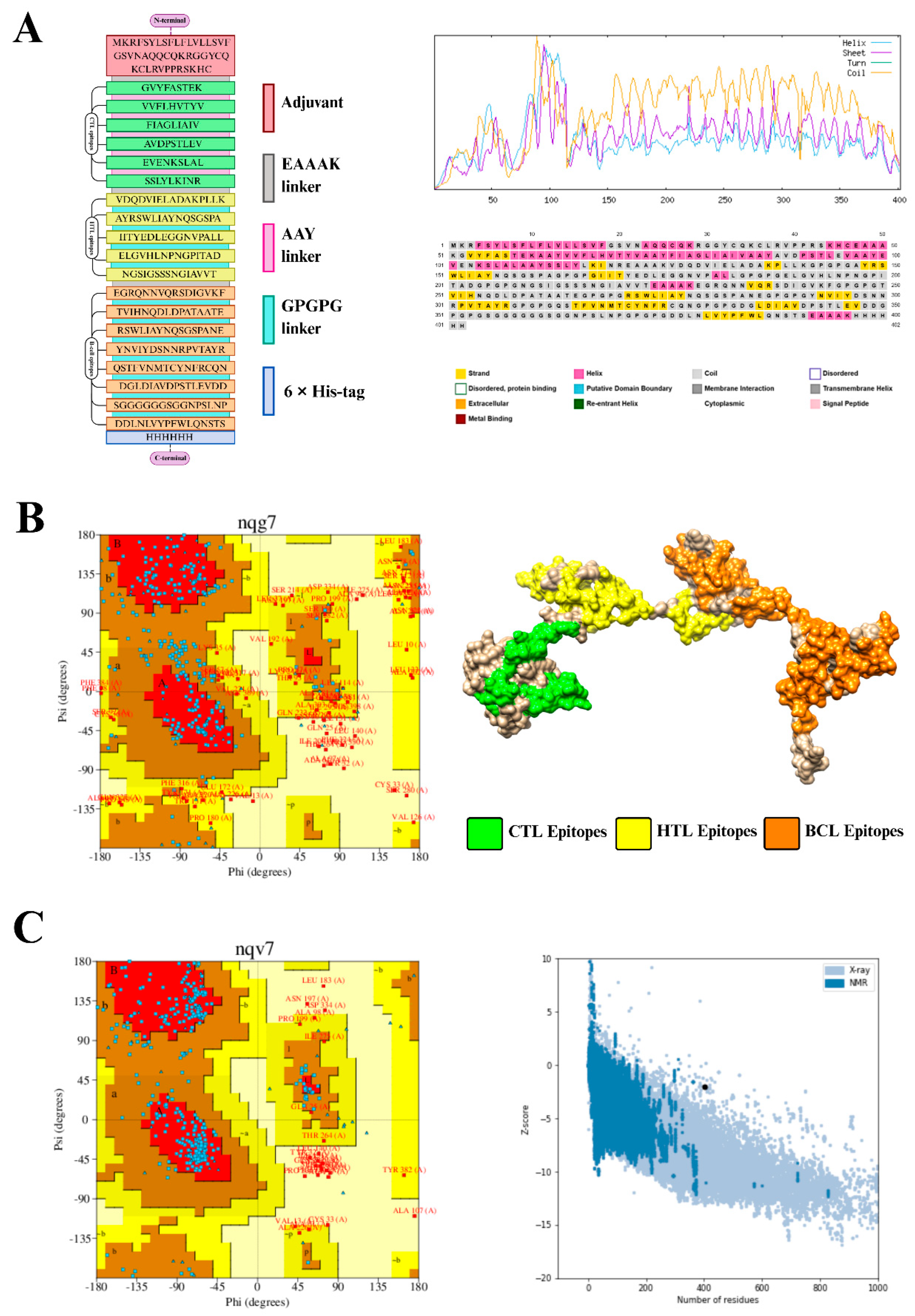
Figure 2.
Molecular docking analysis for the FADV-11 vaccine construct with host immune receptors. (A) Results of the molecular interaction/docking analysis between the FADV-11 vaccine construct and the TLR2 receptor. The 3D docked pose of the complex is shown at the top, while the bottom panel depicts the precise residue-to-residue contacts analyzed using the PDBsum web server. Here, chain A represents the vaccine construct and chain B represents the receptor molecule. The interacting amino acid residues of each respective chain are shown in shaded boxes, with the corresponding lines indicating specific intermolecular forces such as hydrogen bonds, salt bridges, and non-bonded contacts. (B) Results of the molecular interaction/docking analysis between the FADV-11 vaccine construct and the TLR4 receptor. The top panel shows the 3D docked configuration of the complex, and the bottom panel highlights the interacting residues between the vaccine construct (chain A) and the receptor molecule (chain B) analyzed via PDBsum. Shaded boxes designate the interacting amino acid residues in each chain, while the connecting lines illustrate the structural and chemical interactions. (C) Results of the molecular interaction/docking analysis between the FADV-11 vaccine construct and the heterodimeric TLR1 and TLR2 receptor complex. The top panel presents the 3D docked structure, while the bottom panel details the residue-level interactions between the vaccine construct (chain A) and the heterodimeric receptor chains (chain B). The shaded boxes and connecting lines map out the specific interacting amino acids and the corresponding intermolecular forces (hydrogen bonds, salt bridges, and hydrophobic interactions) maintaining the stability of the docked complex. The docking results collectively demonstrate highly stable molecular interactions across all three major immune receptor profiles, indicating the robust immunogenic potential of the designed FADV-11 vaccine construct. Note: Abbreviations: PDB: Protein Data Bank; TLR1: Toll-like receptor 1; TLR2: Toll-like receptor 2; TLR4: Toll-like receptor 4.
Figure 2.
Molecular docking analysis for the FADV-11 vaccine construct with host immune receptors. (A) Results of the molecular interaction/docking analysis between the FADV-11 vaccine construct and the TLR2 receptor. The 3D docked pose of the complex is shown at the top, while the bottom panel depicts the precise residue-to-residue contacts analyzed using the PDBsum web server. Here, chain A represents the vaccine construct and chain B represents the receptor molecule. The interacting amino acid residues of each respective chain are shown in shaded boxes, with the corresponding lines indicating specific intermolecular forces such as hydrogen bonds, salt bridges, and non-bonded contacts. (B) Results of the molecular interaction/docking analysis between the FADV-11 vaccine construct and the TLR4 receptor. The top panel shows the 3D docked configuration of the complex, and the bottom panel highlights the interacting residues between the vaccine construct (chain A) and the receptor molecule (chain B) analyzed via PDBsum. Shaded boxes designate the interacting amino acid residues in each chain, while the connecting lines illustrate the structural and chemical interactions. (C) Results of the molecular interaction/docking analysis between the FADV-11 vaccine construct and the heterodimeric TLR1 and TLR2 receptor complex. The top panel presents the 3D docked structure, while the bottom panel details the residue-level interactions between the vaccine construct (chain A) and the heterodimeric receptor chains (chain B). The shaded boxes and connecting lines map out the specific interacting amino acids and the corresponding intermolecular forces (hydrogen bonds, salt bridges, and hydrophobic interactions) maintaining the stability of the docked complex. The docking results collectively demonstrate highly stable molecular interactions across all three major immune receptor profiles, indicating the robust immunogenic potential of the designed FADV-11 vaccine construct. Note: Abbreviations: PDB: Protein Data Bank; TLR1: Toll-like receptor 1; TLR2: Toll-like receptor 2; TLR4: Toll-like receptor 4.
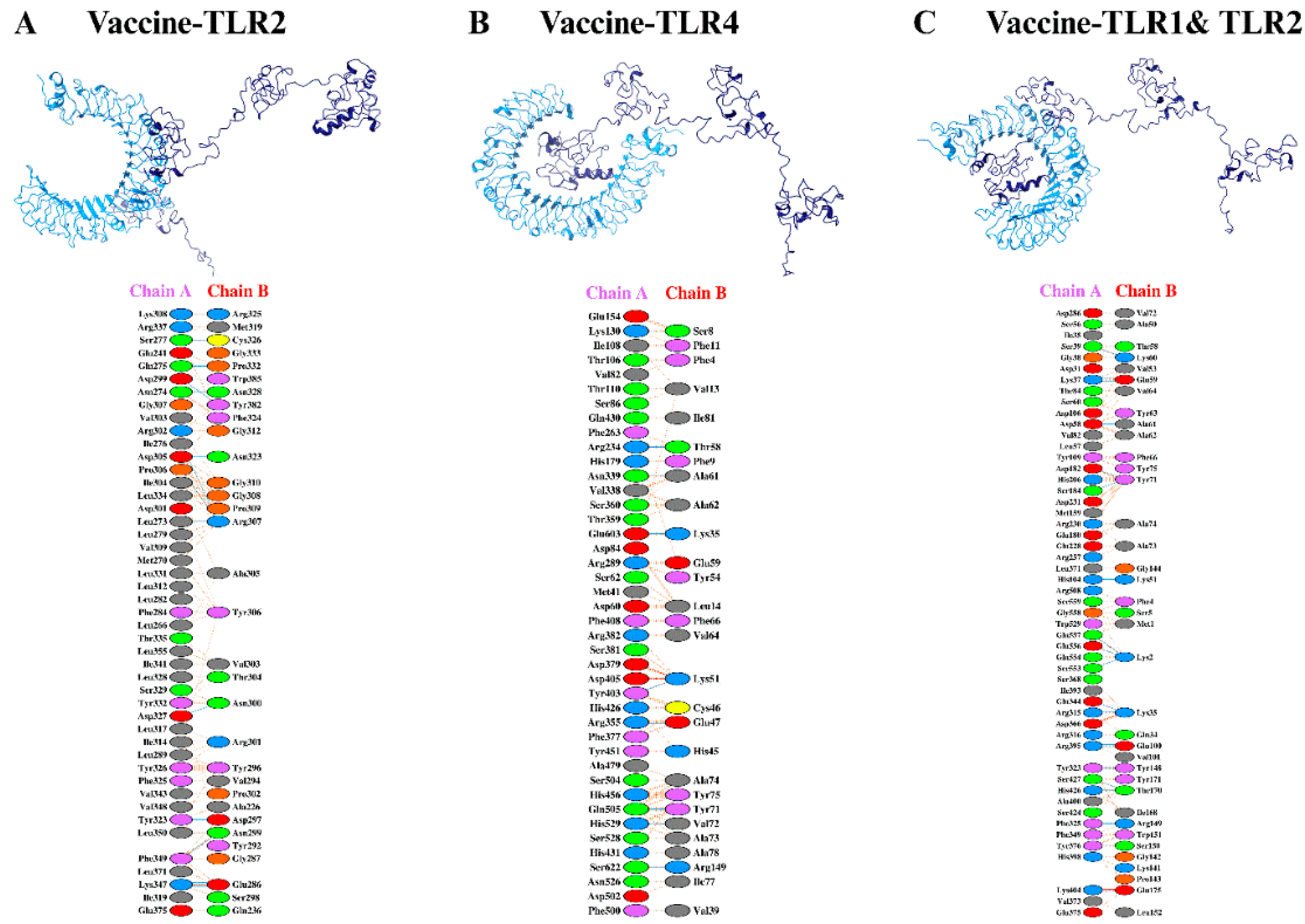
Figure 3.
Molecular dynamics (MD) simulation analysis of the FADV-11 vaccine construct in complex with host immune receptors. The trajectory of the molecular dynamics simulations was analyzed over a 100 ns production run to evaluate the structural stability, flexibility, and compactness of the engineered FADV-11 vaccine construct in complex with three chicken immune receptors: TLR2, TLR4, and the TLR1/TLR2 heterodimer. (A) Root mean square deviation (RMSD) plot of the FADV-11 vaccine construct (orange) and the TLR1/TLR2 heterodimer (blue) complex over the 100 ns simulation period. (B) RMSD plot showing the structural equilibrium of the FADV-11 vaccine construct (orange) in complex with the TLR2 receptor (blue). (C) RMSD plot illustrating the stability of the FADV-11 vaccine construct (orange) in complex with the TLR4 receptor (blue). (D) Root mean square fluctuation (RMSF) profiles plotted against residue numbers, comparing the individual residue-level fluctuations of the TLR2 (blue), TLR1/TLR2 (orange), and TLR4 (green) complexes to identify highly flexible and stable regions. (E) Radius of gyration Rg profiles of the TLR2 (blue), TLR1/TLR2 (orange), and TLR4 (green) complexes over the course of the 100 ns simulation, indicating the overall structural compactness and folding stability of the docked complexes. (F) Solvent accessible surface area (SASA) trajectories for the TLR2 (blue), TLR1/TLR2 (orange), and TLR4 (green) complexes, showing the relative stability and exposure of the hydrophilic/hydrophobic surface areas over time. Note: Abbreviations: MD: Molecular dynamics; Rg: Radius of gyration; RMSD: Root mean square deviation; RMSF: Root mean square fluctuation; SASA: Solvent accessible surface area; TLR: Toll-like receptor.
Figure 3.
Molecular dynamics (MD) simulation analysis of the FADV-11 vaccine construct in complex with host immune receptors. The trajectory of the molecular dynamics simulations was analyzed over a 100 ns production run to evaluate the structural stability, flexibility, and compactness of the engineered FADV-11 vaccine construct in complex with three chicken immune receptors: TLR2, TLR4, and the TLR1/TLR2 heterodimer. (A) Root mean square deviation (RMSD) plot of the FADV-11 vaccine construct (orange) and the TLR1/TLR2 heterodimer (blue) complex over the 100 ns simulation period. (B) RMSD plot showing the structural equilibrium of the FADV-11 vaccine construct (orange) in complex with the TLR2 receptor (blue). (C) RMSD plot illustrating the stability of the FADV-11 vaccine construct (orange) in complex with the TLR4 receptor (blue). (D) Root mean square fluctuation (RMSF) profiles plotted against residue numbers, comparing the individual residue-level fluctuations of the TLR2 (blue), TLR1/TLR2 (orange), and TLR4 (green) complexes to identify highly flexible and stable regions. (E) Radius of gyration Rg profiles of the TLR2 (blue), TLR1/TLR2 (orange), and TLR4 (green) complexes over the course of the 100 ns simulation, indicating the overall structural compactness and folding stability of the docked complexes. (F) Solvent accessible surface area (SASA) trajectories for the TLR2 (blue), TLR1/TLR2 (orange), and TLR4 (green) complexes, showing the relative stability and exposure of the hydrophilic/hydrophobic surface areas over time. Note: Abbreviations: MD: Molecular dynamics; Rg: Radius of gyration; RMSD: Root mean square deviation; RMSF: Root mean square fluctuation; SASA: Solvent accessible surface area; TLR: Toll-like receptor.
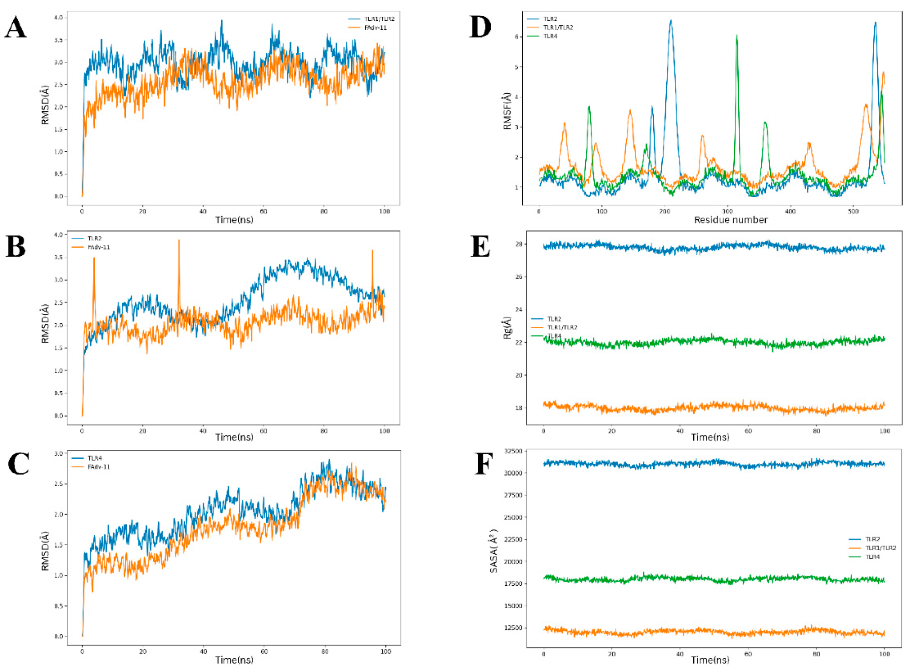
Figure 4.
Immune simulations for the FADV-11 vaccine construct. The outcomes of the immune simulations were generated using the designed vaccine construct via the online C-ImmSim server. Different parameters, including B cell, helper and cytotoxic T cell populations, were determined, along with the concentration of memory cells, cytokine levels, and active antibody titers.
Figure 4.
Immune simulations for the FADV-11 vaccine construct. The outcomes of the immune simulations were generated using the designed vaccine construct via the online C-ImmSim server. Different parameters, including B cell, helper and cytotoxic T cell populations, were determined, along with the concentration of memory cells, cytokine levels, and active antibody titers.
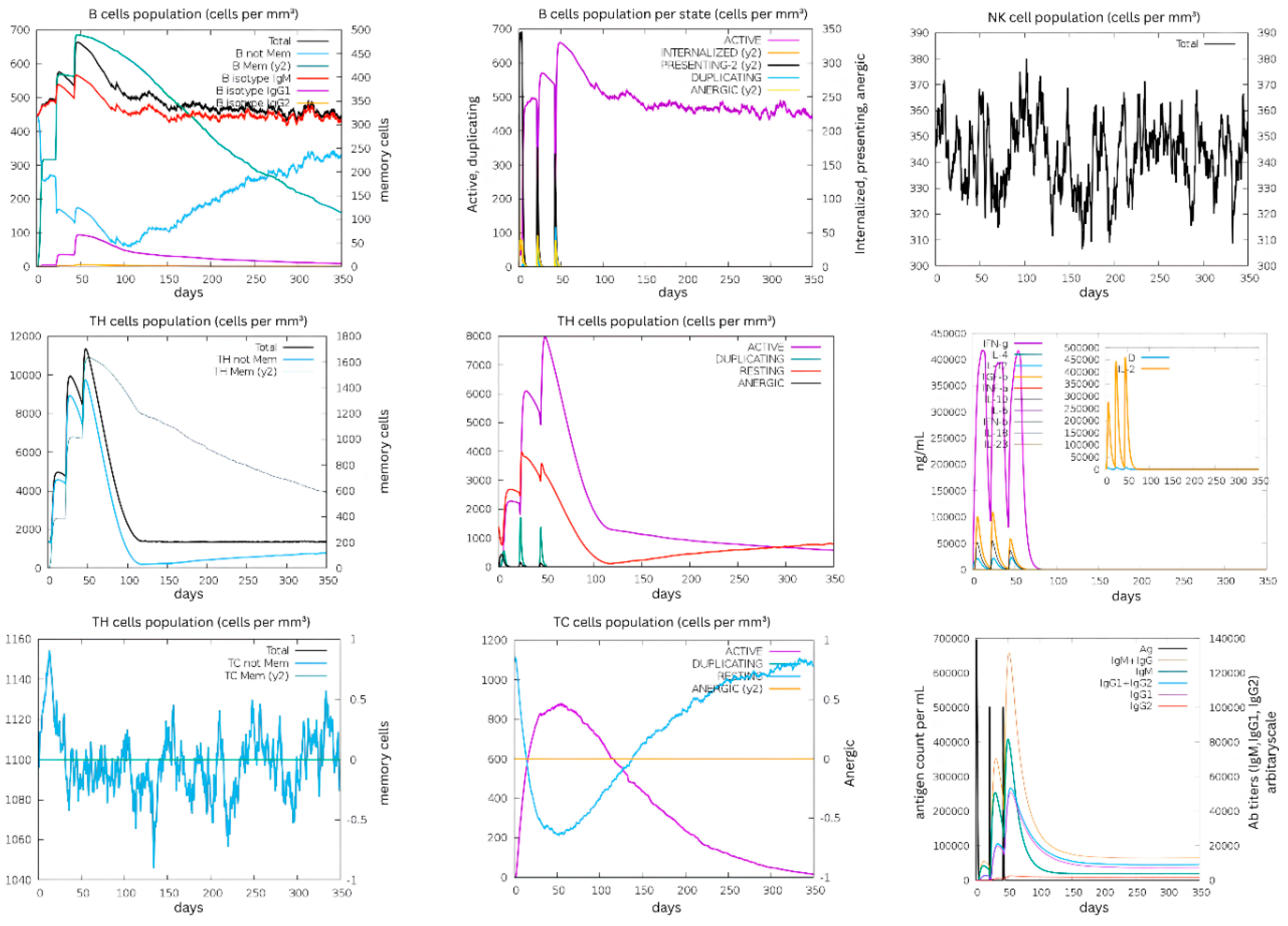
Figure 5.
In silico cloning and codon optimization for the FADV-11 vaccine construct and the results of in silico cloning. First, the coding sequence was optimized using the JCat server; later, the vaccine construct was cloned into the commercially available E. coli expression vector using the SnapGene tool (the inserted vaccine construct sequence is highlighted in red). The generated recombinant plasmid clone was 6,541 base pairs long.
Figure 5.
In silico cloning and codon optimization for the FADV-11 vaccine construct and the results of in silico cloning. First, the coding sequence was optimized using the JCat server; later, the vaccine construct was cloned into the commercially available E. coli expression vector using the SnapGene tool (the inserted vaccine construct sequence is highlighted in red). The generated recombinant plasmid clone was 6,541 base pairs long.
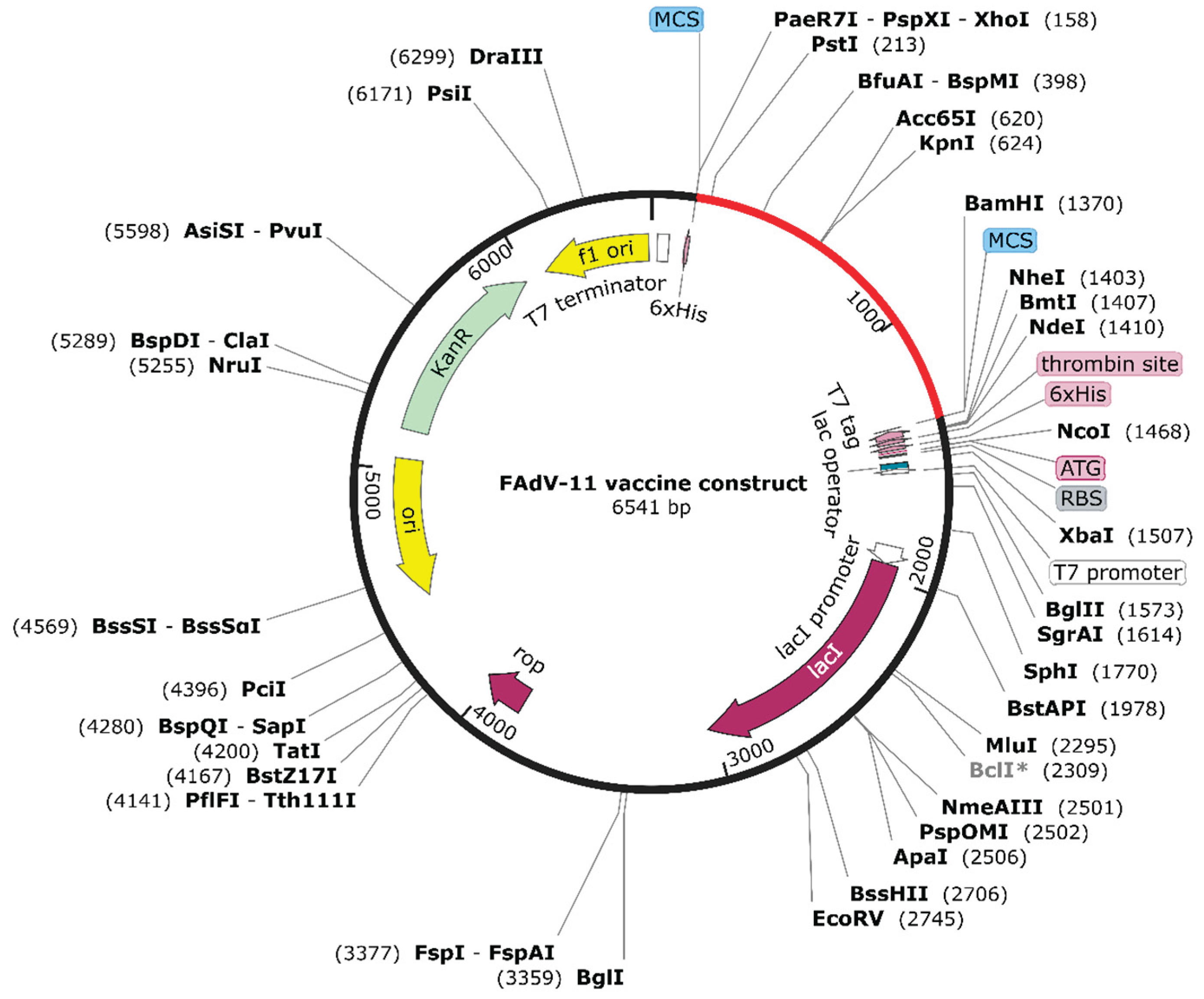
Table 2.
List of B-cell epitopes predicted Fiber & Penton protein of FAdV-11 isolates.
| Protein type | Sequence | Score | VaxiJen | AllerTop | ToxinPred |
|---|---|---|---|---|---|
| Fiber | SGGGGGGSGGNPSLNP | 0.95 | Immunogen (66%) | Non-allergen | Non-toxin |
| NGSIGSSSNGIAVVTD | 0.9 | Immunogen (66%) | Non-allergen | Non-toxin | |
| QSTFVNMTCYNFRCQN | 0.79 | Immunogen (100%) | Non-allergen | Non-toxin | |
| DGLDIAVDPSTLEVDD | 0.7 | Immunogen (100%) | Non-allergen | Non-toxin | |
| NSSNYKFNCAYFLQSW | 0.56 | Immunogen (66%) | Non-allergen | Non-toxin | |
| DDLNLVYPFWLQNSTS | 0.53 | Immunogen (66%) | Non-allergen | Non-toxin | |
| Penton | RSWLIAYNQSGSPANE | 0.89 | Immunogen (66%) | Non-allergen | Non-toxin |
| PDTFVAPTGFKEDNTT | 0.88 | Immunogen (66%) | Non-allergen | Non-toxin | |
| EILKQAPPINVSAVCD | 0.87 | Immunogen (66%) | Non-allergen | Non-toxin | |
| EGRQNNVQRSDIGVKF | 0.82 | Immunogen (100%) | Non-allergen | Non-toxin | |
| GVVQIYLKEGRQNNVQ | 0.78 | Immunogen (66%) | Non-allergen | Non-toxin | |
| YNVIYDSNNRPVTAYR | 0.74 | Immunogen (66%) | Non-allergen | Non-toxin | |
| YEDLEGGNVPALLDVE | 0.68 | Immunogen (66%) | Non-allergen | Non-toxin | |
| DSAADDTSELFVPVQR | 0.67 | Immunogen (66%) | Non-allergen | Non-toxin | |
| GYHPDVVLLPGCAVDF | 0.67 | Immunogen (66%) | Non-allergen | Non-toxin | |
| TVIHNQDLDPATAATE | 0.63 | Immunogen (100%) | Non-allergen | Non-toxin |
The table lists BCL epitopes predicted for fiber and penton proteins of FAdV-11. The epitope sequence is given in the second column whereas the following column contains the score for each epitope. Antigenicity, allergenicity and toxic characteristic of the epitopes are mentioned in the last three column respectively.
Table 3.
List of CTL Epitopes of FAdV-11 Fiber & Penton protein.
| Protein type | Epitope Sequence | Allele | Score | Percentile Rank | Aller-TOP | VaxiJen | Toxin-Pred |
|---|---|---|---|---|---|---|---|
| Fiber | AVDPSTLEV | HLA-A*02:01 | 0.87496 | 0.05 | Non-allergen | Immunogen (100%) |
Non-toxin |
| TPTITPSSV | HLA-B*07:02 | 0.706462 | 0.12 | Non-allergen | Immunogen (66%) | Non-toxin | |
| EVENKSLAL | HLA-B*08:01 | 0.462583 | 0.18 | Non-allergen | Immunogen (100%) |
Non-toxin | |
| VPTYESMNL | HLA-B*07:02 | 0.546975 | 0.21 | Non-allergen | Immunogen (66%) |
Non-toxin | |
| SSLYLKINR | HLA-A*11:01 | 0.40446 | 0.42 | Non-allergen | Immunogen (66%) |
Non-toxin | |
| Penton |
GVYFASTEK | HLA-A*11:01 | 0.920846 | 0.01 | Non-allergen | Immunogen (100%) | Non-toxin |
| SPRRARSVA | HLA-B*07:02 | 0.918799 | 0.04 | Non-allergen | Immunogen (66%) | Non-toxin | |
| INITRFQTL | HLA-B*08:01 | 0.740901 | 0.05 | Non-allergen | Immunogen (66%) | Non-toxin | |
| HLMSFPQSA | HLA-A*02:01 | 0.798454 | 0.08 | Non-allergen | Immunogen (66%) | Non-toxin | |
| VVFLHVTYV | HLA-A*02:01 | 0.74167 | 0.11 | Non-allergen | Immunogen (100%) | Non-toxin | |
| YQPYRVVVL | HLA-B*08:01 | 0.570928 | 0.11 | Non-allergen | Immunogen (66%) | Non-toxin | |
| FIAGLIAIV | HLA-A*02:01 | 0.641405 | 0.17 | Non-allergen | Immunogen (100%) | Non -toxin | |
| TLADAGFIK | HLA-A*11:01 | 0.639216 | 0.18 | Non-allergen | Immunogen (66%) | Non-toxin | |
| GLTVLPPLL | HLA-A*02:01 | 0.622173 | 0.18 | Non-allergen | Immunogen (100%) | Non-toxin |
The table enlists MHC-I epitopes for the fiber and penton proteins of FAdV-11. The epitope sequence is given in the second column whereas the alleles are mentioned in the following column. The fourth column contains the score for each epitope. Allergenicity, antigenicity and toxic characteristic of the epitopes are mentioned in the rest of the columns respectively.
Table 4.
List of HTL Epitopes for Fiber & Penton protein derived from FAdV-11.
| Protein type | Epitope Sequence | Allele | Score | Percentile Rank | Aller-TOP | VaxiJen | Toxin-Pred |
|---|---|---|---|---|---|---|---|
| Fiber | GLDIAVDPSTLEVDD | HLA-DRB1*04:01 | 0.804244 | 0.68 | Non-allergen | Immunogen (66%) |
Non-toxic |
| ELGVHLNPNGPITAD | HLA-DRB1*15:01 | 0.354772 | 3.9 | Non-allergen | Immunogen (66%) |
Non-toxic | |
| ELGVHLNPNGPITAD | HLA-DRB1*07:01 | 0.252852 | 6.9 | Non-allergen | Immunogen (66%) |
Non-toxic | |
| NGSIGSSSNGIAVVT | HLA-DRB1*04:01 | 0.273118 | 7 | Non-allergen | Immunogen (66%) |
Non-toxic | |
| Penton | VDQDVIELADAKPLLK | HLA-DRB1*09:01 | 0.810958 | 0.2 | Non-allergen | Immunogen (100%) | Non-toxin |
| AYRSWLIAYNQSGSPA | HLA-DRB1*04:01 | 0.875289 | 0.3 | Non-allergen | Immunogen (66%) | Non-toxin | |
| VDQDVIELADAKPLLK | HLA-DRB1*07:01 | 0.724949 | 0.88 | Non-allergen | Immunogen (100%) | Non-toxin |
|
| VDQDVIELADAKPLLK | HLA-DRB1*01:01 | 0.795971 | 1.1 | Non-allergen | Immunogen (100%) | Non-toxin |
|
| IITYEDLEGGNVPALL | HLA-DRB1*01:01 | 0.738836 | 1.3 | Non-allergen | Immunogen (66%) | Non-toxin |
|
| DIDTFNPEANHSNFRT | HLA-DRB1*04:01 | 0.660108 | 1.6 | Non-allergen | Immunogen (66%) | Non-toxin |
|
| HPDVVLLPGCAVDFTY | HLA-DRB1*01:01 | 0.668346 | 1.9 | Non-allergen | Immunogen (66%) | Non-toxin |
|
| AYRSWLIAYNQSGSPA | HLA-DRB1*01:01 | 0.514877 | 3.1 | Non-allergen | Immunogen (66%) | Non-toxin |
|
| GVVQIYLKEGRQNNVQ | HLA-DRB1*11:01 | 0.521617 | 3.5 | Non-allergen | Immunogen (66%) | Non-toxin |
The table enlists MHC-II epitopes of FAdV-11Fiber and Penton protein. The epitope sequence is given in the second column whereas the alleles are mentioned in the following column. The fourth column contains the score for each epitope. Allergenicity, antigenicity and toxic characteristic of the epitopes are mentioned in the rest of the columns respectively.
Disclaimer/Publisher’s Note: The statements, opinions and data contained in all publications are solely those of the individual author(s) and contributor(s) and not of MDPI and/or the editor(s). MDPI and/or the editor(s) disclaim responsibility for any injury to people or property resulting from any ideas, methods, instructions or products referred to in the content. |
© 2026 by the authors. Licensee MDPI, Basel, Switzerland. This article is an open access article distributed under the terms and conditions of the Creative Commons Attribution (CC BY) license (http://creativecommons.org/licenses/by/4.0/).
Copyright: This open access article is published under a Creative Commons CC BY 4.0 license, which permit the free download, distribution, and reuse, provided that the author and preprint are cited in any reuse.


