

Submitted:
15 July 2026
Posted:
16 July 2026
You are already at the latest version
Abstract
Background: Primary malignant tumors of the inferior vena cava (IVC) are extremely rare. Objectives: To evaluate the results of surgical treatment of patients with primary leiomyosarcoma of the IVC. Methods: The study included 6 patients (mean age 56.8 ± 5.9 years) diagnosed with “primary leiomyosarcoma of the inferior vena cava” who underwent radical surgery between 2020 and 2025. Complication rate, graft patency, disease progression, and survival were analyzed. Results: In one case, a lateral resection of > 50 % of the circumference with patch plasty was performed; in the remaining cases, circular resections of 8–15 cm length were carried out. Biological vascular grafts made of bovine xenopericardium were used for reconstruction; in 3 patients, renal veins were additionally grafted. Combined resections (right nephroadrenalectomy, liver resection) were performed in 5 patients. Radicality (R0) was achieved in all cases. Complications included retroperitoneal hematoma and bleeding in 2 patients; thrombosis of the infrarenal IVC segment in 1; and partial thrombosis of the right renal vein graft in 1 patient (after 12 months). Kidney injury and venous insufficiency did not develop. Graft patency was preserved in the remaining cases. There was no mortality and no local recurrences were detected. Disease progression was observed in 1 patient after surgery (lung metastases after 6 months and liver metastases after 26 months). Mean follow-up was 32.5 months. Conclusions: Radical resection and reconstruction of the inferior vena cava using biological vascular grafts is a technically feasible, safe, and effective surgical approach for primary leiomyosarcoma.
Keywords:
leiomyosarcoma
; inferior vena cava
; vascular reconstruction
; combined resection
; vascular graft
; biological vascular graft
; bovine xenopericardium
; oncovascular surgery
1. Introduction
Primary malignant neoplasms of blood vessels, originating directly from their walls, are an extremely rare pathology in clinical practice. These mesenchymal tumors develop more frequently in large veins than in arteries. Among them, venous leiomyosarcoma is the most common, primarily localized in the inferior vena cava (IVC), which accounts for over 60% of all reported cases. Despite being the most common malignant tumor of the IVC, leiomyosarcoma remains a rare disease, with most publications limited to case reports or small retrospective series.
According to epidemiological data, the incidence of this pathology in the adult population is less than 0.001% of all malignancies, and about 0.5% of the total number of soft tissue sarcomas [1]. The first description of leiomyosarcoma of the IVC was reported by L. Perl and R. Virchow in 1871 [2]. According to systematic reviews, approximately 450 cases have been reported in the world literature to date [3], which highlights the exceptional rarity of this disease and the need for further accumulation of clinical experience to develop standardized approaches to diagnosis and treatment.
Primary leiomyosarcoma of the IVC develops from smooth muscle cells of the tunica media of the vascular wall and can have an intraluminal, extraluminal, or mixed type of growth. The tumor is characterized by aggressive clinical behavior with an unfavorable prognosis. According to some researchers, the true incidence of vascular leiomyosarcomas is underestimated [4].
For decades, tumors with invasion of the IVC were considered unresectable. However, the accumulated clinical experience, the introduction of modern technologies, and the improvement of surgical techniques have made it possible to change the treatment strategy. In modern clinical practice, radical resection and reconstruction of the IVC are considered the most effective way to achieve long-term survival. Nevertheless, there are still a number of unresolved issues related to the choice of the optimal option for venous reconstruction, the optimal plastic material, the role of multivisceral interventions, and multimodal approaches (chemotherapy and radiation therapy in combination with surgery).
This article presents a series of six patients who underwent surgical treatment for primary leiomyosarcoma of the IVC. The features of IVC resection are described, and options for its reconstruction using biological vascular grafts from bovine xenopericardium are proposed.
2. Materials and Methods
The study was carried out in specialized cardiovascular and oncology centers. Inclusion criteria were: patients with a definitively established, morphologically confirmed diagnosis of primary leiomyosarcoma of the IVC originating from the smooth muscle cells of the venous wall. Exclusion criteria were: patients with other morphological variants of malignancies involving the IVC that are different from primary leiomyosarcoma.
The study included 6 patients with primary leiomyosarcoma of the IVC (5 females and 1 male) who underwent surgery between January 2020 and November 2025. The median age was 56.8 ± 5.9 years. All participants signed free and informed consent forms after being given information about the study. The morphological structure of the tumor was determined by biopsy material obtained before and after surgery. The preoperative examination included: clinical and laboratory tests; ultrasound examination of the abdominal cavity and retroperitoneal space; computed tomography angiography (CTA); magnetic resonance angiography (MRA); and preoperative puncture biopsy.
Intraoperative data recorded the localization, nature, and extent of the IVC lesion, as well as the involvement of surrounding organs and tissues in the tumor process. To standardize surgical approach, the IVC was classified into five anatomical segments: infrarenal (below the confluence of the renal veins), pararenal (at the level of the renal veins confluence), suprarenal (above the confluence of the renal veins), retrohepatic (at the level of the main hepatic veins), suprahepatic (from the diaphragm to the right atrium).
Venous reconstruction was performed using biological vascular grafts made of bovine xenopericardium. Synthetic grafts were not used. Reconstruction options included: IVC resection and patch plasty; resection and prosthetic replacement of the IVC with a biological vascular graft; resection and prosthetic replacement of the IVC with a biological graft including reimplantation of the renal veins; and resection of the IVC and renal veins with IVC prosthesis and reimplantation of the renal vein(s) into the graft.
Some technical aspects of radical resection and reconstruction of the IVC for primary malignant tumors have been described in the literature earlier [5]. The choice of surgical approach was determined by tumor location and extent according to the segmental classification of the IVC. When the infra- and pararenal segments were involved, a right lateral laparotomy was performed. When isolation of the retrohepatic portion of the IVC was required, a right-sided thoracoabdominal incision with mobilization of the right lobe of the liver was used. When resection involved more than 50% of the IVC diameter, reconstruction was carried out using a xenopericardial patch to avoid hemodynamically significant stenosis. In cases of circumferential resection, tubular biological vascular grafts were used, created intraoperatively from bovine xenopericardial sheets according to the diameter and length of the resected segment. The anastomosis was performed from the upper edge of the resection to the lower edge, which allowed for earlier restoration of venous outflow through the renal veins. In organ-preserving procedures, prosthetic replacement and/or reimplantation of the renal veins into the IVC graft was performed to prevent venous hypertension and renal injury. In cases of severe IVC compression and the development of hemodynamic instability due to impaired venous return to the heart, parallel cardiopulmonary bypass with peripheral cannulation (femoral vessels and right internal jugular vein) was used. Before removing the clamps and restoring blood flow through the graft and veins, prophylaxis against thromboembolism and air embolism was carried out.
Early postoperative mortality was defined as death within 30 days postoperatively or prior to discharge. Follow-up was conducted by vascular surgeons and surgical oncologists every 3 months during the first year, and every 6 months thereafter. Graft patency, as well as IVC and renal vein patency, was assessed clinically and by abdominal and renal ultrasound, abdominal CTA/MRA. Standard biomedical statistics methods were used to analyze demographic data, oncological characteristics, and perioperative and postoperative outcomes. Statistical analyses were performed using R software version 4.5.2 within the RStudio integrated development environment.
3. Results
In most patients, the disease manifested with nonspecific symptoms: general malaise, weakness, decreased appetite, and abdominal pain. In two patients (33.3%), the tumor was detected incidentally during diagnostic studies – abdominal ultrasound, magnetic resonance angiography, and computed tomography angiography of the abdominal cavity. Mild edema of the lower extremities was noted in three patients (50%). One patient experienced marked weight loss of 26 kg (24.5% of baseline body weight) over a six-month period.
All patients underwent a comprehensive non-invasive examination. Ultrasound of the abdominal cavity and triphasic multislice computed tomography angiography were performed in all patients (100%). Magnetic resonance angiography was performed in three patients (50%). The frequency and localization of leiomyosarcoma according to the segmental classification of the IVC are shown in Figure 1.
In one case, the tumor was located in the pararenal segment of the IVC; in two cases, it was mainly in the infrarenal segment with spread to the pararenal segment; and in three cases, it was in the suprarenal segment with involvement of the pararenal region. There were no patients with tumor localization or spread to the retrohepatic or suprahepatic segments of the IVC in the study group.
Regarding the pattern of tumor growth, the following was observed: intraluminal growth in one patient (16.7%), extraluminal growth in two patients (33.3%), and mixed growth in three patients (50%). In one patient (16.7%), complete obstruction of the IVC and left renal vein was caused by the tumor with local occlusive thrombosis in the infrarenal segment and non-occlusive thrombosis of both common iliac veins. The mean maximum tumor diameter was 14.3 cm (range, 10.5–16.4 cm). Preoperative morphological verification of the tumor was performed in three patients (50%) by ultrasound-guided percutaneous puncture biopsy. Neoadjuvant chemotherapy and radiation therapy were not used in the study group.
Abdominal approach (right-sided oblique laparotomy) and the thoracoabdominal approach were used equally often – three cases each. All patients underwent radical resection and reconstruction of the IVC. Biological vascular grafts made of bovine xenopericardium were used as the reconstructive material.
In one of the six cases, resection of the anterolateral wall of the IVC and a portion of the ostia of the renal veins was performed, with repair of the defect using a xenopericardial patch (Figure 2).
Five patients underwent extended circular resection of the IVC (8 to 15 cm) and prosthetic replacement with a biological vascular graft made of bovine xenopericardium. Resection of the pararenal segment of the IVC required restoration of renal venous outflow in three patients (50%). In one case, the left renal vein was implanted end-to-side into the IVC graft, which ensured preservation of adequate venous drainage and prevention of renal venous hypertension and kidney injury (Figure 3).
Following the principles of oncological radicality, organ-preserving surgery with kidney preservation was achieved in two patients. Vascular reconstruction included resection and prosthetic replacement of the renal veins with subsequent reimplantation into the IVC graft (Figure 4).
The chosen surgical approach preserved renal venous drainage and prevented the development of acute kidney injury in the perioperative period. Five patients underwent combined resection of the IVC. Two patients underwent resection of segment IV of the liver, and three underwent right-sided nephradrenalectomy. This approach achieved a radical surgical resection (Figure 5 and Figure 6).
Morphological examination confirmed R0 resection in all patients, indicating the radical nature of the interventions and complete removal of tumor.
In two cases, resection of segments II–III of the IVC caused systemic hemodynamic instability, which required the use of parallel extracorporeal bypass. This approach ensured the safety of the surgical intervention and maintained adequate organ perfusion during complex vascular reconstruction.
Postoperative complications were noted in two patients: bleeding and retroperitoneal hematoma, both requiring reoperation. Graft thrombosis occurred in one case (16.7%) two days after reoperation in a patient who had complete tumor occlusion of the IVC and occlusive thrombosis of the infrarenal vena cava. Treatment of the thrombosis was conservative, as a collateral venous network had already formed due to tumor occlusion, and no clinical manifestations of venous insufficiency of the pelvis or lower extremities were observed. At 12-month follow-up, one additional patient had partial thrombosis of the right renal vein graft, but no clinical manifestations or laboratory signs of kidney injury were detected. In all other cases, patency of the IVC and vascular grafts was maintained throughout the follow-up.
There was no mortality in the early or late postoperative periods. No local tumor recurrence was detected; however, one patient developed systemic disease progression after surgery (lung metastases at 6 months and liver metastases at 26 months). Two patients received adjuvant chemotherapy with doxorubicin-based regimens as recommended by the oncologist. The median follow-up duration was 36 months. Clinical and pathological characteristics of the patients are presented in Table 1.
4. Discussion
Primary leiomyosarcoma of the IVC is a rare malignant tumor characterized by relatively slow growth but aggressive biological behavior. According to the literature, leiomyosarcoma of the IVC accounts for approximately 60% of all vascular leiomyosarcomas [6]. The tumor is more commonly diagnosed in older women (in the sixth decade of life), although isolated cases of leiomyosarcoma have been reported in pediatric patients [7]. Clinical symptoms in the early stages of the disease remain nonspecific, which makes timely diagnosis difficult. Most patients presented with nonspecific complaints – malaise, weakness, loss of appetite, and abdominal or lumbar pain. According to the literature, up to 30% of cases of leiomyosarcoma of the IVC are diagnosed incidentally [8].
Analysis of the topographic location of the tumor showed predominant involvement of the suprarenal and infrarenal segments of the IVC, which corresponds to the results of series reported in the literature. In some cases, multisegmented vein involvement was observed. As a rule, this significantly complicates surgical decision-making and requires extended reconstructive interventions – an opinion shared by most researchers [9].
A key point of discussion remains the radicality of surgical resection. According to Hollenbeck et al. (2003) and Jeong et al. (2019), incomplete tumor removal (R1/R2 resection) is associated with a high risk of local recurrence and systemic progression, which directly impacts overall survival [10]. Recent multicenter studies highlight the importance of achieving R0 resection, which provides improved long-term outcomes, including a five-year survival rate of over 90% [11]. In our series, radical resection with vascular reconstruction was performed in all cases. This approach achieved R0 status even in multivisceral resections. These results are consistent with those of other authors and confirm the importance of a radical surgical approach as the standard of care for leiomyosarcoma of the inferior vena cava.
Radical resection (R0) of primary leiomyosarcoma of the IVC is associated with significant technical difficulties due to the close anatomical relationship of the IVC with the hepatic, renal, and visceral veins. The greatest challenges arise with extensive involvement of two or more segments of the IVC, particularly the renal segment. In such clinical situations, the optimal approach may be to perform combined resections with restoration of adequate venous outflow from the kidneys and lower extremities.
The high frequency of combined interventions in this series (83.3%) reflects the aggressive biological behavior of leiomyosarcoma of the IVC and the need for an advanced surgical approach. These results are consistent with those of other authors. Hollenbeck et al. (2003) reported that combined resections were performed in the majority of their patients, dictated by the anatomical proximity of the tumor to the liver, kidneys, and adrenal glands. Similarly, Kieffer et al. (2006) performed combined resections in more than half of their patients [12]. These findings are supported by a multicenter analysis by Nooromid et al. (2021), which emphasizes the impossibility of radical tumor removal without resection of adjacent organs and vascular structures. The high frequency of combined resections in the present series is a natural consequence of the topographic features of the IVC and the biological behavior of the tumor.
Vascular reconstruction remains the most complex and technically demanding stage of surgical treatment for patients with primary leiomyosarcoma of the IVC. It requires a highly skilled surgeon, a significant time investment, and is associated with the risk of several complications, such as infection, graft thrombosis, and hemodynamic disorders. In contrast, resection of the infrarenal segment of the IVC may be safer. According to some authors, this approach minimizes surgical risks and provides acceptable outcomes both in terms of perioperative complications and overall and recurrence-free survival [13,14]. However, most studies emphasize that combining radical resection with mandatory restoration of venous outflow increases the likelihood of achieving R0 resection and improves long-term outcomes [15,16].
In our opinion, radical resection with mandatory vascular reconstruction should be considered the standard of surgical care for primary leiomyosarcoma of the IVC. In certain clinical situations where the tumor completely obstructs the IVC lumen and thrombosis of the iliocaval segment is present, ligation of the infrarenal IVC may be considered a permissible surgical option. The main prerequisite for the relative safety of IVC ligation is the presence of well-developed collateral venous outflow from the lower extremities and pelvic organs. Nevertheless, such an approach requires a thorough preoperative assessment of the venous bed and the degree of collateral circulation, as well as mandatory postoperative follow-up.
The choice of optimal graft material for IVC reconstruction remains a subject of debate in vascular surgery. Despite the availability of various options, synthetic vascular grafts are traditionally preferred in most specialized centers. Polytetrafluoroethylene (PTFE) and polyethylene terephthalate (PET) grafts are the most commonly used, with reinforced PTFE grafts being employed more frequently in certain cases [17]. However, the use of synthetic vascular grafts in clinical practice is associated with several limitations, including the risk of infectious and thrombotic complications [18], as well as the formation of a duodenocaval fistula – a rare but extremely dangerous complication resulting from pressure necrosis of the intestinal wall in direct contact with a vascular graft [19]. These concerns have prompted interest in alternative materials with higher biocompatibility, low thrombogenicity, and greater resistance to infectious complications.
In the present series, biological vascular grafts made of bovine xenopericardium were used for reconstruction of the IVC and renal veins. The results demonstrated satisfactory patency rates and a low complication rate, consistent with data from international centers where similar grafts have shown comparable efficacy [20].
Biological materials have certain advantages over synthetic analogues, including better integration into the vascular wall and a potential reduction in the risk of infectious complications. According to some literature sources, when infectious complications develop postoperatively following the use of biological grafts for IVC resection, the clinical course is generally more favorable and can often be managed without the need for reoperation [21].
The use of biological vascular grafts should be considered a promising direction in reconstructive surgery of large veins, particularly in oncovascular surgery, where radical intervention must be combined with minimizing the risk of severe complications. The results of our study, demonstrating preserved patency of the reconstructed IVC segments in the long term, confirm the viability of this technique. However, it should be noted that a definitive evaluation of the long-term efficacy of xenopericardial biological grafts requires multicenter studies with larger patient cohorts. Such studies will enable the development of standards for their use in oncovascular surgery.
The results of surgical treatment of patients with primary leiomyosarcoma of the IVC indicate a relatively low incidence of postoperative complications, comparable to data from the world literature. In the present series, complications in the form of bleeding and retroperitoneal hematoma occurred in two patients (33.3%), which corresponds to the range of 30–40% reported in the literature.
Thrombotic complications occurred in two cases. In one patient, graft thrombosis developed two days after repeat intervention for bleeding, probably due to mechanical compression of the graft by gauze swabs placed against the liver for hemostasis in the setting of consumption coagulopathy. In the second case, partial thrombosis of the biological right renal vein graft was detected after 12 months of follow-up and was apparently due to compression by a biloma that had formed in the subhepatic space in the postoperative period. Both episodes were attributable to external factors rather than primary graft failure. These findings are consistent with reports from other authors indicating that thrombotic complications following IVC reconstruction can occur with both biological and synthetic grafts, and that the main causes are often technical factors such as compression, kinking, or hemodynamic disturbances [22]. In both cases, no clinical manifestations of venous insufficiency or acute/chronic kidney injury were observed.
Adjuvant chemotherapy with doxorubicin was administered to two patients in accordance with current guidelines for the treatment of soft tissue sarcomas, including leiomyosarcomas of the IVC. However, there is evidence in the literature suggesting a limited effect of doxorubicin on the overall survival of patients in this category [23]. However, a multicenter study (LMS-04) demonstrated that the combination of doxorubicin and trabectedin, compared with doxorubicin alone, improved progression-free survival in patients with metastatic or unresectable leiomyosarcoma, although it was associated with greater toxicity [24].
The use of parallel extracorporeal bypass in two patients made it possible to stabilize hemodynamics during resection of segments II–III of the IVC. Venovenous bypass and total hepatic vascular exclusion are widely used to improve safety in complex vascular reconstructions. Combined procedures including nephroadrenalectomy and liver resection achieved radical resection in patients with advanced disease. Despite the extended scope of surgical intervention, no deaths occurred in the study group. These findings confirm that combined resection with vascular reconstruction can be performed safely, provided that patients are carefully selected and the principles of oncological radicality are observed.
Organ-preserving interventions, including resection and prosthetic replacement of the renal veins with reimplantation into the IVC graft, preserved functional renal integrity and prevented the development of venous hypertension and kidney injury. The importance of restoring physiological renal venous outflow, particularly from the right kidney, to prevent renal injury is also emphasized in other publications [25].
The absence of mortality in both the early and late postoperative periods is of paramount importance. This finding demonstrates that such complex surgical interventions can be performed safely, provided they are carried out in specialized centers with the necessary experience and technical resources.
The long-term results of our study demonstrate the absence of local recurrence and a low incidence of systemic progression. The median follow-up was 36 months, which is comparable to or higher than that reported in other series, where median recurrence-free survival ranged from 7 to 12 months. Cumulative analysis of contemporary treatment outcomes supports an integrated approach to primary IVC leiomyosarcoma, in which resection and reconstruction represent a justified strategy with satisfactory results and acceptable risk.
It should also be emphasized that successful treatment of patients with primary leiomyosarcoma of the IVC is possible only within the context of a multidisciplinary approach. The team should include a pathologist, radiology specialists, a vascular surgeon with experience in oncovascular surgery, a surgical oncologist, a radiation oncologist, and a medical oncologist. Such an integrated approach ensures not only the radicality of the intervention but also minimizes the risk of complications, contributing to the achievement of optimal oncological and functional outcomes.
5. Conclusions
Surgical treatment of patients with primary leiomyosarcoma of the inferior vena cava using biological xenopericardial grafts can be considered a safe and effective method that achieves high radicality, satisfactory survival rates, and good quality of life for patients.
Author Contributions
Conceptualization, H.A.P. and I.A.M.; methodology, H.A.P. and I.A.M.; validation, I.A.M., V.A.Y. and A.A.K.; formal analysis, I.A.M. and V.A.Y.; investigation, H.A.P., I.A.M. and A.A.K.; data curation, H.A.P., I.A.M. and A.A.K.; writing—original draft preparation, I.A.M.; writing—review and editing, all authors; visualization, I.A.M. and V.A.Y.; supervision, H.A.P.; project administration, H.A.P. All authors have read and agreed to the published version of the manuscript.
Funding
This research received no external funding.
Institutional Review Board Statement
The study was conducted in accordance with the Declaration of Helsinki and approved by the Institutional Review Board of Vascular Surgery Research Laboratory (protocol code 014V2020; originally approved on January 15, 2020 with subsequent annual renewals).
Informed Consent Statement
Informed consent was obtained from all subjects involved in the study.
Data Availability Statement
The data are not publicly available due to ethical reasons. Further information is available from the corresponding authors upon reasonable request, subject to institutional review board approval and applicable Belarusian data protection regulations.
Conflicts of Interest
The authors declare no conflict of interest.
References
- Wang, M.X.; Menias, C.O.; Elsherif, S.B.; Segaran, N.; Ganeshan, D. Current update on IVC leiomyosarcoma. Abdom Radiol (NY). 2021, 46, 5284-5296. [CrossRef]
- Perl, L.; Virchow, R. Ein Fall von Sarkom der Vena cava inferior [Leiomyosarcoma of inferior vena cava]. Virchows Arch Pathol Anat. 1871, 53, 378–383. [CrossRef]
- Gonzalez, J.; Garcia, L.E.; Luna, M.; Cruz, L.; Barragan, A.; González, M. Leiomyosarcoma of the inferior vena cava: Case report and review of the literature. Eur J Surg Oncology. 2024, 50, 109231. [CrossRef]
- Mingoli, A.; Feldhaus, R.J.; Cavallaro, A.; Stipa, S. Leiomyosarcoma of the inferior vena cava: analysis and search of world literature on 141 patients and report of three new cases. J Vasc Surg. 1991, 14, 688-699. [CrossRef]
- Popel, H.A.; Maiseyenka, I.A.; Popel, A.N.; Yermachenka, V.A. Epithelioid Hemangioendothelioma of the Inferior Vena Cava, a Rare Vascular Tumor: A Brief Literature Review with a Case Report. Surg East Eur. 2025, 14, 522-538. [CrossRef]
- Di Pilla, M.A.; Capuano, M.A.; Rossi, M.; Di Pilla, G.; Minelli, R.; Pizzicato, P.; Rossi, A.; Paviglianiti, G.; Irace, D.; Vallone, G.; Salvia, A.A.H.; Smaldone, M.C.; Cariello, V.; Zeccolini, R.; Rossi, E. Leiomyosarcoma of the inferior vena cava. Radiol Case Rep. 2023, 19, 382-386. [CrossRef]
- Yoshizawa, K.; Ohno, Y.; Kurata, T.; Takagi, Y.; Kasai, T.; Takizawa, M.; Soejima, Y. Primary leiomyosarcoma of the inferior vena cava in a pediatric case: a case report and literature review. Surg Case Rep. 2023, 9, 52. [CrossRef]
- Hollenbeck, S.T.; Grobmyer, S.R.; Kent, K.C.; Brennan, M.F. Surgical treatment and outcomes of patients with primary inferior vena cava leiomyosarcoma. J Am Coll Surg. 2003, 197, 575-579. [CrossRef]
- Nooromid, M.; De Martino, R.; Squizzato, F.; Benedetto, F.; De Caridi, G.; Chou, E.L.; Conrad, M.F.; Pantoja, J.; Abularrage, C.; Sorber, R.; Garcia-Ortega, D.Y.; Luna-Ortiz, K.; Eichler, C.; Zarkowsky, D.; Chia, M.; Kalluri, A.; Cohnert, T.; Szeberin, Z.; Grotemeyer, D.; Shalhub, S.; Fagg, D.; Jackson, M.J.; Charlton-Ouw, K.; Gombert, A.; Jacobs, M.; Boyd, A.; Motaganahalli, R.; Uceda, D.; Woo, K.; Eskandari, M.K.; Vascular Low Frequency Disease Consortium. Surgical resection and graft replacement for primary inferior vena cava leiomyosarcoma: A multicenter experience. J Vasc Surg Venous Lymphat Disord. 2022, 10, 617-625. [CrossRef]
- Jeong, S.; Han, Y.; Cho, Y.P.; Kwon, T.W. Clinical Outcomes of Surgical Resection for Leiomyosarcoma of the Inferior Vena Cava. Ann Vasc Surg. 2019, 61, 377-383. [CrossRef]
- Shafique, H.S.; Commander, S.J.; Blazer, D.G. 3rd; Kim, Y.; Southerland, K.W.; Williams, Z.F. Surgical outcomes of patients with inferior vena cava leiomyosarcoma. J Vasc Surg Venous Lymphat Disord. 2024, 12, 101885. [CrossRef]
- Kieffer, E.; Alaoui, M.; Piette, J.C.; Cacoub, P.; Chiche, L. Leiomyosarcoma of the inferior vena cava: experience in 22 cases. Ann Surg. 2006, 244, 289-295. [CrossRef]
- Daylami, R.; Amiri, A.; Goldsmith, B.; Troppmann, C.; Schneider, P.D.; Khatri, V.P. Inferior vena cava leiomyosarcoma: is reconstruction necessary after resection? J Am Coll Surg. 2010, 210, 185-190. [CrossRef]
- Goel, M.; Mohan, A.; Patkar, S.; Gala, K.; Shetty, N.; Kulkarni, S.; Dhareshwar, J. Leiomyosarcoma of inferior vena cava (IVC): do we really need to reconstruct IVC post resection? Single institution experience. Langenbecks Arch Surg. 2022, 407, 1209-1216. [CrossRef]
- Lazar, D.; Stefan, D.; Marko, D.; Zlatanovic, P.; Sladojevic, M.; Ilijas, C.; Grubor, N.; Andreja, D. Case series of the inferior vena cava primary leiomyosarcoma treatment. J Surg Case Rep. 2024, 2024, rjad546. [CrossRef]
- Hanif, H.; Rana, M.A.; Marek, L.; Risotto-Urbanowicz, E.; Clark, R.; Fahy, B.; Nir, I. Operative Considerations and Long-Term Outcomes after Resection of Primary Inferior Vena Cava Leiomyosarcomas with Caval Reconstruction. Ann Vasc Surg. 2026, 124, 283-291. [CrossRef]
- Kalluri, A.G.; Jain, A.K.; Rodriguez, H.E.; Eskandari, M.K. Polytetrafluoroethylene Is a Safe and Effective Interposition Conduit for Caval Reconstruction After Resection of Primary Leiomyosarcoma of the Inferior Vena Cava. Ann Vasc Surg. 2019, 58, 289-294. [CrossRef]
- Costa, D.; Andreucci, M.; Ielapi, N.; Serraino, G.F.; Mastroroberto, P.; Bracale, U.M.; Serra, R. Infection of Vascular Prostheses: A Comprehensive Review. Prosthesis. 2023, 5, 148-166. [CrossRef]
- Ippolito, D.; Querques, G.; Drago, S.G.; Bonaffini, P.A.; Sironi, S. Duodenocaval Fistula in a Patient with Inferior Vena Cava Leiomyosarcoma Treated by Surgical Resection and Caval Polytetrafluoroethylene Prosthesis. Case Rep Radiol. 2015, 2015, 575961. [CrossRef]
- Ito, R.; Ono, Y.; Fujii, T.; Takahashi, A.; Kobayashi, K.; Oba, A.; Ito, H.; Inoue, Y.; Saiura, A.; Takahashi, Y. Tubularized Bovine Pericardium Graft for Inferior Vena Cava Reconstruction in Abdominal Malignant Tumor. Ann Surg Oncol. 2026, 33, 1719-1720. [CrossRef]
- Younan, E.Z.; Risbey, C.W.G.; Ye, L.; Karunaratne, S.; Seyfi, D.; Steffens, D.; Lee, P.J.; Laurence, J.; Sandroussi, C. Outcomes following inferior vena cava reconstruction at an advanced surgical unit. Eur J Surg Oncol. 2025, 51, 109740. [CrossRef]
- Pulitanó, C.; Crawford, M.; Ho, P.; Gallagher, J.; Joseph, D.; Stephen, M.; Sandroussi, C. The use of biological grafts for reconstruction of the inferior vena cava is a safe and valid alternative: results in 32 patients in a single institution. HPB (Oxford). 2013, 15, 628-632. [CrossRef]
- Gama, J.M.; Almeida, R.; Oliveira, R.C.; Casanova, J. When Vessels and Sarcomas Combine: A Review of the Inferior Vena Cava Leiomyosarcoma. Journal of Vascular Diseases. 2024, 3, 34-48. [CrossRef]
- Pautier, P.; Italiano, A.; Piperno-Neumann, S.; Chevreau, C.; Penel, N.; Firmin, N.; Boudou-Rouquette, P.; Bertucci, F.; Balleyguier, C.; Lebrun-Ly, V.; Ray-Coquard, I.; Kalbacher, E.; Bardet, A.; Bompas, E.; Collard, O.; Isambert, N.; Guillemet, C.; Rios, M.; Archambaud, B.; Duffaud, F.; French Sarcoma Group. Doxorubicin alone versus doxorubicin with trabectedin followed by trabectedin alone as first-line therapy for metastatic or unresectable leiomyosarcoma (LMS-04): a randomised, multicentre, open-label phase 3 trial. Lancet Oncol. 2022, 23, 1044-1054. [CrossRef]
- Liu, D.; Ren, H.L.; Liu, B.; Shao, J.; Chen, Y.X.; Song, X.J.; Liu, Z.L.; Chen, Y.; Li, Y.J.; Liu, C.W.; Zheng, Y.H. Renal Function Preservation in Surgical Resection of Primary Inferior Vena Cava Leiomyosarcoma Involving the Renal Veins. Eur J Vasc Endovasc Surg. 2018, 55, 229-239. [CrossRef]
Figure 1.
Localization and spread of leiomyosarcoma according to the segmental classification of the inferior vena cava into five segments.
Figure 1.
Localization and spread of leiomyosarcoma according to the segmental classification of the inferior vena cava into five segments.
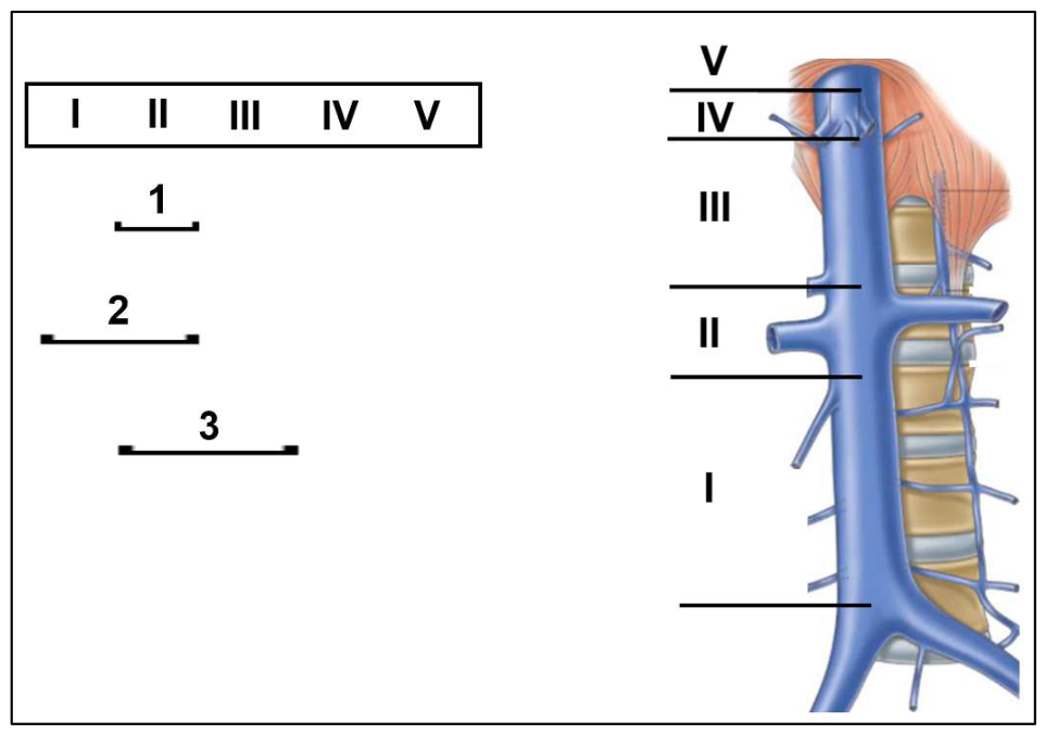
Figure 2.
Resection of the anterolateral wall of the IVC with xenopericardial patch plasty. (A) Leiomyosarcoma arising from the IVC at the level of the renal veins; (B) IVC plasty with a xenopericardial patch (arrow).
Figure 2.
Resection of the anterolateral wall of the IVC with xenopericardial patch plasty. (A) Leiomyosarcoma arising from the IVC at the level of the renal veins; (B) IVC plasty with a xenopericardial patch (arrow).
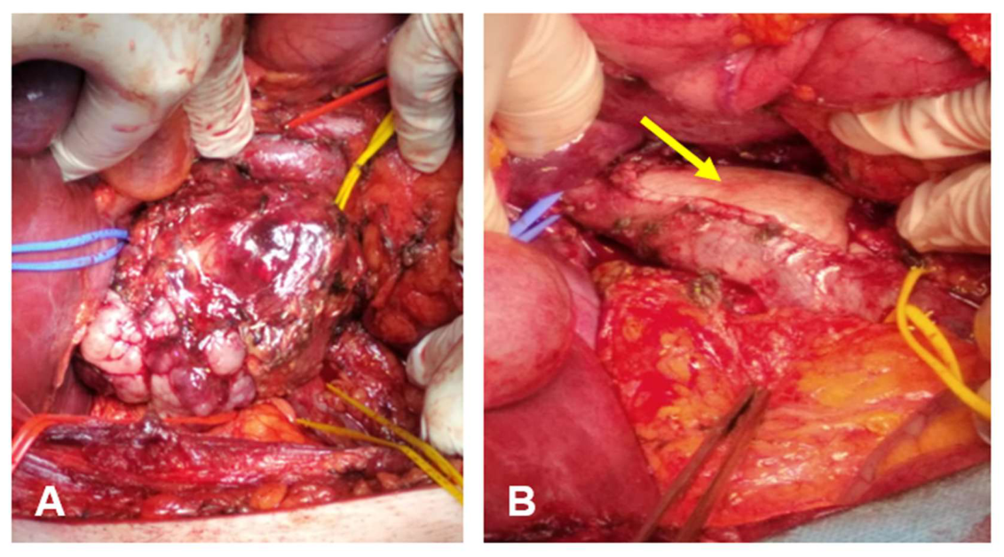
Figure 3.
Implantation of the left renal vein into a biological graft (arrow).
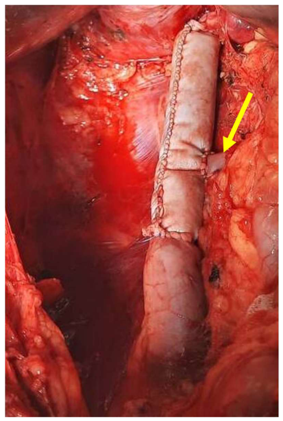
Figure 4.
Resection and prosthetic replacement of the IVC and renal veins using biological vascular grafts, with reimplantation of the renal vein prostheses into the IVC graft. (A) Implantation of a right renal vein prosthesis into the IVC graft; (B) Implantation of a left renal vein prosthesis into the IVC graft.
Figure 4.
Resection and prosthetic replacement of the IVC and renal veins using biological vascular grafts, with reimplantation of the renal vein prostheses into the IVC graft. (A) Implantation of a right renal vein prosthesis into the IVC graft; (B) Implantation of a left renal vein prosthesis into the IVC graft.
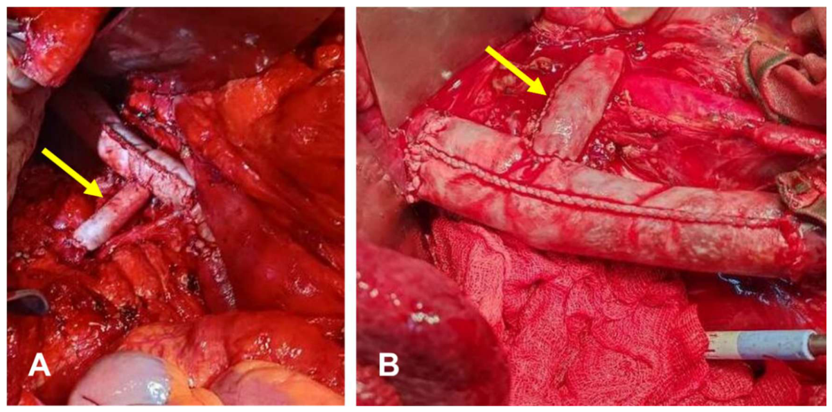
Figure 5.
Abdominal CT scan demonstrating tumor invasion into the vessels and capsule of the right kidney. (A) Axial view; (B) Coronal view.
Figure 5.
Abdominal CT scan demonstrating tumor invasion into the vessels and capsule of the right kidney. (A) Axial view; (B) Coronal view.
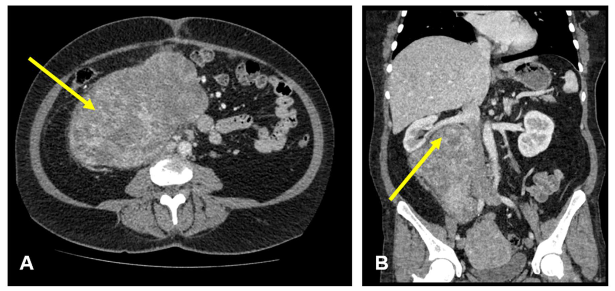
Figure 6.
Reconstruction of the IVC with a biological vascular graft (arrow).
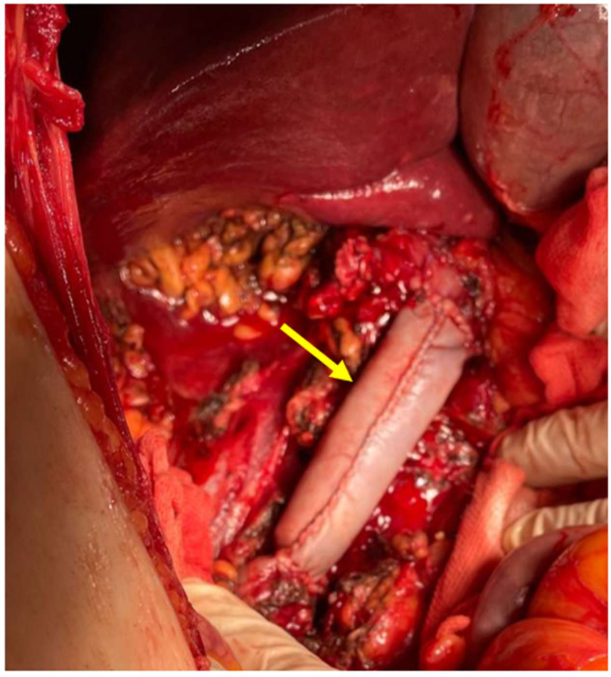

Table 1.
Clinical and pathological characteristics of patients with primary leiomyosarcoma of the inferior vena cava.
Table 1.
Clinical and pathological characteristics of patients with primary leiomyosarcoma of the inferior vena cava.
| Patient | 1 | 2 | 3 | 4 | 5 | 6 |
|---|---|---|---|---|---|---|
| Gender | male | female | female | female | female | female |
| IVC segment | II | I-II | I-II | II-III | II-III | II-III |
| Reconstruction | Patch (Biocard) | IVC graft | IVC graft | IVC graft | IVC graft | IVC graft |
| Multivisceral resection | Liver | Kidney, adrenal gland | Kidney, adrenal gland | Kidney, adrenal gland | Kidney, adrenal gland | No |
| R Status | R0 | R0 | R0 | R0 | R0 | R0 |
| FNCLCC1 Grade | G2 | G2 | G1 | G3 | G3 | G2 |
| Complications | No | Hematoma | No | No | No | Hematoma |
| Progression | Yes | No | No | No | No | No |
| Adjuvant chemotherapy | Yes | Yes | No | No | No | No |
| Follow-up (months) | 54 | 51 | 39 | 33 | 15 | 3 |
1 French Federation Nationale des Centres de Lutte Contre le Cancer.
Disclaimer/Publisher’s Note: The statements, opinions and data contained in all publications are solely those of the individual author(s) and contributor(s) and not of MDPI and/or the editor(s). MDPI and/or the editor(s) disclaim responsibility for any injury to people or property resulting from any ideas, methods, instructions or products referred to in the content. |
© 2026 by the authors. Licensee MDPI, Basel, Switzerland. This article is an open access article distributed under the terms and conditions of the Creative Commons Attribution (CC BY) license (http://creativecommons.org/licenses/by/4.0/).
Copyright: This open access article is published under a Creative Commons CC BY 4.0 license, which permit the free download, distribution, and reuse, provided that the author and preprint are cited in any reuse.


