
Submitted:
15 July 2026
Posted:
16 July 2026
You are already at the latest version
Abstract
Severe anaemia remains a major public health challenge, disproportionately affecting children and women of reproductive age in sub-Saharan Africa. In high-burden settings, the aetiology of severe anaemia is multifactorial, encompassing nutritional deficiencies, infections, and host genetic factors. Beyond its direct clinical consequences, severe anaemia is also associated with increased susceptibility to invasive bacterial infections, particularly those caused by enteric pathogens such as non-typhoidal Salmonella and Escherichia coli. In this review, we synthesise current evidence on the bidirectional interactions between severe anaemia, iron homeostasis, and the gut–immune axis, and how this mediates susceptibility to invasive bacterial infection. We present an integrated biological framework linking severe anaemia, gut dysbiosis, intestinal barrier permeability, and invasive bacterial infection. We examine context-specific modifiers in endemic settings, including iron deficiency, malnutrition, malaria, sickle cell disease, and environmental enteric dysfunction. We also discuss how management of severe anaemia, including iron supplementation and blood transfusion, reshapes the gut microbiome, with direct implications on microbial translocation, invasive bacterial infection, and clinical outcomes. Finally, we identify key knowledge gaps and research priorities to guide safer and more effective prevention and management of severe anaemia in high-burden settings.
Keywords:
1. Introduction
2. The Gut Microbiome in Health
3. Severe Anaemia and the Gut Microbiota
4. Mechanisms Linking Severe Anaemia to Gut Dysbiosis and Invasive Infection
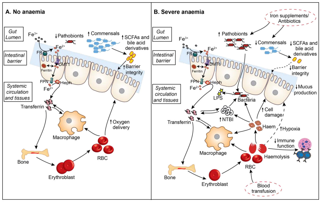
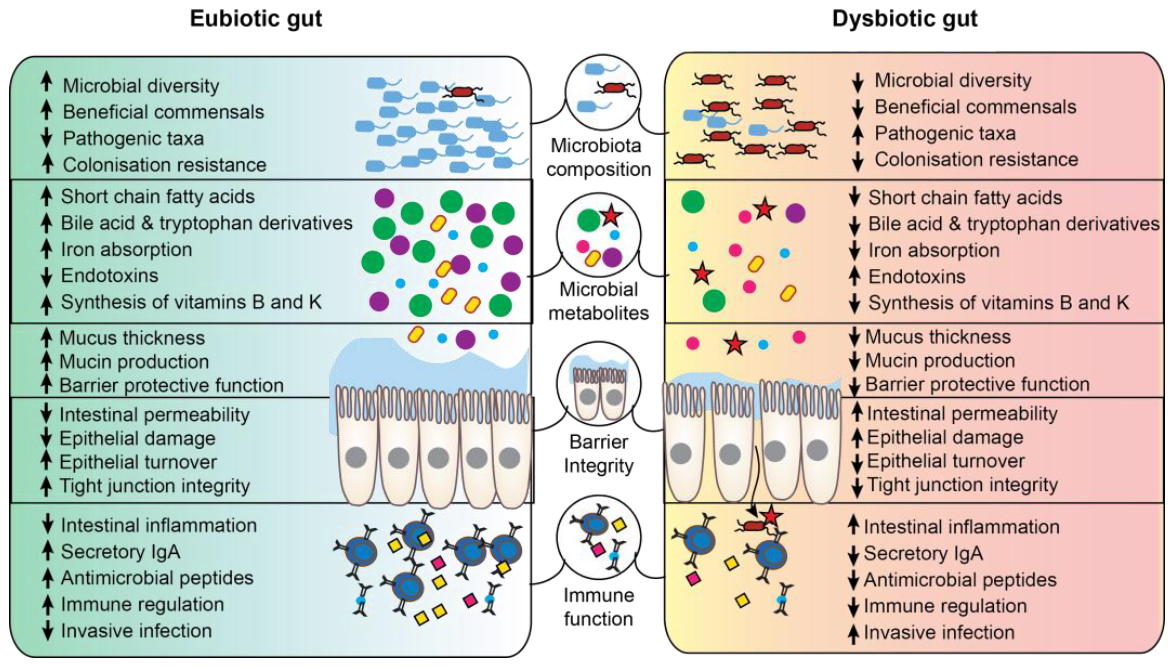
- Reduced microbial diversity
- Reduced abundance of commensal and beneficial taxa
- Expansion of facultative anaerobes and enteric pathobionts
- Altered microbial metabolic capacity
- Reduced production of SCFAs, bile acid, indole, and amino acid-derived metabolites
- Impaired colonisation resistance against enteric pathogens
- Altered microbial iron acquisition and siderophore-mediated competition
- Reduced oxygen delivery to the intestinal mucosa
- Reduced mucus production and mucus layer integrity
- Increased epithelial cell damage
- Impaired intestinal barrier integrity and increased permeability
- Impaired epithelial cell turnover and repair
- Increased intestinal inflammation
- Reduced mucosal immune function, including secretory IgA and antimicrobial peptide production
- Increased microbial translocation and endotoxaemia
- Increased susceptibility to invasive bacterial infection
- Impaired innate and adaptive immune cell function
- Increased systemic inflammation
- Dysregulated iron homeostasis and hepcidin signalling
- Increased NTBI and free haem availability for invasive pathogens
- Altered gut–immune signalling
5. Therapeutic Interventions in Severe Anaemia
5.1. Iron Supplementation
5.2. Blood Transfusion
6. Conclusion and Recommendations
- What are the temporal relationships between severe anaemia, gut dysbiosis, intestinal barrier dysfunction, and invasive infection?
- Are alterations of the gut microbiome reversible following correction of severe anaemia?
- How do changes in luminal and systemic iron availability influence microbial community structure, function, and pathogen colonisation?
- What are the relative contributions of iron deficiency, hepcidin-mediated iron sequestration, haemolysis, and dietary iron exposure to gut dysbiosis?
- Which microbial functional pathways are most sensitive to disruptions in iron metabolism?
- How do microbial metabolic functions compare across major severe anaemia aetiologies, including iron deficiency, malaria-associated anaemia, and malnutrition?
- How does EED modify severe anaemia–gut microbiome interactions?
- Are the mechanisms linking severe anaemia and gut dysbiosis consistent across different aetiologies, epidemiological settings, and age groups?
- How do iron supplementation, blood transfusion, and emerging iron-modulating therapies influence gut microbial ecology, intestinal barrier function, and infection risk?
- Can microbiome-directed interventions (prebiotics, probiotics, synbiotics, postbiotics, or faecal microbiota transplantation) improve anaemia recovery or reduce invasive bacterial infection?
Author Contributions
Funding
Acknowledgments
Conflicts of Interest
Abbreviations
| EED | Environmental enteric dysfunction |
| IDA | Iron deficiency anaemia |
| IgA | Immunoglobulin A |
| NTBI | Non-transferrin-bound iron |
| NTS | Non-typhoidal Salmonella |
| RBC | Red blood cell |
| SCA | Sickle cell anaemia |
| SCFA | Short-chain fatty acid |
| SMA | Severe malarial anaemia |
References
- World Health Organization. Guideline on haemoglobin cutoffs to define anaemia in individuals and populations; World Health Organization, 2024. [Google Scholar]
- Stevens, G.A.; Paciorek, C.J.; Flores-Urrutia, M.C.; Borghi, E.; Namaste, S.; Wirth, J.P.; Suchdev, P.S.; Ezzati, M.; Rohner, F.; Flaxman, S.R.; et al. National, regional, and global estimates of anaemia by severity in women and children for 2000-19: a pooled analysis of population-representative data. Lancet Glob. Health 2022, 10, e627–e639. [Google Scholar] [CrossRef] [PubMed]
- Global Burden of Disease Anaemia Collaborators. Prevalence, years lived with disability, and trends in anaemia burden by severity and cause, 1990-2021: findings from the Global Burden of Disease Study 2021. Lancet Haematol. 2023, 10, e713–e734. [Google Scholar] [CrossRef] [PubMed]
- Chami, N.; Hau, D.K.; Masoza, T.S.; Smart, L.R.; Kayange, N.M.; Hokororo, A.; Ambrose, E.E.; Moschovis, P.P.; Wiens, M.O.; Peck, R.N. Very severe anemia and one year mortality outcome after hospitalization in Tanzanian children: A prospective cohort study. PLoS ONE 2019, 14, e0214563. [Google Scholar] [CrossRef] [PubMed]
- Simbauranga, R.H.; Kamugisha, E.; Hokororo, A.; Kidenya, B.R.; Makani, J. Prevalence and factors associated with severe anaemia amongst under-five children hospitalized at Bugando Medical Centre, Mwanza, Tanzania. BMC Hematol. 2015, 15, 13. [Google Scholar] [CrossRef] [PubMed]
- Abuga, K.M.; Muriuki, J.M.; Mutua, A.M.; Wambui, K.M.; Mturi, N.; Mohammed, S.; Fondo, J.K.; Mwarumba, S.; Ngetsa, C.J.; Newton, C.R.; et al. Severe anaemia and invasive bacterial infections in Kenyan children: a 26-year hospital surveillance observational study. Lancet Glob. Health 2026, 103976. [Google Scholar] [CrossRef]
- Calis, J.C.; Phiri, K.S.; Faragher, E.B.; Brabin, B.J.; Bates, I.; Cuevas, L.E.; de Haan, R.J.; Phiri, A.I.; Malange, P.; Khoka, M.; et al. Severe anemia in Malawian children. N. Engl. J. Med. 2008, 358, 888–899. [Google Scholar] [CrossRef] [PubMed]
- Abuga, K.M.; Nairz, M.; MacLennan, C.A.; Atkinson, S.H. Severe anaemia, iron deficiency, and susceptibility to invasive bacterial infections. Wellcome Open Res. 2023, 8, 48. [Google Scholar] [CrossRef] [PubMed]
- Mooney, J.P.; Galloway, L.J.; Riley, E.M. Malaria, anemia, and invasive bacterial disease: A neutrophil problem? J. Leukoc. Biol. 2019, 105, 645–655. [Google Scholar] [CrossRef] [PubMed]
- Choi, G.; Bessman, N.J. Iron at the crossroads of host-microbiome interactions in health and disease. Nat. Microbiol. 2025, 10, 1282–1293. [Google Scholar] [CrossRef] [PubMed]
- Thaiss, C.A.; Zmora, N.; Levy, M.; Elinav, E. The microbiome and innate immunity. Nature 2016, 535, 65–74. [Google Scholar] [CrossRef] [PubMed]
- Adak, A.; Khan, M.R. An insight into gut microbiota and its functionalities. Cell Mol. Life Sci. 2019, 76, 473–493. [Google Scholar] [CrossRef] [PubMed]
- Ho, T.T.B.; Kumar, A.; Louis-Jacques, A.F.; Dishaw, L.J.; Yee, A.L.; Groer, M.W. The development of intestinal dysbiosis in anemic preterm infants. J. Perinatol. 2020, 40, 1066–1074. [Google Scholar] [CrossRef] [PubMed]
- Gibbons, J.A.; Worthington, L.M.; Chiu, E.G.; Kates, H.R.; Carter, R.R.; Nelson, R.; Zhang, M.; Garrett, T.J.; Ho, T.T.B. Severe anemia in preterm infants associated with increased bacterial virulence potential and metabolic disequilibrium. Pediatr. Res. 2025, 97, 2415–2422. [Google Scholar] [CrossRef] [PubMed]
- MohanKumar, K.; Namachivayam, K.; Sivakumar, N.; Alves, N.G.; Sidhaye, V.; Das, J.K.; Chung, Y.; Breslin, J.W.; Maheshwari, A. Severe neonatal anemia increases intestinal permeability by disrupting epithelial adherens junctions. Am. J. Physiol. Gastrointest. Liver Physiol. 2020, 318, G705–G716. [Google Scholar] [CrossRef] [PubMed]
- Arthur, C.M.; Nalbant, D.; Feldman, H.A.; Saeedi, B.J.; Matthews, J.; Robinson, B.S.; Kamili, N.A.; Bennett, A.; Cress, G.A.; Sola-Visner, M.; et al. Anemia induces gut inflammation and injury in an animal model of preterm infants. Transfusion 2019, 59, 1233–1245. [Google Scholar] [CrossRef] [PubMed]
- Pham, V.T.; Dold, S.; Rehman, A.; Bird, J.K.; Steinert, R.E. Vitamins, the gut microbiome and gastrointestinal health in humans. Nutr. Res. 2021, 95, 35–53. [Google Scholar] [CrossRef] [PubMed]
- Makki, K.; Deehan, E.C.; Walter, J.; Bäckhed, F. The Impact of Dietary Fiber on Gut Microbiota in Host Health and Disease. Cell Host Microbe 2018, 23, 705–715. [Google Scholar] [CrossRef] [PubMed]
- van Eijk, L.T.; Kroot, J.J.; Tromp, M.; van der Hoeven, J.G.; Swinkels, D.W.; Pickkers, P. Inflammation-induced hepcidin-25 is associated with the development of anemia in septic patients: an observational study. Crit. Care 2011, 15, R9. [Google Scholar] [CrossRef] [PubMed]
- Ekregbesi, P.; Shankar-Hari, M.; Bottomley, C.; Riley, E.M.; Mooney, J.P. Relationship between Anaemia, Haemolysis, Inflammation and Haem Oxygenase-1 at Admission with Sepsis: a pilot study. Sci. Rep. 2018, 8, 11198. [Google Scholar] [CrossRef] [PubMed]
- Kempe, D.S.; Akel, A.; Lang, P.A.; Hermle, T.; Biswas, R.; Muresanu, J.; Friedrich, B.; Dreischer, P.; Wolz, C.; Schumacher, U.; et al. Suicidal erythrocyte death in sepsis. J. Mol. Med. (Berl.) 2007, 85, 273–281. [Google Scholar] [CrossRef] [PubMed]
- Sanz, Y.; Cryan, J.F.; Deschasaux-Tanguy, M.; Elinav, E.; Lambrecht, R.; Veiga, P. The gut microbiome connects nutrition and human health. Nat. Rev. Gastroenterol. Hepatol. 2025, 22, 534–555. [Google Scholar] [CrossRef] [PubMed]
- Round, J.L.; Mazmanian, S.K. The gut microbiota shapes intestinal immune responses during health and disease. Nat. Rev. Immunol. 2009, 9, 313–323. [Google Scholar] [CrossRef] [PubMed]
- Bonder, M.J.; Kurilshikov, A.; Tigchelaar, E.F.; Mujagic, Z.; Imhann, F.; Vila, A.V.; Deelen, P.; Vatanen, T.; Schirmer, M.; Smeekens, S.P.; et al. The effect of host genetics on the gut microbiome. Nat. Genet. 2016, 48, 1407–1412. [Google Scholar] [CrossRef] [PubMed]
- Tierney, B.T.; Yang, Z.; Luber, J.M.; Beaudin, M.; Wibowo, M.C.; Baek, C.; Mehlenbacher, E.; Patel, C.J.; Kostic, A.D. The Landscape of Genetic Content in the Gut and Oral Human Microbiome. Cell Host Microbe 2019, 26, 283–295.e288. [Google Scholar] [CrossRef] [PubMed]
- Sender, R.; Fuchs, S.; Milo, R. Revised Estimates for the Number of Human and Bacteria Cells in the Body. PLoS Biol. 2016, 14, e1002533. [Google Scholar] [CrossRef] [PubMed]
- Sender, R.; Fuchs, S.; Milo, R. Are We Really Vastly Outnumbered? Revisiting the Ratio of Bacterial to Host Cells in Humans. Cell 2016, 164, 337–340. [Google Scholar] [CrossRef] [PubMed]
- Moeller, A.H.; Caro-Quintero, A.; Mjungu, D.; Georgiev, A.V.; Lonsdorf, E.V.; Muller, M.N.; Pusey, A.E.; Peeters, M.; Hahn, B.H.; Ochman, H. Cospeciation of gut microbiota with hominids. Science 2016, 353, 380–382. [Google Scholar] [CrossRef] [PubMed]
- Ley, R.E.; Lozupone, C.A.; Hamady, M.; Knight, R.; Gordon, J.I. Worlds within worlds: evolution of the vertebrate gut microbiota. Nat. Rev. Microbiol. 2008, 6, 776–788. [Google Scholar] [CrossRef] [PubMed]
- Robertson, R.C.; Manges, A.R.; Finlay, B.B.; Prendergast, A.J. The Human Microbiome and Child Growth—First 1000 Days and Beyond. Trends Microbiol. 2019, 27, 131–147. [Google Scholar] [CrossRef] [PubMed]
- Zhang, D.; Jian, Y.P.; Zhang, Y.N.; Li, Y.; Gu, L.T.; Sun, H.H.; Liu, M.D.; Zhou, H.L.; Wang, Y.S.; Xu, Z.X. Short-chain fatty acids in diseases. Cell Commun. Signal 2023, 21, 212. [Google Scholar] [CrossRef] [PubMed]
- Collins, S.L.; Stine, J.G.; Bisanz, J.E.; Okafor, C.D.; Patterson, A.D. Bile acids and the gut microbiota: metabolic interactions and impacts on disease. Nat. Rev. Microbiol. 2023, 21, 236–247. [Google Scholar] [CrossRef] [PubMed]
- Lin, R.; Liu, W.; Piao, M.; Zhu, H. A review of the relationship between the gut microbiota and amino acid metabolism. Amino Acids 2017, 49, 2083–2090. [Google Scholar] [CrossRef] [PubMed]
- Bailey, L.B.; Stover, P.J.; McNulty, H.; Fenech, M.F.; Gregory, J.F., 3rd; Mills, J.L.; Pfeiffer, C.M.; Fazili, Z.; Zhang, M.; Ueland, P.M.; et al. Biomarkers of Nutrition for Development-Folate Review. J. Nutr. 2015, 145, 1636S–1680S. [Google Scholar] [CrossRef] [PubMed]
- Allen, L.H.; Miller, J.W.; de Groot, L.; Rosenberg, I.H.; Smith, A.D.; Refsum, H.; Raiten, D.J. Biomarkers of Nutrition for Development (BOND): Vitamin B-12 Review. J. Nutr. 2018, 148, 1995S–2027S. [Google Scholar] [CrossRef] [PubMed]
- Bouglé, D.; Vaghefi-Vaezzadeh, N.; Roland, N.; Bouvard, G.; Arhan, P.; Bureau, F.; Neuville, D.; Maubois, J.L. Influence of short-chain fatty acids on iron absorption by proximal colon. Scand. J. Gastroenterol. 2002, 37, 1008–1011. [Google Scholar] [CrossRef] [PubMed]
- Agrizzi Verediano, T.; Agarwal, N.; Juste Contin Gomes, M.; Martino, H.S.D.; Tako, E. Effects of dietary fiber on intestinal iron absorption, and physiological status: a systematic review of in vivo and clinical studies. Crit. Rev. Food Sci. Nutr. 2023, 63, 9017–9032. [Google Scholar] [CrossRef] [PubMed]
- Salovaara, S.; Sandberg, A.S.; Andlid, T. Combined impact of pH and organic acids on iron uptake by Caco-2 cells. J. Agric. Food Chem. 2003, 51, 7820–7824. [Google Scholar] [CrossRef] [PubMed]
- Skaar, E.P. The battle for iron between bacterial pathogens and their vertebrate hosts. PLoS Pathog. 2010, 6, e1000949. [Google Scholar] [CrossRef] [PubMed]
- Murdoch, C.C.; Skaar, E.P. Nutritional immunity: the battle for nutrient metals at the host-pathogen interface. Nat. Rev. Microbiol. 2022, 20, 657–670. [Google Scholar] [CrossRef] [PubMed]
- Aksoyalp, Z.S.; Temel, A.; Erdogan, B.R. Iron in infectious diseases friend or foe?: The role of gut microbiota. J. Trace Elem. Med. Biol. 2023, 75, 127093. [Google Scholar] [CrossRef] [PubMed]
- Nairz, M.; Weiss, G. Iron in infection and immunity. Mol. Asp. Med. 2020, 75, 100864. [Google Scholar] [CrossRef] [PubMed]
- Shanmugam, N.K.; Chen, K.; Cherayil, B.J. Commensal Bacteria-induced Interleukin 1beta (IL-1beta) Secreted by Macrophages Up-regulates Hepcidin Expression in Hepatocytes by Activating the Bone Morphogenetic Protein Signaling Pathway. J. Biol. Chem. 2015, 290, 30637–30647. [Google Scholar] [CrossRef] [PubMed]
- Bessman, N.J.; Mathieu, J.R.R.; Renassia, C.; Zhou, L.; Fung, T.C.; Fernandez, K.C.; Austin, C.; Moeller, J.B.; Zumerle, S.; Louis, S.; et al. Dendritic cell-derived hepcidin sequesters iron from the microbiota to promote mucosal healing. Science 2020, 368, 186–189. [Google Scholar] [CrossRef] [PubMed]
- Nemeth, E.; Ganz, T. Hepcidin and Iron in Health and Disease. Annu. Rev. Med. 2023, 74, 261–277. [Google Scholar] [CrossRef] [PubMed]
- de Vos, W.M.; Tilg, H.; Van Hul, M.; Cani, P.D. Gut microbiome and health: mechanistic insights. Gut 2022, 71, 1020–1032. [Google Scholar] [CrossRef] [PubMed]
- León, E.D.; Francino, M.P. Roles of Secretory Immunoglobulin A in Host-Microbiota Interactions in the Gut Ecosystem. Front. Microbiol. 2022, 13, 880484. [Google Scholar] [CrossRef] [PubMed]
- Mason, K.L.; Huffnagle, G.B.; Noverr, M.C.; Kao, J.Y. Overview of gut immunology. Adv. Exp. Med. Biol. 2008, 635, 1–14. [Google Scholar] [CrossRef] [PubMed]
- Wiertsema, S.P.; van Bergenhenegouwen, J.; Garssen, J.; Knippels, L.M.J. The Interplay between the Gut Microbiome and the Immune System in the Context of Infectious Diseases throughout Life and the Role of Nutrition in Optimizing Treatment Strategies. Nutrients 2021, 13, 886. [Google Scholar] [CrossRef] [PubMed]
- Caballero-Flores, G.; Pickard, J.M.; Núñez, G. Microbiota-mediated colonization resistance: mechanisms and regulation. Nat. Rev. Microbiol. 2023, 21, 347–360. [Google Scholar] [CrossRef] [PubMed]
- Josefsdottir, K.S.; Baldridge, M.T.; Kadmon, C.S.; King, K.Y. Antibiotics impair murine hematopoiesis by depleting the intestinal microbiota. Blood 2017, 129, 729–739. [Google Scholar] [CrossRef] [PubMed]
- Coll, E.; Cigarran, S.; Portoles, J.; Cases, A. Gut Dysbiosis and Its Role in the Anemia of Chronic Kidney Disease. Toxins 2024, 16. [Google Scholar] [CrossRef] [PubMed]
- Tesoi, D.F.; Trandafir, L.M.; Bozomitu, L.; Frasinariu, O.E.; Filip, N.; Mircea, C.; Hancianu, M.; Badulescu, O.V. Molecular Mechanisms Underlying the Higher Prevalence of Anemia in Crohn’s Disease Compared with Ulcerative Colitis: A Systematic Review. Int. J. Mol. Sci. 2026, 27. [Google Scholar] [CrossRef] [PubMed]
- Zhou, H.; Fan, Z.; Da, Y.; Liu, X.; Wang, C.; Zhang, T.; Zhang, J.; Wu, T.; Liang, J. Causal Relationships Between Iron Deficiency Anemia, Gut Microbiota, and Metabolites: Insights from Mendelian Randomization and In Vivo Data. Biomedicines 2025, 13. [Google Scholar] [CrossRef] [PubMed]
- Lei, W.; Liu, Z.; Lai, H.P.; Fu, R. Gut microbiota and risk of iron deficiency anemia: A two-sample Mendelian randomization study. Medicine 2025, 104, e41617. [Google Scholar] [CrossRef] [PubMed]
- Su, T.; Peng, X.; Gan, Y.; Wu, H.; Ma, S.; Zhi, M.; Lu, Y.; Dai, S.; Yao, J. Associations of genetically predicted iron status with 24 gastrointestinal diseases and gut microbiota: a Mendelian randomization study. Front Genet 2024, 15, 1406230. [Google Scholar] [CrossRef] [PubMed]
- Zheng, X.; Shen, W.; Yin, J. The relationship between gut microbiota, plasma metabolites, and iron deficiency anemia in European populations: a three-sample Mendelian randomization analysis. Hematology 2025, 30, 2549962. [Google Scholar] [CrossRef] [PubMed]
- McClorry, S.; Zavaleta, N.; Llanos, A.; Casapía, M.; Lönnerdal, B.; Slupsky, C.M. Anemia in infancy is associated with alterations in systemic metabolism and microbial structure and function in a sex-specific manner: an observational study. Am. J. Clin. Nutr. 2018, 108, 1238–1248. [Google Scholar] [CrossRef] [PubMed]
- Diaz-Rodriguez, K.; Pacheco-Aranibar, J.; Manrique-Sam, C.; Ita-Balta, Y.; Carpio-Toia, A.M.D.; Lopez-Casaperalta, P.; Chocano-Rosas, T.; Fernandez, F.F.; Villanueva-Salas, J.; Bernabe-Ortiz, J.C. Intestinal Microbiota in Children with Anemia in Southern Peru through Next-Generation Sequencing Technology. Children 2022, 9. [Google Scholar] [CrossRef] [PubMed]
- Seyoum, Y.; Greffeuille, V.; Kouadio, D.K.D.; Kuong, K.; Turpin, W.; M’Rabt, R.; Chochois, V.; Fortin, S.; Perignon, M.; Fiorentino, M.; et al. Faecal microbiota of schoolchildren is associated with nutritional status and markers of inflammation: a double-blinded cluster-randomized controlled trial using multi-micronutrient fortified rice. Nat. Commun. 2024, 15, 5204. [Google Scholar] [CrossRef] [PubMed]
- Balamurugan, R.; Mary, R.R.; Chittaranjan, S.; Jancy, H.; Shobana Devi, R.; Ramakrishna, B.S. Low levels of faecal lactobacilli in women with iron-deficiency anaemia in south India. Br. J. Nutr. 2010, 104, 931–934. [Google Scholar] [CrossRef] [PubMed]
- Muleviciene, A.; D’Amico, F.; Turroni, S.; Candela, M.; Jankauskiene, A. Iron deficiency anemia-related gut microbiota dysbiosis in infants and young children: A pilot study. Acta Microbiol. Immunol. Hung. 2018, 65, 551–564. [Google Scholar] [CrossRef] [PubMed]
- Chen, H.; Wu, W.; Tang, S.; Fu, R.; Gong, X.; Hou, H.; Xu, J. Altered fecal microbial and metabolic profile reveals potential mechanisms underlying iron deficiency anemia in pregnant women in China. Bosn. J. Basic Med. Sci. 2022, 22, 923–933. [Google Scholar] [CrossRef] [PubMed]
- Seo, H.; Yoon, S.Y.; Ul-Haq, A.; Jo, S.; Kim, S.; Rahim, M.A.; Park, H.A.; Ghorbanian, F.; Kim, M.J.; Lee, M.Y.; et al. The Effects of Iron Deficiency on the Gut Microbiota in Women of Childbearing Age. Nutrients 2023, 15. [Google Scholar] [CrossRef] [PubMed]
- Goosen, C.; Proost, S.; Baumgartner, J.; Mallick, K.; Tito, R.Y.; Barnabas, S.L.; Cotton, M.F.; Zimmermann, M.B.; Raes, J.; Blaauw, R. Associations of HIV and iron status with gut microbiota composition, gut inflammation and gut integrity in South African school-age children: a two-way factorial case-control study. J. Hum. Nutr. Diet. 2023, 36, 819–832. [Google Scholar] [CrossRef] [PubMed]
- Zhong, H.J.; Chen, W.R.; Lu, X.J.; Hu, D.X.; Lin, D.J.; Liu, T.; Wu, L.; Wu, L.H.; He, X.X. Washed microbiota transplantation improves haemoglobin levels in anaemia of chronic disease. Eur. J. Clin. Invest. 2023, 53, e14072. [Google Scholar] [CrossRef] [PubMed]
- Wang, H.; Xue, W.; Cheng, J.; He, Y.; Song, Y.; Hu, D.; Peng, A.; Li, C.; Bao, H. Altered fecal microbial and metabolic profiles reveal potential mechanisms underlying anemia in patients with chronic renal failure. Microbiol. Spectr. 2025, 13, e0316624. [Google Scholar] [CrossRef] [PubMed]
- Shao, Y.; Qi, W.; Zhang, X.; Ran, N.; Liu, C.; Fu, R.; Shao, Z. Plasma Metabolomic and Intestinal Microbial Analyses of Patients With Severe Aplastic Anemia. Front Cell Dev. Biol. 2021, 9, 669887. [Google Scholar] [CrossRef] [PubMed]
- Zhang, X.; Xie, J.; Wang, H.; Li, J. Analysis of fecal microbiome in Chinese patients with non-severe aplastic anemia. Ann. Hematol. 2025, 104, 4013–4027. [Google Scholar] [CrossRef] [PubMed]
- Lim, S.H.; Morris, A.; Li, K.; Fitch, A.C.; Fast, L.; Goldberg, L.; Quesenberry, M.; Sprinz, P.; Methé, B. Intestinal microbiome analysis revealed dysbiosis in sickle cell disease. Am. J. Hematol. 2018, 93, E91–e93. [Google Scholar] [CrossRef] [PubMed]
- Delgadinho, M.; Ginete, C.; Santos, B.; Mendes, J.; Miranda, A.; Vasconcelos, J.; Brito, M. Microbial gut evaluation in an angolan paediatric population with sickle cell disease. J. Cell Mol. Med. 2022, 26, 5360–5368. [Google Scholar] [CrossRef] [PubMed]
- Karahan, F.; Yilmaz, S.S.; Bayrakdar, F.; Tezol, O.; Kuyucu, N.; Kilic, S.; Turkegun, M.; Unal, S. Evaluation of Intestinal Microbiota in Children With Sickle Cell Disease. J. Pediatr. Hematol. Oncol. 2023, 45, e904–e909. [Google Scholar] [CrossRef] [PubMed]
- Brim, H.; Taylor, J.; Abbas, M.; Vilmenay, K.; Daremipouran, M.; Varma, S.; Lee, E.; Pace, B.; Song-Naba, W.L.; Gupta, K.; et al. The gut microbiome in sickle cell disease: Characterization and potential implications. PLoS ONE 2021, 16, e0255956. [Google Scholar] [CrossRef] [PubMed]
- Mandal, R.K.; Denny, J.E.; Namazzi, R.; Opoka, R.O.; Datta, D.; John, C.C.; Schmidt, N.W. Dynamic modulation of spleen germinal center reactions by gut bacteria during Plasmodium infection. Cell Rep. 2021, 35, 109094. [Google Scholar] [CrossRef] [PubMed]
- Kalteren, W.S.; Bos, A.F.; van Oeveren, W.; Hulscher, J.B.F.; Kooi, E.M.W. Neonatal anemia relates to intestinal injury in preterm infants. Pediatr. Res. 2022, 91, 1452–1458. [Google Scholar] [CrossRef] [PubMed]
- Maheshwari, A. Severe anemia predisposes very premature infants to transfusion-associated necrotizing enterocolitis. Semin. Fetal Neonatal Med. 2025, 30, 101615. [Google Scholar] [CrossRef] [PubMed]
- Camaschella, C. Iron deficiency. Blood 2019, 133, 30–39. [Google Scholar] [CrossRef] [PubMed]
- Soriano-Lerma, A.; Garcia-Burgos, M.; Barton, W.; Alferez, M.J.M.; Crespo-Perez, J.V.; Soriano, M.; Lopez-Aliaga, I.; Cotter, P.D.; Garcia-Salcedo, J.A. Comprehensive insight into the alterations in the gut microbiome and the intestinal barrier as a consequence of iron deficiency anaemia. BioMed J. 2024, 47, 100701. [Google Scholar] [CrossRef] [PubMed]
- Ippolito, J.R.; Piccolo, B.D.; Robeson, M.S.; Barney, D.E., Jr.; Ali, J.; Singh, P.; Hennigar, S.R. Iron deficient diets modify the gut microbiome and reduce the severity of enteric infection in a mouse model of S. Typhimurium-induced enterocolitis. J. Nutr. Biochem. 2022, 107, 109065. [Google Scholar] [CrossRef] [PubMed]
- Soriano-Lerma, A.; Garcia-Burgos, M.; Alferez, M.J.M.; Perez-Carrasco, V.; Sanchez-Martin, V.; Linde-Rodriguez, A.; Ortiz-Gonzalez, M.; Soriano, M.; Garcia-Salcedo, J.A.; Lopez-Aliaga, I. Gut microbiome-short-chain fatty acids interplay in the context of iron deficiency anaemia. Eur. J. Nutr. 2022, 61, 399–412. [Google Scholar] [CrossRef] [PubMed]
- Soriano-Lerma, A.; Soriano-Suarez, J.S.; Garcia-Rodriguez, M.; Alferez, M.J.; Soriano, M.; Salcedo, J.A.G.; Lopez-Aliaga, I. Molecular study of the small intestine dysbiosis derived from iron deficiency anaemia. Sci. Rep. 2026. [Google Scholar] [CrossRef] [PubMed]
- Dostal, A.; Chassard, C.; Hilty, F.M.; Zimmermann, M.B.; Jaeggi, T.; Rossi, S.; Lacroix, C. Iron depletion and repletion with ferrous sulfate or electrolytic iron modifies the composition and metabolic activity of the gut microbiota in rats. J. Nutr. 2012, 142, 271–277. [Google Scholar] [CrossRef] [PubMed]
- Pereira, D.I.; Aslam, M.F.; Frazer, D.M.; Schmidt, A.; Walton, G.E.; McCartney, A.L.; Gibson, G.R.; Anderson, G.J.; Powell, J.J. Dietary iron depletion at weaning imprints low microbiome diversity and this is not recovered with oral Nano Fe(III). Microbiologyopen 2015, 4, 12–27. [Google Scholar] [CrossRef] [PubMed]
- Dostal, A.; Lacroix, C.; Bircher, L.; Pham, V.T.; Follador, R.; Zimmermann, M.B.; Chassard, C. Iron Modulates Butyrate Production by a Child Gut Microbiota In Vitro. mBio 2015, 6, e01453-01415. [Google Scholar] [CrossRef] [PubMed]
- Soriano-Lerma, A.; Garcia-Burgos, M.; Alferez, M.J.M.; Crespo-Perez, J.V.; Perez-Carrasco, V.; Ortiz-Gonzalez, M.; Linde-Rodriguez, A.; Sanchez-Martin, V.; Soriano, M.; Garcia-Salcedo, J.A.; et al. Fermented Goat’s Milk Contributes to the Recovery of Iron Deficiency Anemia via Modulation of the Gut Microbiome. J. Agric. Food Chem. 2023, 71, 15668–15679. [Google Scholar] [CrossRef] [PubMed]
- Celis, A.I.; Relman, D.A.; Huang, K.C. The impact of iron and heme availability on the healthy human gut microbiome in vivo and in vitro. Cell Chem. Biol. 2023, 30, 110–126 e113. [Google Scholar] [CrossRef] [PubMed]
- Jaeggi, T.; Kortman, G.A.; Moretti, D.; Chassard, C.; Holding, P.; Dostal, A.; Boekhorst, J.; Timmerman, H.M.; Swinkels, D.W.; Tjalsma, H.; et al. Iron fortification adversely affects the gut microbiome, increases pathogen abundance and induces intestinal inflammation in Kenyan infants. Gut 2015, 64, 731–742. [Google Scholar] [CrossRef] [PubMed]
- Zimmermann, M.B.; Chassard, C.; Rohner, F.; N’Goran E, K.; Nindjin, C.; Dostal, A.; Utzinger, J.; Ghattas, H.; Lacroix, C.; Hurrell, R.F. The effects of iron fortification on the gut microbiota in African children: a randomized controlled trial in Cote d’Ivoire. Am. J. Clin. Nutr. 2010, 92, 1406–1415. [Google Scholar] [CrossRef] [PubMed]
- Dike, C.R.; Hanson, C.; Davies, H.D.; Obaro, S.; Yu, F.; Harper, J.; Grace, H.; Lebensburger, J.; Raulji, C.; Ma, J.; et al. The relationship between nutrition, gut dysbiosis, and pediatric sickle cell pain outcomes: A pilot study. Pediatr. Blood Cancer 2023, 70, e30397. [Google Scholar] [CrossRef] [PubMed]
- World Health Organization. World Malaria Report 2025: Addressing the Threat of Antimalarial Drug Resistance; World Health Organization, 2025. [Google Scholar]
- White, N.J. Anaemia and malaria. Malar. J. 2018, 17, 371. [Google Scholar] [CrossRef] [PubMed]
- Perkins, D.J.; Were, T.; Davenport, G.C.; Kempaiah, P.; Hittner, J.B.; Ong’echa, J.M. Severe malarial anemia: innate immunity and pathogenesis. Int. J. Biol. Sci. 2011, 7, 1427–1442. [Google Scholar] [CrossRef] [PubMed]
- Olupot-Olupot, P.; Urban, B.C.; Jemutai, J.; Nteziyaremye, J.; Fanjo, H.M.; Karanja, H.; Karisa, J.; Ongodia, P.; Bwonyo, P.; Gitau, E.N.; et al. Endotoxaemia is common in children with Plasmodium falciparum malaria. BMC Infect. Dis. 2013, 13, 117. [Google Scholar] [CrossRef] [PubMed]
- Abuga, K.M.; Muriuki, J.M.; Uyoga, S.M.; Mwai, K.; Makale, J.; Mogire, R.M.; Macharia, A.W.; Mohammed, S.; Muthumbi, E.; Mwarumba, S.; et al. Hepcidin regulation in Kenyan children with severe malaria and non-typhoidal Salmonella bacteremia. Haematologica 2022, 107, 1589–1598. [Google Scholar] [CrossRef] [PubMed]
- van Santen, S.; de Mast, Q.; Swinkels, D.W.; van der Ven, A.J. The iron link between malaria and invasive non-typhoid Salmonella infections. Trends Parasitol. 2013, 29, 220–227. [Google Scholar] [CrossRef] [PubMed]
- Church, J.; Maitland, K. Invasive bacterial co-infection in African children with Plasmodium falciparum malaria: a systematic review. BMC Med. 2014, 12, 31. [Google Scholar] [CrossRef] [PubMed]
- Wilairatana, P.; Meddings, J.B.; Ho, M.; Vannaphan, S.; Looareesuwan, S. Increased gastrointestinal permeability in patients with Plasmodium falciparum malaria. Clin. Infect. Dis. 1997, 24, 430–435. [Google Scholar] [CrossRef] [PubMed]
- Sarangam, M.L.; Namazzi, R.; Datta, D.; Bond, C.; Vanderpool, C.P.B.; Opoka, R.O.; John, C.C.; Conroy, A.L. Intestinal Injury Biomarkers Predict Mortality in Pediatric Severe Malaria. mBio 2022, 13, e0132522. [Google Scholar] [CrossRef] [PubMed]
- Piel, F.B.; Steinberg, M.H.; Rees, D.C. Sickle Cell Disease. N. Engl. J. Med. 2017, 376, 1561–1573. [Google Scholar] [CrossRef] [PubMed]
- Lewis, C.V.; Sellak, H.; Sawan, M.A.; Joseph, G.; Darby, T.M.; VanInsberghe, D.; Naudin, C.R.; Archer, D.R.; Jones, R.M.; Taylor, W.R. Intestinal barrier dysfunction in murine sickle cell disease is associated with small intestine neutrophilic inflammation, oxidative stress, and dysbiosis. FASEB Bioadv 2023, 5, 199–210. [Google Scholar] [CrossRef] [PubMed]
- Poplawska, M.; Dutta, D.; Jayaram, M.; Salifu, M.; Chong, N.S.; Lim, S.H. Intestinal pathophysiological abnormalities in steady state and after vaso-occlusive crisis in murine sickle cell disease. Br. J. Haematol. 2022, 196, 777–780. [Google Scholar] [CrossRef] [PubMed]
- Dutta, D.; Methe, B.; Amar, S.; Morris, A.; Lim, S.H. Intestinal injury and gut permeability in sickle cell disease. J. Transl. Med. 2019, 17, 183. [Google Scholar] [CrossRef] [PubMed]
- Brandow, A.M.; Atkinson, S.N.; Manjarres, Z.; Ehlers, V.L.; Pratt, M.L.; Mehta, I.; Mudunuri, S.; Kappagantu, A.; Shiers, S.I.; Mazhar, K.; et al. Gut microbiota and metabolites drive chronic sickle cell disease pain in mice. Cell Host Microbe 2025, 33, 1703–1714 e1708. [Google Scholar] [CrossRef] [PubMed]
- Xu, C.; Lee, S.K.; Zhang, D.; Frenette, P.S. The Gut Microbiome Regulates Psychological-Stress-Induced Inflammation. Immunity 2020, 53, 417–428.e414. [Google Scholar] [CrossRef] [PubMed]
- Li, H.; Kazmi, J.S.; Lee, S.; Zhang, D.; Gao, X.; Maryanovich, M.; Torres, L.; Verma, D.; Kelly, L.; Ginzburg, Y.Z.; et al. Dietary iron restriction protects against vaso-occlusion and organ damage in murine sickle cell disease. Blood 2023, 141, 194–199. [Google Scholar] [CrossRef] [PubMed]
- Delgadinho, M.; Ginete, C.; Santos, B.; Fernandes, C.; Silva, C.; Miranda, A.; Vasconcelos, J.N.; Brito, M. How Hydroxyurea Alters the Gut Microbiome: A Longitudinal Study Involving Angolan Children with Sickle Cell Anemia. Int. J. Mol. Sci. 2022, 23. [Google Scholar] [CrossRef] [PubMed]
- Suhas, H.S.; Kumar, M.; Choudhary, T.; Katewa, V.; Sharma, P. Prevalence of Iron Deficiency Anemia and Iron Deficiency Without Anemia Among Moderate and Severely Acute Malnourished Children. Cureus 2024, 16, e65633. [Google Scholar] [CrossRef] [PubMed]
- Tatli, M.M.; Vural, H.; Koc, A.; Kosecik, M.; Atas, A. Altered anti-oxidant status and increased lipid peroxidation in marasmic children. Pediatr. Int. 2000, 42, 289–292. [Google Scholar] [CrossRef] [PubMed]
- Manary, M.J.; Leeuwenburgh, C.; Heinecke, J.W. Increased oxidative stress in kwashiorkor. J. Pediatr. 2000, 137, 421–424. [Google Scholar] [CrossRef] [PubMed]
- Subramanian, S.; Huq, S.; Yatsunenko, T.; Haque, R.; Mahfuz, M.; Alam, M.A.; Benezra, A.; DeStefano, J.; Meier, M.F.; Muegge, B.D.; et al. Persistent gut microbiota immaturity in malnourished Bangladeshi children. Nature 2014, 510, 417–421. [Google Scholar] [CrossRef] [PubMed]
- Bisht, A.; Ahn-Jarvis, J.; Corbin, K.; Harris, S.; Troncoso-Rey, P.; Olupot-Olupot, P.; Calder, N.; Walsh, K.; Maitland, K.; Frost, G.; et al. Gut microbial diversity impacts carbohydrate fermentation by children with severe acute malnutrition. iScience 2026, 29, 114640. [Google Scholar] [CrossRef] [PubMed]
- Nabwera, H.M.; Espinoza, J.L.; Worwui, A.; Betts, M.; Okoi, C.; Sesay, A.K.; Bancroft, R.; Agbla, S.C.; Jarju, S.; Bradbury, R.S.; et al. Interactions between fecal gut microbiome, enteric pathogens, and energy regulating hormones among acutely malnourished rural Gambian children. EBioMedicine 2021, 73, 103644. [Google Scholar] [CrossRef] [PubMed]
- Smith, M.I.; Yatsunenko, T.; Manary, M.J.; Trehan, I.; Mkakosya, R.; Cheng, J.; Kau, A.L.; Rich, S.S.; Concannon, P.; Mychaleckyj, J.C.; et al. Gut microbiomes of Malawian twin pairs discordant for kwashiorkor. Science 2013, 339, 548–554. [Google Scholar] [CrossRef] [PubMed]
- McCormick, B.J.J.; Murray-Kolb, L.E.; Lee, G.O.; Schulze, K.J.; Ross, A.C.; Bauck, A.; Lima, A.A.M.; Maciel, B.L.L.; Kosek, M.N.; Seidman, J.C.; et al. Intestinal permeability and inflammation mediate the association between nutrient density of complementary foods and biochemical measures of micronutrient status in young children: results from the MAL-ED study. Am. J. Clin. Nutr. 2019, 110, 1015–1025. [Google Scholar] [CrossRef] [PubMed]
- Lauer, J.M.; Ghosh, S.; Ausman, L.M.; Webb, P.; Bashaasha, B.; Agaba, E.; Turyashemererwa, F.M.; Tran, H.Q.; Gewirtz, A.T.; Erhardt, J.; et al. Markers of Environmental Enteric Dysfunction Are Associated with Poor Growth and Iron Status in Rural Ugandan Infants. J. Nutr. 2020, 150, 2175–2182. [Google Scholar] [CrossRef] [PubMed]
- Regassa, R.; Duguma, M.; Belachew, T.; Tamiru, D. Environmental Enteropathy and Anaemia Status Among Under-Five Children, in Slum Areas of Jimma Town, Ethiopia. Pediatr. Health Med. Ther. 2023, 14, 33–43. [Google Scholar] [CrossRef] [PubMed]
- Vonaesch, P.; Winkel, M.; Kapel, N.; Nestoret, A.; Barbot-Trystram, L.; Pontoizeau, C.; Barouki, R.; Rakotondrainipiana, M.; Kandou, K.; Andriamanantena, Z.; et al. Putative Biomarkers of Environmental Enteric Disease Fail to Correlate in a Cross-Sectional Study in Two Study Sites in Sub-Saharan Africa. Nutrients 2022, 14. [Google Scholar] [CrossRef] [PubMed]
- Crane, R.J.; Jones, K.D.; Berkley, J.A. Environmental enteric dysfunction: an overview. Food Nutr. Bull. 2015, 36, S76–87. [Google Scholar] [CrossRef] [PubMed]
- Taylor, C.T.; Colgan, S.P. Hypoxia and gastrointestinal disease. J. Mol. Med. (Berl.) 2007, 85, 1295–1300. [Google Scholar] [CrossRef] [PubMed]
- Singhal, R.; Shah, Y.M. Oxygen battle in the gut: Hypoxia and hypoxia-inducible factors in metabolic and inflammatory responses in the intestine. J. Biol. Chem. 2020, 295, 10493–10505. [Google Scholar] [CrossRef] [PubMed]
- Litvak, Y.; Byndloss, M.X.; Bäumler, A.J. Colonocyte metabolism shapes the gut microbiota. Science 2018, 362. [Google Scholar] [CrossRef] [PubMed]
- Rivera-Chávez, F.; Lopez, C.A.; Bäumler, A.J. Oxygen as a driver of gut dysbiosis. Free Radic. Biol. Med. 2017, 105, 93–101. [Google Scholar] [CrossRef] [PubMed]
- Shang, T.; Zhang, R.; Liu, Y.; Shi, S. Intestinal oxygen and microbiota crosstalk: implications for pathogenesis of gastrointestinal diseases and emerging therapeutic strategies. Gut Pathog. 2025, 17, 100. [Google Scholar] [CrossRef] [PubMed]
- Stefanova, D.; Raychev, A.; Arezes, J.; Ruchala, P.; Gabayan, V.; Skurnik, M.; Dillon, B.J.; Horwitz, M.A.; Ganz, T.; Bulut, Y.; et al. Endogenous hepcidin and its agonist mediate resistance to selected infections by clearing non-transferrin-bound iron. Blood 2017, 130, 245–257. [Google Scholar] [CrossRef] [PubMed]
- Michels, K.R.; Zhang, Z.; Bettina, A.M.; Cagnina, R.E.; Stefanova, D.; Burdick, M.D.; Vaulont, S.; Nemeth, E.; Ganz, T.; Mehrad, B. Hepcidin-mediated iron sequestration protects against bacterial dissemination during pneumonia. JCI Insight 2017, 2, e92002. [Google Scholar] [CrossRef] [PubMed]
- Latour, C.; Wlodarczyk, M.F.; Jung, G.; Gineste, A.; Blanchard, N.; Ganz, T.; Roth, M.P.; Coppin, H.; Kautz, L. Erythroferrone contributes to hepcidin repression in a mouse model of malarial anemia. Haematologica 2017, 102, 60–68. [Google Scholar] [CrossRef] [PubMed]
- Jonker, F.A.; Calis, J.C.; Phiri, K.; Kraaijenhagen, R.J.; Brabin, B.J.; Faragher, B.; Wiegerinck, E.T.; Tjalsma, H.; Swinkels, D.W.; van Hensbroek, M.B. Low hepcidin levels in severely anemic malawian children with high incidence of infectious diseases and bone marrow iron deficiency. PLoS ONE 2013, 8, e78964. [Google Scholar] [CrossRef] [PubMed]
- Yilmaz, B.; Li, H. Gut Microbiota and Iron: The Crucial Actors in Health and Disease. Pharmaceuticals 2018, 11, 98. [Google Scholar] [CrossRef] [PubMed]
- Vallelian, F.; Buehler, P.W.; Schaer, D.J. Hemolysis, free hemoglobin toxicity, and scavenger protein therapeutics. Blood 2022, 140, 1837–1844. [Google Scholar] [CrossRef] [PubMed]
- Abuga, K.M.; Muriuki, J.M.; Williams, T.N.; Atkinson, S.H. How Severe Anaemia Might Influence the Risk of Invasive Bacterial Infections in African Children. Int. J. Mol. Sci. 2020, 21, 6976. [Google Scholar] [CrossRef] [PubMed]
- Frost, J.N.; Wideman, S.K.; Preston, A.E.; Teh, M.R.; Ai, Z.; Wang, L.; Cross, A.; White, N.; Yazicioglu, Y.; Bonadonna, M.; et al. Plasma iron controls neutrophil production and function. Sci. Adv. 2022, 8, eabq5384. [Google Scholar] [CrossRef] [PubMed]
- Hill, D.L.; Carr, E.J.; Rutishauser, T.; Moncunill, G.; Campo, J.J.; Innocentin, S.; Mpina, M.; Nhabomba, A.; Tumbo, A.; Jairoce, C.; et al. Immune system development varies according to age, location, and anemia in African children. Sci. Transl. Med. 2020, 12. [Google Scholar] [CrossRef] [PubMed]
- Sundd, P.; Gladwin, M.T.; Novelli, E.M. Pathophysiology of Sickle Cell Disease. Annu. Rev. Pathol. 2019, 14, 263–292. [Google Scholar] [CrossRef] [PubMed]
- Atkinson, S.H.; Suchdev, P.S.; Bode, M.; Carducci, B.; Cerami, C.; Mwangi, M.N.; Namaste, S.; Winichagoon, P.; Leung, S.; Mutua, A.M.; et al. Getting back on track to meet global anaemia reduction targets: a Lancet Haematology Commission. Lancet Haematol. 2025, 12, e717–e767. [Google Scholar] [CrossRef] [PubMed]
- Suchdev, P.S.; Jefferds, M.E.D.; Ota, E.; da Silva Lopes, K.; De-Regil, L.M. Home fortification of foods with multiple micronutrient powders for health and nutrition in children under two years of age. Cochrane Database Syst. Rev. 2020, 2, Cd008959. [Google Scholar] [CrossRef] [PubMed]
- World Health Organization. Guideline: Daily Iron Supplementation in Infants and Children; World Health Organization: Geneva, 2016. [Google Scholar]
- Tang, M.; Frank, D.N.; Hendricks, A.E.; Ir, D.; Esamai, F.; Liechty, E.; Hambidge, K.M.; Krebs, N.F. Iron in Micronutrient Powder Promotes an Unfavorable Gut Microbiota in Kenyan Infants. Nutrients 2017, 9. [Google Scholar] [CrossRef] [PubMed]
- Finlayson-Trick, E.; Nearing, J.; Fischer, J.A.; Ma, Y.; Wang, S.; Krouen, H.; Goldfarb, D.M.; Karakochuk, C.D. The Effect of Oral Iron Supplementation on Gut Microbial Composition: a Secondary Analysis of a Double-Blind, Randomized Controlled Trial among Cambodian Women of Reproductive Age. Microbiol. Spectr. 2023, 11, e0527322. [Google Scholar] [CrossRef] [PubMed]
- Paganini, D.; Uyoga, M.A.; Kortman, G.A.M.; Cercamondi, C.I.; Moretti, D.; Barth-Jaeggi, T.; Schwab, C.; Boekhorst, J.; Timmerman, H.M.; Lacroix, C.; et al. Prebiotic galacto-oligosaccharides mitigate the adverse effects of iron fortification on the gut microbiome: a randomised controlled study in Kenyan infants. Gut 2017, 66, 1956–1967. [Google Scholar] [CrossRef] [PubMed]
- Simonyté Sjödin, K.; Domellöf, M.; Lagerqvist, C.; Hernell, O.; Lönnerdal, B.; Szymlek-Gay, E.A.; Sjödin, A.; West, C.E.; Lind, T. Administration of ferrous sulfate drops has significant effects on the gut microbiota of iron-sufficient infants: a randomised controlled study. Gut 2019, 68, 2095–2097. [Google Scholar] [CrossRef] [PubMed]
- Baldi, A.; Braat, S.; Hasan, M.I.; Bennett, C.; Barrios, M.; Jones, N.; Abdul Azeez, I.; Wilcox, S.; Roy, P.K.; Bhuiyan, M.S.A.; et al. Effects of iron supplements and iron-containing micronutrient powders on the gut microbiome in Bangladeshi infants: a randomized controlled trial. Nat. Commun. 2024, 15, 8640. [Google Scholar] [CrossRef] [PubMed]
- Dostal, A.; Baumgartner, J.; Riesen, N.; Chassard, C.; Smuts, C.M.; Zimmermann, M.B.; Lacroix, C. Effects of iron supplementation on dominant bacterial groups in the gut, faecal SCFA and gut inflammation: a randomised, placebo-controlled intervention trial in South African children. Br. J. Nutr. 2014, 112, 547–556. [Google Scholar] [CrossRef] [PubMed]
- Dorsey, A.F.; Roach, J.; Burten, R.B.; Azcarate-Peril, M.A.; Thompson, A.L. Intestinal microbiota composition and efficacy of iron supplementation in Peruvian children. Am. J. Hum. Biol. 2024, 36, e24058. [Google Scholar] [CrossRef] [PubMed]
- Elms, L.; Hand, B.; Skubisz, M.; Best, K.P.; Grzeskowiak, L.E.; Rogers, G.B.; Green, T.J.; Taylor, S.L. The Effect of Iron Supplements on the Gut Microbiome of Females of Reproductive Age: A Randomized Controlled Trial. J. Nutr. 2024, 154, 1582–1587. [Google Scholar] [CrossRef] [PubMed]
- Dekker Nitert, M.; Gomez-Arango, L.F.; Barrett, H.L.; McIntyre, H.D.; Anderson, G.J.; Frazer, D.M.; Callaway, L.K. Iron supplementation has minor effects on gut microbiota composition in overweight and obese women in early pregnancy. Br. J. Nutr. 2018, 120, 283–289. [Google Scholar] [CrossRef] [PubMed]
- John, N.M.; Ashok, B.; John, O.; V, K.; Abraham, D.; Samuel, P.; Sudhakar, Y.; Srai, S.K.S.; Jacob, M. Daily oral iron supplementation produced greater improvements in hematological parameters than alternate day doses—A pilot double-blind randomized control trial in iron-deficient young women. Clin. Nutr. 2026, 56, 106520. [Google Scholar] [CrossRef] [PubMed]
- Shearer, J.; Shah, S.; MacInnis, M.J.; Shen-Tu, G.; Mu, C. Dose-Responsive Effects of Iron Supplementation on the Gut Microbiota in Middle-Aged Women. Nutrients 2024, 16. [Google Scholar] [CrossRef] [PubMed]
- Dong, Z.; Liu, S.; Deng, Q.; Li, G.; Tang, Y.; Wu, X.; Wan, D.; Yin, Y. Role of iron in host-microbiota interaction and its effects on intestinal mucosal growth and immune plasticity in a piglet model. Sci. China Life Sci. 2023, 66, 2086–2098. [Google Scholar] [CrossRef] [PubMed]
- Ellermann, M.; Gharaibeh, R.Z.; Maharshak, N.; Peréz-Chanona, E.; Jobin, C.; Carroll, I.M.; Arthur, J.C.; Plevy, S.E.; Fodor, A.A.; Brouwer, C.R.; et al. Dietary iron variably modulates assembly of the intestinal microbiota in colitis-resistant and colitis-susceptible mice. Gut Microbes 2020, 11, 32–50. [Google Scholar] [CrossRef] [PubMed]
- Chen, S.; Wu, X.; Wang, X.; Shao, Y.; Tu, Q.; Yang, H.; Yin, J.; Yin, Y. Responses of Intestinal Microbiota and Immunity to Increasing Dietary Levels of Iron Using a Piglet Model. Front Cell Dev. Biol. 2020, 8, 603392. [Google Scholar] [CrossRef] [PubMed]
- Alexeev, E.E.; He, X.; Slupsky, C.M.; Lönnerdal, B. Effects of iron supplementation on growth, gut microbiota, metabolomics and cognitive development of rat pups. PLoS ONE 2017, 12, e0179713. [Google Scholar] [CrossRef] [PubMed]
- Lee, T.; Clavel, T.; Smirnov, K.; Schmidt, A.; Lagkouvardos, I.; Walker, A.; Lucio, M.; Michalke, B.; Schmitt-Kopplin, P.; Fedorak, R.; et al. Oral versus intravenous iron replacement therapy distinctly alters the gut microbiota and metabolome in patients with IBD. Gut 2017, 66, 863–871. [Google Scholar] [CrossRef] [PubMed]
- Mahalhal, A.; Frau, A.; Burkitt, M.D.; Ijaz, U.Z.; Lamb, C.A.; Mansfield, J.C.; Lewis, S.; Pritchard, D.M.; Probert, C.S. Oral Ferric Maltol Does Not Adversely Affect the Intestinal Microbiome of Patients or Mice, But Ferrous Sulphate Does. Nutrients 2021, 13. [Google Scholar] [CrossRef] [PubMed]
- Nicholson, S.E.; Burmeister, D.M.; Johnson, T.R.; Zou, Y.; Lai, Z.; Scroggins, S.; DeRosa, M.; Jonas, R.B.; Merrill, D.R.; Zhu, C.; et al. A prospective study in severely injured patients reveals an altered gut microbiome is associated with transfusion volume. J. Trauma Acute Care Surg. 2019, 86, 573–582. [Google Scholar] [CrossRef] [PubMed]
- Patangia, D.V.; Anthony Ryan, C.; Dempsey, E.; Paul Ross, R.; Stanton, C. Impact of antibiotics on the human microbiome and consequences for host health. Microbiologyopen 2022, 11, e1260. [Google Scholar] [CrossRef] [PubMed]
- Yracheta, J.; Muraoka, W.; Wu, X.; Burmeister, D.; Darlington, D.; Zhao, D.; Lai, Z.; Sayyadioskoie, S.; Cap, A.P.; Bynum, J.; et al. Whole blood resuscitation restores intestinal perfusion and influences gut microbiome diversity. J. Trauma Acute Care Surg. 2021, 91, 1002–1009. [Google Scholar] [CrossRef] [PubMed]
- Kirpalani, H.; Zupancic, J.A. Do transfusions cause necrotizing enterocolitis? The complementary role of randomized trials and observational studies. Semin. Perinatol. 2012, 36, 269–276. [Google Scholar] [CrossRef] [PubMed]
- Hay, S.; Zupancic, J.A.; Flannery, D.D.; Kirpalani, H.; Dukhovny, D. Should we believe in transfusion-associated enterocolitis? Applying a GRADE to the literature. Semin. Perinatol. 2017, 41, 80–91. [Google Scholar] [CrossRef] [PubMed]
- Patel, R.M.; Knezevic, A.; Shenvi, N.; Hinkes, M.; Keene, S.; Roback, J.D.; Easley, K.A.; Josephson, C.D. Association of Red Blood Cell Transfusion, Anemia, and Necrotizing Enterocolitis in Very Low-Birth-Weight Infants. JAMA 2016, 315, 889–897. [Google Scholar] [CrossRef] [PubMed]

| Ref | Country | Study design | Study period | Age range | Sample size | Anaemia definition | 16S region sequenced | Gut taxa enriched | Gut taxa reduced |
|---|---|---|---|---|---|---|---|---|---|
| Anaemia | |||||||||
| [60] | Cambodia | RCT | Nov 2012–Jul 2013 | 6–14 years | 76 | NS | V3–V5 | Genus: Anaerostipes, Prevotella, Ruminococcus torques group | Genus: Limosilactobacillus, Erysipelatoclostridiaceae and Klebsiella. |
| [59] | Peru | Case-control | NS | <10 years | 18 | Hb <11 g/dL | V3– V4 | Order: Clostridiales | Clostridia class and Peptostreptococcales-Tissierellales |
| [58] | Peru | Case-control | Aug–Nov 2014 | 12–13 months | 68 | Hb <110 g/L | V4 | No effect | No effect |
| [14] | USA | Case-control | Aug 2016–Aug 2021 | 25–29 weeks (gestational age) | 38 | Hct ≤25% at 14 days after birth | NS | No effect | No effect |
| [13] | USA | Cohort | May 2012–Dec 2013 | 0–8 weeks | 80 |
Anaemia: Hct <30% Severe anaemia: Hct ≤ 25 |
V4–V5 |
Anaemia: Proteobacteria (Baseline, 2–4 weeks; 4–8 weeks); Klebsiella (4–8 weeks) Severe anaemia: Proteobacteria |
Anaemia: Firmicutes (Baseline, 2–4 weeks; 4–8 weeks); Clostridium (baseline) |
| Iron deficiency anaemia | |||||||||
| [63] | China | Case-control | May 2019–Jun 2020 | Pregnant women | 30 | Hb <11 g/dL | NS | Genus:Blautia, Eubacterium, Streptococcus, Olsenella, Massilioclostridium, Bifidobacterium, Actinomyces, Parvimonas, Eggerthia, Rothia, Gemella, Atopobium, Staphylococcus, Massiliomicrobiota | Genus: Bacteroides |
| [61] | India | Case-control | NS | 18–25 years | 34 | Hb ≤100 g/L | NS | Species: Lactobacillus acidophilus | Genus: Bifidobacterium; Species: Bacteroides– Prevotella group, E. rectale and C. leptum |
| [64] | Korea | Case-control | NS | 20–50 years | 31 | NS | V4 | Genus: Veillonella | Genus: Faecalibacterium, Collinsella |
| [62] | Lithuania | Case–control | NS | 6–34 months | 20 | Hb<110 g/L | V3–V4 | Family: Enterobacteriaceae and Veillonellaceae | Family: Coriobacteriaceae, Bifidobacteriaceae/ Enterobacteriaceae ratio |
| [58] | Peru | Case–control | Aug–Nov 2014 | 12–13 months | 68 | Hb <110 g/L | V4 | NS |
Genus (males): Coprococcus, Dorea, Roseburia, Desulfovibrio Genus (Females): Butyricicoccus |
| [65] | South Africa | Case–control | NS | 8–13 years | 166 | Hb <115 g/L | V4 | NS | Genus: Anaerostipes, Anaerotruncus, Fusicatenibacter |
| Severe malarial anaemia | |||||||||
| [74] | Uganda | Cohort | NS | 0.5–4 years | 75 | Hb ≤ 5 g/dL | All 9 hypervariable regions | Species:Escherichia coli, Parabacteroides distasonis, Bacteroides caccae, and Klebsiella pneumoniae | NS |
| Sickle cell anaemia | |||||||||
| [71] | Angola | Case-control | NS | 3–14 years | 72 | NS | NS | Genus: Actinobacteria, Clostridium cluster XI | Genus: Aestuariispira, Campylobacter, Helicobacter, Polaribacter, and Anaerorhabdus |
| [73] | USA | Case-control | Jun–Sep 2016 | 22–58 years | 28 | NS | V3–V4 | Family: Acetobacteraceae, Acidaminococcaceae, Actinomycetaceae, Bacteroidaceae, Bifidobacteriaceae, Fusobacteriaceae, Peptostreptococcaceae, and Veillonellaceae | Family: Pasteurellaceae, Bacillaceae, Desulfovibrionaceae, Christensenellaceae, Victivallaceae, Methanobacteriaceae, Oxalobacteriaceae, Kopriimonadaceae, Verrucomicrobiaceae, Prevotellaceae |
| [89] | USA | Case-control | Nov 2021–Jun 2022 | 4–18 years | 41 | NS | NS | Species: Veillonella dispar, Eubacterium dolichum, Eggerthella lenta, Streptococcus anginosus (Before FDR) | Species: Dorea formicigenerans (Before FDR adjustment) |
| [70] | USA | Case-control | NS | >8 years | 50 | NS | NS | Genus: Escherichia–Shigella | Genus: Pseudobutyrivibrio, Faecalibacterium, Subdoligranulum, Prevotella 9, Alistipes |
| Ref | Country | Study period | Age range | Sample size | Treatment (n) | Duration | Controls (n) | 16S region sequenced | Effect of iron on gut microbiota | Other effect on gut |
|---|---|---|---|---|---|---|---|---|---|---|
| [144] | Australia | NS | 18–45 years | 82 | 65.7 mg ferrous fumarate (n=39) | 21 days | Placebo (n=41) | V4 | No effect | NS |
| [145] | Australia | NS | Pregnant women | 159 | High supplementary Fe intake (≥60mg Fe /d, n=65) | NS | Low supplementary Fe intake (0–10mg Fe /d, n=94) | V6–V8 | No effect | NS |
| [141] | Bangladeshi | Sep 2018–Feb 2019 | 8 months | 1,093 | 1) 12.5mg ferrous sulphate (n=308) 2) 12.5mg ferrous fumarate (n=307) |
3 months | Placebo (n=308) | V4 | No effect (reduced commensals in unadjusted model) | No effect on diarrhoea incidence |
| [138] | Cambodia | Dec 2019–May 2020 | 18–45 years | 172 | 1) 50 mg ferrous sulphate (n=40) 2) 18 mg ferrous bisglycinate (n=46) |
12 weeks | Placebo (n=47) | V6–V8 | Ferrous bisglycinate increased relative abundance of Enterobacteriaceae | NS |
| [88] | Cote d’Ivoire | Nov 2006–Jun 2007 | 6–14 years | 139 | 2 iron fortified biscuits, 20 mg Fe/d (N=70) | 6 months | Unfortified biscuits, (n=69) | V2–V3 | Increased Enterobacteria and decreased Lactobacilli | Elevated mean faecal calprotectin or GI illness |
| [137] | Kenya | Apr 2011–Jan 2013 | 6 months | 33 | MNP + 12.5 mg Fe (n=13) | 3 months | 1) MNP without Fe (n=13) 2) Placebo (n=7) |
V4 | Decreased Escherichia/Shigella in control groups, but not MNP+Fe | No group differences in faecal calprotectin |
| [139] | Kenya | Oct 2014–Dec 2015 | 6.5–9.5 months | 145 | 1) 2.5 mg iron as NaFeEDTA (n=49) 2) GOS + 2.5 mg as ferrous fumarate (n=48) |
4 months | MNP without iron (n=48) | V3–V4 | Addition of GOS mitigated most of the adverse effects of iron on the gut microbiome. | No group differences in faecal calprotectin or diarrhoea treatment |
| [87] | Kenya | Mar 2010–Sep 2011 | 5.5 months | 101 | 1) MNP with 2.5 mg Fe as NaFeEDTA (n=28) 2) MNP with 12.5 mg Fe as ferrous fumarate (n=21) |
4 months | 1) MNP without iron (n=26) 2) MNP without iron (n=26) |
V3–V6 | Increase in the sum of pathogenic E. coli | Greater incidence of treated diarrhoea episodes, and elevated faecal calprotectin in MNP + 12.5 mg Fe group |
| [142] | South Africa | Feb–Nov 2010 | 6–11 years | 73 | 50mg Fe as FeSO4 (n=22) | 38 weeks | 1) Placebo (n=27) 2) Iron sufficient group (n=24) |
NS | No effect | No group differences in faecal calprotectin or diarrhoea illness |
Disclaimer/Publisher’s Note: The statements, opinions and data contained in all publications are solely those of the individual author(s) and contributor(s) and not of MDPI and/or the editor(s). MDPI and/or the editor(s) disclaim responsibility for any injury to people or property resulting from any ideas, methods, instructions or products referred to in the content. |
© 2026 by the authors. Licensee MDPI, Basel, Switzerland. This article is an open access article distributed under the terms and conditions of the Creative Commons Attribution (CC BY) license (http://creativecommons.org/licenses/by/4.0/).


