

Submitted:
14 July 2026
Posted:
15 July 2026
You are already at the latest version
Abstract
RARg is expressed during embryogenesis by stem/primitive progenitor cells indicating a role in controlling their development. Support to this view is that a physiological level of 10 nM of the RARg agonist AGN205327 blocked stem/progenitor cell differentiation during zebrafish embryo-genesis, mouse gastruloid development, and adult chondro- and osteogenesis. Similarly, transgene expression of RARg or the use of the RARg agonist CD437 enhanced the generation of induced pluripotent stem cells (iPSCs) from human and mouse somatic cells. RARg regulates many events that control the behavior of stem/progenitor cells regarding whether they develop to give rise to mature cells. RARg positively regulates the expressions of NOTCH ligands and their receptors, TGFs, and molecules pertaining to cell identity, extracellular matrix communication, and all-trans retinoic acid synthesis and catabolism. The genes that are repressed by RARg include RARg, PPARg, and RXRa. RARg integrates into Wnt/b-catenin and TGFb signaling by acting as a co-factor to the gene coactivator b-catenin and transcription factor Smad3, respectively. Within the cytoplasm, RARg regulates Akt/NF-kB signaling. We consider that ATRA liganded RARg acts as a gatekeeper to stem cell developmental progression during embryogenesis and that this role extends to stem/progenitor cell homeostasis within adult tissues.
Keywords:
retinoic acid receptors
; embryogenesis
; normal stem cells
; cancer stem cells
; cell differentiation
1. Introduction
Retinoic acid receptors (RARs) bind to the cis-acting response elements of target genes as a heterodimer with the retinoid X receptor. When all-trans retinoic acid (ATRA), the most active metabolite of vitamin A, is absent target gene expression is repressed via the recruitment of co-repressors to RARs. ATRA binding to RARs leads to co-repressor release, the binding of co-activators, and gene expression [1,2]. ATRA is an ancient signaling molecule with a single ancestral RAR existing in bilaterian animals such as the annelid Platynereis Dumerilii [3]. To activate gene transcription, ATRA bound with a low (μM) affinity to the RAR via a pocket that is different to that used by vertebrate RARs. Platynereis Dumerilii larvae responded to exogenous ATRA in the medial neuroectoderm leading to the view that RAR-mediated signaling is a spatial trigger to neurogenesis and axon outgrowth. The ancestral RAR was RARα-like, from studies of the pocket structures of RAR paralogs, with the three subtypes RARα, β, and γ emerging in jawed vertebrates (gnathostomes) after their split from jawless vertebrates (cyclostomes, e.g., lamprey). Vertebrate RARα, β, and γ have a high (nM) affinity for ATRA, and their ligand-binding pockets have diverged to accommodate substantially broad roles. The amino acid sequence of the N-terminus ligand-independent transcriptional activation domain (AF-1) varies among RARs, which may underlie sub-type specific biological roles. Conservation of the RARγ amino acid sequence across vertebrates, e.g., human and mouse, is remarkably high and much more than that for RARα, β, and γ within a species, indicating a unique role for RARγ [4].
Indeed, RARs are indispensable for vertebrate embryonic development [5] because they regulate fundamental cell processes including survival, proliferation, differentiation, and migration. Some 160 years ago Virchow postulated that development goes awry in cancer and advances in molecular biology have highlighted drivers that are common to embryogenesis and carcinogenesis whereby the events that regulate normal developmental are dysregulated in cancer (reviewed in [6]). RARγ is a major focus of attention because it is oncogenic in many cancers including some cases of acute myeloid leukemia [7], cholangiocarcinoma [8] and colorectal [9], gastric [10], head and neck [11], hepatocellular [12], ovarian [13], pancreatic [14], prostate [15], renal [16], and thyroid [17] cancers. This review examines unique roles that have been attributed to RARγ focusing on the findings for normal development whereby an understanding is also key to unlocking the role of RARγ in cancer.
2. RARγ Plays a Particular Role During Development
RAR isoforms substitute for one another because double knockout of RARs in mice was needed to mimic some of the developmental defects within embryos from dams that were rendered vitamin A deficient [18,19]. Albeit isoforms play different roles, for example, to regulate ATRA metabolism because loss of RARγ by F9 embryonal carcinoma cells led to reduced metabolism whereas loss of RARα led to increased ATRA metabolism as compared to wild-type cells [20]. Also, RARγ plays a dominant role in a context dependent manner. RARα or RARγ mediated ATRA-induced differentiation of P19 embryonal carcinoma cells into various cell types, whereas only RARγ mediated the differentiation of F9 embryonal carcinoma cells into primitive endoderm [21]. From the use of RAR selective agonists, RARγ was seen to be functionally dominant regarding the development of striatopallidal medium spiny neurons from embryonal carcinoma cells [22]. Findings for the expression of the superfamily of nuclear hormone receptors within embryonic stem cells (ESCs) provided support to the core role of RARγ within ESCs. Undifferentiated human H1 and H9 ESCs and mouse CMT1-1 ESCs expressed mRNAs for 42 nuclear hormone receptors. RARγ was shared by humans and mice and was > 10-fold higher than any other nuclear hormone receptor within mouse ESCs. RARγ mRNA markedly declined at day 1 when ESCs differentiated towards embryoid bodies [23].
Whereas RARα mRNA is expressed broadly by embryonic and adult cells, RARγ mRNA is restricted to stem/primitive progenitor cells. During mouse embryogenesis the stem/progenitor cells that express RARγ include early mesoderm and as these cells developed from the primitive streak, germ cell layers in the posterior embryo region (i.e., frontonasal and pharyngeal mesenchymal derivatives), all pre-cartilaginous mesenchymal condensations, primitive neural ectoderm and endoderm derivatives, epithelia when they had started to differentiate, whisker follicle root cells, and the cells where teeth develop [24]. This pattern indicates that RARγ regulates morphogenesis, chondrogenesis, and squamous epithelial cell differentiation. During zebrafish embryogenesis, mRNAs for RARγa and RARγb paralogs were expressed by the three streams of migrating cranial neural crest cells and cells in the anterior and tailbud regions [25]. Other workers reported RARγ expression in zebrafish by mesodermal and neural crest stem/progenitor cells in the head area, the lateral plate mesoderm, and the presomitic mesoderm of the tail bud [26]. Hematopoietic stem cells in the adult mouse are within lineage−, c-kit+, and Sca-1+ (LKS+) cells and these cells selectively expressed RARγ1 [27]. Hence, HSCs and their immediate offspring express RARγ1 supporting a unique role for RARγ within HSCs.
It is important that whilst stem/primitive progenitor cells express both RARα and RARγ, RARγ can play a dominant role by virtue of its transactivation by sub nM ATRA. The affinities of RARs for ATRA are similar (ED50 values of 3–10 nM) [28], but the level of ATRA that transactivated RARγ and RARβ was 33-fold lower than that for RARα (~ 0.4 versus ~13 nM) [28,29]. This is highly relevant to the behavior of stem/primitive progenitor cells which appear to not synthesize ATRA because they lack the required synthetic enzymes and the specific cell surface receptor stimulated by retinoic acid 6 (STRA6 for all-trans retinol uptake) [30,31]. Nonetheless, cultures of undifferentiated mouse ESCs (a clone of D3 cells) contained ~ 1 nM ATRA (from fetal calf serum) which was enough to transactivate RARγ to influence gene expression. Treatment of ESCs with the pan-RAR inverse agonist BMS493 (for antagonism) decreased the basal expression of the RARβ and Hox1 genes and increased the expression of Foxd3 and Otx2 which are ATRA repressed genes [32].
Findings for the generation of iPSCs from somatic cells highlighted RARγ playing a unique role in the establishment and/or maintenance of naïve pluripotency [33,34,35]. Mouse embryonic fibroblast reprogramming was enhanced and shortened to 4 days by transgene expression of RARγ and the orphan nuclear receptor liver receptor homolog 1 (LRH-1) when the transcription factors Oct4, Sox2, c-Myc, and Klf4 were used to generate iPSCs. Removal of all-trans retinol from the medium or the use of an RARγ agonist confirmed the need for active RARγ and the action of liver receptor homolog 1 was also dependent on liganded RARγ [33]. A dominant-negative form of RARA impeded reprogramming [34]. iPSCs were derived from human dermal fibroblasts by using agonists of RARγ (CD437) and LRH-1 (RJW101), inhibitors of GSK and MEK1/2, and transgene expression of Oct4, Sox2, Klf4, L-Myc, and p53 [35].
RARγ is unique among RARs in localizing to the cytoplasm, via its N-terminal A/B domain, as seen for confluent H460 lung cancer cells. By contrast, RARγ was predominantly nuclear for sub-confluent cells cultured serum-free and localized to the cytoplasm when serum-free cells were released from 24 hours of serum starvation or treated with epidermal growth factor or platelet-derived growth factor. These effects were not seen for RARα. Cytoplasmic retention of RARγ was not dependent on nuclear export because leptomycin B inhibition of Crm-1-dependent nuclear export had no effect [36]. Localization to the cytoplasm may provide a critical regulatory function whereby it is poised to activate signaling cascades in response to ligand presence (discussed later). Hence, the localization of RARγ is germane to whether RARγ is playing a genomic or non-genomic role within cells.
3. Agonism of RARγ Blocks Stem/Progenitor Cell Development
Highly selective agonists and antagonists of RAR sub-types [37] were used to investigate the roles of RARs within zebrafish embryos. Synthetic agonists and antagonists encode a distinct ligand binding domain (LBD) of their RAR subtype [38]. Antagonists fit into the LBD to prevent the adoption of an active conformation and even to favor corepressor recruitment (reviewed in [39]). Treatment of embryos at 4 hours post fertilization (hpf) with 10 nM of the RARγ agonist AGN205327, in the absence of exogenous ATRA, led to substantial disruption to tissue formation which was not seen for the RARα agonist AGN195183 [40]. Defects included severe posterior truncation, fewer tail somites, a lack of the caudal and pectoral fins, craniofacial bones, and anterior neural ganglia, and cardiac oedema. These effects correlate with the expression of RARγ paralogs (see above). T-box transcription factor 5 positive (Tbx5) progenitor cells for pectoral fin formation were present in the agonist treated fish and RARγ agonist washout experiments or changing to medium containing an equimolar amount (10 nM) of the RARγ antagonist AGN205728 at 27 hpf restored fin development. Washout of 10 nM of the RARγ agonist or reversal of the action of the RARγ agonist, via the addition of the RARγ antagonist, significantly increased caudal fin length as compared to RARγ agonist alone treated embryos. The RARγ agonist also blocked caudal fin regrowth when transected at 2 days post embryo fertilization and subsequent agonist washout or additional treatment with the RARγ antagonist one day after transection increased fin length.
RARγ2 is the major isoform that is expressed throughout the caudal axial progenitor domain of vertebrates with its activity in late-stage embryos playing a role in terminating elongation. Treatment of Xenopus embryos with the RARγ agonist NRX204647 at 100 nM or expression of a constitutively active VP16-RARγ2 led to diminished expression of caudal genes and premature termination of axis extension. The investigators concluded that RARγ2 became active due to the proximity of ATRA at the determination front of the caudal domain which had led to apoptosis when the progenitor pool had become exhausted. In the absence of ATRA, RARγ2 repressed transcriptional activity to maintain the caudal progenitor cell pool and the presomitic mesoderm cells, which give rise to future somites and their derivatives, and conversely RARγ2 facilitated somite differentiation when ATRA was present [41]. Inactive RARγ2 had either ensured the survival of the caudal progenitor cell pool and the presomitic mesoderm cells or allowed a primitive cell compartment to develop towards progenitors. During early mesoderm development and from knockdown studies, RARγ1 functioned as a transcriptional activator for mesodermal gene expression and stabilized this fate. RARγ1 was also required for somite boundaries and the differentiation of terminal skeletal muscle [42]. Hence, RARγ1 and RARγ2 play various roles during Xenopus embryogenesis to govern patterning in a spatial-, temporal-, and ligand-dependent manner.
ESC-derived mouse gastruloids recapitulate many aspects of early development [43]. They are generated by aggregating 200-300 ESCs, derived from the inner cell mass of the preimplantation embryo, into spheroids. They are then pulsed with the Wnt/β catenin agonist CHIR99021 between 48-72 hours, and after ~120 hours the spheroids have developed into “gastruloids” which had broken symmetry, formed 3 orthogonal axis, and undergone elongation [44,45,46]. RARγ mRNA was expressed in developing gastruloids predominantly within primitive tissues containing stem/progenitor cells, namely epiblasts, neuromesodermal progenitors, caudal epiblasts, and caudal mesoderm, rostral neuroectoderm, and primitive streak cells. The RARγ positive cells expressed mRNAs for the development associated transcription factors Nanog, Oct4, Sox2, and brachyury, but not for Aldh1a2 (for ATRA synthesis), Cyp26a1 (for ATRA catabolism), nor Sox1 (for neural stem cell maintenance). It has been known for some time that treating CHIR99021-pulsed spheroids with 0.4-33 nM ATRA blocks elongation of mouse and human gastruloids [47]. Recently, it was shown that treatment of mouse gastruloids with 10 nM of the RARγ agonist AGN205327 was sufficient to block axial elongation which was not seen for the RARα antagonist AGN195183. Examination of brachyury and Sox2 expressions confirmed that the RARγ agonist treated gastruloids had failed to break symmetry. Brachyury immuno-positivity was localized to the posterior of control gastruloids that had elongated at 120 hours, and Sox2 expression was often close by. For the RARγ agonist-treated spheroids that had remained as such, brachyury was present in clusters of cells throughout the spheroid and Sox2 expression was also seen in groups of clustered cells [48]. Furthermore, this study showed that a block to gastruloid elongation by 10 nM ATRA was partially reversed by concurrent treatment with 100 nM of the RARγ antagonist AGN205728. As seen from the zebrafish studies, RARγ agonism had blocked the development of stem/progenitor cells.
These results provided evidence that RARγ ligation prevents stem/primitive progenitor cell development. Other findings for tissue specific stem/progenitor cells support such a role of liganded RARγ. RARγ is the major RAR isoform that is expressed in mouse growth plate chondrocytes indicating a role in chondrogenesis and bone formation [49,50]. Treatment of micro-mass cultures of mesenchymal embryo limb cells obtained from day 11.5 embryos with 3-30 nM of the RARγ agonist NRX20467 blocked cartilaginous nodule formation. Ectopic bone mass formation was blocked when cultured mesenchymal cells were treated with the RARγ agonist and then injected into nude mice. The RARγ agonist also blocked heterotopic ossification in a transgenic model of fibrodysplasia ossificans progressiva [51]. Reduced chondrogenesis was seen when forelimbs from E12.5 mice were cultured for 6 days with the RARγ agonist BMS-18996 [52].
In hematopoiesis, HSCs were reduced ~3-fold and very primitive multilineage repopulating cells were profoundly decreased within the bone marrow of femurs of RARγ-/- mice. There was a corresponding increase in differentiated progenitor cells [27]. This was not seen for RARα-/- mice. Active RARγ maintained HSCs because ATRA promoted their maintenance in vitro and this potentiating effect was abrogated by loss of RARγ. LSK+ cells from RARγ+/+ and RARγ-/- mice also failed to show a competitive repopulating potential when cultured without ATRA. A more undifferentiated phenotype was exhibited by primitive hematopoietic precursors that had been transduced to overexpress RARγ1. Mature myeloid cells were increased in conditional RARγ-/- mice and when wild type mice were treated with the pan-RAR antagonist AGN194310 [53] indicating that an absence of the action of RARγ had allowed HSCs to develop.
4. The Role of RARγ Is Multifaceted Involving Interaction with Other Transcriptional Regulators and Key Regulatory Signaling Pathways
RARγ plays a role primarily within the nucleus to regulate hundreds of genes, either directly or indirectly, as seen for ATRA-induced differentiation of F9 ESCs [54]. Target genes have been identified within human oral squamous cell carcinoma cells whereby RARγ is a tumor suppressor. For RARγ knockout cells minus added ligand, mRNAs for NOTCH1, NOTCH3, the NOTCH ligands JAG2 and DLL1, genes pertaining to cell identity and extracellular matrix communication, and retinaldehyde reductase DHRS3 were reduced. The expressions of RARG, PPARG, and RXRA were at a higher level, and these genes appear to be repressed by RARγ [55]. Other workers have reported that activation of RARγ following its ligation increased cell surface β1 integrin and augmented cellular adhesion [56] and inhibited expression of PPARG [57]. Within F9 embryonal carcinoma cells, RAR/RXR dimers bound to gene loci that bound ESRRB, KLF4, NANOG, NR5A2, POU5f1, SOX2, and TFCP2L1, indicating a role for RARs in regulating these stem cell pluripotency-associated transcription factors [58]. OCT4 plays an essential role in maintaining the state of mouse ESCs and directing differentiation. A subset of ATRA responsive genes was co-occupied by RAR/RXR dimers and OCT4, and findings supported the view that OCT4 positively controls the level of RARγ [59].
RARγ regulation of RXRα is significant because RXRα forms a function dimer with multiple steroid hormone nuclear receptors. Additionally, RARγ dimerizes with thyroid hormone receptors which are members of the nuclear hormone receptor superfamily. Figure 1 shows the domain structure of RARγ. The LBD of RARγ is the interface for the formation of a stable dimer with RXR and a strong dimerization interface in helix 9 enables the interaction of RARγ with RXR in the absence of DNA [60,61]. RARs are efficient partners for thyroid hormone receptors by means of the LBD [62]. RARγ signaling also cross talks with steroid hormone nuclear receptors [63], for example, to impact on androgen signaling [64]. Hence, RARγ has a broad range of action.
For lung fibroblasts, RARγ interacted with Smad3, via the D/E/F sequence region that corresponds to the LBD [Figure 1] [65]. The influence of RARγ as a Smad3 co-factor was revealed from examining TGFβ/Smad3-mediated signaling as measured using a (CAGA)9-lux reporter. HepG2 cells were transfected to overexpress RARα, RARβ, or RARγ, with or without overexpression of Smad3. Smad3 resides in the cytoplasm as a latent protein that is translocated to the nucleus upon phosphorylation by the TGFβ receptor [66]. For HepG2 cells transfected with RARγ plus or minus overexpression of Smad 3 and then treated with TGFβ, antagonism of all RARs (using the pan-antagonist AGN194310) and specific antagonism of RARγ (AGN205728) enhanced reporting. Conversely, agonism of all RARs (AGN191183) and specific agonism of RARγ (AGN205327) decreased reporting. For cells transfected with RARα plus or minus Smad 3 and then treated with TGFβ, antagonism of all RARs and RARα (AGN196886) enhanced reporting, and agonism of all RARs and RARα (AGN195183) decreased reporting. Agonism of RARγ also decreased reporting for cells transfected with RARα [67]. From reporter experiments using lung fibroblasts that had been transfected with RARs, non-liganded RARγ was the most potent among RARs in enhancing cell responsiveness to TGFβ [65]. Hence, RARα and RARγ had enhanced or applied a break to TGFβ responsiveness when antagonized or agonized, respectively [67].
RARγ interacts with β-catenin, via its N-terminal AF-1 domain [Figure 1], as seen from co-immunoprecipitation studies using COS-7 kidney fibroblast-like cells transfected with hemagglutinin-tagged RARγ [68]. Non-liganded RARγ interreacted with β-catenin because complex formation was not seen for ATRA-treated cells. This study also examined interaction between the Wnt/β-catenin and RAR pathways within neonatal mouse epiphyseal cartilage chondrocytes. From lymphoid enhancer factor/T-cell factor/β-catenin reporter and β-catenin nuclear accumulation studies, Wnt/β-catenin signaling increased when chondrocytes were treated with ATRA, in the presence or absence of exogenous Wnt3a, and there was increased expression of Wnt proteins and receptors. For retinoid-free cultures, RARγ overexpression inhibited Wnt/β-catenin signaling and silencing of endogenous RARγ increased signaling which was not seen for RARα or RARβ silencing. Two-hybrid assays were used to examine the influence of RARγ on the association of β-catenin with lymphoid enhancer factor/T cell factor. Non-liganded RARγ, from overexpression within ATRA-free cells, dissociated β-catenin from lymphoid enhancer factor/T cell factor. The investigators concluded that liganded RARγ enhances Wnt/β-catenin signaling as seen for ATRA stimulated expression of Wnt pathway components. Conversely, unliganded RARγ inhibited Wnt/β-catenin signaling because RARγ binding to β-catenin within the nucleus had prevented β-catenin from interacting with lymphoid enhancer factor/T cell factor.
As mentioned above, RARγ localizes to the cell cytoplasm and studies to identify a role within the cytoplasm have focused on cancer cells whereby RARγ is an oncogene. It resides mainly in the cytoplasm of hepatocellular cancer, cholangiocarcinoma, and colorectal cancer cells [8,9,12]. Non-genomic actions of RARγ include activation of the Akt/NF-κB signaling in hepatocellular cancer [12], Akt/NF-κB and Wnt/β-catenin signaling in cholangiocarcinoma [8], and Wnt/β-catenin signaling in colorectal [9] and gastric cancer cells [10]. Conversely, in non-cancerous satellite cells, which are multipotent muscle stem cells, RARγ inactivated Akt. RARγ stimulated the genes that are responsible for Akt dephosphorylation to maintain satellite cell quiescence whereby the initiation of overall protein translation was inhibited. The cells were released from quiescence by alleviation of ATRA signaling [69]. RAR signaling also regulates dormancy of HSCs by restricting protein translation [70]. For mouse embryonic fibroblasts, RARγ has been reported to play a role in the formation of the Riptosome complex within the cytoplasm which is required for necroptosis [71].
5. Longstanding Lessons from Studies of Avian and Mouse Limb Bud Development
For very many years, limb bud development, particularly in the chicken, has provided a key model system for studies of vertebrate morphogenesis. RARα and RARγ mRNAs are present in the interdigital regions of day 9 chick hindlimbs [72]. The findings for mouse hindlimbs are different with RARα and RARγ mRNAs seen to be uniformly distributed in the limb bud at day 10 post-coitum. Cellular retinoic acid-binding protein (CRABP) transcripts showed a graded proximo-distal distribution indicating that its differential expression may establish the ATRA morphogenetic field. At later stages, RARγ mRNA was specific to the cartilage cell lineage and differentiating skin.
ATRA signaling is crucial to initial limb positioning and the establishment of major axes. At stage HH20/21, the posterior half of the chicken limb bud synthesizes ATRA at a higher rate than the anterior half giving rise to a gradient [73]. A signal from the paraxial mesoderm is needed for limb bud formation because placing an impermeable barrier between the somites and the adjacent lateral plate mesoderm led to forelimb and hindlimb absence [74,75]. ATRA activation of RARs had been prevented because limb formation was also blocked when beads soaked in 2.5–5.0 mg/ml of the pan-RAR inverse agonist BMS493 (for antagonism) were applied to the bud. When a barrier to ATRA was applied early, brachyury mRNA expression in the lateral plate mesoderm was downregulated and rescued by the application of an ATRA-soaked bead. ATRA, therefore, regulates brachyury expression at the limb induction stage. Administering disulphiram to chicken embryos to prevent ATRA synthesis prior to limb bud outgrowth also largely abolished limb formation which was rescued by implantation of an ATRA-soaked bead.
When limb formation was blocked by using pan-RAR inverse agonist-soaked beads, the expression of FGF10 mRNA, in the lateral plate mesoderm, and of FGF8 mRNA, in the overlying ectoderm, were not initiated despite normal expression of Tbx5 (for forelimb development) or Tbx4 (for hindlimb development) mRNAs. Hence, FGFs are essential to limb formation. The expression of FGF10 has been shown to be related to RAR signaling and cooperatively with β-catenin/lymphoid enhancer factor/T cell factor and Hox factors [76]. Disulphiram, to prevent ATRA synthesis, lead to an absence of sonic hedgehog (Shh) expression (a determinant of cell fate), despite the normal expression of FGF8 [77,78]. When disulphiram was administered at later stages, reduced ATRA synthesis led to a loss of gene expression of Shh and FGF4, and the expression of FGF8, as seen to be reduced at earlier stage, was unaffected. A feedback mechanism controls limb initiation with ATRA regulating FGFs expression in the lateral plate mesoderm and overlying ectoderm.
Wnt signaling plays a central role by regulating initiation, apical ectodermal ridge (AER) formation, axis specification, stem/progenitor cell maintenance and distal morphogenesis through canonical β-catenin-dependent and non-canonical pathways [79]. Wnt7a signaling from the dorsal ectoderm specifies dorsal limb identity through the induction of Lmx1b and contributes also indirectly to the maintenance of Shh expression within the zone of polarizing activity and regulation of Shh pathway genes [80,81]. Non-canonical Wnt5a signaling regulates distal outgrowth, oriented cell behaviors and tissue polarity during limb elongation through planar cell polarity pathways [82].
Regarding ectoderm development, Wnt3/Wnt3a signaling activated β-catenin/TCF-mediated transcription of FGF8 within the apical ectodermal ridge (AER), establishing the reciprocal FGF10–Wnt–FGF8 feedback loop that sustains limb bud outgrowth and FGF signaling genes were downregulated when Wnt signaling was inhibited [81,83]. Bone morphogenic protein (BMP) signaling interacted with this regulatory circuit by restricting AER maintenance and promoting eventual ridge regression. BMP activity within the distal mesenchyme and ectoderm antagonizes prolonged FGF signaling and contributed to termination of proliferative outgrowth programs and limitation of posterior digit number [84,85]. BMPs are present in the distal mesenchyme and maintained their expression even when distal mesenchymal progenitors were transplanted to earlier stage limb buds. BMPs regulate distal programming via cell cycle parameters as has been demonstrated by implantation of noggin to grafted younger host cells which resulted in a significantly lower level of cells in the G1 to S phase of cell cycle than seen for non-implantation controls [86].
The Wnt/FGF/ATRA signaling pathways also overlap in regulation of distal mesenchymal progenitor competence. When primary mesenchymal cells were cultured in Wnt3a/FGF8/ATRA they remained undifferentiated and the expression of early limb mesenchyme genes was maintained [87]. and supporting studies have shown that ATRA, and cooperatively FGFs and Wnts, maintain mesenchymal progenitors in an undifferentiated, proliferative state [88,89]. This alludes to a role for proximal signals, particularly ATRA, in maintaining stem/progenitor cells. ATRA is depleted from the chick wing bud by around the HH20/21 stage whereby there is activation of an intrinsic distal program of gene expression. BMP signaling subsequently promotes transition away from this proliferative state by regulating cell-cycle kinetics and distal transcriptional timing programs [86,90,91]. The time of ATRA removal can alter the duration that Shh is intrinsically expressed in the polarizing region [92], as well as altering the time that distal Hoxa13 expression is initiated in digit-forming cells [89,93].
To our knowledge, there aren’t studies that have examined directly the role of RARγ in chicken limb development. Nonetheless, recombinant limb studies have provided support to the hypothesis that RARγ is responsible for morphogenic patterning [87,93]. The expressions of RARγ and Cyp26b1 overlap in the developing mouse limb suggesting that RARγ is responsible for ATRA’s teratogenic action. ATRA-induced proximodistal pattern defects were partially rescued by compound knockout of Cyp26-/-/Rarγ-/-, but these mutants retained zeugopod truncation despite increases to stylopod formation and digit numbers. The double knockout mutants showed reduced apoptosis, particularly in anterior and posterior necrotic zones, revealing a mechanism by which RARγ may regulate patterning of the limb [94]. Whilst the precise role of RARγ during limb development is yet uncertain, findings from studies established the prime importance of ATRA and other cardinal principles have emerged. Signaling via Wnts, BMPs, and FGFs coordinate limb initiation, outgrowth, and patterning and the influence of RARγ on these pathways within stem/progenitor cells is considered below. Figure 2 summarizes the importances of these pathways to limb bud development.
6. Findings for the Modes of Action of RARγ Within Stem/Progenitor Cells
RARγ regulates retinoid metabolism from comparison of the mRNAs expressed within ATRA-treated RARγ null and wild-type ESCs. The genes regulated included those that encoded STRA6, lecithin-retinol acyltransferase (LRAT, which converts all-trans retinol to retinyl esters), CRABP2, (which transports ATRA within cells to RARs), and the ATRA catabolizing enzyme Cyp26a1 [95]. RARγ plays a role to reorganize the Hox gene cluster as shown for ESC lines deficient in RARγ [96]. Studies of F9 embryonic teratocarcinoma cells showed that the absence of RARγ led to the loss of ATRA-inducible expression of Hoxa-1 and Hoxa-3 [20] and expressions of Hoxc-11a and Hoxb-13a were lost within the zebrafish embryos treated with the RARγ agonist [40].
Liganded RARγ enhanced the reprogramming of epiblast stem cells into iPSCs as evidenced by adding 0.1 nM ATRA (a concentration that specifically activates RARγ) to all-trans retinol-free culture medium led to enhancement of iPCS formation and/or growth whereas RARγ antagonism (with CD2665) had a deleterious effect [33]. RARγ agonist (CD437) treatment of epiblast stem cells led to enrichment of β−catenin in RARγ immuno-precipitates which were probed with an antibody to β-catenin. A weak interaction of β-catenin with RARγ was seen in the absence of agonist. TOPflash experiments, for Wnt signaling, were undertaken to examine crosstalk between RARγ and the Wnt pathway. CHIR99021 increased reporting which was significantly reduced by RARγ agonism (0.1 nM ATRA and CD437) but not by RARγ antagonism. Hence, physical interaction between liganded RARγ and β-catenin had modulated Wnt signaling in a negative way to promote reprogramming [33]. RARγ and LRH1 agonism enhanced the derivation of iPSCs from human dermal fibroblasts. This was attributed to enhanced TGF-β signaling because transcripts for superfamily members and signaling pathway components were prominent within the iPSCs cells and TGF-β substituted for the use of the agonists of RARγ and LRH-1 to induce naive-like pluripotency. When the iPSCs were cultured with the TGF-β signaling inhibitors SB431542 and A83-01 their numbers declined as cells differentiated, indicating that TGF-β plays a role to sustain naive-like pluripotency [35].
From studies of the influence of RARγ agonism on bone formation the investigators concluded that RARγ had blocked the chondrogenic phase of heterotopic ossification [51]. RARγ agonist treated mesenchymal limb cells were unresponsive in vitro to BMP-2 and skeletogenic potential had been either lost or blocked. To confirm, ATD5 chondrogenic progenitor cells were transfected with the reporter plasmid Id1-Luc and BMP-2 increased luciferase activity which was counteracted by the RARγ agonist CD1530. The levels of Smad proteins and phosphorylation of Smad1, Smad5, and Smad8 were decreased. In keeping, BMP-2 is one of the main chondrogenic factors and induces chondrogenic differentiation of various types of stem cells and osteogenic differentiation and ossification in mesenchymal stem cells [97]. In contrast, RARγ regulated the expression of BMPs within F9 embryonal carcinoma cells and induced BMP-2 and reduced BMP-4 expression from the use of RAR selective ligands [98].
As discussed, the defects seen from the RARγ agonist treated zebrafish embryos included truncation, loss of fins, craniofacial bones, and anterior neural ganglia, and cardiac oedema [40]. Agonism of RARγ may have interfered with FGF signaling because 1 nM of the pan-RAR agonist TTNPB, as sufficient for RARγ agonism, downregulated expression of FGF-binding protein which enhances FGF signaling in mE-180 squamous cell carcinoma cells [99]. Additionally, RARγ agonist treatment of zebrafish embryos provoked defects in zebrafish that collectively mimic those for FGF deficiencies in zebrafish with axis shortening attributed to loss of FGF8a plus FGF24 [100] and loss of pectoral fin formation to FGF16 [101] and FGF24 [102]. Furthermore, FGF signaling was needed to regenerate fins [103]. Heart size and chambers also were affected in FGF8 mutants [104].
Whilst FGFs are required for stem/progenitor cell decision-making [105] and play roles in cell survival [106], they also regulate cell mobility [107] whereby migration is important to pattern formation [108,109]. Mutation of FGF8 impaired craniofacial development via an influence on cranial crest cell migration and survival [110] and FGF24 was needed for the migration of Tbx5 expressing cells to the posterior fin bud [102]. It is noteworthy that RARγb mRNA was expressed from the shield to the 80% epiboly stage during early zebrafish embryogenesis when cell migration is a key feature [111]. Nonetheless, the effects of FGFs are, in part, mediated by interplay with Wnt signaling [112,113] whereby crosstalk patterns the early gastrula [114]. The defects seen for double mutants of dishevelled proteins, which mediate Wnt pathway activation, included axis truncation, craniofacial anomalies, skeletal defects, and cardiac oedema [115] and RARγ agonism may have interfered with Wnt signaling.
7. Liganded RARγ Regulates a Gateway to Stem/Progenitor Cell Development
RARγ agonism blocked stem/progenitor cell development for zebrafish embryos, mouse gastruloids and chondrogenesis. Conversely, RARγ antagonism or its absence is conducive to differentiation because mature myeloid cells increased when RARγ was antagonized in mice and HSCs were fewer and more mature progenitors were increased in RARγ knockout mice. The block to zebrafish pectoral and caudal fin development was reversible by the subsequent addition of the RARγ antagonist. Therefore, RARγ agonism had not caused an irreversible loss of developmental potential within stem/progenitor cells, and the potential remained core to the cells. Instead, we consider that liganded RARγ appears to have acted as a gatekeeper to stem cell developmental progression during embryogenesis and chondrogenesis by virtue of interference with signals that are required for development. Alternatively, the action of agonized RARγ may have been to ensure the developmental state of stem/progenitor cells per se, including perhaps quiescence, which would also explain the many defects seen for zebrafish embryos treated with the RARγ agonist. In this case, permissibly or otherwise regarding the generation of mature cells is a dynamic state and in keeping gene regulatory circuits are dynamic [116,117,118].
RARγ plays a modulatory role because HSCs persisted in the RARγ knockout mouse. Agonism of RARγ prevented gastruloid development by virtue of modulation of CHIR99021 provoked and Wnt/β catenin mediated signaling and the block was partially reversible by the RARγ antagonist. Wnt signaling is a core regulator of cell fates [119] and the pattern of defects seen when zebrafish embryos were treated with the RARγ agonist (see above) is compatible with interference with Wnt signaling. Mesenchymal limb cells treated with the RARγ agonist were unable to differentiate in response to BMP-2 which may also be due to a negative influence on Wnt signaling because such activates the downstream targets of BMP [120]. Regarding facilitation of the generation of iPSCs from epiblast cells, agonized RARγ had a negative influence on Wnt signaling presumably to enhance cell stemness [33]. It is intriguing to note that the influence of RARγ on mature cells is different because non-liganded RARγ dissociated β-catenin from lymphoid enhancer/T cell factor within mouse epiphyseal cartilage chondrocytes and agonized RARγ stimulated the expression of Wnt proteins, receptors, and co-receptors [68]. RARγ clearly integrates into the Wnt/β-catenin pathway as seen also from studies of solid tumor progression (reviewed in [121]). Even so, the regulatory role(s) of RARγ is not yet fully elucidated because 19 Wnts and 10 Frizzled receptors regulate cell fate [122] to trigger cells to either maintain their stem-like state or specialize [123]. A threshold model envisages that Wnt influences are summated regarding an outcome influence on cell behavior [124].
RARγ modulated the expression of TGF-β superfamily members which are also core regulators of embryogenesis [125]. Agonized RARγ enhanced the expression of TGF-β signaling pathway components regarding the generation of iPSCs from human dermal fibroblasts. TGF-β maintained the ground state pluripotency of human ESCs [126] and chemically reset human cR-H9 naïve pluripotent stem cells [127] and has been used with activin, a member of the TGF-β superfamily, to establish human iPSCs [128]. Again, it is interesting to note that the effect of agonizing RARγ within mature cells was different because agonizing blocked the responsiveness of HepG2 hepatocytes to TGFβ and antagonism of RARγ rendered cells more responsive to TGFβ. To add to the above, RARγ positively and negatively regulates a very complex network of genes including the expression of NOTCH ligands and their receptors, the Hox gene cluster, RARγ, PPARγ, and RXRα, and genes pertaining to cell identity, extracellular matrix communication, and ATRA synthesis and catabolism. Figure 3 shows an overview of principles of the actions of agonized RARγ.
Whilst RARγ is largely viewed as a transcriptional regulator it is important to note that RARγ plays a role within the cytoplasm. This dual functionality may ensure tight control on the developmental progression of stem/progenitor cells. Additionally, the pathways that RARγ integrates with cross talk with each other. Hence, whether stem/progenitor cells differentiate is regulated by a complex network of signaling events whereby their duration and integration, regarding whether events are cooperative or counteractive, are important [Figure 4]. Cross talk between Wnt signaling and BMPs during mesenchymal limb development [120,129] is often synergistic with both pathways contributing to an outcome that each alone cannot achieve. There is cross talk between Wnts and FGF [112,113] and TGFβ signaling [130,131,132] and a Wnt signaling timing mechanism coordinates ATRA and FGF gradients during development [133]. Figure 4 summarizes the influences of RARγ on some key pathways and that they crosstalk.
Importantly, RARγ and RARα are co-expressed within stem/progenitor cells whereby agonized RARα drives cell differentiation as described for myelopoiesis [134]. A view is that these RARs balance the proper conduct of hematopoiesis by virtue of regulating stemness versus differentiation, respectively [27]. Similarly, RARγ is the predominant RAR within the skin of humans and mice [135,136] with findings indicating that RARα and RARγ play different roles to balance skin homeostasis [137]. As considered above, stem cells do not appear to synthesize ATRA [30,31] nor did the RARγ+ stem cells within developing gastruloids [48]. Perhaps transactivation of RARγ by a sub nM level of ATRA guarantees stem cell maintenance and that transactivation of RARα by a higher level of ATRA is needed for differentiation. Expression of RARγ is lost during myelopoiesis [27] and RARα2 increases dramatically [138]. In this case, progenitor cells that are differentiating towards mature cells would not be subject to a RARγ-mediated restraint to development with RARα playing a dominant role. Hence, the developmental switch from co-expression of RARγ and RARα to expression of just RARα is important to the conduct of hematopoiesis. RARγ expression within cells is highly dynamic [139] as RARγ has a half-life of 8 hours [140] with NTD phosphorylation leading to ubiquitination and degradation [141,142]. The miR-30a-5p rapidly downregulated RARγ protein levels [143] and miR family members regulate organ development [144]. RARγ-mediated transcription is also tightly regulated by vinexin β which is present in the cytoplasm and nucleus and interacted with the non-phosphorylated AF-1 domain of RARγ to suppress mediated transcription [145]. Transcriptional activity of RARγ would also be affected by localization to the cytoplasm. Regarding the loss of RARγ, a lack of activity of RARγ, by means of antagonism, increased the responsiveness of HepG2 hepatocytes to an extracellular signal.
Some matters are yet uncertain regarding the importance of RARγ to development. RARγ appears to influence the behavior of both stem and progenitor cells. From studies of Xenopus axis elongation, RARγ agonism led to progenitor cell death to terminate elongation and in this way influenced tissue development. Might the dual roles to stem and progenitor cell behavior relate to nuclear versus cytoplasmic compartmentalization of RARγ whereby it is required for the formation of the Riptosome complex for cell death? RARγ has been implicated in cancer initiation [121] and aggressive and metastatic disease regarding expression with cancer stem cells [67]. A precise mode of action in either case is yet unclear. Overexpression of RARγ enhances cell proliferation (reviewed in [146]) but may also shift the balance to the behavior of cancer cells in favor of stemness to contribute to the often-impaired maturation of cancer cells. Nonetheless, RARγ plays a pivotal role by virtue of interacting with pathways that are the cornerstones to many cancers, namely Ras-, p53-, and Myc-mediated perturbations. Ras and the CPF6-RARγ fusion protein play a synergistic role in aggressive acute myeloid leukemia [147] the use of acacetin to displace ATRA from RARγ switched AKT-p53 from a pro-survival to pro-apoptosis role [148], there is interplay between RARγ and Myc in breast cancer [149], and RARγ binds to the Myc promoter in pancreatic cancer [14].
8. Concluding Remarks
During embryogenesis, agonism of RARγ prevented the development of stem/progenitor cells and agonism also promoted the generation of iPSCs from somatic cells. There is evidence to support the converse that non-liganded RARγ is permissive for development. In this case, ATRA liganded RARγ guards the transition of stem cells from non-permissive to a permissive state for differentiation pending its ATRA ligation status. This role for RARγ also extends to stem/progenitor homeostasis within adult tissues. The events that are regulated by RARγ play crucial roles in governing stem and progenitor cell behavior, including the expression of transcription factors, modulation of the responsiveness to extracellular factors, and an influence on intracellular signaling pathways. Understanding the roles of RARγ has largely focused on the direct expression and suppression of genes. However, the actions of RARγ are more complex including roles as a co-factor to other nuclear regulators and within the cytoplasm. Whilst the ATRA ligation status of RARγ may be regarded as a gatekeeper to stem/progenitor cell developmental progression, the associated key regulatory events that are blocked or released are yet unclear.
Author Contributions
GB, EJ, and CM wrote the article.
Funding
G.B. is funded by UK Research and Innovation (UKRI) under the UK government’s Horizon Europe funding guarantee EP/Y030818/1 and is an associate partner to eRaDicate.
Institutional Review Board Statement
Not applicable.
Informed Consent Statement
Not applicable.
Data Availability Statement
Not applicable.
Conflicts of Interest
The author declares no conflict of interest.
References
- Chambon, P. A decade of molecular biology of retinoic acid receptors. FASEB J. 1996, 10, 940–954. [Google Scholar] [CrossRef] [PubMed]
- Petkovich, M.; Chambon, P. Retinoic acid receptors at 35 years. J. Mol. Endocrinol. 2022, 69, T13–T24. [Google Scholar] [CrossRef] [PubMed]
- Tomançak, P.; Pierzchalski, K.; Jones, J.W.; Albalat, R.; Kane, M.A.; Bourguet, W.; Laudet, V.; Arendt, D.; Schubert, M. The ancestral retinoic acid receptor was a low-affinity sensor triggering neuronal differentiation. Sci. Adv. 2018, 4, eaao1261. [Google Scholar] [CrossRef]
- Gutierrez-Mazariegos, J.; Nadendla, E.K.; Studer, R.A.; Alvarez, S.; de Lera, A.R.; Kuraku, S.; Bourgue, W.; Schubert, M.; Laudet, V. Evolutionary diversification of retinoic acid receptor ligand-binding pocket structure by molecular tinkering. R Soc. Open Sci. 2016, 3, 150484. [Google Scholar] [CrossRef] [PubMed]
- Mark, M.; Ghyselinck, N.B.; Chambon, P. Function of retinoic acid receptors during embryonic development. Nucl. Recept Signal. 2009, 7, e002. [Google Scholar] [CrossRef] [PubMed]
- Stanger, B.Z.; Wahl, G.M. Cancer as a Disease of Development Gone Awry. Annu Rev. Pathol. 2024, 19, 397–421. [Google Scholar] [CrossRef] [PubMed]
- Conserva, M.R.; Redavid, I.; Anelli, L.; Zagaria, A.; Specchia, G.; Albano, F. RARG gene dysregulation in acute myeloid leukemia. Front Mol. Biosci. 2019, 6, 114. [Google Scholar] [CrossRef] [PubMed]
- Huang, G.L.; Luo, Q.; Rui, G.; Zhang, W.; Zhang, Q.Y.; Chen, Q.X.; Shen, D.Y. Oncogenic activity of retinoic acid receptor γ is exhibited through activation of the Akt/NF-κB and Wnt/β-catenin pathways in cholangiocarcinoma. Mol. Cell Biol. 2013, 33, 3416–3425. [Google Scholar] [CrossRef] [PubMed]
- Huang, G.L.; Song, W.; Zhou, P.; Fu, Q.R.; Lin, C.L.; Chen, Q.X.; Shen, D.Y. Oncogenic retinoic acid receptor γ knockdown reverses multi-drug resistance of human colorectal cancer via Wnt/β-catenin pathway. Cell Cycle. 2017, 16, 685–692. [Google Scholar] [CrossRef] [PubMed]
- Qin, L.; Wen, F.; Guo, S.; Zhang, W.; Cai, W.; Ma, R.; Liu, C.; Ren, W.; Ye, H.; Jiao, Z. RARG promotes gastric cancer progression by activating PMEPA1 transcription and Wnt/β-catenin signalling. Cell. Signal. 2026, 144, 112552. [Google Scholar] [CrossRef] [PubMed]
- Su, Y.C.; Wu, S.Y.; Hsu, K.F.; Jiang, S.S.; Kuo, P.C.; Shiau, A.L.; Wu, C.L.; Wang, Y.K.; Hsiao, J.R. Oncogenic RARγ isoforms promote head and neck cancer proliferation through vinexin-β-mediated cell cycle acceleration and autocrine activation of EGFR signal. Int. J. Biol. Sci. 2025, 21, 1–16. [Google Scholar] [CrossRef] [PubMed]
- Yan, T.D.; Wu, H.; Zhang, H.P.; Lu, N.; Ye, P.; Yu, F.H.; Zhou, H.; Li, W.G.; Cao, X.; Lin, Y.Y.; He, J.Y.; Gao, W.W.; Zhao, Y.; Xie, L.; Chen, J.B.; Zhang, X.K.; Zeng, J.Z. Oncogenic potential of retinoic acid receptor-gamma in hepatocellular carcinoma. Cancer Res. 2010, 70, 2285–2295. [Google Scholar] [CrossRef] [PubMed]
- Xiu, L.; Zhao, Y.; Li, N.; Zeng, J.; Liu, J.; Fu, Y.; Gao, Q.; Wu, L. High expression of RARG accelerates ovarian cancer progression by regulating cell proliferation. Front Oncol. 2022, 12, 1063031. [Google Scholar] [CrossRef] [PubMed]
- Ji, K.; Dou, W.; Zhang, N.; Wen, B.; Zhong, M.; Zhang, Q.; Xu, S.; Zhou, J.; Liu, J. Retinoic acid receptor gamma is required for proliferation of pancreatic cancer cells. Cell Biol. Int. 2023, 47, 144–155. [Google Scholar] [PubMed]
- Petrie, K.; Urban-Wójciuk, Z.; Sbirkov, Y.; Graham, A.; Hamann, A.; Brown, G. Retinoic acid receptor γ is a therapeutically targetable driver of growth and survival in prostate cancer. Cancer Rep. 2020, 3, e1284. [Google Scholar] [CrossRef]
- Kudryavtseva, A.V.; Nyushko, K.M.; Zaretsky, A.R.; Shagin, D.A.; Kaprin, A.D.; Alekseev, B.Y.; Snezhkina, A.V. Upregulation of Rarb, rarg, and rorc genes in clear cell renal cell carcinoma. Biomed. Pharmacol. J. 2016, 9, 967–975. [Google Scholar] [CrossRef]
- Zhang, F.X.; Xu, P.; Zhang, L.J.; Fan, R.; Zhang, H.X.; Liu, D.H.; Liu, K.; Shen, D.Y. RARγ promotes the invasion and metastasis of thyroid carcinoma by activating the JAK1-STAT3-CD24/MMPs axis. Int. Immunopharmacol. 2023, 125, 111129. [Google Scholar] [CrossRef] [PubMed]
- Lohnes, D.; Mark, M.; Mendelsohn, C.; Dollé, P.; Dierich, A.; Gorry, P.; Gansmuller, A.; Chambon, P. Function of the retinoic acid receptors (RARs) during development (I). Craniofacial and skeletal abnormalities in RAR double mutants. Development 1994, 120, 2723–2748. [Google Scholar] [CrossRef] [PubMed]
- Berenguer, M.; Duester, G. Retinoic acid, RARs and early development. J. Mol. Endocrinol. 2022, 69, T59–T67. [Google Scholar] [CrossRef] [PubMed]
- Boylan, J.F.; Lufkin, T.; Achkar, C.C.; Taneja, R.; Chambon, P.; Gudas, L.J. Targeted disruption of retinoic acid receptor alpha (RAR alpha) and RAR gamma results in receptor-specific alterations in retinoic acid-mediated differentiation and retinoic acid metabolism. Mol. Cell Biol. 1995, 15, 843–851. [Google Scholar] [CrossRef] [PubMed]
- Taneja, R.; Roy, B.; Plassat, J.L.; Zusi, C.F.; Ostrowski, J.; Reczek, P.R.; Chambon, P. Cell-type and promoter-context dependent retinoic acid receptor (RAR) redundancies for RAR beta 2 and Hoxa-1 activation in F9 and P19 cells can be artefactually generated by gene knockouts. Proc. Natl. Acad. Sci. USA 1996, 93, 6197–6202. [Google Scholar] [CrossRef] [PubMed]
- Podleśny-Drabiniok, A.; Sobska, J.; de Lera, A.R.; Gołembiowska, K.; Kamińska, K.; Dollé, P.; Cebrat, M.; Krężel, W. Distinct retinoic acid receptor (RAR) isotypes control differentiation of embryonal carcinoma cells to dopaminergic or striatopallidal medium spiny neurons. Sci. Rep. 2017, 7, 13671. [Google Scholar] [CrossRef] [PubMed]
- Xie, C.Q.; Jeong, Y.; Fu, M.; Bookout, A.L.; Garcia-Barrio, M.T.; Sun, T.; Kim, B.H.; Xie, Y.; Root, S.; Zhang, J.; Xu, R.H.; Chen, Y.E.; Mangelsdorf, D.J. Expression profiling of nuclear receptors in human and mouse embryonic stem cells. Mol. Endocrinol. 2009, 23, 724–733. [Google Scholar] [CrossRef] [PubMed]
- Ruberte, E.; Dolle, P.; Krust, A.; Zelent, A.; Morriss-Kay, G.; Chambon, P. Specific spatial and temporal distribution of retinoic acid receptor gamma transcripts during mouse embryogenesis. Development 1990, 108, 213–222. [Google Scholar] [CrossRef] [PubMed]
- Waxman, J.S.; Yelon, D. Comparison of the expression patterns of newly identified zebrafish retinoic acid and retinoid X receptors. Dev. Dyn. 2007, 236, 587–595. [Google Scholar] [PubMed]
- Hale, L.A.; Tallafuss, A.; Yan, Y.L.; Dudley, L.; Eisen, J.S.; Postlethwait, J.H. Characterization of the retinoic acid receptor genes raraa, rarab and rarg during zebrafish development. Gene Expr. Patterns. 2006, 6, 546–555. [Google Scholar] [CrossRef] [PubMed]
- Purton, L.E.; Dworkin, S.; Olsen, G.H.; Walkley, C.R.; Fabb, S.A.; Collins, S.J.; Chambon, P. RARgamma is critical for maintaining a balance between hematopoietic stem cell self-renewal and differentiation. J. Exp. Med. 2006, 203, 1283–1293. [Google Scholar] [CrossRef] [PubMed]
- Idres, N.; Marill, J.; Flexor, M.A.; Chabot, G.G. Activation of retinoic acid receptor-dependent transcription by all-trans-retinoic acid metabolites and isomers. J. Biol. Chem. 2002, 277, 31491–31498. [Google Scholar] [CrossRef] [PubMed]
- Brown, G.; Marchwicka, A.; Cunningham, A.; Toellner, K.M.; Marcinkowska, E. Antagonizing Retinoic Acid Receptors Increases Myeloid Cell Production by Cultured Human Hematopoietic Stem Cells. Arch. Immunol. Ther. Exp. 2017, 65, 69–81. [Google Scholar]
- Khillan, J.S. Vitamin A/retinol and maintenance of pluripotency of stem cells. Nutrients. 2014, 6, 1209–1222. [Google Scholar] [CrossRef] [PubMed]
- Chen, L.; Khillan, J.S. A novel signaling by vitamin A/retinol promotes self renewal of mouse embryonic stem cells by activating PI3K/Akt signaling pathway via insulin-like growth factor-1 receptor. Stem Cells 2010, 28, 57–63. [Google Scholar] [PubMed]
- Simandi, Z.; Balint, B.L.; Poliska, S.; Ruhl, R.; Nagy, L. Activation of retinoic acid receptor signaling coordinates lineage commitment of spontaneously differentiating mouse embryonic stem cells in embryoid bodies. FEBS Lett. 2010, 584, 3123–3130. [Google Scholar] [CrossRef] [PubMed]
- Yang, J.; Wang, W.; Ooi, J.; Campos, L.S.; Lu, L.; Liu, P. Signalling through retinoic acid receptors is required for reprogramming of both mouse embryonic fibroblast cells and epiblast stem cells to induced pluripotent stem cells. Stem Cells 2015, 33, 1390–1404. [Google Scholar] [CrossRef] [PubMed]
- Wang, W.; Yang, J.; Liu, H.; Lu, D.; Chen, X.; Zenonos, Z.; Campos, L.S.; Rad, R.; Guo, G.; Zhang, S.; Bradley, A.; Liu, P. Rapid and efficient reprogramming of somatic cells to induced pluripotent stem cells by retinoic acid receptor gamma and liver receptor homolog 1. Proc. Natl. Acad. Sci. U S A. 2011, 108, 18283–18288. [Google Scholar] [CrossRef] [PubMed]
- Taei, A.; Kiani, T.; Taghizadeh, Z.; Moradi, S.; Samadian, A.; Mollamohammadi, S.; Sharifi-Zarchi, A.; Guenther, S.; Akhlaghpour, A.; Asgari Abibeiglou, B.; Najar-Asl, M.; Karamzadeh, R.; Khalooghi, K.; Braun, T.; Hassani, S.N.; Baharvand, H. Temporal activation of LRH-1 and RAR-γ in human pluripotent stem cells induces a functional naïve-like state. EMBO Rep. 2020, 21, e47533. [Google Scholar] [CrossRef] [PubMed]
- Han, Y.H.; Zhou, H.; Kim, J.H.; Yan, T.D.; Lee, K.H.; Wu, H.; Lin, F.; Lu, N.; Liu, J.; Zeng, J.Z.; Zhang, X.K. A unique cytoplasmic localization of retinoic acid receptor-gamma and its regulations. J. Biol. Chem. 2009, 284, 18503–18514. [Google Scholar] [CrossRef] [PubMed]
- Hughes, P.J.; Zhao, Y.; Chandraratna, R.A.; Brown, G. Retinoid-mediated stimulation of steroid sulfatase activity in myeloid leukemic cell lines requires RARalpha and RXR and involves the phosphoinositide 3-kinase and ERK-MAP kinase pathways. J. Cell Biochem. 2006, 97, 327–350. [Google Scholar] [PubMed]
- Żołek, T.; Sutuła, S.; Kutner, A. Structural and Thermodynamic Discrimination between Agonists and Antagonists of Retinoic Acid Receptor γ and the Vitamin D Receptor. J. Chem. Inf. Model. 2026. [Google Scholar] [CrossRef] [PubMed]
- Song, Y.; Zhao, W.; Huang, X.; Wei, L.; Han, J.; Hou, J.; Li, M.; Cao, X. Structural insights into retinoic acid receptor activation and selective modulators. Clin. Transl. Disc 2025, 5, e70043. [Google Scholar] [CrossRef]
- Wai, H.A.; Kawakami, K.; Wada, H.; Müller, F.; Vernallis, A.B.; Brown, G.; Johnson, W.E. The development and growth of tissues derived from cranial neural crest and primitive mesoderm is dependent on the ligation status of retinoic acid receptor γ: evidence that retinoic acid receptor γ functions to maintain stem/progenitor cells in the absence of retinoic acid. Stem Cells Dev. 2015, 24, 507–519. [Google Scholar] [PubMed]
- Janesick, A.; Nguyen, T.T.; Aisaki, K.; Igarashi, K.; Kitajima, S.; Chandraratna, R.A.; Kanno, J.; Blumberg, B. Active repression by RARγ signaling is required for vertebrate axial elongation. Development 2014, 141, 2260–2270. [Google Scholar] [CrossRef] [PubMed]
- Janesick, A.; Tang, W.; Shioda, T.; Blumberg, B. RARγ is required for mesodermal gene expression prior to gastrulation in Xenopus. Development 2018, 145, dev147769. [Google Scholar] [CrossRef] [PubMed]
- Ren, H.; Jia, X.; Yu, L. The building blocks of embryo models: embryonic and extraembryonic stem cells. Cell Discov. 2025, 11, 40. [Google Scholar] [CrossRef] [PubMed]
- Turner, D.A.; Girgin, M.; Alonso-Crisostomo, L.; Trivedi, V.; Baillie-Johnson, P.; Glodowski, C.R.; Hayward, P.C.; Collignon, J.; Gustavsen, C.; Serup, P.; Steventon, B.; Lutolf, M.P.; Arias, A.M. Anteroposterior polarity and elongation in the absence of extra-embryonic tissues and of spatially localised signalling in gastruloids: mammalian embryonic organoids. Development 2017, 144, 3894–3906. [Google Scholar] [PubMed]
- Siggia, E.D.; Warmflash, A. Modeling mammalian gastrulation with embryonic stem cells. Curr. Top. Dev. Biol. 2018, 129, 1–23. [Google Scholar] [CrossRef] [PubMed]
- Suppinger, S.; Zinner, M.; Aizarani, N.; Lukonin, I.; Ortiz, R.; Azzi, C.; Stadler, M.B.; Vianello, S.; Palla, G.; Kohler, H.; Mayran, A.; Lutolf, M.P.; Liberali, P. Multimodal characterization of murine gastruloid development. Cell Stem Cell 2023, 30, 867–884. [Google Scholar] [CrossRef] [PubMed]
- Mantziou, V.; Baillie-Benson, P.; Jaklin, M.; Kustermann, S.; Arias, A.M.; Moris, N. In vitro teratogenicity testing using a 3D, embryo-like gastruloid system. Reprod. Toxicol. 2021, 105, 72–90. [Google Scholar] [CrossRef] [PubMed]
- Olanipekun, J.T.; Edginton-White, B.; McQueen, C.; Brown, G.; Johnson, W.E.B. Retinoic acid receptor γ activity plays a critical role in regulating early mouse gastruloid development. Int. J. Mol. Sci. 2026, 27, 3995. [Google Scholar] [CrossRef] [PubMed]
- Koyama, E.; Golden, E.B.; Kirsch, T.; Adams, S.L.; Chandraratna, R.A.; Michaille, J.J.; Pacifici, M. Retinoid signaling is required for chondrocyte maturation and endochondral bone formation during limb skeletogenesis. Dev. Biol. 1999, 208, 375–391. [Google Scholar] [CrossRef] [PubMed]
- Williams, J.A.; Kondo, N.; Okabe, T.; Takeshita, N.; Pilchak, D.M.; Koyama, E.; Ochiai, T.; Jensen, D.; Chu, M.L.; Kane, M.A.; Napoli, J.L.; Enomoto-Iwamoto, M.; Ghyselinck, N.; Chambon, P.; Pacifici, M.; Iwamoto, M. Retinoic acid receptors are required for skeletal growth, matrix homeostasis and growth plate function in postnatal mouse. Dev. Biol. 2009, 328, 315–327. [Google Scholar] [CrossRef] [PubMed]
- Shimono, K.; Tung, W.E.; Macolino, C.; Chi, A.H.; Didizian, J.H.; Mundy, C.; Chandraratna, R.A.; Mishina, Y.; Enomoto-Iwamoto, M.; Pacifici, M.; Iwamoto, M. Potent inhibition of heterotopic ossification by nuclear retinoic acid receptor-γ agonists. Nat. Med. 2011, 17, 454–460. [Google Scholar] [CrossRef] [PubMed]
- Galdones, E.; Hales, B.F. Retinoic acid receptor gamma-induced misregulation of chondrogenesis in the murine limb bud in vitro. Toxicol. Sci. 2008, 106, 223–232. [Google Scholar] [CrossRef] [PubMed]
- Chee, L.C.; Hendy, J.; Purton, L.E.; McArthur, G.A. The granulocyte-colony stimulating factor receptor (G-CSFR) interacts with retinoic acid receptors (RARs) in the regulation of myeloid differentiation. J. Leukoc. Biol. 2013, 93, 235–243. [Google Scholar] [CrossRef] [PubMed]
- Mendoza-Parra, M.A.; Walia, M.; Sankar, M.; Gronemeyer, H. Dissecting the retinoid-induced differentiation of F9 embryonal stem cells by integrative genomics. Mol. Syst. Biol. 2011, 7, 538. [Google Scholar] [CrossRef] [PubMed]
- Hoxie, H.R.; Tang, X.-H.; Gudas, L.J. Multiple modes of transcription regulation by the nuclear hormone receptor RARγ in human squamous cell carcinoma. J. Biol. Chem. 2025, 302, 110905. [Google Scholar] [CrossRef]
- Kelley, M.D.; Phomakay, R.; Lee, M.; Niedzwiedz, V.; Mayo, R. Retinoic acid receptor gamma impacts cellular adhesion, Alpha5Beta1 integrin expression and proliferation in K562 cells. PLoS ONE 2017, 12, e0178116. [Google Scholar] [CrossRef] [PubMed]
- Wang, X.; Yang, P.; Liu, J.; Wu, H.; Yu, W.; Zhang, T.; Fu, H.; Liu, Y.; Hai, C. RARγ-C-Fos-PPARγ2 signaling rather than ROS generation is critical for all-trans retinoic acid-inhibited adipocyte differentiation. Biochimie. 2014, 106, 121–130. [Google Scholar] [CrossRef] [PubMed]
- Chatagnon, A.; Veber, P.; Morin, V.; Bedo, J.; Triqueneaux, G.; Sémon, M.; Laudet, V.; d’Alché-Buc, F.; Benoit, G. RAR/RXR binding dynamics distinguish pluripotency from differentiation associated cis-regulatory elements. Nucleic Acids Res. 2015, 43, 4833–4854. [Google Scholar] [CrossRef] [PubMed]
- Simandi, Z.; Horvath, A.; Wright, L.C.; Cuaranta-Monroy, I.; De Luca, I.; Karolyi, K.; Sauer, S.; Deleuze, J.F.; Gudas, L.J.; Cowley, S.M.; Nagy, L. OCT4 Acts as an Integrator of Pluripotency and Signal-Induced Differentiation. Mol. Cell. 2016, 63, 647–661. [Google Scholar] [CrossRef] [PubMed]
- Rachez, C.; Sautière, P.; Formstecher, P.; Lefebvre, P. Identification of amino acids critical for the DNA binding and dimerization properties of the human retinoic acid receptor alpha. Importance of lysine 360, lysine 365, and valine 361. J. Biol. Chem. 1996, 271, 17996–18006. [Google Scholar] [CrossRef] [PubMed]
- Depoix, C.; Delmotte, M.H.; Formstecher, P.; Lefebvre, P. Control of retinoic acid receptor heterodimerization by ligand-induced structural transitions. A novel mechanism of action for retinoid antagonists. J. Biol. Chem. 2001, 276, 9452–9459. [Google Scholar] [CrossRef] [PubMed]
- Lee, S.; Privalsky, M.L. Heterodimers of retinoic acid receptors and thyroid hormone receptors display unique combinatorial regulatory properties. Mol. Endocrinol. 2005, 19, 863–878. [Google Scholar] [CrossRef] [PubMed]
- Hantusch, B.; Kenner, L.; Stanulović, V.S.; Hoogenkamp, M.; Brown, G. Targeting androgen, thyroid hormone, and vitamin A and D receptors to treat prostate cancer. Int. J. Mol. Sci. 2024, 25, 9245. [Google Scholar] [CrossRef] [PubMed]
- Long, M.D.; Singh, P.K.; Russell, J.R.; Llimos, G.; Rosario, S.; Rizvi, A.; van den Berg, P.R.; Kirk, J.; Sucheston-Campbell, L.E.; Smiraglia, D.J.; Campbell, M.J. The miR-96 and RARγ signaling axis governs androgen signaling and prostate cancer progression. Oncogene 2019, 38, 421–444. [Google Scholar] [PubMed]
- Pendaries, V.; Verrecchia, F.; Michel, S.; Mauviel, A. Retinoic acid receptors interfere with the TGF-beta/Smad signaling pathway in a ligand-specific manner. Oncogene 2003, 22, 8212–8120. [Google Scholar] [CrossRef] [PubMed]
- Fukuchi, M.; Imamura, T.; Chiba, T.; Ebisawa, T.; Kawabata, M.; Tanaka, K.; Miyazono, K. Ligand-dependent degradation of Smad3 by a ubiquitin ligase complex of ROC1 and associated proteins. Mol. Biol. Cell. 2001, 12, 1431–1443. [Google Scholar] [CrossRef] [PubMed]
- Brown, G. The Influences of RARγ on the behavior of normal and cancer stem cells. Int. J. Mol. Sci. 2026, 27, 1291. [Google Scholar] [CrossRef] [PubMed]
- Yasuhara, R.; Yuasa, T.; Williams, J.A.; Byers, S.W.; Shah, S.; Pacifici, M.; Iwamoto, M.; Enomoto-Iwamoto, M. Wnt/beta-catenin and retinoic acid receptor signaling pathways interact to regulate chondrocyte function and matrix turnover. J. Biol. Chem. 2010, 285, 317–327. [Google Scholar] [CrossRef] [PubMed]
- Luo, W.; Xu, Y.; Liu, R.; Liao, Y.; Wang, S.; Zhang, H.; Li, X.; Wang, H. Retinoic acid and RARγ maintain satellite cell quiescence through regulation of translation initiation. Cell Death Dis. 2022, 13, 838. [Google Scholar] [CrossRef] [PubMed]
- Cabezas-Wallscheid, N.; Buettner, F.; Sommerkamp, P.; Klimmeck, D.; Ladel, L.; Thalheimer, F.B.; Pastor-Flores, D.; Roma, L.P.; Renders, S.; Zeisberger, P.; Przybylla, A.; Schönberger, K.; Scognamiglio, R.; Altamura, S.; Florian, C.M.; Fawaz, M.; Vonficht, D.; Tesio, M.; Collier, P.; Pavlinic, D.; Geiger, H.; Schroeder, T.; Benes, V.; Dick, T.P.; Rieger, M.A.; Stegle, O.; Trumpp, A. Vitamin A-retinoic acid signaling regulates hematopoietic stem cell dormancy. Cell 2017, 169, 807–823. [Google Scholar] [CrossRef] [PubMed]
- Kadigamuwa, C.; Choksi, S.; Xu, Q.; Cataisson, C.; Greenbaum, S.S.; Yuspa, S.H.; Liu, Z.G. Role of retinoic acid receptor-γ in DNA damage-induced necroptosis. iScience 2019, 17, 74–86. [Google Scholar] [CrossRef] [PubMed]
- Duarte-Olivenza, C.; Moran, G.; Hurle, J. M.; Montero, J. A.; Lorda-Diez, C. I. Retinoic acid regulates the proliferation, differentiation, and cell death of limb skeletal progenitors, contributing to establish the size and identity of the digits. EvoDevo 2025, 16, 11. [Google Scholar] [CrossRef] [PubMed]
- Thaller, C.; Eichele, G. Identification and spatial distribution of retinoids in the developing chick limb bud. Nature 1987, 327, 625–628. [Google Scholar] [CrossRef] [PubMed]
- Stephens, T. D.; McNulty, T. R. Evidence for a metameric pattern in the development of the chick humerus. Development 1981, 61, 191–205. [Google Scholar] [CrossRef]
- Stratford, T.; Horton, C.; Maden, M. Retinoic acid is required for the initiation of outgrowth in the chick limb bud. Curr. Biol. 1996, 6, 1124–1133. [Google Scholar] [CrossRef] [PubMed]
- Nishimoto, S.; Wilde, S. M.; Wood, S.; Logan, M. P. O. Retinoic acid acts in a coherent feed-forward mechanism with Tbx5 to control limb bud induction and initiation. Cell Rep. 2015, 12, 879–891. [Google Scholar] [CrossRef] [PubMed]
- Helms, J. A.; Kim, C. H.; Eichele, G.; Thaller, C. Retinoic acid signaling is required during early chick limb development. Development 1996, 122, 1385–1394. [Google Scholar] [CrossRef] [PubMed]
- Stratford, T.; Horton, C.; Maden, M. Retinoic acid is required for the initiation of outgrowth in the chick limb bud. Curr. Biol. 1996b, 6, 1124–1133. [Google Scholar] [CrossRef]
- Geetha-Loganathan, P.; Nimmagadda, S.; Scaal, M. Wnt signaling in limb organogenesis. Organogenesis 2008, 4, 109–115. [Google Scholar] [CrossRef] [PubMed]
- Parr, B.; McMahon, A. Dorsalizing signal Wnt-7a required for normal polarity of D–V and A–P axes of mouse limb. Nature 1995, 374, 350–353. [Google Scholar] [CrossRef] [PubMed]
- Malkmus, J.; Morabito, A.; Lopez-Delisle, L.; Avino-Esteban, L.; Mayran, A.; Zuniga, A.; Sharpe, J.; Zeller, R.; Sheth, R. WNT signaling coordinately controls mouse limb bud outgrowth and establishment of the digit-interdigit pattern. Development 2025, 152, dev204606. [Google Scholar] [CrossRef] [PubMed]
- Yamaguchi, T.P.; Bradley, A.; McMahon, A.P.; Jones, S. A Wnt5a pathway underlies outgrowth of multiple structures in the vertebrate embryo. Development 1999, 126, 1211–1223. [Google Scholar] [CrossRef] [PubMed]
- Kawakami, Y.; Capdevila, J.; Büscher, D.; Itoh, T.; Rodríguez Esteban, C.; Izpisúa Belmonte, J.C. WNT signals control FGF-dependent limb initiation and AER induction in the chick embryo. Cell 2001, 104, 891–900. [Google Scholar] [CrossRef] [PubMed]
- Pizette, S.; Niswander, L. BMPs negatively regulate structure and function of the limb apical ectodermal ridge. Development 1999, 126, 883–894. [Google Scholar] [CrossRef] [PubMed]
- Patel, R.; Mackem, S. Dual Bmp-negative feedback loops modulate function of both AER and ZPA to buffer and constrain postaxial digit number. Proc. Natl. Acad. Sci. U S A 2025, 122. [Google Scholar]
- Pickering, J.; Rich, C.A.; Stainton, H.; Aceituno, C.; Chinnaiya, K.; Saiz-Lopez, P.; Ros, M.A.; Towers, M. An intrinsic cell cycle timer terminates limb bud outgrowth. Elife 2018, 7, e37429. [Google Scholar] [CrossRef] [PubMed]
- Cooper, K.L.; Hu, J.K.; ten Berge, D.; Fernandez-Teran, M.; Ros, M.A.; Tabin, C.J. Initiation of proximal-distal patterning in the vertebrate limb by signals and growth. Science 2011, 332, 1083–1086. [Google Scholar] [CrossRef] [PubMed]
- ten Berge, D.; Brugmann, S.A.; Helms, J.A.; Nusse, R. Wnt and FGF signals interact to coordinate growth with cell fate specification during limb development. Development 2008, 135, 3247–3257. [Google Scholar] [CrossRef] [PubMed]
- Stainton, H.; Towers, M. Retinoic acid influences the timing and scaling of avian wing development. Cell Rep. 2022, 38, 110288. [Google Scholar] [CrossRef] [PubMed]
- Pickering, J.; Chinnaiya, K.; Towers, M. An autoregulatory cell cycle timer integrates growth and specification in chick wing digit development. Elife 2019, 8, e47625. [Google Scholar] [CrossRef] [PubMed]
- Saiz-Lopez, P.; Chinnaiya, K.; Campa, V.M.; Delgado, I.; Ros, M.A.; Towers, M. An intrinsic timer specifies distal structures of the vertebrate limb. Nat. Commun. 2015, 6, 8108. [Google Scholar] [CrossRef] [PubMed]
- Chinnaiya, K.; Tickle, C.; Towers, M. Sonic hedgehog-expressing cells in the developing limb measure time by an intrinsic cell cycle clock. Nat. Commun. 2014, 5, 4230. [Google Scholar] [CrossRef] [PubMed]
- Roselló-Díez, A.; Ros, M. A.; Torres, M. Diffusible signals, not autonomous mechanisms, determine the main proximodistal limb subdivision. Science 2011, 332, 1086–1088. [Google Scholar] [CrossRef] [PubMed]
- Pennimpede, T.; Cameron, D. A.; MacLean, G. A.; Petkovich, M. Analysis of Cyp26b1/Rarg compound-null mice reveals two genetically separable effects of retinoic acid on limb outgrowth. Dev. Biol. 2010, 339, 179–186. [Google Scholar] [CrossRef] [PubMed]
- Kashyap, V.; Laursen, K.B.; Brenet, F.; Viale, A.J.; Scandura, J.M.; Gudas, L.J. RARγ is essential for retinoic acid induced chromatin remodeling and transcriptional activation in embryonic stem cells. J. Cell Sci. 2013, 126, 999–1008. [Google Scholar] [PubMed]
- Kashyap, V.; Gudas, L.J.; Brenet, F.; Funk, P.; Viale, A.; Scandura, J.M. Epigenomic reorganization of the clustered Hox genes in embryonic stem cells induced by retinoic acid. J. Biol. Chem. 2011, 286, 3250–3260. [Google Scholar] [CrossRef] [PubMed]
- Zhou, N.; Li, Q.; Lin, X.; Hu, N.; Liao, J.Y.; Lin, L.B.; Zhao, C.; Hu, Z.M.; Liang, X.; Xu, W.; Chen, H.; Huang, W. BMP2 induces chondrogenic differentiation, osteogenic differentiation and endochondral ossification in stem cells. Cell Tissue Res. 2016, 366, 101–111. [Google Scholar] [CrossRef] [PubMed]
- Rogers, M.B. Receptor-selective retinoids implicate retinoic acid receptor alpha and gamma in the regulation of bmp-2 and bmp-4 in F9 embryonal carcinoma cells. Cell Growth Differ. 1996, 7, 115–122. [Google Scholar] [PubMed]
- Boyle, B.J.; Harris, V.K.; Liaudet-Coopman, E.D.; Riegel, A.T.; Wellstein, A. Differential regulation of a fibroblast growth factor-binding protein by receptor-selective analogs of retinoic acid. Biochem Pharmacol. 2000, 60, 1677–1684. [Google Scholar] [CrossRef] [PubMed]
- Draper, B.W.; Stock, D.W.; Kimmel, C.B. Zebrafish fgf24 functions with fgf8 to promote posterior mesodermal development. Development 2003, 130, 4639–4654. [Google Scholar] [CrossRef] [PubMed]
- Nomura, R.; Kamei, E.; Hotta, Y.; Konishi, M.; Miyake, A.; Itoh, N. Fgf16 is essential for pectoral fin bud formation in zebrafish. Biochem Biophys. Res. Commun. 2006, 347, 340–346. [Google Scholar] [CrossRef] [PubMed]
- Fischer, S.; Draper, B.W.; Neumann, C.J. The zebrafish fgf24 mutant identifies an additional level of Fgf signaling involved in vertebrate forelimb initiation. Development 2003, 130, 3515–3524. [Google Scholar] [CrossRef] [PubMed]
- Shibata, E.; Yokota, Y.; Horita, N.; Kudo, A.; Abe, G.; Kawakami, K.; Kawakami, A. Fgf signalling controls diverse aspects of fin regeneration. Development 2016, 143, 2920–2929. [Google Scholar] [CrossRef] [PubMed]
- Marques, S.R.; Lee, Y.; Poss, K.D.; Yelon, D. Reiterative roles for FGF signaling in the establishment of size and proportion of the zebrafish heart. Dev. Biol. 2008, 321, 397–406. [Google Scholar] [CrossRef] [PubMed]
- Roehl, H.; Nüsslein-Volhard, C. Zebrafish pea3 and erm are general targets of FGF8 signaling. Curr. Biol. 2001, 11, 503–507. [Google Scholar] [CrossRef] [PubMed]
- Farooq, M.; Khan, A.W.; Kim, M.S.; Choi, S. The Role of Fibroblast Growth Factor (FGF) Signaling in Tissue Repair and Regeneration. Cells 2021, 10, 3242. [Google Scholar] [CrossRef] [PubMed]
- Yun, Y.R.; Won, J.E.; Jeon, E.; Lee, S.; Kang, W.; Jo, H.; Jang, J.H.; Shin, U.S.; Kim, H.W. Fibroblast growth factors: biology, function, and application for tissue regeneration. J. Tissue Eng. 2010, 2010, 218142. [Google Scholar] [CrossRef] [PubMed]
- Ho, R.K. Cell movements and cell fate during zebrafish gastrulation. Dev. Suppl. 1992, 65–73. [Google Scholar] [CrossRef] [PubMed]
- Fulton, T.; Verd, B.; Steventon, B. The unappreciated generative role of cell movements in pattern formation. R Soc. Open Sci. 2022, 9, 211293. [Google Scholar] [CrossRef] [PubMed]
- Gebuijs, I.G.E.; Raterman, S.T.; Metz, J.R.; Swanenberg, L.; Zethof, J.; Van den Bos, R.; Carels, C.E.L.; Wagener, F.A.D.T.G.; Von den Hoff, J.W. Fgf8a mutation affects craniofacial development and skeletal gene expression in zebrafish larvae. Biol. Open. 2019, 8, bio039834. [Google Scholar] [CrossRef] [PubMed]
- Bhattacharya, D.; Zhong, J.; Tavakoli, S.; Matsudaira, P. Strain maps characterize the symmetry of convergence and extension patterns during zebrafish gastrulation. Sci. Rep. 2021, 11, 19357. [Google Scholar] [CrossRef] [PubMed]
- Dailey, L.; Ambrosetti, D.; Mansukhani, A.; Basilico, C. Mechanisms underlying differential responses to FGF signaling. Cytokine Growth Factor Rev. 2005, 16, 233–247. [Google Scholar] [CrossRef] [PubMed]
- Israsena, N.; Hu, M.; Fu, W.; Kan, L.; Kessler, J.A. The presence of FGF2 signaling determines whether beta-catenin exerts effects on proliferation or neuronal differentiation of neural stem cells. Dev. Biol. 2004, 268, 220–231. [Google Scholar] [CrossRef] [PubMed]
- Cooper, E.J.; Scholpp, S. Transport and gradient formation of Wnt and Fgf in the early zebrafish gastrula. Curr. Top. Dev. Biol. 2024, 157, 125–153. [Google Scholar] [CrossRef] [PubMed]
- Xing, Y.Y.; Cheng, X.N.; Li, Y.L.; Zhang, C.; Saquet, A.; Liu, Y.Y.; Shao, M.; Shi, D.L. Mutational analysis of dishevelled genes in zebrafish reveals distinct functions in embryonic patterning and gastrulation cell movements. PLoS Genet. 2018, 14, e1007551. [Google Scholar] [CrossRef] [PubMed]
- Santos-Moreno, J.; Tasiudi, E.; Stelling, J.; Schaerli, Y. Multistable and dynamic CRISPRi-based synthetic circuits. Nat. Commun. 2020, 11, 2746. [Google Scholar] [CrossRef] [PubMed]
- Angeli, D.; Ferrell, J.E., Jr.; Sontag, E.D. Detection of multistability, bifurcations, and hysteresis in a large class of biological positive-feedback systems. Proc. Natl. Acad. Sci. U S A 2004, 101, 1822–1827. [Google Scholar] [CrossRef]
- Harrington, H.A.; Feliu, E.; Wiuf, C.; Stumpf, M.P. Cellular compartments cause multistability and allow cells to process more information. Biophys. J. 2013, 104, 1824–1831. [Google Scholar] [CrossRef] [PubMed]
- Komiya, Y.; Habas, R. Wnt signal transduction pathways. Organogenesis 2008, 4, 68–75. [Google Scholar] [CrossRef] [PubMed]
- Arya, P.N.; Saranya, I.; Selvamurugan, N. Crosstalk between Wnt and bone morphogenetic protein signaling during osteogenic differentiation. World J. Stem Cells 2024, 16, 102–113. [Google Scholar] [CrossRef] [PubMed]
- Guo, S.; Han, H.; Ding, B.; Zhang, W.; Qin, L. The role of RARG in solid tumor progression and therapeutic potential. Discov. Oncol. 2026, 17, 493. [Google Scholar] [CrossRef] [PubMed]
- Qin, K.; Yu, M.; Fan, J.; Wang, H.; Zhao, P.; Zhao, G.; Zeng, W.; Chen, C.; Wang, Y.; Wang, A.; Schwartz, Z.; Hong, J.; Song, L.; Wagstaff, W.; Haydon, R.C.; Luu, H.H.; Ho, S.H.; Strelzow, J.; Reid, R.R.; He, T.C.; Shi, L.L. Canonical and noncanonical Wnt signaling: Multilayered mediators, signaling mechanisms and major signaling crosstalk. Genes Dis. 2023, 11, 103–134. [Google Scholar] [PubMed]
- Lien, W.H.; Fuchs, E. Wnt some lose some: transcriptional governance of stem cells by Wnt/β-catenin signaling. Genes Dev. 2014, 28, 1517–1532. [Google Scholar] [CrossRef] [PubMed]
- Katoh, M. Regulation of WNT signaling molecules by retinoic acid during neuronal differentiation in NT2 cells: threshold model of WNT action (review). Int. J. Mol. Med. 2002, 10, 683–687. [Google Scholar] [CrossRef] [PubMed]
- Mullen, A.C.; Wrana, J.L. TGF-β Family Signaling in Embryonic and Somatic Stem-Cell Renewal and Differentiation. Cold Spring Harb. Perspect. Biol. 2017, 9, a022186. [Google Scholar] [CrossRef] [PubMed]
- Brons, I.G.; Smithers, L.E.; Trotter, M.W.; Rugg-Gunn, P.; Sun, B.; Chuva de Sousa Lopes, S.M.; Howlett, S.K.; Clarkson, A.; Ahrlund-Richter, L.; Pedersen, R.A.; Vallier, L. Derivation of pluripotent epiblast stem cells from mammalian embryos. Nature. 2007, 448, 191–195. [Google Scholar] [CrossRef] [PubMed]
- Osnato, A.; Brown, S.; Krueger, C.; Andrews, S.; Collier, A.J.; Nakanoh, S.; Quiroga Londoño, M.; Wesley, B.T.; Muraro, D.; Brumm, A.S.; Niakan, K.K.; Vallier, L.; Ortmann, D.; Rugg-Gunn, P.J. TGFβ signalling is required to maintain pluripotency of human naïve pluripotent stem cells. Elife 2021, 10, e67259. [Google Scholar] [CrossRef] [PubMed]
- Qin, H.; Hejna, M.; Liu, Y.; Percharde, M.; Wossidlo, M.; Blouin, L.; Durruthy-Durruthy, J.; Wong, P.; Qi, Z.; Yu, J.; Qi, L.S.; Sebastiano, V.; Song, J.S.; Ramalho-Santos, M. YAP induces human naive pluripotency. Cell Rep. 2016, 14, 2301–2312. [Google Scholar] [CrossRef] [PubMed]
- Nakashima, A.; Katagiri, T.; Tamura, M. Cross-talk between Wnt and bone morphogenetic protein 2 (BMP-2) signaling in differentiation pathway of C2C12 myoblasts. J. Biol. Chem. 2005, 280, 37660–37688. [Google Scholar] [CrossRef] [PubMed]
- Działo, E.; Tkacz, K.; Błyszczuk, P. Crosstalk between the TGF-β and WNT signalling pathways during cardiac fibrogenesis. Acta Biochim Pol. 2018, 65, 341–349. [Google Scholar] [CrossRef] [PubMed]
- Webber, H.C.; Bermudez, J.Y.; Sethi, A.; Clark, A.F.; Mao, W. Crosstalk between TGFβ and Wnt signaling pathways in the human trabecular meshwork. Exp. Eye Res. 2016, 148, 97–102. [Google Scholar] [CrossRef] [PubMed]
- Warner, D.R.; Greene, R.M.; Pisano, M.M. Cross-talk between the TGFbeta and Wnt signaling pathways in murine embryonic maxillary mesenchymal cells. FEBS Lett. 2005, 579, 3539–46. [Google Scholar] [CrossRef] [PubMed]
- Olivera-Martinez, I.; Storey, K.G. Wnt signals provide a timing mechanism for the FGF-retinoid differentiation switch during vertebrate body axis extension. Development 2007, 134, 2125–2135. [Google Scholar] [CrossRef] [PubMed]
- Kastner, P.; Chan, S. Function of RARalpha during the maturation of neutrophils. Oncogene 2001, 20, 7178–7185. [Google Scholar] [CrossRef] [PubMed]
- Elder, J.T.; Fisher, G.J.; Zhang, Q.Y.; Eisen, D.; Krust, A.; Kastner, P.; Chambon, P.; Voorhees, J.J. Retinoic acid receptor gene expression in human skin. J. Invest Dermatol. 1991, 96, 425–433. [Google Scholar] [CrossRef] [PubMed]
- Finzi, E.; Blake, M.J.; Celano, P.; Skouge, J.; Diwan, R. Cellular localization of retinoic acid receptor-gamma expression in normal and neoplastic skin. Am. J. Pathol. 1992, 140, 1463–1471. [Google Scholar] [PubMed]
- Gericke, J.; Ittensohn, J.; Mihály, J.; Alvarez, S.; Alvarez, R.; Töröcsik, D.; de Lera, A.R.; Rühl, R. Regulation of retinoid-mediated signaling involved in skin homeostasis by RAR and RXR agonists/antagonists in mouse skin. PLoS ONE 2013, 8, e62643. [Google Scholar] [CrossRef] [PubMed]
- Zhu, J.; Heyworth, C.M.; Glasow, A.; Huang, Q.H.; Petrie, K.; Lanotte, M.; Benoit, G.; Gallagher, R.; Waxman, S.; Enver, T.; Zelent, A. Lineage restriction of the RARalpha gene expression in myeloid differentiation. Blood 2001, 98, 2563–2567. [Google Scholar] [CrossRef] [PubMed]
- Rochette-Egly, C.; Germain, P. Dynamic and combinatorial control of gene expression by nuclear retinoic acid receptors (RARs). Nucl. Recept Signal. 2009, 7, e005. [Google Scholar] [CrossRef] [PubMed]
- Tsou, H.C.; Lee, X.; Si, S.P.; Peacocke, M. Regulation of retinoic acid receptor expression in dermal fibroblasts. Exp. Cell Res. 1994, 211, 74–81. [Google Scholar] [CrossRef] [PubMed]
- Giannì, M.; Bauer, A.; Garattini, E.; Chambon, P.; Rochette-Egly, C. Phosphorylation by p38MAPK and recruitment of SUG-1 are required for RA-induced RAR gamma degradation and transactivation. EMBO J. 2002, 21, 3760–3769. [Google Scholar] [CrossRef] [PubMed]
- Kopf, E.; Plassat, J.L.; Vivat, V.; de Thé, H.; Chambon, P.; Rochette-Egly, C. Dimerization with retinoid X receptors and phosphorylation modulate the retinoic acid-induced degradation of retinoic acid receptors alpha and gamma through the ubiquitin-proteasome pathway. J. Biol. Chem. 2000, 275, 33280–33288. [Google Scholar] [CrossRef] [PubMed]
- Barrett, A.; Shi, J.Y.; Shirkov, Y.; Brown, G.; Zelent, A.; Petrie, K. Expression of retinoic acid receptor gamma is regulated by miR-30a. Klin. Pediatr. 2023, 235, 0002. [Google Scholar]
- Mao, L.; Liu, S.; Hu, L.; Jia, L.; Wang, H.; Guo, M.; Chen, C.; Liu, Y.; Xu, L. miR-30 family: a promising regulator in development and disease. BioMed Res. Int. 2018, 2018, 9623412. [Google Scholar] [CrossRef] [PubMed]
- Bour, G.; Plassat, J.L.; Bauer, A.; Lalevée, S.; Rochette-Egly, C. Vinexin beta interacts with the non-phosphorylated AF-1 domain of retinoid receptor gamma (RARgamma) and represses RARgamma-mediated transcription. J. Biol. Chem. 2005, 280, 17027–17037. [Google Scholar] [CrossRef] [PubMed]
- Brown, G. Deregulation of All-Trans Retinoic Acid Signaling and Development in Cancer. Int. J. Mol. Sci. 2023, 24, 12089. [Google Scholar] [CrossRef] [PubMed]
- Liu, T.; Wang, T.; Qi, L.; Liu, Y.; Shan, M.; Wang, F.; Fang, Y.; Liu, S.; Wen, L.; Chen, S.; Wu, D.; Xu, Y. CPSF6-RARγ interacts with histone deacetylase 3 to promote myeloid transformation in RARG-fusion acute myeloid leukemia. Nat. Commun. 2025, 16, 616. [Google Scholar] [CrossRef] [PubMed]
- Zeng, W.; Zhang, C.; Cheng, H.; Wu, Y.-L.; Liu, J.; Chen, Z.; Huang, J.-g.; Erickson, R.E.; Chen, L.; Zhang, H.; Han, W.; Zeng, J.Z. Targeting to the non-genomic activity of retinoic acid receptor-gamma by acacetin in hepatocellular carcinoma. Sci. Rep. 2017, 7, 348. [Google Scholar] [CrossRef] [PubMed]
- Bosch, A.; Bertran, S.P.; Lu, Y.; Garcia, A.; Jones, A.M.; Dawson, M.I.; Farias, E.F. Reversal by RARα agonist Am580 of c-Myc-induced imbalance in RARα/RARγ expression during MMTV-Myc tumorigenesis. Breast Cancer Res. 2012, 14, R121. [Google Scholar] [CrossRef] [PubMed]
Figure 1.
RARγ domains bind various molecules. RARγ domains include the N-terminal region (NTD), the DNA-binding domain (DBD), a hinge region (HR), the ligand (ATRA) binding domain (LBD), and two activation function (AF) domains (AF-1 and AF-2). The figure shows the binding sites for all-trans retinoic acid (ATRA), the transcription factors retinoid X receptor (RXR) and Smad 3, and the transcriptional co-activator β-catenin. The cytoplasmic localization domain is also shown. Created with Biorender.com.
Figure 1.
RARγ domains bind various molecules. RARγ domains include the N-terminal region (NTD), the DNA-binding domain (DBD), a hinge region (HR), the ligand (ATRA) binding domain (LBD), and two activation function (AF) domains (AF-1 and AF-2). The figure shows the binding sites for all-trans retinoic acid (ATRA), the transcription factors retinoid X receptor (RXR) and Smad 3, and the transcriptional co-activator β-catenin. The cytoplasmic localization domain is also shown. Created with Biorender.com.
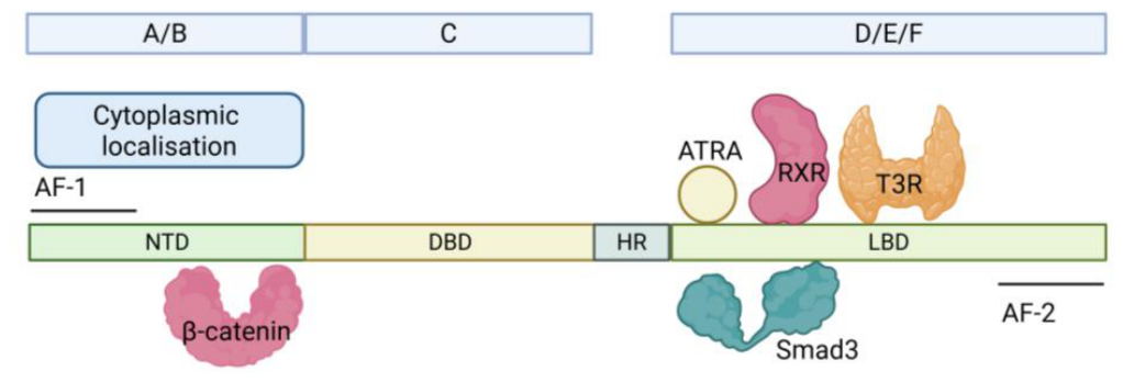
Figure 2.
Overview of the temporal progression of the major signaling pathways that regulate chick limb bud initiation, outgrowth and maturation. Left (limb induction and early outgrowth): proximally derived ATRA is required for limb induction and initiates expression of FGF10 in the lateral plate mesoderm and Shh in the posterior mesenchyme. Reciprocal signaling between mesenchymal FGF10 and apical ectodermal ridge (AER) FGF8, reinforced by Wnt3a, establishes the positive feedback loop that sustains limb bud outgrowth. During this stage, ATRA maintains proximal identity through Meis1/2 expression, promotes maintenance of undifferentiated mesenchymal progenitor cells, and restrains BMP activity. Middle (progressive outgrowth): as ATRA becomes depleted from the distal limb bud (approximately HH20/21), the intrinsic distal developmental program is initiated. Wnt3a-dependent FGF signaling continues to support AER function and proliferative outgrowth, whereas BMP signaling progressively restricts FGF signaling and contributes to developmental timing while Shh expression is maintained. Right (late outgrowth and growth termination): continued BMP activity promotes AER regression, suppresses FGF signaling, reduces progression through the G1-to-S phase of the cell cycle, and terminates proliferative outgrowth as the limb transitions towards differentiation and digit patterning. Arrows indicate activation or promotion, blunt-ended lines indicate inhibition, and circular arrows denote the reciprocal positive feedback between FGF10 and FGF8 during early limb outgrowth. Abbreviations: A, anterior; P, posterior; Px, proximal; D, distal; AER, apical ectodermal ridge; ATRA, all-trans retinoic acid; BMP, bone morphogenetic protein; FGF, fibroblast growth factor; Shh, Sonic hedgehog; Wnt, Wingless/Integrated. Created with Biorender.com.
Figure 2.
Overview of the temporal progression of the major signaling pathways that regulate chick limb bud initiation, outgrowth and maturation. Left (limb induction and early outgrowth): proximally derived ATRA is required for limb induction and initiates expression of FGF10 in the lateral plate mesoderm and Shh in the posterior mesenchyme. Reciprocal signaling between mesenchymal FGF10 and apical ectodermal ridge (AER) FGF8, reinforced by Wnt3a, establishes the positive feedback loop that sustains limb bud outgrowth. During this stage, ATRA maintains proximal identity through Meis1/2 expression, promotes maintenance of undifferentiated mesenchymal progenitor cells, and restrains BMP activity. Middle (progressive outgrowth): as ATRA becomes depleted from the distal limb bud (approximately HH20/21), the intrinsic distal developmental program is initiated. Wnt3a-dependent FGF signaling continues to support AER function and proliferative outgrowth, whereas BMP signaling progressively restricts FGF signaling and contributes to developmental timing while Shh expression is maintained. Right (late outgrowth and growth termination): continued BMP activity promotes AER regression, suppresses FGF signaling, reduces progression through the G1-to-S phase of the cell cycle, and terminates proliferative outgrowth as the limb transitions towards differentiation and digit patterning. Arrows indicate activation or promotion, blunt-ended lines indicate inhibition, and circular arrows denote the reciprocal positive feedback between FGF10 and FGF8 during early limb outgrowth. Abbreviations: A, anterior; P, posterior; Px, proximal; D, distal; AER, apical ectodermal ridge; ATRA, all-trans retinoic acid; BMP, bone morphogenetic protein; FGF, fibroblast growth factor; Shh, Sonic hedgehog; Wnt, Wingless/Integrated. Created with Biorender.com.
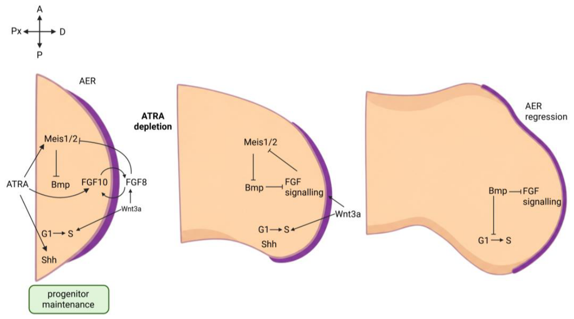
Figure 3.
Some principles to the actions of RARγ. Agonism of RAR blocked stem/progenitor cell development as seen from studies of zebrafish embryogenesis, gastruloid development, and chondrogenesis, and enhanced the generation of iPSCs from somatic cells. RARγ knockout in mice reduced hematopoietic stem cells and there was an increase in more mature progenitors and mature myeloid cells increased when RARγ was antagonized in mice. The absence of active RARγ appears to be permissive to differentiation. RARγ plays a role in the nucleus to regulate the expression of genes that are important to the control of stem cell behavior and influences key intracellular signaling pathways. Created with Biorender.com.
Figure 3.
Some principles to the actions of RARγ. Agonism of RAR blocked stem/progenitor cell development as seen from studies of zebrafish embryogenesis, gastruloid development, and chondrogenesis, and enhanced the generation of iPSCs from somatic cells. RARγ knockout in mice reduced hematopoietic stem cells and there was an increase in more mature progenitors and mature myeloid cells increased when RARγ was antagonized in mice. The absence of active RARγ appears to be permissive to differentiation. RARγ plays a role in the nucleus to regulate the expression of genes that are important to the control of stem cell behavior and influences key intracellular signaling pathways. Created with Biorender.com.
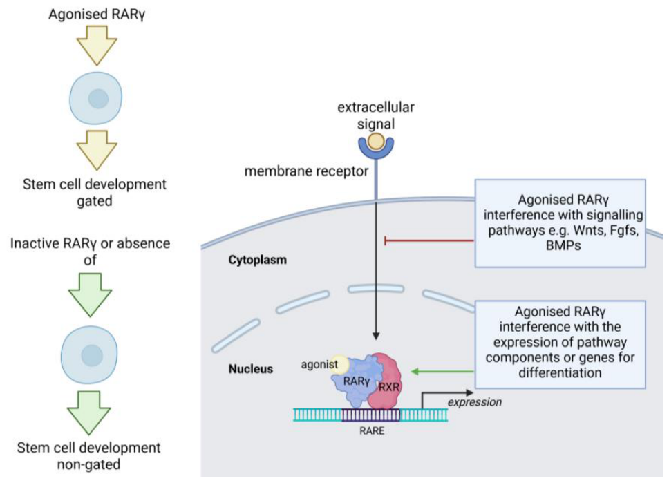
Figure 4.
Crosstalk between the regulatory and signaling pathways of interest. For events regulated by agonized RARγ, the red and green arrows indicate a positive and negative influence, respectively, on other pathways. A negative influence (red arrows) was seen for Wnts signaling from studies of the generation of iPSCs, for BMP studies of chondrogenesis, and for FGFs because the RARγ agonist-mediated defects seen during zebrafish embryogenesis mimicked defects seen in FGF mutant zebrafish. A positive effect (green arrow) of agonized RARγ was seen regarding TGFβ signaling and the generation of iPSCs. The blue double-ended arrows indicate crosstalk between pathways. Created with Biorender.com.
Figure 4.
Crosstalk between the regulatory and signaling pathways of interest. For events regulated by agonized RARγ, the red and green arrows indicate a positive and negative influence, respectively, on other pathways. A negative influence (red arrows) was seen for Wnts signaling from studies of the generation of iPSCs, for BMP studies of chondrogenesis, and for FGFs because the RARγ agonist-mediated defects seen during zebrafish embryogenesis mimicked defects seen in FGF mutant zebrafish. A positive effect (green arrow) of agonized RARγ was seen regarding TGFβ signaling and the generation of iPSCs. The blue double-ended arrows indicate crosstalk between pathways. Created with Biorender.com.
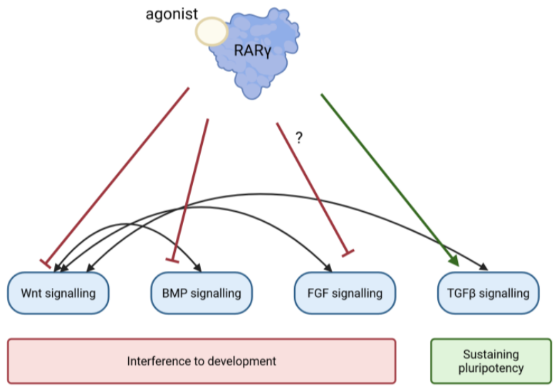
Disclaimer/Publisher’s Note: The statements, opinions and data contained in all publications are solely those of the individual author(s) and contributor(s) and not of MDPI and/or the editor(s). MDPI and/or the editor(s) disclaim responsibility for any injury to people or property resulting from any ideas, methods, instructions or products referred to in the content. |
© 2026 by the authors. Licensee MDPI, Basel, Switzerland. This article is an open access article distributed under the terms and conditions of the Creative Commons Attribution (CC BY) license (http://creativecommons.org/licenses/by/4.0/).
Copyright: This open access article is published under a Creative Commons CC BY 4.0 license, which permit the free download, distribution, and reuse, provided that the author and preprint are cited in any reuse.


