
Submitted:
13 July 2026
Posted:
15 July 2026
You are already at the latest version
Abstract
Epidemiological and clinical evidence indicates that obesity-associated metabolic dysfunction increases the risk of depression, which is often resistant to conventional antidepressant therapies. Elucidating targeted mechanisms underlying obesity-induced depression is therefore essential for developing effective interventions. Accumulating evidence identifies neuroinflammation as a central link between obesity and depressive disorders. Obesity-driven inflammatory signaling disrupts multiple neurobiological processes implicated in depression, thereby contributing to the onset and progression of inflammatory depression. Polyphenols, a diverse class of plant-derived secondary metabolites, have demonstrated neuroprotective properties, largely attributed to their anti-neuroinflammatory effects. By modulating inflammatory pathways and restoring neurobiological homeostasis, polyphenols may represent a promising therapeutic strategy for obesity-related depression. This review summarizes the pathophysiological mechanisms through which obesity induces neuroinflammation and subsequently promotes depressive symptoms. It further evaluates current evidence regarding the efficacy and effective dosing of polyphenols in attenuating inflammatory signaling and alleviating obesity-associated inflammatory depression. Given their anti-neuroinflammatory potential, polyphenols may serve as a valuable adjunctive approach for the management of obesity-induced depression.
Keywords:
obesity
; depression
; neuroinflammation
; polyphenols
; antidepressant
1. Introduction
According to the World Health Organization (WHO), 2.5 billion adults (18 years and older) are overweight globally in 2022, with 890 million of them suffering from obesity. Furthermore, WHO estimates that by 2035, there will be 1.9 billion obese people globally, with 1 in 4 persons on average being obese (World Health Organization (WHO), 2022).
Obesity substantially increases the risk of multiple chronic conditions, including cancer (Lamabadusuriya et al., 2025), diabetes (Milito et al., 2026), depression (Monsalve et al., 2025), respiratory syndrome (Zhao et al., 2026) and cardiovascular disease (Wang et al., 2025). In recent years, the association between obesity and depression has attracted growing attention. Meta-analyses indicate that obesity increases the risk of depression by 65% in older adults and 32% in the general adult population (Guo et al., 2023; Pereira-Miranda et al., 2017). Among children and adolescents, the prevalence of depression is 1.34 times higher in those with obesity than in their normal-weight peers. (Quek et al., 2017). Clinically, obesity-related depression often presents with atypical features, such as increased appetite or weight gain, hypersomnia, leaden paralysis, and heightened sensitivity to interpersonal rejection—symptoms that differ from the appetite loss and insomnia commonly observed in melancholic depression (Lamers et al., 2018). Epidemiological and clinical studies suggest that obesity-induced depression is closely linked to a chronic low-grade inflammatory state driven by immune activation, with neuroinflammation emerging as a central mechanism (Fulton et al., 2022). Current antidepressant therapies primarily target monoamine neurotransmitters, including serotonin (5-HT), norepinephrine (NE), and dopamine (DA). However, these treatments often show limited efficacy in patients with obesity-related depression (Kloiber et al., 2007), suggesting that mechanisms of obesity-related depression are different from those of neurotransmitter-mediated depression. Therefore, it is particularly important to explore in depth the relationship between obesity, neuroinflammation and depression.
Polyphenols, a diverse group of plant-derived bioactive compounds, have attracted considerable interest due to their anti-inflammatory, antioxidant, and neuroprotective properties (Koca et al., 2025). They modulate immune cell activity, regulate the expression of pro-inflammatory cytokines, and influence multiple signaling pathways implicated in depression (Mijailović et al., 2025). Although numerous reviews have examined the role of polyphenols in stress-induced depression, relatively few have focused specifically on obesity-associated inflammatory depression. Moreover, the effective doses required to achieve therapeutic benefits in this context remain unclear. Accordingly, this review synthesizes current evidence on the mechanistic interplay between obesity, neuroinflammation, and depression. It further evaluates the therapeutic potential and effective dosing of polyphenols in modulating inflammatory pathways and mitigating obesity-induced inflammatory depression. This review aims to offer new sights into dietary interventions for mood disorders, particularly obesity-induced inflammatory depression.
2. Causes of Obesity-Induced Neuroinflammation
Microglia are resident immune cells in the central nervous system (CNS) and play an important role in neuroinflammation. Under normal physiological conditions, microglia exist in a resting state. In response to tissue injury, pathogens, or metabolic stress, they become activated and adopt an amoeboid morphology (Cancela et al., 2026). Activated microglia are broadly categorized into a pro-inflammatory (M1-like) phenotype and an anti-inflammatory or reparative (M2-like) phenotype. M1-like microglia secrete pro-inflammatory cytokines that contribute to neuronal dysfunction, whereas M2-like microglia promote debris clearance, neuronal protection, and tissue repair (Jia et al., 2021). Several studies have demonstrated that obese patients show significant activation of microglia in the hippocampus and hypothalamic sites, accompanied by elevated inflammatory cytokines (Cope et al., 2018; Miller & Spencer, 2014). These findings suggest that obesity functions as a chronic immunometabolic stressor that skews microglia toward a sustained pro-inflammatory phenotype. In this section, we explore the main pathways by which obesity activates microglia and summarize the relevant mechanisms that may be involved (Fig. 1).
2.1. Peripheral Inflammation and Blood-Brain Barrier Disruption
Obesity is known to be associated with inflammation of peripheral tissues due to altering the gut microbiota and thus elevating circulating levels of lipopolysaccharide (LPS) produced by gram-negative bacteria (Monsalve et al., 2025). Concurrently, the rapid expansion of adipose tissue in obesity induces local hypoxia, adipocyte death, and activation of hypoxia-inducible factor-1α (HIF-1α), which in turn promotes nod-like receptor family pyrin domain containing 3 (NLRP3) inflammasome activation (Gonzalez et al., 2019). These processes drive the release of pro-inflammatory cytokines and stimulate hepatic production of C-reactive protein (CRP) (Stanimirovic et al., 2022). Large cohort studies have demonstrated that depressed patients have higher CRP concentrations, which makes elevated CRP levels one of the best predictors of the onset of depression in obesity (Hickman et al., 2014). The blood-brain barrier (BBB) is the main pathway through which peripheral inflammatory and immune responses interact with the brain microenvironment. It has been shown that peripheral inflammatory cytokines can down-regulate the expression of tight junction proteins, such as claudin-5 and zonula occludens-1 (ZO-1), thus disrupting tight junctions between neighboring cells and leading to increased BBB permeability (Huang et al., 2021; Wu et al., 2020). Furthermore, it is discovered that LPS inhibites P-glycoprotein activity and induces the secretion of matrix metalloproteinases, which resultes in endoplasmic reticulum stress, mitochondrial damage, and apoptosis in endothelial cells (Bernhart et al., 2018). Impaired BBB integrity caused by endothelial cell apoptosis further exacerbates the infiltration of peripheral inflammatory cytokines in brain tissue. The continuous influx of peripheral inflammatory cytokines into the CNS stimulates microglia over time, leading them to adopt a pro-inflammatory phenotype and trigger neuroinflammation. It has been shown that the mechanism by which peripheral inflammation activates microglia is that inflammatory cytokines, such as interleukin-1β (IL-1β), stimulate the release of the microglia damage-associated molecular pattern (DAMP) molecule adenosine triphosphate (ATP), and then ATP-mediated Ca2+ signaling facilitates the assembly of the NLRP3 inflammasome, thus triggering a series of inflammatory cytokine production (Garaschuk, 2021). Furthermore, Toll-like receptor 4 (TLR4) on the surface of microglia can be bound by LPS, saturated fatty acids (SFA), and adipokine resistin, which might intensify neuroinflammation by starting an inflammatory cascade (An et al., 2020; Benomar & Taouis, 2019; Lu et al., 2021).
2.2. Dietary Saturated Fatty Acids as Direct Microglial Activators
Deliciousness and high energy density are the main features of the diets of obese people. These diets are rich in high saturated fats (e.g., lard or palm oil) that significantly elevate SFA levels in the blood (Nakajima et al., 2020). It is shown that consumption of saturated fats (Lai et al., 2016) and serum concentrations of the SFA palmitic acid (PA) (Tsuboi et al., 2013) are positively associated with depressive symptoms and serum levels of the peripheral inflammatory marker CRP. Indeed, SFA can cross the BBB via transporter- and receptor-mediated mechanisms, allowing them to directly interact with microglia (Sanchez-Cano et al., 2021). Zhuang et al. (2022) demonstrate that high-fat diet (HFD) can lead to PA deposition in the hippocampus of mice and lead to activation of microglia, as evidenced by a smaller microglia area and an increased immunological response to cluster of differentiation 68 (CD68). In vitro, PA and stearic acid (SA) cause BV2 microglia to produce pro-inflammatory cytokines through the TLR4/NF-ĸB pathway, which impaires neurogenesis (Wang et al., 2012). In addition, Beaulieu et al. (2021) demonstrate that PA can activate N9 microglia to trigger neuroinflammation via mediating the nuclear factor kappa-B (NF-ĸB) pathway and phosphorylation of the c-Jun. These findings indicate that dietary SFAs not only exacerbate systemic inflammation but also directly trigger neuroinflammatory signaling within the CNS.
2.3. Lipid Droplet Accumulation in Microglia
Recent evidence highlights lipid droplet (LD) accumulation in microglia as an emerging mechanism linking metabolic stress to neuroinflammation. HFD exposure during adolescence induces emotional behavioral abnormalities in mice and promotes LD accumulation in hippocampal microglia. Importantly, LD burden correlates positively with microglial activation status (Yao et al., 2024). Similar lipid-laden microglia have been identified in the aging brain and are considered representative of dysfunction and pro-inflammatory states in the aging brain (Marschallinger et al., 2020). In vivo and in vitro studies show that innate inflammation induces de novo synthesis of LDs in microglia through activation of the TLR4 signaling pathway (Khatchadourian et al., 2012; Marschallinger et al., 2020). Perilipin-2 (PLIN 2), located on the surface of LDs, can bind to the inflammation amplifying factor triggering receptor expressed on myeloid cells-1 (TREM 1), leading to an abnormal accumulation of TREM 1 inside microglia LDs. TREM 1 subsequently promotes neuroinflammation through the NLRP3 inflammasome pathway, and the elevated pro-inflammatory cytokines in turn activate the TLR4 signaling pathway to induce LD formation, thus constituting a vicious cycle (Li et al., 2023). This explains the long lasting neuroinflammatory state in the brain of obese patients (Henn et al., 2022). Interventions targeting LD metabolism support a causal role for LD accumulation in neuroinflammation. According to Khatchadourian et al. (2012), inhibition of long-chain acyl-CoA synthetase with triacsin C suppresses LD formation and reduces reactive oxygen species and cytokine production in microglia. Similar findings are reported by Li et al. (Li et al., 2023). They find that high glucose can induce lipophagy damage in microglia, leading to the LDs accumulation, and triggering an inflammatory cascade response. In contrast, rapamycin, an autophagy activator, is successful in reversing the defective lipophagy in microglia caused by high glucose, reducing LDs accumulation and inflammation production. These findings suggest that intracellular LD accumulation represents a critical metabolic driver of sustained microglial activation in obesity.
3. Pathways of Neuroinflammation Triggering Depression
3.1. Abnormalities of the Hypothalamic-Pituitary-Adrenal Axis
Neuroinflammation is closely linked to dysregulation of the hypothalamic-pituitary-adrenal (HPA) axis, a central stress-response system that is frequently hyperactivated in obesity (Dymek et al., 2025). Pro-inflammatory cytokines released by activated microglia modulate HPA axis activity, with IL-1β acting as a primary driver. IL-1β stimulates the release of corticotropin-releasing hormone from the hypothalamus and adrenocorticotropic hormone from the pituitary gland, ultimately increasing glucocorticoid (GC) secretion—cortisol in humans and corticosterone in rodents (Dionysopoulou et al., 2021; Fu et al., 2023) (Figure 2). GC is the ultimate effector of the HPA axis, and elevated levels of GC are considered a biological risk factor for depression (Cheiran Pereira et al., 2022). Notably, the relationship between the HPA axis and obesity appears to be bidirectional. Exposure to high levels of cortisol can induce obesity through a variety of pathways: (a) increased appetite with a preference for energy-dense food; (b) stimulation of adipogenesis and adipocyte hypertrophy, especially in visceral fat; (c) inhibition of thermogenesis in brown adipose tissue with relative reduction of energy expenditure (Milano et al., 2020). It is obvious that there is a cycle between obesity and high levels of cortisol. This cycle increases the susceptibility of obesity to depression. Elevated corticosterone can decrease immature neuron activation and may have long-term effects on the integration or function of cells (Workman et al., 2015). In db/db mice, increased corticosterone suppresses hippocampal brain-derived neurotrophic factor (BDNF) expression via activation of glucocorticoid receptors (Wosiski-Kuhn et al., 2014). In contrast, lentiviral suppression of the glucocorticoid receptor expression restores BDNF expression and rescues hippocampal neurogenesis in db/db mice. Therefore, obesity-related depression may be associated with impaired hippocampal neurogenesis induced by elevated corticosterone.
3.2. Defects in Adult Hippocampal Neurogenesis
Rodent studies demonstrate that obesity-induced neuroinflammation inhibits the proliferation, differentiation and survival of newborn cells in the hippocampal dentate gyrus, which in turn prevents the production of newborn neurons (Baghdadchi et al., 2026; Kwon et al., 2026) (Figure 2). Furthermore, HFD-fed obese mice exhibit significantly fewer DCX (a marker of newly generated neurons within the last 2-3 weeks) and Ki67 (a proliferating cell marker) positive cells in the hippocampus, indicating reduced neurogenesis. These structural deficits are accompanied by depression-like behaviors, including reduced sucrose preference and altered exploratory behavior, supporting a functional link between impaired neurogenesis and mood disturbances (Yao et al., 2022). It is reported that pro-inflammatory cytokines (IL-6, IL-1β and TNF-α) appear to inhibit adult hippocampal neurogenesis by activating the signal transducer and activator of transcription 3 (STAT3) pathway in hippocampal neural progenitor cells (Chen et al., 2013; Kong et al., 2019), whereas interferon ɑ and interferon γ inhibit adult hippocampal neurogenesis by activating the STAT1 pathway (Borsini et al., 2018; Zhang et al., 2020). Meanwhile, in a model of LPS-induced neuroinflammation, pharmacological attenuation of microglial activation restores hippocampal neurogenesis (Domínguez-Rivas et al., 2021). Excessive microglial activation not only releases inflammatory mediators but also increases phagocytosis of newborn neurons (Domínguez-Rivas et al., 2021). In HFD-fed mice, hyperactivated microglia excessively engulf immature neurons, further impairing neurogenesis (Yao et al., 2022). Treatment with anti-inflammatory agents such as minocycline reduces microglial overactivation, rescues neurogenesis, and alleviates depressive-like behaviors (Bassett et al., 2021). These findings indicate that neuroinflammation-driven disruption of hippocampal neurogenesis represents a key mechanism linking obesity to depression
3.3. Resistance of Central Insulin and Leptin
Central insulin and leptin resistance is another mechanism connecting metabolic dysfunction to depressive symptoms. Large cohort studies demonstrate that reduced insulin sensitivity correlates with increased depression and anxiety symptoms in individuals with obesity (Perry et al., 2020). Preclinical studies also support this concept because it has been repeatedly demonstrated that HFD-induced insulin resistance in mice or rats leads to anxiety and depression-like behaviors (Hassan et al., 2019). Moreover, it has been demonstrated that knockdown of insulin receptor (InsR) in astrocytes also promotes the development of anxiety and depression-like behaviors in mice (Cai et al., 2018). These studies highlight the critical role that central insulin resistance plays in the association between obesity and depression. It has been shown that inflammatory cytokines in the brain can trigger hypothalamic insulin resistance by impairing insulin-dependent phosphorylation and downstream signaling at InsR via the IKKβ/NF-ĸB signaling pathway (Benzler et al., 2015), the JNK signaling pathway (Feng et al., 2020) and the TLR4 signaling pathway (Benomar & Taouis, 2019) (Figure 2). In rodents, HFD intake is considered an important factor in obesity-induced insulin resistance. Dietary SFA can activate TLR4/MyD88 signaling to enhance endoplasmic reticulum stress and hypothalamic insulin resistance (Li et al., 2020). Leptin is a receptor signal from adipose tissue to the brain that conveys energy storage. It regulates the body's energy balance and exerts antidepressant-like effects by acting on the leptin receptor (LepR) in the hypothalamus (Peng et al., 2021). Studies have shown that obesity-induced inflammation can inhibit the expression of leptin transporter proteins in the BBB, leading to a reduced capacity of the leptin transport system. This prevents high levels of leptin in the periphery from entering the brain to exert antidepressant activity (Sáinz et al., 2015). Meanwhile, obesity-induced inflammation can also impair hippocampal leptin receptor activity, resulting in the inability of leptin to activate downstream signals to exert antidepressant effect. Yamada et al. (2011) demonstrate that diet-induced obese mice with impaired hippocampal leptin receptor activity fail to respond to leptin administration in terms of the forced swimming test (FST) and the biochemical changes in the hippocampus. These studies suggest that inflammation-driven insulin and leptin resistance disrupt central metabolic signaling and neurotrophic support, thereby contributing to the development of obesity-associated depression.
4. Mechanisms of Polyphenols Ameliorating Obesity-Induced Inflammatory Depression
Polyphenols are one of the most abundant and widely distributed natural products in plants. Although their structures vary widely, they all have aromatic rings with one or more hydroxyl substituents (Mehra & Mittal, 2026). Phenolic compounds are traditionally divided into two groups: flavonoids and non-flavonoids (Figure 3). In chemical structure, flavonoids have three rings (C6-C3-C6) as their basic skeleton (labeled A, B, and C in Figure 3) (Shen et al., 2022). Based on the number of phenolic rings and the structural components linking them, flavonoids can be subdivided into several subclasses, including flavanols, flavones, flavonols, anthocyanidins, flavanones, isoflavones, and chalcones (Bessa et al., 2021). Non-flavonoid polyphenols primarily include phenolic acids, stilbenes, lignans, and hydrolysable tannins. Among them, phenolic acids are the most significant non-flavonoid polyphenols and can be further classified into hydroxybenzoic and hydroxycinnamic acids (Wang et al., 2022). Growing evidence indicates that polyphenols exert antidepressant and neuroprotective effects (Fekete et al., 2025). The subsequent sections explore how phenolic compounds alleviate inflammatory depression via the specific signaling pathways (Figure 4).
4.1. Modulation of Hippocampal NF-ĸB Pathway
Rodent studies demonstrate that HFD feeding increases hippocampal expression of NF-κB-related proteins (p-IKKα/β, p-IκBα, p65, p-p65) and is accompanied by a marked prolongation of immobility time in both the tail suspension test and the forced swimming test (Xie et al., 2024). Furthermore, LPS, TNF-α, and PA are all found to activate the microglial NF-κB signaling pathway and trigger neuroinflammation in cellular models (Brás et al., 2020; Evans et al., 2023). These studies confirm the important role of the NF-κB pathway in obesity-induced neuroinflammation. Polyphenols can ameliorate obesity-induced depressive-like behaviors by modulating the expression of the NF-κB signaling pathway in microglia. Mechanistically, this suppression is associated with reduced HPA axis hyperactivity, restoration of hippocampal BDNF expression, and normalization of serotonin metabolism. For example, apple polyphenols inhibit NF-κB-related gene expression in hippocampal microglia, reduce circulating corticosterone and adrenocorticotropic hormone levels, and alleviate depressive-like behaviors in obese mice (Xie et al., 2024). Baicalein (Liu et al., 2022) and salidroside (Zhu et al., 2015) enhance hippocampal BDNF expression through NF-κB inhibition, thereby improving neurogenesis and synaptic plasticity associated with depression. Fisetin suppresses NF-κB activation and indoleamine 2,3-dioxygenase-1 (IDO-1) expression, restoring serotonin synthesis pathways and exerting antidepressant effects (Choubey et al., 2019).
4.2. Modulation of MAPK Pathway in CNS
Mitogen-activated protein kinase (MAPK) is a group of evolutionarily conserved serine-threonine kinases. It contains four classical pathways: ERK1/2, p38, JNK1/2/3, and ERK5 (Behl et al., 2022). HFD and high-fat/high-fructose diet (HFFD) activate JNK and p38 pathways in the brain, leading to neuroinflammation and cognitive impairment (Li et al., 2018; Lu et al., 2021). In LPS-induced depression models, pharmacological inhibition of p38 restores inflammatory markers and ameliorates depressive-like behaviors (Y.-W. Zhao et al., 2018). This result reveals the fact that inhibition of the MAPK pathway can ameliorate inflammatory depression. Activation of the MAPK pathway is a cascading phosphorylation process, and polyphenols can act at various steps in the activation process. Phenolics compounds such as quercitrin (Sun et al., 2021), baicalin (Li et al., 2022), sinapic acid (Huang et al., 2023) and sophoraflavanone G (Guo et al., 2016) all down-regulate the phosphorylation of JNK, p38 and ERK, thereby inhibiting the MAPK signaling pathway and exerting anti-neuroinflammatory effects. In LPS-activated BV2 microglial cells, fisetin reduces the release of inflammatory cytokines, inducible nitric oxide synthase (iNOS) and cyclooxygenase (COX)-2, by inhibiting phosphorylation of ERK but not JNK (Cho et al., 2013). This suggests that the inhibition of the MAPK pathway by polyphenols is specific. Certain polyphenols simultaneously target MAPK and NF-κB pathways. For instance, isorhamnetin attenuates HFFD-induced neuroinflammation by inhibiting the activation of JNK, p38, and NF-ĸB signaling pathways (Mulati et al., 2021). Punicalagin similarly reduces phosphorylation of ERK, JNK, and p38 while inhibiting NF-κB activity. (Lo et al., 2022). Additionally, similar to the findings in the NF-ĸB pathway, the modulation of the MAPK pathway by polyphenols can promote the expression of the CREB/BDNF pathway, which ameliorates the depressive-like behaviors triggered by neuroinflammation (Sun et al., 2021).
4.3. Modulation of Hippocampal PI3K/Akt Pathway
The phosphatidylinositol-3-kinase (PI3K)/protein kinase B (Akt) pathway integrates metabolic and inflammatory signaling and is closely associated with obesity-related depression. Under normal physiological conditions, insulin activates insulin receptor substrates (IRS), initiating PI3K/Akt signaling to support neuronal survival and plasticity (Savova et al., 2023). But in obesity, insulin resistance reduces the activation of the IRS/PI3K/Akt signaling pathway, which exacerbates the risk of obesity suffering from depression (De Paiva et al., 2024). It is shown that genistein (Li et al., 2020) and ferulic acid (Wang et al., 2017) can exert neuroprotective effects in obese diabetic mice and rats by modulating the levels of p-IRS, p-Akt, and p-GSK3β. In addition, epigallocatechin gallate (EGCG) (Mi et al., 2017) and sea-buckthorn flavonoids (Mulati et al., 2020) can significantly ameliorate HFFD-induced insulin resistance and neuronal damage in obese mice by up-regulating IRS/PI3K/Akt and ERK/CREB/BDNF signaling pathways. In inflammatory depression models, PI3K/Akt signaling also interacts with NF-κB. Intraperitoneal injection of LPS activates the hippocampal PI3K/Akt/NF-ĸB and MEK/ERK signaling pathways, which triggers neuroinflammation and impairs hippocampal plasticity. Curcumin (Cianciulli et al., 2016), EGCG (Payne et al., 2023), and eriodictyol (He et al., 2018) can attenuate LPS-induced pro-inflammatory cytokines release in BV2 microglial cells by inhibiting the PI3K/AKT/NF-ĸB pathway in a dose-dependent manner, and the effect of curcumin is comparable to that of the PI3K inhibitor LY294002. However, due to the low solubility of curcumin in plasma and the high rate of degradation in acidic, alkaline and oxidative environments, clinical studies have not yielded good therapeutic results (Parikh et al., 2008).
4.4. Modulation of Other Pathways
In addition to the previously mentioned NF-ĸB, MAPK, and PI3K/Akt pathways, polyphenols can also ameliorate obesity-induced inflammatory depression through specific pathways. Gut microbial composition has been suggested as a potential contributor to the neurobehavioral abnormalities associated with HFD consumption (Paiva et al., 2024). EGCG can restore HFD-induced gut dysbiosis and exert antidepressant effects through the gut-brain axis. Specifically, EGCG can ameliorate HFD-induced intestinal inflammation by increasing the abundance of Muribaculaceae and Alloprevotella, which prevents SCFAs, PPAR-γ, and NF-ĸB secreted by the intestine from entering the CNS through the circulation of blood to affect the expression of hypothalamic neurotransmitters (Zhou et al., 2023). By modulating the gut-brain axis, (-)-epicatechin (EC) also ameliorates obesity-induced depression-like behaviors. However, the efficacy of EC is mainly achieved by increasing the abundance of Lactobacillus and Enterobacter (Kang et al., 2022). Lactobacillus has been known to play a role in HFD-induced depression. It can exert antidepressant effects by modulating hippocampal γ-aminobutyric acid (GABA) expression and HPA axis activity via the vagus nerve (Bravo et al., 2011). Additionally, blueberry polyphenol extract inhibits LPS-induced neuroinflammation and depressive-like behaviors in mice through modulation of brain enzymes activities, such as monoamine oxidase-A (MAO-A), acetylcholinesterase (AChE) and Na+/K+-ATPase (Spohr et al., 2023). This finding is further confirmed in a study conducted by Luduvico et al (2022), who discover that tannic acid can exert neuroprotective and antidepressant effects by modulating brain Na+/K+-ATPase and Ca2+-ATPase activities. However, the modulating effect of polyphenols on brain enzyme activities has only been reported in the LPS-induced depression model, and still needs to be further explored in the HFD-induced depression model.
5. Effective Doses of Polyphenols in Ameliorating Obesity-Induced Inflammatory Depression
Although numerous studies have confirmed the anti-inflammatory and neuroprotective potential of polyphenols, their effective doses in ameliorating obesity-related depression remains unclear. The dose-response relationship is a core parameter for evaluating the pharmacological activity of polyphenols and guiding the development of dietary supplements. Determining the effective doses of polyphenols not only help to explain the discrepancies in results across different studies but also provide a theoretical basis for future personalized and precision interventions (Table 1).
5.1. In Vitro Model
In in vitro models, the ameliorative effects of polyphenols on obesity-related depression are primarily evaluated by simulating neuroinflammation, such as by stimulating microglia with LPS or PA. As shown in Table 1, the effective doses of different polyphenolic compounds in in vitro models vary significantly, and this variation is likely closely related to their chemical structures. For example, the flavonoid baicalin (Li et al., 2022) and its aglycone baicalein (Ran et al., 2021) exhibit different inhibitory potencies against LPS-induced IL-1β release in BV2 microglia. Baicalin significantly reduced IL-1β release at 22.5 μM, whereas baicalein required 45 μM to achieve comparable effects. This result revealed an intrinsic relationship between the activity of flavonoids and their structural characteristics, namely that glycosylation (forming glycosides) may enhance the biological activity of the molecule. Further analysis of the effective doses of polyphenols reveals that the effective doses of polyphenol extracts are generally higher, such as blueberry polyphenol extracts (Carey et al., 2013). This is likely due to their complex composition and the relatively low content of active ingredients. Regarding dose-effect relationships, most studies confirmed that polyphenols inhibited the production of pro-inflammatory factors in microglia in a dose-dependent manner. For instance, sinapic acid (Huang et al., 2023), within the range of 5-20 μM, showed increasing inhibitory effects on iNOS and IL-6 expression as the concentration rised. However, some polyphenols exhibit hormetic effects, characterized by beneficial actions at low concentrations and cytotoxicity at higher doses. EGCG significantly inhibited BV-2 cell proliferation at 175-300 μM (Payne et al., 2023) and hesperetin reduced microglial viability at 200 μM (Evans et al., 2023). These results indicate that the biological effects of polyphenols are not simply linear and may shift from protective to toxic once a certain concentration threshold is exceeded.
5.2. In Vivo Model
The ameliorative effects of polyphenols on obesity-related depression have been validated in various rodent models, including high-fat/high-sucrose diet induced depression and diabetes-associated depression. The effective administration doses, intervention durations, and primary mechanisms of action vary among different polyphenolic compounds. Similar to the patterns observed in in vitro models, the effective doses of different polyphenols in animal models exhibit a wide range. Generally, crude extracts (such as apple polyphenol extract and sea-buckthorn flavonoids) require relatively higher effective doses, typically between 120-500 mg/kg/d, which is attributed to their complex composition and relatively low content of active ingredients (Mulati et al., 2020). In contrast, purified monomeric compounds (such as EGCG and hydroxytyrosol) have relatively lower effective doses, mostly in the range of 25-100 mg/kg/d, benefiting from their defined chemical structures and higher target affinity (Liu et al., 2024; Zhou et al., 2023). Regarding the dose-effect relationship, multiple studies confirmed that polyphenols ameliorated obesity-related depressive-like behaviors and molecular biomarkers in a dose-dependent manner. In an HFD-induced obese mouse model, 20 mg/kg EC produced greater improvements in open-field behavior, hippocampal BDNF expression, and glucocorticoid receptor regulation compared with 2 mg/kg (Kang et al., 2022). Similarly, hydroxytyrosol at 100 mg/kg was more effective than 25 mg/kg in improving cognition and reducing neuroinflammation (Liu et al., 2024). These findings not only confirm the dose-dependent effects of polyphenols but also provide important references for determining the optimal effective dosage window. It is noteworthy that the administration method and intervention duration also significantly impact the effective dose of polyphenols. Currently, most studies employ oral gavage, with intervention periods ranging from 4 to 14 weeks, a timeframe typically aligned with the duration of diet-induced obesity. For instance, continuous intragastric administration of apple polyphenol extract (500 mg/kg/d) for 8 weeks significantly ameliorated depressive-like behaviors in obese mice induced by a high-sucrose diet (Xie et al., 2024). In contrast, sea-buckthorn flavonoids required a longer intervention period of 14 weeks (administered via diet incorporation at 0.06%-0.31% w/w) to exert significant protective effects in a model of cognitive impairment induced by a high-fat and high-fructose diet (Mulati et al., 2020). Furthermore, the impact of administration timing (preventive versus therapeutic administration) on effective dose warrants attention. Theoretically, therapeutic administration may require higher doses or longer intervention periods to achieve effects comparable to preventive administration. However, systematic studies directly comparing the dosage requirements for these two administration timings are still relatively limited. Future research needs to further clarify the optimal dose windows for different administration timings.
6. Conclusion and Future Perspectives
With the rapid socio-economic development and the drastic evolution of dietary patterns, overweight, obesity and related metabolic problems caused by unbalanced diets have emerged. The incidence of depression triggered by obesity is also increasing annually. Due to the lack of response to conventional antidepressants in obese depressed patients, exploring a more targeted approach to ameliorate obesity-induced depression is particularly important. This review elucidated the pathophysiological mechanisms by which obesity triggers neuroinflammation and thus depression. Meanwhile, we summarized the findings from both in vivo and in vitro studies on the ameliorative effects of polyphenols against obesity-related depression, as well as their effective doses. So, given their significant anti-neuroinflammatory properties, polyphenols could be a promising therapeutic for treating obesity-induced depression.
However, the detailed and more targeted of obesity-induced depression are needed to be explored in the future. More work on the difference of obesity-induced depression and conventional depression are also needed. In addition, although polyphenols are reported to have significant anti-neuroinflammatory effects, direct evidence of polyphenols on ameliorating obesity-induced depression are limited. Meanwhile, since neurological disorders are often accompanied by the co-involvement of different brain regions and cells, in vitro studies employing polyphenols to intervene in microglia are no longer sufficient for the exploration of specific neurological diseases. In recent years, brain organoids have been a popular area of study. The integration of microglia with different neuronal cell types can be used to construct specific neurological disease models (Sabate-Soler et al., 2024). Hence, it may be more accurate to explore the therapeutic mechanism of polyphenols for inflammatory depression by constructing the microglia containing brain organoids. Last but not least, although animal and cellular experiments have assessed the antidepressant efficacy of phenolic compounds, clinical studies have not yet demonstrated the ameliorative effect of polyphenols in patients with obesity-induced depression. Therefore, more comprehensive clinical studies are still needed to verify the antidepressant efficacy of polyphenols.:
Author Contributions
Yawei Xu: Writing, Review, Conceptualisation and Editing; Yajie Zhang: Writing, Review and Editing; Yunfei Huang: Review and Proofreading; Lu Li: Review, Editing and Proofreading; Mengna Shi: Writing, Review, Editing and Proofreading; Chunmei Li: Conceptualisation, Supervision, Writing, Review and Editing. All authors have read and agreed to the published version of the manuscript.
Funding
The author(s) reported there is no funding associated with the work featured in this article.
Data Availability
No new data were created or analyzed in this study. Data sharing is not applicable to this article.
Acknowledgments
The authors gratefully acknowledge Muhammad Asif Ismail for correcting the grammar and vocabulary of this review.
Conflicts of interest
The authors declare no conflicts of interest.
Abbreviations
Akt: 2. cyclooxygenase-2; CRP: C-reactive protein; GC: glucocorticoids; HFD: high-fat diet; HFFD: high-fat and high-fructose diet; HPA: hypothalamic-pituitary-adrenal; IĸB : inhibitory ĸB protein; IKK: IĸB kinase; IL-1β: interleukin-1β; IL-6: interleukin-6; iNOS: inducible nitric oxide synthase; InsR: insulin receptor; IRS: insulin receptor substrate; JNK: c-Jun N-terminal kinase; LDs: lipid droplets; LepR: leptin receptor; LPS: lipopolysaccharide; MAPK: mitogen-activated protein kinase; NF-ĸB: nuclear factor kappa-B; NLRP3: nod-like receptor family pyrin domain containing 3; PA: palmitic acid; SFA: saturated fatty acids; STAT3: signal transducer and activator of transcription 3; TLR4: Toll-like receptor 4; TNF-α: tumor necrosis factor-α; PI3K: phosphatidylinositol-3-kinase; ZO-1: zonula occludens-1.
References
- An, J., Chen, B., Kang, X., Zhang, R., Guo, Y., Zhao, J., & Yang, H. (n.d.). Neuroprotective effects of natural compounds.
- Baghdadchi, Y., White, C., & Dimitrov, E. (2026). Obesity suppresses hippocampal neurogenesis and impairs memory and executive function in a weight and sex-specific manner. Physiology & Behavior, 303, 115141. [CrossRef]
- Bassett, B., Subramaniyam, S., Fan, Y., Varney, S., Pan, H., Carneiro, A. M. D., & Chung, C. Y. (2021). Minocycline alleviates depression-like symptoms by rescuing decrease in neurogenesis in dorsal hippocampus via blocking microglia activation/phagocytosis. Brain, Behavior, and Immunity, 91, 519–530. [CrossRef]
- Beaulieu, J., Costa, G., Renaud, J., Moitié, A., Glémet, H., Sergi, D., & Martinoli, M.-G. (2021). The Neuroinflammatory and Neurotoxic Potential of Palmitic Acid Is Mitigated by Oleic Acid in Microglial Cells and Microglial-Neuronal Co-cultures. Molecular Neurobiology, 58(6), 3000–3014. [CrossRef]
- Behl, T., Rana, T., Alotaibi, G. H., Shamsuzzaman, Md., Naqvi, M., Sehgal, A., Singh, S., Sharma, N., Almoshari, Y., Abdellatif, A. A. H., Iqbal, M. S., Bhatia, S., Al-Harrasi, A., & Bungau, S. (2022). Polyphenols inhibiting MAPK signalling pathway mediated oxidative stress and inflammation in depression. Biomedicine & Pharmacotherapy, 146, 112545. [CrossRef]
- Benomar, Y., & Taouis, M. (2019). Molecular Mechanisms Underlying Obesity-Induced Hypothalamic Inflammation and Insulin Resistance: Pivotal Role of Resistin/TLR4 Pathways. Frontiers in Endocrinology, 10, 140. [CrossRef]
- Bernhart, E., Kogelnik, N., Prasch, J., Gottschalk, B., Goeritzer, M., Depaoli, M. R., Reicher, H., Nusshold, C., Plastira, I., Hammer, A., Fauler, G., Malli, R., Graier, W. F., Malle, E., & Sattler, W. (2018). 2-Chlorohexadecanoic acid induces ER stress and mitochondrial dysfunction in brain microvascular endothelial cells. Redox Biology, 15, 441–451. [CrossRef]
- Bessa, C., Francisco, T., Dias, R., Mateus, N., Freitas, V. D., & Pérez-Gregorio, R. (2021). Use of Polyphenols as Modulators of Food Allergies. From Chemistry to Biological Implications. Frontiers in Sustainable Food Systems, 5, 623611. [CrossRef]
- Borsini, A., Cattaneo, A., Malpighi, C., Thuret, S., Harrison, N. A., MRC ImmunoPsychiatry Consortium, Zunszain, P. A., & Pariante, C. M. (2018). Interferon-Alpha Reduces Human Hippocampal Neurogenesis and Increases Apoptosis via Activation of Distinct STAT1-Dependent Mechanisms. International Journal of Neuropsychopharmacology, 21(2), 187–200. [CrossRef]
- Brás, J. P., Bravo, J., Freitas, J., Barbosa, M. A., Santos, S. G., Summavielle, T., & Almeida, M. I. (2020). TNF-alpha-induced microglia activation requires miR-342: Impact on NF-kB signaling and neurotoxicity. Cell Death & Disease, 11(6), 415. [CrossRef]
- Bravo, J. A., Forsythe, P., Chew, M. V., Escaravage, E., Savignac, H. M., Dinan, T. G., Bienenstock, J., & Cryan, J. F. (2011). Ingestion of Lactobacillus strain regulates emotional behavior and central GABA receptor expression in a mouse via the vagus nerve. Proceedings of the National Academy of Sciences, 108(38), 16050–16055. [CrossRef]
- Cai, W., Xue, C., Sakaguchi, M., Konishi, M., Shirazian, A., Ferris, H. A., Li, M. E., Yu, R., Kleinridders, A., Pothos, E. N., & Kahn, C. R. (2018). Insulin regulates astrocyte gliotransmission and modulates behavior. Journal of Clinical Investigation, 128(7), 2914–2926. [CrossRef]
- Cancela, L. M., Mongi-Bragato, B., Avalos, M. P., & Bollati, F. A. (2026). From Stress to Substance Use Disorders: The Expanding Role of Microglia–Astrocyte Crosstalk in Neuroimmune and Glutamate Alterations in the Nucleus Accumbens. International Journal of Molecular Sciences, 27(1), 385. [CrossRef]
- Carey, A. N., Fisher, D. R., Rimando, A. M., Gomes, S. M., Bielinski, D. F., & Shukitt-Hale, B. (2013). Stilbenes and Anthocyanins Reduce Stress Signaling in BV-2 Mouse Microglia. Journal of Agricultural and Food Chemistry, 61(25), 5979–5986. [CrossRef]
- Cheiran Pereira, G., Piton, E., Moreira Dos Santos, B., Ramanzini, L. G., Muniz Camargo, L. F., Menezes Da Silva, R., & Bochi, G. V. (2022). Microglia and HPA axis in depression: An overview of participation and relationship. The World Journal of Biological Psychiatry, 23(3), 165–182. [CrossRef]
- Chen, E., Xu, D., Lan, X., Jia, B., Sun, L., Zheng, J., & Peng, H. (2013). A Novel Role of the STAT3 Pathway in Brain Inflammation-induced Human Neural Progenitor Cell Differentiation. Current Molecular Medicine, 13(9), 1474–1484. [CrossRef]
- Chen, P., Guo, Z., Lei, J., & Wang, Y. (2024). Pomegranate polyphenol punicalin ameliorates lipopolysaccharide-induced memory impairment, behavioral disorders, oxidative stress, and neuroinflammation via inhibition of TLR4-NF-кB pathway. Phytotherapy Research, 38(7), 3489–3508. [CrossRef]
- Cho, N., Lee, K. Y., Huh, J., Choi, J. H., Yang, H., Jeong, E. J., Kim, H. P., & Sung, S. H. (2013). Cognitive-enhancing effects of Rhus verniciflua bark extract and its active flavonoids with neuroprotective and anti-inflammatory activities. Food and Chemical Toxicology, 58, 355–361. [CrossRef]
- Choubey, P., Kwatra, M., Pandey, S. N., Kumar, D., Dwivedi, D. K., Rajput, P., Mishra, A., Lahkar, M., & Jangra, A. (2019). Ameliorative effect of fisetin against lipopolysaccharide and restraint stress-induced behavioral deficits via modulation of NF-κB and IDO-1. Psychopharmacology, 236(2), 741–752. [CrossRef]
- Cianciulli, A., Calvello, R., Porro, C., Trotta, T., Salvatore, R., & Panaro, M. A. (2016). PI3k/Akt signalling pathway plays a crucial role in the anti-inflammatory effects of curcumin in LPS-activated microglia. International Immunopharmacology, 36, 282–290. [CrossRef]
- Cope, E. C., LaMarca, E. A., Monari, P. K., Olson, L. B., Martinez, S., Zych, A. D., Katchur, N. J., & Gould, E. (2018). Microglia Play an Active Role in Obesity-Associated Cognitive Decline. The Journal of Neuroscience, 38(41), 8889–8904. [CrossRef]
- De Paiva, I. H. R., Da Silva, R. S., Mendonça, I. P., De Souza, J. R. B., & Peixoto, C. A. (2024). Semaglutide Attenuates Anxious and Depressive-Like Behaviors and Reverses the Cognitive Impairment in a Type 2 Diabetes Mellitus Mouse Model Via the Microbiota-Gut-Brain Axis. Journal of Neuroimmune Pharmacology, 19(1), 36. [CrossRef]
- De Souza, D. V., Pappis, L., Bandeira, T. T., Sangoi, G. G., Fontana, T., Rissi, V. B., Sagrillo, M. R., Duarte, M. M., Duarte, T., Bodenstein, D. F., Andreazza, A. C., Cruz, I. B. M. D., Ribeiro, E. E., Antoniazzi, A., Ourique, A. F., & Machado, A. K. (2022). Açaí ( Euterpe oleracea Mart.) presents anti-neuroinflammatory capacity in LPS-activated microglia cells. Nutritional Neuroscience, 25(6), 1188–1199. [CrossRef]
- Dionysopoulou, S., Charmandari, E., Bargiota, A., Vlahos, N. F., Mastorakos, G., & Valsamakis, G. (2021). The Role of Hypothalamic Inflammation in Diet-Induced Obesity and Its Association with Cognitive and Mood Disorders. Nutrients, 13(2), 498. [CrossRef]
- Domínguez-Rivas, E., Ávila-Muñoz, E., Schwarzacher, S. W., & Zepeda, A. (2021). Adult hippocampal neurogenesis in the context of lipopolysaccharide-induced neuroinflammation: A molecular, cellular and behavioral review. Brain, Behavior, and Immunity, 97, 286–302. [CrossRef]
- Dymek, A., Zielińska, M., Englert-Bator, A., Dereń, K., & Łuszczki, E. (2025). Generalized Anxiety Disorder and Obesity: Overlapping Neuroendocrine, Metabolic, and Behavioral Pathways. Nutrients, 17(17), 2835. [CrossRef]
- Evans, J. A., Mendonca, P., & Soliman, K. F. A. (2023). Involvement of Nrf2 Activation and NF-kB Pathway Inhibition in the Antioxidant and Anti-Inflammatory Effects of Hesperetin in Activated BV-2 Microglial Cells. Brain Sciences, 13(8), 1144. [CrossRef]
- Fekete, M., Jarecsny, T., Lehoczki, A., Major, D., Fazekas-Pongor, V., Csípő, T., Lipécz, Á., Szappanos, Á., Pázmándi, E. M., Varga, P., & Varga, J. T. (2025). Mediterranean Diet, Polyphenols, and Neuroprotection: Mechanistic Insights into Resveratrol and Oleuropein. Nutrients, 17(24), 3929. [CrossRef]
- Fu, X., Wang, Y., Zhao, F., Cui, R., Xie, W., Liu, Q., & Yang, W. (2023). Shared biological mechanisms of depression and obesity: Focus on adipokines and lipokines. Aging. [CrossRef]
- Fulton, S., Décarie-Spain, L., Fioramonti, X., Guiard, B., & Nakajima, S. (2022). The menace of obesity to depression and anxiety prevalence. Trends in Endocrinology & Metabolism, 33(1), 18–35. [CrossRef]
- Garaschuk, O. (2021). The role of NLRP3 inflammasome for microglial response to peripheral inflammation. Neural Regeneration Research, 16(2), 294. [CrossRef]
- Gonzalez, F. J., Xie, C., & Jiang, C. (2019). The role of hypoxia-inducible factors in metabolic diseases. Nature Reviews Endocrinology, 15(1), 21–32. [CrossRef]
- Guo, C., Yang, L., Wan, C.-X., Xia, Y.-Z., Zhang, C., Chen, M.-H., Wang, Z.-D., Li, Z.-R., Li, X.-M., Geng, Y.-D., & Kong, L.-Y. (2016). Anti-neuroinflammatory effect of Sophoraflavanone G from Sophora alopecuroides in LPS-activated BV2 microglia by MAPK, JAK/STAT and Nrf2/HO-1 signaling pathways. Phytomedicine, 23(13), 1629–1637. [CrossRef]
- Guo, Y. X., Wang, A. Q., Gao, X., Na, J., Zhe, W., Zeng, Y., Zhang, J. R., Jiang, Y. J., Yan, F., Yunus, M., Wang, H., & Yin, Z. X. (2023). Obesity is positively Associated with Depression in Older Adults: Role of Systemic Inflammation*. Biomedical and Environmental Sciences, 36(6), 481–489. [CrossRef]
- Hassan, A. M., Mancano, G., Kashofer, K., Fröhlich, E. E., Matak, A., Mayerhofer, R., Reichmann, F., Olivares, M., Neyrinck, A. M., Delzenne, N. M., Claus, S. P., & Holzer, P. (2019). High-fat diet induces depression-like behaviour in mice associated with changes in microbiome, neuropeptide Y, and brain metabolome. Nutritional Neuroscience, 22(12), 877–893. [CrossRef]
- He, P., Yan, S., Zheng, J., Gao, Y., Zhang, S., Liu, Z., Liu, X., & Xiao, C. (2018). Eriodictyol Attenuates LPS-Induced Neuroinflammation, Amyloidogenesis, and Cognitive Impairments via the Inhibition of NF-κB in Male C57BL/6J Mice and BV2 Microglial Cells. Journal of Agricultural and Food Chemistry, 66(39), 10205–10214. [CrossRef]
- Henn, R. E., Elzinga, S. E., Glass, E., Parent, R., Guo, K., Allouch, A. M., Mendelson, F. E., Hayes, J., Webber-Davis, I., Murphy, G. G., Hur, J., & Feldman, E. L. (2022). Obesity-induced neuroinflammation and cognitive impairment in young adult versus middle-aged mice. Immunity & Ageing, 19(1), 67. [CrossRef]
- Hickman, R. J., Khambaty, T., & Stewart, J. C. (2014). C-reactive protein is elevated in atypical but not nonatypical depression: Data from the National Health and Nutrition Examination Survey (NHANES) 1999–2004. Journal of Behavioral Medicine, 37(4), 621–629. [CrossRef]
- Huang, T., Zhao, D., Lee, S., Keum, G., & Yang, H. O. (2023). Sinapic Acid Attenuates the Neuroinflammatory Response by Targeting AKT and MAPK in LPS-Activated Microglial Models. Biomolecules & Therapeutics, 31(3), 276–284. [CrossRef]
- Huang, X., Hussain, B., & Chang, J. (2021). Peripheral inflammation and blood–brain barrier disruption: Effects and mechanisms. CNS Neuroscience & Therapeutics, 27(1), 36–47. [CrossRef]
- Jia, X., Gao, Z., & Hu, H. (2021). Microglia in depression: Current perspectives. Science China Life Sciences, 64(6), 911–925. [CrossRef]
- Kang, J., Wang, Z., Cremonini, E., Le Gall, G., Pontifex, M. G., Muller, M., Vauzour, D., & Oteiza, P. I. (2022). (-)-Epicatechin mitigates anxiety-related behavior in a mouse model of high fat diet-induced obesity. The Journal of Nutritional Biochemistry, 110, 109158. [CrossRef]
- Khatchadourian, A., Bourque, S. D., Richard, V. R., Titorenko, V. I., & Maysinger, D. (2012). Dynamics and regulation of lipid droplet formation in lipopolysaccharide (LPS)-stimulated microglia. Biochimica et Biophysica Acta (BBA) - Molecular and Cell Biology of Lipids, 1821(4), 607–617. [CrossRef]
- Kloiber, S., Ising, M., Reppermund, S., Horstmann, S., Dose, T., Majer, M., Zihl, J., Pfister, H., Unschuld, P. G., Holsboer, F., & Lucae, S. (2007). Overweight and Obesity Affect Treatment Response in Major Depression. Biological Psychiatry, 62(4), 321–326. [CrossRef]
- Koca, B. E., Sarıtaş, S., Bechelany, M., & Karav, S. (2025). The Functional Role of Polyphenols Across the Human Lifespan. International Journal of Molecular Sciences, 26(22), 11074. [CrossRef]
- Kong, X., Gong, Z., Zhang, L., Sun, X., Ou, Z., Xu, B., Huang, J., Long, D., He, X., Lin, X., Li, Q., Xu, L., & Xuan, A. (2019). JAK2/STAT3 signaling mediates IL-6-inhibited neurogenesis of neural stem cells through DNA demethylation/methylation. Brain, Behavior, and Immunity, 79, 159–173. [CrossRef]
- Kwon, H., Seo, D. S., Ahmad, Y., Park, S., Yoo, J., Lee, J., Bae, H. J., & Jang, Y. (2026). Complementary mechanisms of high-carbohydrate diets and ketogenic diets restore adult hippocampal neurogenesis and cognitive function in high-fat diet induced obesity in mice. The Journal of Nutritional Biochemistry, 150, 110245. [CrossRef]
- Lai, J. S., Oldmeadow, C., Hure, A. J., McEvoy, M., Hiles, S. A., Boyle, M., & Attia, J. (2016). Inflammation mediates the association between fatty acid intake and depression in older men and women. Nutrition Research, 36(3), 234–245. [CrossRef]
- Lamabadusuriya, D. A., Jayasena, H., Bopitiya, A. K., De Silva, A. D., & Jayasekera, P. (2025). Obesity-driven inflammation and cancer risk: A comprehensive review. Seminars in Cancer Biology, 114, 256–266. [CrossRef]
- Lamers, F., Milaneschi, Y., & Penninx, B. W. J. H. (2018). Depression Subtypes and Inflammation: Atypical Rather Than Melancholic Depression Is Linked With Immunometabolic Dysregulations. In Inflammation and Immunity in Depression (pp. 455–471). Elsevier. [CrossRef]
- Lei, Y., Chen, Y., Zhang, S., Wang, W., Zheng, M., & Zhang, R. (2024). Qingzhuan dark tea Theabrownin alleviates hippocampal injury in HFD-induced obese mice through the MARK4/NLRP3 pathway. Heliyon, 10(5), e26923. [CrossRef]
- Li, B., Wang, M., Chen, S., Li, M., Zeng, J., Wu, S., Tu, Y., Li, Y., Zhang, R., Huang, F., & Tong, X. (2022). Baicalin Mitigates the Neuroinflammation through the TLR4/MyD88/NF-κB and MAPK Pathways in LPS-Stimulated BV-2 Microglia. BioMed Research International, 2022, 1–15. [CrossRef]
- Li, J., Shi, Z., & Mi, Y. (2018). Purple sweet potato color attenuates high fat-induced neuroinflammation in mouse brain by inhibiting MAPK and NF-κB activation. Molecular Medicine Reports. [CrossRef]
- Li, Q., Zhao, Y., Guo, H., Li, Q., Yan, C., Li, Y., He, S., Wang, N., & Wang, Q. (2023). Impaired lipophagy induced-microglial lipid droplets accumulation contributes to the buildup of TREM1 in diabetes-associated cognitive impairment. Autophagy, 19(10), 2639–2656. [CrossRef]
- Li, R., Ding, X.-W., Geetha, T., Al-Nakkash, L., Broderick, T. L., & Babu, J. R. (2020). Beneficial Effect of Genistein on Diabetes-Induced Brain Damage in the ob/ob Mouse Model. Drug Design, Development and Therapy, Volume 14, 3325–3336. [CrossRef]
- Liu, H.-T., Lin, Y.-N., Tsai, M.-C., Wu, Y.-C., & Lee, M.-C. (2022). Baicalein Exerts Therapeutic Effects against Endotoxin-Induced Depression-like Behavior in Mice by Decreasing Inflammatory Cytokines and Increasing Brain-Derived Neurotrophic Factor Levels. Antioxidants, 11(5), 947. [CrossRef]
- Liu, S., Lu, Y., Tian, D., Zhang, T., Zhang, C., Hu, C. Y., Chen, P., & Meng, Y. (2024). Hydroxytyrosol Alleviates Obesity-Induced Cognitive Decline by Modulating the Expression Levels of Brain-Derived Neurotrophic Factors and Inflammatory Factors in Mice. Journal of Agricultural and Food Chemistry, 72(12), 6250–6264. [CrossRef]
- Liu, Y., Hu, Z., Wang, J., Liao, Y., & Shu, L. (2023). Puerarin alleviates depressive-like behaviors in high-fat diet-induced diabetic mice via modulating hippocampal GLP-1R/BDNF/TrkB signaling. Nutritional Neuroscience, 26(10), 997–1010. [CrossRef]
- Lo, J., Liu, C.-C., Li, Y.-S., Lee, P.-Y., Liu, P.-L., Wu, P.-C., Lin, T.-C., Chen, C.-S., Chiu, C.-C., Lai, Y.-H., Chang, Y.-C., Wu, H.-E., Chen, Y.-R., Huang, Y.-K., Huang, S.-P., Wang, S.-C., & Li, C.-Y. (2022). Punicalagin Attenuates LPS-Induced Inflammation and ROS Production in Microglia by Inhibiting the MAPK/NF-κB Signaling Pathway and NLRP3 Inflammasome Activation. Journal of Inflammation Research, Volume 15, 5347–5359. [CrossRef]
- Lu, Z., Liu, S., Lopes-Virella, M. F., & Wang, Z. (2021). LPS and palmitic acid Co-upregulate microglia activation and neuroinflammatory response. Comprehensive Psychoneuroendocrinology, 6, 100048. [CrossRef]
- Luduvico, K. P., Spohr, L., De Aguiar, M. S. S., Teixeira, F. C., Bona, N. P., De Mello, J. E., Spanevello, R. M., & Stefanello, F. M. (2022). LPS-induced impairment of Na+/K+-ATPase activity: Ameliorative effect of tannic acid in mice. Metabolic Brain Disease, 37(6), 2133–2140. [CrossRef]
- Marschallinger, J., Iram, T., Zardeneta, M., Lee, S. E., Lehallier, B., Haney, M. S., Pluvinage, J. V., Mathur, V., Hahn, O., Morgens, D. W., Kim, J., Tevini, J., Felder, T. K., Wolinski, H., Bertozzi, C. R., Bassik, M. C., Aigner, L., & Wyss-Coray, T. (2020). Lipid-droplet-accumulating microglia represent a dysfunctional and proinflammatory state in the aging brain. Nature Neuroscience, 23(2), 194–208. [CrossRef]
- Mehra, A., & Mittal, A. (2026). Polyphenols in Cancer Therapy: Recent Advances, Mechanistic Pathways, and Therapeutic Insights. Current Pharmaceutical Design. [CrossRef]
- Mi, Y., Qi, G., Fan, R., Qiao, Q., Sun, Y., Gao, Y., & Liu, X. (2017). EGCG ameliorates high-fat– and high-fructose-induced cognitive defects by regulating the IRS/AKT and ERK/CREB/BDNF signaling pathways in the CNS. The FASEB Journal, 31(11), 4998–5011. [CrossRef]
- Mijailović, N. R., Milojević-Rakić, M., & Mihajlović, K. (2025). Polyphenols: A top-down approach to nutrition and depression. World Journal of Psychiatry, 15(9), 107828. [CrossRef]
- Milano, W., Ambrosio, P., Carizzone, F., De Biasio, V., Di Munzio, W., Foia, M. G., & Capasso, A. (2020). Depression and Obesity: Analysis of Common Biomarkers. Diseases, 8(2), 23. [CrossRef]
- Milito, M. M., Chiesa, M., Mallia, A., Papaianni, G. G., Regalado, J. T., Tiribelli, C., Bonazza, D., Rosso, N., Palmisano, S., Banfi, C., & Giraudi, P. J. (2026). Inflammatory Proteomic Heterogeneity Beyond Glycemia Status in Severe Obesity. International Journal of Molecular Sciences, 27(9), 4152. [CrossRef]
- Miller, A. A., & Spencer, S. J. (2014). Obesity and neuroinflammation: A pathway to cognitive impairment. Brain, Behavior, and Immunity, 42, 10–21. [CrossRef]
- Monsalve, F. A., Fernández-Tapia, B., Arriagada, O. C., González, D. R., & Delgado-López, F. (2025). Obesity and Depression: A Pathophysiotoxic Relationship. International Journal of Molecular Sciences, 26(23), 11590. [CrossRef]
- Mulati, A., Ma, S., Zhang, H., Ren, B., Zhao, B., Wang, L., Liu, X., Zhao, T., Kamanova, S., Sair, A. T., Liu, Z., & Liu, X. (2020). Sea-Buckthorn Flavonoids Alleviate High-Fat and High-Fructose Diet-Induced Cognitive Impairment by Inhibiting Insulin Resistance and Neuroinflammation. Journal of Agricultural and Food Chemistry, 68(21), 5835–5846. [CrossRef]
- Mulati, A., Zhang, X., Zhao, T., Ren, B., Wang, L., Liu, X., Lan, Y., & Liu, X. (2021). Isorhamnetin attenuates high-fat and high-fructose diet induced cognitive impairments and neuroinflammation by mediating MAPK and NFκB signaling pathways. Food & Function, 12(19), 9261–9272. [CrossRef]
- Nakajima, S., Fukasawa, K., Gotoh, M., Murakami-Murofushi, K., & Kunugi, H. (2020). Saturated fatty acid is a principal cause of anxiety-like behavior in diet-induced obese rats in relation to serum lysophosphatidyl choline level. International Journal of Obesity, 44(3), 727–738. [CrossRef]
- Paiva, I. H. R. D., Maciel, L. M., Silva, R. S. D., Mendonça, I. P., Souza, J. R. B. D., & Peixoto, C. A. (2024). Prebiotics modulate the microbiota–gut–brain axis and ameliorate anxiety and depression-like behavior in HFD-fed mice. Food Research International, 182, 114153. [CrossRef]
- Parikh, A. R., Thatcher, B. T., Tamano, E. A., & Liskow, B. I. (2008). Suicidal ideation associated with duloxetine use: A case series. Journal of Clinical Psychopharmacology, 28(1), 101–102. [CrossRef]
- Payne, A., Taka, E., Adinew, G. M., & Soliman, K. F. A. (2023). Molecular Mechanisms of the Anti-Inflammatory Effects of Epigallocatechin 3-Gallate (EGCG) in LPS-Activated BV-2 Microglia Cells. Brain Sciences, 13(4), 632. [CrossRef]
- Peng, J., Yin, L., & Wang, X. (2021). Central and peripheral leptin resistance in obesity and improvements of exercise. Hormones and Behavior, 133, 105006. [CrossRef]
- Pereira-Miranda, E., Costa, P. R. F., Queiroz, V. A. O., Pereira-Santos, M., & Santana, M. L. P. (2017). Overweight and Obesity Associated with Higher Depression Prevalence in Adults: A Systematic Review and Meta-Analysis. Journal of the American College of Nutrition, 36(3), 223–233. [CrossRef]
- Perry, B. I., Khandaker, G. M., Marwaha, S., Thompson, A., Zammit, S., Singh, S. P., & Upthegrove, R. (2020). Insulin resistance and obesity, and their association with depression in relatively young people: Findings from a large UK birth cohort. Psychological Medicine, 50(4), 556–565. [CrossRef]
- Qiu, W.-Q., Pan, R., Tang, Y., Zhou, X.-G., Wu, J.-M., Yu, L., Law, B. Y.-K., Ai, W., Yu, C.-L., Qin, D.-L., & Wu, A.-G. (2020). Lychee seed polyphenol inhibits Aβ-induced activation of NLRP3 inflammasome via the LRP1/AMPK mediated autophagy induction. Biomedicine & Pharmacotherapy, 130, 110575. [CrossRef]
- Quek, Y., Tam, W. W. S., Zhang, M. W. B., & Ho, R. C. M. (2017). Exploring the association between childhood and adolescent obesity and depression: A meta-analysis. Obesity Reviews, 18(7), 742–754. [CrossRef]
- Ran, Y., Qie, S., Gao, F., Ding, Z., Yang, S., Tian, G., Liu, Z., & Xi, J. (2021). Baicalein ameliorates ischemic brain damage through suppressing proinflammatory microglia polarization via inhibiting the TLR4/NF-κB and STAT1 pathway. Brain Research, 1770, 147626. [CrossRef]
- Sáinz, N., Barrenetxe, J., Moreno-Aliaga, M. J., & Martínez, J. A. (2015). Leptin resistance and diet-induced obesity: Central and peripheral actions of leptin. Metabolism, 64(1), 35–46. [CrossRef]
- Sanchez-Cano, F., Hernández-Kelly, L. C., & Ortega, A. (2021). The Blood–Brain Barrier: Much More Than a Selective Access to the Brain. Neurotoxicity Research, 39(6), 2154–2174. [CrossRef]
- Savova, M. S., Mihaylova, L. V., Tews, D., Wabitsch, M., & Georgiev, M. I. (2023). Targeting PI3K/AKT signaling pathway in obesity. Biomedicine & Pharmacotherapy, 159, 114244. [CrossRef]
- Shen, N., Wang, T., Gan, Q., Liu, S., Wang, L., & Jin, B. (2022). Plant flavonoids: Classification, distribution, biosynthesis, and antioxidant activity. Food Chemistry, 383, 132531. [CrossRef]
- Spohr, L., De Aguiar, M. S. S., Bona, N. P., Luduvico, K. P., Alves, A. G., Domingues, W. B., Blödorn, E. B., Bortolatto, C. F., Brüning, C. A., Campos, V. F., Stefanello, F. M., & Spanevello, R. M. (2023). Blueberry Extract Modulates Brain Enzymes Activities and Reduces Neuroinflammation: Promising Effect on Lipopolysaccharide-Induced Depressive-Like Behavior. Neurochemical Research, 48(3), 846–861. [CrossRef]
- Stanimirovic, J., Radovanovic, J., Banjac, K., Obradovic, M., Essack, M., Zafirovic, S., Gluvic, Z., Gojobori, T., & Isenovic, E. R. (2022). Role of C-Reactive Protein in Diabetic Inflammation. Mediators of Inflammation, 2022, 1–15. [CrossRef]
- Sun, Y., Zhang, H., Wu, Z., Yu, X., Yin, Y., Qian, S., Wang, Z., Huang, J., Wang, W., Liu, T., Xue, W., & Chen, G. (2021). Quercitrin Rapidly Alleviated Depression-like Behaviors in Lipopolysaccharide-Treated Mice: The Involvement of PI3K/AKT/NF-κB Signaling Suppression and CREB/BDNF Signaling Restoration in the Hippocampus. ACS Chemical Neuroscience, 12(18), 3387–3396. [CrossRef]
- Tao, W., Hu, Y., Chen, Z., Dai, Y., Hu, Y., & Qi, M. (2021). Magnolol attenuates depressive-like behaviors by polarizing microglia towards the M2 phenotype through the regulation of Nrf2/HO-1/NLRP3 signaling pathway. Phytomedicine, 91, 153692. [CrossRef]
- Tsuboi, H., Watanabe, M., Kobayashi, F., Kimura, K., & Kinae, N. (2013). Associations of depressive symptoms with serum proportions of palmitic and arachidonic acids, and α-tocopherol effects among male population – A preliminary study. Clinical Nutrition, 32(2), 289–293. [CrossRef]
- Wang, C., He, S., Xie, G., Zhang, S., Xiong, Z., Lu, H., Wang, Q., Xie, L., Wang, W., Zou, Y., & Li, X. (2025). Associations of longitudinal trajectories of triglyceride-glucose index combined with classical and novel obesity indices and cardiovascular disease: Evidence from a nationwide prospective cohort study in China. Cardiovascular Diabetology, 24(1), 431. [CrossRef]
- Wang, H., Sun, X., Zhang, N., Ji, Z., Ma, Z., Fu, Q., Qu, R., & Ma, S. (2017). Ferulic acid attenuates diabetes-induced cognitive impairment in rats via regulation of PTP1B and insulin signaling pathway. Physiology & Behavior, 182, 93–100. [CrossRef]
- Wang, R., Zhu, W., Peng, J., Li, K., & Li, C. (2022). Lipid rafts as potential mechanistic targets underlying the pleiotropic actions of polyphenols. Critical Reviews in Food Science and Nutrition, 62(2), 311–324. [CrossRef]
- Wang, Z., Liu, D., Wang, F., Liu, S., Zhao, S., Ling, E.-A., & Hao, A. (2012). Saturated fatty acids activate microglia via Toll-like receptor 4/NF-κB signalling. British Journal of Nutrition, 107(2), 229–241. [CrossRef]
- Workman, J. L., Chan, M. Y. T., & Galea, L. A. M. (2015). Prior high corticosterone exposure reduces activation of immature neurons in the ventral hippocampus in response to spatial and nonspatial memory: Cort and New Neuron Activation. Hippocampus, 25(3), 329–344. [CrossRef]
- World Health Organization. (2022). Obesity and Overweight. https://www.who.int/news-room/fact-sheets/detail/obesity-and-overweight.
- Wosiski-Kuhn, M., Erion, J. R., Gomez-Sanchez, E. P., Gomez-Sanchez, C. E., & Stranahan, A. M. (2014). Glucocorticoid receptor activation impairs hippocampal plasticity by suppressing BDNF expression in obese mice. Psychoneuroendocrinology, 42, 165–177. [CrossRef]
- Wu, T., Wang, X., Zhang, R., Jiao, Y., Yu, W., Su, D., Zhao, Y., & Tian, J. (2020). Mice with pre-existing tumors are vulnerable to postoperative cognitive dysfunction. Brain Research, 1732, 146650. [CrossRef]
- Xie, Y., Wu, Z., Qian, Q., Yang, H., Ma, J., Luan, W., Shang, S., & Li, X. (2024). Apple polyphenol extract ameliorates sugary-diet-induced depression-like behaviors in male C57BL/6 mice by inhibiting the inflammation of the gut–brain axis. Food & Function, 15(6), 2939–2959. [CrossRef]
- Yamada, N., Katsuura, G., Ochi, Y., Ebihara, K., Kusakabe, T., Hosoda, K., & Nakao, K. (2011). Impaired CNS Leptin Action Is Implicated in Depression Associated with Obesity. Endocrinology, 152(7), 2634–2643. [CrossRef]
- Yang, H., Song, R., Xie, Y., Qian, Q., Wu, Z., Han, S., & Li, X. (2023). Apple Polyphenol Extract Ameliorates Atherosclerosis and Associated Cognitive Impairment through Alleviating Neuroinflammation by Weakening TLR4 Signaling and NLRP3 Inflammasome in High-Fat/Cholesterol Diet-Fed LDLR–/– Male Mice. Journal of Agricultural and Food Chemistry, 71(42), 15506–15521. [CrossRef]
- Yao, X., Yang, C., Jia, X., Yu, Z., Wang, C., Zhao, J., Chen, Y., Xie, B., Zhuang, H., Sun, C., Li, Q., Kang, X., Xiao, Y., & Liu, L. (2024). High-fat diet consumption promotes adolescent neurobehavioral abnormalities and hippocampal structural alterations via microglial overactivation accompanied by an elevated serum free fatty acid concentration. Brain, Behavior, and Immunity, 119, 236–250. [CrossRef]
- Yao, X., Yang, C., Wang, C., Li, H., Zhao, J., Kang, X., Liu, Z., Chen, L., Chen, X., Pu, T., Li, Q., & Liu, L. (2022). High-Fat Diet Consumption in Adolescence Induces Emotional Behavior Alterations and Hippocampal Neurogenesis Deficits Accompanied by Excessive Microglial Activation. International Journal of Molecular Sciences, 23(15), 8316. [CrossRef]
- Yu, C.-I., Cheng, C.-I., Kang, Y.-F., Chang, P.-C., Lin, I.-P., Kuo, Y.-H., Jhou, A.-J., Lin, M.-Y., Chen, C.-Y., & Lee, C.-H. (2020). Hispidulin Inhibits Neuroinflammation in Lipopolysaccharide-Activated BV2 Microglia and Attenuates the Activation of Akt, NF-κB, and STAT3 Pathway. Neurotoxicity Research, 38(1), 163–174. [CrossRef]
- Zhang, J., He, H., Qiao, Y., Zhou, T., He, H., Yi, S., Zhang, L., Mo, L., Li, Y., Jiang, W., & You, Z. (2020). Priming of microglia with IFN -γ impairs adult hippocampal neurogenesis and leads to depression-like behaviors and cognitive defects. Glia, 68(12), 2674–2692. [CrossRef]
- Zhao, L., Li, Y., Lin, X., Cui, Q., Liu, J., Yang, F., Wang, Y., Wang, J., Liu, Y., Shi, X., Cao, D., Li, J., Su, F., Zhang, H., & Xie, K. (2026). Obesity and early sepsis-associated acute respiratory distress syndrome: A prospective multicenter study. Respiratory Medicine, 261, 109007. [CrossRef]
- Zhao, Y.-W., Pan, Y.-Q., Tang, M.-M., & Lin, W.-J. (2018). Blocking p38 Signaling Reduces the Activation of Pro-inflammatory Cytokines and the Phosphorylation of p38 in the Habenula and Reverses Depressive-Like Behaviors Induced by Neuroinflammation. Frontiers in Pharmacology, 9, 511. [CrossRef]
- Zhou, J., Ding, L., Chen, W., & Wang, Y. (2023). Green tea catechin epigallocatechin gallate alleviates high-fat diet-induced obesity in mice by regulating the gut–brain axis. Food Frontiers, 4(3), 1413–1425. [CrossRef]
- Zhu, L., Wei, T., Gao, J., Chang, X., He, H., Miao, M., & Yan, T. (2015). Salidroside attenuates lipopolysaccharide (LPS) induced serum cytokines and depressive-like behavior in mice. Neuroscience Letters, 606, 1–6. [CrossRef]
- Zhuang, H., Yao, X., Li, H., Li, Q., Yang, C., Wang, C., Xu, D., Xiao, Y., Gao, Y., Gao, J., Bi, M., Liu, R., Teng, G., & Liu, L. (2022). Long-term high-fat diet consumption by mice throughout adulthood induces neurobehavioral alterations and hippocampal neuronal remodeling accompanied by augmented microglial lipid accumulation. Brain, Behavior, and Immunity, 100, 155–171. [CrossRef]
Figure 1.
A major pathway for obesity-induced neuroinflammation. Rapid expansion of adipose tissue, altered gut microbiota, and SFA in HFD increase peripheral inflammation levels in obesity. Under the constant stimulation of inflammation, the integrity of the BBB is disrupted, which permits peripheral inflammatory cytokines and SFA to cross the BBB to activate microglia triggering neuroinflammation.
Figure 1.
A major pathway for obesity-induced neuroinflammation. Rapid expansion of adipose tissue, altered gut microbiota, and SFA in HFD increase peripheral inflammation levels in obesity. Under the constant stimulation of inflammation, the integrity of the BBB is disrupted, which permits peripheral inflammatory cytokines and SFA to cross the BBB to activate microglia triggering neuroinflammation.
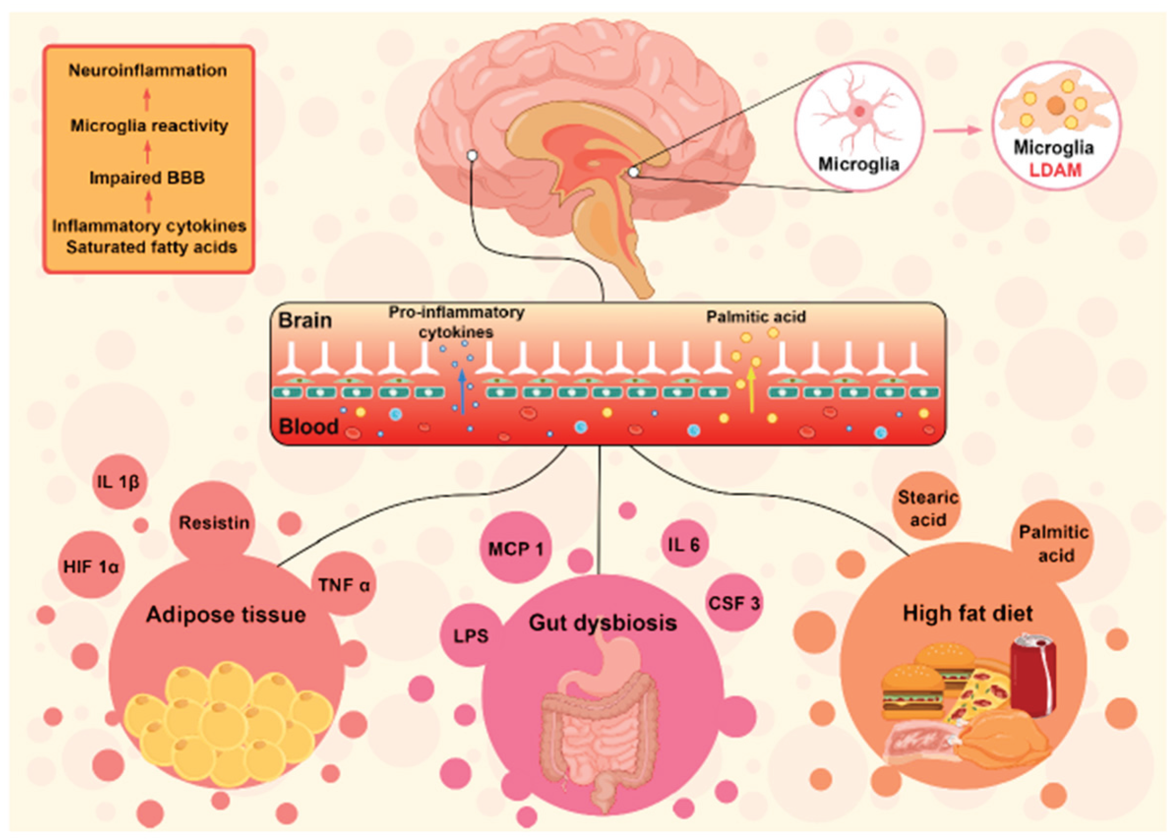
Figure 2.
Mechanisms implicated in obesity-induced inflammatory depression. (i) abnormalities of the HPA axis; (ii) insulin and leptin resistance in the hypothalamus and hippocampus; (iii) defects in adult hippocampal neurogenesis. Each of these mechanisms has been tied to obesity or poor diet-induced neuroinflammation.
Figure 2.
Mechanisms implicated in obesity-induced inflammatory depression. (i) abnormalities of the HPA axis; (ii) insulin and leptin resistance in the hypothalamus and hippocampus; (iii) defects in adult hippocampal neurogenesis. Each of these mechanisms has been tied to obesity or poor diet-induced neuroinflammation.
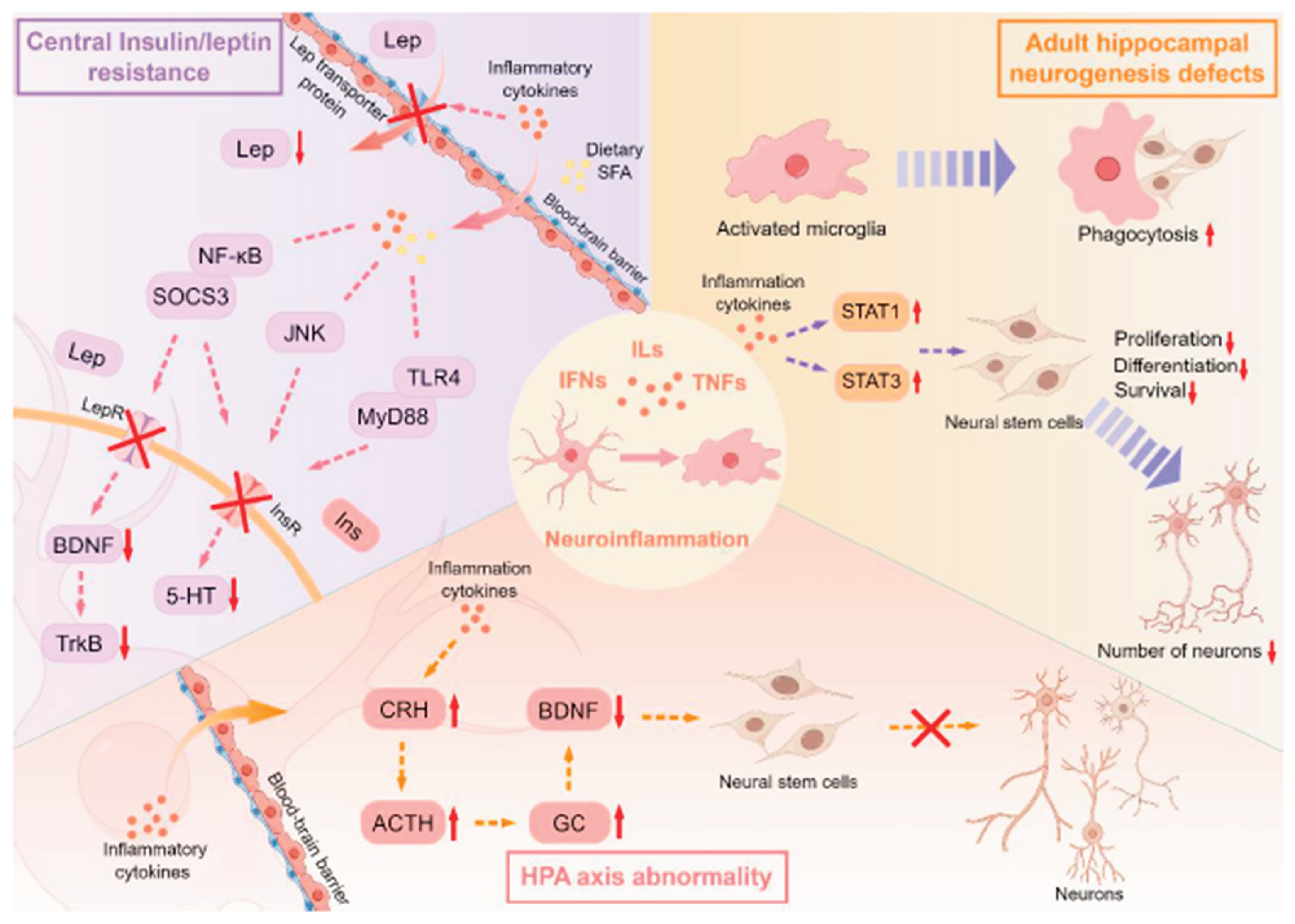
Figure 3.
Classification, chemical structure and main sources of polyphenols.
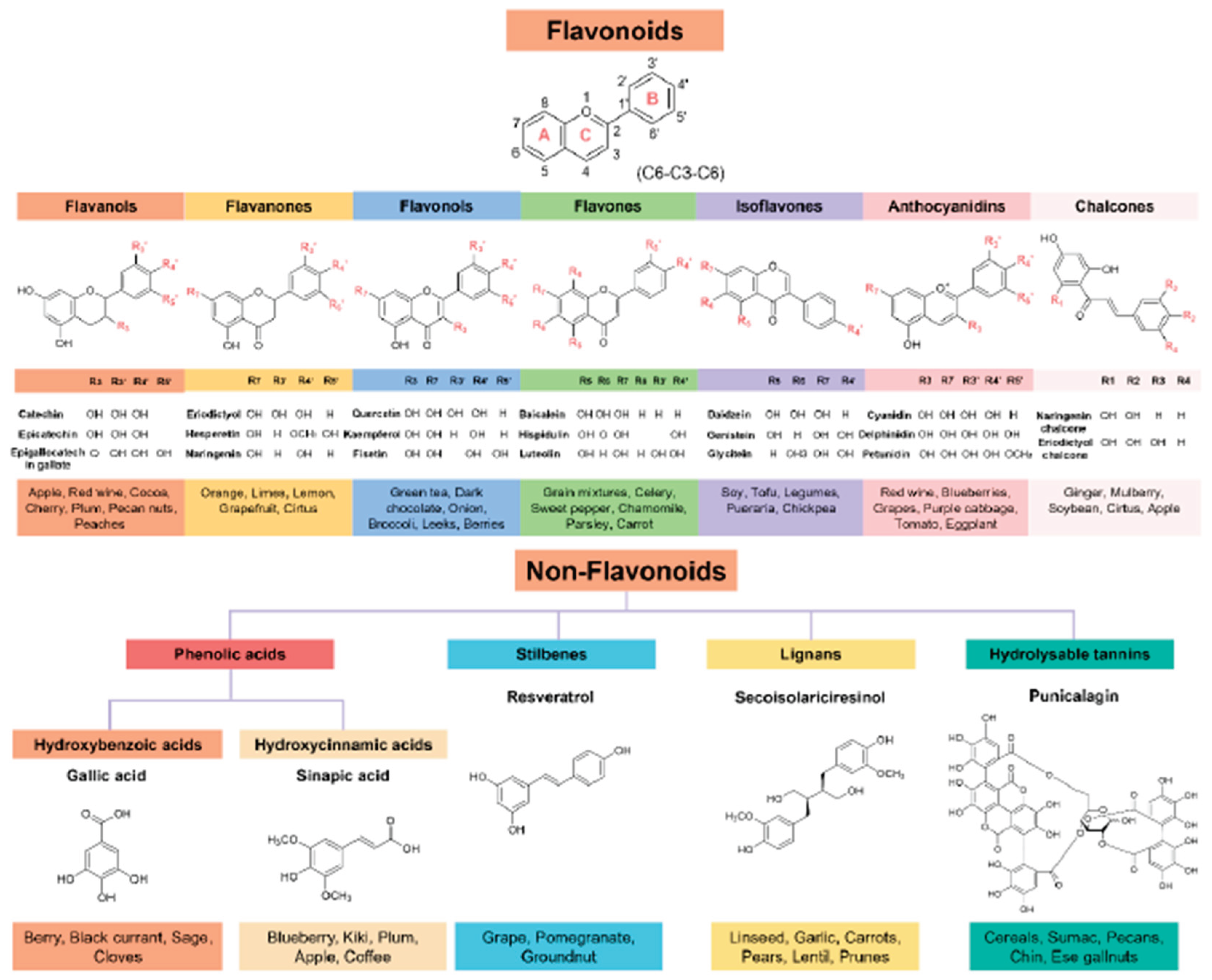
Figure 4.
Summary of potential anti-neuroinflammatory mechanisms implicated in the antidepressant-like effects of polyphenols in obesity and neuroinflammation-induced depression models. Polyphenols can inhibit neuroinflammatory responses via the NF-κB pathway, MAPK pathway, and PI3K/Akt pathway, and ultimately alleviate associated depressive symptoms by modulating HPA axis activity, BDNF expression, and 5-HT synthesis. In addition, polyphenols can exert anti-neuroinflammatory and antidepressant effects by regulating the gut-brain axis and brain enzymes activities.
Figure 4.
Summary of potential anti-neuroinflammatory mechanisms implicated in the antidepressant-like effects of polyphenols in obesity and neuroinflammation-induced depression models. Polyphenols can inhibit neuroinflammatory responses via the NF-κB pathway, MAPK pathway, and PI3K/Akt pathway, and ultimately alleviate associated depressive symptoms by modulating HPA axis activity, BDNF expression, and 5-HT synthesis. In addition, polyphenols can exert anti-neuroinflammatory and antidepressant effects by regulating the gut-brain axis and brain enzymes activities.
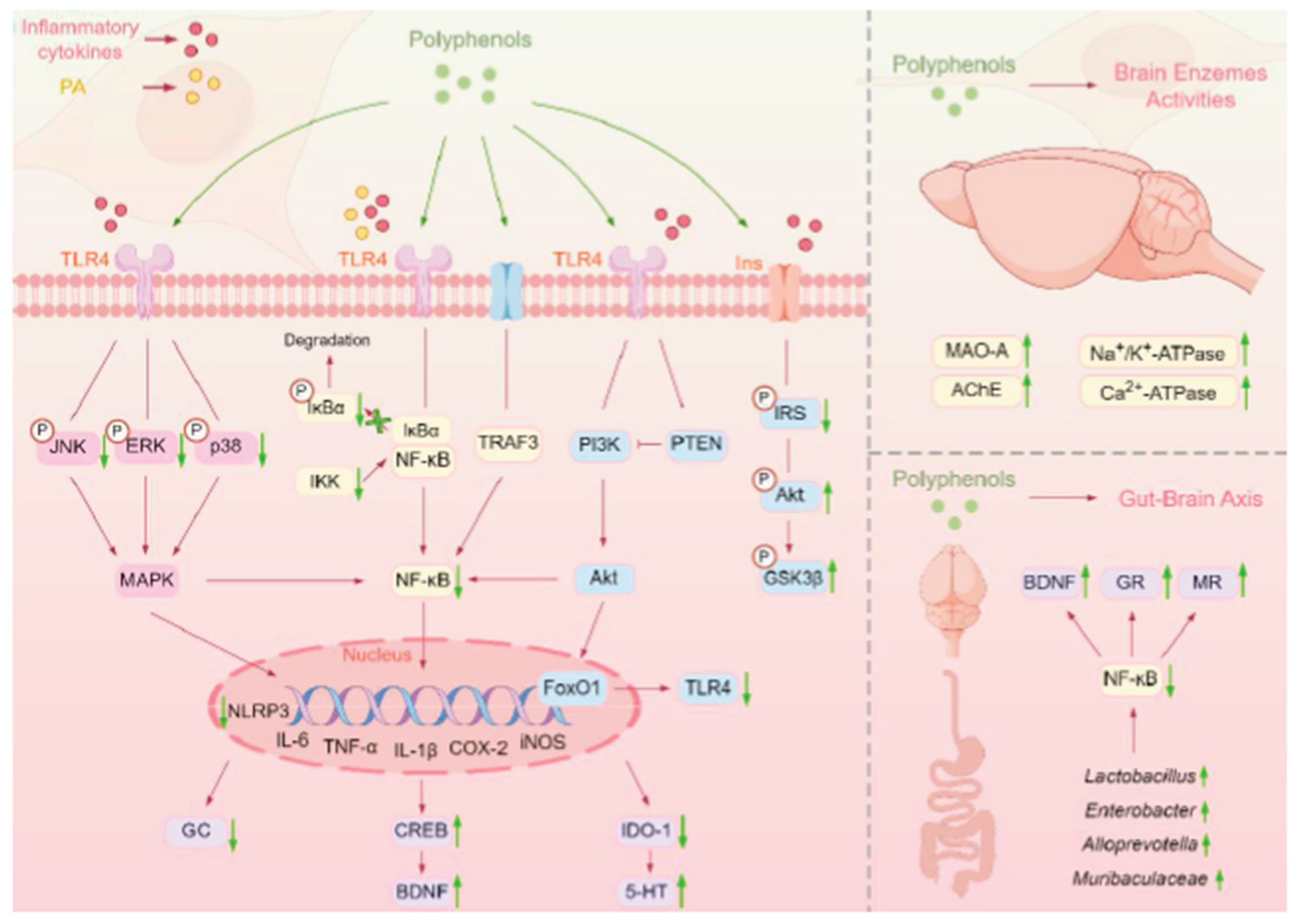

Table 1.
Effective doses of polyphenols for ameliorating obesity-related neuroinflammation in in vitro and in vivo models.
Table 1.
Effective doses of polyphenols for ameliorating obesity-related neuroinflammation in in vitro and in vivo models.
| Compound | Cell line/animal model | Polyphenol doses range | Effective doses |
Mechanisms of action | References |
|---|---|---|---|---|---|
| EGCG | BV-2 cell 1µg/mL LPS for 1h | 0-350 µM for 24h | 150 µM | ↓mTOR, NF-κB, STAT1, Akt, CCL5, SMAD3; ↑Ins2, Pld2, A20/TNFAIP3, GAB1 | (Payne et al., 2023) |
| Baicalin | BV-2 cell 0.1µg/mL LPS for 12h | 2.5, 7.5, 22.5 µM for 1 h | 7.5, 22.5 µM | ↓TLR4/MyD88/NF-κB, MAPK, miR-55 | (Li et al., 2022) |
| Baicalein | BV-2 cell 0.1µg/mL LPS for 24h | 15-200µM for 24h | 45µM | ↓TLR4, NF-kB, STAT1 | (Ran et al., 2021) |
| Punicalagin | BV-2 cell 1µg/mL LPS for 24h | 0-100 µM for 30 min | 25-100 µM | ↓p-ERK, p-JNK, p-STAT3, MAPK/NF-kB, NLRP3, iNOS, COX-2 | (Lo et al., 2022) |
| Hispidulin | BV-2 cell 0.1µg/mL LPS for 24h | 0- 30 µM for 1h | 3-30 µM | ↓NF-κB, STAT3, Akt, ROS, NO | (Yu et al., 2020) |
| Sinapic acid | BV-2 cell 0.1µg/mL LPS for 12h | 0- 20 µM for 1h | 10-20 µM | ↓Akt, MAPK, NO, IL-6 | (Huang et al., 2023) |
| Hesperetin | BV-2 cell 1µg/mL LPS for 24h | 0.78- 200 µM for 24h | 25-100 µM | ↓Keap1/Nrf2, NF-kB; ↑HMOX1, GCLC | (Evans et al., 2023) |
| Açaí extract | BV-2 cell 1µg/mL LPS for 72h | 0.001-1000 µg/mL for 72h | 0.001-10 µg/mL | ↓ROS, IL-1β, IL-6, TNF-α | (De Souza et al., 2022) |
| Magnolol | BV-2 cell LPS/ATP | 0-40 µM for 2h | 10-20 µM | ↓NLRP3, caspase-1, IL-1β; ↑Nrf2, HO-1 | (Tao et al., 2021) |
| Lychee seed polyphenol | BV-2 cell Aβ(1-42) peptide | 2.5, 5, 10 µg/mL for 24h | 5, 10 µg/mL | ↓NLRP3, ASC, caspase-1, IL-1β | (Qiu et al., 2020) |
| Punicalin | BV-2 cell 0.1µg/mL LPS for 24h | 1, 5, 10 µg/mL for 24h | 1, 5, 10 µg/mL | ↓TLR4, NF-kB, IL-1β, IL-6, TNF-α | (Chen et al., 2024) |
| Blueberry extract | BV-2 cell 0.1µg/mL LPS for 24h | 0-2 mg/mL for 1h | 1-2 mg/mL | ↓iNOS, COX-2, TNF-α | (Carey et al., 2013) |
| Apple polyphenol extract | High-fat/cholesterol diet-induced cognitive impairment mice | gavage, 125 and 500 mg/(kg.bw.d), 8 weeks | 125, 500 mg/(kg.bw.d) | ↓NF-κB, MyD88, TRIF, IKKβ; ↑Occludin, ZO-1 | (Yang et al., 2023) |
| High-sugary diet-induced depressed mice | gavage, 500 mg/(kg.bw.d), 8 weeks | 500 mg/(kg.bw.d) | ↓NF-κB, GC, ACTH; ↑IL-10, MUC-2, Occludin, ZO-1 | (Xie et al., 2024) | |
| Isorhamnetin | High-fat and high-fructose diet-induced cognitive impairment mice | 0.03% and 0.06% w/w, mixed in diet, 14 weeks | 0.06% w/w diet | ↓p-JNK, p-p38, p-NF-κB, IL-1β, TNF-α; ↑CREB/BDNF | (Mulati et al., 2021) |
| Genistein | Diabetes-induced brain damage mice | 600 mg/kg, mixed in diet, 4 weeks | 600 mg/kg diet | ↑p-Akt, NGF, BDNF | (Li et al., 2020) |
| EC | High-fat diet-induced depressed mice | gavage, 2 and 20 mg/(kg.bw.d), 24 weeks | 20 mg/(kg.bw.d) | Gut-brain axis modulation; ↑Lactobacillus, Enterobacter; ↑BDNF, GR, MR; ↓11β-HSD1 | (Kang et al., 2022) |
| EGCG | High-fat diet-induced obesity mice | gavage, 25, 50, and 100 mg/(kg.bw.d), 6 weeks | 100 mg/(kg.bw.d) | Gut-brain axis modulation; ↑Muribaculaceae, Alloprevotella; ↓SCFAs, PPAR-γ, NF-ĸB | (Zhou et al., 2023) |
| Sea-buckthorn flavonoids | High-fat and high-fructose diet-induced cognitive impairment mice | 0.06% and 0.31% w/w, mixed in diet, 14 weeks | 0.31% w/w diet | ↑IRS-1/AKT, ERK/CREB/BDNF, PSD-95; ↓NF-kB, IL-1β, iNOS, COX-2 | (Mulati et al., 2020) |
| Puerarin | High-fat diet-induced depressed mice | gavage, 150 mg/kg/day, 6 weeks | 150 mg/kg/day | ↓GC, IL-1β; ↑GLP-1R/BDNF/TrkB, 5-HT | (Liu et al., 2023) |
| Theabrownin | High-fat diet-induced brain damage mice | gavage, 180 and 360 mg/kg/day, 8 weeks | 180, 360 mg/kg/day | ↓MARK4/NLRP3, BAX; ↑PSD95, SYN1, SYP, Bcl-2 | (Lei et al., 2024) |
| Hydroxytyrosol | High-fat and high-fructose diet-induced cognitive impairment mice | gavage, 25, 50 and 100 mg/kg/day, 14 weeks | 50, 100 mg/kg/day | ↓IL-1β, IL-6, TNF-α; ↑PSD95, SNAP-25, BDNF | (Liu et al., 2024) |
Disclaimer/Publisher’s Note: The statements, opinions and data contained in all publications are solely those of the individual author(s) and contributor(s) and not of MDPI and/or the editor(s). MDPI and/or the editor(s) disclaim responsibility for any injury to people or property resulting from any ideas, methods, instructions or products referred to in the content. |
© 2026 by the authors. Licensee MDPI, Basel, Switzerland. This article is an open access article distributed under the terms and conditions of the Creative Commons Attribution (CC BY) license (http://creativecommons.org/licenses/by/4.0/).
Copyright: This open access article is published under a Creative Commons CC BY 4.0 license, which permit the free download, distribution, and reuse, provided that the author and preprint are cited in any reuse.


