

Submitted:
13 July 2026
Posted:
14 July 2026
You are already at the latest version
Abstract
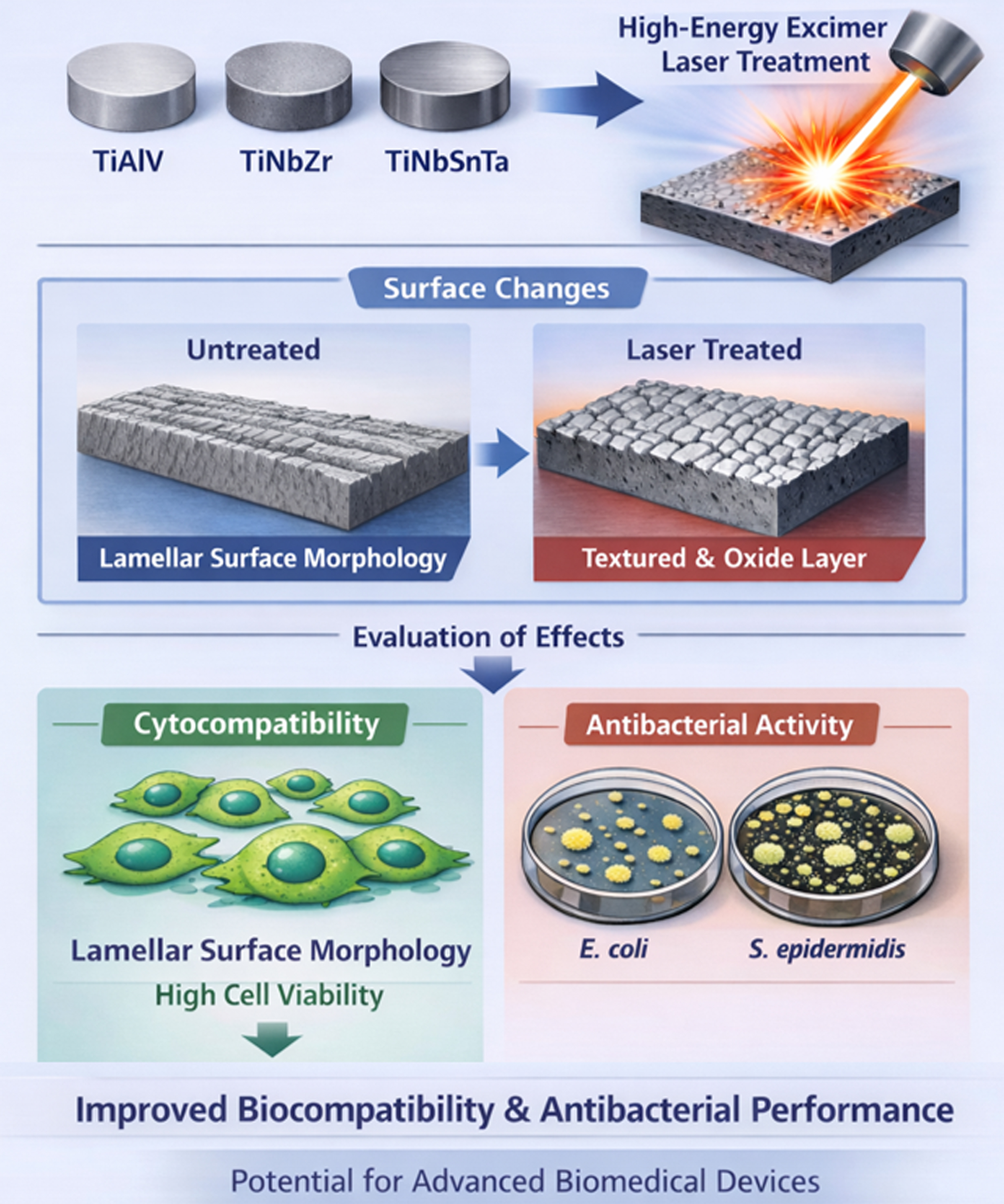
The study investigates the effects of high-energy laser treatment on titanium-based alloys, TiAlV, TiNbZr, and TiNbSnTa, materials of high interest for medical applications such as implants and dental devices due to their exceptional strength-to-weight ratio, corrosion resistance, and biocompatibility. In this research, a unique high-energy laser was used for Ti-based surface activation. The laser exposure induced significant changes in both surface morphology and chemistry while preserving the bulk properties of the substrate. The modified surfaces were evaluated for their impact on cyto-compatibility and antibacterial activity. It was found that viability of U-2 OS cells incubated with laser-treated Ti-based substrates was not negatively affected and was comparable or slightly enhanced than that of control samples, indicating very good cytocompatibility of the prepared materials. Further, antibacterial evaluation against E. coli and S. epidermidis demonstrated that laser-treated samples had improved activity, especially against S. epidermidis, relative to untreated controls. Thus, these results demonstrate that high-energy laser treatment can simultaneously enhance biocompatibility and antibacterial properties of titanium alloys, highlighting its potential as a versatile surface modification strategy for advanced biomedical devices.
Keywords:
titanium
; TiAlV
; surface enhancement
; surface nanopattern
; micropattern
; morphology
; excimer laser
; tissue engineering
; antibacterial properties
1. Introduction
Laser-based surface modification has emerged as one of the most versatile and controllable approaches for tailoring the physicochemical and biological properties of biomedical materials. Advances in laser technology over the past two decades have enabled precise manipulation of surface topography, chemistry, and structure at micro- and nanoscale dimensions. In contrast to conventional chemical or mechanical surface treatments, laser processes offers modifications with negligible bulk damage and excellent reproducibility. These advantages make lase-based techniques particularly attractive for applications in medical implants, biosensors, and tissue engineering scaffolds. A key strength in laser surface modification lies in its ability to generate hierarchical structures that closely mimic natural biological environments. Such structures can modulate protein adsorption, enhance cell-material interactions, and simultaneously impart antibacterial functionality. As a result, laser processing has become a focal point of biomaterial research aimed at understanding how laser-material interactions influence subsequent biological behavior.
Among biomedical materials, metals – and especially titanium and its alloys – have received the greatest attention due to their favorable combination of mechanical strength, corrosion resistance, and biocompatibility. Titanium-based materials are widely used in orthopaedic and dental implants, however, untreated titanium surfaces are intrinsically bioinert and do not optimally promote cell adhesion, proliferation, and differentiation [1,2]. Consequently, a substantial effort has been devoted to developing surface modification strategies that enhance osteointegration and reduce associated infections. One of effective approaches for construction bioactive and antimicrobial macro-/micro-nanoporous selective laser was presented by Pattanayak et al. [3]. Bioinertness to osseointegration and antibacterial activity influenced by a one-step micro-arc oxidation approach for multifunctional Ti implants fabricated by additive manufacturing was presented by Yan et al. [4]. The pure Ti implant was printed using a selective laser melting technique, and a two-layer hierarchical structure was formed on its surface using a one-step micro-arc oxidation (MAO) approach. Laser modification addresses this challenge by reshaping the top surface layer, altering grain structure, introducing oxygen-rich or carbonized layers, or producing controlled periodic patterning [5]. Studies have shown that laser processing can shift titanium from a passive, inert surface into an active interface capable of promoting osteogenesis, vascularization, and strong bone integration, such as laser surface texturing of Ti-cp and Ti6Al4V alloy for the improvement of fibroblast adhesion and alignment as well as to reduce of bacterial adhesion [6]. In addition to tissue engineering benefits, laser treatment can also produce antibacterial properties via nanostructuring or immobilization of metallic nanoparticles such as silver, copper, or gold.
Titanium and titanium alloys (especially Ti6Al4V and beta-titanium systems) remain the gold standard for orthopedic, dental, and craniofacial implants [7]. Nevertheless, their osseointegration behavior is heavily dependent on surface properties. Laser treatment has proven to be a powerful method for improving these characteristics [8]. Depending on processing parameters such as wavelength, pulse duration, fluence, repetition rate, and ambient conditions, laser irradiation can induce surface melting, ablation, controlled oxidation, or formation of laser-induced periodic surface structures (LIPSS) [9]. These structures range from micro-dimples that mechanically anchor cells to sub-100 nm ripples that influence protein adsorption and modulate cell signaling [10,11,12,13,14,15]. Furthermore, femtosecond and nanosecond lasers enable rapid patterning with minimal heat-affected zones, preserving mechanical integrity [1]. Notably, specifically LIPSS geometries have been reported to selectively enhance osteoblast adhesion while inhibiting bacterial colonization, offering a dual function surface design strategy [2]. In dental applications, laser-generated microgrooves have demonstrated improved implant fixation within jawbone tissue, leading to faster healing and reduced failure rates. Additionally, laser- or plasma- induced oxidation [16] produces thin titanium oxide layers that improve corrosion resistance and surface hydrophilicity, thereby enhancing early-stage interactions with serum proteins such as fibronectin and vitronectin – key mediators of cell recruitment and differentiation. The cellular response to laser-modified titanium surfaces is governed by a complex interplay of surface topography, roughness, wettability, chemistry, and mechanical properties [17]. Numerous studies have documented enhanced adhesion and proliferation of osteoblasts, mesenchymal stem cells [18], and fibroblasts on laser-textured surfaces compared to polished or grit-blasted controls. Nanostructures generated using excimer, Nd:YAG, or femtosecond lasers alter the arrangement and conformation of adsorbed proteins, creating a bioactive interface that promotes focal adhesion formation, cytoskeletal organization, and favorable signalling cascades. Endothelial and epithelial cells have also been studied on laser-functionalized titanium, showing faster spreading, increased metabolic activity, and reduced inflammatory signaling [19].
In parallel with improved biocompatibility, laser surface modification has demonstrated significant potential for imparting antibacterial functionality [20,21]. The current advances in bioinspired antibacterial surfaces and their underlying mechanisms have been comprehensively summarized by Zhang et al. [22] . Many studies indicate that laser-generated surface features can mechanically disrupt bacterial membranes, thereby reducing adhesion and biofilm formation. Additional antibacterial effects can be achieved through incorporation of functional layers including carbon-based coatings [23], MXenes [24], and metallic nanoparticles such as silver [25,26,27], for which the antibacterial effect is enhanced with additional laser surface treatment [28].
Several studies have demonstrated the effectiveness of laser-induced alloying or precipitation strategies for antibacterial enhancement. For instance, Li et al. [28] reported improved antibacterial properties and cytocompatibility of laser-modified Ti-20Zr-10Nb-4Ta alloy, while Zhang [29] introduced a laser-induced a Ti2Cu precipitate layer in Ti-13Nb-13Zr-5Cu alloy achieving an optimal balance between low elastic modulus, strong antibacterial activity, corrosion resistance, surface hardness, and cytocompatibility, all of which exhibits great potential for the metal-based implant applications. Laser-patterned micro/nanostructures have also been shown to act as additional storage space for AgNPs, enabling multifunctional Ti temporary anchorage device (TAD) surfaces with both osteogenic and antibacterial properties [30].
Importantly, laser treatment combined with additive layer can serve as a standalone strategy for construction of antibacterial surface. Papa et al. [31] demonstrated effective bacterial adhesion suppression and biofilm inhibition through laser surface texturing alone. Other studies have explored synergistic approaches, such as combining laser patterning with hydroxyapatite coatings on the surface of Ti-based metallic glass [32]. Nanosecond laser was used to prepare different bone surface bionic micropatterns on the surface of Ti-based metallic glasses, and hydroxyapatite (HA) was deposited on the surfaces of different patterned microstructures, thus, HA coatings with different morphologies were obtained [32]. Laser-induced surface modification of Ti implants led to enhanced osseointegration and osteogenesis, laser-induced periodic surface structures (HR-LIPSS) were constructed by direct femtosecond laser exposure [33]. The effects of laser surface treatment on enhancing the antibacterial properties of commercially pure (CP) Ti (Grade 2) and Ti6Al4V (Grade 5) implant materials were studied and compared by Chan et al. [34]. Laser surface treatment performed by a continuous wave (CW) fiber laser with a near-infrared wavelength of 1,064 nm in a nitrogen-containing environment led to a reduction in bacterial adhesion and exhibited a bactericidal effect. Superhydrophobic and superhydrophilic surfaces have also attracted significant attention in fundamental and applied research. Fabrication of the micro/nanostructures with a Q-switched nanosecond pulsed laser on the Ti-6Al-4V surface was presented by Waritanant et al. [35]. Three laser-generated surface topographies on titanium were produced based on three different pitch sizes, thus the hydrophobicity was significantly enhanced.
Additionally, biogenic HA may be enhanced by laser exposure on Ti-based materials, which may strongly affect the surface chemistry and wetting behavior of laser-modified Ti–6Al–4V surgical titanium alloy surfaces, as the basis wet deposition of biogenic HA can be used [36]. Direct laser writing (DLW) was used to modify the titanium surface and facilitate the deposition of biohydroxyapatite (BHA) to optimize their suitability for biomedical implant applications. Fabrication of micro/nanostructured Ti-6Al-4V surface and improved biocompatibility properties of hBMSCs via biomimetic peptide construction [37]. Also some of the chemical based approaches for cytocompatibility enhancement may be applied. Nano-structured surface using chemical pseudo-dealloying by employing an ammonia-based solution (NH4OH and H2O2) of Ti-based alloy was used for alteration of in vitro cytocompatibility and antibacterial response of the treated ribbon samples. This approach for Ti-based amorphous alloy led to increased cytocompatibility of the material in Saos-2 and HfOb cells together with good antibacterial activity against the Pseudomonas aeruginosa [38]. The subsequent procedures, such as nitriding, can effectively produce TiZrNb coating with significantly enhanced mechanical and electrochemical on Ti-6Al-4V substrates [39]. The synergic effect of nanosecond fiber laser surface modification combined with a drug-loaded electrospun PVA coating on the biological performance of Ti-6Al-4V alloy was demonstrated by Rajabi et al. [40]. In their study, laser-textured titanium surfaces were subsequently coated with vancomycin-loaded polyvinyl alcohol fibers via electrospinning, resulting in a multifunctional surface with antibacterial activity while not compromising cell viability.
Overall, laser processing has profoundly advanced surface engineering strategies for biomedical metals, particularly titanium-based substrates. By offering precise and localized control over surface chemistry, morphology, and functional response, laser technologies enable the design to implants that simultaneously promote osseointegration and inhibit bacterial colonization. Despite substantial progress, key challenges remain, including process standardization, scalability to complex implant geometries, and the long-term stability of laser-induced micro- and nanostructures under physiological conditions. Future research is expected to focus on multimaterial laser processing, real-time adaptive surface structuring, and the development of smart, biologically responsive surfaces capable of dynamically interacting with their biological environment.
2. Results and Discussion
2.1. Scanning Electron Microscopy
The morphological changes of the substrate surfaces before and after exposure to a high-energy excimer laser were investigated for TiAlV (TiGr5 ELI), TiNbSnTa, and TiNbZr alloys. High-power laser treatments are commonly employed to fabricate defined surface structures on Ti-based substrates, often affecting relatively deep regions of the material [41] or requiring the application coatings or surface layers [42]. In contrast, surface modification strategies that alter only the outermost layer while preserving the bulk properties of Ti alloys are highly desirable. Therefore, in this study, a high-energy excimer laser treatment was employed to modify the surfaces of Ti-based materials without compromising their bulk characteristics. Conventional excimer laser processing is typically performed at fluences below 100 mJ·cm-2 or even lower, at which it is used for construction of laser-induced periodic surface structures (LIPSS) on polymeric materials, particularly aromatic polymers [43]. However, with these laser types/wavelengths, it is generally insufficient to induce pronounced physicochemical changes on Ti-based substrates due to their high melting temperature and thermal stability.
Here, a unique high-energy excimer laser system capable of delivering fluences exceeding 1,000 mJ·cm-2 over relatively large surface areas was utilized. This system has previously been applied for post-processing and remelting of printed periodic structures in TiAl6V4 alloys [44], for which excimer laser treatment effectively removed loosely sintered particles, thereby reducing the risk of particle detachment in in vivo applications and improving implant reliability. A similar surface-confined modification approach was applied to the Ti-based substrates investigated in the present study. Representative SEM images illustrating the laser-induced surface morphology are shown in Figure 1.
The surface of pristine TiGr5 ELI exhibited a lamellar microstructure with apparent grooves of varying shapes and surface inhomogeneities. For surface modification, a laser fluence of 1,000 mJ·cm-2 combined with 200 laser pulses was applied. These irradiation parameters and the corresponding integral fluence dose were selected based on a preliminary study involving different laser fluences and pulse numbers. Pronounced changes in surface morphology were observed following laser treatment, as documented by the SEM images shown in Figure 1 (bottom row). Laser-induced surface remelting led to the formation of clustered structures separated with troughs. Higher-magnification imaging further revealed a hierarchical morphology, with superimposed micro- and nanoscale features within individual clusters.
Nanoclusters surface features were formed on all laser-exposed samples. As shown in Figure 2, a similar modification of surface morphology was observed also for the TiNbSnTa samples, at which formation of larger surface clusters was clearly visible on the laser-treated substrates. It is important to mention, that owing to the excimer laser wavelength used in this study (248 nm, KrF laser), the absorbed laser energy is focused to the near-surface layer, while the bulk of the substrate remains unaffected. This surface-selective energy deposition is particularly important for tissue engineering applications, as it ensures that the mechanical properties of the modified substrates remain comparable to those of the pristine materials. In comparison with TiGr5 ELI, laser irradiation of the TiNbSnTa substrate resulted in the formation of smaller surface clusters. Quantitative differences in surface roughness, effective surface area, and surface chemistry will be discussed addressed in subsequent sections based on AFM and EDS analyses. The results of SEM analysis of TiNbZr samples are introduced in Figure 3, revealing laser-induced surface patterns that are highly similar to those observed for TiGr5 ELI in Figure 1 and Figure 2.
2.2. Atomic Force Microscopy
The surface morphology of TiGr5 ELI, TiNbSnTa, and TiNbZr foils before and after excimer laser exposure was analyzed using AFM. Figure 4 shows representative AFM images of pristine TiGr5 ELI substrates and samples treated with a laser fluence of 1,000 mJ·cm-2 and 200 pulses. A key advantage of AFM analysis is that, in addition to quantitative evaluation of surface roughness, and effective surface area, it enables detailed visualization of surface morphology across multiple length scales.
Consistent with the results from SEM analysis, AFM analysis of laser-treated TiGr5 ELI samples revealed pronounced cluster formation following excimer laser exposure. We have chosen the presentation of larger 30-micron scans and detailed scans of 1 × 1 m2. Exposure of the samples to the excimer laser “cleaned” the surface, and due to the melting of Ti-based substrate surface, a nanoclustered pattern was induced as superposed on the original microdomain structure, in agreement with data in Figure 1. High-resolution AFM imaging (Figure 4, bottom right) revealed nanocluster pattern with an average diameter of ca. 50 nm, together with larger globular nanostructures, which appear as brighter features in the AFM topography. The formation of this micropattern is accompanied by an increase in surface roughness, from ca. 96 nm for pristine samples to 128 nm after laser treatment. This morphological modification also led to an increase in the effective surface area from 2.2% to 6.1% (30 × 30 μm2). Such changes in surface topography are expected to positively influence cytocompatibility of such material, as discussed in subsequent sections.
Changes in surface morphology, detailed surface structure, surface roughness, and effective surface area were also determined for TiNbSnTa and TiNbZr (Figure 5 and Figure 6, respectively) samples using AFM analysis. Comparison of the pristine samples in Figure 4 and Figure 5 reveals distinct differences between TiGr5 ELI and TiNbSnTa. While both materials exhibited lamellar surface morphology prior to excimer laser treatment, the pristine TiNbSnTa substrate showed significantly higher roughness than TiGr5 ELI. This increased roughness can be attributed to the more pronounced wavy topography present on the surface of pristine TiNbSnTa. Similar to the TiGr5 samples, laser irradiation induced a superimposed nanopattern, which is clearly visible in the high-resolution AFM image shown in Figure 5 (bottom right). In contrast, excimer laser treatment of TiNbZr substrates resulted in a marked increase in surface roughness. A substantial rise from 102 nm to 177 nm was observed, primarily associated with the formation of microdomains, as shown in Figure 6 (bottom row). These results demonstrate that while excimer laser exposure induces comparable hierarchical surface patterning across investigated Ti-based alloys, the extent and direction of roughness modification are strongly dependent on the substrate composition.
The increase in surface roughness, in contrast, is not correlated with effective surface area of the TiNbZr sample. A distinct difference in pristine TiNbZr is also apparent in detail, as only this pristine substrate exhibited a grainy, nanoglobular pattern (upper left image in Figure 6). This feature, however, does not significantly influence the morphology induced by excimer laser exposure. Among the studied Ti-based substrates, the induced microdomain pattern on TiNbZr showed the highest surface roughness (177 nm), exceeding that of TiNbSnTa (136 nm), and TiGr5 ELI (128 nm). These differences in surface roughness and morphology may play and important role in cell attachment and survival.
2.3. Energy Dispersive Spectroscopy
Surface chemical composition plays an important role in surface-cell interactions; therefore, we focused in detail on chemical changes induced on the investigated surfaces by high-energy excimer laser treatment. The pristine samples were analyzed first (first column in Figure 7). As expected, the nominal chemical composition of the pristine materials was confirmed. However, likely due to polishing procedures and subsequent interaction with the ambient atmosphere, oxygen and, in some cases, carbon were detected on the pristine surfaces. Excimer laser treatment induced pronounced changes in surface chemistry. In particular, when carbon was present on the pristine surface, laser exposure led to its effective of the residual carbon from the treated samples. The most significant induced chemical change was an increase in surface oxygen content for all investigated substrates: TiGr5 ELI (from 6.2 to 19.6 wt. %), TiNbZr (from 3.8 to 19.6 wt.%), and TiNbSnTa (from 6.2 to 22.1 wt. %). The presence of oxygen confined to this surface-enhanced layer may influence cell-surface interactions [45].
Another notable change was observed for the TiNbSnTa substrate, for which laser exposure resulted in a reduction of the surface concentrations of Sn and Ta. This effect was confirmed by repeated irradiation and subsequent analyses. The phenomenon is most likely caused by diffusion processes within the laser-induced remelted layer, driven by high-energy annealing and elemental diffusion into greater depths of the material. As discussed later, this effect did not adversely affect the cytocompatibility of the treated samples. As a complementary analytical technique for surface chemical characterization, X-ray photoelectron spectroscopy was employed.
2.4. XPS Analysis
For clarity we present the XPS spectra of the pristine TiNbZr sample alongside those of the sample treated at 1 J·cm-2 with 200 pulses. In addition, we report the atomic concentration of elements that were significantly affected by laser treatment, namely titanium and oxygen (see Figure 8). Consistent with the EDS results, XPS analysis confirmed a substantial increase in oxygen content for all investigated samples following high-energy excimer laser annealing.
This increase in oxygen atomic concentration was accompanied by a pronounced decrease in the titanium concentration at the immediate surface. It should be noted that XPS technique provides information from only the outermost surface layers of both pristine and treated materials. Consequently, direct comparison of absolute concentration values obtained by XPS and EDS is no straightforward. Nevertheless, both techniques consistently confirm the same trend, namely a significant laser-induced increase in surface oxygen content.
2.5. Antibacterial Properties
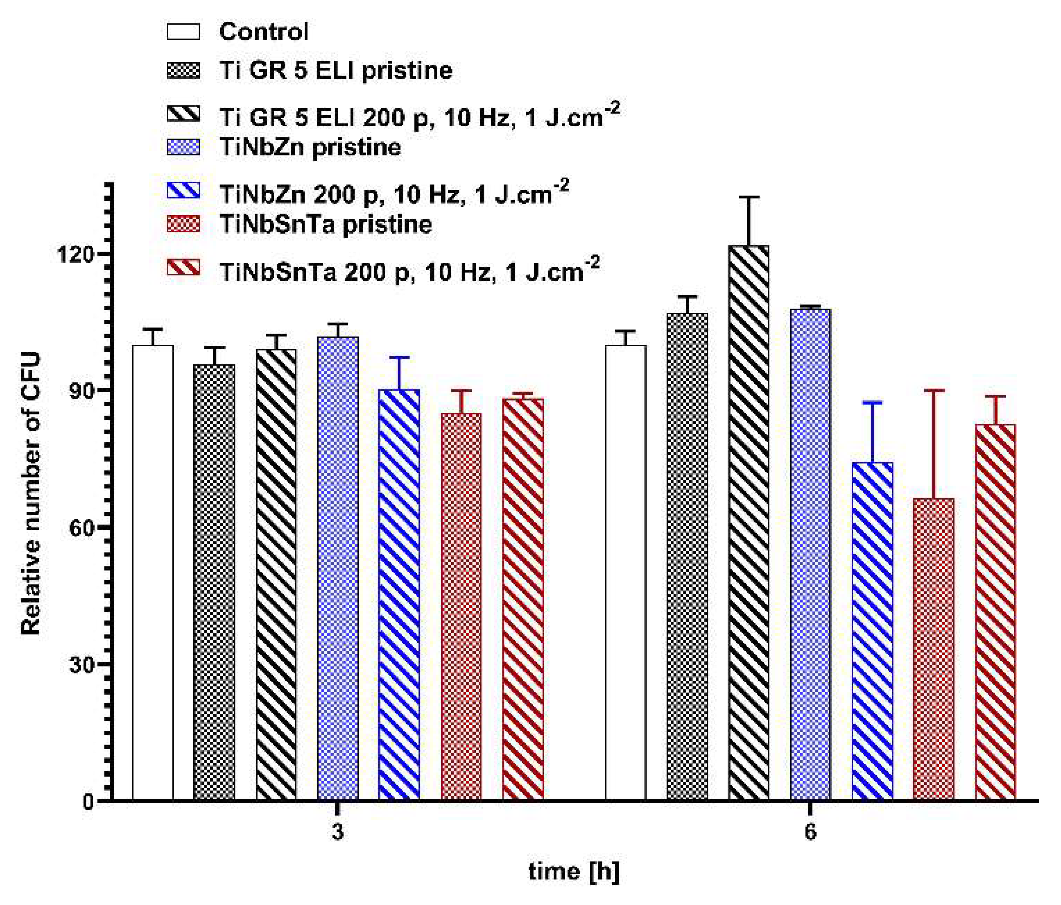
The next aim of this study was to evaluate the antibacterial properties and cytocompatibility of pristine and laser-treated TiGr5 ELI, TiNbZr, and TiNbSnTa substrates. Antibacterial activity was assessed using two representative model strains: Escherichia coli and Staphylococcus epidermidis, corresponding to Gram-negative and Gram-positive bacteria, respectively. The antibacterial response was evaluated after 3 and 6 h of direct contact with the investigated materials. At the early time point (3 h), a reduction in the number of CFU relative to the untreated control was observed exclusively for the TiNbSnTa substrate, for both pristine and laser-treated surfaces, and for both bacterial strains of S. epidermidis and E. coli (Figure 9 and Figure 10, respectively). This antibacterial activity of TiNbSnTa persisted evne after prolonged incubation (6 h) for both microorganisms. In addition, a pronounced reduction in CFU counts of E. coli was detected after incubation with laser-treated TiNbZr for 6 h. In contrast, no reduction in CFU numbers was observed for TiGR5 ELI, regardless of laser-treatment, for either bacterial strain at any time point. Taken together, these results demonstrate that the antibacterial response depends strongly on the substrate composition and surface characteristics. As the effects of high-energy excimer laser treatment differed among the investigated Ti-based alloys, the observed antibacterial behavior cannot be attributed solely to the laser treatment itself.
2.6. Cytocompatibility
The cytocompatibility of both pristine and laser-treated TiGr5 ELI, TiNbZr, and TiNbSnTa substrates was evaluated using U-2 OS cells in order to assess the potential effects of substrate-derived leachates on cell viability over time (24-72 h). Overall, no substantial differences were observed between cells exposed to leachates from any of the tested materials and the control cells cultured in standard cell culture medium alone. This indicates good cytocompatibility of all investigated substrates irrespective of their composition or surface modification.
Figure 11.
Viability of U-2 OS cells following exposure to leachates obtained from pristine and laser-treated TiGr5 ELI, TiNbZr, and TiNbSnTa substrates, evaluated after 24, 48, and 72 h of incubation.
Figure 11.
Viability of U-2 OS cells following exposure to leachates obtained from pristine and laser-treated TiGr5 ELI, TiNbZr, and TiNbSnTa substrates, evaluated after 24, 48, and 72 h of incubation.
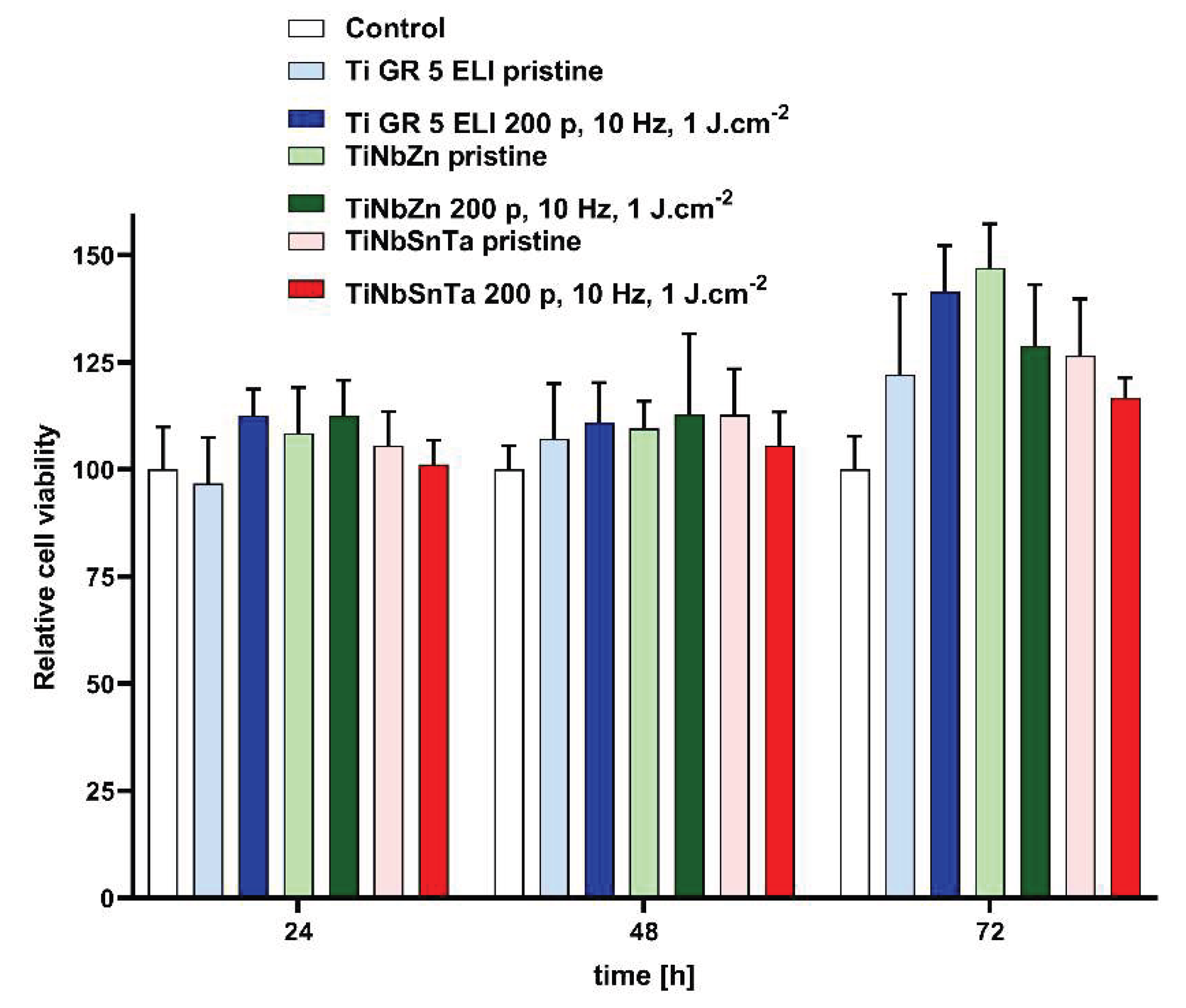
At 24 and 48 h of incubation, U-2 OS cell viability remained comparable across all pristine and laser-treated samples and did not differ significantly from the control. A slight difference was observed only after prolonged incubation (72 h), when leachates from laser-treated TiGR5 ELI and pristine TiNbZr induced a modest increase in cell viability relative to the control. This enhancement, however, was minor and within a range that does not indicate any adverse effects. Taken together, these results demonstrate that both pristine and laser-treated Ti-based substrates exhibited good cytocompatibility.
3. Materials and Methods
3.1. Materials
For the experiments, the following substrates were used: TiGr5 ELI (Titanium Grade 5 Extra Low Interstitial, Ti6Al4V), TiNbZr, and TiNbSnTa. The samples were supplied by New Technologies-Research Centre at the University of West Bohemia. The thickness of the samples was of 1.2 mm. Surface polishing of the samples was applied.
3.2. Excimer Laser Treatment
A high-power excimer KrF laser (Coherent Inc., Santa Clara, California, USA, Leap 100 K) was used with a wavelength of 248 nm, a pulse duration of 20–40 ns, an output energy of up to 1,000 mJ, and beam size with the aperture of 32 × 13 mm2. In all cases, the laser beam acted parallel to the normal of the sample surface (at an angle of 0°). The energy of the laser beam was of 1,000 mJ.cm-2, the number of laser pulses were used in the range between single shot and 500 pulses, finally the number of 200 pulses was selected. The repetition rate of the laser exposure was set to 10 Hz.
3.3. Analytical Methods
3.3.1. Atomic Force Microscopy
Surface morphology and roughness of the pristine and laser-treated films were examined by atomic force microscopy (AFM) technique using Dimension ICON (Bruker Corp., Billerica, MA, USA). The samples were analyzed in the Scan-Assyst® mode using the nitride lever SCANASYST-AIR with a Si tip (the spring constant of 0.4 N·m-1). NanoScope Analysis software was applied for data processing. Surface roughness (Ra) represents the arithmetic mean of the absolute values of the height deviations measured from the central plane.
3.3.2. Scanning Electron Microscopy
The morphology of the sample surfaces was also characterized by a complementary technique using the scanning electron microscope FIB-SEM LYRA3 GMU (Tescan. Brno, Czech Republic). The acceleration voltage was set to 10 kV.
The elemental composition was measured by energy-dispersive X-ray spectroscopy (EDS, analyzer X-ManN, a 20-mm2 SDD detector, Oxford Instruments, United Kingdom), while the accelerating voltage for SEM-EDS analysis was set to 10 kV.
3.3.3. X-Ray Photoelectron Spectrometry
The elemental composition on the material surface was analyzed by X-ray photoelectron spectroscopy (XPS) using spectrometer ESCAProbeP (Omicron Nanotechnology Ldt., Taunusstein, Germany). As a source, a monochromatic X-ray at an energy of 1,486.7 eV was used. Atomic concentrations of elements were determined from the individual peak areas using CasaXPS software.
3.4. Evaluation of Antibacterial Activity
The antibacterial activity of pristine and laser-treated samples was measured using the drop plate method against two bacterial strains: the Gram-positive and Gram-negative bacteria Staphylococcus epidermidis (DBM 2124) and Escherichia coli (DBM 3138), respectively. Overnight cultures (16 h) of both strains were grown in Luria-Bertani (LB; Merck, USA) medium at 37 °C with shaking at 200 rpm. The optical density at 600 nm (OD600) of each culture was measured and adjusted to OD600 = 1 using prewarmed (37 °C) phosphate-buffered saline (PBS). The bacterial suspensions were then further diluted to 4 x 10⁴ and 2 x 10⁴ colony-forming units (CFU) per mL for S. epidermidis and E. coli, respectively. Aliquots of these suspensions were incubated with the tested samples at 37 °C for 3 and 6 h. Then, 25-μL drops were pipetted onto LB agar plates (5 drops per sample, three replicates) and incubated at 37 °C for 24 h. The resulting CFU numbers on plates from each sample were determined and averaged. Bacterial suspensions incubated in PBS without samples served as controls.
3.5. Cytocompatibility Evaluation
The cytocompatibility of the tested samples was evaluated using human osteosarcoma cells (U-2 OS; ATCC, USA). Cells were cultured at 37 °C, 5% CO2, and a humidified atmosphere in high-glucose Dulbecco’s modified Eagle’s medium (DMEM; Merck, USA) with 10% fetal bovine serum (FBS; Merck, USA). Cells were passaged three times per week using trypsin-EDTA and maintained in the exponential growth phase.
For cytocompatibility assessment, pristine and laser-treated samples were first placed into 24-well tissue culture plates (VWR/Avantor, USA), each containing 1 mL of DMEM with 10% FBS, and incubated for 48 h under standard cultivation conditions to obtain material leachates. After incubation, the leachates were collected and used for subsequent cell viability assays.
U-2 OS cells were seeded at the density of 5,000 cells per well into 96-well plates in 100 µL of DMEM with 10% FBS and enabled to grow for 24 h under standard cultivation conditions. Then, the cells were treated with 100 µL of the respective material leachates, and the cells were incubated for additional 24, 48, or 72 h. After each incubation period, the medium was removed and replaced with 100 µL of phenol-red free DMEM (Merck, USA) containing the WST-1 reagent (4% v/v). Following a 1-h incubation, the absorbance of the resulting formazan product was measured at 450 nm with a background reference at 650 nm. Wells with cells cultured in standard medium without exposure to material leachates served as controls.
3.6. Cell Morphology Evaluation
To evaluate the morphology of U-2 OS cells cultured on pristine and laser-treated samples, cells were seeded into 24-well tissue culture plates (VWR/Avantor, USA) containing the respective substrates (in triplicates), which were inserted into the wells prior cell seeding. Cells were cultured in 1 mL of DMEM with 10% FBS and incubated for 1, 3, and 5 days under standard cell culture conditions. After the designated incubation periods, the cells were fixed with a 4% formaldehyde solution in PBS for 20 min (room temperature). After fixation, the samples were washed with PBS and stained for nuclei and F-actin using 4’,6-diamidino-2-phenylindole (DAPI; Merck, USA; 0.5 µg.mL-1) and phalloidin-Atto 565 (ATTO-TEC GmbH, Germany; 1.5 µg.mL-1) for 20 min. Following staining, the cells were washed twice with PBS and imaged using an inverted fluorescence microscope (Olympus IX-81, Olympus, Japan) equipped with xCellence software. Images were acquired using 10, 20, and 40× objectives (numerical apertures of 0.30, 0.45, and 0.60, respectively) with DAPI and TRITC filter sets. Then, image deconvolution was performed using the nearest-neighbor algorithm with a 50% hazel removal factor, and background correction was applied using xCellence software. Wells of the cell culture plates without inserted samples served as controls.
4. Conclusions
In this study, high-excimer laser processing was demonstrated as an effective strategy for tailoring the surface properties of TiGr5 ELI, TiNbZr, and TiNbSnTa alloys. The main aim was to assess the effect of these changes on antibacterial activity and cytocompatibility. Laser irradiation at a fluence 1,000 mJ·cm-2 with 200 pulses induced pronounced modifications in surface morphology and chemistry, as confirmed by AFM, SEM, and XPS analyses. The microdomain pattern on all three studied substrates have been induced, with significant changes of both surface morphology and effective surface area (an increase in at least one from this values) compared to pristine couterparts. Beyond topographical restructuring, high-energy laser treatment caused marked changes in surface chemistry, most notably a significant enrichment of oxygen on the irradiated surfaces, which was confirmed by both EDS and XPS analyses. The combined effects of altered surface morphology and chemistry and the creation of a semi-periodic lamellar structure played a key role in modulating the biological responses. All laser-treated Ti-based substrates exhibited good cytocompatibility comparable to or slightly higher than that of the control samples, indicating no adverse effect on U-2 OS cell viability. Notably, after 72 h, U-2 OS cells incubated with laser-modified TiGr5 ELI exhibited the highest cell viability among all laser-treated samples. In parallel, the modified surfaces displayed enhanced antibacterial performance against both Gram-negative and Gram-positive bacteria, particularly against S. epidermidis. The programmable excimer exposure with steps in selected matrix also opens a possibility of large scale manufacturing of treated substrates. These results indicate that excimer laser-induced surface functionalization can simoultaneously promote mammalian cell viability while suppressing bacterial adhesion and growth, addressing a key challenge in implant material design.
Author Contributions
Conceptualization, P.S. and J.M.; methodology, T.K., J.M. and M.P.; validation, N.S.K, P.S..; formal analysis, S.R., M.P.; investigation, S.R., T.K., J.M., P.Sa., N.S.K. and P.S.; data curation, T.K., Š.H..; writing—original draft preparation, P.S..; writing—review and editing, N.S.K., S.R., Š.H.; supervision, P.S.; funding acquisition, P.S. All authors have read and agreed to the published version of the manuscript.
Funding
This work was supported by the Project OP JAK Mebiosys, No CZ.02.01.01/00/22_008/0004634, of the Ministry of Education, Youth and Sports, which is co-funded by the European Union. This work was also supported by a grant from the Ministry of Health of the Czech Republic (No. NW26-09-00043).
Institutional Review Board Statement
Not applicable.
Informed Consent Statement
Not applicable.
Data Availability Statement
Data will be available on request.
Conflicts of Interest
The authors declare no conflicts of interest.
References
- Hawi, S.; Goel, S.; Kumar, V.; Giusca, C.; Pearce, O.; Nishio Ayre, W. Precision Laser Manufacturing and Metrology of Nature-Inspired Bioactive Surfaces for Antibacterial Medical Implants. Surf. Interfaces 2025, 62, 106267. [Google Scholar] [CrossRef]
- Gupta, R.; Gaddam, A.; Naveena, H.; Prajapati, D.; Dimov, S.; Bhatia, D.; Mishra, A.; Sofronov, Y.; Vadali, M. Improving the cell adhesion and antibacterial behaviour on Ti6Al4V through micro and nano hierarchical laser surface texturing. Surf. Interfaces 2025, 58, 105857. [Google Scholar] [CrossRef]
- Rajendran; Pattanayak, D. K. Bioactive and antimicrobial macro-/micro-nanoporous selective laser melted Ti–6Al–4V alloy for biomedical applications. Heliyon 2022, 8, e09122. [Google Scholar] [CrossRef] [PubMed]
- Tang, J.; Wu, Z.; Yao, X.; Zhou, Y.; Xiong, Y.; Li, Y.; Xu, J.; Dargusch, M. S.; Yan, M. From bio-inertness to osseointegration and antibacterial activity: A one-step micro-arc oxidation approach for multifunctional Ti implants fabricated by additive manufacturing. Mater. Des. 2022, 221, 110962. [Google Scholar] [CrossRef]
- Derya Ince, F.; Ozel, T. Laser surface texturing of materials for surface functionalization: A holistic review. Surf. Coat. Technol. 2025, 498, 131818. [Google Scholar] [CrossRef]
- Ferraris, S.; Cochis, A.; Scalia, A.C.; Tori, A.; Rimondini, L.; Spriano, S. Laser surface texturing of Ti-cp and Ti6Al4V alloy for the improvement of fibroblast adhesion and alignment and the reduction of bacterial adhesion. J. Mater. Res. Technol. 2024, 29, 5464–5472. [Google Scholar] [CrossRef]
- Ozan, S.; Kasman, S.; Can Uçar, I.; Wen, C. Laser surface texturing for enhanced implant biocompatibility and osseointegration: A critical review of its effects on surface properties. Clin. Biomech. 2025, 126, 106540. [Google Scholar] [CrossRef]
- Shivakoti; Kibria, G.; Cep, R.; Pradhan, B. B.; Sharma, A. Laser Surface Texturing for Biomedical Applications: A Review. Coatings 2021, 11, 124. [Google Scholar] [CrossRef]
- Gräf, S. Formation of laser-induced periodic surface structures on different materials: fundamentals, properties and applications. Adv. Opt. Technol. 2020, 9, 11–39. [Google Scholar] [CrossRef]
- Slepička, P.; Chaloupka, A.; Sajdl, P.; Heitz, J.; Hnatowicz, V.; Švorčík, V. Angle dependent laser nanopatterning of poly(ethylene terephthalate) surfaces. Appl. Surf. Sci. 2011, 257, 6021–6025. [Google Scholar] [CrossRef]
- Krajcar, R.; Siegel, J.; Slepička, P.; Fitl, P.; Švorčík, V. Silver nanowires prepared on PET structured by laser irradiation. Mater. Lett. 2014, 117, 184–187. [Google Scholar] [CrossRef]
- Barb, R.-A.; Hrelescu, C.; Dong, L.; Heitz, J.; Siegel, J.; Slepička, P.; Vosmanská, V.; Švorčík, V.; Magnus, B.; Marksteiner, R.; Schernthaner, M.; Groschner, K. Laser-induced periodic surface structures on polymers for formation of gold nanowires and activation of human cells. Appl. Phys. A 2014, 117, 295–300. [Google Scholar] [CrossRef]
- Štofik, M.; Semerádtová, A.; Malý, J.; Kolská, Z.; Neděla, O.; Wrobel, D.; Slepička, P. Direct immobilization of biotin on the micro-patterned PEN foil treated by excimer laser. Colloids Surf. B Biointerfaces 2015, 128, 363–369. [Google Scholar] [CrossRef] [PubMed]
- Slepicka, P.; Siegel, J.; Lyutakov, O.; Kasalkova, N. Slepickova; Kolska, Z.; Bacakova, L.; Svorcik, V. Polymer nanostructures for bioapplications induced by laser treatment. Biotechnol. Adv. 2018, 36, 839–855. [Google Scholar] [CrossRef] [PubMed]
- Fajstavr, D.; Michaljaničová, I.; Slepička, P.; Neděla, O.; Sajdl, P.; Kolská, Z.; Švorčík, V. Surface instability on polyethersulfone induced by dual laser treatment for husk nanostructure construction. React. Funct. Polym. 2018, 125, 20–28. [Google Scholar] [CrossRef]
- Kobayashi, M.; Shimabukuro, M.; Aoki, S.; Kishida, R.; Kawashita, M. Micro-arc oxidation for balancing antibacterial activity and bone formation on titanium surface. Mater. Lett. 2024, 377, 13753. [Google Scholar] [CrossRef]
- Polozov; Nefyodova, V.; Zolotarev, A.; Sokolova, V.; Gracheva, A.; Popovich, A. Optimizing selective laser melting of Ti-13Nb-13Zr-5Cu alloy: Processing parameters, microstructure evolution, and enhanced mechanical properties. J. Alloys Compd. 2025, 1035, 181587. [Google Scholar] [CrossRef]
- Wan, Y.; Zhao, Z.; Yu, M.; Ji, Z.; Wang, T.; Cai, Y.; Liu, C.; Liu, Z. Osteogenic and antibacterial ability of micro-nano structures coated with ZnO on Ti-6Al-4V implant fabricated by two-step laser processing. J. Mater. Sci. Technol. 2022, 131, 240–252. [Google Scholar] [CrossRef]
- Ziegelmeyer, T.; Martins de Sousa, K.; Liao, T. Y.; Lartizien, R.; Delay, A.; Vollaire, J.; Josserand, V.; Linklater, D.; Le, P. H.; Coll, J. L.; Bettega, G.; Ivanova, E. P.; Martel-Frachet, V. Multifunctional micro/nano-textured titanium with bactericidal, osteogenic, angiogenic and anti-inflammatory properties: Insights from in vitro and in vivo studies. Mater. Today Bio 2025, 32, 101710. [Google Scholar] [CrossRef] [PubMed]
- Su, Q.; Xue, Y.; Wang, C.; Zhou, Q.; Zhao, Y.; Su, J.; Zhu, B. Strategies and applications of antibacterial surface-modified biomaterials. Bioact. Mater. 2025, 53, 114–140. [Google Scholar] [CrossRef] [PubMed]
- Georgakopoulos-Soares; Papazoglou, E. L.; Karmiris-Obratanski, P.; Karkalos, N. E.; Markopoulos, A. P. Surface antibacterial properties enhanced through engineered textures and surface roughness: A review. Colloids Surf. B Biointerfaces 2023, 231, 113584. [Google Scholar] [CrossRef] [PubMed]
- Zhang; Chen, Y.; Dub, S.; Du, M.; Eui Jeong, H.; Jiang, R.; Zhao, J.; Ren, L. Addressing bacterial threats in a post-antibiotic era: Bioinspired strategies for antibacterial surface design. Adv. Bonics 2025, 1, 92–112. [Google Scholar] [CrossRef]
- Slepička, P.; Hurtuková, K.; Rimpelová, S.; Trhoňová, Š.; Martan, J.; Procházka, M.; Švorčík, V.; Kasálková, N. Slepičková. Ti and TiAlV foils enhanced with PLD and flash-deposited carbon: On cytocompatibility and antibacterial activity. J. Adv. Join. Process. 2025, 11, 100274. [Google Scholar] [CrossRef]
- Gnanasekar, S.; He, X.; Nagay, B. E.; Xu, K.; Rao, X.; Duan, S.; Murugesan, S.; Barao, V. A.R.; Kang, E. T. Antibacterial MXenes: An emerging non-antibiotic paradigm for surface engineering of orthopedic and dental implants. Bioact. Mater. 2025, 51, 150–176. [Google Scholar] [CrossRef] [PubMed]
- Pryjmaková; Vokatá, B.; Šlouf, M.; Hubáček, T.; Martínez-García, P.; Rebollar, E.; Slepička, P.; Siegel, J. Silver-enriched microdomain patterns as advanced bactericidal coatings for polymer-based medical devices. Colloids Surf. B Biointerfaces 2024, 242, 114067. [Google Scholar] [CrossRef] [PubMed]
- Slepička, P.; Vašinová, T.; Frýdlová, B.; Kutová, A.; Havlíčková, Š.; Švorčík, V.; Kasálková, N. Slepičková. Antibacterial properties of bimetallic nanopattern induced by excimer laser on PTFE nanotextile. Heliyon 2025, 11, e42775. [Google Scholar] [CrossRef] [PubMed]
- Sedaghat, S.; Krishnakumar, A.; Selvamani, V.; Barnard, J. P.; Nejati, S.; Wang, H.; Detwiler, D. A.; Seleem, M. N.; Rahimi, R. Laser-assisted surface alloying of titanium with silver to enhance antibacterial and bone-cell mineralization properties of orthopedic implants. J. Mater. Chem. B 2024, 22, 4489–4501. [Google Scholar] [CrossRef]
- Xue, X.; Lu, L.; He, D.; Guan, Y.; Li, Y. Antibacterial properties and cytocompatibility of Ti-20Zr-10Nb-4Ta alloy surface with Ag microparticles by laser treatment. Surf. Coat. Technol. 2021, 425, 127716. [Google Scholar] [CrossRef]
- Song, X.; Wang, J.; Qin, G.; Yang, Y.; Yang, L.; Zhang, E. Construction of a Ti2Cu precipitate layer by laser surface treatment to endow Ti-13Nb-13Zr-5Cu alloy with low elastic modulus and strong antibacterial property. J. Alloys Compd. 2025, 1044, 184324. [Google Scholar] [CrossRef]
- Guan, X.; Ni, N.; Yang, Ch.; Yang, Z.; Du, T. Layer-by-layer assembly of silver nanoparticle coating on laser-patterned titanium temporary anchorage device for enhanced osteogenic and antibacterial properties. Mater. Lett. 2025, 392, 138544. [Google Scholar] [CrossRef]
- Papa; Mezzapesa, F. P.; Volpe, A.; Gaudiuso, C.; Ancona, A. Bacterial adhesion eradication and biofilm inhibition through laser surface texturing. Appl. Mater. Today 2025, 45, 102801. [Google Scholar] [CrossRef]
- Zhang, W.; Zhang, P.; Song, S.; Sun, T.; Shi, H.; Yan, H.; Lu, Q.; Chen, G. Effect of laser patterning treatment andhydroxyapatite coating on the surface biocompatibility of Ti-based metallic glass. Opt. Lasers Eng. 2025, 186, 108798. [Google Scholar] [CrossRef]
- Ferraria, M.; Diedkova, K.; Kopanchuk, S.; Tsekhmister, Y.; Veiksina, S.; Polewczyk, V.; Pogorielov, M. Enhanced osteointegration and osteogenesis of osteoblast cells by laser-induced surface modification of Ti implants. Nanomed. Nanotechnol. Biol. Med. 2024, 62, 102785. [Google Scholar] [CrossRef]
- Chan, W.; Carson, L.; Smith, G. C.; Morelli, A.; Lee, S. Enhancing the antibacterial performance of orthopaedic implant materials by fibre laser surface engineering. Appl. Surf. Sci. 2017, 404, 67–81. [Google Scholar] [CrossRef]
- Haryono, B.; Lin, K. W. Y.; Waritanant, Tanant. Fast fabrication of superhydrophobic Ti-6Al-4V surface using Q-switched nanosecond pulsed laser at 1064 nm and cyclohexane. Heliyon 2024, 10, e37808. [Google Scholar] [CrossRef] [PubMed]
- Bernal-Alvarez, L. R.; Ramirez-Gutierrez, C. F.; Gomez-Vazquez, O. M.; Correa-Piña, B. A.; Zubieta-Otero, L. F.; Millán-Malo, B. M.; Rodriguez-Garcia, M. E. Enhancing surface chemistry and wetting behavior of laser-modified Ti–6Al–4V surgical titanium alloy surfaces through wet deposition of biogenic hydroxyapatite. Surf. Coat. Technol. 2024, 489, 131065. [Google Scholar] [CrossRef]
- Li, P.; Zhang, Y.; Zhou, T.; Guo, W.; Cui, Y.; Xie, Q.; Liu, P.; Zhou, X.; Wu, G.; Guo, Y. Fabrication of micro/nanostructured Ti-6Al-4V surface and improved biocompatibility properties of hBMSCs via biomimetic peptide construction, Fabrication of micro/nanostructured Ti-6Al-4V surface and improved biocompatibility properties of hBMSCs via biomimetic peptide construction. Surf. Interfaces 2025, 72, 107276. [Google Scholar] [CrossRef]
- Tiwari; Blanquer, A.; Zermeno Perez, D.; Pavan, C.; Tomatis, M.; Fernandez Navas, N.; Scaglione, F.; Rodriguez, B. J.; Gebert, A.; Turci, F.; Croinin, T. O.; Nogues, C.; Rizzi, P. Cytocompatible and antibacterial efficacy of surface designed Ti40Cu40Zr11Fe3Sn3Ag3 amorphous alloy for potential biomedical applications. J. Alloys Compd. 2025, 1014, 178584. [Google Scholar] [CrossRef]
- Meng, Y.; Chen, G.; Yang, Z.; Zhang, Q.; Kurp, P.; Tofil, S.; Wang, B.; Yao, J. Laser cladded and laser surface nitrided TiZrNb coatings on Ti-6Al-4 V:Microstructure, Tribological, and corrosion behavior. Opt. Laser Technol. 2025, 192, 114064. [Google Scholar] [CrossRef]
- Rajabi, T.; Naffakh-Moosavy, H.; Bagheri, F. The synergic effect of nanosecond fiber laser and drug-loaded electrospun PVA coating on metallurgical and biological characteristics of Ti-6Al-4V alloy. Appl. Surf. Sci. Adv. 2024, 22, 100619. [Google Scholar] [CrossRef]
- Fedyaj, V.; Bronnikov, K.; Terentiev, V.; Simonov, V.; Simanchuk, A.; Babin, S. A.; Yakubov, A.; Lazarenko, P.; Lapidas, V.; Pavlov, D.; Zhizhchenko, A.; Kuchmizhak, A. A.; Dostovalov, A. Formation of 2D laser-induced periodic surface structures on titanium films with femtosecond laser pulses. Surf. Interfaces 2025, 78, 108162. [Google Scholar] [CrossRef]
- Silva, P. F.; Crispim, T. R.; Carvalho, Ó.; Fernandes, M. P.; Moreira, J.; Oliveira, S.; Fernandes, M.; Zille, A.; Silva, F.; Fernandes, M. M. Electroactive coatings on laser-textured titanium-based substrates for an improved antibacterial activity. Surf. Coat. Technol. 2026, 519, 132969. [Google Scholar] [CrossRef]
- Rodríguez-Rodríguez, Á.; Rebollar, E.; Soccio, M.; Ezquerra, T. A.; Rueda, D. R.; Garcia-Ramos, J. V.; Castillejo, M.; Garcia-Gutierrez, M. C. Laser-induced periodic surface structures on conjugated polymers: Poly (3-hexylthiophene). Macromolecules 2015, 48, 4024–4031. [Google Scholar] [CrossRef]
- Školáková; Pinc, J.; Kubík, R.; Hosová, K.; Fojt, J.; Jablonská, E.; Slepička, P.; Tesař, K.; Drahokoupil, J.; Hybášek, V.; Čech, J.; Blažek, J.; Kučerová, E.; Sobola, D.; Straková, M.; Vojtěch, D. The effect of pulsed laser on the surface state of 3D-printed triply periodic structures in TiAl6V4 alloy. Prog. Addit. Manuf. 2025, 10, 10627–10647. [Google Scholar] [CrossRef]
- Gentleman, M.; Gentleman, E. The role of surface free energy in osteoblast–biomaterial interactions. Int. Mater. Rev. 2014, 59, 417–430. [Google Scholar] [CrossRef]
Figure 1.
SEM images of pristine and laser-treated TiGr5 ELI substrate. The laser treatment was performed using high-energy laser exposure with 1 J·cm-2 with 200 pulses. The scanned areas were of 100 × 100, 30 × 30, and 10 × 10 μm2.
Figure 1.
SEM images of pristine and laser-treated TiGr5 ELI substrate. The laser treatment was performed using high-energy laser exposure with 1 J·cm-2 with 200 pulses. The scanned areas were of 100 × 100, 30 × 30, and 10 × 10 μm2.
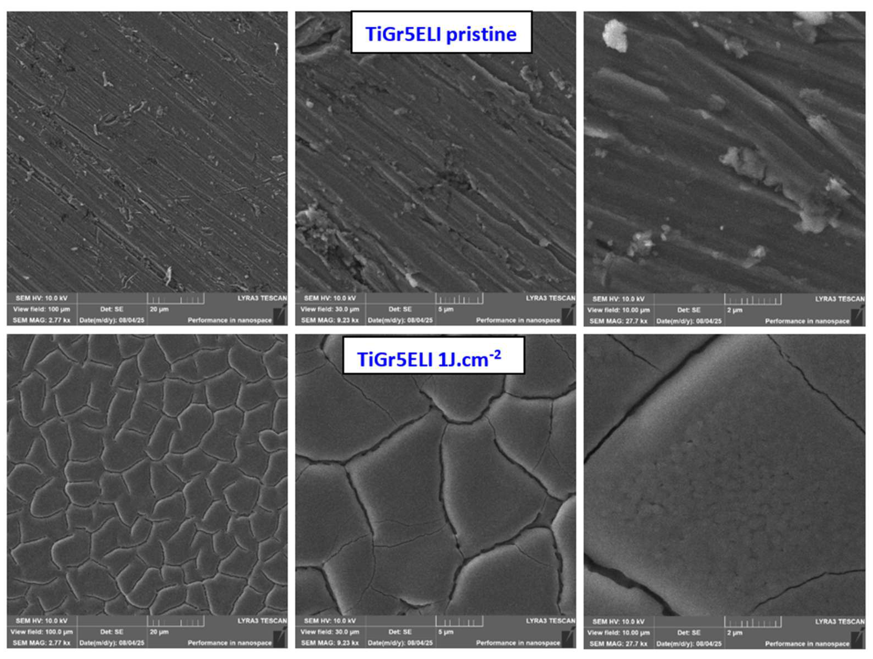
Figure 2.
SEM images of pristine TiNbSnTa and this substrate after high-energy laser treatment with 1 J·cm-2 and 200 pulses. The scanned areas were of 100 × 100, 30 × 30, and 10 × 10 μm2.
Figure 2.
SEM images of pristine TiNbSnTa and this substrate after high-energy laser treatment with 1 J·cm-2 and 200 pulses. The scanned areas were of 100 × 100, 30 × 30, and 10 × 10 μm2.
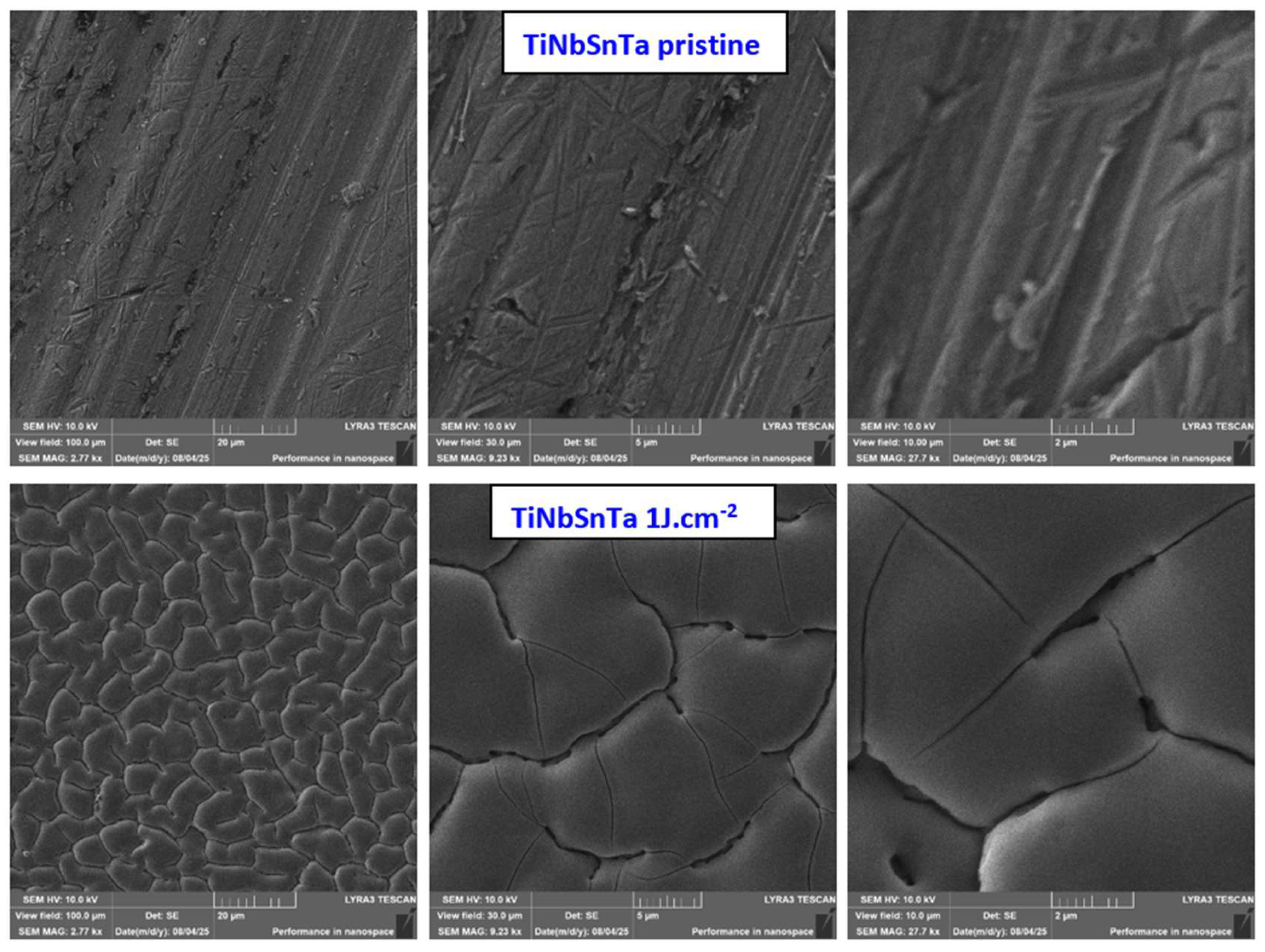
Figure 3.
SEM images of pristine TiNbZr and this substrate after high-energy laser treatment with 1 J·cm-2 and 200 pulses. The scanned areas were of 100 × 100, 30 × 30, and 10 × 10 μm2.
Figure 3.
SEM images of pristine TiNbZr and this substrate after high-energy laser treatment with 1 J·cm-2 and 200 pulses. The scanned areas were of 100 × 100, 30 × 30, and 10 × 10 μm2.
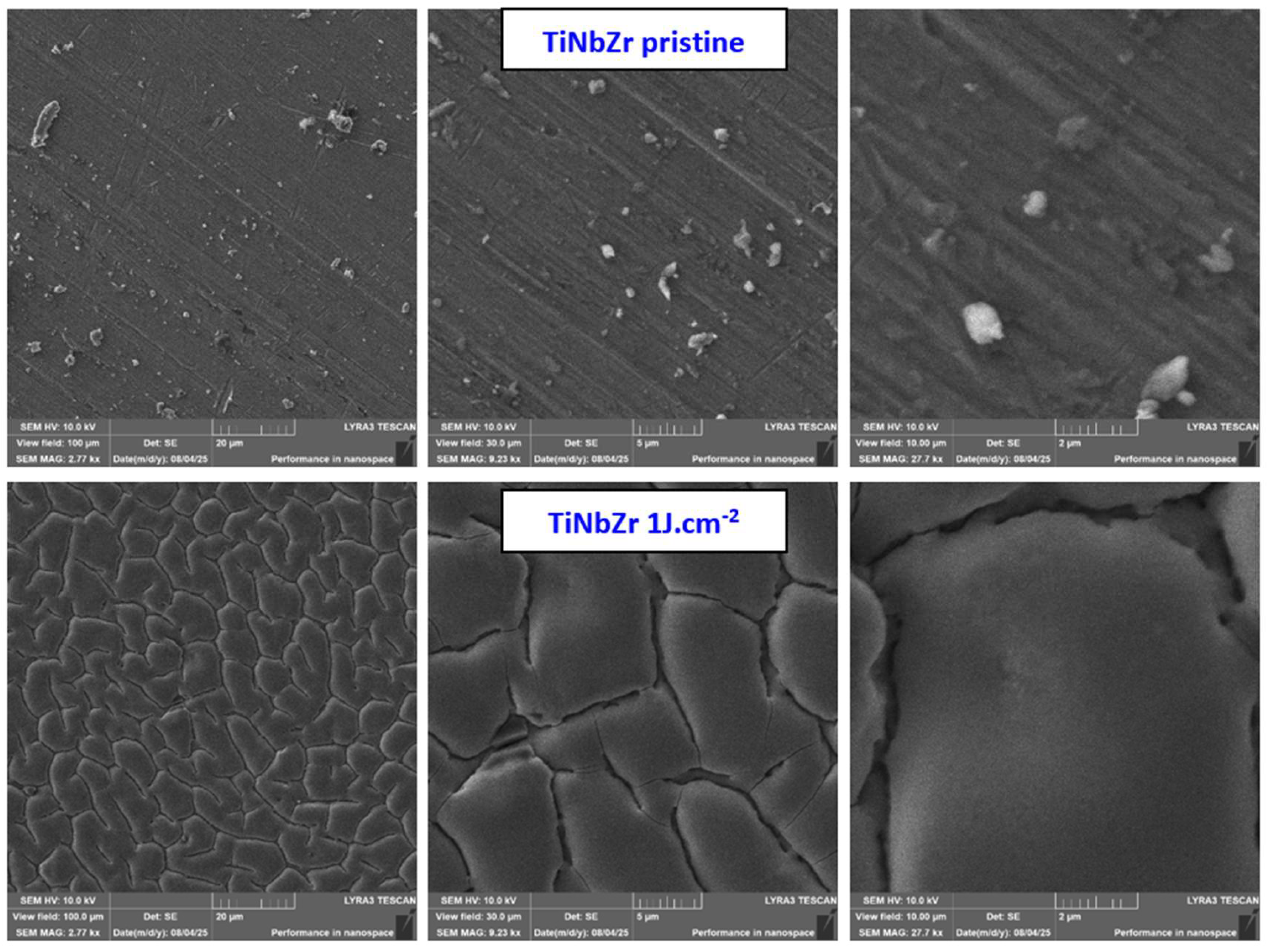
Figure 4.
AFM images of pristine TiGr5 ELI and its form treated with high-energy laser treatment with 1 J·cm-2 and 200 pulses. The measured areas were of 30 × 30 and 1 × 1 μm2. Ra represents average surface roughness in nm, S represents an effective surface area (with difference compared to base area in %).
Figure 4.
AFM images of pristine TiGr5 ELI and its form treated with high-energy laser treatment with 1 J·cm-2 and 200 pulses. The measured areas were of 30 × 30 and 1 × 1 μm2. Ra represents average surface roughness in nm, S represents an effective surface area (with difference compared to base area in %).
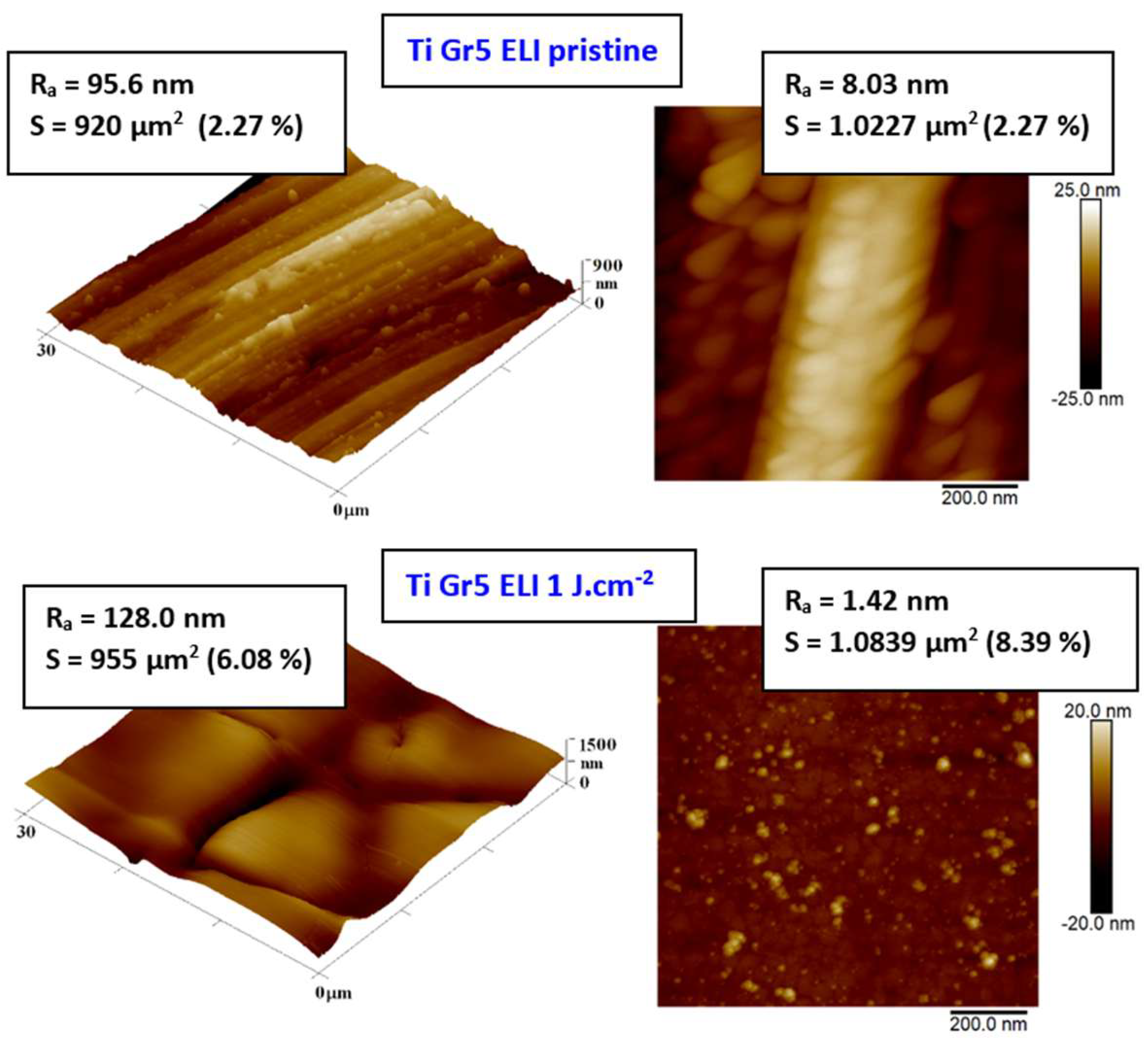
Figure 5.
AFM images of pristine TiNbSnTa and this substrate after high-energy laser treatment with 1 J·cm-2 and 200 pulses. The measured areas were of 30 × 30 and 1 × 1 μm2. Ra represents average surface roughness in nm, S represents an effective surface area (with difference compared to base area in %).
Figure 5.
AFM images of pristine TiNbSnTa and this substrate after high-energy laser treatment with 1 J·cm-2 and 200 pulses. The measured areas were of 30 × 30 and 1 × 1 μm2. Ra represents average surface roughness in nm, S represents an effective surface area (with difference compared to base area in %).
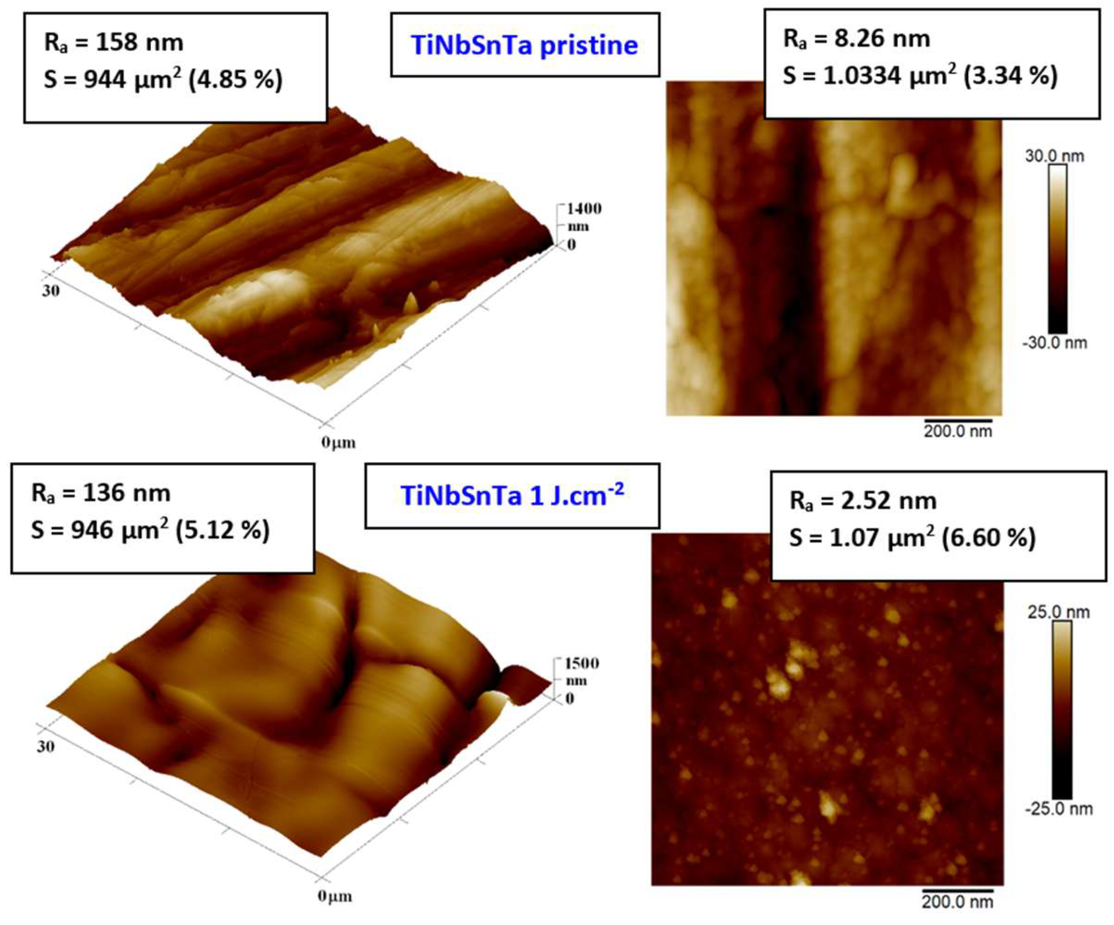
Figure 6.
AFM images of pristine TiNbZr and this substrate after high-energy laser treatment with 1 J·cm-2 and 200 pulses. The measured areas were of 30 × 30 and 1 × 1 μm2. Ra represents average surface roughness in nm, S represents an effective surface area (with difference compared to base area in %).
Figure 6.
AFM images of pristine TiNbZr and this substrate after high-energy laser treatment with 1 J·cm-2 and 200 pulses. The measured areas were of 30 × 30 and 1 × 1 μm2. Ra represents average surface roughness in nm, S represents an effective surface area (with difference compared to base area in %).
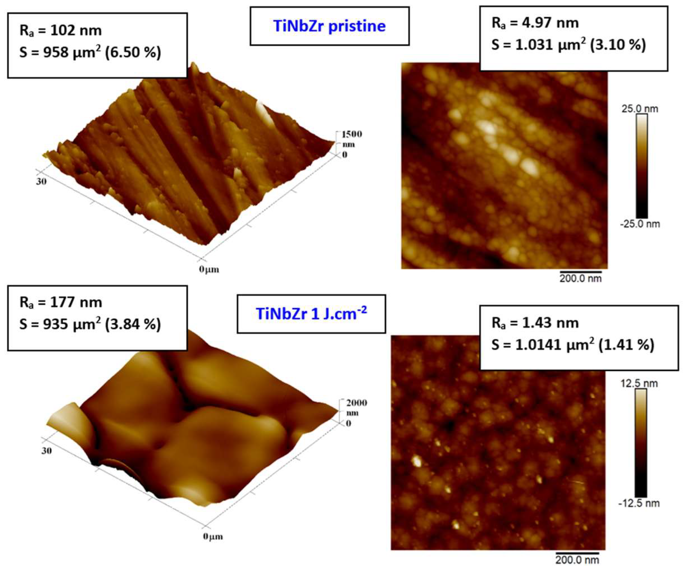
Figure 7.
EDS spectra of pristine TiGr5 ELI, TiNbZr, and TiNbSnTa (left column) and these substrates after high-energy laser exposure with 1 J·cm-2 and 200 pulses (right column). At each spectrum, the weight concentration of particular elements is introduced.
Figure 7.
EDS spectra of pristine TiGr5 ELI, TiNbZr, and TiNbSnTa (left column) and these substrates after high-energy laser exposure with 1 J·cm-2 and 200 pulses (right column). At each spectrum, the weight concentration of particular elements is introduced.
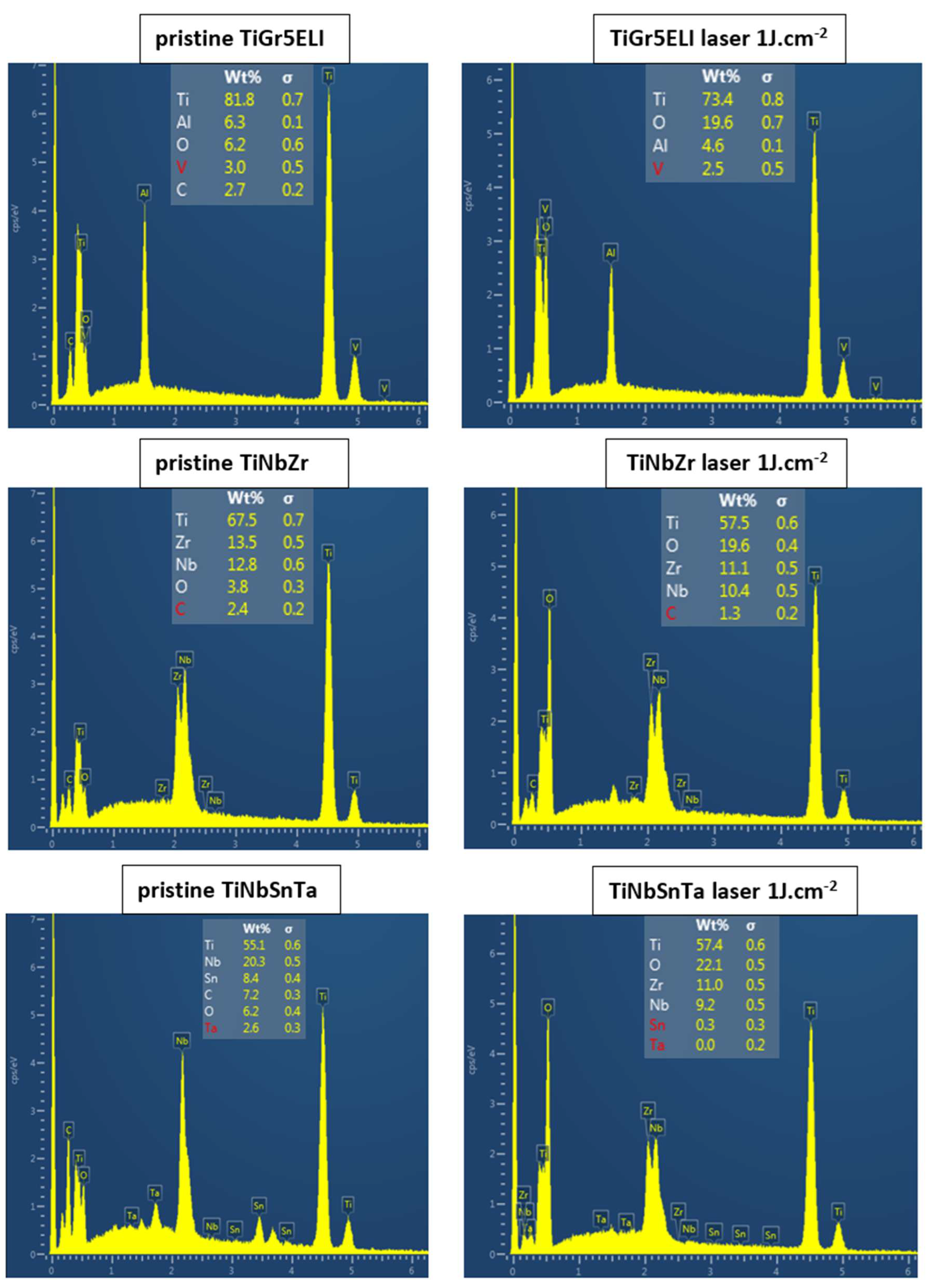
Figure 8.
Atomic concentration from XPS spectra of Ti and C for pristine TiGr5 ELI, TiNbZr, and TiNbSnTa and these substrates after high-energy laser exposure with 1 J·cm-2 and 200 pulses (upper Table). Particular XPS spectra for pristine TiNbZr and exposed TiNbZr with excimer laser 1 J·cm-2 and 200 pulses are introduced at the bottom part.
Figure 8.
Atomic concentration from XPS spectra of Ti and C for pristine TiGr5 ELI, TiNbZr, and TiNbSnTa and these substrates after high-energy laser exposure with 1 J·cm-2 and 200 pulses (upper Table). Particular XPS spectra for pristine TiNbZr and exposed TiNbZr with excimer laser 1 J·cm-2 and 200 pulses are introduced at the bottom part.
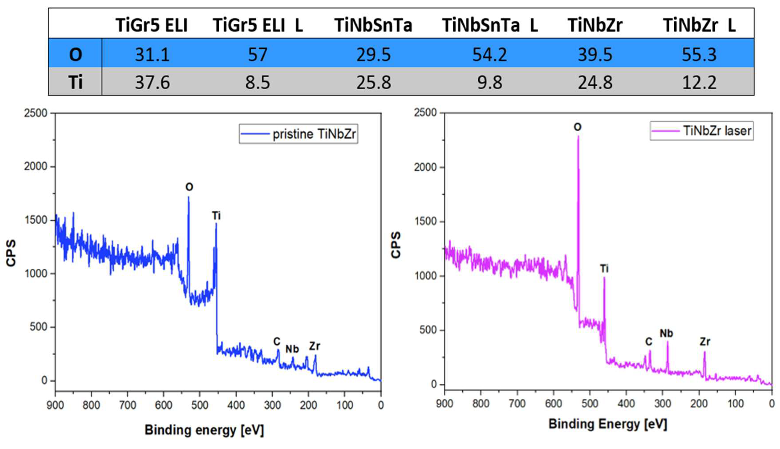
Figure 9.
Antibacterial activity of pristine TiGr5 ELI, TiNbZr, and TiNbSnTa and these substrates after high-energy laser treatment with 1 J·cm-2 and 200 pulses against S. epidermidis after 3 and 6 h of incubation. CFU - colony-forming units.
Figure 9.
Antibacterial activity of pristine TiGr5 ELI, TiNbZr, and TiNbSnTa and these substrates after high-energy laser treatment with 1 J·cm-2 and 200 pulses against S. epidermidis after 3 and 6 h of incubation. CFU - colony-forming units.
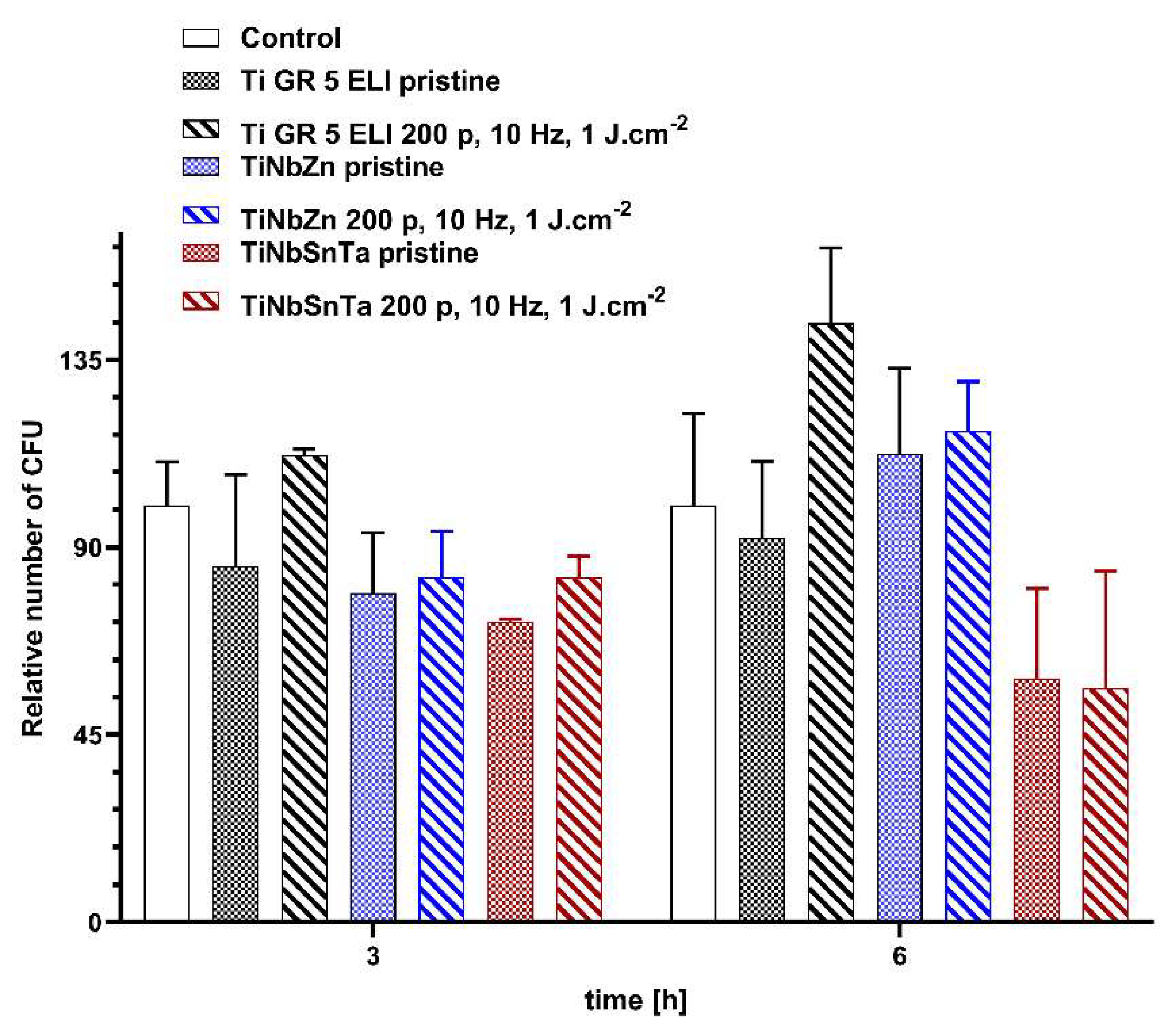
Figure 10.
Antibacterial activity of pristine TiGr5 ELI, TiNbZr, and TiNbSnTa and these substrates after high-energy laser treatment with 1 J·cm-2 and 200 pulses against E. coli after 3 and 6 h of incubation. CFU - colony-forming units.
Figure 10.
Antibacterial activity of pristine TiGr5 ELI, TiNbZr, and TiNbSnTa and these substrates after high-energy laser treatment with 1 J·cm-2 and 200 pulses against E. coli after 3 and 6 h of incubation. CFU - colony-forming units.
Disclaimer/Publisher’s Note: The statements, opinions and data contained in all publications are solely those of the individual author(s) and contributor(s) and not of MDPI and/or the editor(s). MDPI and/or the editor(s) disclaim responsibility for any injury to people or property resulting from any ideas, methods, instructions or products referred to in the content. |
© 2026 by the authors. Licensee MDPI, Basel, Switzerland. This article is an open access article distributed under the terms and conditions of the Creative Commons Attribution (CC BY) license (http://creativecommons.org/licenses/by/4.0/).
Copyright: This open access article is published under a Creative Commons CC BY 4.0 license, which permit the free download, distribution, and reuse, provided that the author and preprint are cited in any reuse.


