

Submitted:
13 July 2026
Posted:
14 July 2026
You are already at the latest version
Abstract
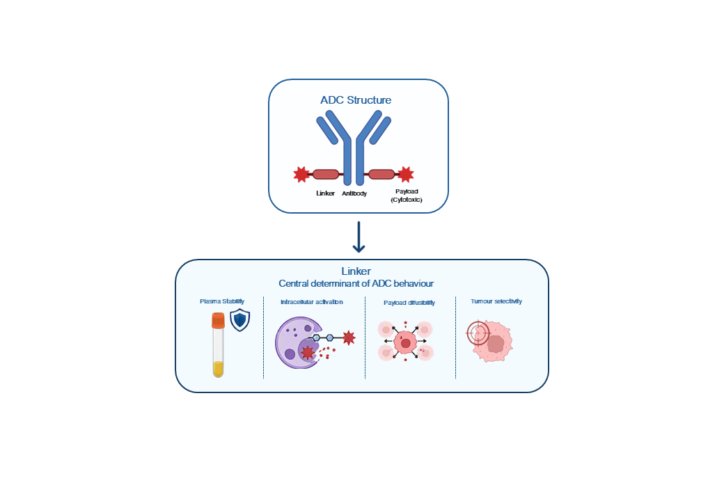
Antibody-drug conjugates (ADCs) have emerged as a powerful class of targeted therapeutics in many clinical areas, such as in oncology. Despite their efficacy, the onset of adverse events has been a major drawback in their clinical use. Among other explanations, the clinical performance of the ADCs has been associated with the chemistry of the linker connecting antibody and payload. Linkers determine plasma stability, intracellular activation, and payload diffusibility, thereby influencing the therapeutic index, off-tumour toxicity, and bystander activity. Mechanistic insights increasingly show that liker-payload properties govern catabolite permeability and intratumoral distribution, particularly in antigen-heterogeneous settings. Current developments include enzyme-cleavable and tumour-selective linkers, polarity -modulating masking strategies, alternative self-immolative spacers, and dual-trigger systems designed to enhance selectivity and decouple efficacy from toxicity. In parallel, linker behaviour intersects with broader mechanisms of tumour resistance. This review focuses on understanding these processes, which are essential for designing the next-generation of linkers capable of improving stability, safety, and long-term therapeutic effectiveness across diverse tumour contexts.
Keywords:
antibody-drug conjugates
; peptide linker design
; tumour-associated enzymes
; therapeutic index
; oncology
1. Introduction
Cancer remains among one of the leading causes of morbidity and mortality worldwide, with the latest GLOBOCAN 2022 estimates indicating approximately 20 million new cases and 9.7 million cancer-related deaths in a single year [1,2]. Despite advances in cancer therapeutics, the effective delivery of highly potent cytotoxic agents continues to be limited by insufficient tumour selectivity and dose-limiting toxicities. This highlights a central challenge in oncology: how to maximize tumour cell killing while minimizing systemic damage.
Conventional systemic chemotherapy has long been the most common therapeutic option for cancer treatment. However, owing to its lack of specificity towards tumour cells/tissues, it is associated with significant adverse effects on healthy tissues [3]. Dose intensity is often constrained by bone marrow suppression, gastrointestinal and neurological toxicity, and cumulative organ damage, which will limit long-term disease control despite potent antitumour activity. These constraints have driven efforts to develop therapeutics that retain the cytotoxic activity of chemotherapy while improving selective delivery to tumour cells [3,4,5].
Antibody-drug conjugates (ADCs) have appeared as a promising solution to address these limitations through the conceptualization of chemotherapy as a targeted biologic-small-molecule hybrid (Figure 1). ADCs are modular constructs composed of 3 key elements: a targeting monoclonal antibody (mAb), cytotoxic payloads, and a chemical linker. While the antibody assures selectivity by recognizing a tumour-associated antigen, decreasing off-target effects, and the payload provides potent cell-killing activity, allowing an efficient cancer cell elimination, the linker governs the spatial and temporal control of drug release [3,6,7,8]. By delivering the highly cytotoxic drugs directly to cancer cells while minimizing the exposure of normal tissues, ADCs are intended to improve the therapeutic index relative to traditional systemic chemotherapy [3,8,9]. Over the past decade, this strategy has been increasingly validated in the clinic, with a growing number of approvals across hematologic malignancies and solid tumours, establishing ADCs as a major pillar for modern oncology [3,10]. Several ADCs have been approved by the Food and Drug Administration (FDA) and the European Medicines Agency (EMA) [11,12] (Table 1), and many others in Phase III clinical trials (Table 2).
The clinical development of ADCs has revealed design challenges, particularly regarding linker stability and drug release (Figure 2). First-generation ADCs, such as Gemtuzumab ozogamicin, relied on acid-labile hydrazone linkers and reducible disulfides, which were prone to premature cleavage in circulation or mildly acidic normal tissues. This instability led to inadequate tumour selectivity, significant off-target toxicity, and a narrow therapeutic window [6,13]. These early limitations established a fundamental principle in ADC design: an optimal balance between systemic stability must be achieved, so that the intact conjugate reaches the tumour, and efficient intracellular drug release occurs [3,6,13]. Subsequent generations of ADCs have addressed these challenges through improved linker chemistries. Second-generation ADCs introduced more stable non-cleavable thioether linkers that minimized premature drug release while still enabling effective payload release after lysosomal degradation of the antibody. More recently, third-generation ADCs combined optimized antibody engineering, highly potent next-generation payloads, and carefully tuned cleavable linkers that remain stable in circulation, but are efficiently activated within tumour cells, leading to improved clinical performance and expanded therapeutic indications. [3,6,14].
These advances collectively emphasize the central role that linkers play in determining ADC behaviour in vivo. Rather than acting as a passive connector, the linker is now recognized as a critical design element that influences multiple aspects. Linker chemistry works as a primary design force that needs to be carefully optimized with antibody engineering, payload selection, and conjugation technology to obtain robust therapeutic indices [13,15]. The role of the linker extends across all critical aspects of ADC function: it determines systemic stability and the risk of premature drug release, thereby influencing systemic toxicity and therapeutic index. It also affects internalization and intracellular processing, because of its influence on how ADC is trafficked and processed within endosomes and lysosomes, where many cleavable linkers are activated [16,17]. Linkers also modulate the bystander effect, and hence the extent to which surrounding antigen low-tumour cells and normal cells are exposed to the drug, through controlling the form, charge, and diffusibility of the released payload. Finally, linker structure contributes to overall hydrophobicity and aggregation propensity, shaping tissue distribution, off-target uptake, and safety, and it also interacts with conjugation strategies to influence drug-to-antibody ratio (DAR) and product heterogeneity [3,6,13,18,19].
Taken together, these considerations highlight the central role of linker chemistry in shaping ADC behaviour, stability, and therapeutic index. This review focuses on enzyme-cleavable and tumour-selective linker strategies, and examines their biological rationale, design principles, and translational implications for modern ADC development.
2. ADC Architecture and the Central Role of the Linker
2.1. Core Components of ADCs
ADCs are modular biologics composed of three main components, each contributing with distinct functional properties to the final therapeutic agent [8] (Figure 1). The mAb, usually a human or humanized IgG1 or IgG4, defines target selectivity and provides a pharmacokinetic (PK) framework. The payload, a small-molecule drug, is typically selected from well-characterized chemotype classes, such as microtubule-disrupting agents, topoisomerase inhibitors, or DNA-alkylating agents, engineered to exhibit sub-nanomolar potency against tumour cells while maintaining suitable physical and chemical properties compatible with conjugation [3,8,13,20]. Finally, the linker, which constitutes the chemical bond connecting the mAb and payload [6,13].
The linker composition and reactivity directly impact how and where the payload is released, with structural features determining plasma stability and susceptibility to specific cleavage mechanisms [6,18]. Linkers are usually short (often 5-20 atoms) and can range from simple aliphatic chains to complex synthetic scaffolds with enzymatic labile motifs, hydrophilic spacers and self-immolative trigger elements. Critically, linkers need to be compatible with the chemical functional groups on the mAb but not significantly impair antibody binding or biophysical properties. They must also endure both the manufacturing process and systemic circulation long enough to allow efficient tumour uptake without releasing the payload prematurely into the bloodstream [6,17,18].
Linker’s structure affects almost every stage of ADC disposition and function. It affects the spatial relationship between mAb and payload, altering tumour penetration and intracellular trafficking. For example, early DM1-ADCs incorporating cyclohexyl-based SMCC linker were characterised by the hydrophobic nature of the resulting linker-payload construct, resulting in increased aggregation and impaired intracellular trafficking, leading to accelerated hepatic clearance. In contrast, more flexible linkers such as SPP or sulfo-SMCC improved solubility and internalization efficiency. Biophysical properties, such as hydrophobicity, length, and rigidity of the linker scaffold contribute to the overall biophysical profile of the conjugate, affecting clearance kinetics, tissue distribution, and the propensity to form high-molecular-weight aggregates [3,6,13]. The use of hydrophobic linkers used in MMAE-ADCs increased aggregation and liver uptake, prompting the development of PEGylated linkers. It determines the route and kinetics of payload release in different cellular compartments, and it can alter the presentation of antibody epitopes, Fc domain interactions, and off-target pharmacology [3,6,17,20]. Therefore, linker engineering appears to be a great opportunity to fine-tune ADC developability and the therapeutic index, independent of target selection and payload optimization.
2.2. Drug-to-Antibody Ratio, Conjugation Strategy, and Heterogeneity
The DAR is a quantitative descriptor of ADC composition defined as the average number of drug molecules covalently attached to each mAb molecule. Traditional conjugation methods targeting multiple lysine residues or interchain disulfide cysteines typically yield heterogeneous mixtures comprising species with DAR values from 0, unconjugated antibodies, to 8 or higher, heavily loaded molecules, with each subpopulation presenting distinct stability, clearance, and potency [3,19,21]. A well-established example is Trastuzumab-DM1, whose lysine-based conjugation produces a broad DAR 0-8 distribution, with each subpopulation exhibiting distinct stability, clearance, and potency. Although high-DAR species can deliver more payload, they often exhibit increased hydrophobicity, greater aggregation propensity, faster plasma clearance, and higher off-target uptake. Conversely, low-DAR species often exhibit excellent plasma stability and prolonged half-lives but may have insufficient payload killing all contacted tumour cells [3,19]. This heterogeneous distribution of DAR within a population can result in a “mixed population” whose average behaviour may not accurately reflect the properties of any single species and complicate the interpretation of results [19,21].
Recent studies highlight that linker chemistry and conjugation strategies are inextricably connected, due to both the number and position of conjugation sites, combined with the physicochemical properties of the linker itself, determining the potential DAR distribution and the overall biophysical characteristics of the ADC [15,21]. In the case of Brentuximab vedotin, the hydrophobic MMAE payload combined with the linker limits manufacturable DAR to approximately 4, as higher-DAR species aggregate and are cleared rapidly. Conversely, hydrophilic linkers can support higher DAR values: Trastuzumab-deruxtecan achieves a DAR of 8 due to its highly hydrophilic tetrapeptide-based linker, which offsets payload hydrophobicity and prevents aggregation. Preclinical studies using polysacaride, or PEG-based linkers, further demonstrated that hydrophilic scaffolds can sustain DAR values of 6-10 without compromising their solubility or stability.
Site-specific conjugation methods, such as engineered cysteines (THIOMABs), glycan remodelling, enzymatic tags or biorthogonal click handles, enable the production of ADCs with defined DAR values, usually 2 or 4, and fixed conjugation sites, yielding more homogeneous products with improved PK predictability [3,7,15,21]. In these strategies, linker design needs to be tuned to offset any hydrophobic burden introduced by diverse payloads because hydrophobic linkers can impair the benefits of site-specific attachment through promoting aggregation and rapid clearance, whereas hydrophilic linkers can enable site-specific ADCs with higher DAR without compromising solubility or stability [3,6,15]. Emerging computational approaches further underscore that DAR, hydrophobicity and linker structure are tightly coupled design dimensions that can be optimized together [22].
2.3. Classification and Mechanistic Roles of Linkers
From a mechanistic perspective, linkers are usually classified into two main categories, non-cleavable and cleavable (Figure 3), with distinct implications for where and how the payload becomes active [6,13,15].
Non-cleavable linkers are designed to remain intact throughout systemic circulation and extracellular processing, with payload release occurring only after the internalized ADC undergoes complete proteolytic degradation of the mAb scaffold in the lysosomal compartment [6,13]. The payload release occurs after lysosomal proteolysis of the mAb scaffolds, generating a charged drug-amino acid catabolite, which potentially impairs membrane permeability and limits bystander effects, and the diffusion of active drug from target cells to neighbouring tumour cells [13,15]. This design offers plasma stability and chemical robustness, and relies on lysosomal degradation as a natural, selective activation mechanism. [13].
In contrast, cleavable linkers are engineered to undergo specific chemical or enzymatic scission under defined intracellular or tumour microenvironment (TME) conditions, such as proteolytic activity, low pH, or redox changes, releasing a more hydrophobic, membrane-permeable drug capable of efficiently diffusing and killing neighbouring cells [6,13,15,16]. Subtypes include:
- pH-sensitive linkers, like hydrazones or acylhydrazones, are stable at neutral pH but are efficiently hydrolysed in the acidic environment of endosomes and lysosomes, where they release free drug for diffusion through lysosomal membranes. These types of linkers were prominent in early-generation ADCs but are now less commonly used because of problems related to premature release in mildly acidic tissues and unpredictable stability throughout different pH ranges [13,15].
- Redox-responsive linkers, usually incorporate disulfide bonds that are reduced in cytoplasm through glutathione or protein disulfide isomerases. However, they remain intact in oxidative extracellular and plasma environments. Although used in some clinical mAbs, concerns about non-selective reduction in normal tissues with glutathione (GSH) levels have reduced their prominence. [6,13].
- Enzyme-cleavable linkers, usually peptide-based motifs, such as Val-Cit or GGFG, that are recognized and hydrolysed by lysosomal cathepsins proteases, cathepsin B, L, and S, following ADC internalization. These linkers exploit the high activity of cysteine proteases in the lysosomal compartment of tumour cells while remaining relatively stable in plasma and normal tissues. Other alternatives for enzyme-responsive linkers include those with glycosidic motifs that are cleaved by β-galactosidase or are designed around matrix metalloproteinase (MMP)-sensitive sequences active in the tumour microenvironment, presenting alternative or complementary enzymatic triggers [3,6,23,24].
Among the cleavable designs, enzyme-cleavable peptide linkers have become the main focus of drug research and development, as they offer an attractive combination of high plasma stability, due to lysosomal cathepsins having low or absent activity in extracellular fluids, efficient intracellular cleavage, and the potential for further optimization toward enhanced tumour selectivity through substrate design and protease profiling [6,15,23]. Mechanistic and structural data showed that amino acid identity and linker length can modulate cathepsin specificity and cleavage kinetics, providing a roadmap for rational design of second- and third-generation peptide linkers with improved selectivity for tumour-associated proteases and reduced off-target activation [6,23]. Consequently, linker choice is not a local chemical detail but a central design decision that must be optimized alongside antibody architecture, antigen biology, internalization kinetics, payload properties, and conjugation method to achieve clinically meaningful improvements in efficacy and safety [6,13,15,25].
3. Tumour-Associated Enzymes as Triggers for Linker Cleavage
Compared to normal tissues, many solid and hematologic tumours exhibit remodelling of their proteolytic and lysosomal machinery, with several enzyme families consistently overexpressed or hyperactivated (Table 3). These enzymes concentrate in enlarged lysosomes, invadopodium and TME, where they contribute to extracellular matrix (ECM) degradation, growth factor activation, invasion, metastasis, angiogenesis and therapy resistance. Crucially, this results in higher and spatially focused enzymatic activity in tumours compared with healthy tissues, creating biochemical niches that differ sharply from the systemic circulation. This dysregulated activity can also be exploited as a biochemical trigger, making tumour-associated enzymes highly attractive as selectivity filters for ADC linkers [24,26,27,28,29].
3.1. Lysosomal Proteases
Lysosomes are central hubs for protein turnover and signalling, and many solid and hematologic tumours exhibit increased lysosomal biogenesis and protease content. Lysosomal proteases are found in tumour cell lysosomes and, sometimes, in pericellular space, and their activity is significantly elevated in highly metastatic tumour variants, such as B16F10 melanoma model or aggressive mammary carcinoma models, compared to the less aggressive ones [30,31]. Recently, this observation was extended to multiple cancer types, in which upregulated lysosomal proteases facilitate degradation of the extracellular matrix, activation of growth factors, and modulation of cell death pathways [26,29].Table 3. Overexpressed enzymes in the tumor microenvironment.

Table 3.
Overexpressed enzymes in the tumor microenvironment.
| Family | Enzyme | Location | Substrate | Tumor | Function | Pathophysiological role | Prognosis | References |
|---|---|---|---|---|---|---|---|---|
|
Cathepsin (Cysteine) |
B | Pericellular space; lysosomes | Broad spectrum | Various | Degradation of ECM components, activation of other proteases amd processing of growth factor and cytokines | General protein turnover in lysosomes, antigen processing, regulation of apoptosis, and autophagy | Increased invasion, metastasis, and poor prognosis in several cancers | [32,33] |
| L | Lysosomes | Broad spectrum | BC, HNC, LC | Degrades ECM proteins and basement membrane components, contributes to EM, invasion and metastasis | Protein turnover, MHCII antigen processing, regulation of cell cycle, and apoptosis | Advanced disease and worse outcome in cancers | [26,33,34] | |
| S | Lysosomes & endosomes | ECM components | Solid tumors | Degrades ECM | Critical for MHCII invariant chain degradation and antigen presenting | More aggressive disease and poor survival in some cohorts | [26] | |
| K | Lysosomes | Collagen | mBone, BC, PC, melanoma | Degrades type I collagens and bone matrix | Potent collagenase | Bone invasion and metastasis | [35] | |
|
Cathepsin (Aspartic) |
D | Lysosomes | Many proteins & pro-hormones | BC, CRC, OC | Degrades protein in lysosomes at an acidic pH; activates precursors of biologically active proteins in pre-lysosomal compartments or specific cells | Pro-enzyme and mature forms both biologically active | Emulates cancer cells proliferation, fibroblast outgrowth, angiogenesis, and metastasis | [29,32,33] |
|
Cathepsin (Serine) |
G | Azurophilic granules of neurophils | ECM components | Carcinomas | Degrades ECM components, activates other proteases | Modulates inflammation | Pro-inflammatory, pro-TME | [36] |
| MMP | 2 | ECM, basement membranes & cell surfaces | Type IV collagen & gelatine | BC, CRC, LC, GC, melanoma | Degrades substrates in basement membranes; releases ECM-bound growth factors | Promotes invasion and angiogenesis | Lymph node metastasis, advanced stage and poor survival | [37,38,39] |
| 9 | ECM, basement membranes & cell surfaces | Type IV collagen & gelatine | BC, CRC, LC, GC, melanoma | Degrades substrates in basement membranes; releases ECM-bound growth factors | Promotes invasion and angiogenesis | Lymph node metastasis, advanced stage and poor survival | [37,38,39] | |
| 11 | ECM | Collagen & ECM components | Carcinomas | Degrades proteoglycans, laminin and fibronectin | Promotes invasion and angiogenesis | Metastasis and poor outcome | [27] | |
| 13 | ECM | Collagen & ECM components | Carcinomas | Degrades collagens in the ECM | Promotes invasion and angiogenesis | Metastasis and poor outcome | [27,38] | |
| 14 | Cell surfaces | Pro-MMP2 & collagen | BC, melanoma | Activates pro-MMP2 and degrades collagen | Pericellular proteolysis and cancer cell invasion | Invasion and poor prognosis | [27,38] | |
| ADAM | 10 | Cell surfaces | Cell-surface proteins | BC, CRC, GC | Enhances the shedding of inflammatory mediators, adhesion molecules, and growth factor ligands | Growth factor signaling and immune modulation | Aggressive behavior and poor prognosis | [40,41,42] |
| 17 | Cell surfaces | Cell-surface proteins | BC, CRC, GC | Enhances the shedding of inflammatory mediators, adhesion molecules, and growth factor ligands | Growth factor signaling and immune modulation | Aggressive behavior and poor prognosis | [40,41,42] | |
| Glycosidase | β-glucuronidase | Lysosomes | β -D-glucuronic acid residues | BC, CRC, CC, pancreatic | Hydrolyses β-D-glucuronic acid residues from glycosaminoglycans and glucuronide conjugates | Deconjugats glucuronic acid | Advanced stage and worse prognosis | [43,44] |
| β-galactosidase | Lysosomes | β -D-galactose | Liver, OC, PC, CRC, BC, gliomas | Removes β-D-galactose from glycoconjugates | Glycan turnover | Not well defined | [45,46] | |
|
Protease (Serine) |
Matriptase | Cell surfaces | Zymogens & ECM components | BC, CRC, Pancreatic | Activates pro-uPA, pro-MMPs and other zymogens; degrades ECM components | Promotes invasion and EMT | Increased invasion, metastasis and poor survival in several cancers | [47,48,49] |
| TMPRSS3 | Cell surfaces | Zymogens & ECM components | BC, CRC, Pancreatic | Activates pro-uPA, pro-MMPs and other zymogens; degrades ECM components | Promotes invasion and EMT | Increased invasion, metastasis and poor survival in several cancers | [47] | |
| TMPRSS4 | Cell surfaces | uPA | Pancreatic, thyroid, CRC | Activates pro-uPA | Promotes invasion | Increased invasion, metastasis and poor survival in several cancers | [48,49,50] | |
| TMPRSS13 | Cell surfaces | Zymogens & ECM components | BC, CRC, Pancreatic | Activates pro-uPA, pro-MMPs and other zymogens; degrades ECM components | Promotes invasion and EMT | Increased invasion, metastasis and poor survival in several cancers | [47,48,49] | |
| uPA | Cell surfaces | Zymogens & ECM components | Various | Activates pro-uPA, pro-MMPs and other zymogens; degrades ECM components | Promotes invasion and EMT | Increased invasion, metastasis and poor survival in several cancers | [47,48,49] | |
|
Specific Protease (Ubiquitin) |
USP4 | Nuclear or cytoplasmic | Ubiquitin | BC | Important determinant for the crosstalk between the TGF-β and AKT signalling pathways | Regulates oncogenic pathways | Link to proliferation, therapy resistance and poor prognosis | [51,52,53] |
| USP39 | Nuclear or cytoplasmic | Ubiquitin | BC | Removes ubiquitin from target proteins; involved in mRNA splicing | Tumor suppressor | Link to proliferation, therapy resistance and poor prognosis | [52,53,54] | |
| BC Breast cancer, CC Cervical cancer, CRC Colorectal cancer, ECM Extracellular matrix, EMT Epithelial to mesenchymal transition, GC Gastric cancer, HNC Head and neck cancer, LC Lung cancer, MHC Major Histocompatibility complex, MMP metalloproteinase, OC Ovarian cancer, PC Prostate cancer, TME Tumor microenvironment. | ||||||||
Due to internalized ADCs being trafficked through endosomes to lysosomes, this compartmental enrichment of proteolytic activity has been directly exploited in lysosomal-cleavable peptide linkers. Linkers with motifs that are recognized by lysosomal proteases are designed to remain intact in plasma and extracellular fluids but be efficiently hydrolysed in the acidic, enzyme-reach lysosomal lumen, triggering self-immolation and payload release [16,55,56]. The elevated lysosomal protease activity in tumours compared with normal tissues promotes a functional selectivity window for linker design, although inter-tumoral heterogeneity and species differences must be considered [16].
3.2. Cathepsins
Cathepsins are a subfamily of lysosomal proteases, including cysteine cathepsins, aspartic cathepsins, and serine cathepsins, that are central to protein turnover and antigen processing. Among them, cathepsin B has received particular attention in oncology, because its expression and activity are elevated in a wide range of tumours, including breast, pancreatic, colorectal, prostate, and melanoma [26,30,35,57].
These enzymes are associated with invasive and metastatic phenotypes, contributing to ECM remodelling, epithelial-mesenchymal transition (EMT), and modulation of apoptosis and autophagy pathways. The expression and activity of these enzymes are “patchiness” expression and activity within tumours, with localized hotspots that may drive focal invasion and metastasis [26,31,58].
From a drug delivery perspective, cathepsins are a primary enzymatic trigger exploited by cathepsin-cleavable ADC linkers. Peptide motifs, like Val-Cit and GGFG, have been shown to be efficiently cleaved by lysosomal cathepsins, and many approved ADCs, such as Brentuximab-vedotin, Polatuzumab-vedotin, Enfortumab-vedotin and Trastuzumab-deruxtecan, rely on these reactions to release their payloads after internalization [55,56]. Medicinal-chemistry design, assisted with protease substrate profiling, demonstrates that a careful selection of P1-P4 residues can bias cleavage toward specific cathepsins and tune stability versus activation across species [16,26,56].
However, cathepsins are not tumour exclusive. They are active in normal lysosomes and can be secreted into the extracellular space under inflammatory conditions [59]. Therefore, tumour selectivity arises from differences in activity level, localization, and trafficking, rather than expression alone [56,59].
3.3. MMPs in the Tumour Microenvironment
MMPs are zinc-dependent endopeptidases that play major roles in ECM remodelling, angiogenesis, and immune cell trafficking. The expression of MMP-2, MMP-9, and MMP-14 are frequently upregulated in tumour cells, stromal fibroblasts, and infiltrating immune cells, particularly in breast cancer, glioblastoma, gastric cancer, and melanoma. Their proteolytic activity enables cancer cells to breach basement membranes, degrade interstitial collagens, shed adhesion molecules, and release ECM-bound growth factor and cytokines, thereby promoting invasion and metastatic dissemination [27,60].
MMP activity is often concentrated at the invasive front of tumours and in perivascular niches, where localized ECM degradation correlates with increased metastatic potential. This spatially and tumour-biased activity overexpression motivated extensive exploration of MMP-cleavable peptides as triggers for prodrugs and nanocarriers, with cleavage in the TME unmasking or releasing active drugs near tumour cells [40,60].
MMP-sensitive motifs have been successfully deployed in peptide-drug conjugates and polymeric drug delivery systems (DDSs), highlighting that MMP-rich microenvironments can be harnessed for tumour-localized drug activation. However, since some MMPs are also upregulated in inflammatory and remodelling processes, it is required to have a careful profiling of MMP expression and activity in tumour versus normal tissues, to avoid off-tumour activation [61,62]
3.4. Glycosidases
Glycosidases are lysosomal exoglycosidases involved in the degradation of glycosaminoglycans and glycoproteins. In several types of tumours, including breast, prostate, lung, and colorectal cancers, b-glucuronidase, and b-galactosidase, are found at elevated levels in tumour tissues and associated extracellular fluids, especially in necrotic regions where lysosomal contents are released due to cell death [28,45,46].
This biology has been exploited in glycosidade-responsive prodrugs, where this active drug is masked with a sugar moiety and remains inactive until the tumour milieu is enzymatically cleaved. B-Glucuronide prodrugs of doxorubicine and camptothecin, for example, showed enhanced tumour selectivity in xenograft models with high b-glucuronidase activity, with activation occurring preferentially in necrotic tumour cores. This principle has been extended to bioconjugates and ADC-like constructs, in which glycosidade-cleavable linkers release the payload only after enzymatic removal of the sugar and subsequent self-immolation of a spacer. Glycosidase—triggered release is particularly attractive for targeting necrosis-rich tumours, where extracellular enzyme levels are highest [16,43,44,45].
However, glycosidases are not tumour exclusive. They can be elevated in inflammatory lesions, sites of tissue remodelling, and certain normal organs with high lysosomal turnover. Their activity is highly heterogeneous within tumours and strongly dependent on the extent of necrosis and hypoxia. As a result, tumour selectivity arises from localized enzyme activity and microenvironmental context, rather than expression alone. These considerations both highlight the potential and the limitations of glycosidase-response linkers as tumour-selective activation strategies.
4. Design Principles for Enzyme-Cleavable Linkers
Design principles for enzyme-cleavable linkers in ADCs revolve around achieving a high differential between stability in circulation and efficient and selective cleavage in tumour or lysosomal compartments, while preserving favourable physicochemical properties and a robust therapeutic index. Enzyme responsiveness typically comprise three modular elements: a recognition motif, usually a peptide or glycosidic unit, that is selectively cleaved by a tumour-associated enzyme, a self-immolative spacer, most commonly para-aminobenzyl carbamate (PABC), that relays the cleavage event to release the free drug, and a flanking structural feature that tunes hydrophobicity, steric, and overall stability [6,18,23,63]. Here, the self-immolative spacers represent the dominant clinical strategy for these linkers, and their optimization has been the focus of extensive structure-activity relationship (SAR) work over the past decade [63].
4.1. Peptide Linker Optimization
Dipeptide linkers of the Val-Cit type, usually coupled with a PABC spacer, are the most commonly enzyme-cleavable motifs and are incorporated in several approved ADCs, including Brentuximab vedotin, Polatuzumab vedotin, and Enfortumab vedotin. In these constructs, cathepsin B and other lysosomal cysteine proteases cleave the amide bond between the citrulline residue in the first position (P1) and the PABC unit (P1’), starting self-immolation of the spacer and release of the attached payload. Early studies revealed that these constructs strike a better balance between plasma stability and lysosomal cleavability, but they can also reveal susceptibility to species-specific enzymes, making preclinical evaluation difficult.
To address these limitations, linker design has expanded beyond simple dipeptides to tri- and tetrapeptide motifs, and modified dipeptides with altered side chains, to fine-tune cathepsin specificity and stability [16,17,55]. Systematic SAR studies have shown that modifying amino acid identity in the P1-P4 positions can significantly alter the rate and specificity of cleavage by lysosomal proteases. For instance, replacing valine at P2 with bulkier or conformationally constrained residues, or introducing β-branched amino acids at P3/P4, can modulate how well the peptide fits cathepsin S against cathepsin B, simultaneously reducing recognition by off-target proteases and esterases [16,18,23,55]. A prominent example is the GGFG tetrapeptide linker used in Trastuzumab-deruxtecan, which is characterized by high plasma stability and efficient cleavage by lysosomal cathepsins, enabling a DAR of 8 without compromising PKs.
These variations have also been used to engineer greater tumour vs. normal tissue selectivity by matching linker motifs to the substrate preferences of proteases enriched in specific tumours [23,24,64]. Substrate profiling and positional scanning libraries identified peptide sequences preferentially cleaved by tumour-associated cathepsins and lysosomal proteases, allowing design of linkers that are processed faster in tumour environments than in those of normal cells [24,65,66].
Critically, peptide linkers need to be optimized and aligned with the payload and overall ADC architecture: heavily hydrophobic payloads require more hydrophilic or longer linker to avoid aggregation, whereas less lipophilic drugs can tolerate shorter linkers without compromising the overall architecture [6,18,23].
4.2. Self-Immolative Spacers and Payload Release
Self-immolative spacers represent a key feature for many enzyme-cleavable ADC linkers, as they decouple the initial enzymatic cleavage from the final release of the active payload, enabling the modular combination of different recognition motifs with diverse drugs [6,16,67]. There are several types of spacers, from variants of the p-aminobenzyl (PAB) scaffold to cyclisation-based spacers and even peptidic and heterocyclic spacers, but the most common and widely used in approved ADCs is the PABC one [68,69,70,71].
Under normal conditions, the protease recognizes and cleaves the peptide bond upstream of the spacer, creating an aniline or related intermediate that, after undergoing a rearrangement to release the payload in its native form, is critical for preserving potency and predictable pharmacology. This two-step process allows the same peptide trigger and spacer architecture to be combined modularly with multiple payloads, as long as the last one can be attached through a self-immolative linkage [16,17,67].
The electronics and substitution patterns of the self-immolative spacers strongly influence the rate of drug release and reduce undesired side reactions, like premature hydrolysis or payload modification, during circulation and manufacturing. The use of more complex self-immolative systems to enable the conditional release of payload under tightly defined biochemical conditions has been explored [67,71].
4.3. Physicochemical Impact
The interplay between payload hydrophobicity, linker composition, and overall ADC architecture represents a critical determinant of aggregation, clearance, and off-target effect. Several clinically used payloads, such as auristatins and maytansinoids, are highly lipophilic, and the direct attachment through short or hydrophobic linkers can significantly increase the hydrophobic surface area of the antibody, leading to aggregation, non-specific binding, rapid hepatic clearance, and a higher uptake in normal tissues [6,18,55].
Linker design provides a powerful tool to try for mitigating these liabilities. The incorporation of hydrophilic elements, such as short PEG chains, charged amino acids or polar spacers, into peptide linkers can reduce aggregation, enable higher DAR values and improve exposure without compromising enzyme access or cleavage [6,17,18,55]. For instance, introduction of PEG7 spacers or charged residues in a Val-Cit-PABC motif has been shown to increase solubility and support DARs of 6 to 8 while still maintaining acceptable pharmacokinetics and efficacy [6,17]. In contrast, truncation of the linker or introduction of more hydrophobic residues often results in increased aggregation and faster clearance, resulting in a finely tuned balance between linker length, composition and developability [6,55].
Hydrophilicity is not only critically for manufacturability and pharmacokinetics but also for maintaining ligand binding and target engagement. The overly hydrophobic linkers and high DARs can stimulate antibody-antigen interaction in non-target tissues or Fc-mediated uptake in liver and spleen, increasing off-target toxicity, whereas hydrophilic linkers can help preserve the native distribution of the unconjugated antibody [18,73]. Overall, hydrophilic linkers are specifically chosen to offset very hydrophobic topoisomerase- or tubulin-targeting payloads and thus maintain good pharmacokinetics and safety profiles.
4.4. Steric Shielding and Unnatural Amino Acids
The gathered expertise on the peptides’ physicochemical properties leading to a better proteolytic stability shall boost the design of stable peptides [25]. Beyond sequence optimization, steric and conformational shielding around the scissile bond provide an additional lever to enhance tumour selectivity and supress off-target cleavage through non-intended proteases or serum enzymes [74]. Strategies include introducing bulky side chains, D-amino acids or non-canonical residues at non-critical positions adjacent to the cleavage site and using non-canonical residues, that maintain recognition with specific tumour-associated proteases but are poorly tolerated by broader-specificity proteases in plasma or normal tissues [16,23,24]. These strategies can restrict access for broad-specificity proteases while preserving efficient processing by the target lysosomal cathepsin or tumour-associated protease [24].
Substitutions at P1’, residue immediately after the cleavage site, or at flanking positions reduces off-target cleavage by serum proteases without significantly impairing cathepsin B or legumain activity [23,72]. Incorporating sterically demanding residues such as cyclohexylalanine (Cha) or norleucine (Nle) at P1’ reduces off-target cleavage by serum proteases without significantly impairing processing by cathepsin B or legumain. Similarly, the incorporation of D-amino acids at positions that are less critical for enzyme recognition, has been also used to reduce cleavage by off-target proteases and prolong plasma stability [16,23,74].
Recent peptide-linker designs have started to use non-canonical amino acids and peptidomimetics to sculpt the local environment around the cleavage site so that only the intended tumour-associated protease can efficiently bind and process the linker [65,66]. These approaches take advantage of small changes in S1-S4 pocket topology between protease isoforms, using side-chain size, polarity and conformational constraints to enhance selectivity. This design, usually results in linkers with markedly improved human plasma stability, reduced interspecies variability and preserved lysosomal processing, supporting their potential as next-generation clinical linkers [16,65,66,72].
4.5. Multi-Trigger Linkers
Emerging designs extend beyond single-enzyme triggers systems to incorporate multi-stimuli or “logic-based” architectures that integrate enzymatic recognitions with other TME cues, like pH, redox state or reactive oxygen species. In these systems, drug release may require sequential or concurrent activation steps, like an initial enzymatic cleavage and a secondary trigger such as acidic pH, reducing conditions or reactive oxygen species, effectively implementation and/or logic to further reduce the probability of payload release in normal tissues [24,63,75,76,77].
Even though, most of these multi-trigger systems have been developed in context of polymeric or nanoparticle drug delivery systems rather than classical ADC, the underlying principles are directly transferable to linker design [75,76,78]. For example, an ADC linker can be engineered to be first cleaved by cathepsin B, exposing a second masked peptide segment that will then be processed through a different lysosomal enzyme, or have a protease cleavage generating a self-immolative spacer that only fragments under acidic pH, thereby ensuring that the payload will only be fully released in the lysosomal compartment [75,77,79]. Likewise, dual-trigger linkers may require both protease activity and high glutathione concentration to finish self-immolation, decreasing the probability of payload release in extracellular or poorly reducing environments [63].
Designing these logic-based systems requires careful consideration of trigger hierarchy, kinetics, spatial localization, knowledge about tumour protease and glycosidase profiling, pH mapping and redox state characterization, as well as leveraging site-specific conjugation and computationally guided linker design. As these tools evolve, the multi-trigger enzyme-responsive linkers acquire the potential to provide an additional layer of specificity above antigen expression and single-enzyme activation, allowing more precise and robust tumour-selective payload release in next-generation ADCs [24,77,80].
4.6. Computational Engineering
Computational engineering is becoming an important tool for generating tumour-selective linkers, complementing experimental profiling approaches. Researchers started to use computational engineering techniques since they reduce the experimental search time and enable an iterative “design-build-test-learn” cycle for enzyme-cleavable ADC linkers that is more systematic and tumour-adapted than traditional trial-and-error medicinal chemistry. Computational engineering methods are generally based on machine learning and optimization techniques.
Machine learning techniques have been highly considered to generate tumour-selective linkers. Rezaee K. provide a comprehensive review that explores how the automated design of linkers can be enhanced by leveraging advanced machine learning and deep learning techniques [81]. Other reviews about the recent progress in linkers’ design using these techniques were provided by Yan J. and Wan F. [82,83]. Data from N-terminomics, PICS-type specificity assays and biochemical cleavage studies provide sets of known proteases substrates which can be used to train machine-learning models that predict cleavage probability for candidate P1-P4 sequences in specific proteases [84,85,86].
Recent work on artificial intelligent (AI)-assisted design demonstrates that these models can propose novel peptide motifs and full linker scaffolds with improved predicted stability and enzyme selectivity, including constraints on hydrophobicity and length. Su A. integrates transfer learning from large-scale molecular datasets and reinforcement learning to iteratively refine molecular linkers’ properties [22]. In parallel, molecular docking and molecular dynamics simulations help rationalize how candidates fit into pockets of tumour-associated proteases, guiding focused modifications to enhance binding to the target protease while discarding off-target enzymes [22,56,86]. Different optimization techniques, namely metaheuristics, are considered.
Metaheuristics have been recognized as having great importance to help researchers in finding solutions for some unsolvable problems or to discover better solutions for those problems that were already solved. Calvet L. discuss how metaheuristics are being applied to solve different bioinformatics optimization problems, namely the molecular docking problem [87]. Muhaxhiri Z. consider a simulated annealing approach [86]. Masoudi-Sobhanzadeh introduce a novel Pareto front-based algorithm for protein-peptide docking [88]. Dao S. propose a combination of Particle Swarm Optimization and Generalized Normal Distribution Optimization to predict antimicrobial peptide toxicity [89]. Metaheuristics are sometimes combined with machine learning techniques, as in Boone K., where a genetic algorithm is combined with machine learning strategies for designing antimicrobial peptides [90].
5. Strategies for Tumour Specificity
Tumour specificity in enzyme-cleavable linkers depends not only on choosing the right substrate but also requires matching the linker sequence and architecture with the specific protease activity landscape of a given tumour type and to the precise type of cleavage. There are three key ways to achieve this specificity: i) deep protease profiling, to define tumour-based substrate preferences, ii) using tri- and tetrapeptides instead of simple dipeptides, to better exploit these preferences, and iii) control of cleavage mode (exo or endo) [16,23,56,91].
5.1. Deep Profiling (TAILS, PILS, and Related Methods)
Deep profiling strategies aim to move from guesswork to quantitative maps of protease specificity and activity in specific tissues and are subsequently used to design tumour-biased linker sequences [84,85].
N-terminomic approaches, like TAILS, can capture and enrich newly formed N-termini in complex samples, allowing mass-spectrometry (MS) identification of endogenous protease cleavage sites in their native context [92,93,94]. These kinds of strategies have been applied to diverse systems, including human tissues and cell models, to profile substrates and cleavage motifs of MMP and other proteases under physiological or disease conditions [84,92]. In both cancer and inflammatory disease, TAILS has revealed distinct N-terminal and substrate signatures that differ between disease and healthy tissues, demonstrating that disease-associated proteases can reshape the pattern of proteolysis in a way that can be mined for selective substrates [93,94].
These strategies complement PICS and related global specificity assays, which use proteome-derived peptide libraries to define the P4-P4’ preferences of a protease’s active site [65,85]. Combining PICS-style specific data with TAILS-derived in-tissue cleavage sites enables the identification of motifs that are both intrinsically preferred by a specific protease and demonstrably cleaved only in a disease context [56,65,94]. With respect to ADC linker design, this yields candidate P1-P4 sequences that should be cleaved efficiently in tumour lysosomes or the TME but less in normal tissues, increasing the likelihood of a tumour-biased activation [16,56].
Another approach, PILS, was a strategy initially developed to characterize peptide ligase specificity; however, it is also used in these situations to map positional preferences at P’ sites, using proteome-derived peptide libraries and LC-MS/MS [95,96]. Together, all these methodologies collectively enable high-resolution mapping of protease “fingerprints” across tumour types and species, allow the identification of tumour-specific or tumour-enriched cleavage motifs, guide the choice of P1-P4 residues in peptide linkers, and flag motifs that would be unsafe due to being efficiently cleaved in plasma or normal tissues [84,85].
5.2. Tri-/Tetrapeptides Versus Dipeptides
Most approved ADCs with enzyme-cleavable linkers still rely on short dipeptide motifs, like Val-Cit coupled with a PABC spacer, which are recognized by lysosomal cathepsins. These linkers provide a good baseline performance, however they present relatively broad sensitivity to multiple cathepsins and, sometimes, to non-target enzymes, which can reduce stability and difficult translation [16,55].
Recently, researchers have started looking at tri- and tetrapeptides that provide a much richer design space for tumour specificity. Extending the peptide sequence allows to engage the full S1-S4 subsite architecture of the protease and to incorporate specific residues to exploit subtle differences in pocket topology between protease isoforms or between tumour and normal tissues [3,23,56,97]. Some ADCs already started using these types of linkers, like Trastuzumab deruxtecan, which uses the tetrapeptide GGFG, and it contributed to the ADC’s strong bystander effect in solid tumours [23]. Other types of tetrapeptide motifs, like EVCit or EEVCit, presented enhanced stability and tuned specificity relative to the classical Val-Cit dipeptide [23,56].
Tri- and tetrapeptide linkers also allow incorporation of tumour-biased motifs identified by substrate profiling. For instance, sequences enriched in solid tumours by TAILS or MSP-MS can be placed at P3-P4, while maintaining a cathepsin-preferred P1 residue, to create linkers that are cleaved quicker in tumour lysosomes than in normal cells [56,65,98]. Having linkers that go beyond simple dipeptides is critical to exploit the full discriminatory potential of cathepsin B against other cathepsins and off-target proteases.
Another important aspect is species selectivity, and here some engineered tri-/tetrapeptides, usually including acidic residues or non-canonical side chains, are less susceptible to rodent esterases while still retaining human cathepsin B or L cleavage, improving the predictive value of preclinical models [16,56]. Exo-linker platforms that reposition extended tetrapeptides to more solvent-exposed positions further enhance this effect by reducing unwanted interactions with off-target enzymes [91,99]. Overall, moving from di- to tri-/tetrapeptides enables more nuanced control over enzyme selectivity, plasma stability, and tumour versus normal cleavage profiles than the one is possible with minimal dipeptide motifs.
5.3. Exo- Versus Endo-Cleavage
Recently, advances in linker engineering have expanded the focus of ADC design beyond linker stability alone to encompass the precise topology of protease-mediated cleavage, being processed by proteases that cleave on the internal bond between P1 and P1’, whereas new “exo-cleavable” linkers reposition the scissile bond and peptide segment relative to the self-immolative spacer [16,91].
In classical Val-Cit-PABC linkers, cathepsins cleave the peptide bond between Cit and PABC, forming an aniline intermediate that self-immolates to release the payload. This design works properly but can suffer from some liabilities: the cleavage site is relatively accessible to multiple cathepsins, and the local environment around the scissile bond is constrained by the need to keep both enzyme recognition and efficient self-immolation [16,55,56].
By contrast, exo-cleavable linkers present an alternative structure where the cleavable peptide is repositioned at an “exo” location relative to the PABC motif. This structure aims to better expose the peptide to lysosomal cathepsins while shielding it from plasma enzymes and reducing steric constraints on the self-immolative step [91,99]. These types of linkers have been reported to have increased plasma stability and reduced neutrophil elastase-mediated cleavage, while still retaining efficient intracellular processing and payload release. These linkers also appear to maintain or even improve antitumour efficacy, reduce off-target activation, and support higher DARs with hydrophobic payloads, by mitigating local hydrophobic clustering [91,100,101].
The distinction between “exo” and “endo” cleavage is not only geometric but has specificity implications. By repositioning the cleavage motif away from the tightly packed PABC-payload junction and altering its presentation, exo-linkers may alter which proteases can physically access and efficiently process the peptide [99,101]. This creates another adaptable parameter for tumour specificity: exo-positioned tetrapeptides can be optimized to be ideal substrates for specific enzymes in tumour cells, but poor substrate for extracellular proteases or plasma enzymes [99]. Moreover, due to exo-linker being able to exploit longer, more hydrophilic sequences without compromising self-immolation, they are well suited to incorporate tumour-biased motifs defined by deep profiling while maintaining favourable ADC development [91,99,101].
Taken together, strategies for tumour specificity in enzyme-cleavable linkers now act on multiple levels, that when converge, they offer a path toward truly tumour-adaptative linkers that go beyond generic Val-Cit motifs to exploit the specific degradome of each tumour type.
6. Challenges and Limitations
The transition from conventional linkers to increasingly sophisticated linkers requires more than just optimizing cleavage kinetics in a buffer. Clinically viable linker platforms must also demonstrate robust manufacturability, reproducible behaviour across species, favourable safety profiles and resistance to premature activation in vivo. These challenges are particularly pronounced for enzyme-cleavable linkers because of their dependence on biological triggers, including proteases, pH gradients or redox gradients, which differ across different tissues, tumour types and species [16,24,55].
6.1. Off-Target Cleavage
The main challenge is the off-target activation of cleavable linkers in healthy tissues or plasma, that can lead to premature payload release and systemic toxicity. Classical linkers, such as Val-Cit, were initially designed to be stable in circulation and specifically processed by cathepsin B or related proteases in lysosome [55,102]. However, subsequent studies demonstrated that these linkers might be susceptible to cleavage by other enzymes, like Ces1c in rodents, resulting in marked instability in rodent plasma. Such species-specific enzymatic liabilities complicate the interpretation of preclinical PK, efficacy and toxicology studies, as linker stability observed in animal models may not accurately predict clinical behaviour in humans [103].
Several engineering strategies, like adding polar or acidic residues N-terminal or rearranging the linker structure to exo-cleavable linkers, were developed to try to mitigate these differences, making the linkers more stable in Ces1c-containing plasma while maintaining cathepsin-mediated activation in tumour cells [91,99,102]. These changes need to be guided with comparative stability tests in both human and animal extracts, to avoid over- or under-estimating clinical stability based on animal data alone.
Nevertheless, off-target cleavage does not occur only within species, it can also occur within species through extracellular proteases in the TME or in inflamed normal tissues. Some tumour-associated proteases can activate prodrugs and conjugates in the extracellular space, which can be beneficial when bystander killing is desired, but can cause on-target off-tumour and off-target toxicities when these enzymes are active in normal tissues [24,104]. Deep profiling of protease activity across tumour and normal tissues has revealed that some tumour-associated proteases are also active in normal organs, highlighting the need to match linker motifs to truly tumour-biased enzyme activity profiles [66,84,98,105].
This way, in studies about linker stability and cleavage assays, is recommended to use multi-species panels combined with protease-profiling datasets, to de-risk species-dependent off-target activation.
6.2. Synthetic Complexity, Scalability, and Manufacturing
Complex peptide linkers and multi-trigger architectures also present significant chemistry, manufacturing, and controls challenges. Longer tri- and tetrapeptide linkers, especially those incorporating non-canonical amino acids or multiple functional modules, increase synthetic complexity and may require multi-step solid-phase synthesis followed by sophisticated purification to achieve GMP-grade quality [18,23]. Each additional step introduces potential impurities and errors that need to be controlled and characterized, raising analytical burden and cost [106].
From a regulatory perspective, ADCs are viewed as complex biological-chemical combination products and are expected to have a full characterization of both the linker-payload intermediate and the final conjugate. Critical quality characteristics include DAR and its distribution, the identity and abundance of positional isomers, levels of free payload and linker, payload bond under storage and physiological conditions [19,107]. Multi-trigger links, that contain multiple bonds, increase the challenge for consistent manufacture and long-term stability, since each function group must be shown to behave predictably within the target shelf-life and under stress conditions [106,107,108].
A critical aspect in ADC production is the consistency of the process, because variability in linker synthesis or conjugation can shift DAR distributions or create new degradation pathways that alter exposure and toxicity. The linkers are supposed to present comparable DAR profiles, impurity patterns, and linker-payload integrity, both in clinical and commercial batches, and sometimes, for that to happen, it’s needed to optimize the formulation, to maintain product quality and manufacturability [106,107,108].
Finally, it is important to consider the supply chain and cost. The production of custom linkers increases lead times and cost of good, which can be particularly challenging for indications requiring high doses or chronic administration. As a result, industrial ADC programs tend to choose motifs that can be manufactured reproducibly at scale and integrated into platform conjugation processes [106,107,108].
6.3. Safety, Immunogenicity and Resistance
Linker behaviour is intrinsically related to ADC safety profiles, especially for cleavable linkers that generate membrane-permeable payloads capable of bystander effects [4]. Cleavable linkers that are too labile in plasma or normal tissues are more prone to increase systemic free payloads levels, which in turn will accumulate in sensitive organs exacerbating toxicity [104]. Contrarily, non-cleavable linkers generally present more favourable tolerability, but at the cost of reduced bystander activity and reduced efficacy in heterogenous solid tumours [103].
Although advantageous in some cases, bystander effects can be a double-edge sword. Lipophilic payloads released by cleavable linkers can diffuse from antigen-positive tumour cells into neighbouring antigen-negative cells, being beneficial for treating antigen heterogeneous tumours, but it is also risk damaging normal cells in the tumour proximity [104]. Linker properties need to be tuned with payload, through linker modifications that adjust payload polarity or charge upon release, to balance intra-tumoral coverage with minimization of collateral damage in normal tissues [4,6,20].
Although clinical experience suggests that most modern ADCs present a low immunogenic profile, when based on humanized or fully human antibodies, it is necessary to take into account immunogenicity because linkers and linker-payload constructs can create novel epitopes or alter antigen processing and presentation [4,108]. This way, monitoring the formation of anti-drug antibodies and assessing their potential impact on pharmacokinetic, efficacy, and safety over time is essential [107,108].
Finally, tumour resistance mechanisms can appear at multiple steps, such as downregulation or mutation of the target antigen, reduced internalization, altered endocytic routing, impaired lysosomal fusion or acidification, changes in protease expression, and upregulation of drug efflux pumps [51,109,110]. Therefore, linker design needs to anticipate these adaptative responses by introducing some redundancy, like dual protease sensitivity or microenvironment-responsive elements, to sustain long-term therapeutic efficacy. Pairing ADCs with efflux inhibitors or lysosome modulators further mitigates resistance and enhances clinical durability across diverse tumour types [111,112].
7. Conclusion
Over the past two decades, intense research in the development of ADCs has been performed, which made clear that the linker is not just a mere connection between the payload and the antibody, but the central control element of the entire conjugate. What started with just simple hydrazone and disulfide linkers has now matured into a sophisticated design space of enzyme-cleavable peptides, self-immolative spacers, hydrophilically balanced scaffolds, and multi-trigger architectures that together control when, where and how a payload is released. As clinical experience with ADCs has improved, the main lesson is consistent: meaningful improvements in efficacy and safety rarely come from payload potency alone; it comes from co-optimization of target biology, conjugation strategies, and, critically, linker chemistry.
Enzyme-cleavable linkers are the centre of this progress. Owing to their ability to harness lysosomal proteases, cathepsins, MMPs, and glycosidases that are overexpressed or mislocalized in tumours, these linkers encode a biochemical layer of selectivity that complements antigen targeting. Modern peptide linkers go beyond generic dipeptide motifs, to tri- and tetrapeptides, allowing them to be tailored to the S1-S4 preferences of specific proteases, improving plasma stability and influencing cleavage toward tumour lysosomes or microenvironments. Self-immolative spacers decouple enzymatic recognition from payload release, promoting modular attachment of diverse drugs whilst preserving tight control over fragmentation and catabolite structure. Simultaneously, a better understanding of how linker hydrophobicity and charge govern aggregation, DAR distribution, and clearance has driven the adoption of more hydrophilic linkers, enabling them to support higher DARs for hydrophobic payloads without compromising developability.
The main translational obstacles are now better defined, enabling new developments in linker design. Species-dependent off-target cleavage has prompted the development of Glu-Val-Cit and exo-cleavable structures, which have improved cross-species stability and highlighted the need to have systematic multi-species protease and stability panels. Manufacturing control and regulatory expectations around ADCs as complex combination products have forced the linkers to be not only pharmacologically right and elegant but also synthetically robust and analytically controllable, with consistent DAR, linker-payload integrity, and impurity profiles across clinical and commercial lots. Through safety analyses, was discovered how linker choice and payload permeability determine the extent and radius of bystander effects, and thus the balance between killing antigen-heterogeneous tumours and sparing normal tissues. Lastly, the tumour resistance mechanisms, like modified internalization, lysosomal trafficking and protease expression, further highlight the need to consider cellular processing pathways when linkers are designed.
Forward looking, linker design is becoming increasingly tumour-adaptive and data-driven, for which it needs deep profiling of tumour degradomes and glycosidase landscapes, combined with computational models trained on substrate libraries and proteomic datasets, to enable more rational selection of P1-P4 motifs and linker architectures with desired cleavage probabilities in specific enzymes and tissues. New exo-cleavable and multi-trigger linkers show how spatial repositioning and logical integration of enzymatic, pH and redox triggers can promote linker stability and selectivity beyond what single-trigger systems can achieve. When all of these tools and considerations are taken into account and integrated with site-specific conjugation, bispecific antibodies and dual-payload conjugates, enzyme-cleavable tumour-selective linker are prompted to transform ADCs from broadly targeted biological deliveries into programmable precision devices capable of delivering distinct payloads with unprecedented biochemical and spatial control.
Abbreviations
The following abbreviations are used in this manuscript:
| ADC | Antibody-drug conjugate |
| AI | Artificial intelligence |
| DAR | Drug-to-antibody ratio |
| DDS | Drug delivery system |
| ECM | Extracellular matrix |
| EMA | European Medicines Agency |
| EMT | Epithelial-to-mesenchymal transition |
| FDA | Food and Drug Administration |
| mAb | Monoclonal antibody |
| MS | Mass spectroscopy |
| MMP | Metalloproteinase |
| SAR | Structure-activity relationship |
| PAB | p-aminobenzyl |
| PABC | para-aminobenzyl carbamate |
| PK | Pharmacokinetic |
| TME | Tumour microenvironment |
References
- Bray F, Laversanne M, Sung H, Ferlay J, Siegel RL, Soerjomataram I, et al. Global cancer statistics 2022: GLOBOCAN estimates of incidence and mortality worldwide for 36 cancers in 185 countries. CA Cancer J Clin. 2024 May 4;74(3):229–63. [CrossRef]
- Siegel RL, Miller KD, Fuchs HE, Jemal A. Cancer statistics, 2022. CA Cancer J Clin. 2022 Jan 12;72(1):7–33. [CrossRef]
- Sasso JM, Tenchov R, Bird R, Iyer KA, Ralhan K, Rodriguez Y, et al. The Evolving Landscape of Antibody-Drug Conjugates: In Depth Analysis of Recent Research Progress. Bioconjugate Chemistry. American Chemical Society; 2023. p. 1951–2000. [CrossRef] [PubMed]
- Cheng Y, Lu J, Zhang C, Yan W, Zhu P, Qin Q, et al. Overview of antibody-drug conjugates nonclinical and clinical toxicities and related contributing factors. Antib Ther. 2025 Mar 28;8(2):124–44. [CrossRef]
- Li W, Guo H, Li L, Zhang Y, Cui J. The promising role of antibody drug conjugate in cancer therapy: Combining targeting ability with cytotoxicity effectively. Cancer Med. 2021 Jul 24;10(14):4677–96. [CrossRef]
- Su Z, Xiao D, Xie F, Liu L, Wang Y, Fan S, et al. Antibody–drug conjugates: Recent advances in linker chemistry. Acta Pharmaceutica Sinica B. Chinese Academy of Medical Sciences; 2021. p. 3889–907. [CrossRef]
- Cavaco M, Castanho MARB, Neves V. The Use of Antibody-Antibiotic Conjugates to Fight Bacterial Infections. Front Microbiol. 2022 Mar 7;13. [CrossRef]
- Pettinato MC. Introduction to Antibody-Drug Conjugates. Antibodies. 2021 Oct 27;10(4):42. [CrossRef]
- Long R, Zuo H, Tang G, Zhang C, Yue X, Yang J, et al. Antibody-drug conjugates in cancer therapy: applications and future advances. Frontiers in Immunology. Frontiers Media SA; 2025. [CrossRef] [PubMed]
- Wei H, Zhang Y, Lu Y, Zou Y, Zhou L, Qin X, et al. Is ADC a rising star in solid tumor? An umbrella review of systematic reviews and meta-analyses. BMC Cancer. 2025 Dec 1;25(1). [CrossRef] [PubMed]
- U.S. Food and Drug Administration [Internet]. [cited 2026 Jan 13]. Available from: https://www.fda.gov/.
- European Medicines Agency (EMA) [Internet]. [cited 2026 Jan 13]. Available from: https://www.ema.europa.eu/en/homepage.
- McCombs JR, Owen SC. Antibody Drug Conjugates: Design and Selection of Linker, Payload and Conjugation Chemistry. AAPS Journal. 2015 Mar 1;17(2):339–51. [CrossRef] [PubMed]
- Tsuchikama K, Anami Y, Ha SYY, Yamazaki CM. Exploring the next generation of antibody–drug conjugates. Nat Rev Clin Oncol. 2024 Mar 8;21(3):203–23. [CrossRef]
- Matsuda Y, Chang JR, Mendelsohn BA. Advanced Antibody–Drug Conjugates Design: Innovation in Linker Chemistry and Site-Specific Conjugation Technologies. ChemBioChem. 2025 Nov 17;26(22). [CrossRef]
- Balamkundu S, Liu CF. Lysosomal-Cleavable Peptide Linkers in Antibody–Drug Conjugates. Biomedicines. 2023 Nov 1;11(11). [CrossRef]
- Jain N, Smith SW, Ghone S, Tomczuk B. Current ADC Linker Chemistry. Pharmaceutical Research. Springer New York LLC; 2015. p. 3526–40. [CrossRef] [PubMed]
- Lei Y, Zheng M, Chen P, Seng Ng C, Peng Loh T, Liu H. Linker Design for the Antibody Drug Conjugates: A Comprehensive Review. ChemMedChem. 2025 Aug 2;20(15). [CrossRef]
- Dong W, Wang W, Cao C. The Evolution of Antibody-Drug Conjugates: Toward Accurate DAR and Multi-specificity. ChemMedChem. 2024 Sep 2;19(17). [CrossRef]
- Wang Z, Li H, Gou L, Li W, Wang Y. Antibody–drug conjugates: Recent advances in payloads. Acta Pharmaceutica Sinica B. Chinese Academy of Medical Sciences; 2023. p. 4025–59. [CrossRef]
- Fan Q, Chen H, Wei G, Wei D, Wang Z, Zhang L, et al. A review of conjugation technologies for antibody drug conjugates. Antibody Therapeutics. Oxford University Press; 2025. p. 157–70. [CrossRef]
- Su A, Luo Y, Zhang C, Duan H. Linker-GPT: design of Antibody-drug conjugates linkers with molecular generators and reinforcement learning. Sci Rep. 2025 Jul 1;15(1):20525. [CrossRef]
- Li S, Guo Y, Che J, Dai H, Dong X. Recent Advances in Peptide Linkers for Antibody-Drug Conjugates. Journal of Medicinal Chemistry. American Chemical Society; 2025. p. 19871–92. [CrossRef] [PubMed]
- Li Y, Zhang C, Li G, Deng G, Zhang H, Sun Y, et al. Protease-triggered bioresponsive drug delivery for the targeted theranostics of malignancy. Acta Pharm Sin B. 2021 Aug;11(8):2220–42. [CrossRef] [PubMed]
- Cavaco M, Valle J, Flores I, Andreu D, A. R. B. Castanho M. Estimating peptide half-life in serum from tunable, sequence-related physicochemical properties. Clin Transl Sci. 2021 Jul 28;14(4):1349–58. [CrossRef]
- Soond SM, Kozhevnikova M V, Zamyatnin AA. ‘Patchiness’ and basic cancer research: unravelling the proteases. Cell Cycle. 2019 Aug;18(15):1687–701. [CrossRef] [PubMed]
- Kessenbrock K, Plaks V, Werb Z. Matrix metalloproteinases: regulators of the tumor microenvironment. Cell. 2010 Apr 2;141(1):52–67. [CrossRef] [PubMed]
- Tomar D, Steinbrueck A, Sedgwick AC, Levine MS, Sessler JL, Metzler-Nolte N. Glycosidase-activated prodrugs of a cytotoxic iron chelator for targeted cancer therapy. RSC Med Chem. 2025;16(8):3507–11. [CrossRef]
- Rakash S. Role of proteases in cancer: A review. Biotechnology and Molecular Biology Reviews. 2012 Oct 31;7(4):90–101. [CrossRef]
- Sloane BF, Dunn JR, Honn K V. Lysosomal Cathepsin B: Correlation with Metastatic Potential. Science (1979). 1981 Jun 5;212(4499):1151–3. [CrossRef]
- Aggarwal N, Sloane BF. Cathepsin B: Multiple roles in cancer. Proteomics Clin Appl. 2014 Jun 26;8(5–6):427–37. [CrossRef]
- Sun T, Jiang D, Zhang L, Su Q, Mao W, Jiang C. Expression profile of cathepsins indicates the potential of cathepsins B and D as prognostic factors in breast cancer patients. Oncol Lett. 2016 Jan;11(1):575–83. [CrossRef] [PubMed]
- Kos J, Šmid A, Krašovec M, Svetic B, Lenarčič B, Vrhovec I, et al. Lysosomal Proteases Cathepsins D, B, H, L and Their Inhibitors Stefins A and B in Head and Neck Cancer. Biol Chem Hoppe Seyler. 1995 Jan;376(7):401–6. [CrossRef]
- Linders DGJ, Bijlstra OD, Fallert LC, Hilling DE, Walker E, Straight B, et al. Cysteine Cathepsins in Breast Cancer: Promising Targets for Fluorescence-Guided Surgery. Mol Imaging Biol. 2023 Feb 24;25(1):58–73. [CrossRef]
- Nomura T, Katunuma N. Involvement of cathepsins in the invasion, metastasis and proliferation of cancer cells. The Journal of Medical Investigation. 2005;52(1–2):1–9. [CrossRef]
- Mehrotra S, Wickremesekera SK, Brasch HD, Van Schaijik B, Marsh RW, Tan ST, et al. Expression and Localization of Cathepsins B, D and G in Cancer Stem Cells in Liver Metastasis From Colon Adenocarcinoma. Front Surg. 2018 Jun 7;5. [CrossRef]
- Fouad FA, Khali MA, Moaz I, Elmasry H, Gheta N, Abdeen A, et al. Prognostic impact of matrix metalloproteinase 2 (MMP-2) and matrix metalloproteinase 9 (MMP-9) in Egyptian breast cancer patients. Int J Immunopathol Pharmacol. 2024 Jan 6;38. [CrossRef]
- Kondratiev S, Gnepp DR, Yakirevich E, Sabo E, Annino DJ, Rebeiz E, et al. Expression and prognostic role of MMP2, MMP9, MMP13, and MMP14 matrix metalloproteinases in sinonasal and oral malignant melanomas. Hum Pathol. 2008 Mar;39(3):337–43. [CrossRef]
- Li H, Qiu Z, Li F, Wang C. The relationship between MMP-2 and MMP-9 expression levels with breast cancer incidence and prognosis. Oncol Lett. 2017 Sep 13. [CrossRef]
- Stetler-Stevenson WG. The tumor microenvironment: regulation by MMP-independent effects of tissue inhibitor of metalloproteinases-2. Cancer and Metastasis Reviews. 2008 Mar 6;27(1):57–66. [CrossRef]
- Badhai C, Singh MR, Dwivedi SD, Singh D. ADAM and ADAMTS Proteases in Breast Cancer: Molecular Mechanisms and Therapeutic Implications. Clin Breast Cancer. 2026 Feb;26(2):17–35. [CrossRef]
- Romero Y, Wise R, Zolkiewska A. Proteolytic processing of PD-L1 by ADAM proteases in breast cancer cells. Cancer Immunology, Immunotherapy. 2020 Jan 3;69(1):43–55. [CrossRef]
- Awolade P, Cele N, Kerru N, Gummidi L, Oluwakemi E, Singh P. Therapeutic significance of β-glucuronidase activity and its inhibitors: A review. Eur J Med Chem. 2020 Feb 1;187:111921. [CrossRef] [PubMed]
- Sperker B, Werner U, Mürdter TE, Tekkaya C, Fritz P, Wacke R, et al. Expression and function of β-glucuronidase in pancreatic cancer: potential role in drug targeting. Naunyn Schmiedebergs Arch Pharmacol. 2000 Jul 18;362(2):110–5. [CrossRef]
- Martin H, Lázaro LR, Gunnlaugsson T, Scanlan EM. Glycosidase activated prodrugs for targeted cancer therapy. Chem Soc Rev. 2022;51(23):9694–716. [CrossRef]
- CARR AJ. Effect of Some Glycosidase Inhibitors on Experimental Tumours in the Mouse. Nature. 1963 Jun;198(4885):1104–5. [CrossRef]
- Murray AS, Hyland TE, Sala-Hamrick KE, Mackinder JR, Martin CE, Tanabe LM, et al. The cell-surface anchored serine protease TMPRSS13 promotes breast cancer progression and resistance to chemotherapy. Oncogene. 2020 Oct 8;39(41):6421–36. [CrossRef]
- Min HJ, Lee MK, Lee JW, Kim S. TMPRSS4 induces cancer cell invasion through pro-uPA processing. Biochem Biophys Res Commun. 2014 Mar;446(1):1–7. [CrossRef]
- Gu J, Huang W, Zhang J, Wang X, Tao T, Yang L, et al. TMPRSS4 Promotes Cell Proliferation and Inhibits Apoptosis in Pancreatic Ductal Adenocarcinoma by Activating ERK1/2 Signaling Pathway. Front Oncol. 2021 Mar 18;11. [CrossRef]
- Li XM, Liu WL, Chen X, Wang YW, Shi DB, Zhang H, et al. Overexpression of TMPRSS4 promotes tumor proliferation and aggressiveness in breast cancer. Int J Mol Med. 2017 Apr;39(4):927–35. [CrossRef]
- Hao Y, Song Z. Mechanisms of resistance to antibody-drug conjugates in cancer therapy: molecular basis and therapeutic strategies. Cancer Drug Resist. 2025;8:59. [CrossRef] [PubMed]
- Yang H, Sun T, Sun Z, Wang H, Liu D, Wu D, et al. Unravelling the role of ubiquitin-specific proteases in breast carcinoma: insights into tumour progression and immune microenvironment modulation. World J Surg Oncol. 2025 Feb 20;23(1):60. [CrossRef]
- Pal A, Donato NJ. Ubiquitin-specific proteases as therapeutic targets for the treatment of breast cancer. Breast Cancer Research. 2014 Oct 25;16(5):461. [CrossRef]
- Huang ML, Shen GT, Li NL. Emerging potential of ubiquitin-specific proteases and ubiquitin-specific proteases inhibitors in breast cancer treatment. World J Clin Cases. 2022 Nov 16;10(32):11690–701. [CrossRef]
- Bargh JD, Isidro-Llobet A, Parker JS, Spring DR. Cleavable linkers in antibody–drug conjugates. Chem Soc Rev. 2019;48(16):4361–74. [CrossRef]
- Giri A, Shin Y, Ha J, Chae S, Shrestha P, Pham KY, et al. Design of cathepsin-sensitive linkers for tumor-selective bioconjugate drug delivery. Journal of Controlled Release. 2025 Nov;387:114197. [CrossRef]
- Liu F, Zhou T, Zhang S, Li Y, Chen Y, Miao Z, et al. Cathepsin B: The dawn of tumor therapy. Eur J Med Chem. 2024 Apr;269:116329. [CrossRef]
- Mijanović O, Branković A, Panin AN, Savchuk S, Timashev P, Ulasov I, et al. Cathepsin B: A sellsword of cancer progression. Cancer Lett. 2019 May;449:207–14. [CrossRef]
- Wang J, Zheng M, Yang X, Zhou X, Zhang S. The Role of Cathepsin B in Pathophysiologies of Non-tumor and Tumor tissues: A Systematic Review. J Cancer. 2023;14(12):2344–58. [CrossRef]
- Niland S, Riscanevo AX, Eble JA. Matrix Metalloproteinases Shape the Tumor Microenvironment in Cancer Progression. Int J Mol Sci. 2021 Dec 23;23(1). [CrossRef] [PubMed]
- Counsell AJ, Walsh SJ, Ashman N, Park M, Dannheim FM, King TA, et al. MMP-cleavable linker platform for tumour-responsive homo- and heterobivalent antibody–drug conjugates. Chem Sci. 2026. [CrossRef]
- Alas M, Saghaeidehkordi A, Kaur K. Peptide-Drug Conjugates with Different Linkers for Cancer Therapy. Journal of Medicinal Chemistry. American Chemical Society; 2021. p. 216–32. [CrossRef] [PubMed]
- Shahriari M, Zahiri M, Abnous K, Taghdisi SM, Ramezani M, Alibolandi M. Enzyme responsive drug delivery systems in cancer treatment. Journal of Controlled Release. 2019 Aug;308:172–89. [CrossRef]
- Proteases for Screening and Validation of ADC Linker | ACROBiosystems [Internet]. [cited 2026 Jan 16]. Available from: https://www.acrobiosystems.com/insights/2737.
- Zhou J, Li S, Leung KK, O’Donovan B, Zou JY, DeRisi JL, et al. Deep profiling of protease substrate specificity enabled by dual random and scanned human proteome substrate phage libraries. Proceedings of the National Academy of Sciences. 2020 Oct 13;117(41):25464–75. [CrossRef]
- Vandooren J, Opdenakker G, Loadman PM, Edwards DR. Proteases in cancer drug delivery. Adv Drug Deliv Rev. 2016 Feb;97:144–55. [CrossRef]
- Edupuganti VVSR, Tyndall JDA, Gamble AB. Self-immolative Linkers in Prodrugs and Antibody Drug Conjugates in Cancer Treatment. Recent Pat Anticancer Drug Discov. 2021 Nov;16(4):479–97. [CrossRef]
- Ermini E, Brai A, Cini E, Finetti F, Giannini G, Padula D, et al. A novel bioresponsive self-immolative spacer based on aza-quinone methide reactivity for the controlled release of thiols, phenols, amines, sulfonamides or amides. Chem Sci. 2024;15(16):6168–77. [CrossRef]
- Huvelle S, Alouane A, Le Saux T, Jullien L, Schmidt F. Syntheses and kinetic studies of cyclisation-based self-immolative spacers. Org Biomol Chem. 2017;15(16):3435–43. [CrossRef]
- Li X, Patel NL, Kalen J, Schnermann MJ. Benzyl Ammonium Carbamates Undergo Two-Step Linker Cleavage and Improve the Properties of Antibody Conjugates. Angew Chem Int Ed. 2025 Feb 3;64(6). [CrossRef]
- Exploring Self-Immolative Linkers in ADCs: Beyond the Classics - SigutLabs [Internet]. [cited 2026 Jan 16]. Available from: https://sigutlabs.com/exploring-self-immolative-linkers-in-adcs-beyond-the-classics/.
- Ojo VT, Bianchi NA, Yi MZ, Chung SH, Gray ME, Rahman MA, et al. Legumain-Cleavable Top1i Antibody-Drug Conjugates without Self-Immolative Spacers Demonstrate Potent Antitumor Activity. J Med Chem. 2025 Dec 11;68(23):25505–22. [CrossRef]
- Park S, Gone GB, Ahn D, Chung SJ. Chemical evolution of antibody–drug conjugates focused on linker design and conjugation strategies in hematology. Semin Hematol. 2025 Nov. [CrossRef]
- Cavaco M, Andreu D, Castanho MARB. The Challenge of Peptide Proteolytic Stability Studies: Scarce Data, Difficult Readability, and the Need for Harmonization. Angew Chem Int Ed. 2021 Jan 25;60(4):1686–8. [CrossRef]
- Wells CM, Harris M, Choi L, Murali VP, Guerra FD, Jennings JA. Stimuli-Responsive Drug Release from Smart Polymers. J Funct Biomater. 2019 Jul 31;10(3). [CrossRef] [PubMed]
- Wang T, Wu C, Hu Y, Zhang Y, Ma J. Stimuli-responsive nanocarrier delivery systems for Pt-based antitumor complexes: a review. RSC Adv. 2023;13(24):16488–511. [CrossRef]
- Rahim MA, Jan N, Khan S, Shah H, Madni A, Khan A, et al. Recent Advancements in Stimuli Responsive Drug Delivery Platforms for Active and Passive Cancer Targeting. Cancers (Basel). 2021 Feb 7;13(4):670. [CrossRef]
- Parshad B, Arora S, Singh B, Pan Y, Tang J, Hu Z, et al. Towards precision medicine using biochemically triggered cleavable conjugation. Commun Chem. 2025 Apr 2;8(1):100. [CrossRef]
- Singh D, Sharma Y, Dheer D, Shankar R. Stimuli responsiveness of recent biomacromolecular systems (concept to market): A review. Int J Biol Macromol. 2024 Mar;261:129901. [CrossRef]
- Majumder J, Minko T. Multifunctional and stimuli-responsive nanocarriers for targeted therapeutic delivery. Expert Opin Drug Deliv. 2021 Feb 1;18(2):205–27. [CrossRef]
- Eslami H, Rezaee K, Rezaee K, Eslami H. Bridging machine learning and peptide design for cancer treatment: a comprehensive review. Artificial Intelligence Review 2025 58:5. 2025 Mar 5;58(5):156-. [CrossRef]
- Yan J, Cai J, Zhang B, Wang Y, Wong DF, Siu SWI. Recent Progress in the Discovery and Design of Antimicrobial Peptides Using Traditional Machine Learning and Deep Learning. Antibiotics (Basel). 2022 Oct 1;11(10). [CrossRef] [PubMed]
- Wan F, Wong F, Collins JJ, de la Fuente-Nunez C. Machine learning for antimicrobial peptide identification and design. Nature reviews bioengineering. 2024 May 1;2(5):392–407. [CrossRef] [PubMed]
- Mintoo M, Chakravarty A, Tilvawala R. N-Terminomics Strategies for Protease Substrates Profiling. Molecules. 2021 Aug 3;26(15). [CrossRef] [PubMed]
- Wang L, Main K, Wang H, Julien O, Dufour A. Biochemical Tools for Tracking Proteolysis. J Proteome Res. 2021 Dec 3;20(12):5264–79. [CrossRef]
- Muhaxhiri Z, Deng L, Shanker S, Sankaran B, Estes MK, Palzkill T, et al. Structural basis of substrate specificity and protease inhibition in Norwalk virus. J Virol. 2013 Apr;87(8):4281–92. [CrossRef] [PubMed]
- Calvet L, Benito S, Juan AA, Prados F. On the role of metaheuristic optimization in bioinformatics. International Transactions in Operational Research. 2023 Nov 1;30(6):2909–44. [CrossRef]
- Masoudi-Sobhanzadeh Y, Jafari B, Parvizpour S, Pourseif MM, Omidi Y. A novel multi-objective metaheuristic algorithm for protein-peptide docking and benchmarking on the LEADS-PEP dataset. Comput Biol Med. 2021 Nov 1;138:104896. [CrossRef] [PubMed]
- Dao SVT, Phan QNX, Tran L Van, Le TM, Tran HM. A hybrid metaheuristic algorithm for antimicrobial peptide toxicity prediction. Scientific Reports 2024 14:1. 2024 Nov 16;14(1):28260-. [CrossRef] [PubMed]
- Boone K, Wisdom C, Camarda K, Spencer P, Tamerler C. Combining genetic algorithm with machine learning strategies for designing potent antimicrobial peptides. BMC Bioinformatics. 2021 Dec 1;22(1). [CrossRef] [PubMed]
- Watanabe T, Arashida N, Fujii T, Shikida N, Ito K, Shimbo K, et al. Exo-Cleavable Linkers: Enhanced Stability and Therapeutic Efficacy in Antibody–Drug Conjugates. J Med Chem. 2024 Oct 24;67(20):18124–38. [CrossRef]
- Gordon MH, Anowai A, Young D, Das N, Campden RI, Sekhon H, et al. N-Terminomics/TAILS Profiling of Proteases and Their Substrates in Ulcerative Colitis. ACS Chem Biol. 2019 Nov 15;14(11):2471–83. [CrossRef]
- Lualdi M, Ronci M, Zilocchi M, Corno F, Turilli ES, Sponchiado M, et al. Exploring the Mitochondrial Degradome by the TAILS Proteomics Approach in a Cellular Model of Parkinson’s Disease. Front Aging Neurosci. 2019 Jul 31;11. [CrossRef]
- MacDonald BT, Keshishian H, Mundorff CC, Arduini A, Lai D, Bendinelli K, et al. TAILS Identifies Candidate Substrates and Biomarkers of ADAMTS7, a Therapeutic Protease Target in Coronary Artery Disease. Mol Cell Proteomics. 2022 Apr;21(4):100223. [CrossRef] [PubMed]
- Weeks AM, Wells JA. Engineering peptide ligase specificity by proteomic identification of ligation sites. Nat Chem Biol. 2018 Jan 20;14(1):50–7. [CrossRef]
- Bridge HN, Leiter W, Frazier CL, Weeks AM. An N terminomics toolbox combining 2-pyridinecarboxaldehyde probes and click chemistry for profiling protease specificity. Cell Chem Biol. 2024 Mar;31(3):534-549.e8. [CrossRef]
- Ha SYY, Anami Y, Yamazaki CM, Xiong W, Haase CM, Olson SD, et al. An Enzymatically Cleavable Tripeptide Linker for Maximizing the Therapeutic Index of Antibody–Drug Conjugates. Mol Cancer Ther. 2022 Sep 6;21(9):1449–61. [CrossRef]
- Slapak EJ, Zwijnenburg DA, Koster J, Bijlsma MF, Spek CA. Identification of pancreatic cancer-specific protease substrates for protease-dependent targeted delivery. Oncogenesis. 2024 Nov 20;13(1):40. [CrossRef]
- Exo-Linker: Positional Reconfiguration Driving Significant Advances in ADC Stability and Efficacy » ADC Review [Internet]. [cited 2026 Jan 16]. Available from: https://www.adcreview.com/exo-linker-positional-reconfiguration-driving-significant-advances-in-adc-stability-and-efficacy/.
- Watanabe T, Iwai Y, Shikida N, Stofleth JT, Fujii T, Seki T, et al. Biological Evaluation of Cleavable Linkers in Exatecan-Based Antibody-Drug Conjugates: A Comparative Study of DXd and Exo-Linker Platforms [Internet]. [CrossRef]
- Exo-Cleavable Linkers: A Promising Architecture for Next-Generation ADCs - SigutLabs [Internet]. [cited 2026 Jan 16]. Available from: https://sigutlabs.com/exo-cleavable-linkers-next-generation-adcs/.
- Anami Y, Yamazaki CM, Xiong W, Gui X, Zhang N, An Z, et al. Glutamic acid-valine-citrulline linkers ensure stability and efficacy of antibody-drug conjugates in mice. Nat Commun. 2018 Jun 28;9(1):2512. [CrossRef] [PubMed]
- Nguyen TD, Bordeau BM, Balthasar JP. Mechanisms of ADC Toxicity and Strategies to Increase ADC Tolerability. Cancers (Basel). 2023 Jan 24;15(3). [CrossRef] [PubMed]
- Wang Y, Cheng X, Li X, Chen W, Zhang X, Liu Y. Bystander effect in antibody-drug conjugates: Navigating the fine line in tumor heterogeneity. Crit Rev Oncol Hematol. 2025 Dec;216:104979. [CrossRef]
- Qi S, Mazumdar S, Zou Y, Yang Q, Stael S, James M. Four guidelines to identify bona fide protease–substrate pairs. J Exp Bot. 2025 Oct 1;76(15):4313–25. [CrossRef]
- CMC Regulatory Considerations for ADCs - BOC Sciences [Internet]. [cited 2026 Jan 16]. Available from: https://www.bocsci.com/blog/cmc-regulatory-considerations-for-adcs/.
- Navigating CMC Complexities in ADC Development [Internet]. [cited 2026 Jan 16]. Available from: https://premier-research.com/perspectives/the-process-is-the-product-navigating-cmc-complexities-in-adc-development/.
- Bioanalytical Strategies for CMC Characterization of Antibody-Drug Conjugates | BioAgilytix [Internet]. [cited 2026 Jan 16]. Available from: https://www.bioagilytix.com/resources/videos/bioanalytical-strategies-for-cmc-characterization-of-antibody-drug-conjugates/.
- Chang HL, Schwettmann B, McArthur HL, Chan IS. Antibody-drug conjugates in breast cancer: overcoming resistance and boosting immune response. Journal of Clinical Investigation. 2023 Sep 15;133(18). [CrossRef]
- Li S, Zhao X, Fu K, Zhu S, Pan C, Yang C, et al. Resistance to antibody–drug conjugates: A review. Acta Pharm Sin B. 2025 Feb;15(2):737–56. [CrossRef]
- Sheyi R, de la Torre BG, Albericio F. Linkers: An Assurance for Controlled Delivery of Antibody-Drug Conjugate. Pharmaceutics. 2022 Feb 11;14(2):396. [CrossRef]
- Wang D, Yin F, Li Z, Zhang Y, Shi C. Current progress and remaining challenges of peptide–drug conjugates (PDCs): next generation of antibody-drug conjugates (ADCs)? J Nanobiotechnology. 2025 Apr 22;23(1):305. [CrossRef]
Figure 1.
Structure and Mechanism of Action of ADCs. a) ADC combines three key components: a monoclonal antibody that binds the antigen, a cytotoxic payload that targets specific components and a covalent linker that connects the antibody and the payload. b) ADC mechanism of action, including the key sequential steps.
Figure 1.
Structure and Mechanism of Action of ADCs. a) ADC combines three key components: a monoclonal antibody that binds the antigen, a cytotoxic payload that targets specific components and a covalent linker that connects the antibody and the payload. b) ADC mechanism of action, including the key sequential steps.
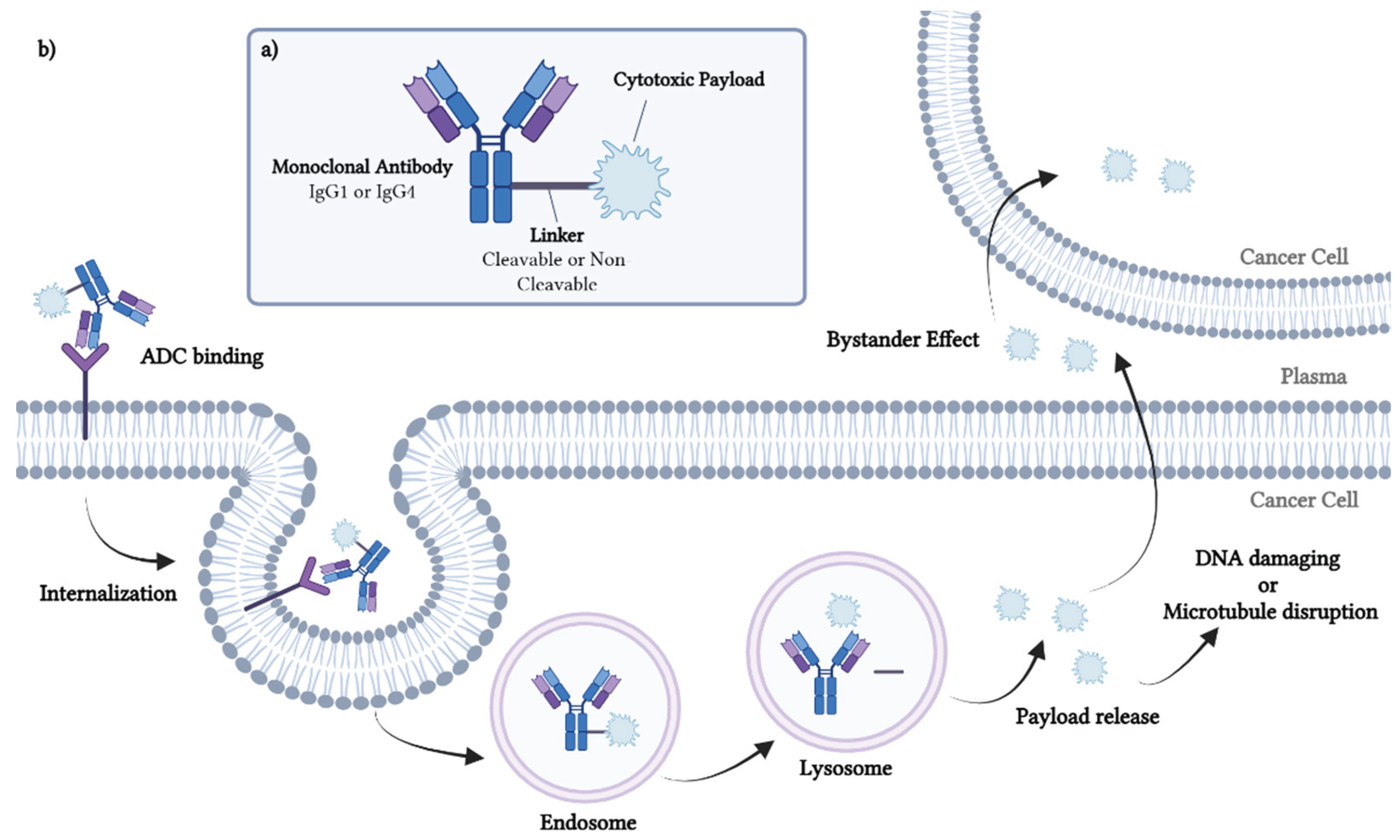
Figure 2.
The development of antibody-drug conjugates (ADCs). (Top) The development stages of ADCs and the next. (Bottom) Key characteristics of approved ADCs. DAR, drug-to-antibody ratio; PK, Pharmacokinetic; Fc, Fragment crystallizable; Fab, Fragment antigen-binding.
Figure 2.
The development of antibody-drug conjugates (ADCs). (Top) The development stages of ADCs and the next. (Bottom) Key characteristics of approved ADCs. DAR, drug-to-antibody ratio; PK, Pharmacokinetic; Fc, Fragment crystallizable; Fab, Fragment antigen-binding.
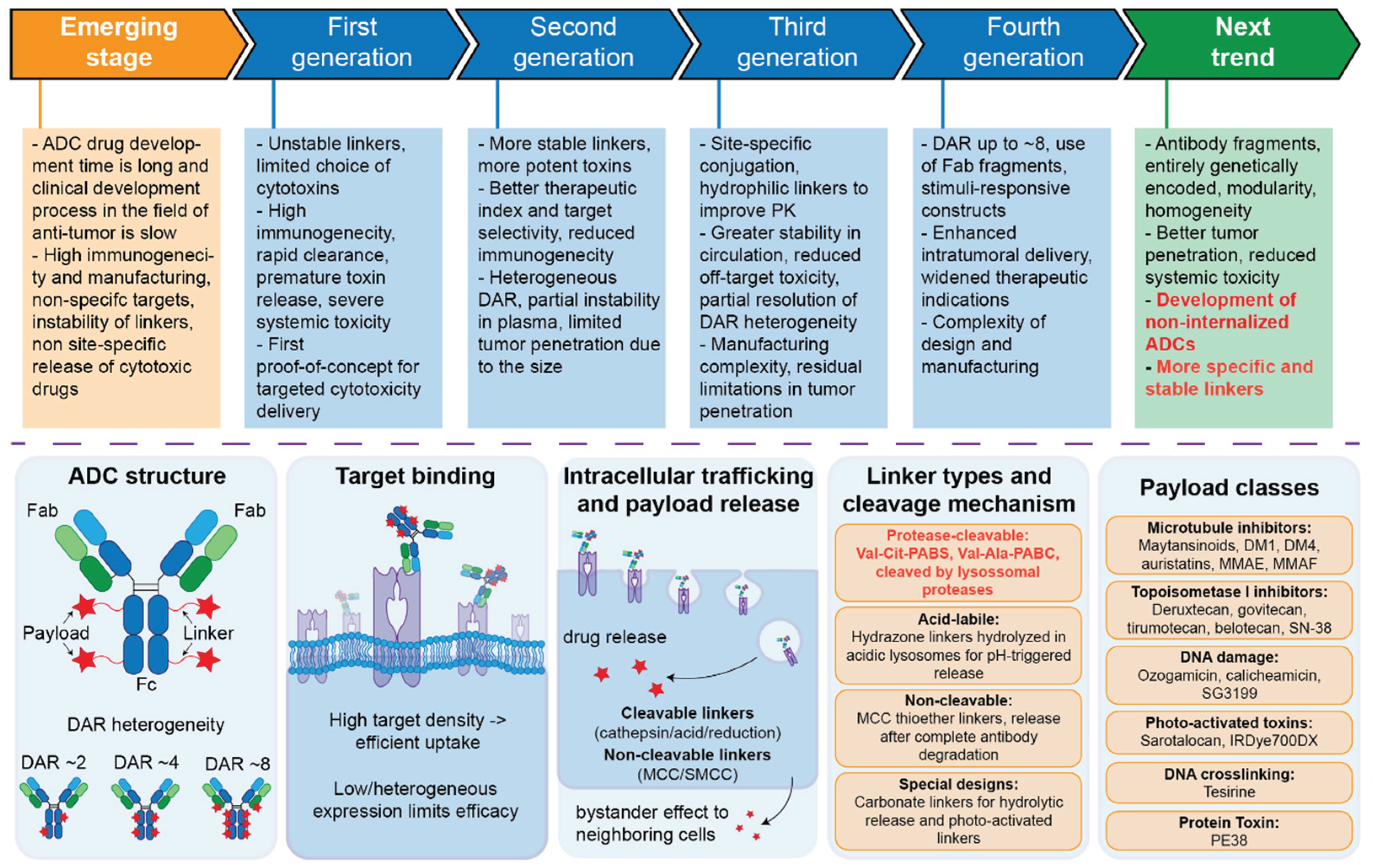
Figure 3.
Classification of linkers in the antibody-drug conjugates (ADCs). Linkers of ADCs are classified into two categories: cleavable and non-cleavable linkers. Cleavable linkers consist of seven subtypes, which can be further divided into chemical cleavable and enzymatic cleavable linker.
Figure 3.
Classification of linkers in the antibody-drug conjugates (ADCs). Linkers of ADCs are classified into two categories: cleavable and non-cleavable linkers. Cleavable linkers consist of seven subtypes, which can be further divided into chemical cleavable and enzymatic cleavable linker.
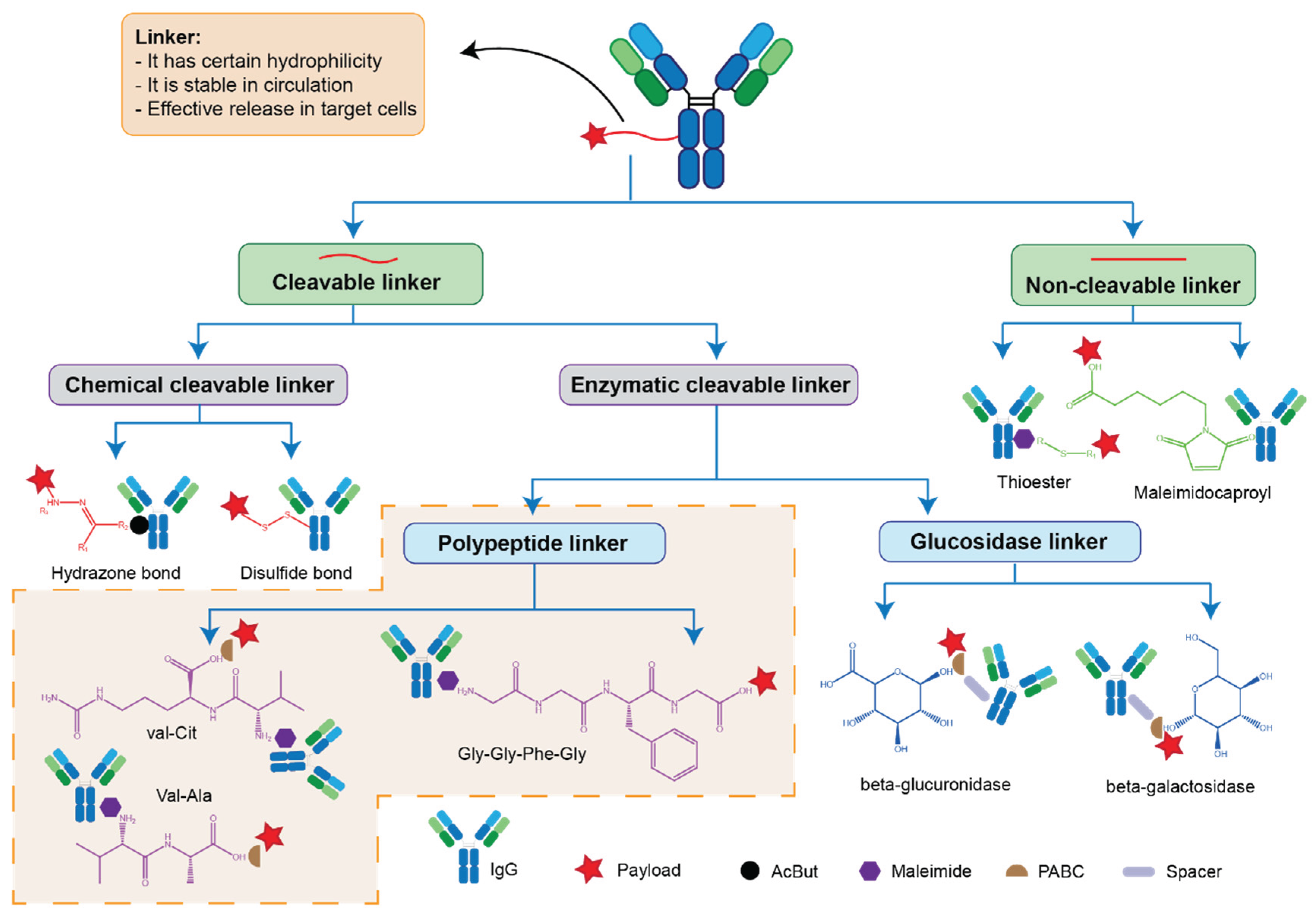
Table 1.
Characteristics of approved ADCs.
| Common name | Trade name | Target | mAb | Linker | Linker type | Linking technology | Payload | Payload type | DAR | Approved indications | Approval Date | Approval Institution |
|---|---|---|---|---|---|---|---|---|---|---|---|---|
| Brentuximab vedotin | Adcetris | CD30 | IgG1 | Mc-Val-Cit-PBAC | Cleavable | Cysteine | MMAE | Microtubule inhibitor | 4 | R/R HL, sALCL, CTCL, MF, PTCL | 2011 | FDA |
| Polatuzumab vedotin | Polivy | CD79b | IgG1 | Mc-Val-Cit-PBAC | Cleavable | Cysteine | MMAE | Microtubule inhibitor | 3.5 | R/R DLBCL | 2019 | FDA |
| Enfortumab vedotin | Padcev | Nectin4 | IgG1 | Mc-Val-Cit-PBAC | Cleavable | Cysteine | MMAE | Microtubule inhibitor | 3.8 | UC | 2019 | FDA |
| Disitamab vedotin | Aidixi | HER2 | IgG1 | Mc-Val-Cit-PBAC | Cleavable | Cysteine | MMAE | Microtubule inhibitor | 4 | HER2+ GC, HER2+ UC | 2021 | NMPA |
| Tisotumab vedotin | Tivdak | TF | IgG1 | Mc-Val-Cit-PBAC | Cleavable | Cysteine | MMAE | Microtubule inhibitor | 4 | R/M CC | 2021 | FDA |
| Trastuzumab deruxtecan | Enhertu | HER2 | IgG1 | MC-Gly-Gly-Phe-Gly-AM | Cleavable | Cysteine | Dxd | TOP1 inhibitor | 8 | GC, GEJC, HER2+ BC, HER2+ GC, HER (low) BC, HER2 (mut) NSCL | 2019 | FDA |
| Datapotamab deruxtecan | Datroway | TROP2 | IgG1 | MC-Gly-Gly-Phe-Gly-AM | Cleavable | Cysteine | Dxd | TOP1 inhibitor | 4 | mBC | 2025 | FDA |
| Loncastuximab tesirine | Zynlonta | CD19 | IgG1 | Mal-PEG8-Val-Ala-PABC | Cleavable | Cysteine | SG3199 | DNA cleavage | 2.3 | BCL, R/R DLBCL | 2021 | FDA |
| Gemtuzumab ozogamicin | Mylotarg | CD33 | IgG4 | AcBut | Cleavable | Lysine | Ozogamicin | DNA cleavage | 2-3 | R/R CD33+AML | 2000 | FDA |
| Inotuzumab ozogamicin | Besponsa | CD22 | IgG4 | AcBut | Cleavable | Lysine | Ozogamicin | DNA cleavage | 6 | R/R ALL | 2017 | FDA |
| Sacituzumab govitecan | Trodelvy | TROP2 | IgG1 | CL2A | Cleavable | Cysteine | SN-38 | TOP1 inhibitor | 7.6 | BC, TNBC, UC | 2020 | FDA |
| Sacituzumab tirumotecan | Jiatailai | TROP2 | IgG1 | Pytimidine-CL2A-carbonate | Cleavable | Cysteine | Belotecan-derivative | TOP1 inhibitor | 7.4 | EGFRm NSCLC | 2024 | FDA |
| Mirvetuximab soravtansine | Elahere | FRα | IgG1 | Sulfo-SPDB | Cleavable | Lysine | DM4 | Microtubule inhibitor | 3.5 | PROC, PRFTC, PRPC | 2022 | FDA |
| Trastuzumab emtansine | Kadcyla | HER2 | IgG1 | SMCC | Non-cleavable | Lysine | DM1 | Microtubule inhibitor | 3.5 | BC, HER2+ BC, mBC | 2013 | FDA |
| Belantamab mafodotin | Blenrep | BCMA | IgG1 | Maleimido-caproyl | Non-cleavable | Cysteine | MMAF | Microtubule inhibitor | 4 | R/R MM | 2020 | FDA |
| Cetuxumab saratolacan | Akalux | EGFR | IgG1 | Linear alkyl/alkoxy linker | Non-cleavable | Lysine | IRDYE700 | Membrane damage | 1.3-3.8 | HNSCC | 2020 | PMDA |
| Moxetumomab pasudotox | Lumoxiti | CD22 | IgG1 | N/A | N/A | N/A | PE38 | Protein synthesis inhibition | 2 | R/R HCL | 2018 | FDA |
| ADCs Antibody‒drug conjugates, ALL Acute lymphoblastic leukemia, AML Acute myeloid leukemia, BCL B-cell lymphoma, BCMA B cell maturation antigen, CTCL Cutaneous T-cell lymphoma, DAR Drug-antibody ratio, DLBCL Diffuse large B-cell lymphoma, DM1 emtansine, DM4 ravtansine, DXd Deruxtecan, EGFR Epidermal growth factor receptor, FDA Food and Drug Administration, FRα Folate receptor alpha, GC Gastric cancer, GEJC Gastro esophageal junction cancer, HCL Hairy cell leukemia, HER2 Human epidermal growth factor receptor 2, HER2 + HER2-positive, HER2 (low) low HER2 expression, HER2 (mut) activating HER2 mutations, HL Hodgkin lymphoma, HNSCC Head and neck squamous carcinoma, mBC metastatic breast cancer, mAb Monoclonal antibody, MM Multiple myeloma, MMAE Monomethyl auristatin E, MMAF Monomethyl auristatin F, MF Mycosis fungoides, Nectin-4 Nectin cell adhesion molecule-4, NMPA National Medical Products Administration, NSCLC Non-small cell lung cancer, PMDA Pharmaceuticals and Medical Devices Agency, PRPC Platinum-resistant peritoneal cancer, PRFTC Platinum-resistant fallopian tube cancer, PROC Platinum-resistant ovarian cancer, PTCL Peripheral T-cell lymphomas, R/M CC Recurrent or metastatic cervical cancer, R/R Relapsed or refractory, sALCL systemic anaplastic large cell lymphoma, SMCC Succinimidyl trans-4-(maleimidylmethyl) cyclohexane-1-carboxylate, SN-38 7-ethyl-10-hydroxycamptothecin, TF Tissue factor, TNBC Triple negative breast cancer, TROP2 Trophoblast cell surface antigen 2, UC Urothelial cancer, | ||||||||||||
Table 2.
ADCs in Phase III clinical trials.
| Common name | Target | mAb | Linker | Linker type | Payload | Payload type | DAR | Representative indications | Phase | NCT Number | Outcome |
|---|---|---|---|---|---|---|---|---|---|---|---|
| Zilovertamab vedotin | ROR1 | IgG1 | mc-Val-Cit-PABC | Cleavable | MMAE | Tubulin binder | 4 | DLBCL | II/III | NCT06717347 NCT05139017 |
Ongoing |
| BNY323/DB-1303 | HER2 | IgG1 | mc-Gly-Gly-Phe-Gly | Cleavable | P1003 | TOP1 inhibitor | 8 | HER2+ BC, mBC, EC | III | NCT06265428 NCT0601833 NCT06340568 |
Ongoing |
| TQB-2102 | HER2 | IgG1 | N/A | N/A | N/A | TOP1 inhibitor | 5.8 | BC | III | NCT06561607 | Ongoing |
| Trastuzumab duo-carmazine/SYD985 | HER2 | IgG1 | mc-PEG2-Val-Cit-PABA-Cys | Cleavable | Seco-DUBA | DNA-damaging | 2.8 | mBC | III | NCT03262935 | PFS, 7.0vs4.9m OS, 20.4vs16.3m ORR, 27.8%vs29.5% |
| ARX-788 | HER2 | IgG1 | Hydroxylamine-PEG4 | Non-cleavable | MMAF | Tubulin binder | 1.9 | HER2+ BC | II/III | NCT05426486 | Ongoing |
| FS-1502 | HER2 | IgG1 | Geranyl ketone pyrophosphate oxime ligation | cleavable | MMAF | Tubulin binder | 2 | BC | III | NCT05755048 | Ongoing |
| MRG-002 | HER2 | IgG1 | mc-Val-Cit-PABC | Cleavable | MMAE | Tubulin binder | 3.8 | aBC, mBC, aCU, mCU | II/III | NCT04924699 NCT05754853 |
Ongoing |
| DP303c | HER2 | IgG1 | PEG2-Val-Cit-PABC | Cleavable | MMAE | Tubulin binder | 2 | HER2+ BC, HER2+ aBC | III | NCT06313086 NCT05901935 |
Ongoing |
| SHR-A1811 | HER2 | IgG1 | mc-Gly-Gly-Phe-Gly | Cleavable | SHR9265 | TOP1 inhibitor | 5.7 | HER2+ BC, OC, CRC, HER2+ r/mBC, NSCLC, HER2+ GC/GEJC | III | NCT05814354 NCT06828354 NCT06057610 NCT06199973 NCT06430437 NCT06126640 NCT05424835 NCT06123494 |
Ongoing |
| Patritumab deruxtecan | HER3 | IgG1 | mc-Gly-Gly-Phe-Gly | Cleavable | Dxd | TOP1 inhibitor | 8 | NSCLC | III | NCT05338970 | Ongoing |
| MRG-003 | EGFR | IgG1 | mc-Val-Cit-PABC | Cleavable | MMAE | Tubulin binder | 3.8 | HNSCC | III | NCT05751512 | Ongoing |
| Depatuxizumab mafodotin/ABT-414 | EGFR | IgG1 | Maleimidocaproxyl | Non-cleavable | MMAF | Tubulin binder | 3.8 | GBM, GSM | III | NCT2573324 | OS, 18.7vs18.9m |
| FDA018 | TROP2 | IgG1 | N/A | N/A | SN-38 | TOP1 inhibitor | 7.6 | TNBC | III | NCT06519370 | Ongoing |
| Tusamitamab ravtansine/SAR408701 | CEACAM5 | IgG1 | SPDB | Non-cleavable | DM4 | Tubulin binder | 3-4 | BCa | III | NCT02449239 | PFS, 75.39vs45.85m OS, 12.8vs11.5m ORR, 21.%vs24.1m |
| Telisotuzumab vedotin | MET | IgG1 | mc-val-Cit-PABC | Cleavable | MMAE | Tubulin binder | 3.1 | NSCLC | III | NCT06758401 NCT06012435 |
Ongoing |
| Telisotuzumab adizutecan/ABBV-400 | MET | IgG1 | N/A | N/A | Adizutecan | TOP1 inhibitor | / | SCLC, ESCC | III | NCT06203210 NCT06644781 |
Ongoing |
| Oportuzumab monatox/Vicinium | EpCAM | scFv | N/A | N/A | ETA-252-608 | Tubulin binder | / | BCa | III | NCT02449239 | CRR, 40% DOR, 9.4m |
| Sigvitatug vedotin/SGN-B6 A | ITGB6 | IgG1 | mc-Val-Cit-PABC | Cleavable | MMAE | Tubulin binder | 4 | NSCLCL | III | NCT06758401 NCT06012435 |
Ongoing |
| Ifinatamab deruxtecan | CD276 | IgG1 | mc-Gly-Gly-Phe-Gly | Cleavable | Dxd | TOP1 inhibitor | 6 | SCLC, ESCC | III | NCT06203210 NCT06644781 |
Ongoing |
| Raludotatug deruxtecan | CDH6 | IgG1 | mc-Gly-Gly-Phe-Gly | Cleavable | Dxd | TOP1 inhibitor | 8 | Solid Cancer | II/III | NCT06161025 | Ongoing |
| Luveltamab tazevibulin/STRO-002 | FRα | IgG1 | Val-Cit-PABA | Cleavable | SC209 | Tubulin binder | 4 | OC, FTC, PC | II/III | NCT05870748 | Ongoing |
| Rinatabart sesutecan | FRα | IgG1 | Cys-11 | Non-cleavable | Exatecan | TOP1 inhibitor | 8 | PROC | III | NCT06619236 | Ongoing |
| aBC advanced breast cancer, ADCs Antibody‒drug conjugates, AF-HPA Auri-statin hydrophile-polymer, aUC advanced urothelium cancer, BCa Bladder cancer, CC Cervical cancer, CDH6 Cadherin 6, CEACAM5 CEA cell adhesion molecule 5, CRC Colorectal cancer, DAR Drug-to-antibody ratio, DLBCL Diffuse large B-cell lymphoma, DM4 ravtansine, DXd Deruxtecan, EC Endometrial cancer, EF-2 Elongation factor 2, EGFR Epidermal growth factor receptor, EpCAM Epithelial cell adhesion molecule, ESCC Esophageal squamous cell carcinoma, FRα Folate receptor alpha, FTC Fallopian tube cancer, GBM Glioblastoma, GSM Gliosarcoma, GEJC Gastroesophageal junction cancer, HER2 Human epidermal growth factor receptor 2, HER2+aBC HER2-positive advanced breast cancer, HER2+GC/GEJC HER2-positive gastric cancer or gastroesophageal junction adenocarcinoma, HER2+BC HER2-positive breast cancer, HER2+r/mBC HER2- positive recurrent or metastatic breast cancer, HER3 Human epidermal growth factor receptor 3, HNSCC Head and neck squamous carcinoma, ITGB6 Integrin alpha V beta 6, mAb monoclonal antibody, mBC metastatic breast cancer, mCRC metastatic colorectal cancer, MET Mesenchymal-epithelial transition factor, MMAE Monomethyl auristatin E, MMAF Monomethyl auristatin F, mUC metastatic urothelium cancer, NSCLC Non-small cell lung cancer, OC Ovarian cancer, PC Peritoneal cancer, PROC Platinum-resistant ovarian cancer, ROR1 Receptor tyrosine kinase-like orphan receptor 1, SCLC Small cell lung cancer, seco-DUBA seco-duobamycin hydroxybenzamide-azaindole, SPDB N-succinimidyl 4-(2-pyridyldithio) butanoate, TNBC Triple negative breast cancer (http://clinicaltrials.gov) | |||||||||||
Disclaimer/Publisher’s Note: The statements, opinions and data contained in all publications are solely those of the individual author(s) and contributor(s) and not of MDPI and/or the editor(s). MDPI and/or the editor(s) disclaim responsibility for any injury to people or property resulting from any ideas, methods, instructions or products referred to in the content. |
© 2026 by the authors. Licensee MDPI, Basel, Switzerland. This article is an open access article distributed under the terms and conditions of the Creative Commons Attribution (CC BY) license (http://creativecommons.org/licenses/by/4.0/).
Copyright: This open access article is published under a Creative Commons CC BY 4.0 license, which permit the free download, distribution, and reuse, provided that the author and preprint are cited in any reuse.