

Submitted:
11 July 2026
Posted:
13 July 2026
You are already at the latest version
Abstract
Existing vectored immunoprophylaxis (VIP) approaches have primarily focused on IgG, which provides systemic protection but is less specialized for mucosal immunity. In contrast, secretory IgA (sIgA) plays a central role at epithelial surfaces, promoting pathogen neutralization while limiting inflammation. Although monoclonal IgA therapies are effective, their short half-life requires repeated dosing. Thus, VIP strategies enabling sustained sIgA expression at mucosal sites could transform mucosal infection prevention and treatment. This review outlines the key challenges associated with in vivo IgA expression and discusses critical considerations for VIP-mediated IgA delivery at mucosal surfaces, with emphasis on its potential for clinical translation. We provide a detailed overview of platforms for targeted IgA expression, including adeno-associated virus (AAV), adenoviral and lentiviral vectors, and lipid nanoparticle-based systems, alongside relevant routes of administration. Additionally, we examine emerging strategies to enhance the robustness, durability, and localization of IgA expression in vivo. Overall, VIP-enabled IgA expression represents an emerging strategy for enhancing mucosal immunity, with continued advances required to establish its role in the prevention and treatment of mucosal infections.
Keywords:
adeno-associated virus (AAV)
; vectored immunoprophylaxis
; secretory IgA
; mucosal immunity
; monoclonal antibody
; antibody engineering
1. Introduction
The discovery of antibodies at the end of the 19th century marked a turning point in the understanding of how the body protects itself from infection.[1] In 1890, Emil von Behring and Shibasaburo Kitasato demonstrated that immunity to infection could be transferred between animals in the form of cell-free serum revealing the existence of protective factors in the serum later termed antibodies by Paul Ehrlich.[1] Although the existence of antibodies was known at the beginning of the 20th century, it was not until the improvement of protein electrophoresis techniques in the 1960s that the structure of these antibodies was elucidated and isotypes assigned.[2] It was during this time that a new class of immunoglobulin named IgA was identified as the prominent antibody in secretions such as saliva.[3] Subsequent studies showed that IgA is produced in abundance at mucosal surfaces where it plays a major role in humoral mucosal immunity. In contrast, IgG is the predominant antibody in serum and tissue fluids and mediates systemic immune protection, although it can also contribute to immunity at select mucosal sites.[3] Further advancements in immunology in the 1970s led to the development of monoclonal antibodies in mice using a hybridoma method characterized by Kohler and Milstein in which therapeutic antibodies could be produced in mice.[4] However, initial clinical studies in humans were plagued by strong immune responses to the foreign murine antibodies limiting effectiveness of monoclonal antibody (mAb) treatments.[4] This led to the development of human-mouse chimeric antibodies and eventually humanized antibodies in the 1980s making mAb treatments viable for the first time.[5] Monoclonal IgA treatments are a recent advancement and remain largely experimental as challenges in large scale production have limited their clinical development.[6] Nonetheless, experimental studies have demonstrated their potential as therapeutics most notably the development of an IgA mAb specific for H5 hemagglutinin which provided complete protection of mice challenged with highly pathogenic avian H5N1 influenza virus.[6] Similarly, monoclonal IgA antibody treatments have shown strong protective effects in murine models of infection caused by Campylobacter jejuni, Enterotoxigenic E. coli, and Salmonella enterica serovar Typhimurium (S. Typhimurium) further highlighting their promise as future therapeutics.[7] Despite the effectiveness of mAb treatments, a major limitation of these treatments is the need for repeat dosing due to the short half-life of IgA antibodies in serum,[8] thereby highlighting the need for innovative approaches to achieve sustained IgA expression and targeted delivery at mucosal surfaces.
2. Biological Rationale for Using sIgA as a Therapeutic
2.1. Structure of IgA and Transcytosis at Mucosal Surfaces
IgA is the most highly produced antibody in the human body containing unique structural features that allow it to remain stable in harsh mucosal environments and provide immune protection at mucosal surfaces.[9] It has been calculated that a 70 kg human produces an estimated 4 grams of IgA every day to cover the large surface area of mucosal epithelium present in the body.[9] Following the general structure of antibodies, IgA contains two identical light chains and two identical heavy chains containing variable Fragment antigen-binding (Fab) regions at the N-terminus that are antigen binding and constant Fragment crystallizable (Fc) regions that interact with immune receptors.[10] Each light chain consists of two globular domains including a variable domain and constant domain in the Fab region of IgA.[10] The heavy chains consist of four globular domains including a variable domain and constant domain Cα1 located in the Fab region followed by two constant domains Cα2 and Cα3 located in the Fc region of IgA.[10] IgA structure differs from other immunoglobulins in that it contains a hinge region between Cα1 and Cα2 that connects the Fab and Fc regions providing flexibility to the antigen binding arms, enabling more effective engagement with spatially diverse epitopes, promoting antigen cross-linking, and enhancing immune exclusion at mucosal surfaces.[10] Each globular domain of IgA forms a recognizable immunoglobulin-like fold characteristic of all antibodies consisting of 110 amino acids arranged in two beta-sheets composed of anti-parallel β-strands.[10] These beta-sheets are further stabilized by internal disulfide bonds between the variable domain of the heavy chain and variable domain of the light chain, Cα1 and the constant region of the light domain, along with the Cα2-Cα3 domains of both heavy chains.[10] There are two types of IgA antibodies found in humans including IgA1 and IgA2 with IgA1 being found primarily in serum and IgA2 being found in mucosal secretions such as intestinal mucus.[11] The IgA1 contains an extensive hinge region absent in IgA2, containing repeats of proline, threonine and serine with these residues containing multiple O-linked oligosaccharides.[11] This O-linked glycosylation on the hinge region is thought to modulate its conformation and increase flexibility but has also been found to make the region more susceptible to proteolysis.[11] Both subtypes of IgA contain N-linked oligosaccharides in the Cα2 domain providing further structural integrity and protection from proteolysis.[11]
The IgA secreted at mucosal surfaces primarily exists in a dimeric form in which two IgA monomers are covalently attached through a joining chain or J-chain polypeptide.[12] The J-chain is a small polypeptide of approximately 15 kDa and is produced by antibody producing plasma cells in tandem with monomeric IgA in the lamina propria of mucosal surfaces.[10] The J-chain creates dimeric IgA (dIgA) using two specific cysteine residues that link the J-chain to the heavy chain of two IgA monomers.[10] This process is enhanced by the marginal zone B-1 cell specific protein (MZB1) chaperone through the binding of the heavy chain of IgA where the J-chain is bound preventing the degradation of dimeric IgA.[13] MZB1 deficient mice showed a measurable decrease in IgA production in response to external inflammatory stimuli highlighting the crucial role of MZB1 in promoting dIgA assembly.[13] The proper formation of dIgA is crucial for its interaction with the polymeric immunoglobulin receptor (pIgR) present on the basolateral surface of mucosal epithelial cells.[14] The pIgR receptor is a type I transmembrane protein consisting of an extracellular region that protrudes from the basolateral surface of epithelial cells, a single membrane spanning region and a cytoplasmic tail.[14] The extracellular ligand region of the pIgR comprises five domains with domain 1 having three separate complementary determining regions (CDRs) that function to form covalent bonds with dIgA produced from plasma cells.[14] Domains 2 to 4 of the pIgR increase the binding efficiency of the receptor by creating the optimal spacing between domains 1 and 5 to allow these domains to bind dimeric IgA.[15] Interestingly, domains 3 and 4 have also been found to play an important role in the binding of the SpsA surface protein of Streptococcus pneumonia allowing this bacterium to hijack the receptor and attach to host epithelial cells.[14] Domain 5 of the pIgR forms a disulfide bond between the receptor and the Fc region of dIgA and acts as a bridge between the extracellular domain and the transmembrane domain of the receptor.[14] Following the binding of dIgA to the pIgR, the complex is internalized into a mucosal epithelial cell through the process of endocytosis and transcytosed to the apical side of the cell in a vesicle.[14] A 22 amino cleavage site is recognized by a pIgR-specific proteinase that cleaves domain 5 of the pIgR receptor, though the identity of this proteinase remains unclear.[16] The cleaved portion of the pIgR bound to IgA is termed the secretory component (SC) and consists of domain 1 through 5 of the original receptor.[15] The domains of the SC fold back on each other and form a condensed triangular shape with a solvent accessible gap in the middle.[17] The SC also contains seven N-linked glycosylation attachment sites for carbohydrate chains involved in mediating interactions with bacteria and host cell lectins.[17] The cleaving of the dIgA-SC complex leads to the creation of the final form of IgA termed secretory IgA (sIgA).[15] The concluding cytoplasmic tail region of pIgR contains signals that are crucial for the transcytosis of IgA across mucosal epithelial cells and for ensuring the receptor is not targeted for lysosomal degradation.[14] The cleaved sIgA complex is released at the apical surface into luminal secretions, such as mucus, where it remains localized and carries out its immunoprotective functions.[15]
2.2. IgA Function and Mechanism of Action at Mucosal Surfaces
When a mature B cell encounters an antigen in the peripheral lymphoid tissues, it becomes activated and induces the expression of activation-induced cytidine deaminase (AID) which is an enzyme vital in the class switching recombination (CSR) process of antibody producing genes.[18] In the instance of IgA expression, the AID enzyme initiates CSR from the Cµ gene which encodes IgM to the Cα gene which encodes IgA.[18] This process occurs through the cytokine-mediated activation of the Iα promotor located upstream of the Cα gene leading to the production of a non-coding mRNA transcript called Iα- Cα which binds to a switch region or S-region upstream of Cα.[18] The binding of the Iα- Cα mRNA to the S-region opens its chromatin structure making cysteine residues accessible to the AID which generates double stranded breaks in the DNA.[19] The non-homologous end joining (NEHJ) DNA repair machinery joins the double stranded breaks and loop out a portion of the DNA in the S-region.[19] As a result, an antibody variable region that determines antigen specificity becomes linked to the Cα gene which encodes a constant antibody region. This fusion event creates a sequence that encodes the IgA protein.[19]
The production and secretion of SIgA at mucosal surfaces in response to antigens allows the antibody to perform a variety of effector functions with the most notable being immune exclusion.[20] The immune exclusion process mediated by SIgA prevents pathogens and foreign antigens from adhering to, penetrating, and infecting mucosal epithelial surfaces.[20] The first step of immune exclusion involves the agglutination or cross-linking of invading pathogens forming clumps of bacteria in the mucus layer of the intestinal mucosa, see Figure 1.[21]
Due to its multivalent structure, SIgA can agglutinate invading pathogens containing polyvalent surface antigens or many copies of the same epitope on their surface.[21] This allows a single SIgA to bind multiple pathogens such as bacteria at the same time facilitating the cross-linking process.[21] Remarkably, research has shown that agglutination does not normally harm bacteria with bacterial growth rates remaining unchanged after cross-linking.[21] However, studies of the binding of the O-antigen of S. Typhimurium to SIgA found that this event leads to a degree of physical distortion of the outer membrane and changes in gene expression detrimental to the bacterium.[21] Consequently, the crosslinking of S. Typhimurium with SIgA antibodies targeted to the flagella of the bacterium did not lead to any structural changes in its membrane.[21] This shows that structural effects and gene expression changes in bacteria as a result of agglutination are likely dependent on the epitope recognized by the SIgA .[21]
The second step of immune exclusion is the physical entrapment of pathogens by SIgA in the mucus layers present at mucosal surfaces.[22] Before SIgA agglutinates bacteria, it binds to the proteins present in the mucus layers most notably mucin-2 (Muc2) and this process is enhanced by the presence of the SC component which contains seven carbohydrate side chains that anchor the antibody at mucosal surfaces (18,19).[22,23] This process allows IgA to trap pathogens in mucus layers leading to them being cleared by the natural process of mucus turnover and peristaltic movement at mucosal surfaces.[22]
In addition to immune exclusion, IgA can also mediate a number of additional effector functions through interactions with a variety of host cell receptors on different cell types.[24] IgA interacts with the FcαRI (CD89) receptor expressed on neutrophils and other myeloid cells, triggering effector functions such as phagocytosis, degranulation, reactive oxygen species production, and cytokine release that contribute to pathogen clearance.[24] Indeed, the binding of IgA to the FcαRI receptor on neutrophils leads to the production of the LTB4 chemoattractant through the ITAM signalling pathway, which attracts additional immune cells resulting in an amplification of this process.[24] After the binding of IgA to FcαRI on neutrophils, this leads to pathogens being phagocytosed and eliminated by an NADPH oxidase respiratory burst in the neutrophil interior.[25] It is important to note that the partial binding of IgA to the FcαRI can cause incomplete phosphorylation of the receptor leading to the downregulation of neutrophil recruitment in which case an inhibitory ITAM signalling pathway is engaged to reduce inflammation.[26] IgA can also bind to the Dectin-1 receptor located on the apical surface of M cells in Peyer’s patches of the intestines allowing the reverse transcytosis of IgA-antigen complexes to occur,.[27] Specifically, antigen-bound SIgA2 binds to Dectin-1 through its glycosylated cα1 domain allowing the endocytosis of the IgA-antigen complex from the apical surface of mucosal epithelial cells to the basolateral surface.[27] The IgA-antigen complex is then released into gut associated lymphoid tissues at the basolateral surface where the complex can be taken up and eliminated by resident dendritic cells, see Figure 1.[27]
In contrast to the reverse transcytosis process, IgA has also been found to harbor an immune excretion process specific to pathogens that replicate in the lamina propria.[28] In addition to mediating immune exclusion at mucosal surfaces, dIgA can facilitate the removal of pathogens that have already crossed the epithelial barrier. For example, dIgA has been shown to bind HIV virions within the lamina propria and form immune complexes that engage pIgR on the basolateral surface of epithelial cells. These complexes are subsequently transcytosed to the apical surface and released into the lumen, thereby removing virus from subepithelial tissues and limiting access to susceptible CD4⁺ T cells and other target cells, see Figure 1.[28] IgA also plays a role in the intracellular neutralization of bacteria and viruses.[29] When the IgA-pIgR complex is undergoing transcytoses across epithelial cells, dIgA can colocalize with endocytosed lipopolysaccharide (LPS), which is a toxic outer membrane component of bacteria.[29] This colocalization allows IgA to inhibit the ability of LPS to trigger an NF-κB mediated cytokine response limiting inflammation and maintaining homeostasis in epithelial cells.[29] Unlike immune excretion of luminal antigens such as LPS, transcytosing dIgA specific for the Sendai virus hemagglutinin-neuraminidase (HN) protein mediates intracellular neutralization by intercepting viral components within infected epithelial cells during pIgR-mediated transport.[30] The pIgR bound dIgA enters the same protein trafficking pathways as the HN causing colocalization to occur and preventing the proper assembly and budding of the Sendai virus at the apical surface of epithelial cells.[30]
Maternal SIgA plays a central role in the establishment of newborn commensal microbiota following exposure to the environment and maternal microbes.[21] Newborns acquire maternal SIgA through breast milk with the antibody guiding the establishment of the newborn microbiota through binding commensal bacteria and facilitating controlled colonization.[21] The colonization of the newborn microbiota in turn leads to the maturation of gut-associated lymphoid tissue (GALT) with the limited production of IgA against commensal microbiota to limit redundant inflammation.[21] A deficiency in the production of IgA in humans characterized as selective IgA deficiency (SIgAd) leads to a reduction in the diversity and an imbalance in proportions of microbes present in the microbiota.[31] The clinical signs of this disease are often masked by the compensatory production of IgM which also binds and keeps commensal bacteria in check.[31] However, individuals with SIgAd still possess a shifted microbiota composition with IgM not being able to fully compensate for the absence in production of IgA or its specialized role in maintaining homeostasis.[31]
2.3. Advantages of SIgA over IgG for Mucosal Pathogens
Although both SIgA and IgG play roles in defending against mucosal pathogens, SIgA is uniquely adapted to mucosal environments whereas IgG is largely involved in whole-body immunity, see Table 1.
As mentioned before, dIgA is produced locally by plasma cells in the lamina propria allowing the transcytosis and secretion of SIgA directly at mucosal surfaces where it can induce immune exclusion before pathogens are able to invade. In contrast, IgG antibodies are primarily produced in the serum by the plasma cells of the spleen with the function of encountering pathogens that have already invaded epithelial tissues and entered the bloodstream.[32] It is important to note that IgG can undergo transcytosis from the lamina propria to the surface of mucosal epithelial cells using the neonatal Fc receptor (FcRn).[33] Experiments have shown that IgG can then bind antigens present at mucosal surfaces and move back through epithelial cells into the lamina propria to encounter dendritic cells for processing.[33] Though IgG is present in mucosal secretions, it is released in significantly smaller concentrations then IgA and is only reactionary to infection rather than preventive.[33] Discussed previously, dIgA is able neutralize a variety of pathogens intracellularly when being transported by pIgR to the apical surface of epithelial cells compared to IgG which can only perform this function for specific pathogens during transport by FcRn.[34] IgG has only been loosely characterized in the literature to perform intracellular neutralization against the VP6 capsid protein of rotavirus in which the Fc region of IgG is able to bind TRIM21 bound pathogens and target them for degradation.[34] In contrast, IgA has been well characterized to be involved in the intracellular neutralization of multiple viruses including Sendai virus, influenza virus and HIV.[30,35] The broad intracellular neutralizing ability of IgA to inhibit viral replication provides it with an advantage over IgG which is largely restricted to extracellular effector functions.[32] The immune exclusion process used by IgA elicits a considerably reduced pro-inflammatory response in comparison to the IgG effector function of binding Fcγ-receptors on immune cell surfaces and promoting phagocytosis.[36] While the IgG proinflammatory response is vital in the clearing of pathogens, this same response can cause damage to tissues when misdirected towards the body’s own cells in the case of autoimmune diseases.[32] Specifically, IgG can mistakenly bind to self-receptors and form immune complexes that can lead to the activation of immune cells that attack the body’s own tissues.[37] These immune complexes can additionally activate complement signaling cascades including the classical, alternative and lectin pathways leading to additional damaging inflammatory responses in the body.[37] Since IgA primarily uses the process of immune exclusion to neutralize pathogens in the mucosa, immune cell activating pathways are not induced at levels comparable to IgG meaning the risk of collateral tissue damage is reduced.[38] Research has shown that due to its distinct glycosylation profile, IgA does not efficiently activate the classical complement pathway like IgG and instead engages the lectin pathway, leading to weaker overall complement activation.[39] As referenced earlier, IgA is crucial in maintaining mucosal homeostasis by preventing excessive inflammatory responses against commensal microbes compared to IgG which can elicit elevated immune responses.[31,40] In a study related to ulcerative colitis, a murine model was created that mimicked the effects of the disease notably the increase in production of IgG from B cells located in the gut normally dominated by IgA.[40] This led to an elevated number of commensal microbes in the gut being coated with IgG with these complexes activating harmful immune responses that would normally not be induced by IgA opsonization.[40] It should be noted that the increased production of and binding of IgA to commensal bacteria has also been observed to induce harmful immune responses in a small subset of ulcerative colitis patients though the correlation is drastically reduced compared to IgG.[41]
As was highlighted earlier, IgA can interact with proteins such as mucin-2 at mucosal surfaces using the SC to anchor itself in the mucosa in contrast to IgG which largely diffuses through the mucosa.[42] IgG has been found to anchor itself in the mucosa through the binding of mucin proteins using the IgG Fc-binding protein (FcGBP) protein through this interaction is limited in its ability to prevent the diffusion of IgG.[42,43] Conversely, the interaction of SIgA with mucin proteins is strong allowing the anchoring of the antibody at mucosal surfaces for longer periods of time with little diffusion.[43] The enhanced ability of SIgA to remain anchored in the mucosa over IgG provides the advantage of increased contact and neutralization of pathogens at mucosal surfaces.[43] Although SIgA has a shorter half-life in serum than IgG, the structural SC component provides IgA with increased resistance to proteases at mucosal surfaces as mentioned earlier. [44,45] In an experiment in which SIgA, dIgA and IgG were digested with purified Proteus Mirabilis protease, it was observed that SIgA was degraded at a slower rate than IgG and dIgA showing the importance of the SC component in protease resistance.[44]
The dimeric structure of IgA provides it with an increased ability to bind and crosslink pathogens over IgG, which only exists in a monomeric structure.[46] A study was conducted in which the avidity and affinity of monomeric IgA, dIgA and monomeric IgG to bind to the M2 protein of influenza was evaluated.[47] It was found that dIgA more effectively inhibited viral plaque formation then monomeric IgA and IgG since it was able to bind a larger subset of M2 proteins.[47] However, there was no significant difference in the affinity of dIgA to bind M2 compared to monomeric IgA and IgG meaning the polymeric nature of IgA only increases its avidity for binding antigens and not the strength in which it can bind antigens.[47] Additionally, IgA is less likely to induce antibody dependent enhancement (ADE) compared to IgG in which non-neutralizing antibodies can bind pathogens and facilitate their increased uptake into host cells.[48] Unlike IgG, which can promote FcγR-mediated uptake of virus–antibody complexes and facilitate ADE when antibodies are non-neutralizing, IgA engages FcαRI in a way that is less efficient at promoting viral entry and therefore is much less likely to drive ADE.[48] Research has found that an IgA version of a dengue virus reactive antibody was able to interfere with the ability of the IgG version to induce ADE due to competition for antigen binding sites.[49] These findings indicate that the amount of dengue specific IgA produced during infection can influence the severity of IgG mediated ADE and serve as a biomarker for disease susceptibility.[49]
3. Strategies for SIgA Production and Platforms for Vectorized Expression In Vivo
3.1. Challenges with Engineering SIgA Expression
Despite the significant potential of SIgA as a first line of defense at mucosal surfaces, there are many complexities associated with its recombinant expression in vivo.[50] One of the main challenges of expressing SIgA lies in using expression vectors to reproduce the precise stoichiometric co-expression ratios of its components including the heavy chain, light chain, J chain and SC that occurs naturally at mucosal surfaces.[50] SIgA is the product of two separate cell types with the SC being a cleaved product from the pIgR on epithelial cells and the other components originating from plasma cells in the lamina propria.[51] This means that each of the components of dIgA need to be expressed at precise ratios to form complexes with the pIgR on epithelial cell surfaces for proper assembly to occur.[51] SIgA was notably produced in a dIgA expressing mammalian cell line in 1997 with the transfection of an SC expression vector proving the viability of recombinant SIgA production in vitro.[52] However, of the IgA components produced in single cells, it was found that only a fraction of the components assembled into SIgA with many forming monomeric IgA or dIgA.[52] It is thought that this anomaly could be a result of suboptimal expression levels of the dIgA components with SC and that using promotors of varying strengths or increasing gene copy numbers could improve SIgA expression.[53] The formation of SIgA is also dependent on proper post-translational modifications of its components including O- and N-linked glycosylation with these modifications being host-cell dependent.[54] A study looking at the difference in IgA glycosylation after expression in plant and HEK293 cells found significant differences in glycosylation patterns with the glycosylation of IgA in HEK293 cells providing improved stability.[55] These findings are significant since the glycosylation patterns of IgA could be altered when being expressed in vivo depending on the location of expression with these alterations affecting proper IgA formation and stability.[55] Differences in glycosylation can also influence IgA interactions with the J-chain and pIgR adding an additional hurdle to in vivo expression.[54] Another challenge associated with SIgA expression in vivo is the tissue specific targeting of dIgA expression at mucosal epithelial cells where it can interact with the pIgR and be expressed at mucosal surfaces. The expression of dIgA mainly occurs in the plasma cells of the lamina propria adjacent to mucosal epithelial cells in the gastrointestinal tract and respiratory tract. This means that expression of plasmids encoding the dIgA components must be delivered to mucosal epithelial cells in a highly specific manner using transport vectors such as viruses or nanoparticles. Previous work has shown that the engineered adeno-associated virus capsid AAV6.2FF has strong tropism for lung epithelial cells, in particular, alveolar type II cells, and was able to efficiently expression of IgG antibodies in the lungs of murine models.[56] These findings support the feasibility of tissue-targeted delivery using vectors such as AAV to achieve sustained antibody expression.[56] The use of transport vectors for in vivo expression of recombinant antibodies can, however, be complicated by host immune responses notably the activation of complement and cytokine signaling pathways.[57] Studies have shown that the administration of AAV vectors can lead to the activation of host immune responses against the viral capsid proteins along with therapeutic transgenes.[57] An ongoing area of research is focused on the removal of the major pathogen associated molecular patterns (PAMPS) from AAV vectors, though it is unlikely that this will completely eliminate the stimulation of host immune responses.[58] Evidence shows that lipid nanoparticles specifically the ionizable lipids also lead to the activation of cytokine signalling pathways showing the widespread issues of immune responses towards expression vectors.[59] In addition, the use of gamma globulin therapy, in which IgA rich preparations derived from donor plasma are given to IgA-deficient patients, has been shown to induce anti-drug antibody (ADA) responses.[60] Specifically, IgG anti-IgA antibodies that were already present in the IgA-deficient patients reacted to the increased levels of IgA causing complement activation in a subset of patients.[60] Although ADA responses to administered IgA have been reported, it is not yet clear whether in vivo IgA expression will elicit comparable responses, underscoring the need to evaluate and mitigate potential immunogenicity.[60] AAV mediated expression of broadly neutralizing IgG antibodies against HIV has been shown to induce ADA responses in non-human primates and humans, providing evidence that this phenomenon could occur for AAV-expressed IgA as well.[61]
3.2. Overview of Platforms for the Expression of IgG and SIgA In Vivo
AAV-based gene delivery has emerged as a leading strategy for the long-term in vivo expression of IgG antibodies.[62] AAV gene therapy vectors have an icosahedral structure composed of three viral capsid proteins along with a genome capable of encoding a maximum of 4.7kb of DNA flanked by inverted terminal repeats (ITR), with all other components of the original viral genome removed making the vectors replication deficient.[63] At least thirteen distinct natural serotypes of AAV have been identified, each with characteristic tissue tropisms.[64,65] Following infection of host cells, AAV delivers its genome to the nucleus where it forms stable, transcriptionally active episomes that express target genes without integration into the host genome.[63] AAV-mediated gene delivery has been extensively investigated for the in vivo expression of IgG monoclonal antibodies targeting a wide range of viral, bacterial, and parasitic pathogens. Across numerous preclinical studies, AAV-mAb expression has consistently demonstrated protective efficacy and the capacity for durable, long-term IgG expression, supporting its potential as an alternative to traditional vaccination or passive immunization approaches. While an extensive body of work spans multiple infectious disease models, representative studies have shown that AAV-directed expression of antiviral IgG can confer complete protection in small animals and sustain circulating antibody levels for the lifespan of the host.[66,67,68,69] For example, AAV-mediated delivery of IgG against filoviruses such as Marburg and Ebola has achieved complete protection in mice and guinea pigs, with antibody expression maintained long term.[66,67,68,69,70] Similarly, translation to large-animal models has demonstrated the scalability and durability of this approach; AAV6.2FF-mediated IgG expression in lambs resulted in sustained antibody production for over 1100 days.[71] Notably, although anti-drug antibodies (ADA) against the expressed IgG and anti-capsid immune responses have been observed, these responses were low and remained stable over time in this model, suggesting that immunogenicity may not preclude prolonged transgene expression in all settings.
Replication-deficient adenoviral vectors, which encompass multiple serotypes with distinct tissue tropisms, represent an in vivo expression system for IgG antibodies.[72] Adenoviral vector icosahedral capsids are approximately 100nm in diameter and can package up to 36kb of DNA with the viral genome being maintained as an episome similar to AAV.[73] There are three generations of adenoviral vector systems with the most widely used being the third-generation system in which the genome has been mostly depleted of adenoviral DNA with the exception of the ITRs and packaging signals required for vector production.[72] An adenoviral vector system was used for the expression of recombinant IgG antibodies against the TcdA and TcdB toxins produced by clostridium difficile and fully protected mice against lethal challenge by the bacterium with strong antibody expression lasting for over a month.[74] Despite the success of this study, use of these systems has been complicated by systemic inflammatory responses and modest duration of IgG expression.[75]
Beyond viral platforms, lipid nanoparticle (LNP) delivery systems, which are composed of four distinct lipid types, offer an alternative method for in vivo expression of IgG antibodies.[76] Current LNPs consist of ionizable lipids important in the encapsulation of nucleic acids and endosomal uptake, helper lipids which support LNP stability, cholesterol which limit nonspecific protein binding to the LNP surface limiting inflammatory responses, PEG-lipids which give LNPs consistent size and prevent aggregation and mRNA of variable size encoding proteins of interest.[76] A significant challenge of using LNPs for tissue specific expression of antibodies is the fact that they are naturally taken up by cells in the liver and targeted expression is difficult.[77] However, recent advances incorporating selective organ targeting (SORT) lipids into LNPs have enabled tissue-specific expression, with positively charged SORT lipids directing delivery to the lungs and negatively charged SORT lipids targeting the spleen.[77] LNPs have been shown to induce sustained expression of an anti-SARS-CoV-2 human IgG mAb in mice providing protection against lethal challenge for over 63 days.[78] Despite the advent of SORT methods, targeted delivery of LNPs to tissues remains a significant barrier for gene delivery.[77]
Regarding in vivo expression of SIgA, there have currently been two methods used including lentiviral vector-mediated gene delivery and LNP-mediated mRNA delivery. Lentiviral vectors have a ~10kb packaging capacity.[79] The third-generation lentiviral system, which is widely used due to its improved safety profile, employs four plasmids: one encoding gag/pol, one encoding the envelope protein for cell-specific targeting, one carrying the transgene under a promoter, and one encoding rev for nuclear export; these systems are typically designed as self-inactivating (SIN) through deletion of the U3 region in the 3′ long terminal repeat (LTR).[79] The co-transfection of these plasmids in vitro leads to the production of lentiviral vectors that can target and integrate their transgenes into host cell genomes for pro-longed expression.[80] Lentiviral vectors containing anti-HIV SIgA transgenes were used for the ex vivo transduction of hematopoietic stem cells, after which these stem cells were transplanted into the bone marrow of humanized mice.[81] It was found that the anti-HIV SIgA was expressed in plasma cells in lymphoid tissues preventing their depletion following lethal HIV challenge.[81] This study demonstrates an indirect approach in which SIgA is expressed in vivo using stem cell therapy, in contrast to the direct delivery methods used for IgG expression by adenoviral and AAV vectors.[81] Despite the success of lentiviral vectors in expressing IgA in vivo, the risk of transgene integration near proto-oncogenes leading to cancer remains a major hurdle to the widespread use of these systems along with the challenges of stem cell therapy.[82] A recently conducted study investigated the use of LNPs for the delivery and expression of SIgA mAbs into mucosal secretions for protection against pseudomonas bacteria.[83] Mice were IV injected with 1mg/kg of LNPs containing mRNA encoding the IgA heavy chain, light chain, and J chain alongside the injection of mice with 5 mg/kg of recombinant IgA produced in cell culture and 4.5 mg/kg of polyclonal IgA isolated from serum.[83] It was found that the recombinant IgA had the shortest serum half-life of 0.64 days with expression dropping of after 2 days, followed by serum derived IgA with a half-life of 0.93 days with expression dropping of after ~12 days, and mRNA derived IgA having a half-life of 1.63 days with expression dropping off after ~16 days.[83] The authors speculated that the reduced half-life of serum- and cell culture–derived IgA may be due to differences in glycosylation patterns arising from expression in different murine cell types.[83] Additionally, it was found that the expression of mRNA derived IgA protected mice against lethal challenge with Salmonella enterica in the intestines and Pseudomonas aeruginosa in the lungs.[83] The conclusions of this work show the feasibility of the production of recombinant SIgA at mucosal surfaces in vivo and the importance of host glycosylation patterns for the stability of expressed antibodies. A major limitation of this study is that LNP-mediated IgA expression peaked 24 hours after administration before gradually declining until day 16 paving the way for longer term expression using vectors such as AAV.[83]
3.3. Design Considerations for the Correct Assembly of SIgA In Vivo
The correct assembly of SIgA in vivo remains a significant challenge requiring the careful consideration of plasmid design, expression balance of components and vector delivery systems.[50] For the proper in vivo expression of SIgA to occur, the heavy chain, light chain, J chain must be expressed for proper assembly to occur as mentioned previously.[50] If AAV is to be used, the genes encoding all four components of SIgA would not fit into a single plasmid, but it would be possible to express the IgA-J chain components using a polycistronic system linking each gene with 2A self-cleaving peptides.[84] The 2A self-cleaving peptides originate from viral genomes and induce ribosome skipping during translation producing two separate proteins from a single mRNA.[84] An advantage of using 2A self-cleaving peptides over other strategies for multi gene co-expression is that they are only 18-22 amino acids in length meaning they add minimal size to plasmids, and the proteins are expressed in equimolar amounts provided ribosomal skipping is 100% efficient.[85] Many different 2A peptides exist including P2A, T2A, F2A, and E2A with each having different cleavage efficiency and effects on downstream expression.[85] Experiments have demonstrated that the T2A led to the highest expression of a second gene in a bicistronic system while the combination of using T2A first, P2A second and E2A third led to the highest expression of the fourth gene in a quad-cistronic system.[85] For the expression of SIgA from a single plasmid, a tri-cistronic system could be used in which the heavy and light chains are separated by T2A, the J-chain separated by P2A, see Figure 2.[85]
The components of this single plasmid could be expressed under a single CASI promotor and packaged in a single viral vector such as AAV.[86] The CASI promotor consists of cytomegalovirus enhancer (CMV) and a chicken beta actin promotor and has been shown to elicit strong expression of proteins from a range of tissues, including lung and muscle, following AAV transduction.[86] A previous study has used a method similar to above for the expression of IgG in which a bicistronic system was used for the expression of the heavy and light chains from the same AAV vector.[87] Despite the advantages of using a single polycistronic system for sIgA expression, it has been demonstrated that the translation of genes further from the promoter decreases due to ribosome drop off.[85] As a result, the genes placed at the end of the proposed polycistronic system for IgA expression could be drastically under expressed.[88] In light of this, the components for IgA expression could be separated with the heavy and light chain genes being placed on a plasmid creating a bicistronic system and the J-chain being placed on an separate plasmid, see Figure 2.[89] Each of these plasmids could be packaged into two separate vectors such as AAV and co-administered in vivo for the expression of IgA mitigating the effects of ribosome drop off.[89] Previous research has shown that using dual AAV systems provided stronger expression of IgG antibodies in fibroblast cells though showed no enhanced expression in other cell types such as HEK293T cells compared to a single vector approach.[89] The use of dual AAV vector systems provides an additional advantage of allowing transgenes to be placed under different promotors and enhancer elements allowing more precise stoichiometric expression.[90] However, a notable disadvantage of dual AAV vector systems is decreased transduction in certain tissues thought to be due to receptor competition.[90] In contrast to using single promotor bicistronic systems, dual promotor systems could be used allowing precise stoichiometric expression of SIgA components without ribosome drop-off.[91] Dual promotor systems have been successful in the production of IgG antibodies from a single plasmid though interference of transgene expression due to the close proximity of promotors remains a significant challenge.[91,92] In addition to using the CASI promotor for expression of IgA, the woodchuck hepatitis virus post transcriptional enhancer (WPRE) could also be included in the plasmid design as this was shown to increase IgG expression 2-fold.[93] A furin cleavage site upstream of the 2A sequences could also be utilized for the removal of 2A derived residues at the end of translated proteins allowing more efficient folding to occur.[94] The additional use of linker sequences such as GSG between 2A sequences and transgenes has been found to increase the efficiency of protein folding through limiting steric hindrance.[94] Since SIgA is a secreted protein, a human growth hormone (HGH) signalling peptide could be included in the plasmid design to enhance the secretion of the heavy and light chains.[62] Specifically, an HGH sequence could be placed in front of the IgA heavy chain and a different HGH DNA sequence encoding the same protein placed in front of the light chain to mitigate recombination-mediated deletion.[62] A polyadenylation signal such as the SV40 polyA is required to direct proper cleavage and polyadenylation of the mRNA, resulting in the addition of a poly-A tail.[95] As mentioned earlier, the expression of IgA can induce ADA responses which could be dampened through incorporating miRNA binding sites in the plasmid design.[96] The mir-142-3p miRNA is naturally expressed by antigen presenting cells (APCs) and the incorporation of binding sites in plasmids allows for transgene suppression.[96] The inhibition of transgene expression in APCs is favourable as this prevents the presentation of expressed proteins to T-cells triggering an immune response.[96] Another approach to combat ADA responses is the inclusion of a PD-L1 expressing gene in the plasmid. PD-L1 is an immune checkpoint ligand that binds the PD-1 receptor on activated T cells, leading to inhibition of T-cell activation and reduced cytokine production.[97] A previous study found that the co-expression of an immunogenic luciferase transgene with PD-L1 increased transgene expression over time compared to non-PD-L1 expressing controls.[97,98] Existing literature has shown that the overexpression of recombinant antibodies in Chinese hamster ovary (CHO) cells does not trigger an upregulation of the chaperone proteins disulfide isomerase (PDI) or heavy chain-binding protein (BiP), both of which are required for proper antibody folding.[99] Notably, co-transfection of plasmids encoding PDI and BiP into antibody-producing CHO cells increased antibody folding efficiency and secretion highlighting an additional plasmid-design consideration for SIgA.[99] Furthermore, studies have shown that the production of recombinant antibodies in CHO cells led to incomplete glycosylation due to these cells not expressing all the necessary glycosyltransferase enzymes.[100] To combat this, glycosyltransferase enzyme genes were engineered into the CHO cell genome allowing the overexpression of these enzymes and the proper glycosylation of recombinant antibodies to occur.[100] This approach could be applied to enhance the stability of SIgA in vivo by the addition of glycosyltransferase enzyme genes in the plasmid design or on a separate plasmid in a dual vector system for proper glycosylation.[100] It is important to note that all sequences incorporated in the plasmid design mentioned above could be codon optimized for the species in which protein expression will occur to improve translation efficiency.[101] Codon optimization is completed through the adaptation of nucleotide sequences to encode a host species most abundant tRNA and can increase transgene expression by 44-fold.[101] Overall, incorporating the plasmid design and vector delivery strategies above could provide a potential route for the production of SIgA in vivo.[50].
4. Clinical Translation Potential and Future Directions in Vectored IgA Immunoprophylaxis
4.1. Lessons from Previous VIP Studies and Relevance to SIgA
Lessons from previous studies focused on the delivery of monoclonal antibodies through VIP provide an important foundation for understanding the challenges and benefits of expressing SIgA in vivo. One of the first major studies focused on VIP involved the AAV-mediated expression of broadly neutralizing mAbs against HIV.[62] This work established that a single intramuscular administration of an AAV vector could drive long-term antibody expression in vivo, eliminating the need for repeated antibody dosing. Importantly, AAV-mediated expression of broadly neutralizing mAbs protected humanized mice from HIV challenge.[62] The implications of this research for IgA suggests that vectored platforms are capable of sustained expression of monoclonal antibodies in vivo. Another important study in this field focused on the comparison of the intramuscular and intranasal routes of administration for AAV encoding respiratory syncytial virus (RSV) specific IgG antibodies.[102] It was observed that IM administration resulted in high concentrations of antibody expression at mucosal surfaces in the lungs and intestines while IN administration resulted in higher antibody concentrations in the lungs but no expression in the intestines.[102] Additionally, this study included the use of a kill switch system in which the IgG coding sequence was flanked by lox-p sites allowing the selective excision and repression of the coding sequence by the administration of an AAV encoding Cre recombinase.[102] These results illustrate that administration of AAV directly to the respiratory tract is a viable strategy for driving SIgA expression at the respiratory mucosa, while also highlighting the clinical importance of incorporating a kill switch to terminate antibody expression in the event of adverse effects. A subsequent study explored the correlation between the dose of IgG encoding AAV vectors given to mice and the corresponding transgene expression levels.[103] In particular, three doses of AAV8 were IV administered to mice including 2x10¹⁰, 1x10¹¹ and 2x10¹¹ vector genomes per mouse (vg/mouse) with blood and tissues samples collected at multiple time points post injection.[103] The concentrations of vg and IgG were quantified in mouse samples and data revealed that concentrations of both components were highest in the lungs over a period of 25 days after a 1x10¹¹ vg dose was administered.[103] These results provided valuable insight for SIgA VIP, demonstrating that a 1x10¹¹ vg dose of AAV may be optimal for the targeted expression of monoclonal antibodies in the lungs of mice, with higher doses providing diminishing returns. An recent study explored AAV-mediated expression of a bispecific IgG antibody against two different Pseudomonas aeruginosa target antigens using a bicistronic expression cassette system.[104] Lopes et al. (2024) extended vectored antibody delivery to bacterial infection by engineering an AAV vector to encode the bispecific anti-PcrV and anti-Psl monoclonal antibody, MEDI3902.[104] The AAV genome was designed to express a single antibody molecule incorporating specificity for both the PcrV component of the type III secretion system and the Psl exopolysaccharide, enabling simultaneous blockade of bacterial virulence and enhanced opsonophagocytic clearance.[105] Following a single intramuscular administration, this AAV construct supported sustained systemic expression of the bispecific antibody in vivo. In a lethal pneumonia model, mice expressing the bispecific mAb exhibited 87.5% survival following challenge with 4.47 × 10⁷ CFU of Pseudomonas aeruginosa (PAO1), demonstrating substantial protection, although slightly lower than that observed with the monospecific anti-PcrV mAb (100%). Notably, despite lower circulating antibody levels, the bispecific construct was associated with enhanced control of bacterial dissemination to peripheral organs, highlighting the functional advantage of dual-targeting over monospecific approaches.[104] A further study examined the immune responses caused by the expression of anti-simian immunodeficiency virus (SIV) IgG antibodies from AAV8 in non-human primates (NHPs).[106] Experiments showed that AAV genomes encoding IgG antibodies naturally generated in NHPs in response to SIV infection resulted in significantly lower anti-drug antibody (ADA) responses than AAV-mediated expression of simianized antibodies derived from human sequences.[106] Additional experiments showed that animals with pre-existing immunity to AAV and re-dosed animals produced significant anti-capsid responses against administration.[106] These findings suggest that expressing host-matched antibodies (e.g., human-derived monoclonal antibodies expressed in humans) may be advantageous over heterologous antibodies, which can be more immunogenic, and underscore the importance of species-matched antibody design alongside the use of low-immunogenic AAV capsids to support sustained in vivo expression.
4.2. Emerging Innovations for SIgA Production In Vivo
Given that SIgA is difficult to produce and deliver in vivo because of its multimeric structure, numerous new strategies have been developed to improve its expression and stability, see Table 2.
An emerging innovation in SIgA production is the introduction of mutations in heavy and light chain domains for increased stability and expression in vivo.[107] The IgA2 isotype expressed at mucosal surfaces lacks stabilizing disulfide bonds limiting its half-life in vivo compared to the IgA1 isotype expressed in the serum.[107] Research has shown that the introduction of a proline to arginine mutation at position 221 (P221R) in the heavy chain of IgA2 allows disulfide bound formation between the heavy and light chains resulting in higher SIgA expression yields and stabilization in Nicotiana benthamiana.[107] This work establishes a basis for employing stabilizing mutations in SIgA for in vivo production potentially overcoming the challenges associated with its short half-life. Another recent innovation in the improvement of SIgA production involves the creation of IgA-IgG fusion antibodies for the extension of serum half-life.[108] A polymeric IgA-IgG fusion antibody was produced through the combining of the Fc regions of IgG1 and IgA2 and the incorporation of a cysteine mutation at position 242 (C242) for the deletion of the destabilizing hinge region of IgA2.[108] The administration of this fusion antibody to mice resulted in its stabilization in the serum for up to 4 days which is a notable improvement over recombinant IgA administration.[108] Though much work must be done to improve IgA-IgG stabilization in vivo, this research provides a promising strategy for increasing IgA half-life in vivo. Further advancements in SIgA production have focused on enhancing pIgR expression levels in animal models to improve IgA transcytosis and mucosal secretion.[109] Experiments have shown that transgenic mice overexpressing pIgR in mammary epithelial cells by up to 270-fold above basal levels produced SIgA levels up to 2-fold higher than normal levels in milk.[109] Additional findings suggested that the relationship between pIgR and SIgA expression is not proportional and a limit of natural SIgA expression is reached despite increased pIgR levels.[109] These conclusions are significant as they imply that increased pIgR levels could increase the transport and expression of vector produced SIgA beyond basal levels of natural SIgA expression. Recent studies of SIgA expression have also started to explore the use of an 46 amino acid albumin binding domain (ABD) for the extension of IgA half-life through promoting interactions with the FcRn receptor.[110,111] IgA antibodies were engineered to contain variable regions from Her2 specific IgG antibodies, and an ABD enabling these new chimeric antibodies to bind serum albumin and subsequently interact with the FcRn receptor.[110] The binding of the chimeric IgA albumin complex to the FcRn is significant as this prevents the lysosomal degradation of the antibody and allows its recycling back into the bloodstream.[110] This innovation could be crucial in extending the half-life of vector produced SIgA in vivo which normally lacks the ability to be recycled resulting in rapid clearance. In addition, ongoing advances in SIgA production have mitigated the challenge of short half-life by engineering IgA with fewer glycosylation sites reducing receptor mediated clearance.[112] An epidermal growth factor receptor specific IgA was engineered to encode the N166 and N337 N-glycosylation site mutations which reduced its overall glycosylation profile and limited recognition by the asialoglyprotein receptor (ASGPR) in the liver.[112] The loss of glycosylation sites in IgA prevented binding of the ASGPR and targeting for degradation resulting in increased antibody half-life in the serum of mice while preserving antigen binding properties.[112] These findings represent an important step forward in the development of less immunogenic SIgA antibodies that retain full functionality and can be expressed in vivo for longer periods of time.
4.3. Considerations for Clinical Translation AAV-Vectored SIgA Expression in Vivo
To date, there have only been two phase 1 AAV-VIP clinical trials conducted in humans with both designed to express broadly neutralizing IgG antibodies against HIV from AAV1 or AAV8 vectors. The first clinical trial involved AAV1-mediated expression of the PG9 IgG1 broadly neutralizing antibody against HIV envelope proteins.113 This trial consisted of 21 healthy men aged 18-24 who were evaluated over 48 weeks after being IM administered different doses of the therapeutic AAV.[113] Specifically, dose escalation was conducted in which four cohorts of participants were administered doses of the AAV ranging from 4x10¹² vg to 1.2x10¹⁴ vg to determine tolerability.[113] The collection of patient serum and use in HIV neutralization assays showed that only 4 patients displayed low neutralizing activity with no PG9 IgG antibody able to be detected using ELISAs.[113] Additionally, almost all participants produced ADAs against the therapeutic antibody by week 4 and anti-capsid responses by week 6 post AAV administration leading to reduction of the therapeutic antibody in patient sera.[113] Interestingly, it was found that these immune responses did not readily cause harm to participants in the study and only contributed to lower efficiency of the therapeutic AAV.[113] The second clinical trial employed AAV8-mediated expression of the broadly neutralizing HIV mAb,VRC07, with dose escalation in eight participants already living with HIV.114 Similar to the first clinical trial, it was found that serum levels of the therapeutic antibody decreased over time due to anti-capsid and ADA responses, and as before, there was no evidence of toxicity.[114] Though HIV viral loads were not reduced in participants, this clinical trial showed feasibility of AAV VIP mediated antibody expression and tolerability in human participants.[114] Overall, these clinical trials show that VIP-mediated expression of antibodies in humans is safe and feasible and suggest that for SIgA, overcoming immune responses in patients could be a significant obstacle, see Figure 3.
4.4. Potential of IgA Therapeutics in Preventing Respiratory and Enteric Infections
To date, there have only been two phase 1 AAV-VIP clinical trials conducted in humans with both designed to express broadly neutralizing IgG antibodies against HIV from AAV1 or AAV8 vectors. The first clinical trial involved AAV1-mediated expression of the PG9 IgG1 broadly neutralizing antibody against HIV envelope proteins.[113] This trial consisted of 21 healthy men aged 18-24 who were evaluated over 48 weeks after being IM administered different doses of the therapeutic AAV.[113] Specifically, dose escalation was conducted in which four cohorts of participants were administered doses of the AAV ranging from 4x10¹² vg to 1.2x10¹⁴ vg to determine tolerability.[113] The collection of patient serum and use in HIV neutralization assays showed that only 4 patients displayed low neutralizing activity with no PG9 IgG antibody able to be detected using ELISAs.[113] Additionally, almost all participants produced ADAs against the therapeutic antibody by week 4 and anti-capsid responses by week 6 post AAV administration leading to reduction of the therapeutic antibody in patient sera.[113] Interestingly, it was found that these immune responses did not readily cause harm to participants in the study and only contributed to lower efficiency of the therapeutic AAV.[113] The second clinical trial employed AAV8-mediated expression of the broadly neutralizing HIV mAb,VRC07, with dose escalation in eight participants already living with HIV.[114] Similar to the first clinical trial, it was found that serum levels of the therapeutic antibody decreased over time due to anti-capsid and ADA responses, and as before, there was no evidence of toxicity.[114] Though HIV viral loads were not reduced in participants, this clinical trial showed feasibility of AAV VIP mediated antibody expression and tolerability in human participants.[114] Overall, these clinical trials show that VIP-mediated expression of antibodies in humans is safe and feasible and suggest that for SIgA, overcoming immune responses in patients could be a significant obstacle, see Figure 3.
3.4. Potential of IgA Therapeutics in Preventing Respiratory and Enteric Infections
Given the global burden of respiratory and enteric infections, IgA therapeutics offer a promising strategy to enhance mucosal immunity and prevent disease. P. aeruginosa is a pathogen of global importance most notably causing respiratory infections in people with cystic fibrosis (pwCF) with 98% of patients being culture positive for infection by the age of three.[115] Infection of pwCF by P. aeruginosa causes progressive deterioration in lung function due to the structural damage of airways leading to high mortality rates and shorter life expectancies.[115] The use of antibiotics remains one of the most effective treatments for the elimination of P. aeruginosa infections, though their prevalent use has caused the emergence of muti-drug resistant strains.[116] The development of live-attenuated vaccines against P. aeruginosa have shown success in inducing elevated levels of IgA at mucosal surfaces in clinical trials.[117] Although initial results have been promising, it is unclear if these vaccines effectively prevent P. aeruginosa infection and are safe for use in immunocompromised individuals such as pwCF.[117] Monoclonal antibody therapy is another potential avenue for the prevention of P. aeruginosa infections in pwCF with IgG antibodies shown to be effective in protecting mice against infection.[118] While recombinant mAbs are effective in preventing infection, they are often limited by having a short half-life in vivo requiring frequent re-dosing which can cause elevated ADA responses.[119] The limitations of these previous methods have laid the groundwork for a AAV VIP approach in which IgA monoclonal antibodies can be produced in CF patients. The major advantage of the VIP method over antibiotic and mAb treatments is that pro-longed in vivo IgA expression can be maintained in without the need for repeat dosing.[120] The VIP method is also particularly advantageous for use in pwCF who are immunocompromised and thus unable to generate robust immune responses upon vaccination.[120] As referenced earlier, the pre-clinical LNP mediated expression of SIgA in mice was shown to prevent lung colonization by P. Aeruginosa demonstrating the potential of this approach.[83] Moreover, clinical trials using VIP have shown no harmful immune responses elicited by patients suggesting this approach could be ideal for pwCF.[113] The salmonella typhimurium serotype of salmonella enterica causes systemic infection of the GI tract resulting in the development of enteric fever which can cause serious illness in immunocompromised individuals.[121] Outbreaks of salmonella are responsible for 1.35 million illnesses each year with over 26,500 hospitalizations in the United States alone making this pathogen of global importance.[121] Like P. aeruginosa, antibiotics are the most common treatment of salmonella infection though antibiotic resistant strains are becoming ever more prevalent.[121] Live attenuated vaccines of S. enterica have been proven effective in eliciting immune responses in mice and providing protection against lethal infection.[122] Nonetheless, the safety of these vaccines have not been validated in clinical trials or immunocompromised individuals most prone to infection.[122] Furthermore, the oral administration of monoclonal IgA antibodies in mice were shown to be effective in limiting invasion of Peyer’s patches in the intestines by S. enterica.[123] Despite these results, the use of mAb treatments in the GI tract is challenging due to the highly acidic environment drastically limiting their half-life to only a few minutes.[123] The use of VIP for sustained IgA expression and subsequent localization to the intestines could overcome the challenges associated with oral administration by inducing mucosal surface expression and avoiding the stomach altogether. Like in P. aeruginosa, the pre-clinical LNP-mediated expression of SIgA at intestinal mucosal surfaces was successful in preventing infection by S. enterica.[83] The VIP mediated expression of SIgA could also be used for the treatment of inflammatory bowel disease (IBD) caused by imbalances in the intestinal microflora.[124] There are 2 types of IBD consisting of Crohn’s disease and ulcerative colitis with these diseases affecting approximately 0.5% of the global population.[124] As stated previously, IgA plays a critical role in the homeostasis of the intestinal microbiota by limiting the growth of potentially pathogenic microbes.[125] Studies have demonstrated that orally administered IgA antibodies targeting colitogenic bacteria in gnotobiotic mice colonized with human IBD microbiota effectively reduced gut inflammation.[125] While effective, this treatment is limited by the short half-life of mAbs in the gut meaning repeated dosing would be a requirement.[125] AAV VIP mediated expression of IgA targeting colitogenic bacteria at intestinal mucosal surfaces could provide prolonged expression potentially limiting the need for repeated dosing. Taken together, these findings highlight the potential of VIP-mediated IgA therapeutics to provide long lasting and targeted expression at mucosal surfaces offering a novel strategy for the prevention of mucosal infections.
5. Conclusion
IgA antibodies offer an advantage over IgG because they mediate immune exclusion at mucosal surfaces, preventing pathogen adherence and invasion while limiting inflammation. Their resistance to proteolytic degradation and ability to neutralize pathogens in secretions make them highly effective for mucosal immunity. The development of IgA VIP therapeutics offers a promising approach for the prevention of a broad range of respiratory and enteric mucosal infections. This novel approach has the potential to produce therapeutic IgA antibodies in vivo for extended periods of time eliminating the need for the repeated dosing as is required for recombinant mAb treatments. However, advancement of this approach has been limited by the complexity of in vivo SIgA production and the generation of host ADA and anti-AAV capsid immune responses. Hence, it is imperative that the optimal stoichiometric expression of SIgA components in vivo is achieved and host therapeutic immune responses dampened. Multiple approaches to produce recombinant IgG in vivo have been explored with great success and could be applied to SIgA, including the implementation of polycistronic expression systems, enhancer sequences, stabilizing mutations to improve folding and secretion, and glycosylation site modifications to optimize function. Additionally, many different gene delivery vehicles could be employed for targeted SIgA at mucosal surfaces such as AAV, adenoviruses, lentiviruses, and LNPs. To combat therapeutic immune responses, a variety of approaches from previous studies could be applied consisting of immunosuppressants, inclusion of APC miRNA binding sites for transgene suppression, and reduction of immunoreactive epitopes of expression vectors. Addressing the production and immune related challenges of IgA VIP therapeutics will be essential for unlocking their full potential and translation to clinical studies.
Author Contributions
Conceptualization, B.J.M. and S.K.W.; investigation, B.J.M.; writing—original draft preparation, B.J.M.; writing—review and editing, S.K.W, J.L.G., S.S., L.S..; funding acquisition, S.K.W. All authors have read and agreed to the published version of the manuscript.
Funding
This work was supported in part by funding from the Natural Sciences and Engineering Research Council of Canada (RGPIN-2018-04737) and Cystic Fibrosis Canada (3017). The authors are solely responsible for the content of this work. B.M. is the recipient of an OVC MSc and PhD Scholarship and an Ontario Graduate Scholarship.
Institutional Review Board Statement
Not applicable.
Informed Consent Statement
Not applicable.
Data Availability Statement
No new data were created or analyzed in this study. Data sharing is not applicable to this article.
Acknowledgments
In this section, you can acknowledge any support given which is not covered by the author contribution or funding sections. This may include administrative and technical support, or donations in kind (e.g., materials used for experiments). Where GenAI has been used for purposes such as generating text, data, or graphics, or for study design, data collection, analysis, or interpretation of data, please add “During the preparation of this manuscript/study, the author(s) used [tool name, version information] for the purposes of [description of use]. The authors have reviewed and edited the output and take full responsibility for the content of this publication.”.
Conflicts of Interest
S.K.W. is a scientific and business founder, respectively, of Avamab Pharma Inc., a pre-clinical, pre-revenue stage company dedicated to research and development of AAV gene therapies for the treatment and prevention of infectious diseases. S.K.W. is a co-founder of Inspire Biotherapeutics, a pre-clinical, pre-revenue stage company dedicated to research and development of AAV gene therapies for the treatment of monogenic lung diseases. S.K.W. is an inventor on a U.S. patent for the AAV6.2FF capsid, which is owned by the University of Guelph. This patent (US20190216949) is licensed to Avamab Pharma Inc. and Inspire Biotherapeutics. The funders had no role in the design of the study; in the collection, analyses, or in the writing of the manuscript, or in the decision to publish.
Abbreviations
The following abbreviations are used in this manuscript:
| Abbreviation | Full Name |
| 2A | Viral 2A Self-Cleaving Peptide Sequence |
| AAV | Adeno-Associated Virus |
| AAV-mAb | Adeno-Associated Virus–Monoclonal Antibody |
| AAV6.2FF | Engineered Adeno-Associated Virus Serotype 6 Variant (6.2FF Capsid) |
| ABD | Albumin Binding Domain |
| ADA | Anti-Drug Antibodies |
| ADE | Antibody-Dependent Enhancement |
| AID | Activation-Induced Cytidine Deaminase |
| APC | Antigen-Presenting Cell |
| ASGPR | Asialoglycoprotein Receptor |
| β-actin | Beta Actin (housekeeping gene promoter element) |
| BiP | Binding Immunoglobulin Protein (also GRP78) |
| CASI | CMV Enhancer/Chicken β-Actin/Hybrid Promoter System |
| CD | Cluster of Differentiation |
| CD4+ | Cluster of Differentiation 4 Positive (T Helper Cell Marker) |
| CD89 | Cluster of Differentiation 89 |
| CFU | Colony Forming Units |
| CHO | Chinese Hamster Ovary Cells |
| CMV | Cytomegalovirus (Promoter/Enhancer) |
| CDRs | Complementarity-Determining Regions |
| Cre | Cyclization Recombination Enzyme (Cre recombinase) |
| CSR | Class Switch Recombination |
| C242 | Cysteine Substitution at Position 242 |
| Dectin-1 | Dendritic Cell-Associated C-Type Lectin-1 |
| dIgA | Dimeric Immunoglobulin A |
| DNA | Deoxyribonucleic Acid |
| EGFR | Epidermal Growth Factor Receptor |
| ELISA | Enzyme-Linked Immunosorbent Assay |
| E2A | Equine Rhinitis A Virus 2A Peptide |
| Fab | Fragment Antigen-Binding |
| Fc | Fragment Crystallizable |
| FcαRI | Fc Alpha Receptor I |
| FcγR | Fc Gamma Receptor |
| FcGBP | Fc Gamma Binding Protein |
| FcRn | Neonatal Fc Receptor |
| F2A | Foot-and-Mouth Disease Virus 2A Peptide |
| GAG/POL | Group-specific Antigen / Polymerase Genes |
| GALT | Gut-Associated Lymphoid Tissue |
| GI | Gastrointestinal |
| GRP78 | Glucose-Regulated Protein 78 (if used separately in manuscript) |
| GSG | Glycine-Serine-Glycine Linker |
| HGH | Human Growth Hormone |
| HN | Hemagglutinin-Neuraminidase |
| Her2 | Human Epidermal Growth Factor Receptor 2 |
| HEK293 | Human Embryonic Kidney 293 Cells |
| HIV | Human Immunodeficiency Virus |
| IBD | Inflammatory Bowel Disease |
| IgA | Immunoglobulin A |
| IgA-IgG | Immunoglobulin A–Immunoglobulin G Fusion Antibody |
| IgA2 | Immunoglobulin A2 Isotype |
| IgG | Immunoglobulin G |
| IgM | Immunoglobulin M |
| ITAM | Immunoreceptor Tyrosine-Based Activation Motif |
| ITR | Inverted Terminal Repeats |
| IV | Intravenous |
| J-chain | Joining Chain |
| kb | Kilobase |
| kDa | Kilodalton |
| loxP | Locus of X-over P1 (Cre recombinase recognition site) |
| LNP | Lipid Nanoparticle |
| LPS | Lipopolysaccharide |
| LTB4 | Leukotriene B4 |
| LTR | Long Terminal Repeat |
| M2 | Matrix Protein 2 (Influenza A Virus) |
| mAb | Monoclonal Antibody |
| MEDI3902 | Bispecific Monoclonal Antibody |
| MHC II | Major Histocompatibility Complex Class II |
| miR-142-3p | MicroRNA 142-3p |
| mRNA | Messenger Ribonucleic Acid |
| Muc2 | Mucin 2 |
| MZB1 | Marginal Zone B and B-1 Cell-Specific Protein |
| NADPH | Nicotinamide Adenine Dinucleotide Phosphate |
| NF-κB | Nuclear Factor Kappa B |
| NHEJ | Non-Homologous End Joining |
| NHPs | Non-Human Primates |
| N166 | Asparagine 166 (amino acid position) |
| N337 | Asparagine 337 (amino acid position) |
| PAO1 | Pseudomonas aeruginosa Strain PAO1 |
| PAMPs | Pathogen-Associated Molecular Patterns |
| PcrV | Pseudomonas aeruginosa Type III Secretion System Protein V |
| PD-1 | Programmed Cell Death Protein 1 |
| PD-L1 | Programmed Death-Ligand 1 |
| PDI | Protein Disulfide Isomerase |
| PG9 | Broadly Neutralizing Antibody (HIV Envelope-Targeting Antibody) |
| pIgR | Polymeric Immunoglobulin Receptor |
| P221R | Proline-to-Arginine Substitution at Position 221 |
| P. aeruginosa | Pseudomonas aeruginosa |
| Psl | Pseudomonas aeruginosa Polysaccharide Psl |
| P2A | Porcine Teschovirus-1 2A Peptide |
| pwCF | People with Cystic Fibrosis |
| rev | Regulator of Virion Expression (HIV-1 accessory protein) |
| RSV | Respiratory Syncytial Virus |
| S. enterica | Salmonella enterica |
| S. typhimurium | Salmonella enterica serovar Typhimurium |
| SC | Secretory Component |
| SIgAd | Selective Immunoglobulin A Deficiency |
| sIgA | Secretory Immunoglobulin A |
| SIN | Self-Inactivating |
| SIV | Simian Immunodeficiency Virus |
| SORT | Selective Organ Targeting |
| SV40 | Simian Virus 40 Polyadenylation Signal |
| TcdA | Clostridioides difficile Toxin A |
| TcdB | Clostridioides difficile Toxin B |
| TCR | T-Cell Receptor |
| T2A | Thosea asigna Virus 2A Peptide |
| tRNA | Transfer Ribonucleic Acid |
| TRIM | Tripartite Motif Protein Family |
| TRIM21 | Tripartite Motif Containing 21 |
| vg | Vector Genomes |
| VIP | Vectored Immunoprophylaxis |
| VP6 | Viral Protein 6 |
| VRC07 | Broadly Neutralizing Antibody (HIV-Targeting Monoclonal Antibody) |
| WPRE | Woodchuck Hepatitis Virus Post-Transcriptional Regulatory Element |
References
- Packer, D. The History of the Antibody as a Tool. Acta Histochem. 2021, 123, 151710. [Google Scholar] [CrossRef] [PubMed]
- Black, C.A. A Brief History of the Discovery of the Immunoglobulins and the Origin of the Modern Immunoglobulin Nomenclature. Immunol. Cell Biol. 1997, 75, 65–68. [Google Scholar] [CrossRef] [PubMed]
- Macpherson, A.J.; McCoy, K.D.; Johansen, F.-E.; Brandtzaeg, P. The Immune Geography of IgA Induction and Function. Mucosal Immunol. 2008, 1, 11–22. [Google Scholar] [CrossRef] [PubMed]
- Lu, R.-M.; Hwang, Y.-C.; Liu, I.-J.; Lee, C.-C.; Tsai, H.-Z.; Li, H.-J.; Wu, H.-C. Development of Therapeutic Antibodies for the Treatment of Diseases. J. BioMed Sci. 2020, 27, 1. [Google Scholar] [CrossRef] [PubMed]
- Nissim, A.; Chernajovsky, Y. Historical Development of Monoclonal Antibody Therapeutics. In Therapeutic Antibodies;Handbook of Experimental Pharmacology; Chernajovsky, Y., Nissim, A., Eds.; Springer Berlin Heidelberg: Berlin, Heidelberg, 2008; Vol. 181, pp. 3–18. ISBN 978-3-540-73258-7. [Google Scholar]
- Ye, J.; Shao, H.; Hickman, D.; Angel, M.; Xu, K.; Cai, Y.; Song, H.; Fouchier, R.A.M.; Qin, A.; Perez, D.R. Intranasal Delivery of an IgA Monoclonal Antibody Effective against Sublethal H5N1 Influenza Virus Infection in Mice. Clin. Vaccine Immunol. 2010, 17, 1363–1370. [Google Scholar] [CrossRef] [PubMed]
- Richards, A.; Baranova, D.; Mantis, N.J. The Prospect of Orally Administered Monoclonal Secretory IgA (SIgA) Antibodies to Prevent Enteric Bacterial Infections. Hum. Vaccines Immunother. 2022, 18, 1964317. [Google Scholar] [CrossRef] [PubMed]
- Ko, S.; Jo, M.; Jung, S.T. Recent Achievements and Challenges in Prolonging the Serum Half-Lives of Therapeutic IgG Antibodies Through Fc Engineering. BioDrugs 2021, 35, 147–157. [Google Scholar] [CrossRef] [PubMed]
- Mestecky, J.; Russell, M.W.; Jackson, S.; Brown, T.A. The Human IgA System: A Reassessment. Clin. Immunol. Immunopathol. 1986, 40, 105–114. [Google Scholar] [CrossRef] [PubMed]
- De Sousa-Pereira, P.; Woof, J.M. IgA: Structure, Function, and Developability. Antibodies 2019, 8, 57. [Google Scholar] [CrossRef] [PubMed]
- Woof, J.M.; Russell, M.W. Structure and Function Relationships in IgA. Mucosal Immunol. 2011, 4, 590–597. [Google Scholar] [CrossRef] [PubMed]
- Kumar Bharathkar, S.; Parker, B.W.; Malyutin, A.G.; Haloi, N.; Huey-Tubman, K.E.; Tajkhorshid, E.; Stadtmueller, B.M. The Structures of Secretory and Dimeric Immunoglobulin A. eLife 2020, 9, e56098. [Google Scholar] [CrossRef] [PubMed]
- Xiong, E.; Li, Y.; Min, Q.; Cui, C.; Liu, J.; Hong, R.; Lai, N.; Wang, Y.; Sun, J.; Matsumoto, R.; et al. MZB1 Promotes the Secretion of J-Chain–Containing Dimeric IgA and Is Critical for the Suppression of Gut Inflammation. Proc. Natl. Acad. Sci. U.S.A. 2019, 116, 13480–13489. [Google Scholar] [CrossRef] [PubMed]
- Kaetzel, C.S. The Polymeric Immunoglobulin Receptor: Bridging Innate and Adaptive Immune Responses at Mucosal Surfaces. Immunol. Rev. 2005, 206, 83–99. [Google Scholar] [CrossRef] [PubMed]
- Wei, H.; Wang, J.-Y. Role of Polymeric Immunoglobulin Receptor in IgA and IgM Transcytosis. IJMS 2021, 22, 2284. [Google Scholar] [CrossRef] [PubMed]
- Asano, M.; Takenouchi-Ohkubo, N.; Matsumoto, N.; Ogura, Y.; Nomura, H.; Suguro, H.; Moro, I. Multiple Cleavage Sites for Polymeric Immunoglobulin Receptor. Immunology 2004, 112, 583–589. [Google Scholar] [CrossRef] [PubMed]
- Stadtmueller, B.M.; Huey-Tubman, K.E.; López, C.J.; Yang, Z.; Hubbell, W.L.; Bjorkman, P.J. The Structure and Dynamics of Secretory Component and Its Interactions with Polymeric Immunoglobulins. eLife 2016, 5, e10640. [Google Scholar] [CrossRef] [PubMed]
- Cerutti, A. The Regulation of IgA Class Switching. Nat. Rev. Immunol. 2008, 8, 421–434. [Google Scholar] [CrossRef] [PubMed]
- Chorny, A.; Puga, I.; Cerutti, A. Innate Signaling Networks in Mucosal IgA Class Switching. In Advances in Immunology; Elsevier, 2010; Vol. 107, pp. 31–69. ISBN 978-0-12-381300-8. [Google Scholar]
- Pietrzak, B.; Tomela, K.; Olejnik-Schmidt, A.; Mackiewicz, A.; Schmidt, M. Secretory IgA in Intestinal Mucosal Secretions as an Adaptive Barrier against Microbial Cells. IJMS 2020, 21, 9254. [Google Scholar] [CrossRef] [PubMed]
- Mantis, N.J.; Rol, N.; Corthésy, B. Secretory IgA’s Complex Roles in Immunity and Mucosal Homeostasis in the Gut. Mucosal Immunol. 2011, 4, 603–611. [Google Scholar] [CrossRef] [PubMed]
- Rogier, E.; Frantz, A.; Bruno, M.; Kaetzel, C. Secretory IgA Is Concentrated in the Outer Layer of Colonic Mucus along with Gut Bacteria. Pathogens 2014, 3, 390–403. [Google Scholar] [CrossRef] [PubMed]
- Phalipon, A.; Cardona, A.; Kraehenbuhl, J.-P.; Edelman, L.; Sansonetti, P.J.; Corthésy, B. Secretory Component. Immunity 2002, 17, 107–115. [Google Scholar] [CrossRef] [PubMed]
- Van Der Steen, L.; Tuk, C.W.; Bakema, J.E.; Kooij, G.; Reijerkerk, A.; Vidarsson, G.; Bouma, G.; Kraal, G.; De Vries, H.E.; Beelen, R.H.J.; et al. Immunoglobulin A: FcαRI Interactions Induce Neutrophil Migration Through Release of Leukotriene B4. Gastroenterology 2009, 137, 2018–2029.e3. [Google Scholar] [CrossRef] [PubMed]
- Pleass, R.J.; Lang, M.L.; Kerr, M.A.; Woof, J.M. IgA Is a More Potent Inducer of NADPH Oxidase Activation and Degranulation in Blood Eosinophils than IgE. Mol. Immunol. 2007, 44, 1401–1408. [Google Scholar] [CrossRef] [PubMed]
- Aleyd, E.; Heineke, M.H.; Van Egmond, M. The Era of the Immunoglobulin A Fc Receptor Fcα RI ; Its Function and Potential as Target in Disease. Immunol. Rev. 2015, 268, 123–138. [Google Scholar] [CrossRef] [PubMed]
- Rochereau, N.; Drocourt, D.; Perouzel, E.; Pavot, V.; Redelinghuys, P.; Brown, G.D.; Tiraby, G.; Roblin, X.; Verrier, B.; Genin, C.; et al. Dectin-1 Is Essential for Reverse Transcytosis of Glycosylated SIgA-Antigen Complexes by Intestinal M Cells. PLoS Biol. 2013, 11, e1001658. [Google Scholar] [CrossRef] [PubMed]
- Wright, A.; Lamm, M.E.; Huang, Y.T. Excretion of Human Immunodeficiency Virus Type 1 through Polarized Epithelium by Immunoglobulin A. J. Virol. 2008, 82, 11526–11535. [Google Scholar] [CrossRef] [PubMed]
- Fernandez, M.I.; Pedron, T.; Tournebize, R.; Olivo-Marin, J.-C.; Sansonetti, P.J.; Phalipon, A. Anti-Inflammatory Role for Intracellular Dimeric Immunoglobulin A by Neutralization of Lipopolysaccharide in Epithelial Cells. Immunity 2003, 18, 739–749. [Google Scholar] [CrossRef] [PubMed]
- Fujioka, H.; Emancipator, S.N.; Aikawa, M.; Huang, D.S.; Blatnik, F.; Karban, T.; DeFife, K.; Mazanec, M.B. Immunocytochemical Colocalization of Specific Immunoglobulin A with Sendai Virus Protein in Infected Polarized Epithelium. J. Exp. Med. 1998, 188, 1223–1229. [Google Scholar] [CrossRef] [PubMed]
- Catanzaro, J.R.; Strauss, J.D.; Bielecka, A.; Porto, A.F.; Lobo, F.M.; Urban, A.; Schofield, W.B.; Palm, N.W. IgA-Deficient Humans Exhibit Gut Microbiota Dysbiosis despite Secretion of Compensatory IgM. Sci. Rep. 2019, 9, 13574. [Google Scholar] [CrossRef] [PubMed]
- Kapur, R.; Einarsdottir, H.K.; Vidarsson, G. IgG-Effector Functions: “The Good, The Bad and The Ugly.”. Immunol. Lett. 2014, 160, 139–144. [Google Scholar] [CrossRef] [PubMed]
- Yoshida, M.; Claypool, S.M.; Wagner, J.S.; Mizoguchi, E.; Mizoguchi, A.; Roopenian, D.C.; Lencer, W.I.; Blumberg, R.S. Human Neonatal Fc Receptor Mediates Transport of IgG into Luminal Secretions for Delivery of Antigens to Mucosal Dendritic Cells. Immunity 2004, 20, 769–783. [Google Scholar] [CrossRef] [PubMed]
- Caddy, S.L.; Vaysburd, M.; Wing, M.; Foss, S.; Andersen, J.T.; O‘Connell, K.; Mayes, K.; Higginson, K.; Iturriza-Gómara, M.; Desselberger, U.; et al. Intracellular Neutralisation of Rotavirus by VP6-Specific IgG. PLoS Pathog. 2020, 16, e1008732. [Google Scholar] [CrossRef] [PubMed]
- Mazanec, M.B.; Kaetzel, C.S.; Lamm, M.E.; Fletcher, D.; Nedrud, J.G. Intracellular Neutralization of Virus by Immunoglobulin A Antibodies. Proc. Natl. Acad. Sci. U.S.A. 1992, 89, 6901–6905. [Google Scholar] [CrossRef] [PubMed]
- Hogarth, P.M.; Pietersz, G.A. Fc Receptor-Targeted Therapies for the Treatment of Inflammation, Cancer and Beyond. Nat. Rev. Drug Discov. 2012, 11, 311–331. [Google Scholar] [CrossRef] [PubMed]
- Karsten, C.M.; Köhl, J. The Immunoglobulin, IgG Fc Receptor and Complement Triangle in Autoimmune Diseases. Immunobiology 2012, 217, 1067–1079. [Google Scholar] [CrossRef] [PubMed]
- Scheurer, S.; Junker, A.-C.; He, C.; Schülke, S.; Toda, M. The Role of IgA in the Manifestation and Prevention of Allergic Immune Responses. Curr. Allergy Asthma Rep. 2023, 23, 589–600. [Google Scholar] [CrossRef] [PubMed]
- Demir, S.; Yet, I.; Sardan Ekiz, M.; Sag, E.; Bilginer, Y.; Celikbicak, O.; Lay, I.; Ozen, S. Plasma Proteomic Analysis Reveals the Potential Role of Lectin and Alternative Complement Pathways in IgA Vasculitis Pathogenesis. Diagnostics 2023, 13, 1729. [Google Scholar] [CrossRef] [PubMed]
- Castro-Dopico, T.; Dennison, T.W.; Ferdinand, J.R.; Mathews, R.J.; Fleming, A.; Clift, D.; Stewart, B.J.; Jing, C.; Strongili, K.; Labzin, L.I.; et al. Anti-Commensal IgG Drives Intestinal Inflammation and Type 17 Immunity in Ulcerative Colitis. Immunity 2019, 50, 1099–1114.e10. [Google Scholar] [CrossRef] [PubMed]
- Lin, R.; Chen, H.; Shu, W.; Sun, M.; Fang, L.; Shi, Y.; Pang, Z.; Wu, W.; Liu, Z. Clinical Significance of Soluble Immunoglobulins A and G and Their Coated Bacteria in Feces of Patients with Inflammatory Bowel Disease. J. Transl. Med. 2018, 16, 359. [Google Scholar] [CrossRef] [PubMed]
- Fahrbach, K.M.; Malykhina, O.; Stieh, D.J.; Hope, T.J. Differential Binding of IgG and IgA to Mucus of the Female Reproductive Tract. PLoS ONE 2013, 8, e76176. [Google Scholar] [CrossRef] [PubMed]
- Schaefer, A.; Lai, S.K. The Biophysical Principles Underpinning Muco-Trapping Functions of Antibodies. Hum. Vaccines Immunother. 2022, 18, 1939605. [Google Scholar] [CrossRef] [PubMed]
- Challacombe, S.J.; Russell, M.W. Estimation of the Intravascular Half-Lives of Normal Rhesus Monkey IgG, IgA and IgM. Immunology 1979, 36, 331–338. [Google Scholar] [PubMed]
- Loomes, L.M.; Senior, B.W.; Kerr, M.A. A Proteolytic Enzyme Secreted by Proteus Mirabilis Degrades Immunoglobulins of the Immunoglobulin A1 (IgA1), IgA2, and IgG Isotypes. Infect. Immun. 1990, 58, 1979–1985. [Google Scholar] [CrossRef] [PubMed]
- Okuya, K.; Eguchi, N.; Manzoor, R.; Yoshida, R.; Saito, S.; Suzuki, T.; Sasaki, M.; Saito, T.; Kida, Y.; Mori-Kajihara, A.; et al. Comparative Analyses of the Antiviral Activities of IgG and IgA Antibodies to Influenza A Virus M2 Protein. Viruses 2020, 12, 780. [Google Scholar] [CrossRef] [PubMed]
- Berdoz, J.; Corthésy, B. Human Polymeric IgA Is Superior to IgG and Single-Chain Fv of the Same Monoclonal Specificity to Inhibit Urease Activity Associated with Helicobacter Pylori. Mol. Immunol. 2004, 41, 1013–1022. [Google Scholar] [CrossRef] [PubMed]
- Wegman, A.D.; Waldran, M.J.; Bahr, L.E.; Lu, J.Q.; Baxter, K.E.; Thomas, S.J.; Waickman, A.T. DENV-Specific IgA Contributes Protective and Non-Pathologic Function during Antibody-Dependent Enhancement of DENV Infection. PLoS Pathog. 2023, 19, e1011616. [Google Scholar] [CrossRef] [PubMed]
- Wegman, A.D.; Fang, H.; Rothman, A.L.; Thomas, S.J.; Endy, T.P.; McCracken, M.K.; Currier, J.R.; Friberg, H.; Gromowski, G.D.; Waickman, A.T. Monomeric IgA Antagonizes IgG-Mediated Enhancement of DENV Infection. Front. Immunol. 2021, 12, 777672. [Google Scholar] [CrossRef] [PubMed]
- Reinhart, D.; Kunert, R. Upstream and Downstream Processing of Recombinant IgA. Biotechnol. Lett. 2015, 37, 241–251. [Google Scholar] [CrossRef] [PubMed]
- Kumar Bharathkar, S.; Stadtmueller, B.M. Structural and Biochemical Requirements for Secretory Component Interactions with Dimeric IgA. J. Immunol. 2024, 213, 226–234. [Google Scholar] [CrossRef] [PubMed]
- Chintalacharuvu, K.R.; Morrison, S.L. Production of Secretory Immunoglobulin A by a Single Mammalian Cell. Proc. Natl. Acad. Sci. U.S.A. 1997, 94, 6364–6368. [Google Scholar] [CrossRef] [PubMed]
- Virdi, V.; Juarez, P.; Boudolf, V.; Depicker, A. Recombinant IgA Production for Mucosal Passive Immunization, Advancing beyond the Hurdles. Cell. Mol. Life Sci. 2016, 73, 535–545. [Google Scholar] [CrossRef] [PubMed]
- Pan, S.; Manabe, N.; Ohno, S.; Komatsu, S.; Fujimura, T.; Yamaguchi, Y. Each N-Glycan on Human IgA and J-Chain Uniquely Affects Oligomericity and Stability. Biochim. Et. Biophys. Acta (BBA) -General. Subj. 2024, 1868, 130536. [Google Scholar] [CrossRef] [PubMed]
- Göritzer, K.; Maresch, D.; Altmann, F.; Obinger, C.; Strasser, R. Exploring Site-Specific N-Glycosylation of HEK293 and Plant-Produced Human IgA Isotypes. J. Proteome Res. 2017, 16, 2560–2570. [Google Scholar] [CrossRef] [PubMed]
- Van Lieshout, L.P.; Rghei, A.D.; Cao, W.; He, S.; Soule, G.; Zhu, W.; Thomas, S.P.; Sorensen, D.; Frost, K.; Tierney, K.; et al. AAV-Monoclonal Antibody Expression Protects Mice from Ebola Virus without Impeding the Endogenous Antibody Response to Heterologous Challenge. Mol. Ther.-Methods Clin. Dev. 2022, 26, 505–518. [Google Scholar] [CrossRef] [PubMed]
- Flotte, T.R. Revisiting the “New” Inflammatory Toxicities of Adeno-Associated Virus Vectors. Hum. Gene Ther. 2020, 31, 398–399. [Google Scholar] [CrossRef] [PubMed]
- Hamilton, B.A.; Wright, J.F. Challenges Posed by Immune Responses to AAV Vectors: Addressing Root Causes. Front. Immunol. 2021, 12, 675897. [Google Scholar] [CrossRef] [PubMed]
- Lee, Y.; Jeong, M.; Park, J.; Jung, H.; Lee, H. Immunogenicity of Lipid Nanoparticles and Its Impact on the Efficacy of mRNA Vaccines and Therapeutics. Exp. Mol. Med. 2023, 55, 2085–2096. [Google Scholar] [CrossRef] [PubMed]
- Rachid, R.; Bonilla, F.A. The Role of Anti-IgA Antibodies in Causing Adverse Reactions to Gamma Globulin Infusion in Immunodeficient Patients: A Comprehensive Review of the Literature. J. Allergy Clin. Immunol. 2012, 129, 628–634. [Google Scholar] [CrossRef] [PubMed]
- Gardner, M.R.; Fetzer, I.; Kattenhorn, L.M.; Davis-Gardner, M.E.; Zhou, A.S.; Alfant, B.; Weber, J.A.; Kondur, H.R.; Martinez-Navio, J.M.; Fuchs, S.P.; et al. Anti-Drug Antibody Responses Impair Prophylaxis Mediated by AAV-Delivered HIV-1 Broadly Neutralizing Antibodies. Mol. Ther. 2019, 27, 650–660. [Google Scholar] [CrossRef] [PubMed]
- Balazs, A.B.; Chen, J.; Hong, C.M.; Rao, D.S.; Yang, L.; Baltimore, D. Antibody-Based Protection against HIV Infection by Vectored Immunoprophylaxis. Nature 2012, 481, 81–84. [Google Scholar] [CrossRef] [PubMed]
- Wang, J.-H.; Gessler, D.J.; Zhan, W.; Gallagher, T.L.; Gao, G. Adeno-Associated Virus as a Delivery Vector for Gene Therapy of Human Diseases. Sig Transduct. Target Ther. 2024, 9, 78. [Google Scholar] [CrossRef] [PubMed]
- Gao, G.; Vandenberghe, L.H.; Alvira, M.R.; Lu, Y.; Calcedo, R.; Zhou, X.; Wilson, J.M. Clades of Adeno-Associated Viruses Are Widely Disseminated in Human Tissues. J. Virol. 2004, 78, 6381–6388. [Google Scholar] [CrossRef] [PubMed]
- Loeb, E.J.; Havlik, P.L.; Elmore, Z.C.; Rosales, A.; Fergione, S.M.; Gonzalez, T.J.; Smith, T.J.; Benkert, A.R.; Fiflis, D.N.; Asokan, A. Capsid-Mediated Control of Adeno-Associated Viral Transcription Determines Host Range. Cell Rep. 2024, 43, 113902. [Google Scholar] [CrossRef] [PubMed]
- Rghei, A.D.; van Lieshout, L.P.; Santry, L.A.; Guilleman, M.M.; Thomas, S.P.; Susta, L.; Karimi, K.; Bridle, B.W.; Wootton, S.K. AAV Vectored Immunoprophylaxis for Filovirus Infections. Trop. Med. Infect. Dis. 2020, 5, 169. [Google Scholar] [CrossRef] [PubMed]
- Campbell, E.S.B.; Goens, M.M.; Cao, W.; Thompson, B.; Susta, L.; Banadyga, L.; Wootton, S.K. Recent Advancements in AAV-Vectored Immunoprophylaxis in the Nonhuman Primate Model. Biomedicines 2023, 11, 2223. [Google Scholar] [CrossRef] [PubMed]
- Lopes, J.A.; Rghei, A.D.; Thompson, B.; Susta, L.; Khursigara, C.M.; Wootton, S.K. Overcoming Barriers to Preventing and Treating P. Aeruginosa Infections Using AAV Vectored Immunoprophylaxis. Biomedicines 2022, 10, 3162. [Google Scholar] [CrossRef] [PubMed]
- Phelps, M.; Balazs, A.B. Contribution to HIV Prevention and Treatment by Antibody-Mediated Effector Function and Advances in Broadly Neutralizing Antibody Delivery by Vectored Immunoprophylaxis. Front Immunol. 2021, 12, 734304. [Google Scholar] [CrossRef] [PubMed]
- Rghei, A.D.; Cao, W.; He, S.; Lopes, J.A.; Zielinska, N.; Pei, Y.; Thompson, B.; Banadyga, L.; Wootton, S.K. AAV-Vectored Expression of Marburg Virus-Neutralizing Antibody MR191 Provides Complete Protection From Challenge in a Guinea Pig Model. J. Infect. Dis. 2023, 228, S682–S690. [Google Scholar] [CrossRef] [PubMed]
- Rghei, A.D.; Van Lieshout, L.P.; Cao, W.; He, S.; Tierney, K.; Lopes, J.A.; Zielinska, N.; Baracuhy, E.M.; Campbell, E.S.B.; Minott, J.A.; et al. Adeno-Associated Virus Mediated Expression of Monoclonal Antibody MR191 Protects Mice against Marburg Virus and Provides Long-Term Expression in Sheep. Gene Ther. 2025, 32, 50–59. [Google Scholar] [CrossRef] [PubMed]
- Leikas, A.J.; Ylä-Herttuala, S.; Hartikainen, J.E.K. Adenoviral Gene Therapy Vectors in Clinical Use—Basic Aspects with a Special Reference to Replication-Competent Adenovirus Formation and Its Impact on Clinical Safety. IJMS 2023, 24, 16519. [Google Scholar] [CrossRef] [PubMed]
- Kennedy, M.A.; Parks, R.J. Adenovirus Virion Stability and the Viral Genome: Size Matters. Mol. Ther. 2009, 17, 1664–1666. [Google Scholar] [CrossRef] [PubMed]
- Yang, Z.; Shi, L.; Yu, H.; Zhang, Y.; Chen, K.; Saint Fleur, A.; Bai, G.; Feng, H. Intravenous Adenovirus Expressing a Multi-Specific, Single-Domain Antibody Neutralizing TcdA and TcdB Protects Mice from Clostridium Difficile Infection. Pathog. Dis. 2016, 74, ftw078. [Google Scholar] [CrossRef] [PubMed]
- Somanathan, S.; Calcedo, R.; Wilson, J.M. Adenovirus-Antibody Complexes Contributed to Lethal Systemic Inflammation in a Gene Therapy Trial. Mol. Ther. 2020, 28, 784–793. [Google Scholar] [CrossRef] [PubMed]
- Hald Albertsen, C.; Kulkarni, J.A.; Witzigmann, D.; Lind, M.; Petersson, K.; Simonsen, J.B. The Role of Lipid Components in Lipid Nanoparticles for Vaccines and Gene Therapy. Adv. Drug Deliv. Rev. 2022, 188, 114416. [Google Scholar] [CrossRef] [PubMed]
- Sui, Y.; Hou, X.; Zhang, J.; Hong, X.; Wang, H.; Xiao, Y.; Zeng, X. Lipid Nanoparticle-Mediated Targeted mRNA Delivery and Its Application in Cancer Therapy. J. Mater. Chem. B 2025, 13, 10085–10117. [Google Scholar] [CrossRef] [PubMed]
- Deng, Y.-Q.; Zhang, N.-N.; Zhang, Y.-F.; Zhong, X.; Xu, S.; Qiu, H.-Y.; Wang, T.-C.; Zhao, H.; Zhou, C.; Zu, S.-L.; et al. Lipid Nanoparticle-Encapsulated mRNA Antibody Provides Long-Term Protection against SARS-CoV-2 in Mice and Hamsters. Cell Res. 2022, 32, 375–382. [Google Scholar] [CrossRef] [PubMed]
- Jargalsaikhan, B.-E.; Muto, M.; Ema, M. The Era of Gene Therapy: The Advancement of Lentiviral Vectors and Their Pseudotyping. Viruses 2025, 17, 1036. [Google Scholar] [CrossRef] [PubMed]
- Wolff, J.H.; Mikkelsen, J.G. Delivering Genes with Human Immunodeficiency Virus-Derived Vehicles: Still State-of-the-Art after 25 Years. J. BioMed Sci. 2022, 29, 79. [Google Scholar] [CrossRef] [PubMed]
- Hur, E.M.; Patel, S.N.; Shimizu, S.; Rao, D.S.; Gnanapragasam, P.N.P.; An, D.S.; Yang, L.; Baltimore, D. Inhibitory Effect of HIV-Specific Neutralizing IgA on Mucosal Transmission of HIV in Humanized Mice. Blood 2012, 120, 4571–4582. [Google Scholar] [CrossRef] [PubMed]
- Joseph, A.; Zheng, J.H.; Chen, K.; Dutta, M.; Chen, C.; Stiegler, G.; Kunert, R.; Follenzi, A.; Goldstein, H. Inhibition of In Vivo HIV Infection in Humanized Mice by Gene Therapy of Human Hematopoietic Stem Cells with a Lentiviral Vector Encoding a Broadly Neutralizing Anti-HIV Antibody. J. Virol. 2010, 84, 6645–6653. [Google Scholar] [CrossRef] [PubMed]
- Deal, C.E.; Richards, A.F.; Yeung, T.; Maron, M.J.; Wang, Z.; Lai, Y.-T.; Fritz, B.R.; Himansu, S.; Narayanan, E.; Liu, D.; et al. An mRNA-Based Platform for the Delivery of Pathogen-Specific IgA into Mucosal Secretions. Cell Rep. Med. 2023, 4, 101253. [Google Scholar] [CrossRef] [PubMed]
- Minskaia, E.; Nicholson, J.; Ryan, M.D. Optimisation of the Foot-and-Mouth Disease Virus 2A Co-Expression System for Biomedical Applications. BMC Biotechnol. 2013, 13, 67. [Google Scholar] [CrossRef] [PubMed]
- Liu, Z.; Chen, O.; Wall, J.B.J.; Zheng, M.; Zhou, Y.; Wang, L.; Ruth Vaseghi, H.; Qian, L.; Liu, J. Systematic Comparison of 2A Peptides for Cloning Multi-Genes in a Polycistronic Vector. Sci. Rep. 2017, 7, 2193. [Google Scholar] [CrossRef] [PubMed]
- Kang, M.H.; Van Lieshout, L.P.; Xu, L.; Domm, J.M.; Vadivel, A.; Renesme, L.; Mühlfeld, C.; Hurskainen, M.; Mižíková, I.; Pei, Y.; et al. A Lung Tropic AAV Vector Improves Survival in a Mouse Model of Surfactant B Deficiency. Nat. Commun. 2020, 11, 3929. [Google Scholar] [CrossRef] [PubMed]
- Fuchs, S.P.; Martinez-Navio, J.M.; Rakasz, E.G.; Gao, G.; Desrosiers, R.C. Liver-Directed but Not Muscle-Directed AAV-Antibody Gene Transfer Limits Humoral Immune Responses in Rhesus Monkeys. Mol. Ther.-Methods Clin. Dev. 2020, 16, 94–102. [Google Scholar] [CrossRef] [PubMed]
- Sin, C.; Chiarugi, D.; Valleriani, A. Quantitative Assessment of Ribosome Drop-off in E. Coli. Nucleic Acids Res. 2016, 44, 2528–2537. [Google Scholar] [CrossRef] [PubMed]
- Fuchs, S.P.; Martinez-Navio, J.M.; Gao, G.; Desrosiers, R.C. Recombinant AAV Vectors for Enhanced Expression of Authentic IgG. PLoS ONE 2016, 11, e0158009. [Google Scholar] [CrossRef] [PubMed]
- Chen, Z.-R.; Guo, J.-Y.; He, L.; Liu, S.; Xu, J.-Y.; Yang, Z.-J.; Su, W.; Liu, K.; Gong, S.-S.; Wang, G.-P. Co-Transduction of Dual-Adeno-Associated Virus Vectors in the Neonatal and Adult Mouse Utricles. Front. Mol. Neurosci. 2022, 15, 1020803. [Google Scholar] [CrossRef] [PubMed]
- Carrara, S.C.; Fiebig, D.; Bogen, J.P.; Grzeschik, J.; Hock, B.; Kolmar, H. Recombinant Antibody Production Using a Dual-Promoter Single Plasmid System. Antibodies 2021, 10, 18. [Google Scholar] [CrossRef] [PubMed]
- Curtin, J.A.; Dane, A.P.; Swanson, A.; Alexander, I.E.; Ginn, S.L. Bidirectional Promoter Interference between Two Widely Used Internal Heterologous Promoters in a Late-Generation Lentiviral Construct. Gene Ther. 2008, 15, 384–390. [Google Scholar] [CrossRef] [PubMed]
- Moazzami; R.; Biotechnology Research Center; Pasteur Institute of Iran; Tehran; Iran; Mirzahoseini; H.; Biotechnology Research Center; Pasteur Institute of Iran; Tehran; Iran; Nematollahi; L.; Biotechnology Research Center; Pasteur Institute of Iran; Tehran; Iran; Barkhordari; F.; Biotechnology Research Center; Pasteur Institute of Iran; Tehran; Iran; Raigani; M.; Biotechnology Research Center; Pasteur Institute of Iran; Tehran; Iran; et al. Woodchuck Hepatitis Virus Post-Transcriptional Regulation Element (WPRE) Promotes Anti-CD19 BiTE Expression in Expi293 Cells. 2021, 25, 275–283. [Google Scholar] [CrossRef] [PubMed]
- Chng, J.; Wang, T.; Nian, R.; Lau, A.; Hoi, K.M.; Ho, S.C.; Gagnon, P.; Bi, X.; Yang, Y. Cleavage Efficient 2A Peptides for High Level Monoclonal Antibody Expression in CHO Cells. mAbs 2015, 7, 403–412. [Google Scholar] [CrossRef] [PubMed]
- Wang, X.; Du, Q.; Zhang, W.; Xu, D.; Zhang, X.; Jia, Y.; Wang, T. Enhanced Transgene Expression by Optimization of Poly A in Transfected CHO Cells. Front. Bioeng. Biotechnol. 2022, 10, 722722. [Google Scholar] [CrossRef] [PubMed]
- Majowicz, A.; Maczuga, P.; Kwikkers, K.L.; Van Der Marel, S.; Van Logtenstein, R.; Petry, H.; Van Deventer, S.J.; Konstantinova, P.; Ferreira, V. Mir-142-3p Target Sequences Reduce Transgene-directed Immunogenicity Following Intramuscular Adeno-associated Virus 1 Vector-mediated Gene Delivery. J. Gene Med. 2013, 15, 219–232. [Google Scholar] [CrossRef] [PubMed]
- McMurphy, T.B.; Park, A.; Heizer, P.J.; Bottenfield, C.; Kurasawa, J.H.; Ikeda, Y.; Doran, M.R. AAV-Mediated Co-Expression of an Immunogenic Transgene plus PD-L1 Enables Sustained Expression through Immunological Evasion. Sci. Rep. 2024, 14, 28853. [Google Scholar] [CrossRef] [PubMed]
- Kuipa, M.; Koroma, A.A.; Leguizamo, I.; Dhole, P.; Barot, Y.; Lee, M.Y.-H.; Tharp, G.K.; Liang, S.; Chouinard, M.; Ehnert, S.; et al. Co-Delivered PD-L1 Rescues the Protective Efficacy Mediated by an AAV-Expressed HIV-1 bNAb 2026. [CrossRef] [PubMed]
- Borth, N.; Mattanovich, D.; Kunert, R.; Katinger, H. Effect of Increased Expression of Protein Disulfide Isomerase and Heavy Chain Binding Protein on Antibody Secretion in a Recombinant CHO Cell Line. Biotechnol. Prog. 2008, 21, 106–111. [Google Scholar] [CrossRef] [PubMed]
- Nguyen, N.T.B.; Lin, J.; Tay, S.J.; Mariati; Yeo, J.; Nguyen-Khuong, T.; Yang, Y. Multiplexed Engineering Glycosyltransferase Genes in CHO Cells via Targeted Integration for Producing Antibodies with Diverse Complex-Type N-Glycans. Sci. Rep. 2021, 11, 12969. [Google Scholar] [CrossRef] [PubMed]
- Ward, N.J.; Buckley, S.M.K.; Waddington, S.N.; VandenDriessche, T.; Chuah, M.K.L.; Nathwani, A.C.; McIntosh, J.; Tuddenham, E.G.D.; Kinnon, C.; Thrasher, A.J.; et al. Codon Optimization of Human Factor VIII cDNAs Leads to High-Level Expression. Blood 2011, 117, 798–807. [Google Scholar] [CrossRef] [PubMed]
- Rghei, A.D.; Yates, J.G.E.; Lopes, J.A.; Zhan, X.; Guilleman, M.M.; Pei, Y.; Van Lieshout, L.P.; Santry, L.A.; Bridle, B.W.; Karimi, K.; et al. Antibody-Based Protection against Respiratory Syncytial Virus in Mice and Their Offspring through Vectored Immunoprophylaxis. Gene Ther. 2025, 32, 38–49. [Google Scholar] [CrossRef] [PubMed]
- Rajwade, A.; Liu, S.; Kumar, M.; Ghobrial, A.; Hahn, S.; Shah, D.K. Investigation of Dose-Exposure-Response Relationship for Adeno-Associated Virus–Mediated Delivery of Antibody Genes. J. Pharmacol. Exp. Ther. 2025, 392, 103601. [Google Scholar] [CrossRef] [PubMed]
- Lopes, J.A.; Garnier, N.E.; Pei, Y.; Yates, J.G.E.; Campbell, E.S.B.; Goens, M.M.; Hughes, M.E.; Rghei, A.D.; Stevens, B.A.Y.; Guilleman, M.M.; et al. AAV-Vectored Expression of Monospecific or Bispecific Monoclonal Antibodies Protects Mice from Lethal Pseudomonas Aeruginosa Pneumonia. Gene Ther. 2024, 31, 400–412. [Google Scholar] [CrossRef] [PubMed]
- Long, M.B.; Gilmour, A.; Kehl, M.; Tabor, D.E.; Keller, A.E.; Warrener, P.; Gopalakrishnan, V.; Rosengren, S.; Crichton, M.L.; McIntosh, E.; et al. A Bispecific Monoclonal Antibody Targeting Psl and PcrV Enhances Neutrophil-Mediated Killing of Pseudomonas Aeruginosa in Patients with Bronchiectasis. Am. J. Respir. Crit. Care Med. 2024, 210, 35–46. [Google Scholar] [CrossRef] [PubMed]
- Welles, H.C.; Jennewein, M.F.; Mason, R.D.; Narpala, S.; Wang, L.; Cheng, C.; Zhang, Y.; Todd, J.-P.; Lifson, J.D.; Balazs, A.B.; et al. Vectored Delivery of Anti-SIV Envelope Targeting mAb via AAV8 Protects Rhesus Macaques from Repeated Limiting Dose Intrarectal Swarm SIVsmE660 Challenge. PLoS Pathog. 2018, 14, e1007395. [Google Scholar] [CrossRef] [PubMed]
- Göritzer, K.; Strasser, R.; Ma, J.K.-C. Stability Engineering of Recombinant Secretory IgA. IJMS 2024, 25, 6856. [Google Scholar] [CrossRef] [PubMed]
- Lombana, T.N.; Rajan, S.; Zorn, J.A.; Mandikian, D.; Chen, E.C.; Estevez, A.; Yip, V.; Bravo, D.D.; Phung, W.; Farahi, F.; et al. Production, Characterization, and in Vivo Half-Life Extension of Polymeric IgA Molecules in Mice. mAbs 2019, 11, 1122–1138. [Google Scholar] [CrossRef] [PubMed]
- De Groot, N.; Van Kuik-Romeijn, P.; Lee, S.H.; De Boer, H.A. Increased Immunoglobulin A Levels in Milk by Over-expressing the Murine Polymeric Immunoglobulin Receptor Gene in the Mammary Gland Epithelial Cells of Transgenic Mice. Immunology 2000, 101, 218–224. [Google Scholar] [CrossRef] [PubMed]
- Meyer, S.; Nederend, M.; Jansen, J.H.M.; Reiding, K.R.; Jacobino, S.R.; Meeldijk, J.; Bovenschen, N.; Wuhrer, M.; Valerius, T.; Ubink, R.; et al. Improved in Vivo Anti-Tumor Effects of IgA-Her2 Antibodies through Half-Life Extension and Serum Exposure Enhancement by FcRn Targeting. mAbs 2016, 8, 87–98. [Google Scholar] [CrossRef] [PubMed]
- Nilvebrant, J.; Hober, S. THE ALBUMIN-BINDING DOMAIN AS A SCAFFOLD FOR PROTEIN ENGINEERING. Comput. Struct. Biotechnol. J. 2013, 6, e201303009. [Google Scholar] [CrossRef] [PubMed]
- Lohse, S.; Meyer, S.; Meulenbroek, L.A.P.M.; Jansen, J.H.M.; Nederend, M.; Kretschmer, A.; Klausz, K.; Möginger, U.; Derer, S.; Rösner, T.; et al. An Anti-EGFR IgA That Displays Improved Pharmacokinetics and Myeloid Effector Cell Engagement In Vivo. Cancer Res. 2016, 76, 403–417. [Google Scholar] [CrossRef] [PubMed]
- Priddy, F.H.; Lewis, D.J.M.; Gelderblom, H.C.; Hassanin, H.; Streatfield, C.; LaBranche, C.; Hare, J.; Cox, J.H.; Dally, L.; Bendel, D.; et al. Adeno-Associated Virus Vectored Immunoprophylaxis to Prevent HIV in Healthy Adults: A Phase 1 Randomised Controlled Trial. Lancet HIV 2019, 6, e230–e239. [Google Scholar] [CrossRef] [PubMed]
- Casazza, J.P.; Cale, E.M.; Narpala, S.; Yamshchikov, G.V.; Coates, E.E.; Hendel, C.S.; Novik, L.; Holman, L.A.; Widge, A.T.; Apte, P.; et al. Safety and Tolerability of AAV8 Delivery of a Broadly Neutralizing Antibody in Adults Living with HIV: A Phase 1, Dose-Escalation Trial. Nat. Med. 2022, 28, 1022–1030. [Google Scholar] [CrossRef] [PubMed]
- Malhotra, S.; Hayes, D.; Wozniak, D.J. Cystic Fibrosis and Pseudomonas Aeruginosa: The Host-Microbe Interface. Clin. Microbiol. Rev. 2019, 32, e00138-18. [Google Scholar] [CrossRef] [PubMed]
- Sathe, N.; Beech, P.; Croft, L.; Suphioglu, C.; Kapat, A.; Athan, E. Pseudomonas Aeruginosa: Infections and Novel Approaches to Treatment “Knowing the Enemy” the Threat of Pseudomonas Aeruginosa and Exploring Novel Approaches to Treatment. Infect. Med. 2023, 2, 178–194. [Google Scholar] [CrossRef] [PubMed]
- Bumann, D.; Behre, C.; Behre, K.; Herz, S.; Gewecke, B.; Gessner, J.E.; Von Specht, B.U.; Baumann, U. Systemic, Nasal and Oral Live Vaccines against Pseudomonas Aeruginosa: A Clinical Trial of Immunogenicity in Lower Airways of Human Volunteers. Vaccine 2010, 28, 707–713. [Google Scholar] [CrossRef] [PubMed]
- Pier, G.B.; Boyer, D.; Preston, M.; Coleman, F.T.; Llosa, N.; Mueschenborn-Koglin, S.; Theilacker, C.; Goldenberg, H.; Uchin, J.; Priebe, G.P.; et al. Human Monoclonal Antibodies to Pseudomonas Aeruginosa Alginate That Protect against Infection by Both Mucoid and Nonmucoid Strains. J. Immunol. 2004, 173, 5671–5678. [Google Scholar] [CrossRef] [PubMed]
- Awwad, S.; Angkawinitwong, U. Overview of Antibody Drug Delivery. Pharmaceutics 2018, 10, 83. [Google Scholar] [CrossRef] [PubMed]
- Sanders, J.W.; Ponzio, T.A. Vectored Immunoprophylaxis: An Emerging Adjunct to Traditional Vaccination. Trop. Dis. Travel Med. Vaccines 2017, 3, 3. [Google Scholar] [CrossRef] [PubMed]
- Sivanandy, P.; Yuk, L.S.; Yi, C.S.; Kaur, I.; Ern, F.H.S.; Manirajan, P. A Systematic Review of Recent Outbreaks and the Efficacy and Safety of Drugs Approved for the Treatment of Salmonella Infections. IJID Reg. 2025, 14, 100516. [Google Scholar] [CrossRef] [PubMed]
- Sears, K.T.; Nasrin, S.; Baliban, S.M.; Council, D.N.; Pasetti, M.F.; Tennant, S.M. Evaluation of Three Candidate Live-Attenuated Salmonella Enterica Serovar Typhimurium Vaccines to Prevent Non-Typhoidal Salmonella Infection in an Infant Mouse Model. Vaccines 2023, 11, 1562. [Google Scholar] [CrossRef] [PubMed]
- Richards, A.F.; Doering, J.E.; Lozito, S.A.; Varrone, J.J.; Willsey, G.G.; Pauly, M.; Whaley, K.; Zeitlin, L.; Mantis, N.J. Inhibition of Invasive Salmonella by Orally Administered IgA and IgG Monoclonal Antibodies. PLoS Negl. Trop. Dis. 2020, 14, e0007803. [Google Scholar] [CrossRef] [PubMed]
- Qiu, P.; Ishimoto, T.; Fu, L.; Zhang, J.; Zhang, Z.; Liu, Y. The Gut Microbiota in Inflammatory Bowel Disease. Front. Cell. Infect. Microbiol. 2022, 12, 733992. [Google Scholar] [CrossRef] [PubMed]
- Takahashi, K.; Morita, N.; Tamano, R.; Gao, P.; Iida, N.; Andoh, A.; Imaeda, H.; Kurokawa, K.; Tsuboi, M.; Hayakawa, Y.; et al. Mouse IgA Modulates Human Gut Microbiota with Inflammatory Bowel Disease Patients. J. Gastroenterol. 2024, 59, 812–824. [Google Scholar] [CrossRef] [PubMed]
Figure 1.
Overview of SIgA production and mechanisms of action at mucosal surfaces. (1) dIgA is produced from plasma cells in the lamina propria. (2) dIgA binds to pIgR on the basolateral surface of mucosal epithelial cells. (3) The pIgR-dIgA complex undergoes transcytosis across mucosal epithelial cells. (4) Intracellular dIgA can bind pathogens such as HIV and release them at the apical surface of epithelial cells for clearance. (5) The pIgR undergoes cleavage at the apical surface of epithelial cells releasing SIgA which can anchor itself at mucosal surfaces using mucin-2 and crosslink pathogens. (6) Pathogens crosslinked by SIgA can be cleared by peristaltic movement of mucus. (7) SIgA can bind antigens and interact with epithelial cell surface receptors such as Dectin-1. (8) The antigen-SIgA complex undergoes reverse transcytosis to the basolateral side of mucosal epithelial cells. (9) The antigen-SIgA complex encounters dendritic cells in the lamina propria. (10) The dendritic cell uptakes the antigen-SIgA complex. (11) The major histocompatibility complex class II (MHC II) of the dendritic cell presents the antigen to the T-cell receptor (TCR) of a naive T-cell. (12) The naïve T-cell differentiates into a CD4+ T helper cell releasing cytokines. (13) The cytokines released by T-cells bind cytokine receptors on B-cells. (14) The B-cells undergo differentiation into dIgA producing plasma cells. Created with BioRender.com.
Figure 1.
Overview of SIgA production and mechanisms of action at mucosal surfaces. (1) dIgA is produced from plasma cells in the lamina propria. (2) dIgA binds to pIgR on the basolateral surface of mucosal epithelial cells. (3) The pIgR-dIgA complex undergoes transcytosis across mucosal epithelial cells. (4) Intracellular dIgA can bind pathogens such as HIV and release them at the apical surface of epithelial cells for clearance. (5) The pIgR undergoes cleavage at the apical surface of epithelial cells releasing SIgA which can anchor itself at mucosal surfaces using mucin-2 and crosslink pathogens. (6) Pathogens crosslinked by SIgA can be cleared by peristaltic movement of mucus. (7) SIgA can bind antigens and interact with epithelial cell surface receptors such as Dectin-1. (8) The antigen-SIgA complex undergoes reverse transcytosis to the basolateral side of mucosal epithelial cells. (9) The antigen-SIgA complex encounters dendritic cells in the lamina propria. (10) The dendritic cell uptakes the antigen-SIgA complex. (11) The major histocompatibility complex class II (MHC II) of the dendritic cell presents the antigen to the T-cell receptor (TCR) of a naive T-cell. (12) The naïve T-cell differentiates into a CD4+ T helper cell releasing cytokines. (13) The cytokines released by T-cells bind cytokine receptors on B-cells. (14) The B-cells undergo differentiation into dIgA producing plasma cells. Created with BioRender.com.
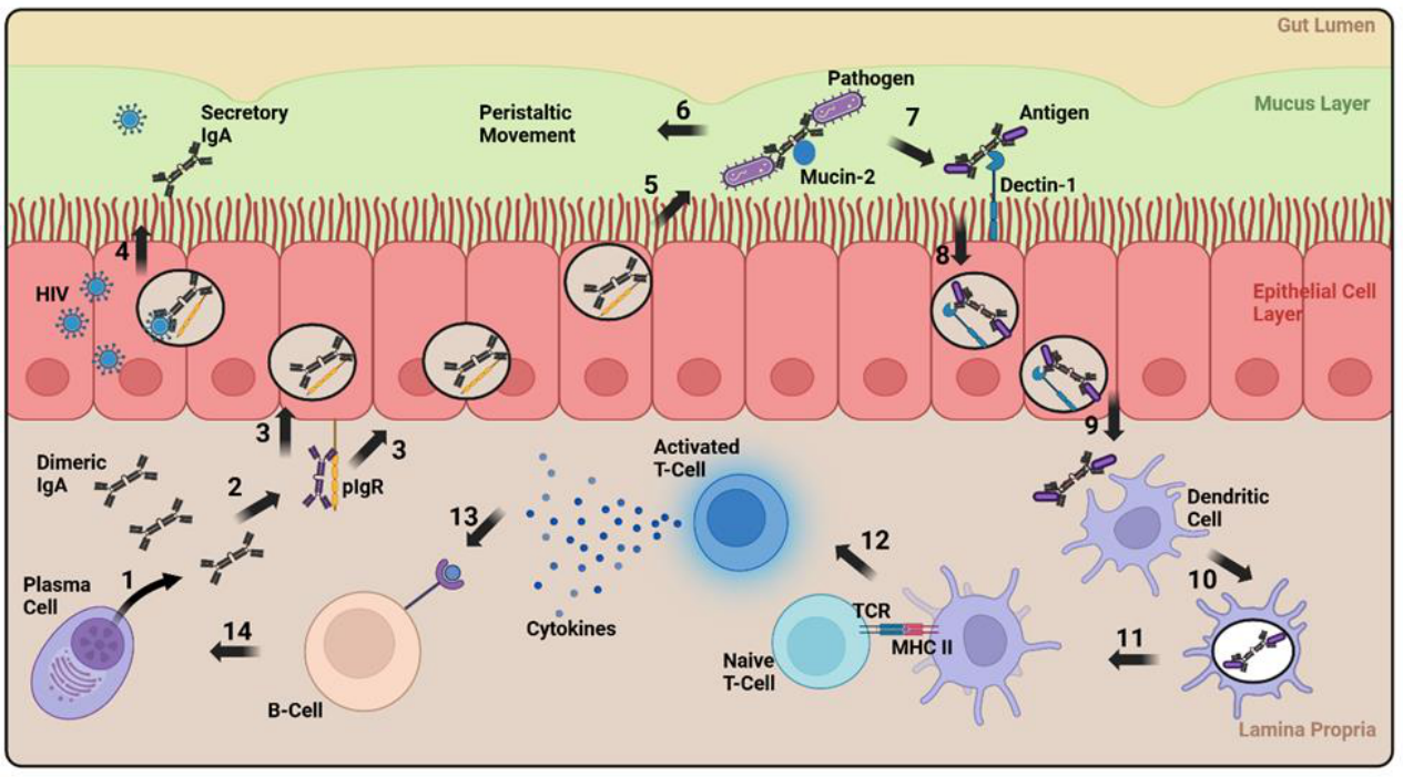
Figure 2.
Overview of dual and single AAV vector systems and mechanisms of action. (a) Potential components of IgA expressing AAV vector genomes for a single and dual vector system. (b) Mechanism of action of single and dual AAV vector systems and expected expression levels. (1) IgA and J-chain expressed from one episome in a single vector system and two episomes in a dual vector system. (2) The dIgA forms in a vesicle which fuses with a pIgR undergoing transcytosis to the basolateral side of a mucosal epithelial cell. (3) The dIgA binds to pIgR. (4) The pIgR is cleaved releasing SIgA at mucosal surfaces with expression predicted to be higher in a dual vector system due to lower ribosome drop-off. Created with BioRender.com.
Figure 2.
Overview of dual and single AAV vector systems and mechanisms of action. (a) Potential components of IgA expressing AAV vector genomes for a single and dual vector system. (b) Mechanism of action of single and dual AAV vector systems and expected expression levels. (1) IgA and J-chain expressed from one episome in a single vector system and two episomes in a dual vector system. (2) The dIgA forms in a vesicle which fuses with a pIgR undergoing transcytosis to the basolateral side of a mucosal epithelial cell. (3) The dIgA binds to pIgR. (4) The pIgR is cleaved releasing SIgA at mucosal surfaces with expression predicted to be higher in a dual vector system due to lower ribosome drop-off. Created with BioRender.com.
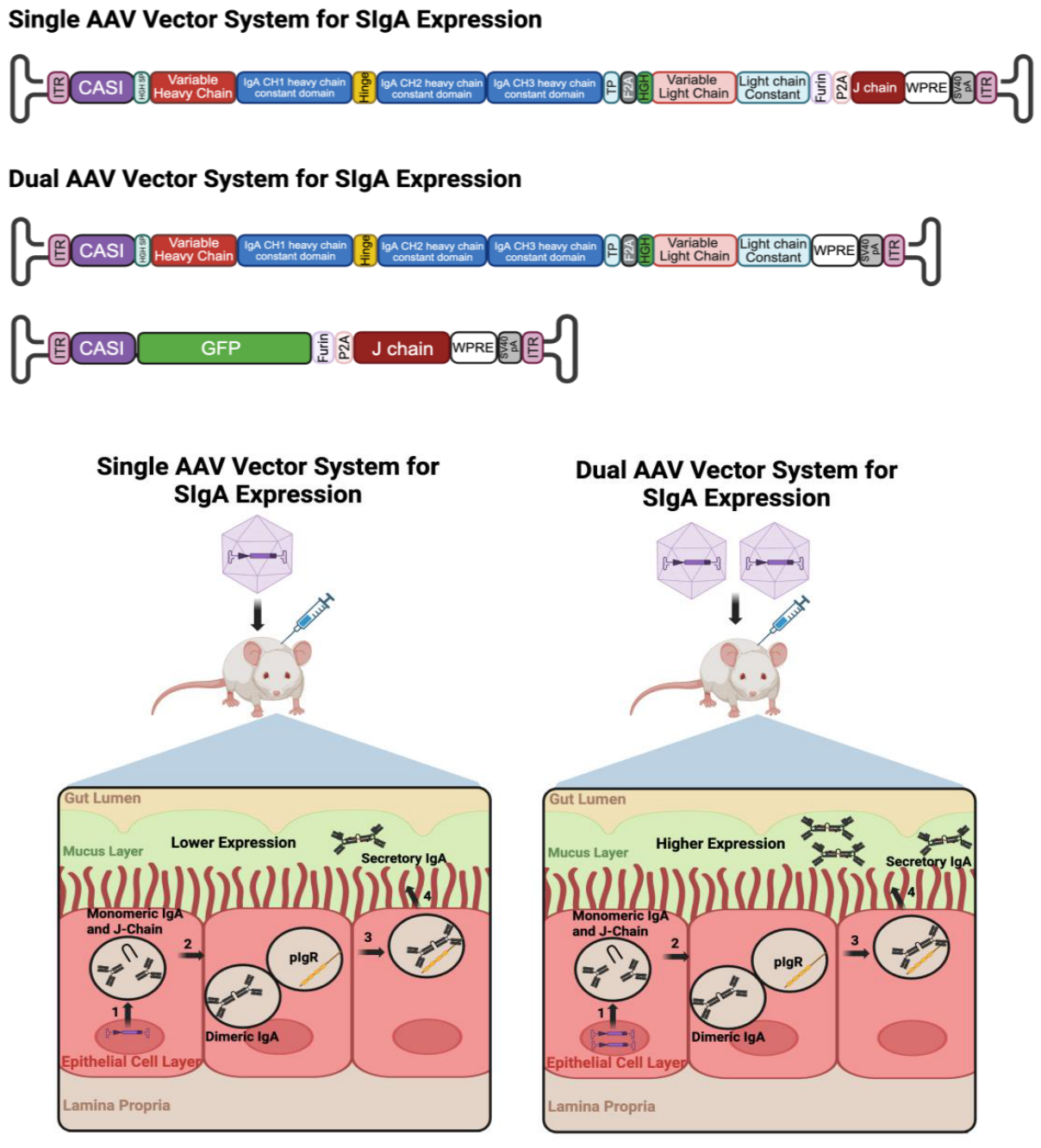
Figure 3.
Simplified schematic of potential workflow required to produce SIgA in pre-clinical animal models, clinical translation of SIgA vectored immunoprophylaxis therapeutics for people with cystic fibrosis, and regulatory approval for commercial use.
Figure 3.
Simplified schematic of potential workflow required to produce SIgA in pre-clinical animal models, clinical translation of SIgA vectored immunoprophylaxis therapeutics for people with cystic fibrosis, and regulatory approval for commercial use.
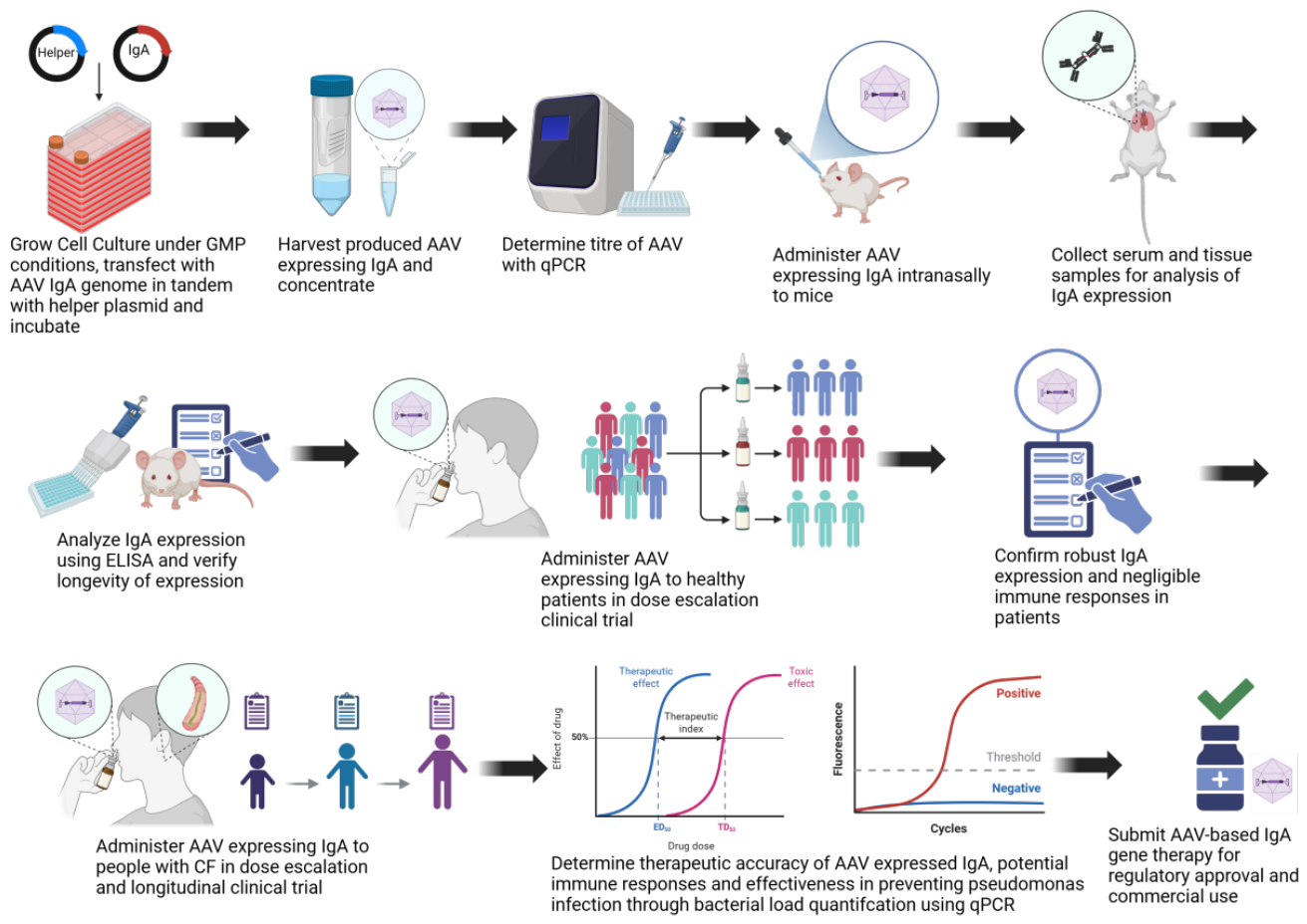

Table 1.
Comparative advantages of SIgA versus IgG at mucosal surfaces.
| Feature | SIgA | IgG | Advantage | Reference |
| Site of production | Produced by plasma cells in the lamina propria, enabling immediate secretion at mucosal surfaces | Primarily produced by plasma cells in the spleen and bone marrow, reaching mucosal surfaces by serum | SIgA can neutralize pathogens before they breach the epithelium | [32] |
| Immune exclusion | Traps pathogens at mucosal surfaces without triggering strong inflammatory responses | Functions after pathogen invasion promoting phagocytosis and inflammation | SIgA neutralizes pathogens while minimizing inflammatory responses which can cause tissue damage | [36,37] |
| Complement activationcF | Limited to the lectin pathway causing minimal activation | Activates classical, alternative and lectin pathways leading to potent inflammation | SIgA reduces excessive complement mediated inflammation thereby preventing collateral tissue damage | [37,38] |
| Interaction with commensal microbes | Prevents overactivation of immune system against commensal microbes | Can induce harmful immune responses when coating commensal microbes | SIgA supports mucosal homeostasis, limiting harmful immune responses | [40,41] |
| Mucosal anchoring | Binds to multiple mucin proteins at mucosal surfaces allowing long-term retention |
Possesses limited ability to bind mucin proteins diffusing more freely at mucosal surfaces | SIgA remains localized at mucosal surfaces for extended periods of time allowing enhanced pathogen neutralization | [42,43] |
| Protease resistance | Resistant to degradation by proteases due to SC | More susceptible to protease degradation | SIgA maintains function in protease rich mucosal environments extending protective effects | [44,45] |
| Pathogen crosslinking | Dimeric structure increases its ability to crosslink multiple pathogens simultaneously | Monomeric structure limits crosslinking activity | SIgA more effectively neutralizes pathogens by immune exclusion using multivalent interactions | [46,47] |
| Antibody dependent enhancement (ADE) | Low risk of ADE when non-neutralizing | High risk of ADE when non-neutralizing | SIgA reduces the risk of ADE, limiting enhanced infection | [48] |
Table 2.
Summary of recent innovations for improving SIgA production and stability.
| Innovation | Engineering Approach | Mechanism of Action | Key Outcome | Reference |
| Stabilizing mutation in IgA heavy chain | P221R mutation introduced into IgA2 heavy chain | Enables disulfide bond formation between heavy and light chain, increasing structural stability | Increased SIgA expression yield and stability | [112] |
| IgA-IgG fusion antibodies | Fusion of IgG1 Fc and IgA2 Fc regions with C242 mutation to delete destabilizing IgA2 hinge | Combines IgA effector functions with IgG serum persistence and FcRn-mediated recycling | Detectable serum IgA levels for a longer period | [113] |
| Enhanced pIgR expression | Overexpression of pIgR in epithelial cells | Increases transcytosis of dIgA across epithelial cells | Increased SIgA expression at mucosal surfaces | [114] |
| Albumin binding domain (ABD) incorporation | Incorporation of a 46 amino acid ABD to IgA antibodies | Enables albumin binding and FcRn-mediated recycling | Reduced clearance of IgA antibodies and prolonged circulation | [115,116] |
| Reduction of IgA glycosylation | Removal of N166 and N337 N-glycosylation sites | Prevents ASGPR-mediated clearance | Increased half-life of IgA without loss of antigen binding efficiency | [117] |
Disclaimer/Publisher’s Note: The statements, opinions and data contained in all publications are solely those of the individual author(s) and contributor(s) and not of MDPI and/or the editor(s). MDPI and/or the editor(s) disclaim responsibility for any injury to people or property resulting from any ideas, methods, instructions or products referred to in the content. |
© 2026 by the authors. Licensee MDPI, Basel, Switzerland. This article is an open access article distributed under the terms and conditions of the Creative Commons Attribution (CC BY) license (http://creativecommons.org/licenses/by/4.0/).
Copyright: This open access article is published under a Creative Commons CC BY 4.0 license, which permit the free download, distribution, and reuse, provided that the author and preprint are cited in any reuse.


