
Submitted:
09 July 2026
Posted:
13 July 2026
You are already at the latest version
Abstract
We characterized the intestinal microbiota of Misgurnus anguillicaudatus using 16S rRNA gene sequencing (V4 region). Strains secreting extracellular digestive enzymes were functionally screened, identified by 16S rRNA sequencing, and evaluated for biosafety. Microbial diversity analysis revealed that the intestinal microbiota of M. anguillicaudatus was dominated by the genera Vibrio, Pseudoalteromonas, and Aeromonas as core predominant taxa. Functional screening yielded four strains. Three non-hemolytic strains secreting amylase, lipase, and protease were identified as Aeromonas hydrophila, Shewanella xiamenensis, and Cedecea neteri, respectively. A. veronii was excluded due to its β-hemolytic activity. Biosafety assessment indicated that all three strains exhibited satisfactory biosafety. The three strains, A. hydrophila, S. xiamenensis, and C. neteri, obtained in this study possess potential probiotic functions in aiding host digestion and promoting nutrient absorption, and provide candidate strains for the development of microecological preparations for aquatic animals.
Keywords:
Misgurnus anguillicaudatus
; intestinal microbiota
; digestive enzymes
; probiotics
1. Introduction
The Chinese loach (Misgurnus anguillicaudatus) , belonging to the family Cobitidae is a freshwater benthic fish species inhabiting the bottom layers of rivers and lakes, with a wide distribution across major river systems in China. Loach meat is highly palatable with a balanced amino acid composition, abundant fatty acid content, and rich in various trace mineral elements and possessing both high nutritional and medicinal value [1]. According to the China Fishery Statistical Yearbook, the national production of loach reached 278,332 tons in 2024. Currently, aquaculture is developing toward intensification and high-density cultivation to increase unit water body yield and economic benefits to meet growing market demands; however, environmental stress and disease prevention and control issues have become increasingly prominent. Probiotics have received extensive attention from academia and industry as a management strategy for improving production performance and health status of aquaculture animals through multiple pathways, including enhancing immune function, promoting digestion, resisting pathogenic bacteria, optimizing water quality, and facilitating growth, development, and reproduction [2]. To date, multiple probiotic species have demonstrated promising application potential in aquaculture, including Lactobacillus, Lactococcus, Leuconostoc, Pseudomonas, Enterococcus, Carnobacterium, Shewanella, Bacillus, Aeromonas, Vibrio, Enterobacter, Clostridium, Pediococcus, Micrococcus, and Saccharomyces [3−8].
In this study, functional strains capable of secreting protease, amylase, and lipase were isolated and screened from intestinal contents of wild loach (M. anguillicaudatus). Four strains with distinct extracellular enzyme secretion characteristics were obtained and identified as Aeromonas veronii, Aeromonas hydrophila, Shewanella xiamenensis, and Cedecea neteri, respectively. Antimicrobial susceptibility testing, hemolytic assays, and safety evaluation demonstrated that A. hydrophila , S. xiamenensis , and C. neteri exhibited no pathogenicity to the host, whereas A. veronii showed β-hemolytic activity and was excluded from further safety assessment. The three strains, A. hydrophila , S. xiamenensis , and C. neteri, are promising candidates for the development of microecological preparations for loach. This study provides candidate strains for the development and application of loach probiotic formulations, and offers theoretical basis and technical support for intensive healthy aquaculture of loach..
2. Materials and Methods
2.1. Experimental Materials
Healthy loach (M. anguillicaudatus) were purchased from the Daxing’anling region, Heilongjiang Province, without acclimation to artificial feed. A total of 30 individuals were used, with a body weight of 15.02±2.01 g and a total length of 15.97±1.32 cm..
2.2. Experimental Reagents and Culture Media
Gentamicin and penicillin were purchased from North China Pharmaceutical Group Corporation; oxytetracycline and florfenicol were purchased from Huamukang Animal Pharmaceutical Co., Ltd.; ampicillin and kanamycin were purchased from Brazilian Institute of Biotechnology and Innovation; Columbia blood agar was purchased from Guangdong Huankai Microbial Sci. & Tech. Co., Ltd.; Lugol’s iodine solution was purchased from Lanbao Medical & Health Products Co., Ltd.; casein and soluble starch were purchased from Shanghai Sangon Biological Engineering Technology & Services Co., Ltd.
Beef extract peptone medium: beef extract 5 g·L−1, peptone 10 g·L−1, NaCl 5 g·L−1, agar 16 g·L−1, pH 7.2–7.4; Protein medium: Solution A: peptone 13.33 g·L−1, yeast extract 4 g·L−1, NaCl 6.67 g·L−1, agar 21.33 g·L−1; Solution B: casein 40 g·L−1; Starch medium: beef extract 3 g·L−1, peptone 10 g·L−1, starch 10 g·L−1, NaCl 5 g·L−1, agar 16 g·L−1, pH 7.2–7.4; Lipid medium: peptone 10 g·L−1, yeast extract 5 g·L−1, CaCl2·H2O 0.1 g·L−1, Tween-80 10 mL·L−1, agar 16 g·L−1, pH 7.2–7.4.
2.3. Experimental Methods
2.3.1. Sample Collection
Healthy loach were selected and euthanized by anesthesia with MS-222 (100 mg·L−1), then placed in clean bench. The body surface was disinfected with 75% ethanol, and the abdominal cavity was opened with sterile dissecting scissors to remove the intestine into a sterile petri dish. After rinsing the intestine with sterile physiological saline (0.85%), the intestinal contents were collected into sterile cryovials, snap-frozen in liquid nitrogen, and stored at –80 °C. Intestinal contents from six loach were pooled as one sample, with five replicates established.
2.3.2. Analysis of Intestinal Content Microbiota Characteristics
Genomic DNA was extracted from loach intestinal contents. The V4 region of 16S rRNA was amplified using specific primers 515F (5’-GTGCCAGCMGCCGCGGTAA-3’) and 806R (5’-GGACTACHVGGGTWTCTAAT-3’). PCR products were purified, pooled in equimolar amounts, ligated with sequencing adapters to construct sequencing libraries, and sequenced on the Illumina NovaSeq 6000 platform.
The raw sequencing data were assembled and filtered to obtain high-quality clean tags. Tags were aligned against the Silva database (https://www.arb-silva.de/for 16S) to generate effective tags. The DADA2 module in QIIME2 (version QIIME2-202202) was used to obtain amplicon sequence variants (ASVs), and QIIME2 was employed for species annotation (Silva 138.1 database). Species abundance tables at phylum, class, order, family, genus, and species levels were generated based on ASV annotation results and sample feature tables. Tax4Fun was used to analyze metabolic activities of the microbiota, and functional prediction of loach intestinal content microbiota was performed according to the Kyoto Encyclopedia of Genes and Genomes (KEGG) database.
2.3.3. Isolation and Purification of Strains
Under aseptic conditions, 0.1 g of intestinal contents was transferred into a sterile centrifuge tube containing 5 mL of pre-cooled sterile physiological saline (pH 7.2), and thoroughly vortexed to prepare the stock solution. The stock solution was serially diluted to 10−3 CFU·mL−1, 10−4 CFU·mL−1, and 10−5 CFU·mL−1. Aliquots of 0.1 mL from each dilution (10−3 CFU·mL−1, 10−4 CFU·mL−1, and 10−5 CFU·mL−1) were inoculated onto beef extract peptone medium. Three replicates were established for each dilution, and incubated at 28 °C for 24–48 h. Single colonies were selected and streaked on plates for three consecutive generations to achieve purification.
2.3.4. Isolation and Identification of Enzyme-Producing Strains
Purified strains were suspended in 200 μL sterile physiological saline to prepare stock solutions. The stock solutions were serially diluted to 10−3 CFU·mL−1, 10−4 CFU·mL−1, and 10−5 CFU·mL−1, and 0.1 mL aliquots were spread onto protease medium, amylase medium, and lipase medium, respectively. Three replicates were established for each dilution and incubated at 28 °C for 24–48 h. Hydrolysis capacity (HC), defined as the ratio of hydrolysis zone diameter (Dh) to colony diameter (Dc) (HC = Dh/Dc), was used to indicate the organic matter decomposition ability of strains. Lipid and protein hydrolysis abilities were determined by direct observation of transparent hydrolysis zones surrounding colonies. Starch hydrolysis ability was assessed by adding appropriate amount of Lugol’s iodine solution onto the medium surface, standing for 1–2 min, discarding the iodine solution, and immediate observation.
2.3.5. Species Identification of Enzyme-Producing Strains
Genomic DNA of enzyme-producing strains was extracted using the Bacterial Genomic DNA Extraction Kit (Sangon Biotech B518255). The 16S rRNA gene was amplified using universal primers 27F (5’-AGAGTTTGATCMTGGCTCAG-3’) and 1492R (5’-TACGGYTACCTTGTTACGACTT-3’). PCR products with correct band sizes were sequenced by Shanghai Sangon Biological Engineering Technology & Services Co., Ltd. The obtained sequences were subjected to Blast analysis on NCBI, and phylogenetic trees were constructed using MEGA 11 software. Species identification was determined based on a gene sequence similarity criterion of ≥99%.
2.3.6. Antimicrobial Resistance Characteristics of Enzyme-Producing Strains
Six aquaculture commonly used antibiotics (oxytetracycline hydrochloride, gentamicin, penicillin, florfenicol, ampicillin, and kanamycin) were selected for antimicrobial susceptibility testing by the disk diffusion method [7]. Purified enzyme-producing strains were inoculated into LB liquid medium and cultured at 28 °C, 220 r/min to logarithmic growth phase (OD600 = 1). One milliliter of bacterial suspension was centrifuged at 4 °C, 8000 r/min for 5 min, the supernatant was discarded, and the pellet was washed three times with sterile physiological saline and resuspended to adjust the bacterial concentration to OD600 = 0.1. One hundred microliters of bacterial suspension was evenly spread onto LB solid medium and stood at room temperature for 5 min. Antimicrobial disks impregnated with oxytetracycline hydrochloride (concentration per disk), gentamicin (10 µg/disk), penicillin (10 µg/disk), florfenicol (30 µg/disk), ampicillin (10 µg/disk), and kanamycin (30 µg/disk) were placed on the medium surface inoculated with enzyme-producing strains, with 4–6 disks per plate, spacing ≥24 mm, and distance from edge ≥15 mm. After incubation at 28 °C for 10 h, the inhibition zone diameter (mm, including disk diameter) was measured and observed. Results were classified as susceptible (S), intermediate (I), or resistant (R) according to standards established by CLSI (Clinical and Laboratory Standards Institute) and EUCAST (European Committee on Antimicrobial Susceptibility Testing), combined with bacterial species, antimicrobial agent type, culture medium, and test conditions (e.g., disk diffusion method).
2.3.7. Hemolytic Assay of Enzyme-Producing Strains
Enzyme-producing strains were inoculated into LB liquid medium and cultured at 28 °C, 220 r·min−1 to logarithmic growth phase, then streaked onto Columbia blood agar medium and incubated at 28 °C in inverted position for 24–48 h to observe whether a transparent hemolytic zone appeared around the colonies.
2.3.8. Safety Assessment of Enzyme-Producing Strains
Enzyme-producing strains were inoculated into 400 μL liquid medium and cultured at 28 °C, 220 r/min to logarithmic growth phase (OD600=1). One milliliter of bacterial suspension was centrifuged at 4 °C, 8000 r/min for 5 min, the supernatant was discarded, and the pellet was washed three times with sterile physiological saline and resuspended to adjust the bacterial concentration to 106 CFU·mL−1, 107 CFU·mL−1, and 108 CFU·mL−1.
Healthy loach were randomly divided into four groups (three experimental groups and one control group), with 30 individuals per group and three replicates (10 individuals per replicate). Intraperitoneal injection was performed with 0.1 mL bacterial suspension per individual (control group injected with 0.1 mL sterile physiological saline), at inoculation doses of 105, 106, and 107 CFU per individual. The fish were reared for 14 d, and mortality was recorded daily to evaluate the safety of potential probiotic strains to the host.
3. Results
3.1. Analysis of Loach Intestinal Community Structure
As shown in Figure 1, the number of bacterial genera in loach intestinal contents (305) was higher than that in loach intestinal wall (293). Vibrio (21.87%), Pseudoalteromonas (18.07%), and Aeromonas (14.43%) were the core dominant genera in the loach intestinal microbiota, representing the major constituent groups. Similar to the intestinal microbiota, Vibrio (22.4%), Pseudoalteromonas (17.13%), Aeromonas (14.11%), and Bacillus (9.86%) exhibited relatively high abundances in the intestinal content microbiota, serving as the core dominant genera. In loach intestinal contents, the relative abundances of Aeromonas, Shewanella, and Cedecea were 14.11%, 4.09%, and 0.3%, respectively.
3.2. Prediction of Enzyme Functions in Loach Intestinal Microbiota
As shown in Figure 2, compared with the intestinal microbiota, the level 3 metabolic functions of enzyme families in the intestinal content microbiota were significantly upregulated in protein, fatty acid, and carbohydrate synthesis and degradation. Enzyme families associated with protein degradation, modification, synthesis, and amino acid metabolism included Peptidases, Proteasome, Amino acid related enzymes, Peptidoglycan biosynthesis and degradation, Protein kinases, Protein phosphatase and associated proteins, Glutathione metabolism, Nitrogen metabolism, Sulfur metabolism, Aminoacyl-tRNA biosynthesis, and One carbon pool by folate. Those associated with polysaccharide degradation, sugar transport, glycosylation, and sugar nucleotide metabolism included Phosphotransferase system (PTS), Glycosyltransferases, Glycosaminoglycan degradation, Other glycan degradation, and Starch and sucrose metabolism. Those associated with fatty acid, glycerophospholipid, sphingolipid, and isoprenoid metabolism included Lipid biosynthesis proteins, Fatty acid degradation, Glycerophospholipid metabolism, Glycerolipid metabolism, Sphingolipid metabolism, Primary bile acid biosynthesis, and Prenyltransferases.
3.3. Isolation and Identification of Enzyme-Producing Strains
Enzyme-producing capabilities were analyzed for strains isolated from the intestine of healthy wild loach, and their abilities to secrete amylase, lipase, and protease were determined (Table 1). Following 16S rRNA identification, A. veronii and A. hydrophila were identified as amylase-producing strains with Dh/Dc values of 1.70 and 1.43, respectively; A. hydrophila and S. xiamenensis as protease-producing strains with Dh/Dc values of 2.82 and 2.46, respectively; and C. neteri as a lipase-producing strain with a Dh/Dc value of 3.0. To verify whether the isolated strains possessed the ability to decompose the other two nutrient types, strains were inoculated onto the other two media. Cross-decomposition testing revealed that A. veronii and A. hydrophila could simultaneously decompose starch and protein, S. xiamenensis could simultaneously decompose protein and lipid, while C. neteri showed no cross-decomposition capability and only produced lipase.
3.4. Antimicrobial Susceptibility Testing
As shown in Table 2, among Aeromonas strains, A. veronii was susceptible to oxytetracycline, florfenicol, ampicillin, and kanamycin, whereas A. hydrophila was susceptible to oxytetracycline, ampicillin, and kanamycin, but intermediate to florfenicol. S. xiamenensis was susceptible to oxytetracycline, ampicillin, and kanamycin, and intermediate to florfenicol. C. neteri was susceptible to oxytetracycline, ampicillin, and kanamycin, but resistant to florfenicol.
3.5. Hemolytic Assay
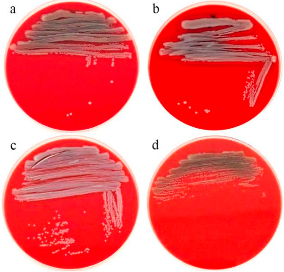
Hemolytic assay results showed that A. veronii produced a clear and transparent hemolytic ring on Columbia blood agar, which exhibited typical β-hemolytic characteristics, indicating that this strain could produce hemolysin and lyse erythrocytes, possessing strong hemolytic activity and suggesting certain virulence potential. In contrast, A. hydrophila , S. xiamenensis, and C. neteri showed no obvious hemolytic phenomenon under the same culture conditions, with no hemolytic ring formation around colonies, and were considered non-hemolytic, indicating that these three strains had low expression levels of hemolysis-related virulence factors and relatively favorable biosafety.
Figure 3.
Hemolytic assay results of the strains. a. Hemolytic assay result of A. veronii; b. Hemolytic assay result of A. hydrophila; c. Hemolytic assay result of C. neterid; d. Hemolytic assay result of S. xiamenensis;.
Figure 3.
Hemolytic assay results of the strains. a. Hemolytic assay result of A. veronii; b. Hemolytic assay result of A. hydrophila; c. Hemolytic assay result of C. neterid; d. Hemolytic assay result of S. xiamenensis;.
3.6. Safety Assessment
Healthy loach were selected; the experimental groups were injected intraperitoneally with 0.1 mL live bacterial suspensions (106 CFU·mL−1, 107 CFU·mL−1, and 108 CFU·mL−1) of A. hydrophila, S. xiamenensis, and C. neteri, respectively, and the control group was injected with an equal volume of physiological saline. After 14 d of rearing, results showed that loach in both experimental and control groups remained in good condition, with no mortality or lesions observed during the experimental period, indicating that A. hydrophila, S. xiamenensis, and C. neteri exhibited favorable safety to juvenile loach. A. veronii was not subjected to safety assessment due to its demonstrated hemolytic toxicity.
4. Discussion
Extracellular digestive enzymes synthesized by microorganisms in the digestive tract are considered key factors assisting host substance metabolism, significantly enhancing host digestive efficiency and growth performance [8−10]. The animal digestive tract represents a potential source of enzyme-producing microorganisms, and digestive tract-derived microbes exhibit stronger niche adaptability and colonization capacity within the host intestinal environment. Consequently, screening potential probiotics with extracellular enzyme secretion capabilities from the intestines of healthy animals has become a common strategy for probiotic acquisition [11]. Previous studies have demonstrated that digestive tract-derived microorganisms can produce amylase, protease, and lipase [12−14], These three enzyme categories represent key functional enzymes in fish digestive physiology, playing central roles in the decomposition and metabolic transformation of carbohydrates, proteins, and lipids in feed [12,13,15]. The exploitation and utilization of digestive enzyme-producing microorganisms from the digestive tracts of healthy animals provide an alternative solution for optimizing feed costs and enhancing aquaculture production [16].
In this study, four bacterial strains with extracellular enzyme secretion capabilities were isolated from loach intestine and identified as A. veronii, A. hydrophila, S. xiamenensis, and C. neteri. Aeromonas belongs to the family Aeromonadaceae , capable of secreting multiple enzyme types [17], its pathogenicity is closely associated with extracellular products [18]. Zeng et al. [19] investigated Aeromonas in the intestine of healthy grass carp (Ctenopharyngodon idella ), isolating 76 Aeromonas strains, of which 50 secreted amylase and 49 secreted protease; all strains exhibited non-pathogenicity upon hemolysin detection. In this study, two Aeromonas strains secreting extracellular enzymes were isolated from loach intestine, namely A. veronii and A. hydrophila. Among them, A. hydrophila possessed the ability to secrete amylase and protease, and exhibited biosafety. Consistent with the results of this study, Zhu Xiaoyan et al. [20] isolated 19 A. hydrophila strains from the intestines of common carp (Cyprinus carpio ), grass carp (Ctenopharyngodon idella ), silver carp (Hypophthalmichthys molitrix ), and blunt snout bream (Megalobrama amblycephala ). These strains did not produce hemolysin but could secrete extracellular enzymes, promoting fish growth. Aeromonas hydrophila is considered an opportunistic pathogen of aquatic animals; however, it exhibits no pathogenicity to the host when intestinal microecological balance is maintained, can assist host digestion, improve feed utilization efficiency, and exerts positive effects on aquatic animal growth [21]. In this study, A. veronii exhibited typical β-hemolytic phenotype on blood agar, demonstrating definitive cytotoxicity. Previous studies have demonstrated that A. veronii represents an important opportunistic pathogen in various aquatic animals [22,23]. Li et al. [24] isolated 87 A. veronii strains from the intestines of healthy crucian carp (Carassius auratus ), common carp (Cyprinus carpio ), grass carp (Ctenopharyngodon idella ), and catfish (Silurus asotus ). The study found that nearly 50% of A. veronii carried at least four or more virulence genes, 25% carried at least five virulence genes, and the number of virulence genes carried was positively correlated with strain virulence.
Shewanella constitutes a component of the gastrointestinal microbiota in naturally cultured fish [28]. As fish probiotics, Shewanella exerts positive effects in promoting growth [27,28,29,30,31] , optimizing intestinal microbiota [32,33], regulating immunity [27,30,31,33,34,35], and enhancing stress resistance [28]. Previous studies have demonstrated that Shewanella can secrete protease [36,37] and lipase [38,39,40]. This study further confirmed that S. xiamenensis possesses lipase and protease secretion activities and exhibits biosafety. Meanwhile, research by Wang et al. [33,35] demonstrated that S. xiamenensis can effectively improve the intestinal microbial community structure of grass carp, reduce the relative abundance of potential pathogens (such as Pseudomonas and Flavobacterium ), and promote the proliferation of potential probiotics (such as Vibrio , Streptococcus , and Enterococcus ). Additionally, S. xiamenensis can enhance innate immunity and disease resistance in grass carp, representing an intestinal-derived candidate probiotic with dual functions of microbiota regulation and immune modulation.
Cedecea neteri belongs to the family Enterobacteriaceae and genus Cedecea , and positive lipase activity represents one of the typical biochemical characteristics of this genus [25]. In this study, one C. neteri strain was screened from loach intestinal contents, which exhibited lipase secretion capability. Antimicrobial susceptibility testing, hemolytic assay, and host safety assessment confirmed that this strain was safe to loach host and devoid of pathogenic effects.
5. Conclusions
In this study, four bacterial strains with extracellular enzyme secretion capabilities were initially isolated from loach intestine, but one (A. veronii ) was excluded due to β-hemolytic activity. The remaining three strains, identified as A. hydrophila , S. xiamenensis , and C. neteri , were preliminarily confirmed as safe candidate probiotics with amylase, protease, and lipase activities. These strains possess potential probiotic functions in assisting host digestion and promoting nutrient absorption, indicating their potential for development and application as probiotics for fish and other aquatic animals. However, further systematic evaluation through feeding experiments is required to verify probiotic efficacy, including growth promotion effects, disease resistance, long-term colonization capacity, and optimal formulation as single or composite agents.
Author Contributions
Sui Zhaoxin and Shu Mingshuai: investigation, data curation, writing – original draft, visualization, and formal analysis. Zhang Ruining, Yu Jianing, Dai Zhuo and Sun Yi: methodology and investigation. Jun Li: conceptualization, methodology, validation, investigation, writing – review and editing, and project administration.
Funding
This study was funded by the Key Laboratory of Loach Genetics and Breeding of Jiangsu Province, Soochow University (KJS22082), and the Study on Laboratory Animalization of Lampetra reissneri (JYTMS20231060).
Institutional Review Board Statement
The animal study was reviewed and approved by the Animal Ethics Committee of Liaoning Normal University (Dalian, China), which approved all experimental protocols used in this study. All animal procedures follow the Guidelines for Ethical Treatment of Experimental Animals prepared by the Ministry of Science and Technology of China.
Informed Consent Statement
.
Data Availability Statement
.
Acknowledgments
.
Conflicts of Interest
The authors declared that they have no conflict of interest.
References
- Wang, Z. R.; Li, Y. A.; Zhou, Q. B.; et al. Analysis of nutritional components in different parts of Paramisgurnus dabryanus and Misgurnus anguillicaudatus from Poyang Lake area [J]. Feed Ind. 2022, 43(02), 56–64. [Google Scholar]
- SAYES, C.; LEYTON, Y.; RIQUELME, C. Probiotic Bacteria as an Healthy Alternative for Fish Aquaculture [M]//SAVIĆ S. Antibiotic Use in Animals; IntechOpen: London, 2017. [Google Scholar]
- LIAO, Z.-H.; IBARRA-ARANA, M. J.; CHEN, J.-C.; et al. The effects of dietary supplement of Ulva fasciata, fermented soybean and probiotics on the growth of Apostichopus japonicus [J]. Aquac. Nutr. 2021, 27, 1363–9. [Google Scholar] [CrossRef]
- LIU, L.; LIU, Y.; QIN, G.; et al. Evaluation of regulatory capacity of three lactic acid bacteria on the growth performance, non-specific immunity, and intestinal microbiota of the sea cucumber Apostichopus japonicus [J]. Aquaculture 2024, 579, 740156. [Google Scholar]
- NAYAK, S. K. Role of gastrointestinal microbiota in fish [J]. Aquac. Res. 2010, 41, 1553–73. [Google Scholar] [CrossRef]
- WANG, G.; CHEN, Y.; XIA, Y.; et al. Characteristics of Probiotic Preparations and Their Applications [J]. Foods 2022, 11, 2472. [Google Scholar] [CrossRef] [PubMed]
- WANG, K. Y.; CHEN, D. F.; HUANG, L. Y.; et al. Isolation and characterization of Streptococcus agalactiae from Nile Tilapia Oreochromis niloticus in China [J]. Afr. J. Microbiol. Res. 2013, 7, 317–23. [Google Scholar] [CrossRef]
- SIMORA C D A, C. R. M. Isolation and Identification of Protease-Producing Pseudomonas sp. PD14 in the Gut of Rabbitfish Siganus guttatus (Bloch 1787) [J]. Asian Fish. Sci. 2016, 29, 82–95. [Google Scholar]
- MAKLED, S. O.; HAMDAN, A. M.; EL-SAYED, A. M. Growth Promotion and Immune Stimulation in Nile Tilapia, Oreochromis niloticus, Fingerlings Following Dietary Administration of a Novel Marine Probiotic, Psychrobacter maritimus S [J]. Probiotics Antimicrob. Proteins 2020, 12, 365–74. [Google Scholar] [PubMed]
- HUDDY, R. J.; COYNE, V. E. Characterisation of the role of an alkaline protease from Vibrio midae SY9 in enhancing the growth rate of cultured abalone fed a probiotic-supplemented feed [J]. Aquaculture 2015, 448, 128–34. [Google Scholar] [CrossRef]
- ZUO, Z.-H.; SHANG, B.-J.; SHAO, Y.-C.; et al. Screening of intestinal probiotics and the effects of feeding probiotics on the growth, immune, digestive enzyme activity and intestinal flora of Litopenaeus vannamei [J]. Fish. Shellfish Immunol. 2019, 86, 160–8. [Google Scholar] [CrossRef] [PubMed]
- FITRIA, A.; RUSMANA, I.; HASTUTI, Y. Isolation and characterization of Bacillus sp. producing amylase, protease, and lipase from the digestive tract of tambaqui (Colossoma macropomum) as potential probiotic bacteria. proceedings of the IOP Conference Series: Earth and Environmental Science, F, 2025 [C]; IOP Publishing. [Google Scholar]
- HORTILLOSA, E. M.; AMAR, M. J. A.; NUñAL, S. N.; et al. Effects of putative dietary probiotics from the gut of milkfish (Chanos chanos) on the growth performance and intestinal enzymatic activities of juvenile Nile tilapia (Oreochromis niloticus) [J]. Aquac. Res. 2022, 53, 98–108. [Google Scholar]
- DUTTA, D.; BANERJEE, S.; MUKHERJEE, A.; GHOSH, K. Potential gut adherent probiotic bacteria isolated from rohu, Labeo rohita (Actinopterygii: Cypriniformes: Cyprinidae): Characterisation, exo-enzyme production, pathogen inhibition, cell surface hydrophobicity, and bio-film formation [J]. Acta Ichthyol. Et. Piscat. 2018. [Google Scholar] [CrossRef]
- RUST, M. B.; Nutritional physiology [M]; HALVER, J. E.; HARDY, R. W. Fish Nutrition; Academic Press: San Diego, 2002. [Google Scholar]
- WANG, Q.; QI, Z.; FU, W.; et al. Research and Prospects of Enzymatic Hydrolysis and Microbial Fermentation Technologies in Protein Raw Materials for Aquatic Feed [J]. Fermentation 2024, 10, 648. [Google Scholar] [CrossRef]
- PEMBERTON, J. M.; KIDD, S. P.; SCHMIDT, R. Secreted enzymes of Aeromonas [J]. FEMS Microbiol. Lett. 1997, 152, 1–10. [Google Scholar] [CrossRef] [PubMed]
- Liu, M. Z.; Ye, X.; Tian, Y. Y.; et al. Cloning and structural prediction of aerolysin and hemolysin genes from Aeromonas hydrophila [J]. Acta Agric. Univ. Jiangxiensis 2010, 32(02), 356–362. [Google Scholar]
- Zeng, Y.; Yuan, Y. Seasonal variation of Aeromonas and investigation of extracellular enzymes in the intestine of healthy grass carp [J]. J. Fish. Sci. China 1999, (02), 80–82. [Google Scholar]
- Zhu, X. Y.; Tang, F. S. Aeromonas hydrophila and its extracellular enzyme distribution in intestinal bacteria of healthy domestic fish [J]. J. Hubei Agric. Coll. 1994, (01), 35–39. [Google Scholar]
- Luo, Z. Screening and characterization of intestinal probiotics from channel catfish (Ictalurus punctatus) [D]; Huazhong Agricultural University, 2007. [Google Scholar]
- ZHU, X.; QIAN, Q.; WU, C.; et al. Pathogenicity of Aeromonas veronii Causing Mass Mortality of Largemouth Bass (Micropterus salmoides ) and Its Induced Host Immune Response [J]. Microorganisms 2022, 10, 2198. [Google Scholar] [CrossRef] [PubMed]
- YOUSSEF, H. A.; AYOUB, H. F.; SOROR, E. I.; et al. Virulence genes contributing to Aeromonas veronii pathogenicity in Nile tilapia (Oreochromis niloticus ): approaching the development of live and inactivated vaccines [J]. Aquac. Int. 2023, 31, 1253–1267. [Google Scholar] [PubMed]
- LI, T.; RAZA, S. H. A.; YANG, B.; et al. Aeromonas veronii Infection in Commercial Freshwater Fish: A Potential Threat to Public Health [J]. Anim. An. Open Access J. From MDPI 2020, 10(4). [Google Scholar] [CrossRef]
- MARTIN-ANTONIO, B.; MANCHADO, M.; INFANTE, C.; et al. Intestinal microbiota variation in Senegalese sole (Solea senegalensis) under different feeding regimes [J]. Aquac. Res. 2007, 38, 1213–22. [Google Scholar] [CrossRef]
- TAPIA-PANIAGUA, S.; LOBO, C.; GARCıA DE LA BANDA, I.; et al. Impact of the intestinal microbiota species of Senegalese sole larvae and fries fed with Shewanella putrefaciens Pdp11 probiotic bioencapsulated in Artemia, effect of one or two intervals of application [J]. CD Abstr. Aquac. Europe2011 2011, 622–3. [Google Scholar]
- VARELA, J. L.; RUIZ-JARABO, I.; VARGAS-CHACOFF, L.; et al. Dietary administration of probiotic Pdp11 promotes growth and improves stress tolerance to high stocking density in gilthead seabream Sparus auratus [J]. Aquaculture 2010, 309(1-4), 265–271. [Google Scholar] [CrossRef]
- SÁENZ DE RODRÍGUEZ, M. A.; DÍAZ-ROSALES, P.; CHABRILLÓN, M.; et al. Effect of dietary administration of probiotics on growth and intestine functionality of juvenile Senegalese sole (Solea senegalensis, Kaup 1858) [J]. Aquac. Nutr. 2009, 15, 177–185. [Google Scholar] [CrossRef]
- GARCÍA DE LA BANDA, I.; LOBO, C.; LEÓN-RUBIO, J. M.; et al. Influence of two closely related probiotics on juvenile Senegalese sole (Solea senegalensis, Kaup 1858) performance and protection against Photobacterium damselae subsp. piscicida [J]. Aquaculture 2010, 306, 281–288. [Google Scholar] [CrossRef]
- GARCÍA DE LA BANDA, I.; LOBO, C.; CHABRILLÓN, M.; et al. Influence of dietary administration of a probiotic strain Shewanella putrefaciens on Senegalese sole (Solea senegalensis, Kaup 1858) growth, body composition and resistance to Photobacterium damselae subsp. piscicida [J]. Aquac. Res. 2012, 43, 662–669. [Google Scholar]
- TAPIA-PANIAGUA, S. T.; CHABRILLÓN, M.; DÍAZ-ROSALES, P.; et al. Intestinal microbiota diversity of the flat fish Solea senegalensis (Kaup, 1858) following probiotic administration [J]. Microb. Ecol. 2010, 60, 310–319. [Google Scholar] [CrossRef] [PubMed]
- HAO, K.; WU, Z. Q.; LI, D. L.; et al. Effects of Dietary Administration of Shewanella xiamenensis A-1, Aeromonas veronii A-7, and Bacillus subtilis, Single or Combined, on the Grass Carp (Ctenopharyngodon idella ) Intestinal Microbiota [J]. Probiotics Antimicrob. Proteins 2017, 9, 386–396. [Google Scholar] [CrossRef] [PubMed]
- SALINAS, I.; DÍAZ-ROSALES, P.; CUESTA, A.; et al. Effect of heat-inactivated fish and non-fish derived probiotics on the innate immune parameters of a teleost fish (Sparus aurata L.) [J]. Vet. Immunol. Immunopathol. 2006, 111(3-4), 279–286. [Google Scholar] [CrossRef] [PubMed]
- WU, Z. Q.; JIANG, C.; LING, F.; et al. Effects of dietary supplementation of intestinal autochthonous bacteria on the innate immunity and disease resistance of grass carp (Ctenopharyngodon idellus ) [J]. Aquaculture 2015, 438, 105–114. [Google Scholar] [CrossRef]
- YANG, X. H.; SONG, J. Y.; LI, K.; et al. The complete genome sequence of proteases-producing Shewanella sp. H8 isolated from Antarctica [J]. Mar. Genom. 2024, 78, 101147. [Google Scholar] [CrossRef]
- ZHUGE, B.; XU, Q.; DU, G. C.; et al. Characteristics and application of degradation enzymes produced by a cold-tolerant bacterium [J]. Chin. J. Appl. Environ. Biol. 2010, 16, 870–874. [Google Scholar]
- JOSEPH, D.; CHAKRABORTY, K. Production and Biotechnological Application of Extracellular Alkalophilic Lipase from Marine Macroalga-Associated Shewanella algae to Produce Enriched C(20-22) n-3 Polyunsaturated Fatty Acid Concentrate [J]. Appl. Biochem. Biotechnol. 2018, 185, 55–71. [Google Scholar] [PubMed]
- YANG, S. H.; LEE, J. H.; RYU, J. S.; et al. Shewanella donghaensis sp. nov., a psychrophilic, piezosensitive bacterium producing high levels of polyunsaturated fatty acid, isolated from deep-sea sediments [J]. Int. J. Syst. Evol. Microbiol. 2007, 57 Pt 2, 208–212. [Google Scholar] [CrossRef] [PubMed]
- SATOMI, M.; OIKAWA, H.; YANO, Y. Shewanella marinintestina sp. nov., Shewanella schlegeliana sp. nov. and Shewanella sairae sp. nov., novel eicosapentaenoic-acid-producing marine bacteria isolated from sea-animal intestines [J]. Int. J. Syst. Evol. Microbiol. 2003, 53, 491–499. [Google Scholar] [CrossRef] [PubMed]
- CHAN, K. G.; TAN, W. S. Insights into Cedecea neteri strain M006 through complete genome sequence, a rare bacterium from aquatic environment [J]. Stand. Genom. Sci. 2017, 12, 40. [Google Scholar] [CrossRef]
Figure 1.
Stacked bar chart of relative abundance at the genus level of gut microbiota and gut content microbiota in M. anguillicaudatus.
Figure 1.
Stacked bar chart of relative abundance at the genus level of gut microbiota and gut content microbiota in M. anguillicaudatus.
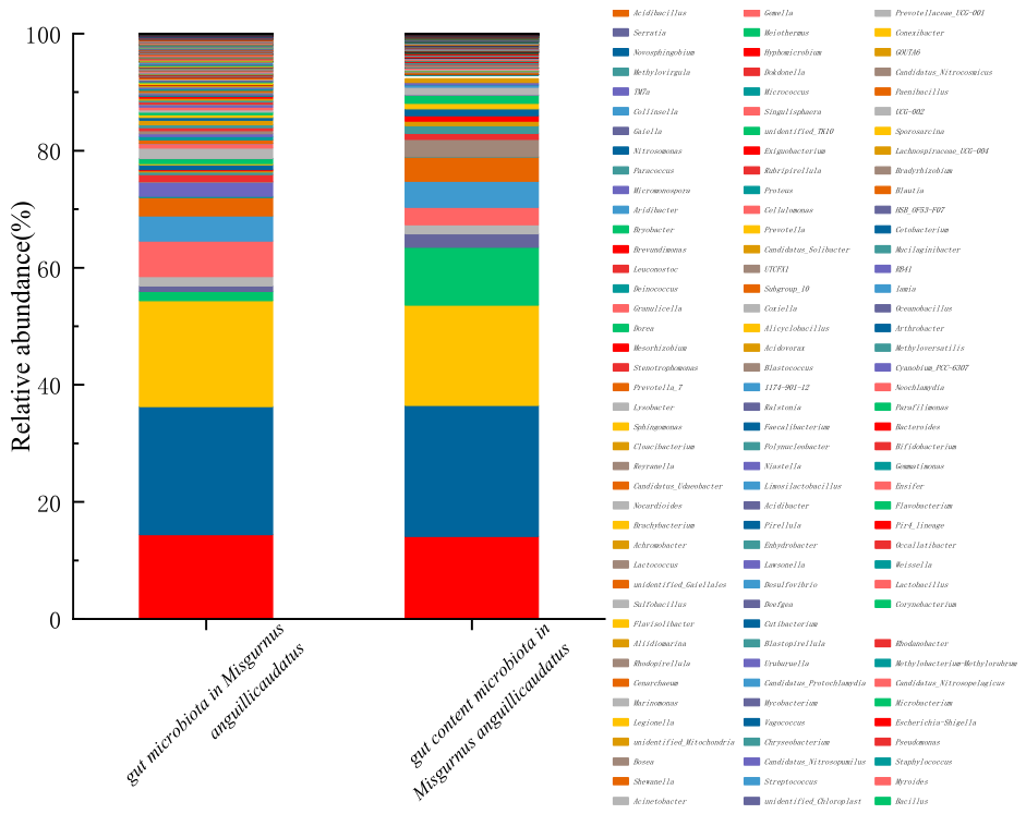
Figure 2.
Cluster heatmap of level 3 enzyme family functions of intestinal microbiota in M. anguillicaudatus predicted by Tax4Fun.
Figure 2.
Cluster heatmap of level 3 enzyme family functions of intestinal microbiota in M. anguillicaudatus predicted by Tax4Fun.
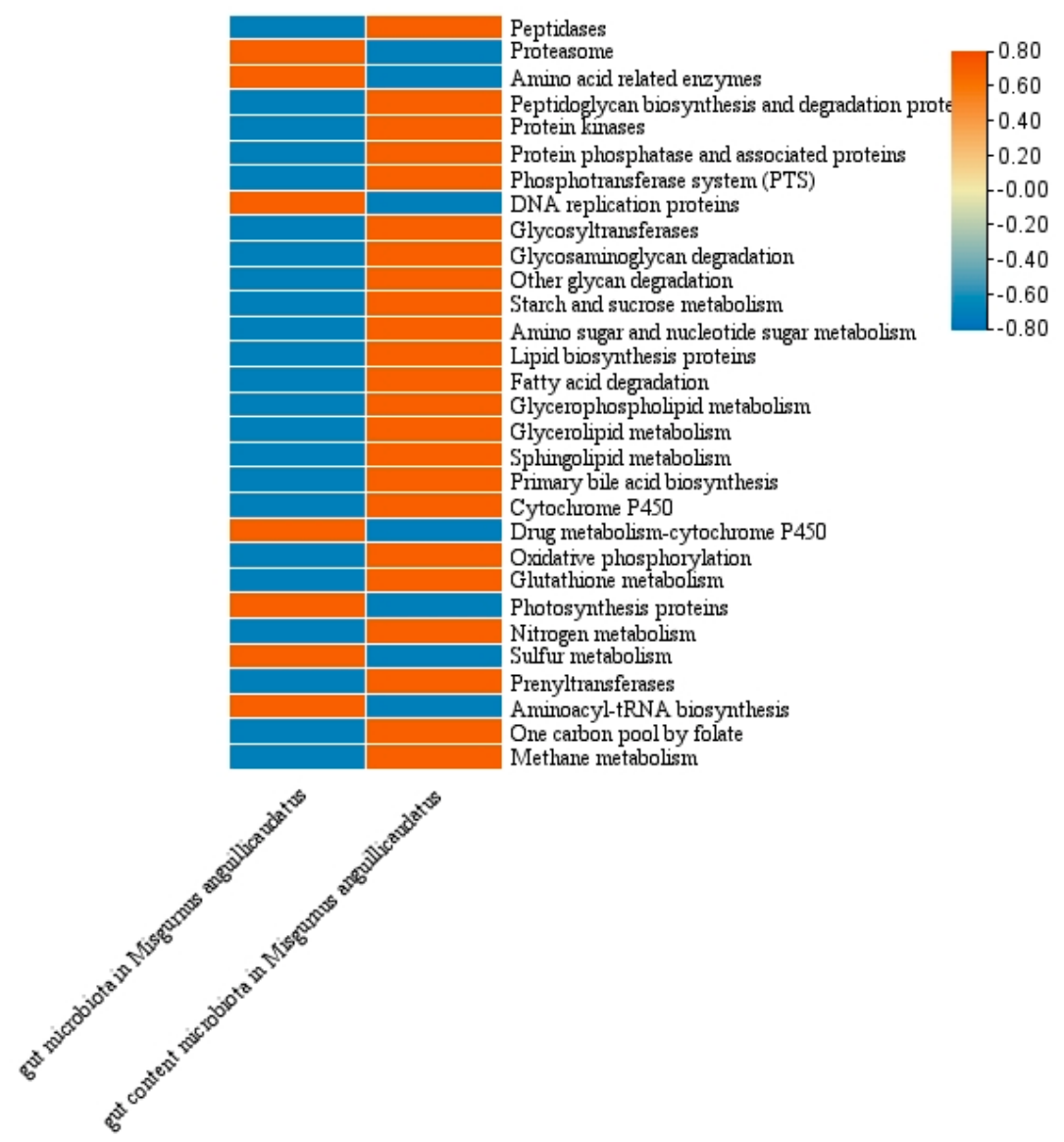

Table 1.
Enzyme production capacity of intestinal bacteria in M. anguillicaudatus.
| Enzyme-producing category | Hydrolysis zone of strains | |||
|---|---|---|---|---|
| A. veronii | A. hydrophila | S. xiamenensis | C. neteri | |
| Amylase (Dh/Dc) |
1.70 | 1.43 | - | - |
| Lipase (Dh/Dc) |
- | - | 1.41 | 3.0 |
| Protease (Dh/Dc) |
2.41 | 2.82 | 2.46 | - |
Note: Dh/Dc represents the ratio of hydrolysis zone diameter (Dh) to colony diameter (Dc).
Table 2.
Antibacterial activity of the strains.
| Antimicrobial Agent | Dosage (μg) | Inhibition Zone Diameter Criteria (mm) | |||||||
|---|---|---|---|---|---|---|---|---|---|
| Resistant (R) | Intermediate (I) | Susceptible (S) | A. veronii | A. hydrophila | S. xiamenensis | C. neteri | |||
| Tetracyclines | Oxytetracycline | 30 | ≤13 | 14-18 | ≥19 | 23.5/S | 37/S | 20/S | 21/S |
| Chloramphenicols | Florfenicol | 30 | ≤12 | 13-17 | ≥18 | 20/S | 16/I | 8/R | 9/R |
| Ampicillin | 10 | ≤13 | 14-16 | ≥17 | 21/S | 20/S | 37/S | 18/S | |
| Aminoglycosides | Kanamycin | 30 | ≤13 | 14-17 | ≥18 | 25/S | 25/S | 36/S | 28/S |
Note: R, resistant; I, intermediate; S, susceptible. Breakpoints were based on CLSI and EUCAST standards for disk diffusion method (28 °C, 10 h). Penicillin was not tested due to intrinsic resistance of aquatic bacteria to this agent.
Disclaimer/Publisher’s Note: The statements, opinions and data contained in all publications are solely those of the individual author(s) and contributor(s) and not of MDPI and/or the editor(s). MDPI and/or the editor(s) disclaim responsibility for any injury to people or property resulting from any ideas, methods, instructions or products referred to in the content. |
© 2026 by the authors. Licensee MDPI, Basel, Switzerland. This article is an open access article distributed under the terms and conditions of the Creative Commons Attribution (CC BY) license (http://creativecommons.org/licenses/by/4.0/).
Copyright: This open access article is published under a Creative Commons CC BY 4.0 license, which permit the free download, distribution, and reuse, provided that the author and preprint are cited in any reuse.