Preprint
Article

This version is not peer-reviewed.

Genetic Diversity and Hospital Circulation of Opportunistic Pathogens in COVID19 ICUs: WholeGenome Sequencing Data

Submitted:

10 July 2026

Posted:

10 July 2026

You are already at the latest version

Abstract

The COVID-19 pandemic led to a dramatic increase in healthcare‑associated infections (HAIs) and antimicrobial resistance (AMR), particularly in intensive care units (ICUs). This study aimed to provide a comprehensive genetic characterisation of opportunistic pathogens (OPs) circulating in the ICUs of two infectious diseases hospitals repurposed for treating severe COVID‑19 patients. Whole‑genome sequencing (WGS) was performed on 175 isolates recovered from patients and the hospital environment (including personal protective equipment, PPE) between 2021 and 2023. The species composition included Klebsiella pneumoniae (n=53), Acinetobacter baumannii (n=60), Staphylococcus aureus (n=20), Enterococcus faecalis (n=20), Escherichia coli (n=10), Proteus mirabilis (n=5), Pseudomonas aeruginosa (n=4), Enterococcus faecium (n=2), and Klebsiella variicola (n=1). Bioinformatic analysis included multilocus sequence typing (MLST), core genome single‑nucleotide polymorphism (SNP) analysis, phylogenetic reconstruction, and in silico detection of resistance and virulence genes and plasmid replicons. Among K. pneumoniae isolates, the high‑risk clone ST512 (47.2%) carrying blaKPC‑3, pmrB(R256G), gyrA(S83I), oqxAB, qacEdelta1 and fosA dominated and showed intra‑hospital spread (9 patients, 7 PPE, 9 surfaces). A. baumannii was represented by ST2 (40.0%, all blaOXA‑23‑positive), ST78 (28.3%, blaOXA‑72/90) and ST19 (3.3%). For E. coli, clonal complex CC14 (including ST1193) carried blaCTX‑M‑15, marR(S3N) and fluoroquinolone resistance mutations. S. aureus isolates were assigned to CC15, CC8, CC22 (MRSA) and CC97. Resistance genes against up to nine antimicrobial classes were identified. Core genome SNP analysis confirmed direct transmission of K. pneumoniae (1–6 SNP differences) between patients and healthcare worker gloves. PPE was a major reservoir (68.4% of environmental isolates). High frequencies of IncF, rep5a/rep16, rep9 and IncC plasmids were detected in E. coli, S. aureus, E. faecalis and P. mirabilis, respectively. The study demonstrates the power of WGS for high‑resolution epidemiological surveillance, confirms the critical role of contaminated PPE in nosocomial transmission, and highlights the dominance of internationally spreading MDR clones in COVID‑19 ICUs.

Keywords: 
;  ;  ;  ;  ;  ;  ;  

1. Introduction

The COVID-19 pandemic, caused by the SARS-CoV-2 virus, posed an unprecedented challenge to healthcare systems worldwide. According to the World Health Organization, by the end of 2023, over 770 million cases and nearly 7 million deaths had been recorded globally. However, beyond the direct consequences of viral infection, the pandemic led to profound and long-term changes in the organisation of medical care, which created favourable conditions for the activation of healthcare-associated infection (HAI) transmission and the spread of antimicrobial resistance (AMR) [1,2].
Of particular concern is the situation in intensive care units (ICUs), which became the epicentre for patients with severe COVID-19 during the pandemic. Extreme workloads on medical staff, resource shortages, the need to rapidly deploy additional beds, and forced deviations from standard infection control protocols created a unique and highly aggressive ecosystem in ICUs [3]. Patients with severe COVID-19, who required invasive mechanical ventilation, received massive immunosuppressive therapy (corticosteroids, interleukin-6 inhibitors) and antimicrobial agents, becoming highly vulnerable to secondary infections by opportunistic pathogens (OPs) [4]. In this context, SARS-CoV-2-induced disruption of respiratory epithelium integrity, dysregulation of immune responses, and alterations of the lung microbiome further increased the risk of bacterial superinfection.
A critical factor contributing to the rise of AMR during the pandemic was the irrational use of antibiotics. Faced with the uncertainty of a novel disease and difficulties in the differential diagnosis of viral and bacterial infections, antimicrobials were widely used empirically, often without clear indications. As noted in an international study involving patients from nine countries, a significant increase in the use of broad-spectrum antibiotics, including meropenem, piperacillin/tazobactam, and azithromycin, was observed during the first 18 months of the pandemic. The most pronounced increase in carbapenem consumption was recorded in countries with high healthcare system burdens, creating strong selective pressure on the microbiota of infectious disease hospitals [5]. According to a systematic review covering studies from Italy, the prevalence of HAIs increased by 9–11% during the pandemic, while the detection rate of multidrug-resistant (MDR) Gram-negative OPs increased in a range from 0.8% to 45.6%, depending on the pathogen and the study population [1].
The scale of the problem is confirmed by data from a retrospective study conducted in Turkey that analysed 1,662 respiratory tract samples collected between 2016 and 2024. Comparison of the pre-pandemic and post-pandemic periods revealed a worrying trend: resistance of Acinetobacter baumannii to gentamicin increased from 70.9% to 91.3%, to colistin from 3.2% to 12.3%, while resistance of Klebsiella pneumoniae to carbapenems (e.g., meropenem: from 50.3% to 77.8%) and colistin (from 23.0% to 62.7%) increased statistically significantly (p < 0.001 for both classes) [2]. Moreover, ICU patient mortality in the post-pandemic period also rose significantly, from 70.5% to 74.9% (p = 0.048), which may be related both to the severity of the underlying disease and to limited treatment options due to high AMR. Notably, despite the overall increase in resistance among most Gram-negative pathogens, the isolation rate of Escherichia coli from respiratory samples decreased (from 11.4% to 7.0%, p < 0.05), which may reflect changes in the spectrum of nosocomial pathogens during the pandemic [2].
At the same time, the impact of the pandemic on AMR was not unidirectional. According to a rapid scoping review, changes in antibiotic use and infection control measures led to heterogeneous effects: while resistance increased in the hospital setting, community and general population settings may have experienced a certain decrease in the prevalence of some resistant strains owing to the introduction of restrictive measures (mask wearing, social distancing, travel restrictions). However, the authors emphasise that most studies had a moderate or high risk of bias, and the analysis of socio-economic determinants and impacts on healthcare systems remained underdeveloped [6].
Particular concern is raised by the role of personal protective equipment (PPE) as a potential reservoir of infection and a factor in its nosocomial transmission. A systematic review evaluating the role of gowns in preventing nosocomial transmission of respiratory viruses showed that the level of PPE contamination reached 77.5%, with the longest preservation of virus infectivity observed on Tyvek suits and plastic gowns [7]. During the pandemic, when staff were forced to work in PPE for extended periods (up to 12 hours or more), the risk of contamination of outer surfaces and subsequent pathogen transmission increased significantly. A study conducted in India involving 3,098 healthcare workers demonstrated that even with extensive training and observer supervision, 12.7% of staff made mistakes in the sequence of donning PPE, with eye protection being the most frequently omitted element (10.1% of cases) [8]. These data highlight that, even with formal adherence to protocols, significant risks associated with the “human factor” remain.
An important addition to the “human factor” problem is the limited informativeness of traditional microbiological control methods. Routine culture and genotypic typing (e.g., multilocus sequence typing, MLST) often lack sufficient resolution to reliably confirm or refute suspected epidemiological links between isolates, especially under conditions of long-term circulation of genetically related isolates in a closed hospital environment [9,10]. Furthermore, taxonomic complexities and limitations of standard identification methods can lead to incorrect conclusions about isolate relatedness, which in turn hinders timely outbreak recognition and effective infection control measures [11]. The COVID-19 pandemic, placing an enormous burden on healthcare systems, led to a shortage of resources for traditional epidemiological surveillance and infection control auditing, further reducing the effectiveness of standard approaches to HAI prevention. These limitations underscore the need for more accurate and informative molecular analysis methods.
In this context, the use of high-throughput sequencing methods for HAI epidemiological surveillance becomes particularly important. Whole-genome sequencing (WGS) has recently been considered the “gold standard” of molecular epidemiological analysis, allowing not only the identification of the pathogen species and sequence type but also the in silico determination of the complete spectrum of AMR and virulence genes, the detection of plasmid replicons responsible for horizontal gene transfer, and, most importantly, core genome single-nucleotide polymorphism (SNP) analysis to establish the degree of relatedness between isolates with resolution down to a single nucleotide substitution [12]. Data obtained by WGS make it possible to reliably confirm or refute cases of direct microorganism transmission, reconstruct transmission pathways, and assess the contribution of various reservoirs to infection spread [12]. Modern approaches to genomic surveillance, such as the Enhanced Detection System for Healthcare-Associated Transmission, demonstrate not only high efficiency in detecting hidden outbreaks but also cost-effectiveness: the cost of sequencing a single sample in routine surveillance can be less than USD 100 [13].
In the Russian Federation, as in many other countries, the COVID-19 pandemic required the rapid deployment of infectious diseases hospitals with “red” and “green” zones. These institutions became unique model systems for studying the circulation of OPs under conditions of maximal isolation and intensive use of antimicrobial agents. However, despite the urgency of the problem, comprehensive studies covering the entire spectrum of leading OPs (K. pneumoniae, A. baumannii, E. coli, Staphylococcus aureus, and others) circulating in such hospitals using WGS have been insufficient to date.
Aim of the study – based on the results of WGS, to provide a comprehensive genetic characterisation of OP isolates (K. pneumoniae, A. baumannii, E. coli, S. aureus, and others) obtained from ICU patients and environment (including PPE) in infectious diseases hospitals repurposed for the treatment of COVID-19 patients during the pandemic.
The data obtained will allow not only to describe the bacterial landscape of the COVID-19 hospital but also to substantiate the need for integrating high-throughput sequencing methods into routine epidemiological surveillance of healthcare-associated infections, as well as to develop more effective strategies for the prevention and control of the spread of MDR hospital OPs.

2. Materials and Methods

2.1. Sampling and Bacterial Isolation

From 2021 to 2023, biological and environmental samples were collected from two large regional infectious diseases hospitals repurposed for treating patients with severe COVID-19. One of the hospitals had a 440-bed capacity, including a 12-bed ICU; the other hospital had an ICU of comparable size (approximately 12 beds). Sampling was performed exclusively within the ICUs of both hospitals, which were thus representative of the larger, strained healthcare system responding to the severe wave of the pandemic. The samples were collected in accordance with a scheme for surface swabs patented by the authors, developed for simultaneous assessment of viral and bacterial contamination [14]. Briefly, two sterile cotton swabs moistened with 0.1% peptone water containing a neutraliser for the oxidising group (0.5% sodium thiosulfate solution) were used. The overall study design, sampling scheme, and sample collection procedures were described in detail previously [15] and were applied in the present study without modification. In brief, surface swabs were collected from the same 20 sampling points grouped into three blocks (PPE of medical workers, patient care environment (PCE), general hospital points (GHP)) every four hours over three consecutive days. Sputum samples were used as clinical specimens. The key differences from our previous work are that the present study (i) was conducted in two infectious diseases hospitals repurposed for severe COVID-19 patients (whereas the previous study involved only one such hospital), (ii) covered a longer time period (2021–2023), and (iii) included a larger and more diverse collection of isolates (n = 175) spanning nine OP species, as detailed in Section 3.1.
For clinical specimens, 28 sputum samples, 14 oropharyngeal swabs, one urine sample, and one urethral swab were collected from patients. Sample collection and processing were performed in accordance with the national methodological guidelines MUK 4.2.2942-11 “Methods for sanitary and bacteriological testing of environmental objects, air, and sterility control in healthcare institutions”, which are effective in the Russian Federation.
A detailed list of each isolate, including its sample ID and the specific sampling point it was obtained from, is provided in Appendix A.1.

2.2. DNA Extraction and Whole-Genome Sequencing

DNA extraction and library preparation for the isolates sequenced on the Illumina MiSeq platform were performed as described previously [15]. The remaining isolates (comprising the majority of K. pneumoniae, E. faecalis, E. faecium, P. aeruginosa, P. mirabilis, and four A. baumannii isolates) were sequenced on the MGI DNBSEQ-G50 platform. For these isolates, DNA extraction was performed using the «MAGNO-sorb-M» kit (FBIS CRIE, Moscow, Russia) according to the manufacturer’s instructions, using an Auto-Pure 96 station (Allsheng). DNA concentration was measured with a Fluo200 fluorometer and the QuDye dsDNA HS assay kit (Lumiprobe, Russia). Library preparation was carried out following the MGIEasy Fast FS Library Prep Set User Manual (v2.0). Briefly, 100–500 ng of genomic DNA was fragmented with Fast FS II enzyme (MGI, Shenzhen, China) for 25 min at 30 °C, followed by a 0.8× size selection using MGI Easy DNA Clean Beads. Universal UDB adapters were ligated to the fragments, and adapter-ligated products were purified with a 0.8× bead cleanup. Indexing PCR was performed with limited cycles (5–8 cycles) using UDB primers. The PCR product was purified with 0.9× beads. Library quality was assessed using a Fluo200 fluorometer and a Qsep1 capillary electrophoresis system (target fragment size ~300–500 bp). DNA nanoballs (DNBs) were prepared using the DNBSEQ One-step DNB Make Reagent Kit V2.0 (MGI, Shenzhen, China) according to the manufacturer’s protocol. Libraries were pooled in equimolar amounts, and the final pool was quantified with the QuDye dsDNA HS kit. Sequencing was performed on a DNBSEQ-G50 instrument using an FCL flow cell and the PE150 reagent kit (300 cycles, paired-end 2 × 150 bp).

2.3. Bioinformatic Analysis

Sequencing data quality was assessed using FastQC 0.12.1, evaluating read counts, maximum/minimum read lengths, GC content, and ambiguous nucleotide proportions for forward and reverse reads.
De novo genome assembly was performed via scaffolding using SPAdes 4.2.0 [16] on the Galaxy server, based on paired-end reads. A taxonomic analysis of scaffolds was conducted using Galaxy’s FCS GX tool 0.5.5 [17] to confirm species identity and remove contaminants.
Assembled OP’s sequences were cleaned of residual adapters using NCBI VecScreen: FCS Adaptor 0.5.5 [16] and filtered by scaffold length with fastp 1.3.3 [18].
Typing was performed via MLST using the PubMLST database (https://pubmlst.org/, accessed on 4 May 2026) or Galaxy’s tool MLST 2.22.0 (https://usegalaxy.org/, accessed on 4 May 2026).
The analysis of assembled genomes was performed using several bioinformatic tools and databases. Antimicrobial resistance (AMR) genes were identified using ResFinder 4.6.0 and AMRFinderPlus 3.12.8 [19,20]. Virulence genes were detected using the ABRicate utility 1.4.0 [21] with the VFDB database. Plasmid replicons were determined using PlasmidFinder 2.1.6 [22]. All searches were conducted with thresholds of ≥90% nucleotide identity and ≥60% coverage.
For phylogenetic analysis, genome annotation was performed using Prokka 1.14.6 [23]. The core genome alignment was constructed with Roary 3.13.0 [24] using a 90% threshold for gene presence (rather than the default 99%) to retain greater genetic diversity. Non-informative columns (e.g., positions containing only undetermined values, which were treated as missing data as identified by RAxML 8.2.12 [25]) were removed using a custom Python script (version 3.14.3). Maximum-likelihood phylogenetic trees were reconstructed with IQ-TREE 2.0.7 [26] using 1000 bootstrap replicates. Reference genomes of the corresponding species and relevant sequence types were obtained from PubMLST (https://pubmlst.org/, accessed on 5 May 2026), NCBI (https://www.ncbi.nlm.nih.gov/, accessed on 6 May 2026), and the VGARus (https://genomenvpn.crie.ru/, accessed on 7 May 2026) database and included in the tree reconstruction.
To assess recent transmission events, a core genome SNP analysis was performed on isolates that showed similarity in their resistance and virulence profiles. The core genome alignment generated by Roary was used to calculate pairwise SNP distances with the SNP distance matrix tool (version 0.8.2) [27] Isolates differing by fewer than 15 SNPs were considered genetically linked, which is consistent with established criteria for recent nosocomial transmission.

2.4. Statistical Analysis

Statistical analysis was performed using Microsoft Excel. The structure of the isolated OPs was described using extensive indicators (percentages). The prevalence of antimicrobial resistance and virulence genes, as well as plasmid replicon types, was determined as their frequency of occurrence.

3. Results

From 2021 to 2023, a total of 175 OP isolates were obtained in the ICUs of infectious diseases hospitals treating patients with COVID-19. Of these, 36 isolates originated from ICU patients, and 139 were collected from the environment ICU. Among the environmental isolates, 76 were recovered from the surface of PPE, 52 from the PCE, and 11 from GHP.
The species composition included nine species: K. pneumoniae (n = 53; 30.3%), A. baumannii (n = 60; 34.3%), S. aureus (n = 20; 11.4%), Enterococcus faecalis (n = 20; 11.4%), E. coli (n = 10; 5.7%), Proteus mirabilis (n = 5; 2.9%), Pseudomonas aeruginosa (n = 4; 2.3%), Enterococcus faecium (n = 2; 1.1%), and Klebsiella variicola (n = 1; 0.6%). K. pneumoniae and A. baumannii played a dominant role in the OP structure both among patients and in the hospital environment.

3.1. Genetic Characterisation of Hospital Pathogens

Detailed genetic profiles (resistance genes, virulence factors and plasmid replicons) for all isolates are presented in Supplementary Tables S1–S3. Below, we highlight the key characteristics of each species.

3.1.1. Acinetobacter baumannii Isolates

Of the 60 A. baumannii isolates, nine (15.0%) were obtained from clinical specimens of patients, and 51 (85.0%) from the environment, of which 23 (45.1%) came from PPE.
Typing and Phylogeny. MLST identified three sequence types (STs) among the 60 A. baumannii isolates: ST2 (n = 24; 40.0%), ST78 (n = 17; 28.3%), and ST19 (n = 2; 3.3%). Seventeen isolates (30.4%) were non-defined. Phylogenetic analysis based on a soft-core genome alignment (Figure 1) distributed all studied isolates into the three STs – ST19, ST78, and ST2. The ST19 isolates (including one non-defined isolate, ab_29) formed a compact cluster together with Russian reference sequences (crie084740, crie087042), separating from European isolates. Within ST78, two main subclades emerged: one consisted exclusively of isolates from the second sampling phase, while the other included a mixed group from the second and third phases, suggesting the simultaneous circulation of two distinct subpopulations. ST2 appeared to be the most heterogeneous. In addition to several separate lineages (basal isolate ab_09, a branch with isolates from Denmark, Kenya, and Nizhny Novgorod, as well as the pair ab_50+ab_06 and isolate ab_56), a large cluster was identified within which two genetically distinct subclades could be distinguished. Both subclades contained isolates from the second and third sampling phases, indicating long-term parallel circulation of two independent ST2 lineages within the hospital. The non-defined isolates were distributed among all three STs, confirming that no other STs were present in the hospital during the study period.
AMR. All A. baumannii isolates carried genes conferring resistance to aminoglycosides and β-lactams. ST2 was characterised by the profile: armA, aph(6)-Id, aph(3′)-Ia, aph(3″)-Ib, blaOXA-23, msr(E), mph(E). The carbapenemase gene blaOXA-23 was present in 100% of ST2 isolates. ST78 was characterised by the presence of armA, blaOXA-72, blaOXA-90, msr(E), mph(E), while blaOXA-23 was absent. ST19 carried aph(3′)-VIa, blaOXA-69, blaADC-25, catA1, sul2, and tet(B). The resistance profile of non-defined isolates was similar to that of ST2, suggesting genetic relatedness.

3.2.2. Klebsiella pneumoniae Isolates

Fifteen isolates (28.3%) were obtained from clinical specimens of ICU patients, and 38 (71.7%) from the ICU environment, of which 15 (39.5%) came from PPE.
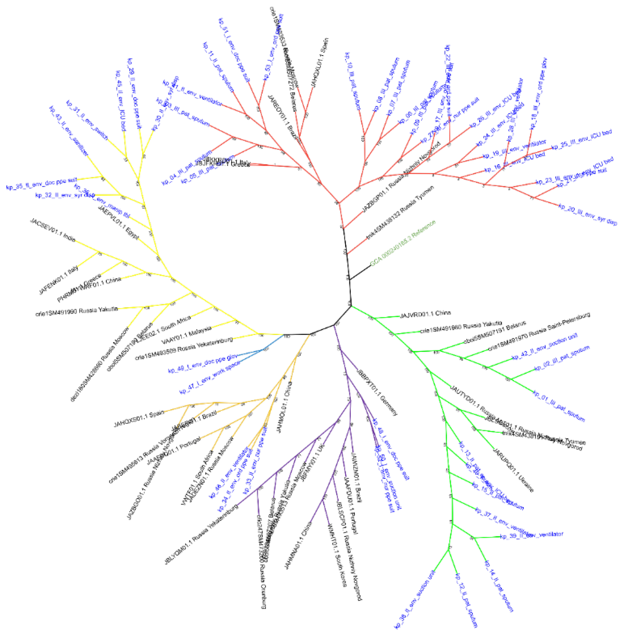
Typing and Phylogeny. A total of 53 isolates were obtained. The dominant clone belonged to ST512 (n = 25; 47.2% of all isolates). Other STs detected included ST101, ST1190, ST15, ST307, ST395, as well as non-defined isolates. Intra-hospital circulation of ST512 was confirmed: isolates were recovered from nine patients and from the hospital environment (seven from PPE, nine from PCE).
Phylogenetic analysis based on a soft-core genome alignment (Figure 2) distributed the isolates into six STs. The ST101 branch was distinct, with two parallel sublines; all ST101 isolates belonged exclusively to the second sampling phase and showed relatedness to an Egyptian reference genome. ST1190 was extremely rare (two isolates), and no closely related reference sequences were found in the databases; no mixing of isolates from different phases was observed. ST15 and ST307 also formed separate clusters without mixing of phases, and ST307 did not show clear affinity to any geographic group (Russian, European, etc.). The ST395 branch split into two subclades: the first contained three isolates (one from the second phase and two from the third) and clustered with Russian and Belarusian isolates; the second subclade consisted exclusively of non-defined isolates from the second phase, close to Russian-Ukrainian references. This structure suggests that ST395 is endemic to the study region. The largest and most heterogeneous group was ST512, within which two major clusters formed: one comprised exclusively isolates from the third phase, while the other (smaller) contained isolates from the first, second and third phases simultaneously. Notably, in this smaller subcluster, the first-phase isolates originated from the first hospital, while the related second-phase isolates came from the second hospital. Considering the bootstrap support (100 within each of the two groups and 88 between them), it can be hypothesised that in the small ST512 subcluster, the first-phase isolates and the related second-phase isolates share a common ancestor. This may indicate either inter-hospital transmission of the strain or a common source of introduction.
AMR. The dominant clone ST512 (n = 25) exhibited a MDR profile including the carbapenemase gene blaKPC-3 (carbapenems, 25/25), the chromosomal colistin resistance mutation pmrB(R256G) (24/25), the fluoroquinolone resistance determinants gyrA(S83I) (24/25) and oqxAB (25/25), the biocide resistance gene qacEdelta1 (24/25), and the fosfomycin resistance gene fosA (25/25).
The other sequence types (ST15, ST101, ST307, ST395 and ST1190) were also MDR but differed markedly from ST512. None of them carried carbapenemase genes (blaKPC-3, blaNDM-1, blaOXA-48), the colistin resistance mutation pmrB(R256G), or the biocide resistance gene qacEdelta1. Instead, their β-lactam resistance was mediated by extended-spectrum β-lactamase (ESBL) genes, predominantly blaCTX-M-15, often in combination with blaOXA-1, blaSHV-28, blaTEM-1 or blaSHV-1. All these isolates possessed various aminoglycoside resistance genes (aac(6')-Ib-cr5, aph(3')-Ia, aph(3″)-Ib, aph(6)-Id, aadA1, aadA2), fluoroquinolone resistance determinants (mutations in gyrA and parC, and/or qnrS1, qnrB), tetracycline resistance (tet(A)), phenicol resistance (catA1, catA2, catB3), sulphonamide resistance (sul1, sul2), and macrolide resistance (erm(B), mph(A)). Fosfomycin resistance (fosA) was detected only in ST395. The resistance profiles of ST101 and ST307 were similar to each other, while ST395 additionally carried blaSHV-1, erm(B), mph(A), catA1, qnrB and fosA. ST1190 harboured blaCTX-M-15, aph(3')-Ia, aph(3″)-Ib, aph(6)-Id, catA1, oqxB19, sul1, sul2, and qnrS1. Thus, genetically diverse K. pneumoniae lineages with different resistance mechanisms co-circulated in the hospital, and the carbapenemase, colistin and biocide resistance markers were exclusive to the dominant ST512 clone.
A wide diversity of plasmid replicons was detected among the isolates. The most frequent were IncFIB(K) (71.7% of isolates), IncFII(K) (67.9%), IncRNAI (64.2%), IncX3 (45.3%) and IncHI1B (34.0%).
In addition, a single isolate of K. variicola ST4101 was recovered from PPE. It carried resistance genes for aminoglycosides (aph(3″)-Ib, aph(6)-Id), β-lactams (blaLEN-2), fluoroquinolones (oqxA, oqxB), fosfomycin (fosA), as well as the plasmid replicons IncFIB(K), IncFIB(pKPHS1), IncFII(K) and Col(pHAD28).

3.2.3. Escherichia coli Isolates

Two isolates (20.0%) were obtained from clinical specimens of ICU patients, and eight (80.0%) from the ICU environment, of which five (63.0%) came from PPE.
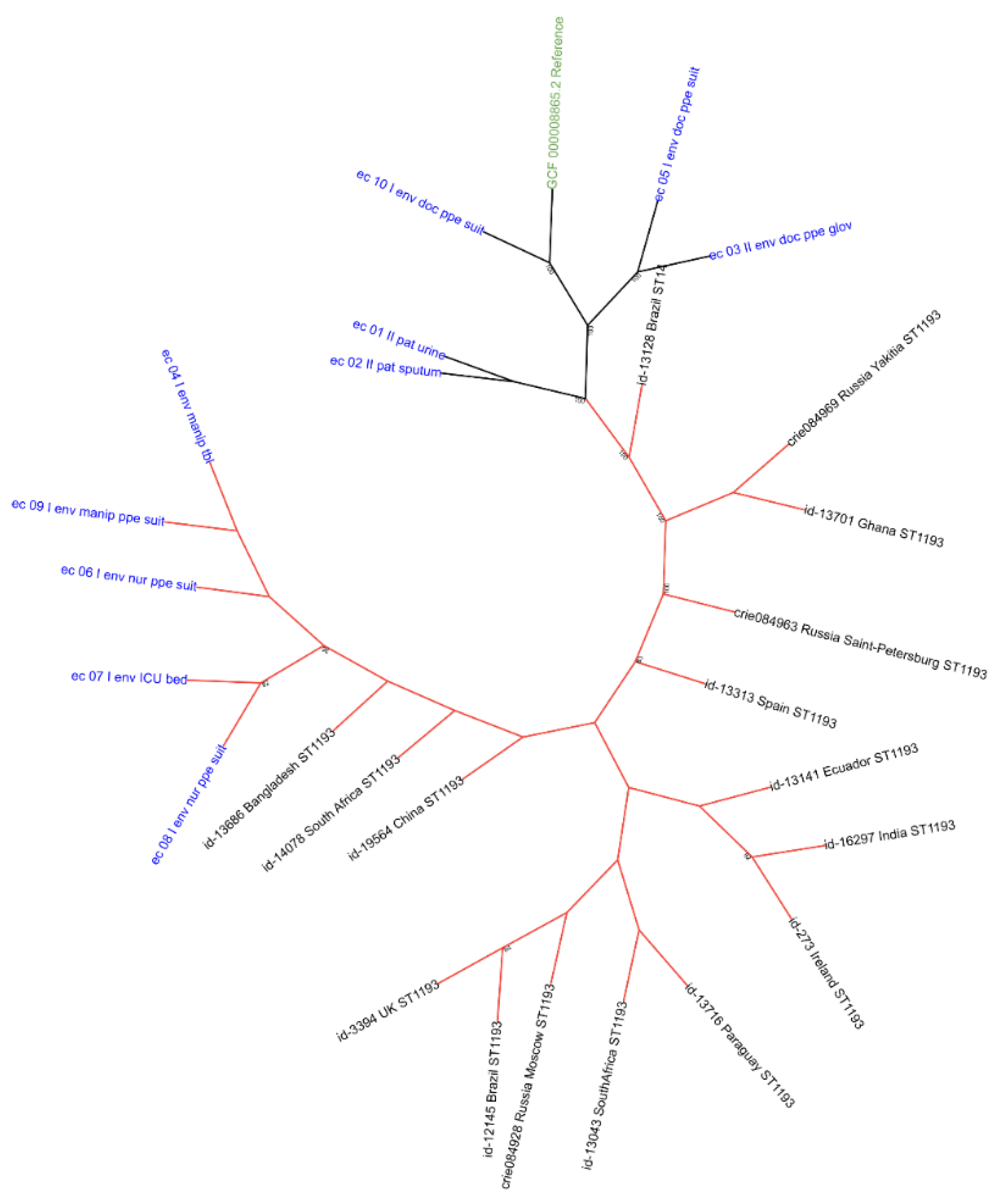
Typing and Phylogeny. Although none of our E. coli isolates (n = 10) could be typed by standard MLST schemes (probably due to technical limitations or loss of target genes), phylogenetic analysis based on a soft-core genome alignment (Figure 3) allowed assessment of their genetic relatedness. Most environmental isolates (first sampling phase) formed a compact subcluster within the ST1193 lineage (Clonal Complex 14, CC14) with high bootstrap support (96–100), indicating their assignment to this ST. Other isolates (including patient samples) showed mixing of phases: some grouped with reference sequences, while others formed separate branches. Thus, the tree structure suggests the circulation of at least two genetically distinct E. coli lineages in the hospital, one of which (ST1193) was represented exclusively by first-phase isolates from the hospital environment.
AMR. In silico analysis revealed resistance determinants against eight classes of antimicrobials: aminoglycosides (aph(3″)-Ib, aph(6)-Id, aadA25), β-lactams (blaCTX-M-15, blaTEM, blaTEM-1, blaTEMp(C32T)), quinolones (qnrS1, mutations parC(80I), gyrA(D87N), gyrA(S83L)), antifolates (sul2), fosfomycins (uhpT(E350Q), glpT(E448K), fosA, cyaA(S352T)), colistin (pmrB(E123D)), nitrofurans (nfsB(W94STOP)), and multidrug resistance (marR(S3N)). This gene group was the most prevalent, indicating a high adaptive potential of the population.
The blaCTX-M-15 gene was detected in 22% of isolates, mainly among ST1193. All CC14 isolates carried marR(S3N); blaTEM group genes were found in 80% of CC14 isolates. Among isolates from patient care surfaces, 100% carried multidrug resistance genes, whereas isolates from PPE showed greater diversity of resistance determinants (16 out of 21 unique genes).
Virulence. The most frequently detected genes were associated with adhesion (fimH – 100%, yehA/B/C/D – up to 90%), invasion (ibeA – 33%) and immune evasion (traT – 78%). Hypothetical ST1193 isolates carried a full set of adhesion genes. The hypothetical ST1380 isolate harboured the rare vat gene encoding a serine protease autotransporter characteristic of some uropathogenic E. coli. The clinical isolate from a patient contained the highest number of virulence genes (15).
Plasmid replicons were found in 100% of isolates; the dominant types were IncFIA, IncFIB and ColBS512.

3.2.4. Staphylococcus aureus Isolates

Three isolates (15.0%) were obtained from clinical specimens of ICU patients, and 17 (85.0%) from the ICU environment, of which 14 (82.4%) came from PPE.
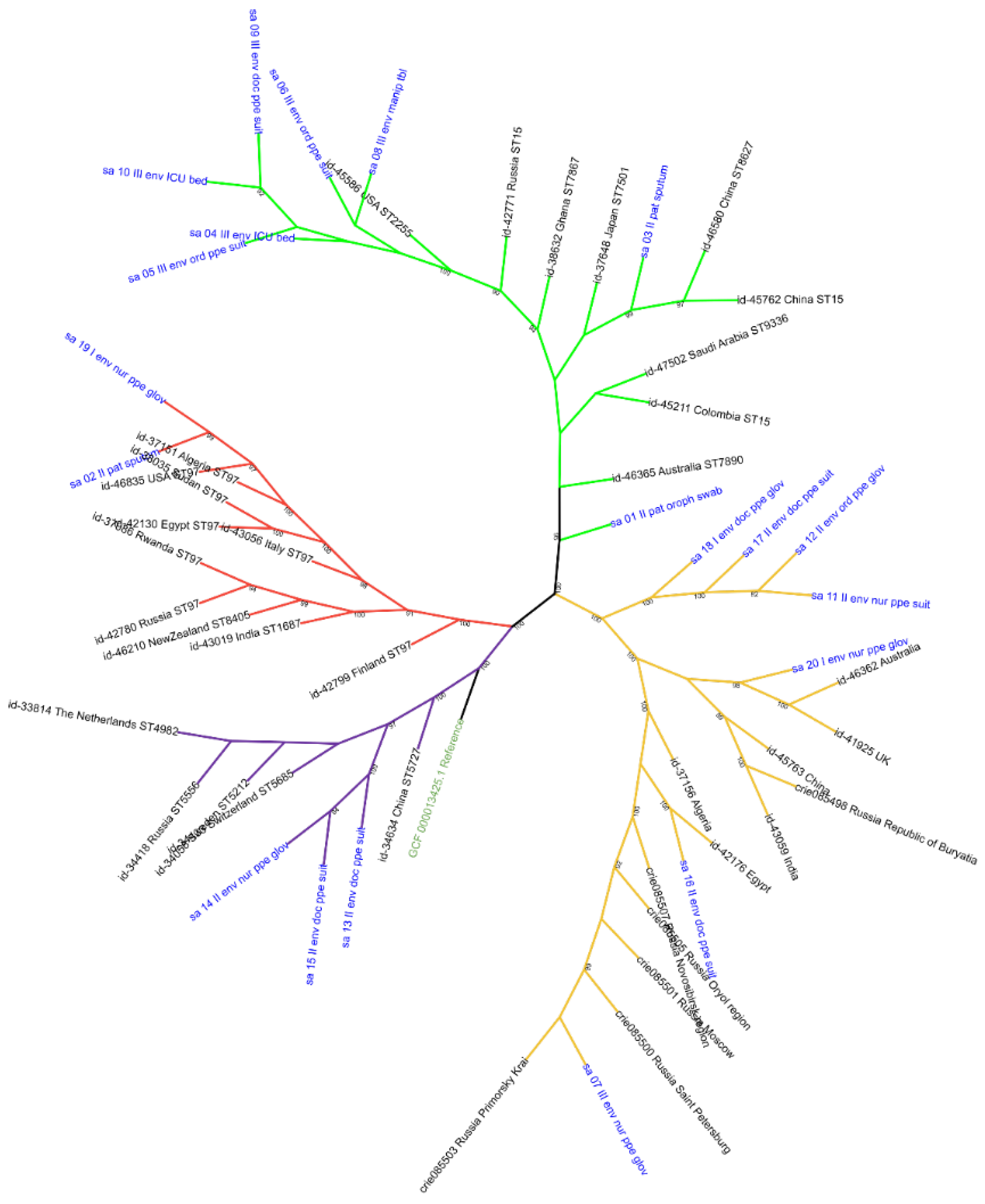
Typing and Phylogeny. Among the 20 S. aureus isolates, a full ST could be assigned for six (30%): ST22 (n = 1), ST97 (n = 1), ST5727 (n = 3), ST7614 (n = 1). For 14 (70%) of isolates, full typing was impossible due to the absence of alleles for one or more housekeeping genes; however, for seven of them, assignment to CCs was possible. Four CCs circulated in the ICU: CC15 (30%, n = 6), CC8 (15%, n = 3), CC22 (10%, n = 2), and CC97 (10%, n = 2). The remaining 7 isolates could not be assigned to any specific ST or CC – non-defined.
Despite the fact that a significant proportion of our S. aureus isolates could not be typed by standard schemes, phylogenetic analysis based on a soft-core genome alignment (Figure 4) reliably assigned them to four clonal complexes – CC8, CC15, CC22 and CC97. CC97 isolates formed a single cluster with isolates from the USA and Algeria. Within CC8, our isolates formed a sister group to European sequences from Russia, Sweden, the Netherlands and Switzerland. CC22 showed the greatest heterogeneity: one lineage clustered with Russian genomes, another with an Egyptian isolate, a third with isolates from Australia and the United Kingdom, and a separate group of non-defined isolates (three from the second phase and one basal isolate from the first phase) also emerged. Within CC15, our isolates were distributed among several subclades without clear mixing by phase in closely related nodes; some grouped with Australian, Chinese, Colombian and Saudi isolates, while others formed independent local branches. Thus, the tree structure confirms the circulation of four S. aureus CCs in the hospital, with the greatest diversity among our isolates observed in CC15.
AMR. Resistance determinants against nine classes of antimicrobials were identified. The most common were β-lactam resistance genes: blaZ (65%), blaI (60%), blaR1 (55%), as well as tetracycline resistance (tet(38) – 95%). Methicillin-resistant S. aureus (MRSA) carrying mecA and mecR1 were detected in 100% of CC22 isolates and in 14.3% of non-defined isolates. All CC8 and CC15 isolates carried fosB (fosfomycin resistance). CC22 showed 100% presence of dfrB(A135T) (trimethoprim resistance). The fusA(A655V) mutation (fusidic acid resistance) was found in 50% of CC97 isolates.
Isolates from PPE displayed the greatest diversity of resistance determinants, whereas isolates from PCE carried β-lactam resistance genes in 100% of cases.
Virulence. Genes encoding exoenzymes (aur – 90%, splA/B/E – 60–70%), host immune evasion factors (sak – 55%, scn – 75%), and toxins (hlgA/B/C – 90%, lukD/E – 50–60%) were widely distributed. Enterotoxins (sea, seb, seg, sei, sem, sen, seo, seu) were detected in 10–30% of isolates, mainly among CC22 and some non-defined isolates. The toxic shock syndrome toxin gene (tst) was found in 5% of isolates.
Plasmid replicons were detected in 85% of isolates; the most frequent were rep16 and rep5a.

3.2.5. Enterococcus faecalis Isolates

Three isolates (15.0%) were obtained from clinical specimens of ICU patients, and 17 (85.0%) from the ICU environment, of which 13 (76.5%) came from PPE.
Typing. A total of 20 isolates were typed by MLST, identifying nine STs: ST30 (n = 5), ST40 (n = 3), ST387 (n = 2), ST832 (n = 2), ST25, ST44, ST179, ST228 and ST2080 (one each), and three non-defined isolates.
AMR. The most frequent genes were lsa(A) (85%), dfrE (80%), emeA (80%), efrA and efrB (each 75%), tetM (60%), erm(B) (45%). Aminoglycoside resistance genes (aad(6), aph(3')-IIIa, sat-4) were found in 40% of isolates, and aac(6')-Ie-aph(2")-Ia in 20%.
Virulence. Nearly all isolates carried genes encoding adhesion factors (ebpA, ebpB, ebpC, srtC, efaA, prgB/asc10), the two-component regulatory system (fsrABC), gelatinase (gelE), serine protease (sprE), as well as capsule biosynthesis genes (cpsAK). Cytolysin operon genes (cyl) were detected in 45% of isolates. The aggregation substance gene asa1 was found in 15% of isolates, and the adhesin bopD in 85%.
Plasmid replicons were detected in 70% of isolates; the most frequent were rep9b and repUS43.

3.2.6. Enterococcus faecium Isolates

Isolates were obtained exclusively from the ICU environment, with two isolates (100%) detected on PPE; no clinical isolates from patients were registered.
Typing. The two obtained isolates belonged to ST52.
AMR. Both isolates carried aac(6')-I (aminoglycosides), msr(C) (macrolides), and eat(A)(T450I). No other resistance determinants were found.
Virulence. No virulence genes were detected.
Both isolates carried identical plasmid replicons: repUS15, rep1, rep14b.

3.2.7. Proteus mirabilis Isolates

Two isolates (40.0%) were obtained from clinical specimens of ICU patients, and three (60.0%) from the ICU environment, of which two (66.7%) came from PPE.
Typing. Five isolates were obtained, belonging to two STs: ST269 (n = 3) and ST234 (n = 2).
AMR. All isolates exhibited multidrug resistance in silico. β-Lactam resistance genes detected: blaCTX-M-3 (in three isolates), blaCTX-M-14 (in two), blaTEM-1 (in four). Additional resistance genes were found against aminoglycosides (aadA2, aph(6)-Id, aph(3″)-Ib, rmtB, aac(3)-IIe, ant(3″)-IIa), sulphonamides (sul1, sul2), tetracyclines (tet(J)), chloramphenicol (catI, catA4), trimethoprim (dfrA1, dfrA12). The streptothricin resistance gene sat-1 was present in four isolates.
Virulence. All isolates carried genes encoding adhesion factors (mrpA, pmfA, fimA, fimH), secretion systems (T6SS: hcp, vgrG, clpV1), the urease operon (ureA–G), flagellar genes, regulatory systems (crp, cpxAR, phoPQ, rcsAB), as well as iron-siderophore transport genes (fyuA, hemR, hmuTUV, exbD). Some isolates harboured toxin genes (hpmA, tdhA, hlyB/tolC).
Plasmid replicons. All isolates carried the IncC replicon. One isolate (ST234) additionally harboured the Col3M replicon.

3.2.8. Pseudomonas aeruginosa Isolates

Two isolates (50.0%) were obtained from clinical specimens of ICU patients, and two (50.0%) from the ICU environment, of which one (50.0%) came from PPE.
Typing. Four isolates were obtained, belonging to three STs: ST654 (n = 2), ST235 (n = 1), ST549 (n = 1).
AMR. The ST235 isolates carried aminoglycoside resistance genes (aph(3')-IIb, aph(3')-XV, aac(6')-Ib4, aadA6), the carbapenemase blaGES-1, oxacillinases blaOXA-488, blaOXA-50, blaPDC-35 and blaPDC-195, the mutation oprD(W417STOP), phenicol resistance genes (catB7, floR2), tetracycline resistance genes (tetR(G), tet(G)), fosA, gyrA(T83I), sul1, and qacEdelta1. While ST654 isolates’ profile included blaVIM-2, blaOXA-396, blaPDC-3, blaPDC-216, strA/strB, aph(3')-IIb, aph(3″)-Ib, aph(6)-Id, aph(3')-VI, catB7, floR2, tet(A), tet(G), fosA, parC(S87L), gyrA(T83I), sul1. ST549: the smallest set – aph(3')-IIb, blaOXA-50, blaPDC-1, blaPDC-212, catB7, fosA, crpP.
Virulence. All isolates carried an extensive set of virulence genes, including regulators (lasI/R, rhlI, exsA-E, algU, mucA-D), adhesion factors (pil genes), secretion systems (T3SS, T6SS), exotoxins (toxA, exoT, exoS, exoY), pigment biosynthesis (phz operon), siderophores (pvd, pch, pvc), and flagellar genes.
Plasmid replicons were not detected in any of the P. aeruginosa isolates.

3.3. Role of the Hospital Environment and PPE in Nosocomial Transmission of Infection

Contaminated Sites

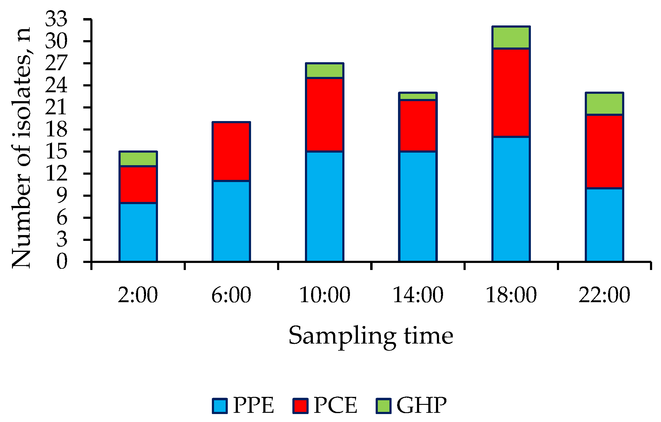
Among the environmental samples (n = 139), the highest level of bacterial contamination was found on the outer surface of the physician’s PPE suit (39.2%), nurses’ PPE suit (25.4%) and orderlies’ PPE suit (29.5%), as well as on handrails and levers of ICU beds (14.8%) and the surface of medical manipulation tables (11.2%). The temporal dynamics of contamination showed peaks at 10:00 and 18:00 (Figure 5).

SNP Analysis and Assessment of Recently Transmission

To confirm or exclude recent transmission of microorganisms between patients and the hospital environment, we used core-genome SNP distance analysis. Species with isolates present both on healthcare staff’s gloves and from patients that showed similar genetic profiles in preliminary typing – K. pneumoniae and A. baumannii – were selected for in-depth SNP analysis.
Interpretation of SNP distances is highly species-specific: the rate of mutation accumulation, core genome size, and the time interval between sampling affect the choice of threshold. In epidemiological investigations, it is recommended to combine genomic data with classical epidemiological methods and to use literature-derived thresholds as a guide (e.g., ≤6–10 SNPs for recent transmission in Enterobacteriaceae and ≤15–20 SNPs for non-fermentative bacteria such as A. baumannii) [28,29,30,31].

Analysis of Acinetobacter baumannii

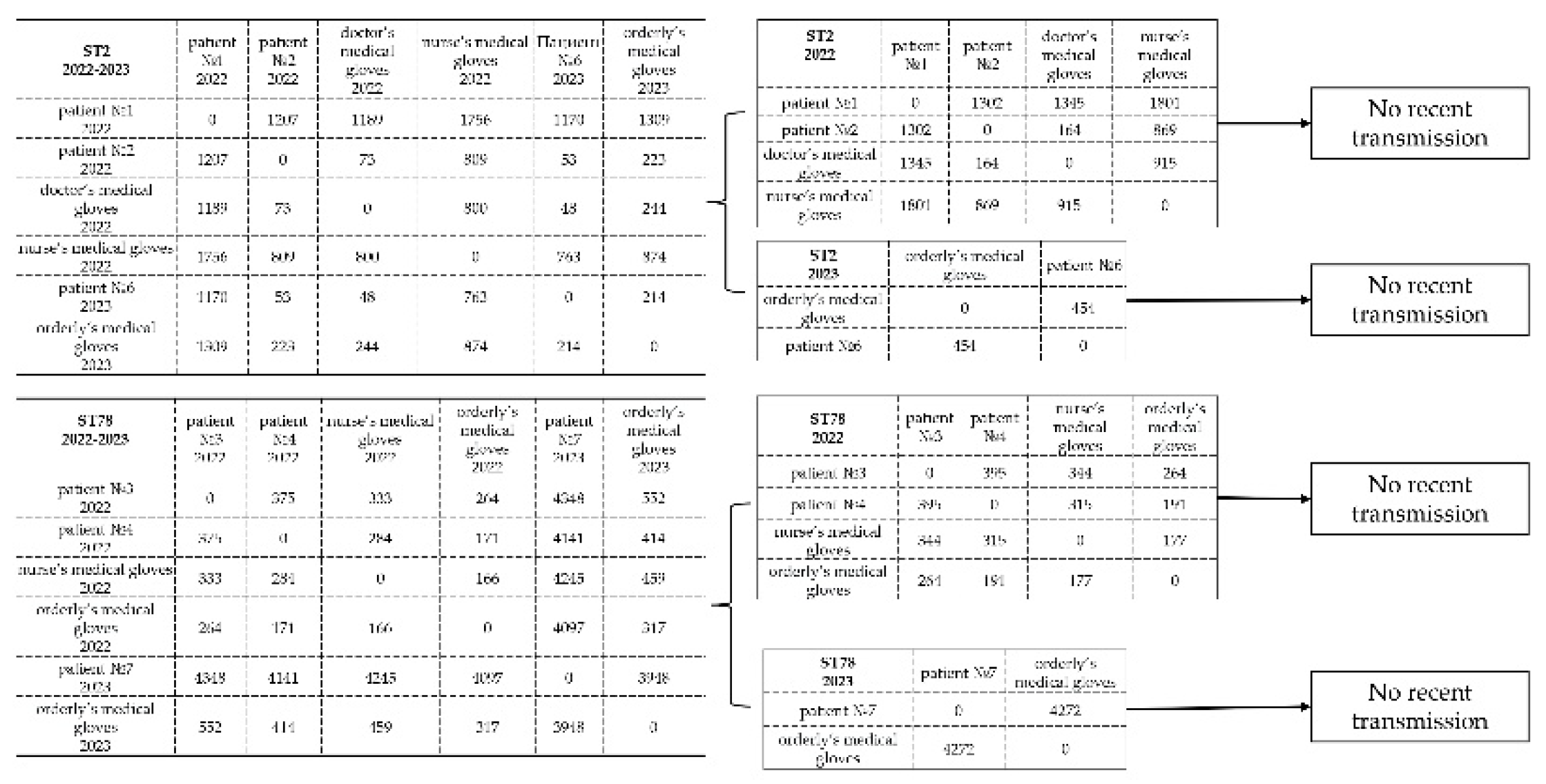
For A. baumannii, pairs of isolates from “patient – healthcare staff hands” were analysed within the two most prevalent sequence types (ST2 and ST78) over both study phases. When separated by year (2022 and 2023), no reliable close relatedness was observed between patient isolates and isolates from healthcare staff hands (distances exceeded threshold values). This indicates that independent subpopulations of A. baumannii circulate in the hospital, despite the high similarity of resistance and virulence profiles among some isolates (Figure 6).

Analysis of Klebsiella pneumoniae

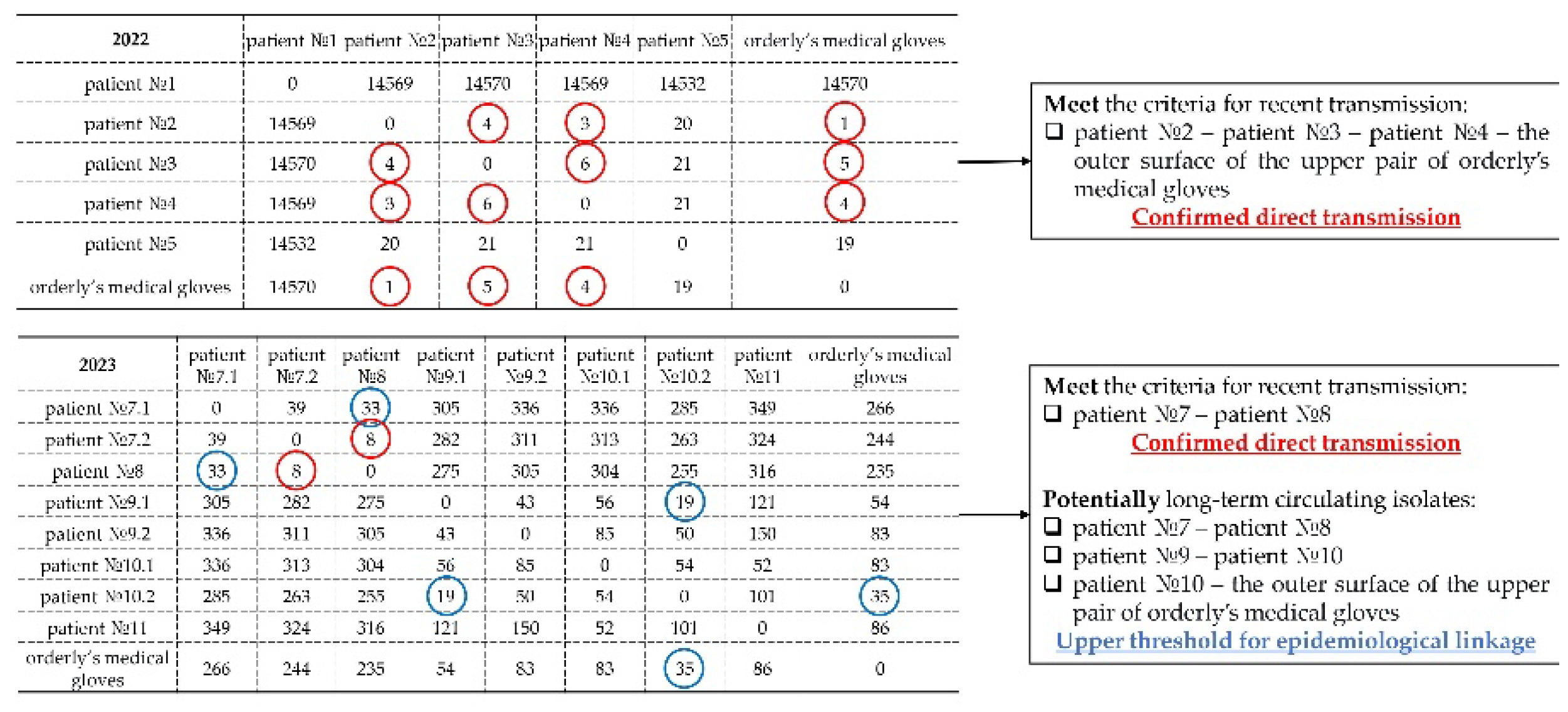
For K. pneumoniae in 2022, a high degree of genomic relatedness was found between isolates from three patients and from the orderly’s gloves (distance 1–6 SNPs), confirming recently transmission. In 2023, cases of recent transmission between two patients were also noted, as well as possible circulation of a K. pneumoniae subpopulation between patients and staff PPE (distances within the upper bound of relatedness). Thus, for K. pneumoniae, not only recently transmission from staff to patients was proven, but also circulation of isolates between patients and healthcare workers’ PPE (with possible subsequent transmission to other patients) (Figure 7).

3.4. Plasmid Pool and Its Role in the Spread of AMR

Plasmids play a key role in the horizontal transfer of AMR and virulence genes, enabling the rapid adaptation of bacterial populations to selective pressure in the hospital environment. In this study, an in silico analysis of plasmid replicons was performed for all isolates that underwent WGS.
All ten E. coli isolates (100%) carried plasmid replicons. The dominant types were IncFIA (60.0%), IncFIB (70.0%) and ColBS512 (50.0%). IncB/O/K/Z (20.0%), IncFII(pRSB107) (10.0%), IncHI2 (10.0%) and others were also detected. IncF group plasmids are widespread among Enterobacteriaceae and are often associated with the transfer of ESBL genes, including blaCTX-M-15, as well as carbapenemase genes [32,33]. In our study, isolates carrying IncFIA and IncFIB simultaneously harboured blaCTX-M-15 or blaTEM-1, confirming the role of these plasmids in the spread of ESBL phenotypes. The presence of ColBS512 (a replicon characteristic of colicinogenic plasmids) in 50.0% of isolates may indicate additional mechanisms of bacterial competition in the hospital setting [32]. The detection of IncB/O/K/Z – a group of plasmids often carrying heavy metal and aminoglycoside resistance genes – points to possible co-selection under the action of disinfectants [34].
Plasmid replicons were found in 17 of S. aureus isolates (85%). The most frequent were rep16 (55%), rep5a (40%), and rep20 (25%), rep21 (20%), repUS5 (20%), rep7c (15%) were also detected. The rep5a and rep16 plasmids are important vectors for the horizontal transfer of resistance genes, as confirmed by recent genomic studies [35]. rep20 and rep21, characteristic of the pT181 plasmid family, were associated with the tet(38) gene, conferring tetracycline resistance [36]. The rep7c replicon (pC194-like plasmid family) was linked to fosB and murA(G257D), confirming its role in the spread of fosfomycin resistance among staphylococci [37]. In isolates carrying repUS5, enterotoxin genes (seg, sei, sen, seo, seu) were additionally detected, suggesting a potential link between plasmids and virulence, although such an association is less common for staphylococci than for Gram-negative bacteria [38].
A wide diversity of plasmid replicons was found among K. pneumoniae isolates, reflecting the complex resistance structure of the population. The most common replicons were IncFIB(K) (71.7%), IncFII(K) (67.9%) and IncRNAI (64.2%). IncF family plasmids are major vectors for the spread of carbapenemases, including blaKPC-3, in Enterobacteriaceae, which is consistent with the high prevalence of IncFIB(K) and IncFII(K) among isolates of the dominant ST512 clone [39,40,41]. The IncX3 replicon, detected in 45.3% of isolates, is often associated with the transfer of blaKPC-2 and blaNDM in various geographical regions [42]. The presence of IncHI1B (34.0%) is of interest because plasmids of this group often carry heavy metal and biocide resistance genes, including qacEdelta1 [43]. IncL/M(pOXA-48) replicons, found in 13.2% of isolates, are classic carriers of blaOXA-48 [44]. Col440II (32.1%) and ColRNAI (64.2%) replicons belong to colicinogenic and low-copy-number plasmids that may participate in horizontal gene transfer, although their contribution to the spread of clinically relevant resistance is generally less pronounced [32].
Plasmid replicons were detected in 70% of E. faecalis isolates. The most frequent were rep9b (45%), repUS43 (35%). Replicons rep9a (25%), rep7a (25%) and rep6 (20%) were also detected. Family rep9 replicons are often involved in horizontal transfer of resistance genes among enterococci [45]. The absence of plasmids in 30% of isolates may indicate a chromosomal location of pathogenicity determinants.
All P. mirabilis isolates carried the IncC replicon, which has a broad host range and is often associated with the transfer of multidrug resistance genes, including blaCTX-M, rmtB and sul [46].
No plasmid replicons were detected in any of the P. aeruginosa isolates. This is typical for this species, as most resistance genes in P. aeruginosa are located on chromosomal integrated elements or on specific plasmids not detectable by standard replicon typing sets [29].

4. Discussion

The COVID-19 pandemic led to an unprecedented increase in HAIs and AMR, particularly in ICUs. Our study represents one of the first comprehensive analyses of the genetic landscape of OPs circulating in the ICUs of infectious diseases hospitals treating COVID-19 patients, using WGS. The data obtained allowed us not only to characterise the structure of the microbial population but also to identify key reservoirs, transmission pathway, and molecular mechanisms of pathogen adaptation within a closed hospital ecosystem.

4.1. Dominance of MDR Clones of Leading OPs

Among the obtained isolates OPs, K. pneumoniae and A. baumannii absolutely predominated (together accounting for 64.3% of isolates), which is consistent with global data on the leading role of these pathogens in the aetiology of HAIs during the pandemic [2,4]. Among K. pneumoniae, the dominant clone was ST512 (56.8% of typed isolates), characterised by the presence of the KPC-3 carbapenemase, a mutation in pmrB(R256G) associated with colistin resistance, and determinants of fluoroquinolone resistance (gyrA(S83I), oqxAB) and fosfomycin resistance (fosA). ST512 is one of the most successful global clones of KPC-producing K. pneumoniae, widespread in Europe, Asia and North America [39,40]. In our study, intra-hospital circulation of ST512 was established: isolates were recovered from nine patients, from seven PPE samples and from nine PCE, indicating insufficient effectiveness of infection control measures. The risk of developing resistance to disinfectants is a serious concern. The detection of the qacEdelta1 gene in the vast majority of isolates of the dominant ST512 clone (96%) is an important signal [47] that, nevertheless, requires careful interpretation.
The presence of the qacE gene or its variant qacEdelta1 alone does not always mean that the bacterium is phenotypically resistant to quaternary ammonium compound (QAC)-based disinfectants. Nevertheless, its high prevalence in the hospital microbiota is an alarming marker, indicating potential selection and the possibility of resistance gene spread among other microorganisms. When such markers are detected, monitoring the resistance of epidemiologically significant strains to the disinfectants in use is necessary – genetic data must be complemented by direct phenotypic studies. As recommended by the Russian national regulations (SanPiN 3.3686-21, clause 3577), such findings should trigger an assessment of the need for disinfectant rotation, i.e., sequential replacement of the active substance from one chemical group with another, following susceptibility testing of the hospital strain to the newly selected agent. Accordingly, it is necessary to determine the minimum inhibitory concentrations of QACs for the isolates and to assess their survival under the working concentrations and exposure times of disinfectants as regulated by the national normative documents.
Among A. baumannii, the dominant STs were ST2 (35.7%) and ST78 (30.4%). ST2 is a “global” clone responsible for most hospital outbreaks of CRAB worldwide [48,49]. In our study, all ST2 isolates carried the OXA-23 carbapenemase, which is consistent with data on the wide spread of blaOXA-23 among ST2 in Europe, Asia and the Americas [50]. Interestingly, the second most frequent type, ST78, was characterised by the absence of blaOXA-23 but the presence of blaOXA-72 and blaOXA-90. ST78 is also known as a successful hospital clone, often associated with outbreaks in European countries [51], and its detection in our hospitals confirms the global spread of this clone.
For E. coli, dominance of clonal complex CC14 (83.3% of typed isolates) was revealed, including the potentially highly virulent and MDR clone ST1193. ST1193 is a recently described globally emerging clone belonging to phylogroup B2, often associated with community-acquired and hospital-acquired urinary tract infections and bacteraemia [52,53]. In our study, ST1193 isolates carried blaCTX-M-15 (ESBL) and mutations in parC(80I) and conferring fluoroquinolone resistance, which is characteristic of this clone [54].
S. aureus in our study was represented by several CCs: CC15 (30%), CC8 (15%), CC22 (10%) and CC97 (10%). MRSA were detected exclusively among CC22 and some non-defined isolates. CC22 (specifically its variant EMRSA-15) is one of the most widespread hospital MRSA clones in Europe and worldwide [55]. During the COVID-19 pandemic, outbreaks of MRSA CC22 in COVID-19 units have been reported, linked to breaches in infection control protocols [56]. CC15 and CC8 (the latter includes the well-known USA300 clone) are more often associated with community-acquired infections; however, their detection on staff PPE indicates a potential role in hospital transmission [57]. Interestingly, CC97, traditionally associated with bovine mastitis, was isolated both from a patient and from PPE, which may indicate possible zoonotic transfer or adaptation of this clone to humans [58].
E. faecalis was characterised by a polyclonal structure with predominance of ST30 (25% of typed isolates) and ST40 (15%), which is consistent with a global meta-analysis showing a wide distribution of various STs of this species in clinical and ecological niches [59]. The high frequency of lsa(A) (85%) and erm(B) (45%) genes reflects the selective pressure typical of ICUs, where lincosamides and macrolides are widely used. The detection of the cytolysin gene (cyl) in 45% of isolates has clinical significance, as cytolysin is associated with increased virulence and mortality in enterococcal infections [60].
Both isolates of E. faecium belonged to ST52 – a hospital clone previously described as associated with nosocomial infections in Europe [61]. In our study, the ST52 isolates were identical in their resistance profile and plasmid pool, suggesting a clonal origin. The absence of virulence genes is typical for E. faecium, where pathogenicity is more linked to antibiotic resistance and persistence capacity than to toxigenicity [62]. The detection of ST52 on staff PPE indicates a potential role of this clone in the development of hospital infections.
Among P. mirabilis, ST269 and ST234 were typed – clones previously described in hospitals in Asia and Europe [63]. All isolates carried ESBL genes (blaCTX-M-3, blaCTX-M-14, blaTEM-1), which is consistent with the global trend of increasing ESBL-producing P. mirabilis strains, especially in Asian countries where the prevalence of blaCTX-M-14 reaches 11.3% [47,64]. The presence of rmtB (16S rRNA methyltransferase) in ST269 isolates is an alarming marker, as this gene confers high-level resistance to all aminoglycosides and has previously been detected in P. mirabilis in Taiwan and Korea [47,65]. The detection of IncC plasmids in all isolates confirms their key role in the spread of multidrug resistance in this species [63].
P. aeruginosa was represented by international high-risk clones: ST235, ST654 and ST549. ST235 is the dominant high-risk clone worldwide, associated with the production of various carbapenemases and high virulence (production of exotoxin ExoU) [66,67]. ST654 is also among the top ten high-risk clones and is often associated with blaVIM-2 metallo-β-lactamase [66,68]. In our study, the ST235 isolate carried blaGES-1, while ST654 isolates carried blaVIM-2, confirming the circulation of different carbapenemases in the same unit. The absence of plasmid replicons in all P. aeruginosa isolates is not unexpected: most resistance genes in this species are located on chromosomal integrated elements or on specific plasmids that are not detectable by standard replicon typing sets [69].

4.2. Personal Protective Equipment and the Hospital Environment as Critical Reservoirs of Pathogens

One of the most significant findings of our study is the high level of contamination of healthcare staffs’ PPE: 68.4% of environmental isolates were obtained from the surfaces of PPE suits and medical gloves. Particularly frequent isolation from PPE was observed for A. baumannii (47–64% of ST2 and ST78 isolates were found on PPE) and S. aureus (60–100% of CC8, CC22, CC15 and non-defined isolates were found on PPE). These data are consistent with a systematic review showing that PPE contamination levels can reach 77.5% and that prolonged PPE use (up to 12 hours or more) during the pandemic significantly increases the risk of pathogen transmission [7].
Phylogenetic and SNP analyses confirmed direct epidemiological links between isolates obtained from PPE and those from patients. Specifically, for K. pneumoniae, cases with core genome differences of 1–6 SNPs were identified, providing strong evidence of recent transmission [29]. These data highlight that PPE acts not only as a passive barrier but also as an active reservoir and vector for nosocomial transmission of pathogens, necessitating enhanced control over their use and replacement [70].
The temporal dynamics of PPE contamination showed peaks at 10:00 and 18:00, coinciding with the periods of most intensive ICU work (shift changes, invasive procedures, ward rounds). Similar patterns have been described in studies of surface contamination in ICUs [71]. The high level of contamination in the evening (18:00, 22:00) may reflect a cumulative effect of microorganism build-up during the work shift and calls for a review of PPE change frequency and disinfection procedures.

4.3. Value of Whole-Genome Sequencing and Core Genome SNP Analysis for Epidemiological Surveillance

Our study demonstrated the advantages of WGS over traditional typing methods. Classical MLST allowed only a rough stratification of isolates but did not enable assessment of microevolutionary changes. SNP analysis revealed significant heterogeneity within A. baumannii ST2 (>100 allele differences), indicating long-term circulation and adaptation of the clone within the hospital. Similar heterogeneity has been described for other hospital clones of A. baumannii [72].
For K. pneumoniae, SNP analysis not only confirmed recent transmission (differences of 1–6 SNPs) but also revealed cases with 35–349 SNP differences, which may correspond to the circulation of genetically related but not identical lineages within a single outbreak or reflect long-term persistence in the ICU [73]. In the literature, a threshold of 10–15 SNPs is commonly used to differentiate recent transmission from background circulation, although different criteria may apply for different species and time scales [29,74].
The application of WGS also allowed a unified characterisation of the resistome, virulome and plasmid pool of all isolates, providing an opportunity to assess the potential for horizontal gene transfer. The high frequency of IncF group plasmid replicons in E. coli and K. pneumoniae, as well as rep5a and rep16 in S. aureus, confirms their key role in the spread of resistance genes in the hospital environment [40]. The detection of biocide resistance determinants (qacEdelta1 in K. pneumoniae) further explains the ability of these isolates to persist for long periods on environmental surfaces [47].

4.4. Comparison with Results of Other Studies During the Pandemic

Our data are consistent with the results of multicentre studies showing an increased frequency of MDR OPs isolation during the COVID-19 pandemic. A study conducted in Italy involving 1,662 patients reported increased resistance of A. baumannii to gentamicin (from 70.9% to 91.3%) and colistin (from 3.2% to 12.3%), as well as increased ICU mortality [2]. Another study covering 41 Brazilian hospitals also showed dominance of A. baumannii and K. pneumoniae on ICU surfaces, with the authors highlighting the role of PPE as a major reservoir [75].
The spread of the A. baumannii ST2 clone carrying blaOXA-23 during the pandemic has been confirmed in studies from Croatia, Switzerland and Mexico [76,77,78], indicating the global nature of this phenomenon. The detection of A. baumannii ST78 with blaOXA-72 is also consistent with data on the spread of this clone in Europe [51].
Regarding E. coli ST1193, its high proportion among isolates in our study (40% of typed isolates) is comparable to data from China and the USA, where ST1193 has become the second most frequent ESBL-producing clone after ST131 [53,54]. Interestingly, in our study ST1193 formed a separate phylogenetic cluster, which may indicate endemic circulation of this lineage in the hospital.
For S. aureus, the dominance of CC22 MRSA and CC15 among isolates from PPE agrees with the results of a recent study from China, where CC22 was the most frequent MRSA clone in COVID-19 units, while CC15 predominated among methicillin-susceptible isolates [57]. The detection of CC97 in our hospital requires further investigation, as this clone is traditionally associated with livestock, but cases of human isolation have been described in recent years [59].

4.5. Study Limitations

Our study has several limitations. First, the study was conducted in two infectious diseases hospitals located in different cities of the Sverdlovsk region, which may limit the generalisability of the results to other regions and types of hospitals. Second, resistance determination was performed in silico based on genomic data, without phenotypic confirmation by disc diffusion or serial dilution methods. However, ResFinder and AMRFinderPlus have high predictive value (sensitivity and specificity >95% for most antibiotic classes) [19,20]. Third, the time frame of the study (2021–2023) limits the ability to assess long-term trends, especially in the post-pandemic period. Fourth, single isolates of some species (K. variicola) do not allow general conclusions to be drawn about their role in the epidemic process. Moreover, for seven out of 175 isolates (4%), complete genome assemblies could not be obtained due to short contigs that did not cover the full genome; consequently, their sequences were not deposited in GenBank, which did not affect the main conclusions of the study.

5. Conclusions

Thus, our study provided a comprehensive genetic characterisation of OPs circulating in the ICUs of COVID-19 hospitals, revealed the dominance of MDR clones of international significance, confirmed the key role of PPE and the hospital environment in infection transmission, and demonstrated the high resolution of WGS and SNP analysis for epidemiological surveillance. The obtained results justify the need to strengthen infection control measures and to implement genomic monitoring to prevent the spread of MDR OPs in infectious disease hospitals.

Supplementary Materials

The following supporting information can be downloaded at: Preprints.org, Table S1: Complete list of antimicrobial resistance genes for all 175 isolates. Table S2: Complete list of virulence determinants for all 175 isolates. Table S3: Complete list of plasmid replicons for all 175 isolates.

Author Contributions

Conceptualization, S.S.S., A.V.S. and N.N.Z.; methodology, S.S.S., A.V.S., D.D.A. and T.M.I.; software, D.D.A.; validation, Y.S.S., A.A.K. and N.N.Z.; formal analysis, D.D.A.; investigation, S.S.S., Y.S.S., A.A.K., N.N.Z. and T.M.I.; data curation, D.D.A.; writing—original draft preparation, D.D.A. and S.S.S.; writing—review and editing, all authors; visualization, D.D.A.; supervision, S.S.S.; project administration, S.S.S.; funding acquisition, A.V.S. All authors have read and agreed to the published version of the manuscript.

Funding

This research was funded by the Russian scientific project “Improving Epidemiological Monitoring of Healthcare-Associated Infections of Viral and Bacterial Etiology in the Ural and Siberian Federal Districts from the Standpoint of Genomic Epidemiological Surveillance”, Reg. No. 126021116957-7.

Institutional Review Board Statement

This study was conducted in accordance with the Declaration of Helsinki and approved by the Local Ethics Committee of the Federal Scientific Research Institute of Viral Infections «Virome» Rospotrebnadzor (protocol code № 1, date of approval 20 February 2026).

Data Availability Statement

The raw sequencing data generated in this study have been deposited in the NCBI SRA database under BioProject accession number PRJNA1165946 (A. baumannii), PRJNA1180224 (E. coli), PRJNA1178453 (S. aureus), PRJNA1348999 (K. pneumoniae and K. variicola), PRJNA1348956 (E. faecalis), PRJNA1348989 (E. faecium), PRJNA1348996 (P. mirabilis), PRJNA1348997 (P. aeruginosa).

Conflicts of Interest

The authors declare no conflicts of interest.

Abbreviations

The following abbreviations are used in this manuscript:
AMR Antimicrobial resistance
CC Clonal complex
CRAB Carbapenem-resistant A. baumannii
cgMLST Core-genome MLST
ICUs Intensive care units
HAIs Healthcare-associated infections
OP Opportunistic pathogen
MDR Multidrug-resistant
MLST Multi-locus sequence typing
PPE Personal protective equipment
SNP Single-nucleotide polymorphism
ST Sequence type
WGS Whole-genome sequencing
PCE Patient care environment
GHP General hospital point

Appendix A

Appendix A.1

Table A1. Sampling points.
ID Stage Selection group Selection point GB accession
ab_01 III Patient sputum JBITPY000000000
ab_02 III Patient sputum JBITPX000000000
ab_03 II Patient urethra JBITPW000000000
ab_04 II Patient sputum JBHYBP000000000
ab_05 II Patient sputum JBHYBQ000000000
ab_06 II Patient sputum JBHYBR000000000
ab_07 II Patient oropharyngeal swab JBITPZ000000000
ab_08 II Patient oropharyngeal swab JBHYBS000000000
ab_09 II Patient oropharyngeal swab JBHYBT000000000
ab_10 III PCE the outer surface or the ventilator JBRYPU000000000
ab_11 III PCE the surface of medical manipulation table JBITQH000000000
ab_12 III PPE the outer surface or the orderly's PPE suit JBITQG000000000
ab_13 III PPE the outer surface or the orderly's PPE suit JBITQA000000000
ab_14 III PPE the outer surface or the upper pair of orderly's medical gloves JBHYBU000000000
ab_15 III PCE handrails and adjustment levers on an ICU bed JBHYBX000000000
ab_16 III PPE the outer surface or the nurse's PPE suit JBITQK000000000
ab_17 III PPE the outer surface or the doctor's PPE suit JBHYBW000000000
ab_18 III PCE handrails and adjustment levers on an ICU bed JBHYCH000000000
ab_19 III PCE handrails and adjustment levers on an ICU bed JBHYCE000000000
ab_20 III PCE the outer surface or the ventilator JBHYBY000000000
ab_21 III PCE the surface of medical manipulation table JBITQZ000000000
ab_22 III PPE the outer surface or the orderly's PPE suit JBITQX000000000
ab_23 III PPE the outer surface or the orderly's PPE suit JBITQW000000000
ab_24 III PPE the outer surface or the upper pair of orderly's medical gloves JBITQV000000000
ab_25 III PPE the outer surface or the nurse's PPE suit JBHYCJ000000000
ab_26 III PPE the outer surface or the nurse's PPE suit JBITRA000000000
ab_27 II PCE the surface of medical manipulation table JBITQB000000000
ab_28 II PCE the outer surface of the medical syringe dispenser JBITQC000000000
ab_29 II PPE the outer surface or the upper pair of doctor's medical gloves JBHYBV000000000
ab_30 II PPE the outer surface or the upper pair of nurse's medical gloves JBITQD000000000
ab_31 II PPE the outer surface or the orderly's PPE suit JBITQE000000000
ab_32 II PPE the outer surface or the doctor's PPE suit JBITQF000000000
ab_33 II PCE handrails and adjustment levers on an ICU bed JBITQI000000000
ab_34 II PCE the outer surface of the medical syringe dispenser
ab_35 II GHP ICU door handles JBITQJ000000000
ab_36 II PCE the surface of medical manipulation table JBITQL000000000
ab_37 II PCE handrails and adjustment levers on an ICU bed
ab_38 II PCE the outer surface or the ventilator JBITQM000000000
ab_39 II PCE handrails and adjustment levers on an ICU bed JBHYBZ000000000
ab_40 II PCE the outer surface or the ventilator JBHYCA000000000
ab_41 II PPE the outer surface or the upper pair of orderly's medical gloves JBHYCB000000000
ab_42 II PPE the outer surface or the upper pair of nurse's medical gloves JBITQN000000000
ab_43 II PPE the outer surface or the nurse's PPE suit JBITQO000000000
ab_44 II PPE the outer surface or the doctor's PPE suit JBHYCC000000000
ab_45 II PCE handrails and adjustment levers on an ICU bed JBHYCD000000000
ab_46 II PCE the outer surface or the ventilator JBRYPS000000000
ab_47 II PCE the surface of medical manipulation table JBITQP000000000
ab_48 II PCE handrails and adjustment levers on an ICU bed JBITQQ000000000
ab_49 II GHP the outer surface of the suction unit JBHYCF000000000
ab_50 II GHP dispensers for liquid soap and hand sanitizer JBITQR000000000
ab_51 II PPE the outer surface or the nurse's PPE suit JBITQS000000000
ab_52 II PPE the outer surface or the orderly's PPE suit JBHYCG000000000
ab_53 III PCE handrails and adjustment levers on an ICU bed JBRYPT000000000
ab_54 II PPE the outer surface or the doctor's PPE suit
ab_55 II PCE handrails and adjustment levers on an ICU bed JBITQT000000000
ab_56 II GHP ICU door handles JBITQU000000000
ab_57 II PPE the outer surface or the upper pair of doctor's medical gloves JBHYCI000000000
ab_58 II PCE the outer surface or the ventilator JBSHXN000000000
ab_59 II PCE the surface of medical manipulation table JBITQY000000000
ab_60 II PPE the outer surface or the orderly's PPE suit JBHYCK000000000
kp_01 III Patient sputum JBRXXF000000000
kp_02 III Patient sputum JBRXXG000000000
kp_03 III Patient sputum JBRXXD000000000
kp_04 III Patient sputum JBRXXE000000000
kp_05 III Patient sputum JBRXXB000000000
kp_06 III Patient sputum JBRXWZ000000000
kp_07 III Patient sputum JBRXXA000000000
kp_08 III Patient sputum JBRXWW000000000
kp_09 III Patient sputum JBRXWV000000000
kp_10 III Patient sputum JBRXWX000000000
kp_11 II Patient sputum JBRXXC000000000
kp_12 II Patient sputum JBRXWY000000000
kp_13 II Patient sputum JBRXWU000000000
kp_14 II Patient sputum JBRXWT000000000
kp_15 II Patient sputum JBRXWS000000000
kp_16 III PCE handrails and adjustment levers on an ICU bed JBRYKD000000000
kp_17 III PPE the outer surface or the orderly's PPE suit JBRYKG000000000
kp_18 III PPE the outer surface or the upper pair of orderly's medical gloves JBRYKH000000000
kp_19 III PCE the outer surface or the ventilator JBRYJY000000000
kp_20 III PCE the outer surface of the medical syringe dispenser JBRYJZ000000000
kp_21 III PCE handrails and adjustment levers on an ICU bed JBRYKA000000000
kp_22 III PPE the outer surface or the orderly's PPE suit JBRYKB000000000
kp_23 III PPE the outer surface or the nurse's PPE suit JBRYKC000000000
kp_24 III PCE handrails and adjustment levers on an ICU bed JBSBGB000000000
kp_25 III PCE handrails and adjustment levers on an ICU bed JBRYJR000000000
kp_26 III PCE handrails and adjustment levers on an ICU bed JBRYJX000000000
kp_27 III PPE the outer surface or the nurse's PPE suit JBRYJL000000000
kp_28 III PCE the outer surface or the ventilator JBRYJQ000000000
kp_29 II PPE the outer surface or the doctor's PPE suit JBRYKI000000000
kp_30 II PCE the outer surface of the medical syringe dispenser JBRYKF000000000
kp_31 II GHP ICU electric light switches JBRYKE000000000
kp_32 II PCE the outer surface of the medical syringe dispenser JBRYJW000000000
kp_33 II PPE the outer surface or the nurse's PPE suit JBRYJV000000000
kp_34 II PPE the outer surface or the orderly's PPE suit JBRYJU000000000
kp_35 II PPE the outer surface or the doctor's PPE suit JBRYJT000000000
kp_36 II PCE the surface of medical manipulation table JBRYJS000000000
kp_37 II PCE the outer surface or the ventilator JBRYJP000000000
kp_38 II GHP the outer surface of the suction unit JBRYJO000000000
kp_39 II PCE the outer surface or the ventilator JBRYJN000000000
kp_40 II PCE handrails and adjustment levers on an ICU bed JBRYJM000000000
kp_41 II PCE the outer surface or the ventilator JBUAPN000000000
kp_42 II GHP the outer surface of the suction unit JBRYJK000000000
kp_43 II GHP dispensers for liquid soap and hand sanitizer JBRYJJ000000000
kp_44 II PPE the outer surface or the upper pair of orderly's medical gloves JBRYJI000000000
kp_45 II PCE handrails and adjustment levers on an ICU bed JBRYJH000000000
kp_46 II PCE the outer surface or the ventilator JBRYJG000000000
kp_47 I GHP clinical doctor's work-space JBRYJF000000000
kp_48 I PPE the outer surface or the doctor's PPE suit JBRYJE000000000
kp_49 I PPE the outer surface or the upper pair of doctor's medical gloves JBRYJD000000000
kp_50 I GHP the outer surface of the suction unit JBRYJC000000000
kp_51 I PPE the outer surface or the doctor's PPE suit JBRYJB000000000
kp_52 I PPE the outer surface or the nurse's PPE suit JBRYJA000000000
kp_53 I PPE the outer surface or the orderly's PPE suit JBRYIZ000000000
sa_01 II Patient oropharyngeal swab JBIYQB000000000
sa_02 II Patient sputum JBIYQA000000000
sa_03 II Patient sputum JBIYPZ000000000
sa_04 III PCE handrails and adjustment levers on an ICU bed JBIYQL000000000
sa_05 III PPE the outer surface or the orderly's PPE suit JBIYQM000000000
sa_06 III PPE the outer surface or the orderly's PPE suit JBIYQN000000000
sa_07 III PPE the outer surface or the upper pair of nurse's medical gloves JBIYQO000000000
sa_08 III PCE the surface of medical manipulation table JBIYQJ000000000
sa_09 III PPE the outer surface or the doctor's PPE suit JBIYQK000000000
sa_10 III PCE handrails and adjustment levers on an ICU bed JBIYQQ000000000
sa_11 II PPE the outer surface or the nurse's PPE suit
sa_12 II PPE the outer surface or the upper pair of orderly's medical gloves JBIYQP000000000
sa_13 II PPE the outer surface or the doctor's PPE suit JBIYRD000000000
sa_14 II PPE the outer surface or the upper pair of nurse's medical gloves JBIYQI000000000
sa_15 II PPE the outer surface or the doctor's PPE suit JBIYQH000000000
sa_16 II PPE the outer surface or the doctor's PPE suit JBIYQG000000000
sa_17 II PPE the outer surface or the doctor's PPE suit JBIYQF000000000
sa_18 I PPE the outer surface or the upper pair of doctor's medical gloves JBIYQE000000000
sa_19 I PPE the outer surface or the upper pair of nurse's medical gloves JBIYQD000000000
sa_20 I PPE the outer surface or the nurse's PPE suit JBIYQC000000000
ec_01 II Patient urine JBRYYL000000000
ec_02 II Patient sputum
ec_03 II PPE the outer surface or the upper pair of doctor's medical gloves
ec_04 I PCE the surface of medical manipulation table JBIYQW000000000
ec_05 I PPE the outer surface or the doctor's PPE suit JBIYQV000000000
ec_06 I PPE the outer surface or the nurse's PPE suit JBRYYK000000000
ec_07 I PCE handrails and adjustment levers on an ICU bed JBIYQT000000000
ec_08 I PPE the outer surface or the nurse's PPE suit JBIYQS000000000
ec_09 I PCE the surface of medical manipulation table JBIYQR000000000
ec_10 I PPE the outer surface or the doctor's PPE suit
efs_01 III Patient sputum JBRXJJ000000000
efs_02 II Patient sputum JBRXJK000000000
efs_03 III PCE handrails and adjustment levers on an ICU bed JBUAPO000000000
efs_04 II Patient sputum JBRXJI000000000
efs_05 I PPE the outer surface or the upper pair of doctor's medical gloves JBRXKA000000000
efs_06 I PPE the outer surface or the upper pair of nurse's medical gloves JBRXJZ000000000
efs_07 I PPE the outer surface or the doctor's PPE suit JBRXJY000000000
efs_08 I PPE the outer surface or the orderly's PPE suit JBRXJX000000000
efs_09 I PCE the surface of medical manipulation table JBRXJW000000000
efs_10 I PPE the outer surface or the doctor's PPE suit JBRXJV000000000
efs_11 I PPE the outer surface or the upper pair of doctor's medical gloves JBRXJU000000000
efs_12 I PPE the outer surface or the doctor's PPE suit JBRXJT000000000
efs_13 I PPE the outer surface or the nurse's PPE suit JBRXJS000000000
efs_14 I PPE the outer surface or the upper pair of doctor's medical gloves JBRXJR000000000
efs_15 I PPE the outer surface or the doctor's PPE suit JBRXJQ000000000
efs_16 I PPE the outer surface or the orderly's PPE suit JBRXJP000000000
efs_17 I PCE the surface of medical manipulation table JBRXJO000000000
efs_18 I PCE handrails and adjustment levers on an ICU bed JBRXJN000000000
efs_19 I PPE the outer surface or the doctor's PPE suit JBRXJM000000000
efs_20 I PPE the outer surface or the nurse's PPE suit JBRXJL000000000
efm_01 I PPE the outer surface or the upper pair of doctor's medical gloves JBRXVA000000000
efm_02 I PPE the outer surface or the upper pair of nurse's medical gloves JBRXUZ000000000
pm_01 II Patient sputum JBWGWX000000000
pm_02 II Patient sputum JBWGWW000000000
pm_03 II PPE the outer surface or the nurse's PPE suit JBWGXA000000000
pm_04 II PCE the surface of medical manipulation table JBWGWZ000000000
pm_05 II PPE the outer surface or the upper pair of orderly's medical gloves JBWGWY000000000
pa_01 II Patient sputum JBRXVJ000000000
pa_02 II Patient sputum JBRXVI000000000
pa_03 II GHP oxygen pipeline valves JBRXVH000000000
pa_04 I PPE the outer surface or the doctor's PPE suit JBRXVG000000000
kv_01 II PPE the outer surface or the upper pair of doctor's medical gloves JBSCEI000000000
Notes: ab – A. baumannii, kp – K. pneumoniae, sa –S. aureus, ec – E. coli, efs – E. faecalis, efm – E. faecium, pm – P. mirabilis, pa – P. aeruginosa, kv – K. variivola ;PPE—personal protective equipment; PCE—patient care environment; GHP—general hospital point.

References

  1. Peconi, C.; Martini, E.; Sarti, D.; Prospero, E. Impact of the COVID-19 pandemic on healthcare-associated infections and MDR microorganisms in Italy: A systematic review. J. Infect. Public Health 2025, 18, 102729. [CrossRef]
  2. Investigation of antibiotic resistance against pathogens isolated from respiratory samples in intensive care units after SARS-CoV-2 pandemic. BMC Infect. Dis. 2025, 25, 1691. [CrossRef]
  3. Kutyrev, V.V.; Popova, A.Y.; Smolensky, V.Y.; Ezhlova, E.B.; Demina, Y.V.; Safronov, V.A.; Karnaukhov, I.G.; Ivanova, A.V.; Shcherbakova, S.A. Epidemiological Peculiarities of New Coronavirus Infection (COVID-19). Message 2. Probl. Osob. Opasnykh Infektsii 2020, 2, 6–12. (In Russian). [CrossRef]
  4. Lansbury, L.; Lim, B.; Baskaran, V.; Lim, W.S. Co-infections in people with COVID-19: A systematic review and meta-analysis. J. Infect. 2020, 81, 266–275. [CrossRef]
  5. Farzana, R.; et al. The impact of the COVID-19 pandemic on antimicrobial usage: An international patient-level cohort study. JAC Antimicrob. Resist. 2025, 7, dlaf037. [CrossRef]
  6. Emdin, F.; Orubu, E.S.F.; Rogers Van Katwyk, S.; Strong, K.; Shaver, N.; Abdullah, K.; Asamoah, G.; Skidmore, B.; Chan, E.; Poirier, M.J.P. COVID-19 impact on AMR: A rapid scoping review, equity analysis and evidence gap map study. BMJ Glob. Health 2025, 10, e018118. [CrossRef]
  7. Orsel, L.M.; Severin, J.A.; Knoester, M.; et al. The role of gowns in preventing nosocomial transmission of respiratory viruses: A systematic review. J. Hosp. Infect. 2025, 163, 57–71. [CrossRef]
  8. Dhandapani, S.; Priyadarshi, K.; Benedict Vinothini, A.; et al. Donning of personal protective equipment under direct supervision–A quality improvement study in COVID-ICUs of a tertiary care facility, South India. IP Int. J. Med. Microbiol. Trop. Dis. 2024, 10, 357–363. [CrossRef]
  9. Shropshire, W.C.; Hanson, B.M.; Shelburne, S.A. Genome-wide approaches to bacterial strain typing: A history and review of recent methodological advances. Curr. Opin. Infect. Dis. 2025, 38, 329–338. [CrossRef]
  10. Szarvas, J.; Bartels, M.D.; Westh, H.; Lund, O. Rapid Open-Source SNP-Based Clustering Offers an Alternative to Core Genome MLST for Outbreak Tracing in a Hospital Setting. Front. Microbiol. 2021, 12, 636608. [CrossRef]
  11. Kaplan, E.; Lurie-Weinberger, M.N.; Rakov, C.; et al. Enterobacter cloacae complex taxonomic incongruence may lead to misidentification of outbreaks and hinder infection control in real time. Eur. J. Clin. Microbiol. Infect. Dis. 2025, 44, 3097–3101. [CrossRef]
  12. Waggle, K.D.; Griffith, M.P.; Rokes, A.B.; et al. Methods for cost-efficient, whole genome sequencing surveillance for enhanced detection of outbreaks in a hospital setting. medRxiv 2024, 2024.02.16.24302955. [CrossRef]
  13. Mustapha, M.M.; Srinivasa, V.R.; Griffith, M.P.; Cho, S.-T.; Evans, D.R.; Waggle, K.; Ezeonwuka, C.; Snyder, D.J.; Marsh, J.W.; Harrison, L.H.; et al. Genomic Diversity of Hospital-Acquired Infections Revealed through Prospective Whole-Genome Sequencing-Based Surveillance. mSystems 2022, 7, e0138421. [CrossRef]
  14. Smirnova, S.S.; Zhuikov, N.N.; Egorov, I.A.; Pushkareva, N.A.; Semenov, A.V. Sampling Scheme of Flushes from Environmental Objects for Simultaneous Assessment of Viral and Bacterial Contamination: No. 2022501675. Russian Federation Industrial Design Patent No. 132971. 5 September 2022. (In Russian).
  15. Smirnova, S.S.; Avdyunin, D.D.; Holmanskikh, M.V.; Stagilskaya, Y.S.; Zhuikov, N.N.; Itani, T.M. Genetic Characteristics of Acinetobacter baumannii Isolates Circulating in an Intensive Care Unit of an Infectious Diseases Hospital During the COVID-19 Pandemic. Pathogens 2025, 14, 961. [CrossRef]
  16. Vasilinetc, I.; Prjibelski, A.D.; Gurevich, A.; Korobeynikov, A.; Pevzner, P.A. Assembling short reads from jumping libraries with large insert sizes. Bioinformatics 2015, 31, 3262–3268. [CrossRef]
  17. Astashyn, A.; Tvedte, E.S.; Sweeney, D.; Sapojnikov, V.; Bouk, N.; Joukov, V.; Mozes, E.; Strope, P.K.; Sylla, P.M.; Wagner, L.; et al. Rapid and Sensitive Detection of Genome Contamination at Scale with FCS-GX. Genome Biol. 2024, 25, 60. [CrossRef]
  18. Chen, S.; Zhou, Y.; Chen, Y.; Gu, J. fastp: An ultra-fast all-in-one FASTQ preprocessor. Bioinformatics 2018, 34, i884–i890. [CrossRef]
  19. Bortolaia, V.; Kaas, R.S.; Ruppe, E.; Roberts, M.C.; Schwarz, S.; Cattoir, V.; Philippon, A.; Allesoe, R.L.; Rebelo, A.R.; Florensa, A.F.; et al. ResFinder 4.0 for predictions of phenotypes from genotypes. J. Antimicrob. Chemother. 2020, 75, 3491–3500. [CrossRef]
  20. Feldgarden, M.; Brover, V.; Gonzalez-Escalona, N.; Frye, J.G.; Haendiges, J.; Haft, D.H.; Hoffmann, M.; Pettengill, J.B.; Prasad, A.B.; Tillman, G.E.; et al. AMRFinderPlus and the Reference Gene Catalog facilitate examination of the genomic links among antimicrobial resistance, stress response, and virulence. Sci. Rep. 2021, 11, 12728. [CrossRef]
  21. Seemann, T. ABRicate: Mass screening of contigs for antibiotic resistance genes. GitHub. 2016. Available online: https://github.com/tseemann/abricate.
  22. Carattoli, A.; Hasman, H. PlasmidFinder and In Silico pMLST: Identification and Typing of Plasmid Replicons in Whole-Genome Sequencing (WGS). Methods Mol. Biol. 2020, 2075, 285–294. [CrossRef]
  23. Seemann, T. Prokka: Rapid prokaryotic genome annotation. Bioinformatics 2014, 30, 2068–2069. [CrossRef]
  24. Page, A.J.; Cummins, C.A.; Hunt, M.; et al. Roary: Rapid large-scale prokaryote pan genome analysis. Bioinformatics 2015, 31, 3691–3693. [CrossRef]
  25. Stamatakis, A. RAxML version 8: A tool for phylogenetic analysis and post-analysis of large phylogenies. Bioinformatics 2014, 30, 1312–1313. [CrossRef]
  26. Kalyaanamoorthy, S.; Minh, B.Q.; Wong, T.K.F.; von Haeseler, A.; Jermiin, L.S. ModelFinder: Fast model selection for accurate phylogenetic estimates. Nat. Methods 2017, 14, 587–589. [CrossRef]
  27. Seemann, T. snp-dists. GitHub. 2019. Available online: https://github.com/tseemann/snp-dists.
  28. Schürch, A.C.; Arredondo-Alonso, S.; Willems, R.J.L.; et al. Whole genome sequencing options for bacterial strain typing and epidemiologic analysis based on single nucleotide polymorphism versus gene-by-gene-based approaches. Clin. Microbiol. Infect. 2018, 24, 350–354. [CrossRef]
  29. Gorrie, C.L.; Mirčeta, M.; Wick, R.R.; et al. Genomic dissection of Klebsiella pneumoniae infections in hospital patients reveals insights into an opportunistic pathogen. Nat. Commun. 2022, 13, 3017. [CrossRef]
  30. Romano-Bertrand, S.; Virieux-Petit, M.; Mauffrey, F.; Senn, L.; Blanc, D.S. Defining a genomic threshold for investigating Pseudomonas aeruginosa hospital outbreak. J. Hosp. Infect. 2025, 161, 119–129. [CrossRef]
  31. Cui, X.; Lin, C.; Zhu, W.; Zhang, X.; Wang, W.; Ye, L.; Liu, W.; Liu, S. Whole-genome sequencing identifies persistent transmission of a high-risk ST2 Acinetobacter baumannii clone in a Guangzhou Hospital. Front. Microbiol. 2026, 17, 1730485. [CrossRef]
  32. Rozwandowicz, M.; Brouwer, M.S.M.; Fischer, J.; Wagenaar, J.A.; Gonzalez-Zorn, B.; Guerra, B.; Mevius, D.J.; Hordijk, J. Plasmids carrying antimicrobial resistance genes in Enterobacteriaceae. J. Antimicrob. Chemother. 2018, 73, 1121–1137. [CrossRef]
  33. Rocha-Gracia, R.D.C.; Lozano-Zarain, P.; Gutiérrez Cázarez, Z.; et al. IncFIB plasmids carrying the resistance gene blaCTX-M-15 in ESBL-producing Escherichia coli clones from pediatric patients. J. Infect. Dev. Ctries. 2022, 16, 500–506. [CrossRef]
  34. Johnson, T.J.; Nolan, L.K. Pathogenomics of the virulence plasmids of Escherichia coli. Microbiol. Mol. Biol. Rev. 2009, 73, 750–774. [CrossRef]
  35. Alhejaili, A.Y.; Zhou, G.; Halawa, H.; et al. Methicillin-resistant Staphylococcus aureus in Saudi Arabia: Genomic evidence of recent clonal expansion and plasmid-driven resistance dissemination. Front. Microbiol. 2025, 16, 1602985. [CrossRef]
  36. Schwarz, S.; Shen, J.; Kadlec, K.; Wang, Y.; Michael, G.B.; Feßler, A.T.; Vester, B. Lincosamides, Streptogramins, Phenicols, and Pleuromutilins: Mode of Action and Mechanisms of Resistance. Cold Spring Harb. Perspect. Med. 2016, 6, a027037. [CrossRef]
  37. Xu, W.; Chen, T.; Wang, H.; Zeng, W.; Wu, Q.; Yu, K.; Xu, Y.; Zhang, X.; Zhou, T. Molecular Mechanisms and Epidemiology of Fosfomycin Resistance in Staphylococcus aureus Isolated From Patients at a Teaching Hospital in China. Front. Microbiol. 2020, 11, 1290. [CrossRef]
  38. Umeda, K.; Nakamura, H.; Yamamoto, K.; et al. Molecular and epidemiological characterization of staphylococcal foodborne outbreak of Staphylococcus aureus harboring seg, sei, sem, sen, seo, and selu genes without production of classical enterotoxins. Int. J. Food Microbiol. 2017, 256, 30–35. [CrossRef]
  39. Mathers, A.J.; Peirano, G.; Pitout, J.D. The role of epidemic resistance plasmids and international high-risk clones in the spread of MDR Enterobacteriaceae. Clin. Microbiol. Rev. 2015, 28, 565–591. [CrossRef]
  40. Carattoli, A. Plasmids and the spread of resistance. Int. J. Med. Microbiol. 2013, 303, 298–304. [CrossRef]
  41. Bi, D.; Zheng, J.; Li, J.; et al. In Silico Typing and Comparative Genomic Analysis of IncFII K Plasmids and Insights into the Evolution of Replicons, Plasmid Backbones, and Resistance Determinant Profiles. Antimicrob. Agents Chemother. 2018, 62, e00764-18. [CrossRef]
  42. Cai, M.; et al. Global spread and evolution of KPC-2 and NDM-1-producing Gram-negative bacteria. Sci. China Life Sci. 2025/2026. [CrossRef]
  43. Mendoza-Guido, B.; Durán-Méndez, S.; Barrantes, K.; et al. Genomic analysis of IncH plasmids reveals their role as drivers of antimicrobial resistance and adaptive traits in enterobacterales. FEMS Microbiol. Lett. 2026, 373. [CrossRef]
  44. Poirel, L.; Bonnin, R.A.; Nordmann, P. Genetic features of the widespread plasmid coding for the carbapenemase OXA-48. Antimicrob. Agents Chemother. 2012, 56, 559–562. [CrossRef]
  45. Osunla, C.A.; et al. Genomic and bioinformatic insights into Enterococcus faecalis from retail meats in Nigeria. Int. J. Microbiol. 2025, 2025, 7325430. [CrossRef]
  46. Park, Y.J.; et al. Chromosome-encoded AmpC and CTX-M extended-spectrum β-lactamases in clinical isolates of Proteus mirabilis from Korea. Antimicrob. Agents Chemother. 2011, 55, 1414–1419. [CrossRef]
  47. Wand, M.E.; Bock, L.J.; Bonney, L.C.; et al. Mechanisms of increased resistance to chlorhexidine and cross-resistance to colistin following exposure of Klebsiella pneumoniae clinical isolates to chlorhexidine. Antimicrob. Agents Chemother. 2017, 61, e01162-16. [CrossRef]
  48. Hamidian, M.; Nigro, S.J. Emergence, molecular mechanisms and global spread of carbapenem-resistant Acinetobacter baumannii. Microb. Genom. 2019, 5, e000306. [CrossRef]
  49. Karah, N.; Sundsfjord, A.; Towner, K.; Samuelsen, Ø. Insights into the global molecular epidemiology of carbapenem non-susceptible clones of Acinetobacter baumannii. Drug Resist. Updat. 2012, 15, 237–247. [CrossRef]
  50. Mugnier, P.D.; Poirel, L.; Naas, T.; Nordmann, P. Worldwide dissemination of the blaOXA-23 carbapenemase gene of Acinetobacter baumannii. Emerg. Infect. Dis. 2010, 16, 35–40. [CrossRef]
  51. Karah, N.; Haldorsen, B.; Hegstad, K.; et al. Species identification and molecular characterization of Acinetobacter spp. blood culture isolates from Norway. J. Antimicrob. Chemother. 2011, 66, 738–744. [CrossRef]
  52. Tchesnokova, V.L.; Rechkina, E.; Chan, D.; et al. Pandemic uropathogenic fluoroquinolone-resistant Escherichia coli have enhanced ability to persist in the gut and cause bacteriuria in healthy women. Clin. Infect. Dis. 2020, 70, 937–939. [CrossRef]
  53. Pitout, J.D.D.; Peirano, G.; Chen, L.; et al. Escherichia coli ST1193: Following in the footsteps of E. coli ST131. Antimicrob. Agents Chemother. 2022, 66, e0051122. [CrossRef]
  54. Zurita, J.; Sevillano, G.; Paz Y Miño, A.; et al. Dominance of ST131, B2, blaCTX-M-15, and papA-papC-kpsMII-uitA among ESBL Escherichia coli isolated from bloodstream infections in Quito, Ecuador: A 10-year surveillance study (2009–2019). J. Appl. Microbiol. 2023, 134, lxad269. [CrossRef]
  55. Turner, N.A.; Sharma-Kuinkel, B.K.; Maskarinec, S.A.; et al. Methicillin-resistant Staphylococcus aureus: An overview of basic and clinical research. Nat. Rev. Microbiol. 2019, 17, 203–218. [CrossRef]
  56. Sato, T.; Yamaguchi, T.; Aoki, K.; et al. Whole-genome sequencing analysis of molecular epidemiology and silent transmissions causing meticillin-resistant Staphylococcus aureus bloodstream infections in a university hospital. J. Hosp. Infect. 2023, 139, 141–149. [CrossRef]
  57. David, M.Z.; Daum, R.S. Community-associated methicillin-resistant Staphylococcus aureus: Epidemiology and clinical consequences of an emerging epidemic. Clin. Microbiol. Rev. 2017, 30, 1–55. [CrossRef]
  58. Feltrin, F.; Alba, P.; Kraushaar, B.; et al. A livestock-associated, MDR, methicillin-resistant Staphylococcus aureus clonal complex 97 lineage spreading in dairy cattle and pigs in Italy. Appl. Environ. Microbiol. 2015, 82, 816–821. [CrossRef]
  59. Guan, L.; Beig, M.; Wang, L.; et al. Global status of antimicrobial resistance in clinical Enterococcus faecalis isolates: Systematic review and meta-analysis. Ann. Clin. Microbiol. Antimicrob. 2024, 23, 80. [CrossRef]
  60. Coburn, P.S.; et al. Enterococcus faecalis cytolysin: A novel toxin and its role in pathogenesis. Curr. Opin. Infect. Dis. 2008, 21, 251–255. [CrossRef]
  61. Top, J.; Willems, R.; Bonten, M. Emergence of CC17 Enterococcus faecium in the Netherlands. J. Clin. Microbiol. 2007, 45, 2534–2539. [CrossRef]
  62. Hegstad, K.; Mikalsen, T.; Coque, T.M.; et al. Mobile genetic elements and their contribution to the emergence of antimicrobial-resistant Enterococcus faecalis and Enterococcus faecium. Clin. Microbiol. Infect. 2010, 16, 541–554. [CrossRef]
  63. Liu, H.; et al. Genomic epidemiology of antimicrobial resistance in Proteus mirabilis: Core genome and plasmid-mediated drivers. BMC Microbiol. 2026, 26, 89. [CrossRef]
  64. Wu, J.J.; et al. Prevalence of extended-spectrum β-lactamases in Proteus mirabilis in a Taiwanese university hospital, 1999 to 2005: Identification of a novel CTX-M enzyme (CTX-M-66). Diagnostic Microbiology and Infectious Disease. 2008, 60, 169–175. [CrossRef]
  65. Doi, Y.; et al. 16S rRNA methylase rmtB in Proteus mirabilis from a US hospital. Antimicrob. Agents Chemother. 2009, 53, 5317–5319. [CrossRef]
  66. Del Barrio-Tofiño, E.; et al. Pseudomonas aeruginosa epidemic high-risk clones and their association with horizontally-acquired β-lactamases: 2020 update. Int. J. Antimicrob. Agents 2020, 56, 106196. [CrossRef]
  67. Treepong, P.; et al. Global emergence of the widespread Pseudomonas aeruginosa ST235 clone. J. Clinical Microbiology and Infection. 2017, 24, 258–266. [CrossRef]
  68. Sherchan, J.B.; et al. Molecular characterisation of carbapenem-resistant Pseudomonas aeruginosa clinical isolates in Nepal. J. Glob. Antimicrob. Resist. 2021, 26, 279–284. [CrossRef]
  69. Botelho, J.; Grosso, F.; Peixe, L. Antibiotic resistance in Pseudomonas aeruginosa – mechanisms, epidemiology and evolution. Drug Resist. Updat. 2019, 44, 100640. [CrossRef]
  70. Otter, J.A.; Yezli, S.; French, G.L. The role played by contaminated surfaces in the transmission of nosocomial pathogens. J. Hosp. Infect. 2021, 107, 5–11. [CrossRef]
  71. de Bastiani, D.C.; Silva, C.V.; Christoff, A.P.; et al. 16S rRNA amplicon sequencing and antimicrobial resistance profile of intensive care units environment in 41 Brazilian hospitals. Front. Public Health 2024, 12, 1378413. [CrossRef]
  72. Zhang, G.; Zhou, Y.; Yan, B.; et al. The evolution of the MDR and globally distributed ST2 Acinetobacter baumannii in China. BMC Microbiol. 2025, 25, 618. [CrossRef]
  73. Duan, Z.; Li, X.; Li, S.; et al. Nosocomial surveillance of MDR Acinetobacter baumannii: A genomic epidemiological study. Microbiol. Spectr. 2024, 12, e0220723. [CrossRef]
  74. Fox, J.M.; Saunders, N.J.; Jerwood, S.H. Economic and health impact modelling of a whole genome sequencing-led intervention strategy for bacterial healthcare-associated infections for England and for the USA. Microb. Genom. 2023, 9, mgen001087. [CrossRef]
  75. Pustijanac, E.; Hrenović, J.; Vranić-Ladavac, M.; et al. Dissemination of clinical Acinetobacter baumannii isolate to hospital environment during the COVID-19 pandemic. Pathogens 2023, 12, 410. [CrossRef]
  76. Bedenić, B.; Bratić, V.; Mihaljević, S.; et al. MDR bacteria in a COVID-19 hospital in Zagreb. Pathogens 2023, 12, 117. [CrossRef]
  77. Zingg, S.; Kuster, S.; von Rotz, M.; et al. Outbreak with OXA-23-producing Acinetobacter baumannii in a COVID-19 ICU cohort: Unraveling routes of transmission. Antimicrob. Resist. Infect. Control 2024, 13, 127. [CrossRef]
  78. Cureño-Díaz, M.A.; Plascencia-Nieto, E.S.; Loyola-Cruz, M.Á.; et al. Gram-negative ESKAPE bacteria surveillance in COVID-19 pandemic exposes high-risk sequence types of Acinetobacter baumannii MDR in a tertiary care hospital. Pathogens 2024, 13, 50. [CrossRef]
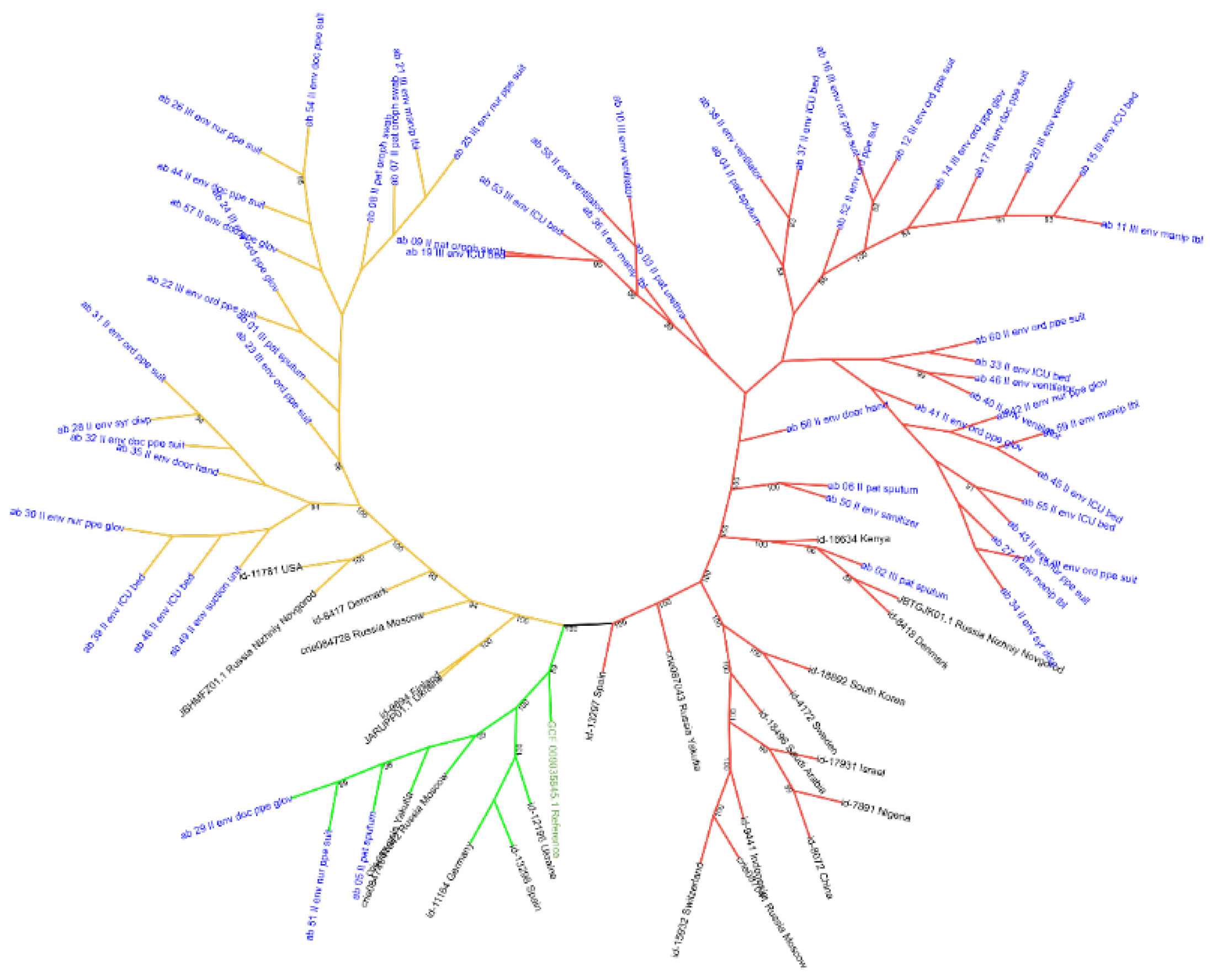
Figure 1. Maximum-likelihood phylogenetic tree of A. baumannii clinical isolates and reference sequences from the PubMLST, NCBI, VGARus databases. Colored clades: red (ST2), light green (ST19), and golden yellow (ST78). Blue: study isolates; Black: PubMLST, NCBI, VGARus databases; Green: GenBank species reference.
Preprints 222557 g001
Figure 2. Maximum-likelihood phylogenetic tree of K. pneumoniae clinical isolates and reference sequences from the PubMLST, NCBI, VGARus databases. Colored clades: red (ST512), light green (ST395), purple (ST307), golden yellow (STST15), blue (ST1190) and yellow (ST101). Blue: study isolates; Black: PubMLST, NCBI, VGARus databases; Green: GenBank species reference.
Preprints 222557 g002
Figure 3. Maximum-likelihood phylogenetic tree of E. coli clinical isolates and reference sequences from the PubMLST, NCBI, VGARus databases. Colored clades: red (CC14). Blue: study isolates; Black: PubMLST, VGARus databases; Green: GenBank species reference.
Preprints 222557 g003
Figure 4. Maximum-likelihood phylogenetic tree of S. aureus clinical isolates and reference sequences from the PubMLST, NCBI, VGARus databases. Colored clades: red (CC97), light green (CC15), purple (CC8), golden yellow (CC22). Blue: study isolates; Black: PubMLST, VGARus databases; Green: GenBank species reference.
Preprints 222557 g004
Figure 5. Temporal dynamics of bacterial isolates obtained from personal protective equipment (PPE), patient care environment (PCE) and general hospital sampling points (GHP).
Preprints 222557 g005
Figure 6. Pairwise SNP distances between A. baumannii isolates of STs: ST2 and ST78 obtained from patients and medical PPE gloves in 2022–2023.
Preprints 222557 g006
Figure 7. Pairwise SNP distances between K. pneumoniae isolates recovered from patients and medical PPE gloves in 2022–2023.
Preprints 222557 g007
Disclaimer/Publisher’s Note: The statements, opinions and data contained in all publications are solely those of the individual author(s) and contributor(s) and not of MDPI and/or the editor(s). MDPI and/or the editor(s) disclaim responsibility for any injury to people or property resulting from any ideas, methods, instructions or products referred to in the content.
Copyright: This open access article is published under a Creative Commons CC BY 4.0 license, which permit the free download, distribution, and reuse, provided that the author and preprint are cited in any reuse.
Prerpints.org logo

Preprints.org is a free preprint server supported by MDPI in Basel, Switzerland.

Subscribe

© 2026 MDPI (Basel, Switzerland) unless otherwise stated

Accessibility

Disclaimer

Terms of Use

Privacy Policy

Privacy Settings