

Submitted:
09 July 2026
Posted:
10 July 2026
You are already at the latest version
Abstract
Thylakoid membranes are highly organized yet dynamic systems in which Photosystem II (PSII) complexes undergo continual assembly, repair and lateral redistribution. Here a perspective is proposed that local lumenal pH - arising from trans-thylakoidal proton gradients and potential sub-micrometer proton microdomains - may contribute to the regulation of protein movement and PSII maturation within the thylakoid membrane. Five mechanistically distinct but potentially coupled routes by which ΔpH could influence protein dynamics are outlined: (i) pH-dependent modulation of lumenal protein-protein affinities; (ii) proton-sensitivity of lumenal proteases (e.g., CtpA) that gate processing and release of PSII subunits; (iii) proton-induced changes in membrane packing and stacking that alter diffusion pathways; (iv) formation of local proton microdomains that create chemically distinct niches; and (v) coupling of proton flux to electrostatic and lipid compositional shifts that bias protein localization. Together, these considerations suggest that ΔpH may act as a co-regulatory parameter integrating biochemical and biophysical cues in thylakoid organization and PSII homeostasis.
Keywords:
ΔpH
; protonation
; thylakoid membrane
; thylakoid lumen
; PSII
In Brief, Function of Thylakoid Membranes and the Role of Photosystem II
Thylakoid membranes are specialized internal membranes found both in cyanobacteria and in the chloroplasts of algae and plants. Their principal function is to host the light-driven reactions of oxygenic photosynthesis and to couple light energy capture to the formation of a transmembrane proton motive force (pmf). Structurally these membranes form an interconnected lamellar network in cyanobacteria, whereas in chloroplasts the thylakoid system is differentiated into stacked regions (grana) and unstacked stroma lamellae. The membrane bilayer is enriched in non-bilayer-promoting galactolipids and anionic lipids (e.g., monogalactosyldiacylglycerol, digalactosyldiacylglycerol and phosphatidylglycerol), which influence the packing, activity and stability of embedded protein complexes (Demé et al., 2014; Fujii et al., 2025; Ostermeier et al., 2024).
The structural and dynamic behaviour of thylakoid membranes is often discussed in the context of the classic fluid-mosaic picture of biological membranes, but thylakoids require important refinements of that simple model (Singer & Nicolson, 1972; Wilhelm et al., 2020). The Singer-Nicolson fluid-mosaic idea - proteins diffusing in a continuous lipid bilayer - captures the notion of lateral mobility, yet thylakoids are unusually protein-dense and organized into large, stable assemblies (for example PSII-LHCII supercomplexes), membrane stacks (grana) and microdomains that create lateral heterogeneity (Anderson, 2012; Andersson & Anderson, 1980; Engel et al., 2015; Fujii et al., 2025). As a result, the membrane is better described as a dynamic mosaic of semi-fluid domains: some components (mobile electron carriers, small peripheral subunits) retain relatively high freedom of movement, whereas larger complexes are sterically constrained, form transient or stable supercomplexes, or become confined by lipid phase behaviour, membrane curvature and protein-protein interactions (Brown, 2012; Wilhelm et al., 2020). Functional processes - reversible phosphorylation, state transitions, assembly/disassembly during PSII repair and biogenesis, and changes in ionic or redox state - further modulate local packing and mobility, allowing the membrane to switch between more fluid and more ordered states as needed. This domain-centric view explains how thylakoids simultaneously support rapid energy transfer and long-lived spatial segregation and provides the physical language used in current work that links membrane composition and curvature to the localization, mobility and lifecycle of photosynthetic complexes.
Four large, multi-subunit protein complexes embedded in the thylakoid membrane carry out the primary energy-conversion chemistry: photosystem II (PSII), the cytochrome b6f complex, photosystem I (PSI), and the ATP synthase. In addition, mobile electron carriers (plastoquinone, plastocyanin) shuttle electrons between complexes and peripheral antenna systems gather sunlight and transfer excitation energy to the reaction centers (Komenda et al., 2024). The organization and antenna architecture differ between cyanobacteria and chloroplasts: cyanobacteria typically use phycobilisomes - large, water-soluble antenna assemblies - attached to the membrane, whereas chloroplasts of green algae and land plants use membrane-intrinsic light-harvesting complexes (LHCs) that participate in grana stacking and dynamic regulation of excitation energy (Neilson & Durnford, 2010; Watanabe & Ikeuchi, 2013).
PSII is the light-driven water:plastoquinone oxidoreductase and is central to oxygenic photosynthesis (Kern & Renger, 2007). It is a dimeric, membrane-integral complex whose core contains the D1 and D2 proteins that bind the primary chlorophylls and the redox cofactors responsible for charge separation and electron transfer to plastoquinone. Two large chlorophyll-binding subunits (CP43 and CP47) form the inner antenna and scaffold additional pigments. On the lumenal side the oxygen-evolving complex (OEC) - a Mn₄CaO₅ cluster - catalyzes water oxidation to molecular oxygen and releases protons into the lumen; the OEC is stabilized by lumenal extrinsic proteins. Through sequential photooxidation of the Mn-cluster PSII extracts electrons from water, reduces plastoquinone to plastoquinol, and contributes protons to the lumenal compartment, thereby initiating the electron-transport chain and feeding into the proton gradient that drives ATP synthesis.
Physiologically PSII is both highly efficient and highly vulnerable: its reaction-center D1 protein is prone to photodamage under excess light, and cells maintain an active D1 repair and turnover cycle that involves selective proteolysis, replacement of damaged subunits, and reassembly of cofactor binding sites. In chloroplasts, PSII complexes are often concentrated in grana stacks where light capture and nonphotochemical quenching are regulated, whereas in cyanobacteria PSII distribution is determined by local membrane architecture and phycobilisome interactions (Krysiak et al., 2025; Rast et al., 2019). Together with PSI, cytochrome b6f and the ATP synthase, PSII forms a dynamic, spatially organized machinery that converts photon energy into chemical energy while balancing light harvesting, photoprotection and repair to maintain photosynthetic performance (Junge, 2019).
The Heterogeneity of Thylakoid Membranes
The current picture is that PSII biogenesis does not occur uniformly across the thylakoid system but is spatially organized into specialized thylakoid subdomains where translation, co-translational membrane insertion and early assembly steps are concentrated (Ostermeier et al., 2025; Sakamoto, 2025; Schottkowski et al., 2012). In cyanobacteria these subdomains have been identified as biogenesis regions or biogenic membranes (Figure 1; thylakoid convergence regions in Synechocystis sp. PCC 6803) that are compositionally and functionally distinct from photosynthetically active membrane regions; they are enriched in assembly factors, ribosomes and early PSII assembly intermediates and often map to particular layers or margins of the thylakoid network rather than to the full lamellar surface (Ostermeier et al., 2025; Rast et al., 2019; Rengstl et al., 2011). In microalgae such as Chlamydomonas reinhardtii, a discrete translation zone (T-zone) adjacent to the pyrenoid serves as a primary locus for the local translation of chloroplast-encoded core proteins (notably D1), for chlorophyll synthesis and for the early steps of PSII assembly; the T-zone concentrates chloroplast mRNAs, ribosomes, pigment-biosynthetic enzymes and imported assembly factors so that nascent subunits and early intermediates accumulate there prior to redistribution (Schottkowski et al., 2012; Uniacke & Zerges, 2007). In chloroplasts of multicellular algae and higher plants, analogous specializations have been localized primarily to stroma-exposed thylakoid regions and to grana margins/curvature zones (Figure 1): many early assembly events and the translation/insertion of chloroplast-encoded core subunits take place on stroma lamellae or at grana margins before mature PSII complexes accumulate within grana stacks (Anderson, 1989; Fujii et al., 2025; Krysiak et al., 2025; Puthiyaveetil et al., 2014; Sakamoto, 2025).
All these subdomains have in common that they are nearly free of chlorophyll and, therefore, are less to non active in photosynthetic reactions (Figure 1) (Lübben et al., 2024).
A Hypothetical New Way of Movement via the pH Gradient
The light-driven water-splitting reaction of mature PSII releases protons into the thylakoid lumen and thereby contributes substantially to the trans-thylakoidal pH gradient (ΔpH) that, together with the membrane potential, comprises the proton-motive force used by ATP synthase (Lubitz et al., 2019; Tommos & Babcock, 2000). Because nascent or immature PSII intermediates either lack a fully functional oxygen-evolving complex or perform water oxidation only weakly (Zabret et al., 2020), they will contribute far less to local lumen acidification than fully active centres - a difference that creates a physiological contrast between active PSII zones and sites of biogenesis/repair such as grana margins, T-zones, biogenic regions, and motivates the following hypotheses (Figure 2). Historically, this idea sits within a long debate over whether thylakoid protons equilibrate rapidly with the bulk lumen or instead form localized, membrane-proximal domains. Early work by Theg and colleagues argued for proton subcompartmentation, while contemporaneous work explicitly framed the issue as localized versus delocalized proton behavior (Dilley et al., 1987; Hong & Junge, 1983; Theg et al., 1982).
- 1.
-
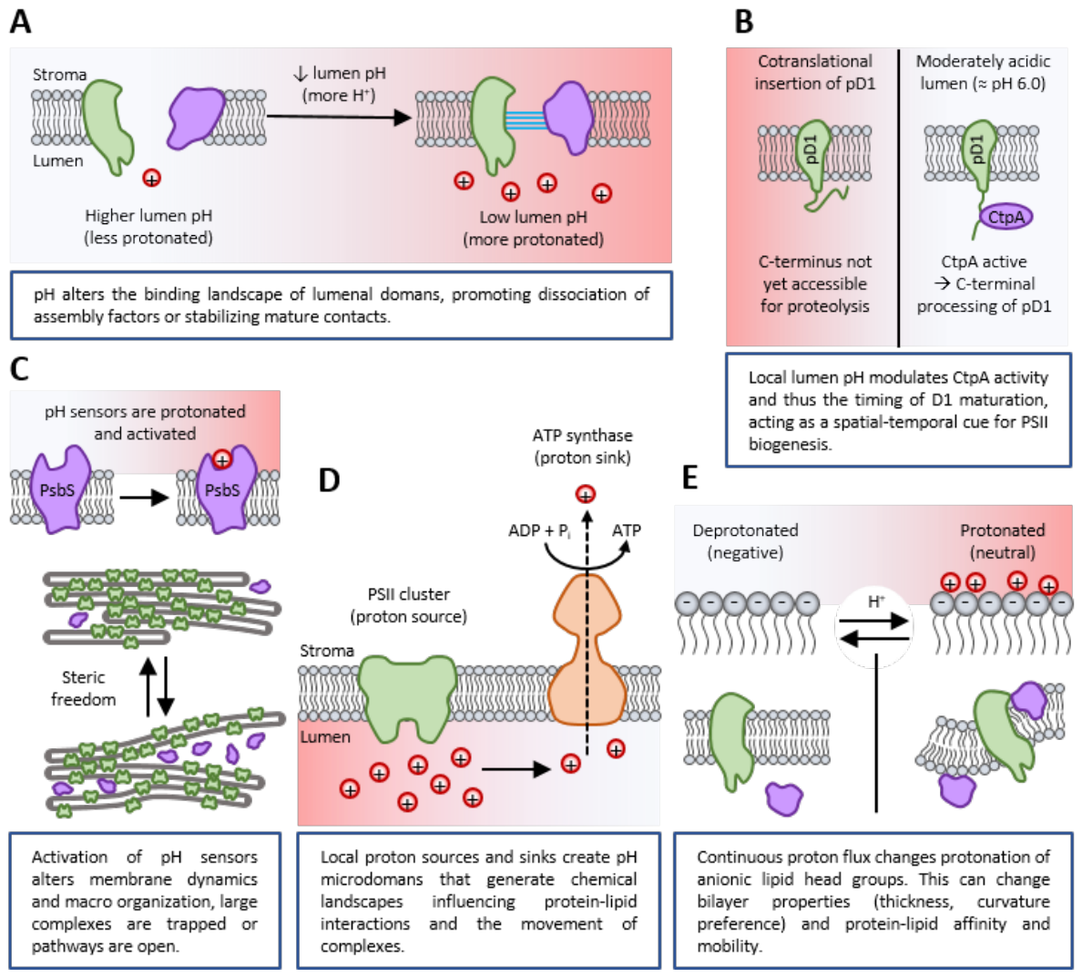
Electrostatic switching of lumen-facing interactions (affinity-switch hypothesis). Protonation of lumen-exposed amino acid side chains at low lumen pH can alter the electrostatic surface of lumenal domains on PSII, assembly factors (e.g. Psb27 (Huang et al., 2021)) and lumenal chaperones. Those pH-dependent changes in charge can alter affinities between components (for example by weakening or strengthening salt bridges or H-bonding networks) (Onufriev & Alexov, 2013), thereby promoting dissociation of assembly factors or, conversely, stabilizing mature contacts. In this way, the local ΔpH could act as a permissive signal (Trinh & Masuda, 2022) that alters the binding landscape as described by Zabret et al. (2021) (Figure 2A).This may affect whether a partially assembled complex remains tethered at a biogenesis site or becomes free to diffuse into neighbouring membrane domains. Direct biochemical precedent for lumen-side pH sensing exists (for example PsbS and other lumen-facing pH sensors are protonated at low lumen pH and change conformation (Chiariello et al., 2023; Krishnan-Schmieden et al., 2021; Liguori et al., 2019)), so the general principle that protonation changes protein-protein interactions in the lumenal environment is well supported.Testing this idea will require correlating controlled lumen-pH changes with the binding dynamics of assembly factors and PSII intermediates in vivo.
- 2.
-
Control of D1 maturation by pH-dependent proteolysis (timing-gate hypothesis). The C-terminal processing protease CtpA, which removes the D1 precursor extension and thereby enables formation of the active Mn-cluster and downstream assembly steps, shows strong pH sensitivity in vitro and in membrane assays; different substrates or assay formats report distinct pH optima but experiments indicate increased activity on membrane-embedded pD1 at moderately acidic conditions (≈ pH 6.0) (Anbudurai et al., 1994; Chang et al., 2021) (Figure 2B). This pH sensitivity is compatible with - and indeed complements - cotranslational insertion of pD1 (Ostermeier et al., 2025): cotranslational membrane insertion positions the pD1 C-terminus in the lumen but does not necessarily render that C-terminus immediately accessible or in the correct conformation for proteolysis (Zhang et al., 2000). Membrane context, substrate presentation and interactions with assembly factors can protect or mask the pD1 extension until other early assembly events are complete. Because biogenesis zones are expected to be less acidified than regions containing active, water-oxidizing PSII (Huokko et al., 2021; Järvi et al., 2013), a relatively alkaline local lumen would both reduce CtpA activity and favour retention of assembly-competent intermediates; conversely, lumen acidification (or changes in conformation/partner binding that expose the C-terminus) would accelerate CtpA-mediated maturation, permitting Mn-cluster formation, extrinsic subunit binding and dimerization and thereby promoting relocation of the complex. Thus, ΔpH can act indirectly as a spatial-temporal cue by modulating CtpA activity in concert with substrate accessibility and assembly cofactors; however, a direct in vivo demonstration that lumen pH alone times CtpA action remains to be conclusively shown.Direct support for this model could come from experiments that simultaneously monitor lumen pH, CtpA activity, and the maturation state of D1 under changing physiological conditions.
- 3.
-
pH-driven reorganization of membrane packing and stacking (mobility-release hypothesis). Lumen acidification is a central trigger of photoprotective responses (e.g., qE) through protonation of pH sensor proteins such as PsbS and LHCSR (Ballottari et al., 2016; Nicol & Croce, 2021; Niyogi et al., 2005); activation of these sensors and of associated pigment/antenna rearrangements is known to modify macro-organization of thylakoid membranes and to influence grana/stroma lamellae dynamics (Kirchhoff, 2014b; Ünnep et al., 2020) (Figure 2C). Because lateral diffusion of large membrane complexes is strongly constrained by membrane packing, changes in stacking and local order induced by low lumen pH can increase or decrease the steric freedom of PSII assembly intermediates (Kirchhoff et al., 2008; Onoa et al., 2020) (Figure 2C). Empirically, macromolecular crowding and packing constraints have been linked to the immobilization of large complexes such as PSII, while smaller or peripheral antennae remain mobile (Kirchhoff et al., 2008; Sarcina et al., 2001). For example, lumen acidification that promotes unstacking or local loosening could create pathways through which matured or repair-competent complexes more readily move into photosynthetically active domains, whereas conditions favouring tighter stacking could trap complexes (Kirchhoff, 2014b; Li et al., 2020; Van Eerden et al., 2017). The established link between lumen pH, PsbS protonation and membrane reorganization supports this hypothesis, though direct observation that pH-induced reorganizations drive directional relocation of individual PSII assembly intermediates is still lacking.Future work combining ΔpH manipulations with high-resolution measurements of membrane architecture and PSII mobility should reveal whether such structural changes facilitate complex relocation.
- 4.
-
Local proton sinks/sources and microdomain formation (microgradient hypothesis). Local proton sinks and sources do not necessarily imply a uniform lumen-wide pH field at sub-micrometre scales (Vershubskii et al., 2017). Rather, they may generate transient, geometry-dependent proton microdomains whose extent depends on membrane architecture, buffering, and the balance between proton production and consumption (Figure 2D). These considerations build on the longstanding concept of localized proton domains in thylakoids (Theg et al., 1982; Theg & Junge, 1983). Comparable lateral pH heterogeneity has been measured in mitochondrial cristae: in live cells, cristae-targeted pH sensors resolved a ~0.3 pH-unit difference between complex IV and F0F1 ATP synthase, and later work extended this to pH profiles at F0F1 ATP synthase, complex IV, and the matrix, demonstrating that local pmf is spatially heterogeneous within a single inner membrane system (Rieger et al., 2021; Rieger et al., 2014). Proton release from active PSII clusters and local consumption by ATP synthase or buffering by lumenal proteins can produce pH microdomains (Armbruster et al., 2017; Farci & Schröder, 2023; Theg et al., 1988). Such microgradients could create local “chemical landscapes” that favour particular protein–lipid interactions (for example protonation-dependent lipid headgroup behaviour) and hence locally modulate membrane fluidity or the affinity of assembly factors for membrane sites (Garab et al., 2017; McKinnon et al., 2020). If biogenesis zones maintain a different luminal pH microenvironment than active photosynthetic patches, this would add a physico-chemical basis for spatial segregation and for the selective release of matured complexes into regions with the appropriate pH (Chotewutmontri & Barkan, 2024; Ostermeier et al., 2025; Rast et al., 2015). There is theoretical and experimental work pointing to limited lateral equilibration of protons in the thylakoid lumen and to lateral heterogeneity of proton potential, but direct in vivo mapping of such microgradients and their effect on protein trajectories remains an open experimental challenge (June & Polle, 1986; Kaňa et al., 2023; Vershubskii et al., 2017).Resolving this question will require direct visualization of lumenal pH landscapes together with spatial tracking of PSII assembly intermediates at sub-micrometre resolution.
- 5.
-
Coupling to ATP synthase activity and lipid/electrostatic environment (force-modulation hypothesis). Proton flux through ATP synthase both consumes local protons and is associated with local electric fields (Sekiguchi et al., 2024); gradients in proton flux (for example where ATP synthase is enriched) may influence the local electrostatic screening of membrane surfaces and alter the packing or lateral mobility of charged complexes (Junge, 1989; Schöttler et al., 2015). In addition, continuous proton flux alters the local protonation state of lipid head groups (notably anionic lipids abundant in thylakoids; Figure 2E) (Yoshihara et al., 2021), which can change bilayer thickness, curvature preference and protein–lipid affinity (Bolik et al., 2022; Kooijman et al., 2003). Through these effects, the steady-state balance between local proton release by PSII and proton consumption by ATP synthase could modulate the physical membrane landscape that determines how easily large complexes diffuse (Dekker & Boekema, 2005; Kirchhoff, 2014a; Onoa et al., 2020). This idea builds on the centrality of ΔpH to chloroplast energetics and on evidence that protein diffusion is sensitive to membrane supramolecular organization; again, direct mechanistic proof that differential proton fluxes drive directed protein migration is not yet available (Johnson & Wientjes, 2020).A critical test would be to determine whether local perturbations of ATP synthase activity produce measurable changes in membrane organization that correlate with altered PSII distribution.
Each of the hypotheses above links physically plausible effects of proton concentration and flux to changes in binding, maturation or membrane organization that could alter mobility (Kaňa et al., 2023; Komenda et al., 2024). However, current research lacks direct, high-resolution in vivo demonstrations of causal chains that run from a measured, localized lumen pH change → a specific molecular change (for instance CtpA activation or a defined change in lipid protonation) → and then to a measurable, directed relocation of PSII intermediates (Bos et al., 2024; Kaňa et al., 2023; Komenda et al., 2024). It is also not known whether ΔpH acts mainly as a local biochemical timing cue (e.g., by modulating CtpA and other lumen-facing enzymes), mainly as a physical modulator of membrane order and diffusion, or as an integrated signal combining both effects - and the relative importance of these routes may differ between cyanobacteria, microalgae (T-zones) and higher plants because of their differing membrane geometries and protein complements (Komenda et al., 2024; Mehra et al., 2024). Finally, quantitative data on lumenal pH microheterogeneity at the spatial and temporal scales relevant to single-complex movements are still sparse (Kaňa et al., 2023; Vershubskii et al., 2017).
Together, these hypotheses suggest that local ΔpH is not merely a consequence of photosynthetic electron transport but may act as a spatial regulator of PSII maturation and localization. While each proposed mechanism is individually plausible, future work will need to determine their relative contribution and physiological relevance.
Conclusion
PSII biogenesis and movement are governed by the interplay between intrinsic changes in the complex during maturation and the dynamic physical structure of the thylakoid membrane. Early assembly occurs in distinct membrane regions enriched in assembly factors, where incomplete PSII forms are retained until they acquire their full complement of cofactors and subunits. As maturation progresses, the complexes become more stable and more compatible with the densely packed, energy-converting domains of the membrane.
The transthylakoid proton gradient (ΔpH) provides a unifying physical parameter that could link energy conversion to this spatial organization. Variations in lumenal pH influence the charge state of exposed protein surfaces, modulate the activity of lumenal enzymes such as CtpA, and affect the packing and curvature of the membrane through protonation of lipids and proteins. These pH-dependent effects are able to locally change the affinity of PSII assembly intermediates for their surrounding environment, thereby promoting their retention in biogenesis zones or their release toward active membrane areas.
Together, these processes describe a coherent picture in which PSII localization is not determined by a single molecular switch but by the combined action of maturation-dependent changes within the complex and pH-driven modulation of the surrounding membrane. This integrated physical and biochemical view provides a consistent hypothesis for how thylakoid membranes coordinate energy conversion with the continuous renewal and spatial redistribution of PSII.
Accordingly, ΔpH should be considered a potential co-regulatory parameter that acts alongside phosphorylation, redox control, proteolysis, and membrane remodelling rather than as a singular explanatory principle. Taken together, these observations suggest that any pH-based contribution to PSII relocation should be treated as a testable extension of the older localized-proton-domain framework rather than as a settled mechanism. The broader question of proton-coupled membrane energetics remained active in later Theg work on cpTat transport and direct ΔpH/electric-field measurements, underscoring that proton-dependent membrane organization is still an experimentally open problem (Alder & Theg, 2003; Theg & Tom, 2011).
Mitochondrial evidence finally supports the broader premise that spatially restricted proton gradients can act as local regulatory parameters at the membrane level, rather than as a strictly bulk-equilibrated signal (Rieger et al., 2021; Rieger et al., 2014).
Future experiments combining live pH reporters and ΔpH manipulations with targeted assays of CtpA activity and high-resolution imaging of PSII intermediates will be required to determine whether pH-dependent membrane physics biases complexes toward biogenesis zones or active domains. Importantly, recent technical advances are beginning to make such experiments feasible. Cryo-electron microscopy and cryo-electron tomography promise unprecedented insight into the spatial organization of PSII assembly and disassembly intermediates within native thylakoid membranes, while the development of improved pH- and pmf-sensitive probes, together with state of the art live-cell and superresolution microscopy, should allow local proton dynamics to be directly linked to PSII remodeling in vivo. These emerging approaches now provide the experimental framework needed to critically test this hypothesis.
Author Contributions
M.O. wrote the manuscript and drew the figures.
Acknowledgments
I thank Jörg Nickelsen for basic support and Helmut Kirchhoff and Steven M. Theg for discussion of ideas.
Competing Interest
The author declares no competing interest.
References
- Albanese, P.; Melero, R.; Engel, B. D.; Grinzato, A.; Berto, P.; Manfredi, M.; Chiodoni, A.; Vargas, J.; Sorzano, C. Ó. S.; Marengo, E. Pea PSII-LHCII supercomplexes form pairs by making connections across the stromal gap. Sci. Rep. 2017, 7(1), 10067. [Google Scholar] [CrossRef] [PubMed]
- Alder, N. N.; Theg, S. M. Energetics of protein transport across biological membranes: a study of the thylakoid ΔpH-dependent/cpTat pathway. Cell 2003, 112(2), 231–242. [Google Scholar] [CrossRef] [PubMed]
- Anbudurai, P.; Mor, T. S.; Ohad, I.; Shestakov, S. V.; Pakrasi, H. B. The ctpA gene encodes the C-terminal processing protease for the D1 protein of the photosystem II reaction center complex. Proc. Natl. Acad. Sci. 1994, 91(17), 8082–8086. [Google Scholar] [CrossRef] [PubMed]
- Anderson, J. M. The grana margins of plant thylakoid membranes. Physiol. Plant. 1989, 76(2), 243–248. [Google Scholar] [CrossRef]
- Anderson, J. M. Lateral heterogeneity of plant thylakoid protein complexes: early reminiscences. Philos. Trans. R. Soc. B Biol. Sci. 2012, 367(1608), 3384–3388. [Google Scholar] [CrossRef]
- Andersson, B.; Anderson, J. M. Lateral heterogeneity in the distribution of chlorophyll-protein complexes of the thylakoid membranes of spinach chloroplasts. Biochim. Et. Biophys. Acta (BBA) -Bioenergetics 1980, 593(2), 427–440. [Google Scholar] [CrossRef]
- Armbruster, U.; Galvis, V. C.; Kunz, H.-H.; Strand, D. D. The regulation of the chloroplast proton motive force plays a key role for photosynthesis in fluctuating light. Curr. Opin. Plant Biol. 2017, 37, 56–62. [Google Scholar] [CrossRef] [PubMed]
- Ballottari, M.; Truong, T. B.; De Re, E.; Erickson, E.; Stella, G. R.; Fleming, G. R.; Bassi, R.; Niyogi, K. K. Identification of pH-sensing sites in the light harvesting complex stress-related 3 protein essential for triggering non-photochemical quenching in Chlamydomonas reinhardtii. J. Biol. Chem. 2016, 291(14), 7334–7346. [Google Scholar] [CrossRef] [PubMed]
- Bolik, S.; Albrieux, C.; Schneck, E.; Demé, B.; Jouhet, J. Sulfoquinovosyldiacylglycerol and phosphatidylglycerol bilayers share biophysical properties and are good mutual substitutes in photosynthetic membranes. Biochim. Et. Biophys. Acta (BBA) -Biomembranes 2022, 1864(12), 184037. [Google Scholar] [CrossRef]
- Bos, P. R.; Berentsen, J.; Wientjes, E. Expansion microscopy resolves the thylakoid structure of spinach. Plant Physiol. 2024, 194(1), 347–358. [Google Scholar]
- Brown, M. F. Curvature forces in membrane lipid–protein interactions. Biochemistry 2012, 51(49), 9782–9795. [Google Scholar] [CrossRef] [PubMed]
- Chang, W.; Li, C.; Cui, Z.; Li, W.; Song, H.; Chang, H.; Fu, W.; Wang, C.; Huang, T.; Luo, Y. Diverged early from CtpB and CtpC, CtpA has evolved to process D1 precursor in oxygenic photosynthetic organisms. Front. Plant Sci. 2021, 12, 676036. [Google Scholar] [CrossRef] [PubMed]
- Chiariello, M. G.; Grünewald, F.; Zarmiento-Garcia, R.; Marrink, S. J. pH-dependent conformational switch impacts stability of the PsbS dimer. J. Phys. Chem. Lett. 2023, 14(4), 905–911. [Google Scholar] [CrossRef] [PubMed]
- Chotewutmontri, P.; Barkan, A. Localization of proteins involved in the biogenesis and repair of the photosynthetic apparatus to thylakoid subdomains in Arabidopsis. Plant Direct 2024, 8(11), e70008. [Google Scholar] [CrossRef] [PubMed]
- Dekker, J. P.; Boekema, E. J. Supramolecular organization of thylakoid membrane proteins in green plants. Biochim. Et. Biophys. Acta (BBA) -Bioenergetics 2005, 1706(1-2), 12–39. [Google Scholar] [CrossRef]
- Demé, B.; Cataye, C.; Block, M. A.; Maréchal, E.; Jouhet, J. Contribution of galactoglycerolipids to the 3-dimensional architecture of thylakoids. FASEB J. 2014, 28(8), 3373–3383. [Google Scholar] [CrossRef] [PubMed]
- Dilley, R. A.; Theg, S. M.; Beard, W. A. Membrane-proton interactions in chloroplast bioenergetics: Localized proton domains. Annu. Rev. Plant Physiol. 1987, 38(1), 347–389. [Google Scholar] [CrossRef]
- Engel, B. D.; Schaffer, M.; Cuellar, L. K.; Villa, E.; Plitzko, J. M.; Baumeister, W. Native architecture of the Chlamydomonas chloroplast revealed by in situ cryo-electron tomography. elife 2015, 4, e04889. [Google Scholar] [CrossRef] [PubMed]
- Farci, D.; Schröder, W. P. Thylakoid Lumen; from “proton bag” to photosynthetic functionally important compartment. Front. Plant Physiol. 2023, 1, 1310167. [Google Scholar] [CrossRef]
- Fujii, S.; Nagata, N.; Kobayashi, K. Membrane lipid–driven thylakoid biogenesis coordinating chlorophyll synthesis and expression of photosynthetic proteins. Plant Cell Physiol. 2025, pcaf130. [Google Scholar]
- Garab, G.; Ughy, B.; Waard, P. d.; Akhtar, P.; Javornik, U.; Kotakis, C.; Šket, P.; Karlický, V.; Materová, Z.; Špunda, V. Lipid polymorphism in chloroplast thylakoid membranes–as revealed by 31P-NMR and time-resolved merocyanine fluorescence spectroscopy. Sci. Rep. 2017, 7(1), 13343. [Google Scholar] [CrossRef] [PubMed]
- Hong, Y. Q.; Junge, W. Localized or delocalized protons in photophosphorylation? On the accessibility of the thylakoid lumen for ions and buffers. Biochim. Et. Biophys. Acta (BBA) -Bioenergetics 1983, 722(1), 197–208. [Google Scholar] [CrossRef]
- Huang, G.; Xiao, Y.; Pi, X.; Zhao, L.; Zhu, Q.; Wang, W.; Kuang, T.; Han, G.; Sui, S.-F.; Shen, J.-R. Structural insights into a dimeric Psb27-photosystem II complex from a cyanobacterium Thermosynechococcus vulcanus. Proc. Natl. Acad. Sci. 2021, 118(5), e2018053118. [Google Scholar] [CrossRef] [PubMed]
- Huokko, T.; Ni, T.; Dykes, G. F.; Simpson, D. M.; Brownridge, P.; Conradi, F. D.; Beynon, R. J.; Nixon, P. J.; Mullineaux, C. W.; Zhang, P. Probing the biogenesis pathway and dynamics of thylakoid membranes. Nat. Commun. 2021, 12(1), 1–14. [Google Scholar] [CrossRef]
- Järvi, S.; Gollan, P. J.; Aro, E.-M. Understanding the roles of the thylakoid lumen in photosynthesis regulation. Front. Plant Sci. 2013, 4, 434. [Google Scholar] [CrossRef] [PubMed]
- Johnson, M. P.; Wientjes, E. The relevance of dynamic thylakoid organisation to photosynthetic regulation. Biochim. Et. Biophys. Acta (BBA) -Bioenergetics 2020, 1861(4), 148039. [Google Scholar] [CrossRef]
- June, W.; Polle, A. Theory of proton flow along appressed thylakoid membranes under both non-stationary and stationary conditions. Biochim. Et. Biophys. Acta (BBA) -Bioenergetics 1986, 848(3), 265–273. [Google Scholar] [CrossRef]
- Junge, W. Protons, the thylakoid membrane, and the chloroplast ATP synthase. Ann. N. Y. Acad. Sci. 1989, 574, 268–286. [Google Scholar] [CrossRef] [PubMed]
- Junge, W. Oxygenic photosynthesis: history, status and perspective. Q. Rev. Biophys. 2019, 52, e1. [Google Scholar] [CrossRef] [PubMed]
- Kaňa, R.; Šedivá, B.; Prášil, O. Microdomains heterogeneity in the thylakoid membrane proteins visualized by super-resolution microscopy. Photosynthetica 2023, 61(4), 483. [Google Scholar] [CrossRef] [PubMed]
- Kern, J.; Renger, G. Photosystem II: Structure and mechanism of the water: plastoquinone oxidoreductase. Photosynth. Res. 2007, 94(2), 183–202. [Google Scholar] [CrossRef] [PubMed]
- Kirchhoff, H. Diffusion of molecules and macromolecules in thylakoid membranes. Biochim. Et. Biophys. Acta (BBA) -Bioenergetics 2014a, 1837(4), 495–502. [Google Scholar] [CrossRef]
- Kirchhoff, H. Structural changes of the thylakoid membrane network induced by high light stress in plant chloroplasts. Philos. Trans. R. Soc. B Biol. Sci. 2014b, 369(1640), 20130225. [Google Scholar] [CrossRef]
- Kirchhoff, H.; Haase, W.; Haferkamp, S.; Schott, T.; Borinski, M.; Kubitscheck, U.; Rögner, M. Structural and functional self-organization of Photosystem II in grana thylakoids. Biochim. Et. Biophys. Acta (BBA) -Bioenergetics 2007, 1767(9), 1180–1188. [Google Scholar] [CrossRef]
- Kirchhoff, H.; Haferkamp, S.; Allen, J. F.; Epstein, D. B.; Mullineaux, C. W. Protein diffusion and macromolecular crowding in thylakoid membranes. Plant Physiol. 2008, 146(4), 1571–1578. [Google Scholar] [CrossRef] [PubMed]
- Komenda, J.; Sobotka, R.; Nixon, P. J. The biogenesis and maintenance of PSII: Recent advances and current challenges. Plant Cell 2024, koae082. [Google Scholar]
- Kooijman, E. E.; Chupin, V.; de Kruijff, B.; Burger, K. N. Modulation of membrane curvature by phosphatidic acid and lysophosphatidic acid. Traffic 2003, 4(3), 162–174. [Google Scholar] [CrossRef] [PubMed]
- Krishnan-Schmieden, M.; Konold, P. E.; Kennis, J. T.; Pandit, A. The molecular pH-response mechanism of the plant light-stress sensor PsbS. Nat. Commun. 2021, 12(1), 2291. [Google Scholar] [CrossRef] [PubMed]
- Krysiak, M.; Oung, H. M. O.; Kirchhoff, H. What are grana in chloroplasts of vascular plants good for? Ann. Bot. 2025, mcaf229. [Google Scholar]
- Li, M.; Mukhopadhyay, R.; Svoboda, V.; Oung, H. M. O.; Mullendore, D. L.; Kirchhoff, H. Measuring the dynamic response of the thylakoid architecture in plant leaves by electron microscopy. Plant Direct 2020, 4(11), e00280. [Google Scholar] [CrossRef] [PubMed]
- Liguori, N.; Campos, S. R.; Baptista, A. n. M.; Croce, R. Molecular anatomy of plant photoprotective switches: the sensitivity of PsbS to the environment, residue by residue. J. Phys. Chem. Lett. 2019, 10(8), 1737–1742. [Google Scholar] [CrossRef] [PubMed]
- Lübben, M. K.; Klingl, A.; Nickelsen, J.; Ostermeier, M. CLEM, a universal tool for analyzing structural organization in thylakoid membranes. Physiol. Plant. 2024, 176(4), e14417. [Google Scholar] [CrossRef] [PubMed]
- Lubitz, W.; Chrysina, M.; Cox, N. Water oxidation in photosystem II. Photosynth. Res. 2019, 142(1), 105–125. [Google Scholar] [CrossRef] [PubMed]
- Mabbitt, P. D.; Wilbanks, S. M.; Eaton-Rye, J. J. Structure and function of the hydrophilic Photosystem II assembly proteins: Psb27, Psb28 and Ycf48. Plant Physiol. Biochem. 2014, 81, 96–107. [Google Scholar] [CrossRef] [PubMed]
- MacGregor-Chatwin, C.; Sener, M.; Barnett, S. F.; Hitchcock, A.; Barnhart-Dailey, M. C.; Maghlaoui, K.; Barber, J.; Timlin, J. A.; Schulten, K.; Hunter, C. N. Lateral segregation of photosystem I in cyanobacterial thylakoids. Plant Cell 2017, 29(5), 1119–1136. [Google Scholar] [CrossRef] [PubMed]
- McKinnon, L. J.; Fukushima, J.; Endow, J. K.; Inoue, K.; Theg, S. M. Membrane chaperoning of a thylakoid protease whose structural stability is modified by the protonmotive force. Plant Cell 2020, 32(5), 1589–1609. [Google Scholar] [CrossRef] [PubMed]
- Mehra, H. S.; Wang, X.; Russell, B. P.; Kulkarni, N.; Ferrari, N.; Larson, B.; Vinyard, D. J. Assembly and Repair of Photosystem II in Chlamydomonas reinhardtii. Plants 2024, 13(6), 811. [Google Scholar] [CrossRef] [PubMed]
- Mullineaux, C.; Mahbub, M. Locations of membrane protein production in a cyanobacterium. J. Bacteriol. 2023. [Google Scholar] [CrossRef]
- Mullineaux, C. W. FRAP analysis of photosynthetic membranes. J. Exp. Bot. 2004, 55(400), 1207–1211. [Google Scholar] [CrossRef] [PubMed]
- Mullineaux, C. W. Factors controlling the mobility of photosynthetic proteins. Photochem. Photobiol. 2008, 84(6), 1310–1316. [Google Scholar] [CrossRef] [PubMed]
- Nath, K.; Jajoo, A.; Poudyal, R. S.; Timilsina, R.; Park, Y. S.; Aro, E.-M.; Nam, H. G.; Lee, C.-H. Towards a critical understanding of the photosystem II repair mechanism and its regulation during stress conditions. FEBS Lett. 2013, 587(21), 3372–3381. [Google Scholar] [CrossRef] [PubMed]
- Neilson, J. A.; Durnford, D. G. Structural and functional diversification of the light-harvesting complexes in photosynthetic eukaryotes. Photosynth. Res. 2010, 106(1), 57–71. [Google Scholar] [CrossRef] [PubMed]
- Nickelsen, J.; Rengstl, B. Photosystem II assembly: from cyanobacteria to plants. Annu Rev. Plant Biol. 2013, 64, 609–635. [Google Scholar] [CrossRef] [PubMed]
- Nicol, L.; Croce, R. The PsbS protein and low pH are necessary and sufficient to induce quenching in the light-harvesting complex of plants LHCII. Sci. Rep. 2021, 11(1), 7415. [Google Scholar] [CrossRef] [PubMed]
- Niyogi, K. K.; Li, X.-P.; Rosenberg, V.; Jung, H.-S. Is PsbS the site of non-photochemical quenching in photosynthesis? J. Exp. Bot. 2005, 56(411), 375–382. [Google Scholar] [PubMed]
- Onoa, B.; Fukuda, S.; Iwai, M.; Bustamante, C.; Niyogi, K. K. Atomic force microscopy visualizes mobility of photosynthetic proteins in grana thylakoid membranes. Biophys. J. 2020, 118(8), 1876–1886. [Google Scholar] [CrossRef] [PubMed]
- Onufriev, A. V.; Alexov, E. Protonation and pK changes in protein–ligand binding. Q. Rev. Biophys. 2013, 46(2), 181–209. [Google Scholar] [CrossRef] [PubMed]
- Ostermeier, M.; Buschmann, I.; Heinz, S.; Nickelsen, J. The subcellular localisation of early photosystem II assembly in Synechocystis sp. PCC 6803. Physiol. Plant. 2025, 177(2), e70234. [Google Scholar] [CrossRef] [PubMed]
- Ostermeier, M.; Garibay-Hernández, A.; Holzer, V. J.; Schroda, M.; Nickelsen, J. Structure, biogenesis, and evolution of thylakoid membranes. Plant Cell 2024, 36(10), 4014–4035. [Google Scholar] [CrossRef] [PubMed]
- Puthiyaveetil, S.; Tsabari, O.; Lowry, T.; Lenhert, S.; Lewis, R. R.; Reich, Z.; Kirchhoff, H. Compartmentalization of the protein repair machinery in photosynthetic membranes. Proc. Natl. Acad. Sci. 2014, 111(44), 15839–15844. [Google Scholar] [CrossRef] [PubMed]
- Rast, A.; Heinz, S.; Nickelsen, J. Biogenesis of thylakoid membranes. Biochim Biophys. Acta 2015, 1847(9), 821–830. [Google Scholar] [CrossRef] [PubMed]
- Rast, A.; Schaffer, M.; Albert, S.; Wan, W.; Pfeffer, S.; Beck, F.; Plitzko, J. M.; Nickelsen, J.; Engel, B. D. Biogenic regions of cyanobacterial thylakoids form contact sites with the plasma membrane. Nat. Plants 2019, 5(4), 436–446. [Google Scholar] [CrossRef] [PubMed]
- Rengstl, B.; Oster, U.; Stengel, A.; Nickelsen, J. An intermediate membrane subfraction in cyanobacteria is involved in an assembly network for Photosystem II biogenesis. J. Biol. Chem. 2011, 286(24), 21944–21951. [Google Scholar] [CrossRef] [PubMed]
- Rieger, B.; Arroum, T.; Villalta, J.; Busch, K. B. pH profiling of mitochondrial sub-compartments reveals that IF1 is essential to prevent ATP hydrolysis under OXPHOS conditions. bioRxiv 2021, 2011.430746. [Google Scholar]
- Rieger, B.; Junge, W.; Busch, K. B. Lateral pH gradient between OXPHOS complex IV and F0F1 ATP-synthase in folded mitochondrial membranes. Nat. Commun. 2014, 5(1), 3103. [Google Scholar] [CrossRef] [PubMed]
- Rühle, T.; Leister, D. Photosystem II assembly from scratch. Front. Plant Sci. 2016, 6, 1234. [Google Scholar] [CrossRef] [PubMed]
- Sakamoto, W. Thylakostasis: key factors in thylakoid membrane organization with emphasis on biogenesis and remodeling proteins in vascular plants. Plant Cell Physiol. 2025, pcaf098. [Google Scholar]
- Sarcina, M.; Mullineaux, C.; Murata, N. Thylakoid membrane fluidity and its crucial importance in photoinhibition. Sci. Access 2001, 3(1). [Google Scholar]
- Schottkowski, M.; Peters, M.; Zhan, Y.; Rifai, O.; Zhang, Y.; Zerges, W. Biogenic membranes of the chloroplast in Chlamydomonas reinhardtii. Proc. Natl. Acad. Sci. 2012, 109(47), 19286–19291. [Google Scholar] [CrossRef] [PubMed]
- Schöttler, M. A.; Tóth, S. Z.; Boulouis, A.; Kahlau, S. Photosynthetic complex stoichiometry dynamics in higher plants: biogenesis, function, and turnover of ATP synthase and the cytochrome b 6 f complex. J. Exp. Bot. 2015, 66(9), 2373–2400. [Google Scholar] [PubMed]
- Sekiguchi, T.; Yoshida, K.; Wakabayashi, K.-i.; Hisabori, T. Proton gradient across the chloroplast thylakoid membrane governs the redox regulatory function of ATP synthase. J. Biol. Chem. 2024, 300(9). [Google Scholar] [CrossRef] [PubMed]
- Singer, S. J.; Nicolson, G. L. The Fluid Mosaic Model of the Structure of Cell Membranes: Cell membranes are viewed as two-dimensional solutions of oriented globular proteins and lipids. Science 1972, 175(4023), 720–731. [Google Scholar] [CrossRef] [PubMed]
- Stengel, A.; Gugel, I. L.; Hilger, D.; Rengstl, B.; Jung, H.; Nickelsen, J. Initial steps of photosystem II de novo assembly and preloading with manganese take place in biogenesis centers in Synechocystis. Plant Cell 2012, 24(2), 660–675. [Google Scholar] [CrossRef] [PubMed]
- Theg, S.; Chiang, G.; Dilley, R. Protons in the thylakoid membrane-sequestered domains can directly pass through the coupling factor during ATP synthesis in flashing light. J. Biol. Chem. 1988, 263(2), 673–681. [Google Scholar] [CrossRef] [PubMed]
- Theg, S. M.; Johnson, J. D.; Homann, P. H. Proton efflux from thylakoids induced in darkness and its effect on photosystem II. FEBS Lett. 1982, 145(1), 25–29. [Google Scholar] [CrossRef]
- Theg, S. M.; Junge, W. The effect of low concentrations of uncouplers on the detectability of proton deposition in thylakoids. Evidence for subcompartmentation and preexisting pH differences in the dark. Biochim. Et. Biophys. Acta (BBA) -Bioenergetics 1983, 723(2), 294–307. [Google Scholar] [CrossRef]
- Theg, S. M.; Tom, C. Measurement of the ΔpH and electric field developed across Arabidopsis thylakoids in the light. In Chloroplast Research in Arabidopsis: Methods and Protocols, Volume Volume II; Springer, 2011; pp. 327–341. [Google Scholar]
- Theis, J.; Schroda, M. Revisiting the photosystem II repair cycle. Plant Signal. Behav. 2016, 11(9), e1218587. [Google Scholar] [CrossRef] [PubMed]
- Tomizioli, M.; Lazar, C.; Brugière, S.; Burger, T.; Salvi, D.; Gatto, L.; Moyet, L.; Breckels, L. M.; Hesse, A.-M.; Lilley, K. S. Deciphering thylakoid sub-compartments using a mass spectrometry-based approach. Mol. Cell. Proteom. 2014, 13(8), 2147–2167. [Google Scholar] [CrossRef]
- Tommos, C.; Babcock, G. T. Proton and hydrogen currents in photosynthetic water oxidation. Biochim. Et. Biophys. Acta (BBA) -Bioenergetics 2000, 1458(1), 199–219. [Google Scholar] [CrossRef]
- Trinh, M. D. L.; Masuda, S. Chloroplast pH homeostasis for the regulation of photosynthesis. Front. Plant Sci. 2022, 13, 919896. [Google Scholar] [CrossRef] [PubMed]
- Uniacke, J.; Zerges, W. Photosystem II assembly and repair are differentially localized in Chlamydomonas. Plant Cell 2007, 19(11), 3640–3654. [Google Scholar] [CrossRef] [PubMed]
- Ünnep, R.; Paul, S.; Zsiros, O.; Kovács, L.; Székely, N. K.; Steinbach, G.; Appavou, M.-S.; Porcar, L.; Holzwarth, A. R.; Garab, G. Thylakoid membrane reorganizations revealed by small-angle neutron scattering of Monstera deliciosa leaves associated with non-photochemical quenching. Open Biol. 2020, 10(9), 200144. [Google Scholar] [CrossRef] [PubMed]
- Van Eerden, F. J.; Van Den Berg, T.; Frederix, P. W.; De Jong, D. H.; Periole, X.; Marrink, S. J. Molecular dynamics of photosystem II embedded in the thylakoid membrane. J. Phys. Chem. B 2017, 121(15), 3237–3249. [Google Scholar] [PubMed]
- Vershubskii, A. V.; Trubitsin, B. V.; Priklonskii, V. I.; Tikhonov, A. N. Lateral heterogeneity of the proton potential along the thylakoid membranes of chloroplasts. Biochim. Et. Biophys. Acta (BBA) -Biomembranes 2017, 1859(3), 388–401. [Google Scholar] [CrossRef]
- Watanabe, M.; Ikeuchi, M. Phycobilisome: architecture of a light-harvesting supercomplex. Photosynth. Res. 2013, 116(2), 265–276. [Google Scholar] [CrossRef] [PubMed]
- Wilhelm, C.; Goss, R.; Garab, G. The fluid-mosaic membrane theory in the context of photosynthetic membranes: Is the thylakoid membrane more like a mixed crystal or like a fluid? J. Plant Physiol. 2020, 252, 153246. [Google Scholar] [CrossRef] [PubMed]
- Wu, J.; Wu, C.; Chen, S.; Huang, C.; Wen, Q.; Lin, W.; Wang, C.; Han, D.; Lu, D.; Xu, X. Assembly mechanism of PSII-LHCII array from higher plants. J. Integr. Plant Biol. 2025. [Google Scholar] [CrossRef] [PubMed]
- Wunder, T.; Xu, W.; Liu, Q.; Wanner, G.; Leister, D.; Pribil, M. The major thylakoid protein kinases STN7 and STN8 revisited: effects of altered STN8 levels and regulatory specificities of the STN kinases. Front. Plant Sci. 2013, 4, 417. [Google Scholar] [CrossRef] [PubMed]
- Yoshihara, A.; Nagata, N.; Wada, H.; Kobayashi, K. Plastid anionic lipids are essential for the development of both photosynthetic and non-photosynthetic organs in Arabidopsis thaliana. Int. J. Mol. Sci. 2021, 22(9), 4860. [Google Scholar] [CrossRef] [PubMed]
- Zabret, J.; Bohn, S.; Schuller, S. K.; Arnolds, O.; Möller, M.; Meier-Credo, J.; Liauw, P.; Chan, A.; Tajkhorshid, E.; Langer, J. D. How to build a water-splitting machine: structural insights into photosystem II assembly. bioRxiv 2020, 2020.2009. 2014.294884. [Google Scholar]
- Zabret, J.; Bohn, S.; Schuller, S. K.; Arnolds, O.; Möller, M.; Meier-Credo, J.; Liauw, P.; Chan, A.; Tajkhorshid, E.; Langer, J. D.; Stoll, R.; Krieger-Liszkay, A.; Engel, B. D.; Rudack, T.; Schuller, J. M.; Nowaczyk, M. M. Structural insights into photosystem II assembly. Nat. Plants 2021, 7(4), 524–538. [Google Scholar] [CrossRef] [PubMed]
- Zhang, L.; Paakkarinen, V.; van Wijk, K. J.; Aro, E.-M. Biogenesis of the chloroplast-encoded D1 protein: regulation of translation elongation, insertion, and assembly into photosystem II. Plant Cell 2000, 12(9), 1769–1781. [Google Scholar] [CrossRef] [PubMed]
Figure 1.
Thylakoid membranes in Cyanobacteria and Chloroplasts. Comparison of different thylakoid architecture. Highlighted in blue analogous specialized regions of low chlorophyll abundance (red) and high biogenic activity (adapted from Lübben et al., 2024).
Figure 1.
Thylakoid membranes in Cyanobacteria and Chloroplasts. Comparison of different thylakoid architecture. Highlighted in blue analogous specialized regions of low chlorophyll abundance (red) and high biogenic activity (adapted from Lübben et al., 2024).
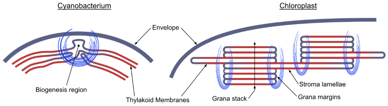
Figure 2.
Hypothetical ways of membrane/protein interaction changes via transthylakoidal pH gradient (ΔpH). A) Electrostatic switching of lumen-facing interactions. B) Control of D1 maturation by pH-dependent proteolysis. C) pH-driven reorganization of membrane packing and stacking. D) Local proton sinks/sources and microdomain formation. E) Coupling to ATP synthase activity and lipid/electrostatic environment.
Figure 2.
Hypothetical ways of membrane/protein interaction changes via transthylakoidal pH gradient (ΔpH). A) Electrostatic switching of lumen-facing interactions. B) Control of D1 maturation by pH-dependent proteolysis. C) pH-driven reorganization of membrane packing and stacking. D) Local proton sinks/sources and microdomain formation. E) Coupling to ATP synthase activity and lipid/electrostatic environment.
Disclaimer/Publisher’s Note: The statements, opinions and data contained in all publications are solely those of the individual author(s) and contributor(s) and not of MDPI and/or the editor(s). MDPI and/or the editor(s) disclaim responsibility for any injury to people or property resulting from any ideas, methods, instructions or products referred to in the content. |
© 2026 by the authors. Licensee MDPI, Basel, Switzerland. This article is an open access article distributed under the terms and conditions of the Creative Commons Attribution (CC BY) license (http://creativecommons.org/licenses/by/4.0/).
Copyright: This open access article is published under a Creative Commons CC BY 4.0 license, which permit the free download, distribution, and reuse, provided that the author and preprint are cited in any reuse.