

Submitted:
08 July 2026
Posted:
10 July 2026
You are already at the latest version
Abstract
Background: Severe burn injuries involving the face and scalp have profound physical and psychosocial consequences, and represent the greatest reconstructive challenge in plastic surgery. One comprehensive solution involves the sequential application of autologous fat grafting, follicular unit excision (FUE) hair transplantation, and scalp micropigmentation (SMP); however, the long-term outcomes of this approach have rarely been documented.
Case Presentation: A male patient sustained catastrophic methane explosion burns at age 27, involving 79% total body surface area (TBSA) with full-thickness involvement of the face and scalp (Revised Baux Score 123; estimated mortality risk 50–80%). Following 65 reconstructive procedures at external institutions over six years, he was referred to our center at age 30. He presented with extensive facial and scalp scarring, complete absence of eyebrow, beard, and mustache hair, and widespread scalp alopecia. Over an eight-year period, the patient received 11 sessions of autologous FUE hair transplantation (7,180 follicular units to the eyebrows, beard, and scalp), four sessions of structural fat grafting using the Coleman technique, and five sessions of SMP.
Conclusions: The patient achieved complete reconstruction of facial hair, a natural scalp hairline, significant scar quality improvement, and full psychosocial recovery, as well as a return to professional and family life. Our findings demonstrate that systematic, long-term multimodal reconstruction, i.e. combining fat grafting, FUE transplantation and SMP, can yield clinically-meaningful aesthetic and psychosocial outcomes, even after the most catastrophic burn injuries.
Keywords:
burn reconstruction
; face
; scalp
; FUE hair transplantation
; fat grafting
; scalp micropigmentation
; alopecia
; scar
; psychosocial recovery
; cicatricial alopecia
1. Introduction
Burn injuries remain a global burden on public health, accounting for an estimated 11 million injuries and approximately 180,000 deaths annually [1]. In Europe, the incidence of severe burns requiring hospitalization is estimated at 0.2–2.9 per 10,000 population per year, with many of the worst cases being associated with industrial and occupational accidents such as coal mining and chemical spills [2]. In many centers, burns affecting more than 40% TBSA (total body surface area) are associated with mortality rates of over 50%, and injuries involving more than 70% TBSA can present a challenge for even the most experienced burn units [3].
The damage associated with major thermal injury extends far beyond local tissue destruction. Full-thickness burns destroy the epidermis, dermis and subcutaneous tissue, resulting in the permanent loss of associated structures, such as sweat glands and sebaceous glands. In addition, in the head and neck areas, scarring can cause cicatricial ectropion, which threatens corneal integrity, as well as perioral contracture, which impairs nutrition and speech, and cervical contracture, which restricts head movement. Damage to the scalp can also result in permanent alopecia, which has a string influence on appearance and social identity [4,5].
Recent decades have seen a considerable expansion in the reconstructive armamentarium available for post-burn sequelae of the head and neck. The range of modern techniques now include local and regional flaps, split-thickness skin grafting, autologous fat grafting, autologous hair transplantation and scalp micropigmentation [6,7,8]. The present study describes the case of a patient with catastrophic pan-facial and scalp burn sequelae, who received sequential application of various treatments over an eight-year period. Its findings demonstrate the restorative potential of such systematic, long-term reconstructive programs.
2. Case Presentation
A 30-year-old male patient with extensive scarring of the head region was admitted to our institution on October 18, 2017 for reconstructive treatment of the hair-bearing areas of the face and scalp.
The patient received the injuries as a result of thermal trauma caused by a methane explosion in a coal mine on October 7, 2014. Immediately after the accident, he was admitted to a regional burn center in critical condition, with deep dermal/full-thickness (IIb/III degree) thermal burns involving approximately 79% total body surface area (TBSA): this area included the face, head, neck, chest, trunk, buttocks, abdomen, perineum, and both upper and lower extremities (Figure 1). The patient also experienced eyelid insufficiency due to facial burns to the eyelids, conjunctivae, and corneas of both eyes. Inhalation injury was also present. Given the extent and depth of burns, the patient’s Revised Baux Score was approximately 123, corresponding to an estimated mortality risk of 50–80% [9].
Over the following six years, the patient underwent 65 reconstructive procedures at five external institutions. The complete procedural list is provided in Supplementary Material, Table S1.
On admission to our institution on October 18, 2017, the patient presented with widespread, rigid facial and scalp scars, and complete loss of hair from the eyebrows, mustache and beard areas. The temporal, frontal, parietal, and partially left occipital regions of the scalp were covered with split-thickness skin grafts. The temporal and occipital areas also presented irregular areas of residual hair-bearing skin (Figure 2). The patient also reported experiencing depressive symptoms, characterized by a ruminative, past-focused cognitive style and markedly reduced engagement with present and future life circumstances.
The patient underwent 11 follicular unit excision (FUE) sessions, during which a total of 7 180 follicular units (FU) were transplanted. In the eyebrow region, two sessions were performed with a cumulative total of 490 FU; single- and double-hair follicular units were used to reconstruct the natural eyebrow architecture. For the mustache and beard, four sessions were performed with a cumulative total of 2 300 FU. For the scalp, five sessions were performed with a cumulative total of 4 390 FU. Donor follicles were harvested from the remaining hair-bearing occipital and temporal scalp using 0.9-mm FUE hybrid punches and the WAW system under local anesthesia. Graft implantation was performed using the stick-and-place technique.
Four sessions of structural fat grafting were performed in the areas of facial and scalp scarring. Adipose tissue was harvested by manual lipoaspiration from the abdominal wall and centrifuged (Coleman technique); the resulting material was then injected into the subdermal and intradermal planes of the scar tissue using blunt-tipped microcannulas in a retrograde fanning pattern [19,20].
To replicate the appearance of shaved follicular units, the FUE transplantation was complemented by five scalp micropigmentation (SMP) sessions. Pigment was deposited at the level of the papillary dermis within alopecic or low-density areas of the scalp using a specialized digital device with a fine needle.
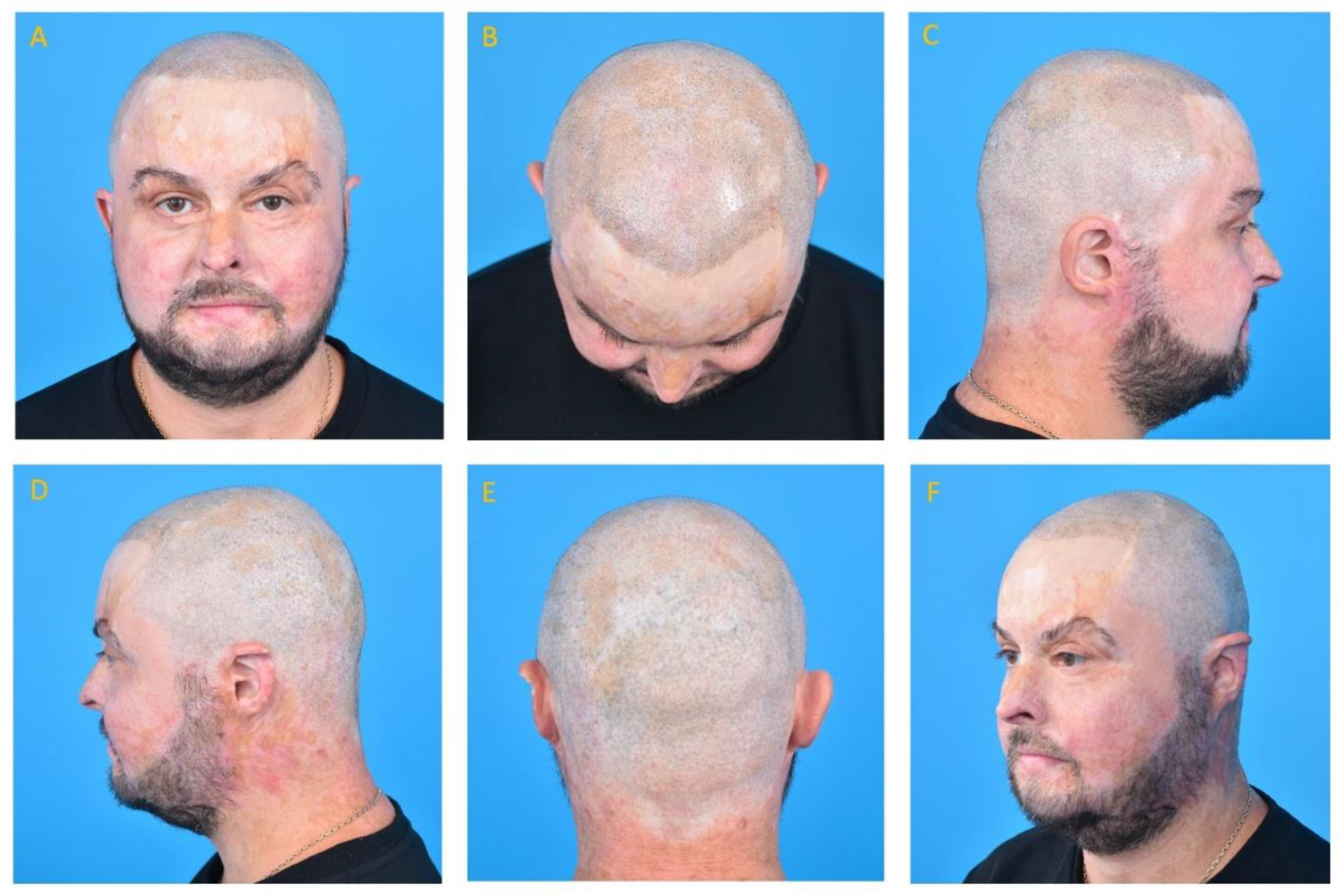
Treatment at our institution was concluded on September 16, 2025 with complete reconstruction of the eyebrow, mustache, and beard hair. The resulting facial hair pattern significantly improved facial identity and gender-appropriate appearance (Figure 3). The patient re-engaged fully in professional and family life: during the treatment period, he became the father of two children. By the conclusion of treatment, he had achieved complete recovery of his psychosocial wellbeing.
3. Discussion
The present report describes the case of a patient who had sustained burns involving 79% TBSA, with an estimated mortality risk of 50 to 80% on the Revised Baux Score [3,9]. His recovery demanded extraordinary clinical resources and the coordinated efforts of specialists and surgeons from burns intensive care units, together with a multidisciplinary team. As a result, the patient had undergone 65 separate procedures at external institutions over the six years preceding his arrival at our center.
Full-thickness burns of the face and scalp carry a unique selection of long-term physical sequelae. The presence of high amounts of graft-covered scar tissue over the face results in a loss of dynamic facial expressiveness, reduces cutaneous sensibility and prevents sebaceous secretion. These features, together with the characteristic inelastic appearance of the face, are characteristic of patients with severe burns [7]. Perioral scarring impairs oral opening, chewing, and speech; in addition, cervical contracture restricts head movement and poses a challenges for airway management [10]. From the aesthetic and social perspectives, these effects are further exacerbated by the loss of hair from the scalp, eyebrows, or beard.
Catastrophic burn injury is known to confer a psychological burden to the patient. Post-traumatic stress disorder (PTSD) has been reported in 30–45% of major burn survivors, with the risk increasing with burn size and facial involvement [11]. Also, an estimated 30–50% of survivors report experiencing depression during the first year after injury [12]. Indeed, as commonly noted in major burn survivors in the late reconstruction phase, the present patient was characterized by a clinically-evident depressive symptomatology and a ruminative, past-focused cognitive style.
Psychologically, the patient achieved complete recovery over the eight-year course of treatment. The successful reconstruction of his eyebrows, beard, and scalp appeared to act as a turning point in self-perception and social engagement. Indeed, facial hair restoration is often perceived by burn survivors as having one of the greatest psychosocial impacts of all reconstructive interventions [13].
The selected reconstructive strategy prioritized interventions most likely to generate both aesthetic and psychological benefit while respecting the biological limitations of the available tissue. Structural fat grafting was used as a preparatory step to assist FUE and transplantation into scarred recipient beds; the grafts improve scar quality and normalize the dermal environment by reducing skin rigidity and improving vascularity [14,15]. During FUE, the best-quality follicular units are selectively harvested from donor zones; in this case, the most socially-visible deficit was addressed first i.e. the eyebrows, followed by the beard area, then the scalp. The scalp presented more extensive alopecia, which was prepared by repeated fat grafting [16,17].
Scalp micropigmentation was introduced at a stage when the transplanted hair had matured sufficiently to assess any deficits in residual density. The aim of the procedure was not to replace hair transplantation, but to improve its appearance by filling out the follicular units and defining the hairline contour [18].
4. Conclusions
This case documents the eight-year reconstructive journey of a patient who survived catastrophic burn injuries involving 79% TBSA, with extensive full-thickness involvement of the face and scalp. The patient ultimately achieved complete restoration of facial hair, a natural scalp hairline, improved scar quality, and full psychosocial recovery.
This outcome was made possible through the sequential application of complementary modalities, viz. fat grafting, FUE hair transplantation and scalp micropigmentation, and their synergic effects. However, in such cases, the true measure of reconstructive success is the return of a severely-burned patient to professional life and family engagement, and the restoration of their full psychosocial wellbeing.
Supplementary Materials
The following supporting information can be downloaded at the website of this paper posted on Preprints.org.
Author Contributions
Conceptualization, J.K.; methodology, J.K.; investigation, J.K.; resources, J.K.; data curation, J.K.; writing—original draft preparation, J.K.; writing—review and editing, J.K.; visualization, J.K.; project administration, J.K. The author has read and agreed to the published version of the manuscript.
Funding
This research received no external funding.
Institutional Review Board Statement
Ethical review and approval were waived for this study in accordance with local legislation and institutional requirements, as this is a single case report and no experimental intervention was performed beyond standard clinical care.
Informed Consent Statement
Written informed consent was obtained from the patient for publication of this case report and all accompanying clinical photographs. The patient reviewed the manuscript and approved its submission for publication.
Data Availability Statement
No new datasets were generated or analyzed in this study. The clinical data supporting the findings are contained within the article.
Acknowledgments
All procedures performed on drscribed patient at our institution were provided free of charge to the patient under the Pro Bono “Operation Restore” Program of the International Society of Hair Restoration Surgery (ISHRS).
Conflicts of Interest
The author declares no conflicts of interest.
References
- World Health Organization. Burns. Fact Sheet. WHO: Geneva, Switzerland, 2018; Available online: https://www.who.int/news-room/fact-sheets/detail/burns (accessed on 1 January 2024).
- Brusselaers, N.; Monstrey, S.; Vogelaers, D.; Hoste, E.; Blot, S. Severe burn injury in Europe: A systematic review of the incidence, etiology, morbidity, and mortality. Crit. Care 2010, 14, R188. [Google Scholar] [CrossRef]
- Ryan, C.M.; Schoenfeld, D.A.; Thorpe, W.P.; Sheridan, R.L.; Cassem, E.H.; Tompkins, R.G. Objective estimates of the probability of death from burn injuries. N. Engl. J. Med. 1998, 338, 362–366. [Google Scholar] [CrossRef]
- Jeschke, M.G.; van Baar, M.E.; Choudhry, M.A.; Chung, K.K.; Gibran, N.S.; Logsetty, S. Burn injury. Nat. Rev. Dis. Prim. 2020, 6, 11. [Google Scholar] [CrossRef] [PubMed]
- Woodson, L.C. Diagnosis and grading of inhalation injury. J. Burn Care Res. 2009, 30, 143–145. [Google Scholar] [CrossRef]
- Gauglitz, G.G.; Korting, H.C.; Pavicic, T.; Ruzicka, T.; Jeschke, M.G. Hypertrophic scarring and keloids: Pathomechanisms and current and emerging treatment strategies. Mol. Med. 2011, 17, 113–125. [Google Scholar] [CrossRef]
- Sheridan, R.L. Comprehensive treatment of burns. Curr. Probl. Surg. 2001, 38, 641–756. [Google Scholar] [CrossRef]
- Hierner, R.; Degreef, H.; Vranckx, J.J.; Garmyn, M.; Massagé, P.; van Brussel, M. Skin grafting and wound healing—The dermato-plastic team approach. Clin. Dermatol. 2005, 23, 343–352. [Google Scholar] [CrossRef] [PubMed]
- Osler, T.; Glance, L.G.; Hosmer, D.W. Simplified estimates of the probability of death after burn injuries: Extending and updating the Baux score. J. Trauma 2010, 68, 690–697. [Google Scholar] [CrossRef]
- Palmieri, T.L.; Greenhalgh, D.G. Topical treatment of pediatric patients with burns: A practical guide. Am. J. Clin. Dermatol. 2002, 3, 529–534. [Google Scholar] [CrossRef] [PubMed]
- Fauerbach, J.A.; Lawrence, J.W.; Schmidt, C.W., Jr.; Munster, A.M.; Costa, P.T., Jr. Personality predictors of injury-related posttraumatic stress disorder. J. Nerv. Ment. Dis. 2000, 188, 510–517. [Google Scholar] [CrossRef] [PubMed]
- Wiechman, S.A.; Patterson, D.R. Psychosocial aspects of burn injuries. BMJ 2004, 329, 391–393. [Google Scholar] [CrossRef] [PubMed]
- Brown, B.C.; McKenna, S.P.; Siddhi, K.; McGrouther, D.A.; Bayat, A. The hidden cost of skin scars: Quality of life after skin scarring. J. Plast. Reconstr. Aesthet. Surg. 2008, 61, 1049–1058. [Google Scholar] [CrossRef] [PubMed]
- Garg, S.; Manchanda, S. Fat grafting as a pre-treatment before hair transplantation in cicatricial alopecia. J. Cutan. Aesthet. Surg. 2019, 12, 154–160. [Google Scholar] [CrossRef] [PubMed]
- Garg, A.K. Autologous fat grafting in scar alopecia for hair transplant. J. Cutan. Aesthet. Surg. 2018, 11, 120–124. [Google Scholar] [CrossRef] [PubMed]
- Rassman, W.R.; Bernstein, R.M.; McClellan, R.; Jones, R.; Worton, E.; Uyttendaele, H. Follicular unit extraction: Minimally invasive surgery for hair transplantation. Dermatol. Surg. 2002, 28, 720–728. [Google Scholar] [CrossRef] [PubMed]
- Beehner, M. Eyebrow and eyelash transplantation with FUE grafts. Hair Transpl. Forum Int. 2017, 27, 100–106. [Google Scholar]
- Traquina, A.C. Micropigmentation as an adjuvant in cosmetic surgery of the scalp. Dermatol. Surg. 2001, 27, 123–128. [Google Scholar] [CrossRef] [PubMed]
- Coleman, S.R. Structural fat grafting: More than a permanent filler. Plast. Reconstr. Surg. 2006, 118 (Suppl. S3), 108S–120S. [Google Scholar] [CrossRef] [PubMed]
- Klinger, M.; Marazzi, M.; Vigo, D.; Torre, M. Fat injection for cases of severe burn outcomes: A new perspective of scar remodeling and reduction. Aesthetic Plast. Surg. 2008, 32, 465–469. [Google Scholar] [CrossRef]
Figure 1.
Acute post-burn state during initial hospitalization (Burn Treatment Center, Siemianowice Śląskie, October 2014). (A) Early post-burn phase: extensive full-thickness facial burns with severe edema, near-complete eyelid closure, and erythematous, exudative wound surfaces involving the entire face, periorbital region, nose, lips, and scalp. The degree of soft tissue destruction and edema is consistent with the documented 79% total body surface area (TBSA) burn extent and the Revised Baux Score of 123. (B) Subacute phase: partial resolution of edema with emerging wound demarcation; extensive full-thickness involvement of the face, neck, and upper chest is evident. These images illustrate the severity of the initial injury from which the subsequent eight-year reconstructive program proceeded.
Figure 1.
Acute post-burn state during initial hospitalization (Burn Treatment Center, Siemianowice Śląskie, October 2014). (A) Early post-burn phase: extensive full-thickness facial burns with severe edema, near-complete eyelid closure, and erythematous, exudative wound surfaces involving the entire face, periorbital region, nose, lips, and scalp. The degree of soft tissue destruction and edema is consistent with the documented 79% total body surface area (TBSA) burn extent and the Revised Baux Score of 123. (B) Subacute phase: partial resolution of edema with emerging wound demarcation; extensive full-thickness involvement of the face, neck, and upper chest is evident. These images illustrate the severity of the initial injury from which the subsequent eight-year reconstructive program proceeded.
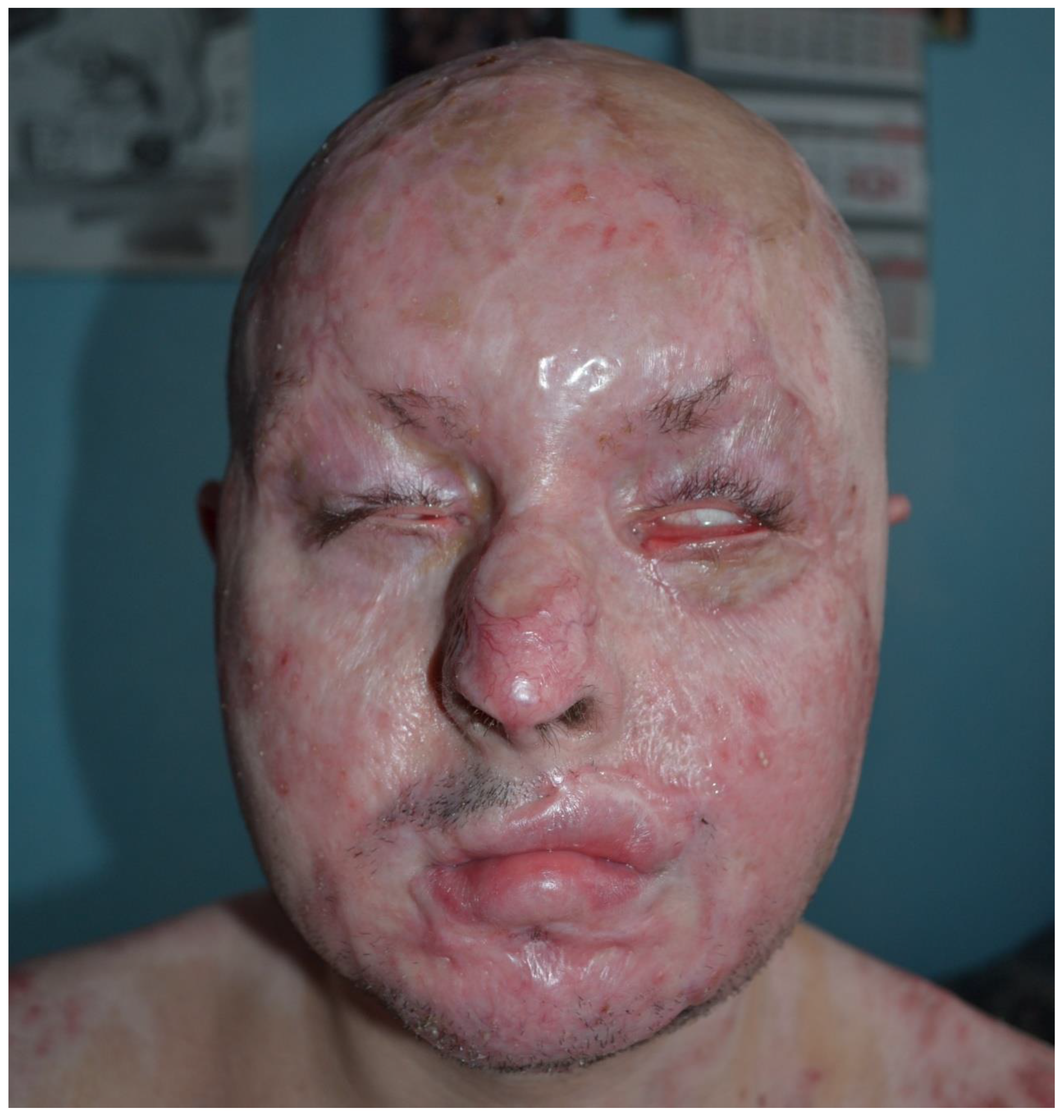
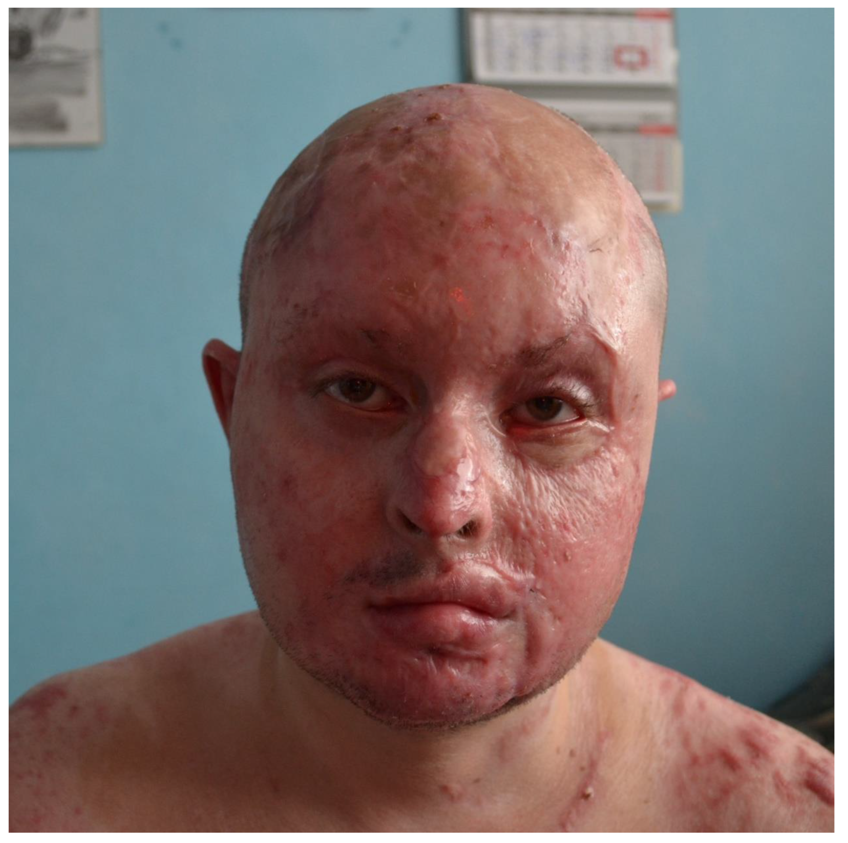
Figure 2.
Patient at initial presentation to our institution (October 2017). (A) Frontal view: extensive post-burn scarring of the face with complete absence of eyebrows, mustache, and beard hair; the frontal and anterior parietal scalp is covered by hypopigmented split-thickness skin grafts (STSG). (B) Superior (vertex) view: the parietal and frontal scalp is almost entirely alopecic, covered by STSG with mottled discoloration; only peripheral follicular islands remain. (C) Right lateral view: residual hair-bearing skin in the right temporal and occipital regions; extensive STSG-covered alopecic area spans the parietal scalp; post-burn scarring of the neck. (D) Left lateral view: sparse residual follicular tissue at the temporal and occipital margins; significant scarring of the preauricular region and neck. (E) Posterior view: extensive STSG-covered alopecia of the parieto-occipital scalp with irregular peripheral islands of surviving follicular tissue. (F) Right oblique view.
Figure 2.
Patient at initial presentation to our institution (October 2017). (A) Frontal view: extensive post-burn scarring of the face with complete absence of eyebrows, mustache, and beard hair; the frontal and anterior parietal scalp is covered by hypopigmented split-thickness skin grafts (STSG). (B) Superior (vertex) view: the parietal and frontal scalp is almost entirely alopecic, covered by STSG with mottled discoloration; only peripheral follicular islands remain. (C) Right lateral view: residual hair-bearing skin in the right temporal and occipital regions; extensive STSG-covered alopecic area spans the parietal scalp; post-burn scarring of the neck. (D) Left lateral view: sparse residual follicular tissue at the temporal and occipital margins; significant scarring of the preauricular region and neck. (E) Posterior view: extensive STSG-covered alopecia of the parieto-occipital scalp with irregular peripheral islands of surviving follicular tissue. (F) Right oblique view.
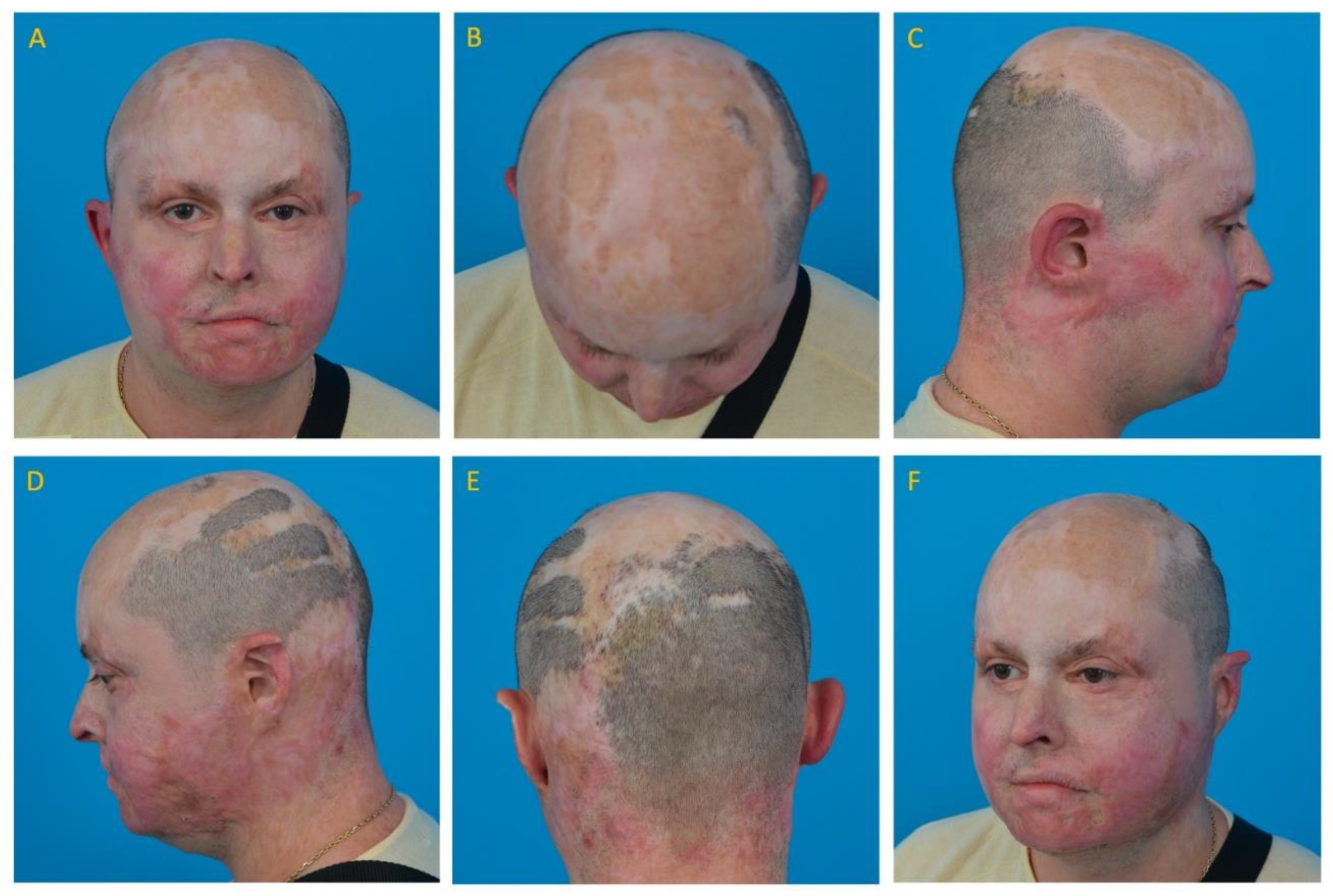
Figure 3.
Patient following completion of the eight-year reconstructive program at our institution (September 2025). (A) Frontal view: complete reconstruction of both eyebrows with natural arch and hair direction; full beard and mustache restoration; improved scar quality and color homogeneity of the face; residual focal alopecia of the anterior scalp partially concealed by scalp micropigmentation (SMP). (B) Superior (vertex) view: extensive follicular unit excision (FUE)-transplanted hair and SMP coverage throughout the parietal and frontal regions, with a small residual alopecic area at the vertex. (C) Right lateral view: natural-appearing temporal hairline; full beard reconstruction extending through the preauricular and submandibular regions; residual hypopigmented scar patches on the parietal scalp. (D) Left lateral view: comparable result bilaterally; improved scar texture and reduced erythema of the neck. (E) Posterior view: uniform short-cropped appearance of the occipital and parietal scalp with combined transplanted hair and SMP coverage; residual focal alopecic area with hypopigmented scar at the mid-occiput. (F) Right oblique view.
Figure 3.
Patient following completion of the eight-year reconstructive program at our institution (September 2025). (A) Frontal view: complete reconstruction of both eyebrows with natural arch and hair direction; full beard and mustache restoration; improved scar quality and color homogeneity of the face; residual focal alopecia of the anterior scalp partially concealed by scalp micropigmentation (SMP). (B) Superior (vertex) view: extensive follicular unit excision (FUE)-transplanted hair and SMP coverage throughout the parietal and frontal regions, with a small residual alopecic area at the vertex. (C) Right lateral view: natural-appearing temporal hairline; full beard reconstruction extending through the preauricular and submandibular regions; residual hypopigmented scar patches on the parietal scalp. (D) Left lateral view: comparable result bilaterally; improved scar texture and reduced erythema of the neck. (E) Posterior view: uniform short-cropped appearance of the occipital and parietal scalp with combined transplanted hair and SMP coverage; residual focal alopecic area with hypopigmented scar at the mid-occiput. (F) Right oblique view.
Disclaimer/Publisher’s Note: The statements, opinions and data contained in all publications are solely those of the individual author(s) and contributor(s) and not of MDPI and/or the editor(s). MDPI and/or the editor(s) disclaim responsibility for any injury to people or property resulting from any ideas, methods, instructions or products referred to in the content. |
© 2026 by the authors. Licensee MDPI, Basel, Switzerland. This article is an open access article distributed under the terms and conditions of the Creative Commons Attribution (CC BY) license (http://creativecommons.org/licenses/by/4.0/).
Copyright: This open access article is published under a Creative Commons CC BY 4.0 license, which permit the free download, distribution, and reuse, provided that the author and preprint are cited in any reuse.


