

Submitted:
09 July 2026
Posted:
10 July 2026
You are already at the latest version
Abstract
Hybridoma technology remains one of the most reliable and widely used platforms for generating highly specific monoclonal antibodies for use in diagnostics, fundamental research, and clinical practice. Moreover, the combination of unlimited proliferative capacity with the preservation of a key B lymphocyte function, antibody production, enables hybridoma cells to support a comprehensive functional evaluation of cellular responses. Thus, by integrating analysis of proliferation, viability, and productivity, hybridoma-based approaches enable the detection of differential modulatory effects and offer a nuanced assessment of compound bioactivity. This review provides a comprehensive analysis of the functional parameters of hybridoma cells: viability, proliferation, and productivity, as well as the methods used for their evaluation. The main stages of hybridoma cell generation, advances in hybridoma technology, and current applications of hybridoma cells are also reviewed. A key aspect of this review is the differential modulation of functional parameters of hybridoma cells. Modulation of culture conditions and bioactive compounds can differentially influence growth dynamics and specific antibody yield, often revealing an inverse relationship between proliferation and productivity.
Keywords:
hybridoma technology
; viability
; proliferation
; productivity
; monoclonal antibody
; functional parameters
1. Introduction
The modern pharmaceutical, biomedical, and biotechnology industries are characterized by a continual increase in the number of synthesized compounds, necessitating rapid and reliable evaluation of their biological activity. The early identification of substances with therapeutic potential, as well as the detection of compounds with undesirable toxic or immunomodulatory effects, represents a key objective of preclinical research.
In vitro studies play a central role in the primary screening of biological activity [1,2]. However, most conventional cell-based assays primarily focus on assessing basic parameters such as proliferation and viability [3,4]. Despite their utility, these parameters reflect general cytotoxic effects and do not fully capture changes in specialized cellular functions. In this regard, functional characteristics, especially the secretory activity of immunocompetent cells, are critical for understanding the mechanisms of action of biologically active compounds.
In this context, hybridoma cell lines are particulary important, as they combine the capacity for unlimited proliferation with the preservation of a key specialized function of B lymphocytes—antibody production [5]. This enables hybridoma cells to support a comprehensive functional evaluation of cellular responses, including the analysis of proliferation, viability, and antibody production. This approach enables the identification of differential modulatory effects of compounds, including cases where inhibition of cell growth is followed by enhanced antibody secretion [6].
This review focuses on the functional characteristics of hybridoma cells, including proliferation, viability, and productivity, as well as the methods used for their evaluation. It also summarizes key advances in hybridoma technology and opportunities for its application. Hybridoma cells are considered not only a source of monoclonal antibodies but also as a promising experimental platform for a comprehensive analysis of cellular response. This analysis includes the evaluation of proliferation, viability, and productivity.
A literature search was conducted using PubMed, ScienceDirect, and Google Scholar databases. Articles published up to and including 2026 were considered. The following keywords were used in various combinations: hybridoma, hybridoma technology, viability, proliferation, productivity, apoptosis, monoclonal antibody, cell sorting, cell cloning, production rate, production stimulating.
2. Hybridoma Technology, General Information
2.1. Key Stages in Hybridoma Generation
Hybridoma technology, originally described by Georges J. F. Köhler and César Milstein in 1975, made it possible to overcome a key challenge: the production of unlimited quantities of antibodies with identical specificity [7]. This breakthrough is widely considered one of the most significant milestones in the field of biotechnology. Hybridoma technology greatly expanded the capacity for both the detection and production of antibodies, thereby laying the foundation for the widespread application of monoclonal antibodies in scientific research, diagnostics, therapeutics, and biotechnology [8].
Classical hybridoma technology is a method based on the fusion of an immortal myeloma cell with a B-lymphocyte that produces monoclonal antibodies of predefined specificity [7]. Thus, hybridoma cell lines feature both stable antibody expression and the ability for continuous proliferation. Generating hybridoma cell lines is a multistep and labor-intensive process, with its key stages shown in Figure 1.
The initial step involves the immunization of laboratory animals, which receive multiple injections of the selected antigen over several weeks to stimulate specific antibody production [9]. Classical murine hybridomas are typically generated using BALB/c mice [10].
During the next step, activated B-lymphocytes are harvested from the spleen of an immunized animal and fused with HGPRT-deficient myeloma cells [5]. Commonly used myeloma partners include P3X63Ag8-U1 [11,12], SP2/0 [12,13,14,15,16], P3U1 [17], P3X63-Ag8.653 [12,18,19,20], and NS1 [13,21]. In practice, non-secreting myeloma lines are preferred. Polyethylene glycol is commonly used to induce cell fusion [7]. Alternatively, viruses such as Sendai virus [7,22] or electrofusion methods may be used [21,23]. Electrofusion often demonstrates higher efficiency [24,25].
After fusion, some intact cells remain in the culture. Hybridoma selection is carried out in HAT medium containing hypoxanthine, aminopterin, and thymidine [7]. Aminopterin inhibits de novo nucleotide synthesis, whereas hypoxanthine and thymidine are utilized in nucleotide production via the salvage pathway in HGPRT-positive cells. Due to their limited in vitro lifespan, unfused B-lymphocytes die within a few days. Myeloma cells that lack functional HGPRT are unable to use the salvage pathway and consequently do not survive. As a result, only hybridoma cells survive in HAT medium; these hybrid cells combine unlimited proliferation capacity inherited from the myeloma partner with the ability to express antibodies of defined specificity derived from B cells [5].
Hybridomas are then cloned by limiting dilution [26] to assess the characteristics of the monoclonal antibodies. In the final stage, selected antibody-secreting clones are expanded and cryopreserved [27] to ensure long-term storage and subsequent application in in vivo and in vitro culture systems [5].
2.2. Advances in Hybridoma Technology
Since its introduction over five decades ago, hybridoma technology has been significantly refined in its key stages. Some of these advances are summarized in Table 1.
A fusion stage modification significantly enhanced hybridoma yield. Preincubation of myeloma cells and lymphocytes in the presence of 0.25% PEG for 90 minutes at 37 °C resulted in an approximately tenfold increase in the proportion of wells containing antibody-secreting hybridomas [28].
Significant progress has been achieved in the field of electrofusion [24]. Optimization of pulse parameters, including the use of nanosecond and microsecond pulses with low peak power, has enabled a 1.5- to 3-fold increase in fusion efficiency compared with conventional high-power microsecond pulses [29]. Enriching cells using fluorescence-activated cell sorting (FACS) prior to electrofusion has also been shown to improve the yield of functional hybridomas [21]. Furthermore, a microfluidic droplet-based platform for hybridoma generation has been developed [43], along with a microfluidic chip that enables cell electrofusion within microdroplets [30].
In addition to these technical advances, modifications of culture media have been proposed to improve cell survival following fusion, including the addition of macrophage-conditioned medium [31]. Similarly, a conditioned medium derived from cultured rat thymocytes effectively replaces feeder cells and significantly enhances hybridoma growth [28].
FACS has become an important tool not only at the stage of cell preparation for fusion but also in the selection of antigen-specific hybridomas [34]. Early strategies involved labeling cells with antigen-coated fluorescent microspheres to identify antibody-secreting clones [32]. Current methods involve multistep fluorescence detection, including antigen binding followed by secondary reporter antibodies [33], as well as genetic modification of hybridomas to express a biotinylated surface marker that captures secreted antibodies on the surface of the producing cell [35]. Furthermore, membrane immunoglobulin-based screening (MIHS), which utilizes antigen binding to the hybridoma B-cell receptor, has been introduced [36]. Overall, FACS-based methods significantly increase the number of analyzed cells and enable the generation of clonal lines with high reliability [22].
Magnetic particles functionalized with the target antigen allow selective isolation of antigen-specific hybridomas, providing up to a 300-fold enrichment of the desired cell population [37].
Cultivation in semisolid media, such as those based on methylcellulose [38] or agar [39], is one of the most effective approaches for hybridoma clone selection. In liquid culture, the lack of spatial isolation leads to overgrowth of rapidly proliferating, low-producing cells and loss of slower-growing but potentially high-producing clones [44]. Semisolid media overcome this limitation through spatial isolation: each hybridoma forms an individual colony, thereby preventing repeated selection of identical variants and enabling independent evaluation of each clone’s productivity [45]. Due to matrix viscosity, secreted antibodies remain localized around the colony, forming a characteristic “halo” of immune complexes that can be evaluated by fluorescence intensity [45].
Automation of this approach is achieved using the ClonePix 2 platform (formerly Genetic ClonePix FL, Molecular Devices) [40,41]. This platform enables in situ screening of colonies and subsequent isolation of antigen-specific clones of a defined isotype [40]. Moreover, it allows selection of the most productive variants and, in many cases, generates high-producing lines after the first post-fusion cultivation step [22,40]. Integration of ClonePix with FACS further enhances selection efficiency and reduces development time [46].
A promising approach involves the use of self-seeding microwell chips coupled with an activated membrane for capturing antibodies secreted by individual cells [42]. This platform enables high-throughput screening and isolation of up to 6,400 clones per day, providing a rapid and scalable solution for early-stage clone selection.
Taken together, these developments have markedly enhanced the efficiency of hybridoma technology, accelerated the generation of monoclonal antibodies, and increased the likelihood of isolating rare, high-producing, antigen-specific clones.
Initially, hybridoma technology was predominantly used for the production of mouse antibodies; however, with further methodological development, it has been widely applied to generate monoclonal antibodies of diverse origin, including those derived from chicken [47], goat [48], rat [49], and rabbit [50]. Rabbit antibodies are of particular interest because the rabbit immune system can generate antibodies with higher affinity and broader molecular recognition, including phosphopeptides, carbohydrates, and antigens that are weakly immunogenic in mice [51].
Despite the success of hybridoma technology in generating high-affinity and highly specific antibodies, the therapeutic application of non-human antibodies is limited because they induce immunogenic responses [52] and adverse effects [53], which can reduce treatment efficacy and negatively affect pharmacokinetic properties [54]. To overcome these challenges, approaches for the development of chimeric [55] and humanized monoclonal antibodies [56] have been established. Concurrently, alternative antibody discovery platforms independent of hybridoma technology have been developed, including phage display [57,58], single B-cell–based methods [59], and de novo approaches based on artificial intelligence [60]. These and other modern antibody generation technologies have been extensively reviewed elsewhere [61,62,63,64].
2.3. Current Applications of Hybridoma Technology
Hybridoma technology remains one of the most reliable and widely utilized platforms for monoclonal antibody generation. Its relevance is evidenced by the continuous increase in publications describing the development and application of novel monoclonal antibodies produced using this approach. This technology enables the generation of highly specific antibodies that are extensively used in diagnostics, fundamental research, veterinary medicine, and clinical practice. Figure 2 illustrates the application of hybridoma technology.
Comprehensive coverage of all publications in this field is beyond the scope of this review. Therefore, the literature was selected based on its relevance, scientific novelty, and practical significance, with a focus on recent studies that reflect key directions in the development and application of monoclonal antibodies and hybridoma technology. The selected studies presented in Table 2 illustrate the significance of hybridoma technology.
Diagnostics represent one of the major application areas of hybridoma-derived antibodies. For example, monoclonal antibodies have been developed for the ELISA-based detection of citrinin in wine, a toxic compound of concern for human health [65]. Similarly, antibodies targeting the natural antioxidant dihydromyricetin have been generated for its detection in Ampelopsis grossedentata [66]. Furthermore, monoclonal antibodies against the denatured monomeric non-structural protein of Zika virus ZNS1 have been proposed as a basis for highly sensitive and specific indirect competitive ELISAs for early flavivirus detection [67]. Monoclonal antibodies are also used in ELISAs for the detection of calreticulin gene (CALR) mutations associated with essential thrombocythemia and primary myelofibrosis [68]. Chinese researchers have generated the first monoclonal antibodies against giant panda (Ailuropoda melanoleuca) vascular endothelial growth factor A (VEGFA), which are considered a novel tool for studying reproductive biology and oncology in this species [14].
Many studies focus on developing diagnostic tools for infectious diseases. For example, monoclonal antibodies against the VP27 protein of goose astrovirus GoAstV-2 have been proposed as promising candidates for both diagnostics and epitope-based vaccine development [69]. Similarly, monoclonal antibodies targeting the VP3 protein of infectious bursal disease virus have enabled the identification of a neutralizing antigenic epitope and provided a basis for designing multiantigen vaccines [15]. In addition, antibodies targeting the SWAP antigen of Schistosoma mansoni have demonstrated high performance in schistosomiasis immunodiagnostics, enhancing sensitivity, specificity, and staging accuracy [11].
Another important research direction is the development of therapeutically and functionally active antibodies. For example, a monoclonal antibody against ITGA4 has been reported to induce cell death in NK/T-cell lymphoma by forming large pores in the cell membrane [17]. Hybridoma-derived neutralizing monoclonal antibodies targeting the beta and delta variants of SARS-CoV-2 are considered promising candidates for therapeutic application after further optimization [70]. Furthermore, monoclonal antibodies specific to peanut-reactive human IgE passively sensitize effector cells and are widely used in in vitro models of the effector phase of food allergy [71].
Hybridoma cells represent an important source of genetic material for antibody engineering. Genes derived from hybridomas are used in transfection-based systems to generate recombinant antibodies, including neutralizing antibodies against papillomavirus genotypes [72] and antibodies targeting the hemagglutinin of highly pathogenic avian influenza virus H5N1 [73]. Furthermore, a single-chain variable fragment (scFv) derived from the 5G2 hybridoma, which produces IgM antibodies against REST, has been developed for the clinical evaluation of REST as a potential biomarker of precancerous lesions and cervical squamous cell carcinoma [18,74].
Hybridoma technology is not limited to producing monoclonal antibodies. T-cell hybridomas have emerged as valuable research tools. These systems enable the investigation of T-cell biology, including the characterization of atypical CD4⁺CD8⁺ populations observed in SATB1-deficient mice with Sjögren’s syndrome [75]. Moreover, T-cell hybridomas have been used as highly sensitive biosensors to measure the functional activity of endoplasmic reticulum aminopeptidases 1 and 2 [76].
Furthermore, hybridoma cell lines are considered a promising source of extracellular vesicles with defined immunological characteristics. For instance, the OKT3 hybridoma, which produces murine IgG specific to human CD3, secretes exosomes displaying these antibodies on its surface. These vesicles trigger strong T-cell cytokine responses and demonstrate increased stability compared to soluble antibodies [77].
Hybridoma cells represent a useful model for evaluating the cytotoxicity and biological activity of various compounds. Using the BAP3 hybridoma cell line, which produces murine IgG specific to human trophoblastic β-1 glycoprotein, the effects of graphene oxide nanoparticles functionalized with linear and branched polyethylene glycol were investigated. Graphene oxide nanoparticles at a concentration of 25 μg/mL decreased cell viability, with cytotoxicity diminishing as particle size increased and correlating with nanoparticle internalization. Due to their high proliferative capacity and plasma cell–like characteristics, hybridomas serve as a relevant model for evaluating the effects of different substances on antibody-producing cells [78]. Furthermore, B-cell hybridoma cells (line 53-6.7 from ATCC) were used to assess the biocompatibility of gadolinium oxide (Gd2O3:Er3+, Yb3+) nanostructures. Following incubation with nanoparticles and nanorods (concentrations up to 100 μg/mL for 48 h), the hybridoma cells consistently showed high viability and proliferation, with no morphology- or size-dependent toxic effects. These findings support the utility of hybridoma cells as a sensitive cellular model for evaluating nanomaterial cytotoxicity [79].
Hybridoma cells have been used as a model system to study the proliferative effects of β-lactoglobulin (LG). It was shown that LG at a concentration of 5 mg/mL stimulates hybridoma proliferation through interaction with membrane-bound IgM, which was identified as the LG receptor. These findings indicate that hybridomas can serve as a convenient cellular model for investigating receptor-mediated effects of proteins on cell proliferation [80].
Hybridoma technology remains a versatile platform for monoclonal antibody generation, with broad applications in diagnostics, fundamental research, and therapeutic development. Furthermore, hybridoma cell lines are used as a source of genetic material for antibody engineering, as model systems for cytotoxicity evaluation, and as producers of extracellular vesicles with specific immunological characteristics.
3. Functional Parameters of Hybridoma Cells
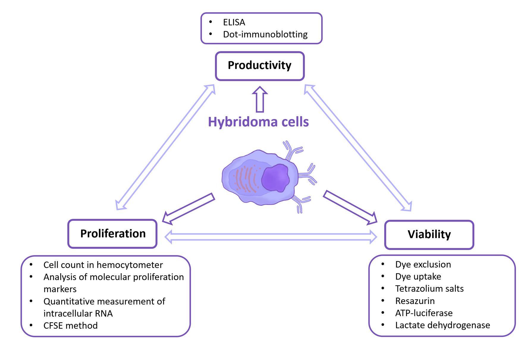
When working with hybridoma cells, it is standard practice to evaluate three key functional parameters: viability, proliferative activity, and productivity [81]. In addition to proliferation analysis, the evaluation of apoptosis is critically important. The high proliferative activity and viability of hybridoma cells are determined by genes derived from the tumor cell, whereas the ability to synthesize antibodies is conferred by genes originating from the mature B cell. A comprehensive evaluation of these three parameters not only characterizes the biological activity of the compounds under investigation but also allows researchers to distinguish substances based on their differential effects on cellular functional parameters. This approach is particularly important because an increase in one parameter may be followed by a decrease in another, necessitating a balanced interpretation of the results.
A wide range of methods is available for evaluating cell proliferation, viability, apoptosis, and productivity. This review primarily presents those most commonly applied to hybridoma cell lines.
3.1. Viability
Cell viability is defined as a quantitative measure of the number of cells that retain functional activity [82]. The methods used for its evaluation, presented in Figure 3, are conventionally classified as colorimetric and metabolic activity-based assays.
Colorimetric methods are among the most accessible and widely applicable ones for hybridoma cells [78,79,83,84]. These include dye exclusion and dye uptake assays [4]. Dye exclusion assays are based on the principle that dead cells lose membrane integrity, allowing dyes to penetrate the cell [85]. The most commonly used dyes include trypan blue, eosin, erythrosine B, Congo red, and propidium iodide [4,85,86]. Cell viability is calculated as the ratio of unstained cells to the total cell number. A modern approach involves the use of Zombie Aqua dye, which provides high-accuracy determination of hybridoma cell viability using flow cytometry and fluorescence microscopy [78].
Dye uptake methods are based on the ability of viable cells to accumulate dyes such as neutral red and Janus Green B [4]. The former is retained within lysosomes due to the pH gradient, whereas the latter is selectively taken up by active mitochondria and reduced by enzymes of the respiratory chain to form a colored product. The amount of dye accumulated is quantified spectrophotometrically [4].
Comparative studies have shown that the differences between trypan blue staining and neutral red staining [87], as well as between trypan blue and fluorescein diacetate (FDA) and propidium iodide (PI) [88], are minimal in the context of hybridoma cell lines. This allows any of these methods to be used in routine practice. However, another study has highlighted limitations associated with the use of trypan blue. When comparing viable cell counts obtained using trypan blue with those obtained by dual fluorescent staining with FDA and PI, it was found that, as hybridoma cultures age, trypan blue may significantly overestimate viability. This results in an underestimation of the proportion of dead cells and an inaccurate assessment of the overall culture status [89]. These findings underscore the need to validate results using additional methods, particularly when analyzing long-term cultivated cell lines.
Metabolic assays can be interpreted in two ways. On the one hand, the level of metabolic activity correlates with the number of viable cells, as only metabolically active cells are capable of reducing substrates. In this context, these methods are regarded as indirect assays of hybridoma cell viability [19,90]. On the other hand, in experiments involving actively proliferating hybridoma cultures, changes in the metabolic signal are frequently used as an indicators of proliferation [13,80,91]. This approach is based on the assumption that an increase in signal reflects not only the maintenance of viability but also an increase in cell number within the population. However, such an interpretation requires caution, as metabolic activity per cell may vary depending on the physiological state, culture conditions, and cell cycle stage.
Thus, metabolic assays cannot always be unambiguously assigned to a single category of functional analyses. Within the scope of this review, we consider them primarily as methods for assessing cell viability.
Metabolic assays are based on measuring enzyme activity or the concentration of metabolites in viable cells. Tetrazolium salts are compounds that, upon reduction, are converted into intensely colored formazans [4,85]. This reduction occurs via mitochondrial enzymes in metabolically active cells [3,4,85,92]. Common tetrazolium salts include MTT, XTT, MTS, and WST, an analog of MTS [3,4,85,92,93]. Absorbance is measured spectrophotometrically at 540 nm for MTT (blue formazan), 450 nm for XTT (orange formazan), and 490–500 nm for MTS (red-orange formazan) [4,85,92].
Resazurin, also known commercially as Alamar Blue, is a non-toxic, cell-permeable dye that is blue in its oxidized form and pink in its reduced form. The oxidized form, resazurin, is reduced by mitochondrial enzymes in viable cells to resorufin. Fluorescence is measured at wavelengths of 570 nm and 630 nm, and the amount of reduced dye correlates with the number of viable cells [4,85].
Intracellular ATP is a reliable indicator of cell viability [85]. ATP concentration is commonly determined using a luciferase-based luminescence assay because ATP is required for the luciferase-catalyzed conversion of luciferin [4,85].
Lactate dehydrogenase (LDH) is a cytoplasmic enzyme that is released upon membrane damage [4,85,86]. Its concentration in the supernatant correlates with the number of dead cells [19]. The enzyme catalyzes the reversible conversion of lactate to pyruvate, coupled with the reduction of NAD+ to NADH and vice versa [4,85,86]. The standard approach involves monitoring the change in NADH absorbance at 340 nm, followed by calculation using an appropriate formula [94].
3.2. Proliferation
Hybridoma cell proliferation proceeds according to the typical mammalian cell cycle. This process consists of four phases: G1 (cell growth and preparation for DNA synthesis), S (DNA replication), G2 (preparation for cell division), and M (mitosis) [95,96,97].
The transition from the G1 phase to the S phase is a critical event that determines subsequent cell proliferation and viability [98]. Studies of antibody synthesis have shown that per-cell productivity is highest during the G1 and G2 phases, whereas it decreases during mitosis and is lowest in the S phase [99]. At the same time, a positive correlation has been observed between overall culture productivity and the fraction of cells in S phase [100].
Under standard culture conditions, hybridoma cells have a doubling time of approximately 1–2 days [101]. Proliferation rates are strongly influenced by culture conditions. One of the limiting factors is glutamine; its exhaustion leads to a reduction in cell growth [102]. Cytokines play a crucial role in regulating hybridoma growth and survival. For many murine hybridomas, interleukin-6 (IL-6) is a key factor. Binding of IL-6 to its receptor activates the Ras/MAPK and PI3K/Akt signaling pathways, promoting the transition from G1 to S phase and supporting cell viability [98,103]. Disruption of these signaling pathways can result in cell cycle arrest and apoptosis induction. For example, IL-6 deprivation leads to G1-phase arrest in hybridoma cells and prevents the accumulation of S-phase cyclins, ultimately resulting in cell death [104]. Similar effects are observed when the cell cycle is arrested by thymidine or by serum starvation [98].
Comprehensive characterization of a cell culture requires the simultaneous evaluation of proliferation, viability, and cell death [105].
Numerous approaches exist for evaluating cell proliferation. The simplest and most cost-effective method is cell counting using a hemocytometer or its analogs in combination with light microscopy [106,107]. However, this method provides only approximate cell number dynamics, is labor-intensive, and loses accuracy at high cell suspension densities [106].
A more informative approach is the analysis of molecular proliferation markers, as shown in Figure 4. The nuclear protein Ki-67 is expressed during all phases of the cell cycle except G0 phase and therefore allows evaluation of the fraction of actively proliferating cells [108,109]. Synthesis of proliferating cell nuclear antigen (PCNA) begins in late G1 phase and reaches its peak during S phase; it subsequently declines during G2 and is virtually absent during mitosis [3,110,111]. Nucleoside analogs such as BrdU and EdU are added to the culture medium and incorporated into newly synthesized DNA, thereby enabling detection of S-phase cells [3,96,112].
An alternative approach to assessing cell cycle status is quantitative measurement of intracellular RNA using dual staining with Hoechst 33342 and Pyronin Y, as actively proliferating cells typically contain higher RNA levels than quiescent cells [113].
Hybridoma cell proliferation can also be evaluated using the carboxyfluorescein succinimidyl ester (CFSE) dilution assay [79]. The CFSE method relies on the principle of fluorescent dye dilution upon cell division. After binding to intracellular proteins, CFSE is equally distributed between daughter cells during symmetric mitosis, resulting in a stepwise twofold decrease in fluorescence intensity with each generation, which can be measured by flow cytometry [114].
Hybridoma cells exhibit high sensitivity to stress [115]; therefore, the assessment of apoptosis is critically important. Apoptosis is a form of programmed cell death. In most cases, cell death proceeds via the mitochondrial apoptotic pathway, followed by caspase activation [104].
One approach to enhancing cellular robustness is overexpression of anti-apoptotic proteins of the Bcl-2 family [116,117]. In Sp2/0 hybridoma cells, this modification approximately doubles the duration of the productive phase and increases antibody production by about 40% [116]. Similarly, overexpression of Bcl-2 increases the maximum viable cell density by 45%, delays the onset of apoptosis, and prolongs culture viability [117]. Pharmacological inhibition of apoptosis can also enhance cell survival; for example, the pan-caspase inhibitors z-VAD-fmk and Ac-DEVD-cho block effector caspases and maintain hybridoma viability under glutamine deprivation for up to 36 h [118].
Apoptosis in hybridoma cells is commonly detected using Annexin V [119], a marker that binds to phosphatidylserine exposed on the outer surface of the cell membrane during the early stages of apoptosis [4,120]. The combined use of Annexin V and PI allows discrimination between early apoptosis (Annexin V⁺/PI⁻) and late apoptosis or necrosis (Annexin V⁺/PI⁺) [4]. In addition, the TUNEL assay (terminal deoxynucleotidyl transferase dUTP nick end labeling) is used to assess apoptosis in hybridoma cells [121]. This method is based on the use of the enzyme terminal deoxynucleotidyl transferase (TdT), which catalyzes the incorporation of labeled deoxynucleotides into free 3′-OH termini of DNA, thereby enabling detection of DNA fragmentation—one of the hallmarks of apoptosis [122,123]. The DNA laddering assay is also used to detect apoptosis in hybridoma cells [124]. This method is based on the identification of a characteristic DNA fragmentation pattern by agarose gel electrophoresis [4].
3.3. Productivity
Productivity represents the capacity of a cell to carry out its specialized function, namely the synthesis and secretion of the target protein. At the molecular level, it is driven by processes associated with expression of the gene encoding the target protein—in hybridomas, the immunoglobulin light- and heavy-chain genes [125]—as well as by post-translational modifications and protein synthesis [126]. A detailed discussion of the molecular mechanisms regulating productivity is beyond the scope of this review.
In the context of hybridoma productivity, it is useful to differentiate parameters such as target product titer and specific productivity. The target product titer represents the final concentration of antibodies in the culture medium. This parameter is equivalent to overall cellular productivity and is typically expressed as antibody mass per unit volume. Mathematical models describing target product titer have been reported in several studies [127,128]:
where is target product titer, is specific productivity, is viable cell density over the cultivation period.
Specific productivity is generally defined as the amount of target product produced by a single cell per unit time. This parameter depends on both the antibody concentration in the medium and the viable cell count. Accordingly, evaluation of specific productivity requires consideration of both the dynamics of viable cell density and the final antibody concentration in the medium (titer). The mathematical formulation can be derived from the model described above [127,128]:
where is specific productivity, is target product titer, is viable cell density over the cultivation period.
Enzyme-linked immunosorbent assay (ELISA) and dot-immunoblotting are commonly used to assess the productivity of hybridoma cell lines. Among these methods, ELISA is the preferred [13,129,130,131], primarily due to its high sensitivity, specificity, and quantitative capacity.
Enzyme-linked immunosorbent assay (ELISA) is a versatile immunological method widely used in fundamental and applied research, as well as in clinical practice and diagnostics. The method relies on the specific interaction between an antigen and a primary antibody targeting the protein of interest. Detection of the antigen–antibody complex is typically achieved using enzyme-conjugated antibodies that catalyze a reaction with an appropriate substrate. The reaction products are quantified using either a luminometer or a spectrophotometer. Depending on the experimental design, ELISA can be classified into direct, indirect, sandwich, and competitive formats, which differ in reagent types, step sequences, and assay conditions [132].
An alternative approach is dot-immunoblotting, which is used for the rapid screening of hybridoma clones and evaluation of their secretory activity [133,134]. In the classical assay format, the nitrocellulose membrane is precoated with affinity-purified anti-mouse immunoglobulins (derived from goat or rabbit), followed by incubation with the hybridoma culture supernatant. Secreted monoclonal antibodies are captured on the membrane surface and subsequently detected using enzyme-labeled reagents, most commonly horseradish peroxidase, which enables signal visualization and comparative assessment of antibody production levels [135].
4. Differential Modulation of Hybridoma Cells Functional Parameters
Various strategies have been developed to enhance target product titers in hybridoma cells, including optimization of culture conditions [136], fed-batch cultivation [137], vitamin supplementation [138], use of antioxidants such as coenzyme Q10 [139], and use of various chemical compounds [6]. In these cases, productivity is enhanced either through an increase in viable cell numbers [140] or through an increase in specific productivity [141]. However, proliferation alone does not always correlate with increased antibody production, which underscores the importance of identifying compounds that stimulate cell growth without reducing specific productivity.
The studies summarized in Table 3 investigate the correlation between proliferation and specific productivity. According to the proposed model, reducing proliferation to a certain level increases specific cell productivity; however, overly suppressed growth also decreases productivity [6]. This finding supports the concept of intracellular resource reallocation, wherein artificial suppression of cell division may redirect metabolic resources toward antibody synthesis and secretion. It has been demonstrated that an increase in specific growth rate results in a reduction in the specific production rates of both ATP and antibodies [142]. The corresponding mathematical model describes the dependence of productivity on the proliferation rate through the regulation of transcription and translation at different stages of the cell cycle [6]. The experimental data below further show that inhibiting proliferation can indeed increase cell-specific productivity. Growth suppression may be induced either by unfavorable culture conditions or by compounds with growth-inhibitory activity.
Osmolarity, defined as the total concentration of osmotically active particles per unit volume, determines the direction of water flux across the cell membrane and represents a key physicochemical parameter governing the balance between cell proliferation and protein synthesis. The use of culture media with elevated osmolarity can enhance hybridoma productivity [143,144]. However, this is typically followed by a decrease in proliferation rate and an increase in cell volume. Effective application of hyperosmotic conditions often requires prior adaptation of the cells. The physiological osmolarity of the medium is approximately 250–300 mOsm. Sodium chloride is commonly used to increase it. Increasing osmolarity to 435 mOsm enhances specific productivity by approximately 2.2-fold but does not improve overall productivity because of reduced cell growth [143]. A combination of moderate osmolarity elevation (350 mOsm) with sodium butyrate (0.1 mM) resulted in a twofold increase in antibody titer at the end of cultivation [144]. Despite its relative simplicity, this strategy requires prolonged adaptation [144] and careful, cell line–specific optimization [153].
A wide range of compounds has been reported as growth inhibitors of hybridoma cells. Among the most extensively studied is sodium butyrate [145,146], commonly applied at a concentration of 1 mM. Notably, sodium butyrate is effective not only in hybridoma systems but also in CHO cell cultures [146]. Colchicine has likewise been proposed as a promising agent for enhancing hybridoma productivity. The addition of colchicine at a concentration of 10 ng/mL to a medium supplemented with 1 mM sodium butyrate resulted in a 43% increase in productivity [147]. The use of colchicine represents an effective strategy for increasing monoclonal antibody yield, particularly in hybridoma lines that respond poorly to conventional productivity enhancers. This effect is related to the ability of colchicine, a microtubule and mitotic inhibitor, to slow cell proliferation [154].
Similar effects have been reported for other regulators of the cell cycle. For example, caffeine can induce cell cycle arrest in the G0/G1 phase [155]. At a concentration of 1.92 mM, caffeine increased hybridoma-specific productivity by 2.8-fold [141] and 1.8-fold [148], although no corresponding increase in final antibody titer was observed. Bacterial lipopolysaccharide (LPS) at a concentration of 100 pg per cell has also been reported to suppress proliferation while increasing overall hybridoma productivity by up to 2.5-fold [149]. However, its practical application is limited by challenges associated with downstream purification. Similarly, low concentrations of DMSO (0.2%) can enhance hybridoma productivity by approximately twofold [150], likely through induction of cell cycle arrest in the G1 phase [156]. Notably, DMSO at low concentrations does not affect monoclonal antibody glycosylation, highlighting its potential for industrial-scale applications [150].
Polyamines are also considered potential regulators capable of enhancing hybridoma productivity and modulating their functional activity. Studies using the human hybridoma cell line HB4C5 have demonstrated that certain polyamines significantly enhance IgM production under serum-free conditions [151,152]. For example, the addition of spermine at a concentration of 7.3 mM and spermidine at 4.5 mM increased IgM production approximately sixfold [151,152]. In both cases, the increase in specific productivity was followed by growth inhibition. Thermine at a concentration of 2 mM also exhibited a pronounced stimulatory effect, increasing IgM production by approximately 5.3-fold without noticeably suppressing cell proliferation [152]. Although the final antibody titer was not reported, it can be assumed that it increases due to enhanced specific productivity while maintaining proliferative activity. In contrast, triethylenetetramine induced only a transient increase in IgM production and exhibited marked cytotoxicity, limiting its applicability in long-term culture [152].
In addition to small molecules, cytokines also play a significant role in regulating hybridoma productivity. For example, recombinant human IL-6 at a concentration of 2 ng/mL increased the specific productivity of hybridomas approximately fivefold in serum-free medium [129]. Furthermore, IL-6 can inhibit apoptosis in hybridoma cells, thereby extending culture duration and increasing overall antibody yield [157].
One should note that inhibition of proliferation combined with stimulation of specific productivity does not always result in a higher antibody titer. This effect can be explained by a substantial reduction in viable cell numbers, which may ultimately lead to lower target product titers. At the same time, it has been demonstrated that the addition of growth inhibitors at high cell densities can increase the yield of the target product by up to twofold [145]. Growth inhibitors that disrupt antibody synthesis reduce both the specific productivity and the target product titer [6].
Thus, optimization of hybridoma cells functional parameters requires a strict balance between proliferation regulation and product yield enhancement, making the identification of reliable productivity-enhancing agents a promising and important area of research.
5. Conclusions
Hybridoma cell lines represent a unique biotechnological model that combines unlimited proliferative capacity with the ability to produce highly specific antibodies. This dual nature underlies their significance not only as a classical tool for generating monoclonal antibodies but also as a versatile platform for comprehensive evaluation of the biological activity of various compounds.
Compared with conventional cell-based assays, hybridoma cell lines enable the simultaneous evaluation of proliferation, viability, and productivity—the last of which represents a specific functional parameter. Since none of these parameters alone can fully reflect the functional state of the culture, their integrated evaluation offers the most accurate insight into the cellular response to the investigated compounds.
Importantly, these parameters are connected through a complex functional interplay. An increase in specific productivity may correlate with cell cycle inhibition, whereas stimulation of proliferation can lead to a reduction in productivity. Such differential modulation effects indicate cellular resource reallocation and underscore the need for comprehensive analysis of functional parameters. Accordingly, evaluation of hybridoma cells functional parameters enables the detection and characterization of these effects. Thus, hybridoma cells should be considered not only a production platform but also a highly informative biological model for both fundamental and applied research.
Nevertheless, hybridoma technology remains a relevant platform for the generation of monoclonal antibody-producing cells, making the identification of productivity-enhancing factors a key strategy for optimizing the biotechnological production of target proteins.
Author Contributions
Conceptualization, M.R; S.Z and T.B.; writing—original draft preparation, T.B.; writing—review and editing, S.Z; M.R., S.L. and T.B.; visualization, T.B.; supervision, M.R. All authors have read and agreed to the published version of the manuscript.
Funding
R&D “Study of mechanisms of regulation of immune system cells and development of methods for their evaluation in normal and pathology”, Reg. number: 124020500027-7 (“IEGM, PFRC of the Ural Branch of RAS”).
Data Availability Statement
No new data was created.
Conflicts of Interest
The authors declare no conflict of interest.
Abbreviations
The following abbreviations are used in this manuscript:
| ATCC | American Type Culture Collection |
| BrdU | 5-bromo-2'-deoxyuridine |
| CALR | Calreticulin |
| CFSE | Carboxyfluorescein succinimidyl ester |
| EdU | 5-ethynyl-2'-deoxyuridine |
| ELISA | Enzyme-linked immunosorbent assay |
| FACS | Fluorescence-activated cell sorting |
| FDA | Fluorescein diacetate |
| HAT | Hypoxanthine, aminopterin, thymidine |
| HGPRT | Hypoxanthine-guanine phosphoribosyltransferase |
| IL-6 | Interleukin-6 |
| LDH | Lactate dehydrogenase |
| LG | β-lactoglobulin |
| LPS | Lipopolysaccharide |
| MIHS | Membrane Ig-based screening |
| MTS | 3-(4,5-dimethylthiazol-2-yl)-5-(3-carboxymethoxyphenyl)-2-(4-sulfophenyl)-2H-tetrazolium |
| MTT | 3-(4,5-dimethylthiazol-2-yl)-2,5-diphenyltetrazolium bromide |
| PCNA | proliferating cell nuclear antigen |
| PEG | Polyethylene glycol |
| PI | Propidium iodide |
| scFv | Single-chain variable fragment |
| TdT | Terminal deoxynucleotidyl transferase |
| TUNEL | Terminal deoxynucleotidyl transferase dUTP nick end labeling |
| VEGFA | Vascular endothelial growth factor A |
| XTT | 2,3-bis(2-methoxy-4-nitro-5-sulfophenyl)-5-[(phenylamino)carbonyl]-2H-tetrazolium hydroxide |
References
- Habib, S.; Gupta, P.; Bhat, S.S.; Gupta, J. In Silico, in-Vitro and in Vivo Screening of Biological Activities of Citral. Int. J. Vitam. Nutr. Res. 2021, 91, 251–260. [Google Scholar] [CrossRef] [PubMed]
- Nair, D.G.; Weiskirchen, R. Advanced In Vitro Models for Preclinical Drug Safety: Recent Progress and Prospects. Curr. Issues Mol. Biol. 2024, 47, 7. [Google Scholar] [CrossRef] [PubMed]
- Adan, A.; Kiraz, Y.; Baran, Y. Cell Proliferation and Cytotoxicity Assays. Curr. Pharm. Biotechnol. 2016, 17, 1213–1221. [Google Scholar] [CrossRef] [PubMed]
- Khalef, L.; Lydia, R.; Filicia, K.; Moussa, B. Cell Viability and Cytotoxicity Assays: Biochemical Elements and Cellular Compartments. Cell Biochem. Funct. 2024, 42, e4007. [Google Scholar] [CrossRef] [PubMed]
- Mitra, S.; Tomar, P.C. Hybridoma Technology; Advancements, Clinical Significance, and Future Aspects. J. Genet. Eng. Biotechnol. 2021, 19, 159. [Google Scholar] [CrossRef] [PubMed]
- Suzuki, E.; Ollis, D.F. Enhanced Antibody Production at Slowed Growth Rates: Experimental Demonstration and a Simple Structured Model. Biotechnol. Prog. 1990, 6, 231–236. [Google Scholar] [CrossRef] [PubMed]
- Köhler, G.; Milstein, C. Continuous Cultures of Fused Cells Secreting Antibody of Predefined Specificity. Nature 1975, 256, 495–497. [Google Scholar] [CrossRef] [PubMed]
- Parray, H.A.; Shukla, S.; Samal, S.; Shrivastava, T.; Ahmed, S.; Sharma, C.; Kumar, R. Hybridoma Technology a Versatile Method for Isolation of Monoclonal Antibodies, Its Applicability across Species, Limitations, Advancement and Future Perspectives. Int. Immunopharmacol. 2020, 85, 106639. [Google Scholar] [CrossRef] [PubMed]
- Greenfield, E.A. Immunizing Animals. Cold Spring Harb. Protoc. 2022, 2022, pdb.top100180. [Google Scholar] [CrossRef] [PubMed]
- Chakravarty, E.; Dorak, M.T. Missed Opportunities to Increase Efficiency of Monoclonal Antibody Development Using Hybridoma Technology and Mice as the Source Animal. Front. Immunol. 2024, 15, 1443726. [Google Scholar] [CrossRef] [PubMed]
- Alajmi, F.E. Cross-Reactivity and Localization of Schistosoma Mansoni Antigen for Immunodiagnosis. Braz. J. Med. Biol. Res. Rev. Bras. Pesqui. Medicas E Biol. 2026, 58, e15126. [Google Scholar] [CrossRef] [PubMed]
- Mariani, E.; Mariani, A.R.; Monaco, M.C.G.; Lalli, E.; Vitale, M.; Facchini, A. Commercial Serum-Free Media: Hybridoma Growth and Monoclonal Antibody Production. J. Immunol. Methods 1991, 145, 175–183. [Google Scholar] [CrossRef] [PubMed]
- Lee, R.-J.; Wu, J.-S.; Juang, R.-H.; Wu, Y.-J.; Lin, C.-Y.; Wu, S.-Y.; Huang, P.-R.; Chu, C.-H.; Chin, L.-T.; Chen, H.-M. Increasing Hybridoma Viability and Antibody Repertoire after the Cell Fusion by the Use of Human Plasma as an Alternative Supplement. J. Immunol. Methods 2010, 361, 89–97. [Google Scholar] [CrossRef] [PubMed]
- Liu, X.; Li, F.; Wang, S.; Zhang, M.; Hu, X.; Liu, Y.; Hou, R.; Cai, K. Prokaryotic Expression of the Giant Panda VEGFA Gene and the Preparation and Characterization of Its Monoclonal Antibodies. Vet. Anim. Sci. 2026, 31, 100551. [Google Scholar] [CrossRef] [PubMed]
- Pan, X.-Y.; Ren, H.; Zi, M.-H.; Fan, J.-H.; Ma, Y.-H.; Shao, H.-C.; Liang, Z.-S.; Zhang, Y.; Han, S.; Zhang, G.-P.; et al. Development and Characterization of Monoclonal Antibodies Against VP3 Protein of Infectious Bursal Disease Virus. Transbound. Emerg. Dis. 2025, 2025, 5915042. [Google Scholar] [CrossRef] [PubMed]
- Shulman, M.; Wilde, C.D.; Köhler, G. A Better Cell Line for Making Hybridomas Secreting Specific Antibodies. Nature 1978, 276, 269–270. [Google Scholar] [CrossRef] [PubMed]
- Takeuchi, S.; Ito, Y.; Matsuoka, S.; Abe, M.; Tsurui, H.; Horimoto, Y.; Fukuhara, T.; Hirano, T.; Mizutani, N.; Ito, T.; et al. Novel Anti-ITGA4 Monoclonal Antibody Induces Cell Death via Large Pore Formation in NK/T-Cell Lymphoma Cells. Sci. Rep. 2025, 15, 45655. [Google Scholar] [CrossRef] [PubMed]
- Cortés-Sarabia, K.; Medina-Flores, Y.; Alarcón-Romero, L.D.C.; Mata-Ruíz, O.; Vences-Velázquez, A.; Rodríguez-Ruíz, H.A.; Valdés, J.; Ortuño-Pineda, C. Production and Characterization of Monoclonal Antibodies against the DNA Binding Domain of the RE1-Silencing Transcription Factor. J. Biochem. 2019, 166, 393–402. [Google Scholar] [CrossRef] [PubMed]
- Duval, D.; Demangel, C.; Geahel, I.; Blondeau, K.; Marcadet, A. Comparison of Various Methods for Monitoring Hybridoma Cell Proliferation. J. Immunol. Methods 1990, 134, 177–185. [Google Scholar] [CrossRef] [PubMed]
- Ozturk, S.S.; Øpalsson, B. Loss of Antibody Productivity During Long-Term Cultivation of a Hybridoma Cell Line in Low Serum and Serum-Free Media. Hybridoma 1990, 9, 167–175. [Google Scholar] [CrossRef] [PubMed]
- Rousseau, F.; Menier, C.; Brochard, P.; Simon, S.; Perez-Toralla, K.; Wijkhuisen, A. Targeted Fusion of Antibody-Secreting Cells: Unlocking Monoclonal Antibody Production with Hybridoma Technology. mAbs 2025, 17, 2510336. [Google Scholar] [CrossRef] [PubMed]
- Smith, S.A.; Crowe, J.E., Jr. Use of Human Hybridoma Technology To Isolate Human Monoclonal Antibodies. Microbiol. Spectr. 2015, 3, 3.1.05. [Google Scholar] [CrossRef] [PubMed]
- Yu, X.; McGraw, P.A.; House, F.S.; Crowe, J.E. An Optimized Electrofusion-Based Protocol for Generating Virus-Specific Human Monoclonal Antibodies. J. Immunol. Methods 2008, 336, 142–151. [Google Scholar] [CrossRef] [PubMed]
- Kou, J.; Shen, J.; Wang, Z.; Yu, W. Advances in Hybridoma Preparation Using Electrofusion Technology. Biotechnol. J. 2023, 18, 2200428. [Google Scholar] [CrossRef] [PubMed]
- Tomita, M.; Tsumoto, K. Hybridoma Technologies for Antibody Production. Immunotherapy 2011, 3, 371–380. [Google Scholar] [CrossRef] [PubMed]
- Greenfield, E.A. Single-Cell Cloning of Hybridoma Cells by Limiting Dilution. Cold Spring Harb. Protoc. 2019, 2019, pdb.prot103192. [Google Scholar] [CrossRef] [PubMed]
- Greenfield, E.A. Liquid Nitrogen Storage of Hybridoma Cells. Cold Spring Harb. Protoc. 2020, 2020, pdb.prot103267. [Google Scholar] [CrossRef] [PubMed]
- Orlik, O.; Altaner, C. Modifications of Hybridoma Technology Which Improve the Yield of Monoclonal Antibody Producing Cells. J. Immunol. Methods 1988, 115, 55–59. [Google Scholar] [CrossRef] [PubMed]
- Wu, M.; Ke, Q.; Bi, J.; Li, X.; Huang, S.; Liu, Z.; Ge, L. Substantially Improved Electrofusion Efficiency of Hybridoma Cells: Based on the Combination of Nanosecond and Microsecond Pulses. Bioengineering 2022, 9, 450. [Google Scholar] [CrossRef] [PubMed]
- Schoeman, R.M.; Van Den Beld, W.T.E.; Kemna, E.W.M.; Wolbers, F.; Eijkel, J.C.T.; Van Den Berg, A. Electrofusion of Single Cells in Picoliter Droplets. Sci. Rep. 2018, 8, 3714. [Google Scholar] [CrossRef] [PubMed]
- Hnasko, R.; Lin, A.V.; Stanker, L.; McGarvey, J. A Bioassay for Optimization of Macrophage-Conditioned Medium as a Culture Supplement to Promote Hybridoma Cell Survival and Growth. Monoclon. Antib. Immunodiagn. Immunother. 2018, 37, 126–133. [Google Scholar] [CrossRef] [PubMed]
- Parks, D.R.; Bryan, V.M.; Oi, V.T.; Herzenberg, L.A. Antigen-Specific Identification and Cloning of Hybridomas with a Fluorescence-Activated Cell Sorter. Proc. Natl. Acad. Sci. 1979, 76, 1962–1966. [Google Scholar] [CrossRef] [PubMed]
- Dippong, M.; Carl, P.; Lenz, C.; Schenk, J.A.; Hoffmann, K.; Schwaar, T.; Schneider, R.J.; Kuhne, M. Hapten-Specific Single-Cell Selection of Hybridoma Clones by Fluorescence-Activated Cell Sorting for the Generation of Monoclonal Antibodies. Anal. Chem. 2017, 89, 4007–4012. [Google Scholar] [CrossRef] [PubMed]
- Greenfield, E.A. Hybridoma Screening by Antibody Capture: Flow Cytometry/FACS with Permeabilized Cells to Detect Intracellular Binding. Cold Spring Harb. Protoc. 2022, 2022, pdb.prot103085. [Google Scholar] [CrossRef] [PubMed]
- Listek, M.; Hönow, A.; Gossen, M.; Hanack, K. A Novel Selection Strategy for Antibody Producing Hybridoma Cells Based on a New Transgenic Fusion Cell Line. Sci. Rep. 2020, 10, 1664. [Google Scholar] [CrossRef] [PubMed]
- Sakaguchi, A.; Tanaka, Y.; Shoji, E.; Takeshima, T.; Sakamaki, R.; Matsuba, T.; Kurihara, Y. Rapid, Simple, and Effective Strategy to Produce Monoclonal Antibodies Targeting Protein Structures Using Hybridoma Technology. J. Biol. Eng. 2023, 17, 24. [Google Scholar] [CrossRef] [PubMed]
- Ossendorp, F.A.; Bruning, P.F.; Vaan Den Brink, J.A.M.; De Boer, M. Efficient Selection of High-Affinity B Cell Hydridomas Using Antigen-Coated Magnetic Beads. J. Immunol. Methods 1989, 120, 191–200. [Google Scholar] [CrossRef] [PubMed]
- Yokoyama, W.M.; Christensen, M.; Santos, G.D.; Miller, D.; Ho, J.; Wu, T.; Dziegelewski, M.; Neethling, F.A. Production of Monoclonal Antibodies. Curr. Protoc. Immunol. 2013, 102. [Google Scholar] [CrossRef] [PubMed]
- Greenfield, E.A. Single-Cell Cloning of Hybridoma Cells by Growth in Soft Agar. Cold Spring Harb. Protoc. 2019, 2019, pdb.prot103200. [Google Scholar] [CrossRef] [PubMed]
- Mann, C.J. Rapid Isolation of Antigen-Specific Clones from Hybridoma Fusions. Nat. Methods 2007, 4, i–ii. [Google Scholar] [CrossRef]
- Stravinskiene, D.; Imbrasaite, A.; Petrikaite, V.; Matulis, D.; Matuliene, J.; Zvirbliene, A. New Monoclonal Antibodies for a Selective Detection of Membrane-Associated and Soluble Forms of Carbonic Anhydrase IX in Human Cell Lines and Biological Samples. Biomolecules 2019, 9, 304. [Google Scholar] [CrossRef] [PubMed]
- Rubben, K.; Vander Plaetsen, A.-S.; Almey, R.; Tytgat, O.; Deserranno, K.; Debaere, J.; Acar, D.D.; Meuleman, P.; Deforce, D.; Van Nieuwerburgh, F. High-Throughput Single-Cell Screening of Viable Hybridomas and Patient-Derived Antibody-Secreting Cells Using Punchable Microwells. Artif. Cells Nanomed. Biotechnol. 2024, 52, 426–436. [Google Scholar] [CrossRef] [PubMed]
- Schoeman, R.M.; Kemna, E.W.M.; Wolbers, F.; Van Den Berg, A. High-throughput Deterministic Single-cell Encapsulation and Droplet Pairing, Fusion, and Shrinkage in a Single Microfluidic Device. ELECTROPHORESIS 2014, 35, 385–392. [Google Scholar] [CrossRef] [PubMed]
- Dharshanan, S.; Hung, C.S. Screening and Subcloning of High Producer Transfectomas Using Semisolid Media and Automated Colony Picker. In Monoclonal Antibodies; Methods in Molecular Biology; Ossipow, V., Fischer, N., Eds.; Humana Press: Totowa, NJ, 2014; Vol. 1131, pp. 105–112. ISBN 978-1-62703-991-8. [Google Scholar]
- Ammar, A.E.S. Single-Cell Cloning and Its Approaches. Front. Cell Dev. Biol. 2025, 13, 1678157. [Google Scholar] [CrossRef] [PubMed]
- Roy, G.; Miro-Quesada, G.; Zhuang, L.; Martin, T.; Zhu, J.; Wu, H.; Marelli, M.; Bowen, M.A. Sequential Screening by ClonePix FL and Intracellular Staining Facilitate Isolation of High Producer Cell Lines for Monoclonal Antibody Manufacturing. J. Immunol. Methods 2017, 451, 100–110. [Google Scholar] [CrossRef] [PubMed]
- Nishinaka, S.; Suzuki, T.; Matsuda, H.; Murata, M. A New Cell Line for the Production of Chicken Monoclonal Antibody by Hybridoma Technology. J. Immunol. Methods 1991, 139, 217–222. [Google Scholar] [CrossRef] [PubMed]
- Flynn, J.N.; Harkiss, G.D.; Hopkins, J. Generation of a Sheep × Mouse Heterohybridoma Cell Line (1C6.3a6T.1D7) and Evaluation of Its Use in the Production of Ovine Monoclonal Antibodies. J. Immunol. Methods 1989, 121, 237–246. [Google Scholar] [CrossRef] [PubMed]
- Fang, J.C.; Bodeus, M.; Burtonboy, G. Study on Rat-Rat Hybridoma Technique and Production of Rat Monoclonal Antibodies against HIV and HBsAg. Chin. J. Biotechnol. 1991, 7, 73–81. [Google Scholar] [PubMed]
- Mei-Chang, K.; Soon, J.A.; Max, E.E.; Kindt, T.J. Rabbit-Mouse Hybridomas Secreting Intact Rabbit Immunoglobulin. Mol. Immunol. 1985, 22, 351–359. [Google Scholar] [CrossRef] [PubMed]
- Zhang, Z.; Liu, H.; Guan, Q.; Wang, L.; Yuan, H. Advances in the Isolation of Specific Monoclonal Rabbit Antibodies. Front. Immunol. 2017, 8, 494. [Google Scholar] [CrossRef] [PubMed]
- Hwang, W.Y.K.; Foote, J. Immunogenicity of Engineered Antibodies. Methods 2005, 36, 3–10. [Google Scholar] [CrossRef] [PubMed]
- Carter, P. Improving the Efficacy of Antibody-Based Cancer Therapies. Nat. Rev. Cancer 2001, 1, 118–129. [Google Scholar] [CrossRef] [PubMed]
- Ober, R.J.; Radu, C.G.; Ghetie, V.; Ward, E.S. Differences in Promiscuity for Antibody–FcRn Interactions across Species: Implications for Therapeutic Antibodies. Int. Immunol. 2001, 13, 1551–1559. [Google Scholar] [CrossRef] [PubMed]
- Morrison, S.L.; Johnson, M.J.; Herzenberg, L.A.; Oi, V.T. Chimeric Human Antibody Molecules: Mouse Antigen-Binding Domains with Human Constant Region Domains. Proc. Natl. Acad. Sci. 1984, 81, 6851–6855. [Google Scholar] [CrossRef] [PubMed]
- Güssow, D.; Seemann, G. [5] Humanization of Monoclonal Antibodies. In Methods in Enzymology; Elsevier, 1991; Vol. 203, pp. 99–121. ISBN 978-0-12-182104-3. [Google Scholar]
- McCafferty, J.; Griffiths, A.D.; Winter, G.; Chiswell, D.J. Phage Antibodies: Filamentous Phage Displaying Antibody Variable Domains. Nature 1990, 348, 552–554. [Google Scholar] [CrossRef] [PubMed]
- Smith, G.P. Filamentous Fusion Phage: Novel Expression Vectors That Display Cloned Antigens on the Virion Surface. Science 1985, 228, 1315–1317. [Google Scholar] [CrossRef] [PubMed]
- Tiller, T.; Meffre, E.; Yurasov, S.; Tsuiji, M.; Nussenzweig, M.C.; Wardemann, H. Efficient Generation of Monoclonal Antibodies from Single Human B Cells by Single Cell RT-PCR and Expression Vector Cloning. J. Immunol. Methods 2008, 329, 112–124. [Google Scholar] [CrossRef] [PubMed]
- Mason, D.M.; Friedensohn, S.; Weber, C.R.; Jordi, C.; Wagner, B.; Meng, S.M.; Ehling, R.A.; Bonati, L.; Dahinden, J.; Gainza, P.; et al. Optimization of Therapeutic Antibodies by Predicting Antigen Specificity from Antibody Sequence via Deep Learning. Nat. Biomed. Eng. 2021, 5, 600–612. [Google Scholar] [CrossRef] [PubMed]
- Aboul-Ella, H.; Gohar, A.; Ali, A.A.; Ismail, L.M.; Mahmoud, A.E.E.-R.; Elkhatib, W.F.; Aboul-Ella, H. Monoclonal Antibodies: From Magic Bullet to Precision Weapon. Mol. Biomed. 2024, 5, 47. [Google Scholar] [CrossRef] [PubMed]
- Devasani, J.R.; Guntuku, G.; Sarabu, P.; Muthyala, M.K.K.; Palla, M.S.; Subrahmanyam Volety, M. Integrative and Emerging Models in Antibody Research: A Comprehensive Review. Antib. Ther. 2025, 8, 317–335. [Google Scholar] [CrossRef] [PubMed]
- Han, K.H.; Li, Y.-C.; Parveen, R.; Venkataraman, S.; Lin, C.-W. Technologies for Monoclonal Antibody Discovery and Development. Int. J. Mol. Sci. 2025, 26, 10470. [Google Scholar] [CrossRef] [PubMed]
- Moraes, J.Z.; Hamaguchi, B.; Braggion, C.; Speciale, E.R.; Cesar, F.B.V.; Soares, G.D.F.D.S.; Osaki, J.H.; Pereira, T.M.; Aguiar, R.B. Hybridoma Technology: Is It Still Useful? Curr. Res. Immunol. 2021, 2, 32–40. [Google Scholar] [CrossRef] [PubMed]
- Yang, X.; Qu, Y.; Wang, C.; Wu, L.; Hu, X. Development of an Enzyme-Linked Immunosorbent Assay Based on a Monoclonal Antibody for the Rapid Detection of Citrinin in Wine. Foods 2024, 14, 27. [Google Scholar] [CrossRef] [PubMed]
- Cui, X.; Cao, Z.; Yu, H.; Li, H.; Ying, Y.; Luo, T.; Wang, J. Development of a Reliable and Convenient Lateral Flow Immunoassay for Dihydromyricetin Detection in Ampelopsis Grossedentata Samples. J. Pharm. Biomed. Anal. 2026, 270, 117299. [Google Scholar] [CrossRef] [PubMed]
- Roldán, J.S.; Cassola, A.; Castillo, D.S. Development of a Novel NS1 Competitive Enzyme-Linked Immunosorbent Assay for the Early Detection of Zika Virus Infection. PLoS ONE 2021, 16, e0256220. [Google Scholar] [CrossRef] [PubMed]
- Tian, J.; Tang, X.; Zhou, W.; Yang, C.; Zhang, Q.; Wu, Q.; Ma, X.; Tang, G.; Guan, M.; Wu, Z. Quantification of Plasma Mutant Calreticulin for Diagnosis and Therapeutic Response Assessment of Peginterferon-α in Myeloproliferative Neoplasms: A Proof-of-Concept Study. Clin. Chim. Acta 2026, 582, 120817. [Google Scholar] [CrossRef] [PubMed]
- Qin, K.; Yin, L.; Zhang, H. Development of Monoclonal Antibodies for GoAstV-2 VP27 Protein and Precise Mapping of Linear Antigenic Epitopes. Poult. Sci. 2026, 105, 106535. [Google Scholar] [CrossRef] [PubMed]
- Wang, Q.; Peng, L.; Nie, Y.; Shu, Y.; Zhang, H.; Song, Z.; Li, Y.; Hu, H.; Li, L.; Wang, X.; et al. Hybridoma-Derived Neutralizing Monoclonal Antibodies against Beta and Delta Variants of SARS-CoV-2 in Vivo. Virol. Sin. 2023, 38, 257–267. [Google Scholar] [CrossRef] [PubMed]
- Suber, J.; Zhang, Y.; Ye, P.; Guo, R.; Burks, A.W.; Kulis, M.D.; Smith, S.A.; Iweala, O.I. Novel Peanut-Specific Human IgE Monoclonal Antibodies Enable Screens for Inhibitors of the Effector Phase in Food Allergy. Front. Immunol. 2022, 13, 974374. [Google Scholar] [CrossRef] [PubMed]
- Duty, J.A.; Kraus, T.A.; Kumar, M.; Tortorella, N.A.; Jimoh, T.O.; Barrall, E.B.; Pastrana, D.V.; Buck, C.B.; Moran, T.M.; Tortorella, D. Broadly Neutralizing Human Monoclonal Antibodies against BK Polyomavirus Genotypes. J. Gen. Virol. 2025, 106. [Google Scholar] [CrossRef] [PubMed]
- Alzua, G.P.; León, A.N.; Yellin, T.; Bhavsar, D.; Loganathan, M.; Bushfield, K.; Brouwer, P.J.M.; Rodriguez, A.J.; Jeevan, T.; Webby, R.; et al. Human Monoclonal Antibodies That Target Clade 2.3.4.4b H5N1 Hemagglutinin. Nat. Commun. 2025, 17, 135. [Google Scholar] [CrossRef] [PubMed]
- Rodríguez-Nava, C.; Cortés-Sarabia, K.; Riaño-Umbarila, L.; Becerril-Luján, B.; Medina-Flores, Y.; Mata-Ruíz, O.; Lloret-Sánchez, L.; Illades-Aguiar, B.; Alarcón-Romero, L.D.C.; Ortuño-Pineda, C. Generation of a scFv Derived from an IgM-Producing Hybridoma for the Detection of REST Expression in Premalignant Lesions and Invasive Squamous Cell Carcinoma. Int. J. Mol. Sci. 2025, 26, 11946. [Google Scholar] [CrossRef] [PubMed]
- Mashimo, S.; Arita, M.; Kuwabara, T.; Naito, T.; Takizawa, S.; Inoue, A.; Ishiko, A.; Kondo, M.; Tanaka, Y. Establishment of Autoreactive CD4+CD8+ T Cell Hybridomas from Sjögren’s Disease Model, SATB1 Conditional Knockout Mice. Int. J. Mol. Sci. 2025, 27, 414. [Google Scholar] [CrossRef] [PubMed]
- Reeves, E.; James, E. Transient Transfection and T Cell Activation in the Assessment of Endoplasmic Reticulum Aminopeptidase 1 and 2 Peptide Trimming Function. In Antigen Processing and Presentation; Methods in Molecular Biology; Stratikos, E., Ed.; Springer US: New York, NY, 2026; Vol. 3007, pp. 275–289. ISBN 978-1-0716-5091-2. [Google Scholar]
- Logozzi, M.; Di Raimo, R.; Properzi, F.; Barca, S.; Angelini, D.F.; Mizzoni, D.; Falchi, M.; Battistini, L.; Fais, S. Nanovesicles Released by OKT3 Hybridoma Express Fully Active Antibodies. J. Enzym. Inhib. Med. Chem. 2021, 36, 175–182. [Google Scholar] [CrossRef] [PubMed]
- Lazarev, S.S.; Bochkova, M.S.; Timganova, V.P.; Rayev, M.B. Effect of Graphene Oxide Nanoparticles on Viability of BAP3 Hybridoma Cells. Russ. J. Immunol. 2022, 25, 245–250. [Google Scholar] [CrossRef]
- Hemmer, E.; Yamano, T.; Kishimoto, H.; Venkatachalam, N.; Hyodo, H.; Soga, K. Cytotoxic Aspects of Gadolinium Oxide Nanostructures for Up-Conversion and NIR Bioimaging. Acta Biomater. 2013, 9, 4734–4743. [Google Scholar] [CrossRef] [PubMed]
- Tai, C.S.; Chen, Y.Y.; Chen, W.L. β-Lactoglobulin Influences Human Immunity and Promotes Cell Proliferation. BioMed Res. Int. 2016, 2016, 7123587. [Google Scholar] [CrossRef] [PubMed]
- Dutton, R.L.; Scharer, J.M.; Moo-Young, M. Hybridoma Growth and Productivity: Effects of Conditioned Medium and of Inoculum Size. Cytotechnology 1999, 29, 1–10. [Google Scholar] [CrossRef] [PubMed]
- Jeong, H.; Tombor, B.; Albert, R.; Oltvai, Z.N.; Barabási, A.-L. The Large-Scale Organization of Metabolic Networks. Nature 2000, 407, 651–654. [Google Scholar] [CrossRef] [PubMed]
- deZengotita, V.M.; Abston, L.R.; Schmelzer, A.E.; Shaw, S.; Miller, W.M. Selected Amino Acids Protect Hybridoma and CHO Cells from Elevated Carbon Dioxide and Osmolality. Biotechnol. Bioeng. 2002, 78, 741–752. [Google Scholar] [CrossRef] [PubMed]
- Greenfield, E.A. Myeloma and Hybridoma Cell Counts and Viability Checks. Cold Spring Harb. Protoc. 2019, pdb.prot103259. [Google Scholar] [CrossRef] [PubMed]
- Kamiloglu, S.; Sari, G.; Ozdal, T.; Capanoglu, E. Guidelines for Cell Viability Assays. Food Front. 2020, 1, 332–349. [Google Scholar] [CrossRef]
- Madorran, E.; Ambrož, M.; Knez, J.; Sobočan, M. An Overview of the Current State of Cell Viability Assessment Methods Using OECD Classification. Int. J. Mol. Sci. 2024, 26, 220. [Google Scholar] [CrossRef] [PubMed]
- Modha, K.; Whiteside, J.P.; Spier, R.E. The Determination of Cellular Viability of Hybridoma Cells in Microtitre Plates: A Colorimetric Assay Based on Neutral Red. Cytotechnology 1993, 13, 227–232. [Google Scholar] [CrossRef] [PubMed]
- Coco-Martin, J.M.; Oberink, J.W.; van der Velden-de Groot, T.A.; Beuvery, E.C. Viability Measurements of Hybridoma Cells in Suspension Cultures. Cytotechnology 1992, 8, 57–64. [Google Scholar] [CrossRef] [PubMed]
- Altman, S.A.; Randers, L.; Rao, G. Comparison of Trypan Blue Dye Exclusion and Fluorometric Assays for Mammalian Cell Viability Determinations. Biotechnol. Prog. 1993, 9, 671–674. [Google Scholar] [CrossRef] [PubMed]
- Cao, T.-T.; Zhang, Y.-Q. Viability and Proliferation of L929, Tumour and Hybridoma Cells in the Culture Media Containing Sericin Protein as a Supplement or Serum Substitute. Appl. Microbiol. Biotechnol. 2015, 99, 7219–7228. [Google Scholar] [CrossRef] [PubMed]
- Irvin, B.J.; Hanson, C.L.; Smith, L.H.; Daniels, C.K. Cyclic AMP- and IL6-Signaling Cross Talk: Comodulation of Proliferation and Apoptosis in the 7TD1 B Cell Hybridoma. Exp. Cell Res. 2001, 265, 73–79. [Google Scholar] [CrossRef] [PubMed]
- Berridge, M.V.; Herst, P.M.; Tan, A.S. Tetrazolium Dyes as Tools in Cell Biology: New Insights into Their Cellular Reduction. Biotechnol. Annu. Rev. 2005, 11, 127–152. [Google Scholar] [CrossRef] [PubMed]
- Mosmann, T. Rapid Colorimetric Assay for Cellular Growth and Survival: Application to Proliferation and Cytotoxicity Assays. J. Immunol. Methods 1983, 65, 55–63. [Google Scholar] [CrossRef] [PubMed]
- Kendig, D.M.; Tarloff, J.B. Inactivation of Lactate Dehydrogenase by Several Chemicals: Implications for in Vitro Toxicology Studies. Toxicol. Vitr. Int. J. Publ. Assoc. BIBRA 2007, 21, 125–132. [Google Scholar] [CrossRef] [PubMed]
- Chao, H.X.; Fakhreddin, R.I.; Shimerov, H.K.; Kedziora, K.M.; Kumar, R.J.; Perez, J.; Limas, J.C.; Grant, G.D.; Cook, J.G.; Gupta, G.P.; et al. Evidence That the Human Cell Cycle Is a Series of Uncoupled, Memoryless Phases. Mol. Syst. Biol. 2019, 15, e8604. [Google Scholar] [CrossRef] [PubMed]
- Ligasová, A.; Frydrych, I.; Koberna, K. Basic Methods of Cell Cycle Analysis. Int. J. Mol. Sci. 2023, 24, 3674. [Google Scholar] [CrossRef] [PubMed]
- Zhang, M.; Zhang, L.; Hei, R.; Li, X.; Cai, H.; Wu, X.; Zheng, Q.; Cai, C. CDK Inhibitors in Cancer Therapy, an Overview of Recent Development. Am. J. Cancer Res. 2021, 11, 1913–1935. [Google Scholar] [CrossRef] [PubMed]
- Côté, S.; Lemieux, R.; Simard, C. The Survival of IL-6-Dependent Myeloma Cells Critically Relies on Their Capability to Transit the G1 to S Phase Interval of the Cell Cycle. Cell. Signal. 2005, 17, 615–624. [Google Scholar] [CrossRef] [PubMed]
- Hayter, P.M.; Kirkby, N.F.; Spier, R.E. Relationship between Hybridoma Growth and Monoclonal Antibody Production. Enzym. Microb. Technol. 1992, 14, 454–461. [Google Scholar] [CrossRef] [PubMed]
- Chong, S.L.; Mou, D.G.; Ali, A.M.; Lim, S.H.; Tey, B.T. Cell Growth, Cell-Cycle Progress, and Antibody Production in Hybridoma Cells Cultivated Under Mild Hypothermic Conditions. Hybridoma 2008, 27, 107–111. [Google Scholar] [CrossRef] [PubMed]
- Simpson, N.H.; Singh, R.P.; Emery, A.N.; Al-Rubeai, M. Bcl-2 over-Expression Reduces Growth Rate and Prolongs G1 Phase in Continuous Chemostat Cultures of Hybridoma Cells. Biotechnol. Bioeng. 1999, 64, 174–186. [Google Scholar] [CrossRef]
- Duval, D.; Demangel, C.; Munier-Jolain, K.; Miossec, S.; Geahel, I. Factors Controlling Cell Proliferation and Antibody Production in Mouse Hybridoma Cells: I. Influence of the Amino Acid Supply. Biotechnol. Bioeng. 1991, 38, 561–570. [Google Scholar] [CrossRef] [PubMed]
- Zhang, J.; Li, Y.; Shen, B. PI3-K/Akt Pathway Contributes to IL-6-Dependent Growth of 7TD1 Cells. Cancer Cell Int. 2003, 3, 1. [Google Scholar] [CrossRef] [PubMed]
- Sabourin, L.A.; Hawley, R.G. Suppression of Programmed Death and G1 Arrest in B-cell Hybridomas by Interleukin-6 Is Not Accompanied by Altered Expression of Immediate Early Response Genes. J. Cell. Physiol. 1990, 145, 564–574. [Google Scholar] [CrossRef] [PubMed]
- Ishaque, A.; Al-Rubeai, M. Use of Intracellular pH and Annexin-V Flow Cytometric Assays to Monitor Apoptosis and Its Suppression by Bcl-2 over-Expression in Hybridoma Cell Culture. J. Immunol. Methods 1998, 221, 43–57. [Google Scholar] [CrossRef] [PubMed]
- Morten, B.C.; Scott, R.J.; Avery-Kiejda, K.A. Comparison of Three Different Methods for Determining Cell Proliferation in Breast Cancer Cell Lines. J. Vis. Exp. 2016, 54350. [Google Scholar] [CrossRef] [PubMed]
- Wiepz, G.J.; Edwin, F.; Patel, T.; Bertics, P.J. Methods for Determining the Proliferation of Cells in Response to EGFR Ligands. In Epidermal Growth Factor; Humana Press: New Jersey, 2005; Vol. 327, pp. 179–188. ISBN 978-1-59745-012-6. [Google Scholar]
- Hintermair, S.; Iser, S.; Varga, A.; Biesinger, M.; Bohanes, T.; Celik, A.; Sayan, M.; Kankoç, A.; Akyurek, N.; Öğüt, B.; et al. Ki67 Tumor Expression Predicts Treatment Benefit Achieved by Macroscopic Radical Lung-Preserving Surgery in Pleural Mesothelioma—A Retrospective Multicenter Analysis. Cancers 2024, 16, 1817. [Google Scholar] [CrossRef] [PubMed]
- Zwager, M.C.; Yu, S.; Buikema, H.J.; de Bock, G.H.; Ramsing, T.W.; Thagaard, J.; Koopman, T.; van der Vegt, B. Advancing Ki67 Hotspot Detection in Breast Cancer: A Comparative Analysis of Automated Digital Image Analysis Algorithms. Histopathology 2025, 86, 204–213. [Google Scholar] [CrossRef] [PubMed]
- González-Magaña, A.; Blanco, F.J. Human PCNA Structure, Function and Interactions. Biomolecules 2020, 10, 570. [Google Scholar] [CrossRef] [PubMed]
- Mansilla, S.F.; De La Vega, M.B.; Calzetta, N.L.; Siri, S.O.; Gottifredi, V. CDK-Independent and PCNA-Dependent Functions of P21 in DNA Replication. Genes 2020, 11, 593. [Google Scholar] [CrossRef] [PubMed]
- Mead, T.J.; Lefebvre, V. Proliferation Assays (BrdU and EdU) on Skeletal Tissue Sections. Methods Mol. Biol. 2014, 1130, 233–243. [Google Scholar] [CrossRef] [PubMed]
- Kim, K.H.; Sederstrom, J.M. Assaying Cell Cycle Status Using Flow Cytometry. Curr. Protoc. Mol. Biol. 2015, 111. [Google Scholar] [CrossRef] [PubMed]
- Bocharov, G.; Luzyanina, T.; Cupovic, J.; Ludewig, B. Asymmetry of Cell Division in CFSE-Based Lymphocyte Proliferation Analysis. Front. Immunol. 2013, 4. [Google Scholar] [CrossRef] [PubMed]
- Solis-Recendez, M.G.; Perani, A.; D’habit, B.; Stacey, G.N.; Maugras, M. Hybridoma Cell Cultures Continuously Undergo Apoptosis and Reveal a Novel 100 Bp DNA Fragment. J. Biotechnol. 1995, 38, 117–127. [Google Scholar] [CrossRef] [PubMed]
- Charbonneau, J.; Gauthier, E. Protection of Hybridoma Cells against Apoptosis by a Loop Domain-Deficient Bcl-xL Protein. Cytotechnology 2001, 37, 41–47. [Google Scholar] [CrossRef] [PubMed]
- Itoh, Y.; Ueda, H.; Suzuki, E. Overexpression of Bcl-2, Apoptosis Suppressing Gene: Prolonged Viable Culture Period of Hybridoma and Enhanced Antibody Production. Biotechnol. Bioeng. 1995, 48, 118–122. [Google Scholar] [CrossRef] [PubMed]
- Tintó, A.; Gabernet, C.; Vives, J.; Prats, E.; Cairó, J.J.; Cornudella, L.; Gòdia, F. The Protection of Hybridoma Cells from Apoptosis by Caspase Inhibition Allows Culture Recovery When Exposed to Non-Inducing Conditions. J. Biotechnol. 2002, 95, 205–214. [Google Scholar] [CrossRef]
- Wang, X.; Xu, J.; Zhang, Y.; Li, L.; Feng, Q.; Mi, L.; Chen, Z. Inducible Expression of Bcl-XL Inhibits Sodium Butyrate-induced Apoptosis in Hybridoma, Resulting in Enhanced Antibody Production. Cell Biol. Int. 2004, 28, 185–191. [Google Scholar] [CrossRef]
- Elmore, S. Apoptosis: A Review of Programmed Cell Death. Toxicol. Pathol. 2007, 35, 495–516. [Google Scholar] [CrossRef] [PubMed]
- Reiners, K.S.; Hansen, H.P.; Krüssmann, A.; Schön, G.; Csernok, E.; Gross, W.L.; Engert, A.; Von Strandmann, E.P. Selective Killing of B-cell Hybridomas Targeting Proteinase 3, Wegener’s Autoantigen. Immunology 2004, 112, 228–236. [Google Scholar] [CrossRef] [PubMed]
- Chimata, A.V.; Deshpande, P.; Mehta, A.S.; Singh, A. Protocol to Study Cell Death Using TUNEL Assay in Drosophila Imaginal Discs. STAR Protoc. 2022, 3, 101140. [Google Scholar] [CrossRef]
- Mirzayans, R.; Murray, D. Do TUNEL and Other Apoptosis Assays Detect Cell Death in Preclinical Studies? Int. J. Mol. Sci. 2020, 21, 9090. [Google Scholar] [CrossRef] [PubMed]
- Franěk, F.; Vomastek, T.; Dolníková, J. Fragmented DNA and Apoptotic Bodies Document the Programmed Way of Cell Death in Hybridoma Cultures. Cytotechnology 1992, 9, 117–123. [Google Scholar] [CrossRef] [PubMed]
- Parola, C.; Mason, D.M.; Zingg, A.; Neumeier, D.; Reddy, S.T. Genome Engineering of Hybridomas to Generate Stable Cell Lines for Antibody Expression. Methods Mol. Biol. 2018, 1850, 79–111. [Google Scholar] [CrossRef] [PubMed]
- Walsh, G. Post-Translational Modifications of Protein Biopharmaceuticals. Drug Discov. Today 2010, 15, 773–780. [Google Scholar] [CrossRef] [PubMed]
- Baek, E.; Kim, C.L.; Park, J.H.; Lee, G.M. Cell Engineering for Therapeutic Protein Production. In Animal Cell Culture;Cell Engineering; Al-Rubeai, M., Ed.; Springer International Publishing: Cham, 2015; Vol. 9, pp. 565–590. ISBN 978-3-319-10319-8. [Google Scholar]
- Li, F.; Vijayasankaran, N.; Shen, A.Y.; Kiss, R.; Amanullah, A. Cell Culture Processes for Monoclonal Antibody Production. mAbs 2010, 2, 466–479. [Google Scholar] [CrossRef] [PubMed]
- Makishima, F.; Terada, S.; Mikami, T.; Suzuki, E. Interleukin-6 Is Antiproliferative to a Mouse Hybridoma Cell Line and Promotive for Its Antibody Productivity. Cytotechnology 1992, 10, 15–23. [Google Scholar] [CrossRef] [PubMed]
- Manna, L.; Di Febo, T.; Armillotta, G.; Luciani, M.; Ciarelli, A.; Salini, R.; Di Ventura, M. Production of Monoclonal Antibodies in Serum-Free Media. Monoclon. Antib. Immunodiagn. Immunother. 2015, 34, 278–288. [Google Scholar] [CrossRef] [PubMed]
- Salah, F.; Maher, K.; Demerdash, Z.; El Baz, H.; Abdallah, H.H.; Maher, S.; El Khateeb, E.M.; Shawky, S.M. Lab-Scale Production of Anti-Schistosoma Monoclonal Antibodies by Hybridoma Culturing in Serum Free Medium: Enhancement of Growth, Cryopreservation Survival Rate, and Diagnostic Accuracy. Exp. Parasitol. 2022, 242, 108390. [Google Scholar] [CrossRef] [PubMed]
- Hayrapetyan, H.; Tran, T.; Tellez-Corrales, E.; Madiraju, C. Enzyme-Linked Immunosorbent Assay: Types and Applications. Methods Mol. Biol. 2023, 2612, 1–17. [Google Scholar] [CrossRef] [PubMed]
- Sulimenko, T.; Dráber, P. A Fast and Simple Dot-Immunobinding Assay for Quantification of Mouse Immunoglobulins in Hybridoma Culture Supernatants. J. Immunol. Methods 2004, 289, 89–95. [Google Scholar] [CrossRef] [PubMed]
- Valle, M.L.; Getaneh, B.; Loveland, J.; Erdjument-Bromage, H.; William, C.; Neubert, T.A.; Rostagno, A.; Ghiso, J. Protocol for Culture, Purification, and Target Validation of a Hybridoma-Generated Monoclonal Antibody Targeting Aβ Truncated Species. STAR Protoc. 2025, 6, 103864. [Google Scholar] [CrossRef]
- Greenfield, E.A. Hybridoma Screening by Antigen Capture: Reverse Dot Blot. Cold Spring Harb. Protoc. 2022, 2022, pdb.prot103135. [Google Scholar] [CrossRef] [PubMed]
- Sen, S.; Roychoudhury, P.K. Development of Optimal Medium for Production of Commercially Important Monoclonal Antibody 520C9 by Hybridoma Cell. Cytotechnology 2013, 65, 233–252. [Google Scholar] [CrossRef] [PubMed]
- Gong, X.; Li, D.; Li, X.; Fang, Q.; Han, X.; Wu, Y.; Yang, S.; Shen, B.Q. Fed-Batch Culture Optimization of a Growth-Associated Hybridoma Cell Line in Chemically Defined Protein-Free Media. Cytotechnology 2006, 52, 25–38. [Google Scholar] [CrossRef] [PubMed]
- Ducommun, P.; Ruffieux, P.A.; von Stockar, U.; Marison, I. The Role of Vitamins and Amino Acids on Hybridoma Growth and Monoclonal Antibody Production. Cytotechnology 2001, 37, 65–73. [Google Scholar] [CrossRef] [PubMed]
- Konno, Y.; Aoki, M.; Takagishi, M.; Sakai, N.; Koike, M.; Wakamatsu, K.; Hosoi, S. Enhancement of Antibody Production by the Addition of Coenzyme-Q(10). Cytotechnology 2011, 63, 163–170. [Google Scholar] [CrossRef] [PubMed]
- Richards, S.M.; Garman, R.D.; Keyes, L.; Kavanagh, B.; McPherson, J.M. Prolactin Is an Antagonist of TGF-Beta Activity and Promotes Proliferation of Murine B Cell Hybridomas. Cell. Immunol. 1998, 184, 85–91. [Google Scholar] [CrossRef] [PubMed]
- Takahashi, K.; Tereda, S.; Ueda, H.; Makishima, F.; Suzuki, E. Growth Rate Suppression of Cultured Mammalian Cells Enhances Protein Productivity. Cytotechnology 1994, 15, 57–64. [Google Scholar] [CrossRef] [PubMed]
- Omasa, T.; Furuichi, K.; Iemura, T.; Katakura, Y.; Kishimoto, M.; Suga, K.-I. Enhanced Antibody Production Following Intermediate Addition Based on Flux Analysis in Mammalian Cell Continuous Culture. Bioprocess Biosyst. Eng. 2010, 33, 117–125. [Google Scholar] [CrossRef] [PubMed]
- Ozturk, S.S.; Palsson, B.O. Effect of Medium Osmolarity on Hybridoma Growth, Metabolism, and Antibody Production. Biotechnol. Bioeng. 1991, 37, 989–993. [Google Scholar] [CrossRef] [PubMed]
- Oh, S.K.; Vig, P.; Chua, F.; Teo, W.K.; Yap, M.G. Substantial Overproduction of Antibodies by Applying Osmotic Pressure and Sodium Butyrate. Biotechnol. Bioeng. 1993, 42, 601–610. [Google Scholar] [CrossRef] [PubMed]
- Cherlet, M.; Marc, A. Stimulation of Monoclonal Antibody Production of Hybridoma Cells by Butyrate: Evaluation of a Feeding Strategy and Characterization of Cell Behaviour. Cytotechnology 2000, 32, 17–29. [Google Scholar] [CrossRef] [PubMed]
- Yee, J.C.; de Leon Gatti, M.; Philp, R.J.; Yap, M.; Hu, W.-S. Genomic and Proteomic Exploration of CHO and Hybridoma Cells under Sodium Butyrate Treatment. Biotechnol. Bioeng. 2008, 99, 1186–1204. [Google Scholar] [CrossRef] [PubMed]
- Lazarev, S.S.; Dolgikh, M.D.; Bezukladnikova, T.K.; Devyatov, S.O.; Raev, M.B. Method For Increasing Productivity of Hybridoma Cells by Cultivating them in the Presence of Colchicine. Patent RU 2828843 C1, 2024. [Google Scholar]
- Suzuki, E.; Takahashi, K.; Terada, S.; Makishima, F. Growth Control of Hybridoma Cultures by Adding or Depriving Interleukin-6, Caffeine, Amino Acids, Growth Factors, or Antisense C-Myc; Animal Cell Technology: Basic & Applied Aspects; Springer: Dordrecht, 1993; Vol. 5. [Google Scholar]
- Martín-López, A.; García-Camacho, F.; Contreras-Gómez, A.; Molina-Grima, E. Enhanced Monoclonal Antibody Production in Hybridoma Cells by LPS and Anti-mIgG. Biotechnol. Prog. 2007, 23, 1447–1453. [Google Scholar] [CrossRef] [PubMed]
- Ling, W.L.W.; Deng, L.; Lepore, J.; Cutler, C.; Cannon-Carlson, S.; Wang, Y.; Voloch, M. Improvement of Monoclonal Antibody Production in Hybridoma Cells by Dimethyl Sulfoxide. Biotechnol. Prog. 2003, 19, 158–162. [Google Scholar] [CrossRef] [PubMed]
- Miyazaki, Y.; Nishimoto, S.; Sasaki, T.; Sugahara, T. Spermine Enhances IgM Productivity of Human-Human Hybridoma HB4C5 Cells and Human Peripheral Blood Lymphocytes. Cytotechnology 1998, 26, 111–118. [Google Scholar] [CrossRef] [PubMed]
- Sugahara, T.; Nishimoto, S.; Miyazaki, Y. Effects of Polyamines on Proliferation and IgM Productivity of Human-Human Hybridoma, HB4C5 Cells. Cytotechnology 2008, 57, 115–122. [Google Scholar] [CrossRef] [PubMed]
- Lee, G.M.; Park, S.Y. Enhanced Specific Antibody Productivity of Hybridomas Resulting from Hyperosmotic Stress Is Cell Line-Specific. Biotechnol. Lett. 1995, 17, 145–150. [Google Scholar] [CrossRef]
- Bhattacharyya, B.; Panda, D.; Gupta, S.; Banerjee, M. Anti-Mitotic Activity of Colchicine and the Structural Basis for Its Interaction with Tubulin. Med. Res. Rev. 2008, 28, 155–183. [Google Scholar] [CrossRef] [PubMed]
- Ku, B.M.; Lee, Y.K.; Jeong, J.Y.; Ryu, J.; Choi, J.; Kim, J.S.; Cho, Y.W.; Roh, G.S.; Kim, H.J.; Cho, G.J.; et al. Caffeine Inhibits Cell Proliferation and Regulates PKA/GSK3β Pathways in U87MG Human Glioma Cells. Mol. Cells 2011, 31, 275–279. [Google Scholar] [CrossRef] [PubMed]
- Ponzio, G.; Loubat, A.; Rochet, N.; Turchi, L.; Rezzonico, R.; Farahi Far, D.; Dulic, V.; Rossi, B. Early G1 Growth Arrest of Hybridoma B Cells by DMSO Involves Cyclin D2 Inhibition and P21[CIP1] Induction. Oncogene 1998, 17, 1159–1166. [Google Scholar] [CrossRef] [PubMed]
- Chung, J.D.; Zabel, C.; Sinskey, A.J.; Stephanopoulos, G. Extension of Sp2/0 Hybridoma Cell Viability through Interleukin-6 Supplementation. Biotechnol. Bioeng. 1997, 55, 439–446. [Google Scholar] [CrossRef]
Figure 1.
Illustrative overview for the key stages in hybridoma technology (Created with BioRender.com).
Figure 1.
Illustrative overview for the key stages in hybridoma technology (Created with BioRender.com).
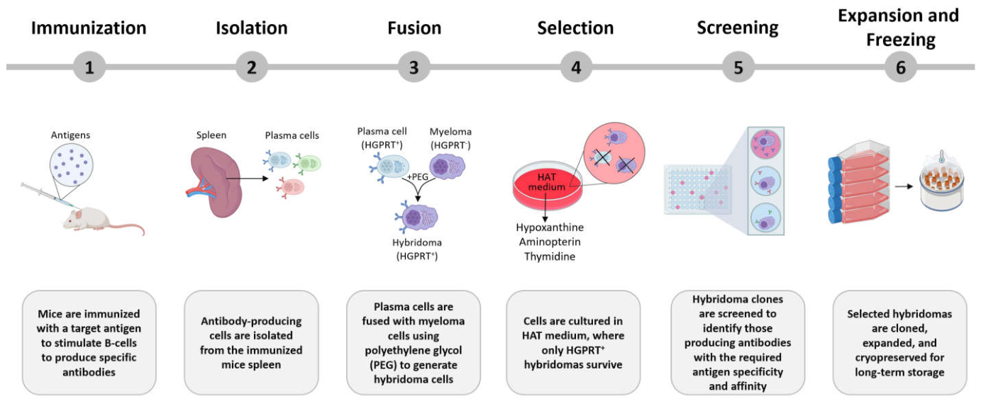
Figure 2.
Illustrative overview of hybridoma cell applications and the uses of their monoclonal antibodies (Created with BioRender.com).
Figure 2.
Illustrative overview of hybridoma cell applications and the uses of their monoclonal antibodies (Created with BioRender.com).
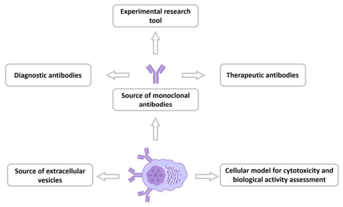
Figure 3.
Common assays for cell viability determination.
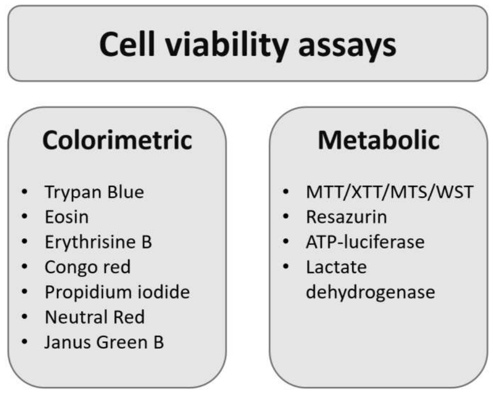
Figure 4.
Cell cycle phases and associated molecular markers (Created with BioRender.com). G1—cell growth and preparation for DNA synthesis, S—DNA replication, G2—preparation for cell division, M—mitosis, G0—quiescent state.
Figure 4.
Cell cycle phases and associated molecular markers (Created with BioRender.com). G1—cell growth and preparation for DNA synthesis, S—DNA replication, G2—preparation for cell division, M—mitosis, G0—quiescent state.
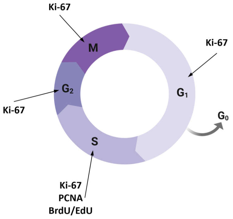

Table 1.
Advances in Hybridoma Technology.
| No | Hybridoma Technology Stage | Optimization Strategy | Description of Improvement | Ref. |
|---|---|---|---|---|
| 1 | Fusion | PEG pre-incubation | Pre-incubation of myeloma cells and lymphocytes with 0.25% PEG for 90 min at 37 °C | [28] |
| 2 | Fusion | Electrofusion pulse optimization | Use of nanosecond and microsecond pulses with low peak power in electrofusion | [29] |
| 3 | Fusion | Fluorescence-activated cell sorting enrichment prior to fusion | Sorting of B-cells by FACS before electrofusion | [21] |
| Fusion | Microfluidic electrofusion chip | Electrofusion in microfluidic droplet-based platform | [30] | |
| 4 | Post-fusion Survival | Macrophage-conditioned medium | Addition of macrophage-conditioned medium | [31] |
| 5 | Post-fusion Survival | Thymocyte-conditioned medium | Replacement of feeder cells | [28] |
| 6 | Selection | FACS-based screening | Antigen-specific sorting using fluorescence labeling | [32,33,34,35] |
| 7 | Selection | Membrane Ig-based screening (MIHS) | Detection via B-cell receptor antigen binding | [36] |
| 8 | Selection | Magnetic particle isolation | Antigen-coated magnetic particles | [37] |
| 9 | Selection | Semisolid media | Spatial isolation of hybridoma colonies | [38,39] |
| 10 | Selection | ClonePix 2 | Automated colony screening | [40,41] |
| 11 | Selection | Microwell chip with antibody capture membrane | Self-seeding chip capturing secreted antibodies | [42] |
| 12 | Fusion | PEG pre-incubation | Pre-incubation of myeloma cells and lymphocytes with 0.25% PEG for 90 min at 37 °C | [28] |
Table 2.
Applications of Hybridoma-Derived Monoclonal Antibodies.
| No | Target | Application | Ref. |
|---|---|---|---|
| Diagnostic antibodies | |||
| 1 | Сitrinin | ELISA-based detection of citrinin in wine | [65] |
| 2 | Dihydromyricetin | ELISA-based detection of dihydromyricetin in Ampelopsis grossedentata | [66] |
| 3 | Denatured monomeric non-structural protein of Zika virus ZNS1 | Early ELISA-based flavivirus detection | [67] |
| 4 | Calreticulin gene (CALR) mutations | ELISA-based detection of calreticulin gene (CALR) mutations associated with essential thrombocythemia and primary myelofibrosis | [68] |
| 5 | Giant panda vascular endothelial growth factor A (VEGFA) | ELISA-based diagnosis of giant panda reproductive disorders and cancer | [14] |
| 6 | VP27 protein of goose astrovirus GoAstV-2 | Specific detection of GoAstV-2 by western blotting, immunofluorescence assay, immunohistochemistry and in vivo viral neutralization | [69] |
| 7 | VP3 protein of infectious bursal disease virus | The development of diagnostic tools and vaccines for infectious bursal disease | [15] |
| 8 | Schistosoma mansoni SWAP antigen | Immunophosphatase and immunoperoxidase staining detection of schistosomiasis | [11] |
| Therapeutic antibodies | |||
| 9 | ITGA4 | Induces cell death in NK/T-cell lymphoma | [17] |
| 10 | S protein of SARS-CoV-2, receptor-binding domain (RBD) | In vitro and in vivo neutralization of SARS-CoV-2 with both prophylactic and therapeutic potential | [70] |
| 11 | Peanut-specific human IgE | In vitro food allergy effector phase models | [71] |
Table 3.
Strategies for enhancing hybridoma cell productivity. ↑—increase, ↓—decrease, =—no effect, N/S—not stated in the article.
Table 3.
Strategies for enhancing hybridoma cell productivity. ↑—increase, ↓—decrease, =—no effect, N/S—not stated in the article.
| No | Cell treatment | Effect on proliferation | Effect on specific productivity | Effect on antibody titer | Ref. |
|---|---|---|---|---|---|
| 1 | Increase in osmolarity | ↓ | ↑ | = | [143] |
| 2 | Increase in osmolarity + Sodium butyrate addition | ↓ | ↑ | ↑ | [144] |
| 3 | Sodium butyrate addition | ↓ | ↑ | ↑ | [145,146] |
| 4 | Colchicine addition | ↓ | ↑ | ↑ | [147] |
| 5 | Caffeine addition | ↓ | ↑ | N/S | [141,148] |
| 6 | LPS addition | ↓ | ↑ | ↑ | [149] |
| 7 | DMSO addition | ↓ | ↑ | N/S | [150] |
| 8 | Spermine addition | ↓ | ↑ | N/S | [151] |
| 9 | Spermidine addition | ↓ | ↑ | N/S | [152] |
| 10 | Thermine addition | = | ↑ | N/S | [152] |
| 11 | Triethylenetetraamine addition | ↓ | ↑ | N/S | [152] |
| 12 | IL-6 addition | ↑ | ↑ | N/S | [129] |
Disclaimer/Publisher’s Note: The statements, opinions and data contained in all publications are solely those of the individual author(s) and contributor(s) and not of MDPI and/or the editor(s). MDPI and/or the editor(s) disclaim responsibility for any injury to people or property resulting from any ideas, methods, instructions or products referred to in the content. |
© 2026 by the authors. Licensee MDPI, Basel, Switzerland. This article is an open access article distributed under the terms and conditions of the Creative Commons Attribution (CC BY) license (http://creativecommons.org/licenses/by/4.0/).
Copyright: This open access article is published under a Creative Commons CC BY 4.0 license, which permit the free download, distribution, and reuse, provided that the author and preprint are cited in any reuse.


