

Submitted:
08 July 2026
Posted:
09 July 2026
You are already at the latest version
Abstract
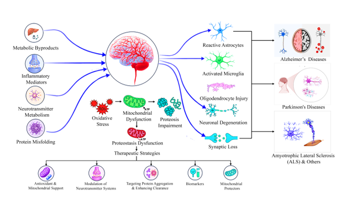
Neurotoxicity is generally thought to result from exogenous agents like environmental chemicals, drugs and biological toxins. However, increasing evidence suggests that many endogenous molecules that play a critical role in normal brain function can become neu-rotoxic when the regulatory mechanism involved in their production, metabolism, com-partmentalization and clearance are disrupted. This shift underlies the basis of endoge-nous neurotoxicity. This review discusses the major endogenous sources of neurotoxicity: metabolic neuro-toxins, dysfunctional neurotransmitters, protein aggregates and inflammatory mediators. These endogenous factors arise from different physiological pathways, but share common pathogenic mechanisms, all of which involve an underlying state of oxidative stress, mi-tochondrial dysfunction, impaired proteostasis, excitotoxic signalling, neurovascular dysfunction and maladaptive neuroglial responses. This is not a singular process but a network of interconnected processes, which work together to progressively diminish neu-ronal resilience and promote synaptic dysfunction and neurodegeneration. The review also underscores the critical role of astrocytes, microglia and other glial cells in the maintenance of neuronal homeostasis. By integrating diverse endogenous neurotoxic pathways within a unified homeosta-sis-centred framework, this review provides a broader perspective on the mechanisms linking metabolic disorders, aging and neurodegenerative diseases. This framework sug-gests that effective therapeutic strategies may require restoration of physiological regula-tory networks rather than targeting individual neurotoxic molecules in isolation. A sys-tems-level understanding of endogenous neurotoxicity may therefore facilitate the devel-opment of earlier biomarkers and more effective interventions aimed at preserving neu-ronal homeostasis and preventing progressive neurological dysfunction.
Keywords:
endogenous neurotoxicity
; neuronal homeostasis
; neurological dysfunction
; neurodegeneration
1. Introduction
The central nervous system (CNS) depends on tightly regulated metabolic and cellular processes to maintain neuronal function throughout life. In physiological conditions, energy metabolism, synaptic transmission, redox status, protein quality control, and tissue repair are regulated by a balance of endogenous metabolite, neurotransmitter, protein and immune mediator. They are thus essential for proper brain activity and are constantly maintained by complex interdependent regulatory mechanisms acting at multiple levels to regulate their synthesis, degradation, cellular localization and elimination. However, physiologically essential molecules can become mediators of neuronal injury when the regulatory systems are disrupted, due to various reasons including aging, genetic susceptibility, metabolic disturbances, oxidative stress, mitochondrial dysfunction, chronic inflammation or impaired protein homeostasis [1,2]. This transition is the foundation of endogenous neurotoxicity.
Endogenous neurotoxicity refers to the pathological process by which naturally occurring metabolites, neurotransmitter derivatives or intrinsic cellular products become harmful to the CNS. This happens when their production, metabolism, compartmentalization or clearance is disrupted [1]. In contrast to exogenous neurotoxins that originate from environmental or xenobiotic sources, endogenous neurotoxins are generated within the body as normal intermediates of physiological metabolism, however, they acquire neurotoxic properties under conditions such as oxidative stress, mitochondrial dysfunction, chronic inflammation, impaired protein homeostasis or metabolic dysregulation [1]. The biological effects of endogenous metabolites are determined by their concentration, duration of exposure, cellular localization and the metabolic resilience of surrounding neural cells. Therefore, neurotoxicity should be considered as a dynamic and context-dependent phenomenon rather than an inherent property of a specific molecule [2].
These principles also go beyond the single neuron and apply to the coordinated functions of the astrocytes, microglia and other types of glia, which control neurotransmitter turnover, metabolic support, antioxidant defense and immune surveillance. These responses are initially responsible for maintaining tissue homeostasis, but chronic stress can lead to maladaptive activation of glia that can exacerbate oxidative damage, metabolic dysfunction and chronic inflammation, making neurons more vulnerable [3,4,5].
The endogenous molecules capable of contributing to neurotoxicity arise from several fundamental biological systems. Reactive intermediates generated by normal cellular metabolism can become trapped and accumulate if normal detoxification and clearance mechanisms are impaired, while abnormally regulated neurotransmitter metabolism can give rise to reactive intermediates which disrupt mitochondrial function and synaptic signaling [1,6]. Similarly, when protein quality control fails, misfolded proteins and toxic oligomers accumulate, disrupting the cellular homeostasis while continued activation of immune and inflammatory pathways leads to the production of mediators which further promote neuronal injury [7,8]. While these endogenous neurotoxic species vary in biochemical origin, they converge on shared mechanisms, such as oxidative stress, mitochondrial dysfunction, impaired proteostasis, excitotoxicity and chronic neuroinflammation, all of which compromise neuronal resilience [9,10]. Furthermore, increasing evidence suggests that neuroglial responses critically influence the transition from physiological signaling to pathological toxicity. Astrocytes and microglia normally maintain metabolic homeostasis and regulate neurotransmitter turnover while limiting oxidative and inflammatory insults. However, persistent injury or disease induces reactive phenotypes that may compromise the protective functions and amplify neuronal vulnerability [3]. In addition, inflammatory activation redirects endogenous metabolic pathways leading to the accumulation of neuroactive metabolites that are capable of disrupting excitatory neurotransmission, mitochondrial function and redox balance [10].
Therefore, a framework that integrates these diverse neurotoxic systems within the broader context of disrupted homeostasis is needed to better understand the shared mechanisms underlying neurodegeneration and to identify therapeutic strategies capable of restoring physiological regulation rather than targeting individual pathogenic molecules in isolation. Therefore, this review examines the main endogenous molecules that induce neurotoxicity, such as metabolic neurotoxins, dysregulated neurotransmitters and protein aggregates and the convergent intracellular and neuroglial mechanisms by which these endogenous molecules contribute to neuronal dysfunction are discussed. Lastly, the review discusses the clinical implications of this homeostasis-centered framework, highlighting emerging biomarkers and therapeutic strategies aimed at restoring the interconnected biological networks that preserve neuronal health.
2. Metabolic Endogenous Neurotoxins and Their Systemic Disease Interfaces
Normal cellular metabolism continuously generates numerous intermediate and end products which are essential for maintaining physiological function. Under healthy conditions, these metabolites are tightly regulated through coordinated metabolic pathways that control their production, utilization and clearance, thereby maintaining metabolic homeostasis [11,12]. Systemic metabolic disorders, however, can interfere with the regulation, resulting in the accumulation of endogenous molecules that can have neurotoxic properties [13,14]. The following sections examine major classes of metabolic endogenous neurotoxins and the mechanisms through which they contribute to neurological dysfunction and neurodegenerative disease.
2.1. Ammonia and Nitrogen Dysregulation
Ammonia is an endogenous neurotoxin that plays a central role in metabolic disorders affecting brain function. Under normal physiological conditions, ammonia is continuously generated during acid metabolism, intestinal bacterial metabolism and glutamine breakdown. However, even slight elevations of ammonia concentrations are toxic to the CNS. So, the liver rapidly converts it into urea through the urea cycle; while a smaller fraction is incorporated into glutamine through glutamine synthetase. These pathways work together to maintain circulating ammonia within a narrow physiological range [12,13,15]. Hyperammonemia results when ammonia production exceeds the body’s capacity for detoxification. This occurs mostly in acute or chronic liver diseases where ammonia bypasses hepatic metabolism and enter the systemic circulation due to impaired urea synthesis and portal-systemic shunting. This can also happen due to inherited urea cycle disorders, severe metabolic disturbances and as an adverse effect of medications that inhibit key enzymes involved in ammonia disposal [12,16,17]. In addition, impaired urea synthesis itself is a fundamental metabolic defect that underlies hyperammonemia since reduced hepatic nitrogen clearance delays ammonia elimination and promotes its systemic accumulation [15].
As circulating ammonia increases, ammonia as well as ammonium can enter the brain through diffusion and transporter-mediated pathways. Astrocytes contain high levels of glutamine synthetase and therefore act as the primary site of cerebral ammonia detoxification. Glutamine synthetase converts ammonia and glutamate into glutamine. This pathway initially protects neurons by lowering free ammonia concentrations. However, persistent hyperammonemia results in excessive intracellular glutamine accumulation within astrocytes [13,18,19]. Glutamine promotes water influx into astrocytes resulting in cellular swelling and low-grade cerebral edema which is characteristic of chronic hepatic encephalopathy. In addition, glutamine also enters astrocytic mitochondria where it is metabolized to produce ammonia which promotes oxidative stress, mitochondrial dysfunction and impairs cellular energy production. This is why ammonia toxicity persists even after its initial detoxification and glutamine accumulation results in astrocyte injury [13,16,19,20]. Astrocyte dysfunction subsequently disrupts neuronal communication. Hyperammonemia impairs the glutamate/glutamine cycle reducing the efficiency of glutamate uptake by astrocytes and altering the balance between excitatory and inhibitory neurotransmission. Ammonia also interferes with many ion transport systems and cellular pH regulation which further compromises excitability and synaptic function [13]. Recent studies emphasize that ammonia toxicity is not the sole act of elevated concentrations of ammonia. Rather, neuroinflammation, oxidative and nitrosative stress, mitochondrial dysfunction and metabolic alterations interact with hyperammonemia to amplify neuronal injury [13,16].
2.2. Homocysteine and One-Carbon Metabolism
Homocysteine is a sulfur-containing amino acid that plays a key role in one-carbon metabolism. Under normal physiological conditions, homocysteine is either remethylated to methionine through folate- and vitamin B12-dependent pathways or converted to cysteine through the vitamin B6-dependent transsulfuration pathway. These reactions are tightly regulated and maintain adequate levels of S-adenosylmethionine (principal methyl donor for DNA, RNA, proteins, phospholipids and neurotransmitters) while preventing homocysteine accumulation [11,21].
These pathways are impaired by nutritional deficiencies of folate, vitamin B6 or B12, genetic variants affecting enzymes such as methylenetetrahydrofolate reductase and age-related metabolic disturbances. This leads to hyperhomocysteinemia and reduced methylation capacity [11,14,22,23]. This imbalance extends beyond elevated concentrations of homocysteine. S-adenosylhomocysteine, a potent inhibitor of methyltransferases, accumulates and further suppresses methylation reactions resulting in altered gene expression, protein function and neuronal homeostasis [21]. Recent studies also reported that disruption of one-carbon metabolism modifies DNA methylation patterns in genes involved in antioxidant defense. Therefore, elevated homocysteine levels promote oxidative stress by increasing the production of reactive oxygen species (ROS) and reducing antioxidant defenses [11,23,24].
Oxidative stress in turn reduces nitric oxide availability, impairs mitochondrial function and activates inflammatory signaling pathways. All of these affect endothelial cell function [23]. The cerebral microvasculature relies on healthy endothelial cells to regulate blood flow and maintain the integrity of the blood-brain barrier (BBB). Therefore, persistent endothelial dysfunction disrupts vascular homeostasis and increases BBB permeability [25,26]. Studies have also reported that hyperhomocysteinemia results in reduced cerebral blood flow, altered extracellular matrix remodeling and weakened neurovascular coupling. This further limits efficient oxygen and nutrient delivery to metabolically active brain areas [26,27]. Furthermore, vascular injury interferes with angiogenic signaling and increases endothelial apoptosis. These accelerate neurovascular dysfunction during aging and neurodegeneration [28].
2.3. Advanced Glycation End Products (AGEs)
Advanced glycation end products (AGEs) are a diverse group of endogenous molecules formed through the non-enzymatic reactions of reducing sugars with proteins, lipids or nucleic acids [29,30]. During the normal aging process, AGE formation occurs slowly. However, it is significantly accelerated under conditions of chronic hyperglycemia and oxidative stress [30,31]. AGE formation is substantially accelerated by highly reactive dicarbonyl intermediates, particularly methylglyoxal, glyoxal and 3-deoxyglucosone. Under normal physiological conditions, the glyoxalase system detoxifies these reactive intermediates, thereby limiting AGE production. However, persistent hyperglycemia overwhelms this protective pathway, leading to increased accumulation of AGEs in both intracellular and extracellular compartments [29,32]. AGE-modified proteins persist even after glycemic control improves due to their long half-lives. This results in continued tissue injury despite normalization of blood glucose levels [31].
In addition to their tissue accumulation, pathogenic effects of AGEs are due to their ability to bind to cell surface receptors such as the receptor for advanced glycation end products (RAGE). When AGEs bind to RAGE, it triggers a series of intracellular signaling pathways that lead to the generation of ROS, activation of nuclear factor-κB (NF-κB) and increased production of pro-inflammatory cytokines [29,30]. This process becomes a vicious cycle because oxidative stress promotes the production of more AGEs which in turn contributes to the chronic inflammation and cellular dysfunction leading to more oxidative stress [29,33]. In the CNS, RAGE is expressed by endothelial cells, astrocytes, microglia and neurons which enables AGE accumulation to affect various populations of cells at once. Continued activation of this pathway leads to impaired function of mitochondria, disruption of cellular energy metabolism and glial activation resulting in loss of neuronal homeostasis [33]. Cerebral endothelial cells are especially sensitive to AGE induced injury. Oxidative stress and inflammatory signaling decreases endothelial integrity, compromises vascular function and leads to increased permeability of BBB. This allows circulating inflammatory mediators to gain access to the brain. Hence, AGE-RAGE signalling is now known as a potential molecular bridge between chronic metabolic dysfunction and neurovascular injury as well as and chronic neuroinflammation [30,31].
3. Neurotransmitter-Derived Toxicity
Neurotransmitters are indispensable for neuronal communication, regulating synaptic transmission, neural plasticity, and the coordinated function of neural networks [34,35]. However, disruption of regulatory processes can lead to excessive receptor activation or the accumulation of reactive neurotransmitter metabolites, transforming essential signaling molecules into endogenous neurotoxic species [17,36]. The following sections examine how dysregulation of glutamatergic and dopaminergic signaling contributes to endogenous neurotoxicity and neurodegeneration.
3.1. Glutamate Excitotoxicity
3.1.1. NMDA/AMPA Receptor Overactivation
Glutamate is the major excitatory neurotransmitter and plays a crucial role in processes such as synaptic transmission, learning and memory and neuronal plasticity in the CNS. Its extracellular concentration is tightly regulated allowing the activation of ionotropic glutamate receptors, especially N-methyl-D-aspartate (NMDA) and α-amino-3-hydroxy-5-methyl-4-isoxazolepropionic acid (AMPA) receptors in a spatially and temporally restricted manner [34,36]. Neurotoxicity occurs when glutamate is overabundant in the extracellular environment or when the receptor becomes activated excessively or for a prolonged period of time thereby leaving the neuron constantly excited. The sustained stimulation mainly target NMDA receptor due to its high permeability to calcium and the calcium-permeable AMPA receptor which further enhances the excitatory signaling under pathological states [36,37]. Importantly, excitotoxicity is not simply caused by the mere presence of glutamate but by the inability to control the activation of the receptors. Recent studies also show that activation of receptors at different locations can affect the fate of the neurons. In general, activation of synaptic NMDA receptors is pro-survival and pro-plasticity while overactivation of extrasynaptic NMDA receptor signaling at the plasma membrane is pro-stress and pro-degeneration [36,38].
3.1.2. Calcium Influx and Mitochondrial Dysfunction
One of the hallmark characteristics of glutamate excitotoxicity is that after sustained activation of glutamate receptors, there is excessive calcium influx into the neurons. Transient increases in intracellular calcium, under normal circumstances, are crucial signaling events that control neurotransmitter release, gene expression and synaptic plasticity. However, constant stimulation of the receptor leads to continual influx of calcium and triggers a chain of events that could lead to damage [34,36]. Initially, mitochondria act as calcium buffers that bind excess calcium in the cytosol to protect cells from damage. However, excessive cytosolic calcium loading gradually damages mitochondrial function, disrupts ATP production and increases the production of ROS [39,40]. When energy levels drop, ion pumps which maintain the membrane potential of neurons start to become less effective which further promotes membrane depolarization, resulting in continued glutamate receptor activation. This leads to a vicious cycle of calcium overload, mitochondrial dysfunction, oxidative stress and energy metabolism impairment [34,39]. Experimental studies also show that the extent of these changes is variable with the concentration and duration of glutamate exposure suggesting that these changes are a dose- and time-dependent process and not the immediate result of glutamate action. Thus, neuronal injury occurs in a progressive manner, as a result of failure of the cellular homeostatic mechanisms rather than a single toxic event [41].
3.1.3. Astrocytic Glutamate Clearance Failure
Astrocytes are the main regulators of extracellular glutamate levels and play a key role in the switch from physiological glutamate signaling to excitotoxicity. Following synaptic transmission, glutamate is quickly taken up from the extracellular space by the excitatory amino acid transporters, then used to synthesize glutamine by the astrocytes and finally recycled to the neurons through the glutamate–glutamine cycle [35]. This highly organized process allows for reduction of the duration of receptor activation and the excessive firing of neurons. If glutamate is not taken up by astrocytes, extracellular glutamate levels rise leading to prolonged activation of NMDA and AMPA receptors thereby increasing the risk of an excitotoxic injury [34,35]. Metabolic stress, oxidative injury, inflammation or energy depletion can all lead to dysfunction of the astrocytes which in turn can lead to impaired glutamate transport and affect neuron–glia interactions [39,42]. At the same time, impaired astrocytes lose the ability to regulate ionic balance and support the metabolism of neurons thus increasing the vulnerability to excitotoxic damage [35,42]. Therefore, the integrity of astrocytic glutamate clearance is an important factor as to whether glutamate is an essential neurotransmitter or an endogenous neurotoxin that causes progressive neuronal damage.
3.2. Dopamine Oxidation and Quinone Formation
3.2.1. Auto-Oxidation and Enzymatic Metabolism
Dopamine is a crucial neurotransmitter which controls voluntary movements, motivation, reward and several cognitive functions. The vesicular monoamine transporter-2 (VMAT2) transports newly synthesized dopamine from the cytoplasm into the synaptic vesicles thereby limiting its exposure in the cytosolic environment and minimizing its chemical instability [43]. Excessive cytosolic dopamine is mostly metabolized by monoamine oxidase (MAO) and catechol-O-methyltransferase (COMT) which prevents accumulation of dopamine in neurons [43]. These tightly regulated processes ensure dopamine homeostasis and normal neurotransmission. When dopamine escapes these mechanisms and leaks into the cytosol, it leads to neurotoxicity. In such conditions, dopamine can spontaneously auto-oxidize or be enzymatically oxidized to various reactive intermediates such as dopamine o-quinone, aminochrome and other dopamine oxidation products [43,44]. Meanwhile, MAO metabolizes to the highly reactive catecholaldehyde 3,4-dihydroxyphenylacetaldehyde (DOPAL) which is normally neutralized by aldehyde dehydrogenase. Therefore, when this detoxification pathway is impaired, DOPAL accumulates. When dopamine homeostasis is disrupted, both dopamine oxidation and enzymatic metabolism can result in the production of endogenous neurotoxic species [45]. So, dopamine is not a neurotoxin by itself; it only becomes toxic when normal storage, metabolism and detoxification processes in the brain become overwhelmed.
3.2.2. Generation of Reactive Quinones and Reactive Oxygen Species
The oxidation of cytosolic dopamine kickstarts a series of chemical reactions that produce reactive quinones and ROS which can cause cellular homeostasis disturbances. Dopamine o-quinone is unstable and easily converted to downstream intermediates which are capable of altering proteins, lipids and nucleic acids through covalent reactions [43,44]. These quinones disrupt the normal function of proteins that are a part of the vesicular dopamine transport system, mitochondrial activity, protein degradation and antioxidant defense thereby increasing intracellular stress [43,46]. In parallel, dopamine metabolism via MAO produces hydrogen peroxide while DOPAL can react further to form more highly reactive hydroxyl radicals. This further increases oxidative damage to organelles and cellular membranes [45]. Studies in human dopaminergic neurons has shown that mitochondrial oxidative stress leads to progressive dopamine oxidation followed by lysosomal dysfunction, loss of protein clearance and accumulation of α-synuclein. This illustrates how oxidative dopamine metabolism initiates multiple interconnected pathways of cellular injury [47]. These results suggest that ROS and dopamine-derived quinones are not simply acting independently of each other, but rather are involved in self-reinforcing cycles in which oxidative stress further accelerates dopamine oxidation resulting in progressive neuronal dysfunction.
3.2.3. Selective Vulnerability of Dopaminergic Neurons
Dopamine can be synthesized in a number of neuronal populations. However, dopaminergic neurons in the substantia nigra are especially vulnerable to dopamine-derived neurotoxicity due to the metabolic needs of dopamine handling. These neurons are constantly producing, storing, releasing and recycling high levels of dopamine while maintaining extensive axonal networks which need to produce a significant amount of energy [46]. Thus, minor changes in vesicular storage or mitochondrial function lead to greater levels of free dopamine in the cytosol and increased dopamine oxidation [43]. The resulting quinones and aldehydes cause damage to proteins that transport dopamine, are involved in mitochondrial function and lysosomal processes and protein quality control. This gradually diminishes the ability of the neuron to maintain its intracellular homeostasis [45,47]. Furthermore, dopaminergic neurons are subjected to extra oxidative stress due to the production of H2O2 during dopamine metabolism and the relatively high iron levels found in the substantia nigra exacerbating the production of highly reactive oxygen radicals [46]. In normal physiology, several mechanisms are present to prevent these harmful reactions including sequestration of dopamine in vesicles, detoxification of reactive metabolites, antioxidant systems and conversion of metabolites into neuromelanin. The failure of these buffering systems, however, leads to a vicious cycle of dopamine oxidation, resulting in progressive and lasting neuronal injury, mitochondrial dysfunction, impaired protein homeostasis and oxidative stress [43].
4. Protein Aggregates as Endogenous Neurotoxic Species
Proteostasis is responsible for the proper synthesis, folding, trafficking and degradation of cellular proteins to keep them active and prevent the buildup of potentially harmful conformations. In physiological conditions, this well-organized network maintains neuronal homeostasis via molecular chaperones, ubiquitin-proteasome system and autophagic-lysosomal pathways [48]. These quality control systems can, however, fail leading to the misfolding, self-association and accumulation of normally soluble proteins into neurotoxic oligomers and aggregates that disrupt synaptic function, mitochondrial integrity and neuronal survival [49,50,51]. The subsequent sections discuss the mechanisms by which dysregulation of amyloid-β, α-synuclein and tau can lead to the generation of physiologically important proteins as endogenous neurotoxic species which lead to progressive neurodegeneration.
4.1. Amyloid-β Toxicity
4.1.1. Imbalance Between Amyloid-β Production and Clearance
Amyloid-β (Aβ) is a naturally occurring peptide produced in the normal metabolism of brain cells that is derived from amyloid precursor protein (APP). However, under physiological conditions, the production of Aβ is countered by efficient degradation and clearance via enzymatic pathways, microglial phagocytosis and transport across the BBB. This allows Aβ to be integrated into normal neuronal functioning without becoming toxic [52,53]. In addition, there is growing evidence that physiological levels of Aβ can be neuro-protective and play a role in regulating synapses as well as promoting neuronal plasticity and protection from excessive neuronal activity [51,54]. The disturbance of the balance between the production and clearance of Aβ causes neurotoxicity. As clearance pathways diminish in efficiency, soluble Aβ monomers gradually convert to oligomers, protofibrils, fibrils and, finally, extracellular plaques [52]. Of these species, soluble oligomers are now known to be the major neurotoxic species as they readily bind to the neuronal membrane and are known to interfere with cellular homeostasis well before extensive deposits of plaques. These factors can further hinder the clearance of Aβ, perpetuating a vicious cycle of protein accumulation and neuronal stress [53,55].
4.1.2. Synaptic Dysfunction, Membrane Disruption and Neuronal Stress
Increasing evidence suggests that soluble Aβ oligomers are most toxic to the synapse, disrupting the communication between neurons before there is any noticeable loss of neurons [56,57]. Oligomers are not only able to affect the function of various membrane receptors but also disrupt calcium signaling and impair synaptic plasticity [57,58]. Persistent calcium imbalance raises metabolic demands on neurons leading to increased production of ROS which may result in mitochondrial dysfunction and reduced energy production [59,60]. Studies also show that Aβ-induced mitochondrial dysfunction is accompanied by diminished ATP production, higher levels of oxidative stress and synaptic protein loss and dysfunction. This supports the close connection between mitochondrial injury and synaptic degeneration [60,61,62]. In addition, oxidative stress and mitochondrial dysfunction feed each other and further damage neurons and activate inflammatory pathways leading to additional synaptic damage [51,59].
4.2. α-Synuclein Misfolding and Proteostatic Stress
4.2.1. Misfolding, Oligomerization and Aggregation
α-Synuclein is a presynaptic protein that is highly expressed and plays roles in synaptic vesicle organization, neurotransmitter release and regulation of normal synaptic function. It is mainly found as a soluble protein whose folding, intracellular distribution and degradation are under tight cellular protein quality control mechanisms [50,63]. These mechanisms keep α-synuclein in its functional conformation and prevent its self-association. Neurotoxicity arises when the balance of proteostasis is lost leading to the structural changes of soluble α-Synuclein that allow it to self-assemble into oligomers, protofibrils and finally into fibrillar aggregates [63,64]. The most biologically active neurotoxic species are the soluble oligomeric intermediates rather than mature fibrillar inclusions because they can interact with intracellular structures and disrupt essential cellular functions [63,65]. Importantly, the conversion of soluble monomer s to toxic oligomers is a gradual process influenced by various parameters such as the concentration of proteins, post-translational modifications, oxidative stress and age-related decline in protein quality control mechanisms [50,66]. After oligomerisation, α-synuclein may continue to aggregate by self-templating, perpetuating a vicious cycle of protein misfolding and proteostatic stress that progressively leads to neuronal homeostasis dysfunctions and neuron death [64,67].
4.2.2. Membrane Interactions, Vesicular Trafficking and Mitochondrial Dysfunction
Physiological α-synuclein is involved in reversible association with synaptic vesicular membranes to modulate membrane trafficking and neurotransmitter release while oligomeric forms gain altered membrane-binding properties that compromise membrane integrity and vesicle trafficking [50,63]. These abnormal membrane interactions affect calcium homeostasis and disrupt the pathways of intracellular trafficking which in turn affects protein degradation and organelle communication [50,68]. These changes seem to take place especially in the mitochondria. Studies have shown that pathogenic oligomers of α-synuclein bind strongly to the mitochondrial membranes, disrupting mitochondrial function, decreasing ATP production, increasing mitochondrial ROS and changing mitochondrial dynamics and quality control [69,70,71]. Recent studies also indicate that mitochondrial membranes could actively promote α-synuclein oligomerization, creating a vicious cycle of mitochondrial dysfunction leading to further α-synuclein oligomerization which in turn contributes to further mitochondrial dysfunction [72]. This self-reinforcing cascade of progressive oxidative stress, impaired energy metabolism and impaired organelle quality control leads to persistent neuronal stress and neuronal vulnerability [68,70].
4.3. Tau Dysfunction and Cytoskeleton Instability
4.3.1. Hyperphosphorylation and Microtubule Destabilization
Tau is a microtubule-associated protein which is critical for supporting neuronal architecture, stabilizing microtubules, facilitating axonal transport and maintaining synaptic function. Under physiological conditions, tau is subjected to reversible phosphorylation, a normal regulatory process required for tau to interact with tubules and keep the neuronal cytoskeleton dynamic without compromising structural integrity [49,73]. An imbalance of this tightly controlled balance causes neurotoxicity. Over-phosphorylated tau no longer binds to microtubules and if it persists, leads to disassociation from the cytoskeleton thereby disrupting the transport of organelles, proteins and synaptic components along axons [49,74]. Detached tau becomes increasingly prone to structural changes that foster self-associations that can give rise to soluble oligomers and larger aggregates, further disrupting neuronal function [75,76]. Multiple protein kinases which enhance tau phosphorylation become activated under conditions of oxidative stress, mitochondrial dysfunction, excitotoxicity and neuroinflammatory signaling, thereby directly linking disturbances of cellular homeostasis with tau pathology [74,77].
4.3.2. Propagation of Tau Pathology Across Neuronal Networks
The production of these misfolded species of tau can progressively spread out of the initially affected cells into interconnected neuronal networks, expanding the pathology. Experimental and pathological studies have suggested that misfolded tau can be released to the extracellular environment, endocytosed by neighbouring neurons and serve as a template for misfolding and aggregation of native intracellular tau [78,79]. This self-propagating process is similar to that of seeded protein aggregation and contributes to the sequential spread of tau pathology across anatomically connected regions of the brain [78]. Abnormal tau proteins are degraded continuously in the cell by degradation pathways such as ubiquitin-proteasome system and autophagic-endolysosomal pathway to maintain protein homeostasis under physiological conditions [48]. The impairment of these clearance mechanisms allow pathogenic tau build up, resulting in both intracellular toxicity and extracellular transmission [48,75]. Under normal circumstances, glial cells are involved in extracellular tau clearance and promote neuronal homeostasis, but chronic tau exposure can impair neuron-glia interactions and create a pro-inflammatory environment [75].
4.3.3. Toxic Oligomers Versus Fibrillar Aggregates
While neurofibrillary tangles are the defining pathological feature of tauopathies, there has been a growing amount of evidence that soluble tau oligomers are the key mediators of neuronal dysfunction. The soluble aggregates are small, form in early tau aggregation and are able to disrupt important cellular functions before the emergence of mature fibrillar inclusions [76,80]. Studies have shown that tau oligomers are toxic to synaptic transmission, are involved in calcium homeostasis disruption, lead to mitochondrial damage, cause impairment of axonal transport and produce neuronal death, while tau monomers do not elicit similar toxic effects [79,80]. In contrast, mature fibrillar aggregates and neurofibrillary tangles could represent relatively less active end-stage deposits [80]. This supports the view that the soluble tau oligomers are the most critical species responsible for neuronal injury. In terms of endogenous neurotoxicity, tau, a physiologically necessary cytoskeletal protein, becomes self-propagating neurotoxic assemblies when impaired by failure of protein quality control leading to gradual impairment of neuronal homeostasis and neurodegeneration [49,75]. Table 1 summarizes the major endogenous neurotoxins and their principal mechanisms.
5. Convergent Cellular Mechanisms of Endogenous Neurotoxicity
While endogenous neurotoxins come from a variety of metabolic, neurotransmitter or proteostatic abnormalities, they have a shared set of cellular mechanisms that impair neuronal functions. Oxidative stress, mitochondrial dysfunction and failure in protein quality control do not act independently, but rather create a vicious cycle of progressive neuronal damage and are interdependent in amplifying the damage sustained by the cell [81,82,83]. When these protective mechanisms become overwhelmed, neurons lose the capacity to maintain redox balance, energy metabolism and protein integrity, thereby causing self-sustaining cycles of dysfunction that lead to synaptic failure and neurodegeneration [84,85,86]. The convergent mechanisms discussed in the following sections are the common cellular pathways by which a variety of endogenous neurotoxins are capable of exerting neurotoxicity.
5.1. Oxidative Stress and Redox Imbalance
5.1.1. Generation of Reactive Oxygen and Nitrogen Species
One of the key pathways of neuronal damage induced by various endogenous neurotoxic agents is oxidative stress. Metabolic by-products, dysregulated neurotransmitters and misfolded proteins are generated via different biological processes, all of which result in increased generation or reduced function of systems that maintain redox homeostasis, including production of ROS and reactive nitrogen species (RNS) [82,87]. ROS and RNS are produced naturally under physiological conditions during mitochondrial respiration, neurotransmitter metabolism and normal cell signaling and play a pivotal role in regulating various cellular functions including gene expression, synaptic plasticity and immune responses [88,89]. At these low concentrations, they act as important signaling molecules and not as a harmful by-product. However, when endogenous neurotoxins increase their production or disrupt their removal, the balance of oxidants and antioxidants tips to the oxidants side, leading to chronic oxidative stress [87,90]. Neurons have high metabolic requirements and have a large number of mitochondria but relatively small reserves of antioxidants, making them highly vulnerable to redox imbalance and making oxidative stress a common mechanism through which various types of endogenous neurotoxicity can occur [82,89].
5.1.2. Oxidative Damage to Lipids, Proteins, and DNA
When the level of production of reactive species is higher than the capacity of the endogenous antioxidant systems, oxidative damage can be found in practically all classes of cellular macromolecules. Neuronal membranes are particularly susceptible because they contain abundant poly unsaturated fatty acids which are easily oxidizable and undergo lipid peroxidation, leading to decreased membrane integrity, change in receptor function and synaptic transmission [82,89]. Reactive species also oxidize amino acid residues and disrupt protein structure causing enzyme inactivation, protein misfolding and disruption of the transport proteins and ion channels [87,90]. At the same time, oxidative damage of nuclear and mitochondrial DNA can induce the formation of breaks, base modifications and mutations that may disrupt gene expression and energy metabolism [82,88]. Molecular injuries are not usually found in isolation. Rather, the lipid, protein and DNA oxidation events reinforce one another in a cumulative fashion and impair the ability of neurons to restore normal cellular homeostasis and function after metabolic or proteostatic stress. Thus, oxidative damage represents one common pathway by which a variety of endogenous neurotoxins lead to synaptic dysfunction and neuronal degeneration [87,89].
5.1.3. Failure of Endogenous Antioxidant Defenses
Neurons have an organized antioxidant defence system that is constantly regulating the production of antioxidants to maintain a redox balance under normal physiological conditions. This system consists of enzymatic antioxidants (such as superoxide dismutase, catalase and glutathione peroxidase) as well as non-enzymatic antioxidants (such as glutathione and other low molecular weight reducing molecules that neutralize reactive intermediates before they can damage cellular components [82,88]. Endogenous neurotoxins, however, may overwhelm these antioxidant mechanisms when they are produced in excess over an extended period of time, leading to the depletion of antioxidant reserves or the impairment of antioxidant enzyme activity [87,90]. The efficiency of these defence systems is further decreased with ageing, chronic inflammation and mitochondrial dysfunction, leading to a progressive inability of neurons to recover redox homeostasis after oxidative challenges [82,91]. As antioxidant capacity wanes, oxidative damage adds to other pathogenic processes such as mitochondrial dysfunction, protein misfolding, faulty degradation mechanisms and neuroinflammatory signaling. The failure of endogenous antioxidant defenses, therefore, marks a crucial point at which physiological redox signaling is converted into a self-sustaining oxidative injury that drives endogenous neurotoxicity [87,88].
5.2. Mitochondrial Dysfunction
5.2.1. Impaired Bioenergetics and ATP Depletion
The central role of mitochondria in endogenous neurotoxicity is that they are responsible for bringing together the disturbances from metabolic dysregulation, abnormal neurotransmission and protein misfolding into a common pattern of cellular dysfunction. In physiological conditions, mitochondria produce ATP through oxidative phosphorylation, thereby providing energy for maintaining ion gradients, neurotransmission, axonal transport and protein quality control [83,92]. Besides generating energy, they undergo dynamic fusion and fission processes and mitophagy to maintain the quality of mitochondria and respond to the metabolic requirements of neurons [83]. Endogenous neurotoxins exert their effects on these homeostatic processes through diverse upstream mechanisms but all share the common endpoint of decreased mitochondrial respiration, lower ATP production and decreased bioenergetic flexibility [83]. The neurons in the brain are highly energy-demanding cells with very little glycolytic reserve, meaning that even a slight decrease of ATP levels causes the loss of the ability to transmit signals to one another, transport molecules and maintain their cellular homeostasis. In the course of bioenergetic failure, neurons gradually lose their capacity to maintain the energy-dependent processes that promote recovery, allowing mitochondrial dysfunction to transform an adaptive response to a major driver of endogenous neurotoxicity [92].
5.2.2. Calcium Dysregulation and Mitochondrial Permeability Transition
In addition to their role in energy metabolism, mitochondria also play a crucial role in the regulation of intracellular calcium levels. During normal neuronal activity, they transiently bind calcium released during synaptic signaling and slowly release it into the cytoplasm to modulate cellular calcium dynamics and also activate ATP production to cope with the increased metabolic demand [84,93]. This buffering capacity is however, limited. In the long run, the excessive influx of calcium due to various endogenous neurotoxic events ultimately exceeds the capacity of mitochondria to regulate calcium, causing calcium overload in the mitochondrial matrix [93]. Excessive calcium accumulation leads to mitochondrial dysfunction, failure in oxidative phosphorylation and opening of the mitochondrial permeability transition pore, which causes an impairment of ATP generation, release of pro-death signaling molecules and mitochondrial membrane potential collapse [84]. Since calcium buffering and energy production are tightly coupled, mitochondrial dysfunction further reduces the capacity of the cell to reestablish calcium homeostasis, creating a vicious cycle whereby calcium dysregulation further increases neuronal injury [84,93]. Therefore, mitochondrial calcium deregulation is a key transitional step in which physiological calcium events become chronic processes that lead to neurotoxicity.
5.2.3. Mitochondrial Reactive Oxygen Species and Amplification of Neurotoxicity
Physiological oxidative phosphorylation releases small amounts of ROS in a continuous manner and is involved in intracellular redox signalling. Impairment of the electron transport chain, however, leads to increased electron leakage leading to overproduction of mitochondrial ROS that further damages mitochondrial proteins, membrane lipids and mitochondrial DNA [83,84,92]. Oxidative damage further hinders ATP production and increases dysregulated calcium buffering which, in turn, increases additional generation of ROS in the mitochondria, thereby establishing a feedback loop between dysregulated calcium and oxidative stress [84]. Concurrently, oxidative damage is detrimental to mitochondrial dynamics and mitophagy, leading to the accumulation of dysfunctional mitochondria instead of being removed through normal quality control mechanisms [83]. As a result, mitochondria are not only passive targets of cellular damage, but are instead active amplifiers of endogenous neurotoxicity. ATP depletion, calcium overload and oxidative stress all form self-reinforcing feedback loops, which further disrupt neuronal homeostasis and create conditions for further failure of protein quality control and cellular stress response mechanisms.
5.3. Proteostasis Failure and Cellular Stress Responses
5.3.1. Ubiquitin-Proteasome Dysfunction
Proteins are being continuously produced, folded, modified and subsequently degraded during the life of the cell and thus it is important to maintain their quality. In physiological states, the ubiquitin-proteasome system (UPS) selectively targets short-lived, misfolded or damaged proteins, tags them with ubiquitin and sends them to the proteasome for degradation, thereby maintaining protein homeostasis and preventing the accumulation of potentially toxic proteins [81,94]. But over time, continuous oxidative stress, mitochondrial dysfunction and persistent damage to proteins from endogenous neurotoxins begin to reduce the effectiveness of this protein-degrading system [85]. The build-up of oxidized and misfolded proteins is a growing burden on the UPS and at the same time, impaired proteasomal activity further diminishes the capacity of the cell to remove damaged proteins. This bi-directional interaction perpetuates itself in a vicious cycle of proteotoxic stress and proteasome dysfunction [81,85]. Impairment of the ubiquitin-mediated protein turnover allows the accumulation of harmful proteins and disrupt normal cellular function, thereby making impairment of the UPS a major aspect of endogenous neurotoxicity [94].
5.3.2. Autophagy-Lysosomal Impairment
The autophagy-lysosomal pathway is a complementary quality-control process that eliminates protein aggregates, damaged organelles and other cell components that are too large or too complex to be efficiently degraded by the proteasome [81]. In normal conditions, the UPS and autophagy operate in a coordinated manner to ensure that damaged protein is continuously recognised and eliminated, thereby preventing the accumulation of damaged protein in cells [94]. But with ongoing stress to the cells, both systems of degradation become overwhelmed with time. Protein aggregation, mitochondrial damage and continuous oxidative damage increase the need for autophagic clearance, but also impair lysosomal function and autophagic flux, making the intracellular recycling process less efficient [81]. Accumulation of defective proteins and dysfunctional organelles continues to disrupt normal mitochondrial function, further contributing to oxidative stress and damaged proteins, creating a further vicious cycle of cellular injury [85]. This is not isolated, but is a progressive loss of an important housekeeping system whose function is to protect against endogenous protein damage and organelle dysfunction [94].
5.3.3. Endoplasmic Reticulum Stress and the Unfolded Protein Response
The endoplasmic reticulum (ER) is the main location of protein folding and quality control where the newly synthesized proteins assume the proper tertiary structure prior to their destination. If oxidative damage, mitochondrial dysfunction or ongoing protein misfolding is greater than the ER's ability to fold proteins, unfolded proteins and misfolded proteins accumulate in the ER lumen, creating ER stress [86,95]. In response, cells activate the unfolded protein response (UPR) which is an adaptive signaling network that temporarily slows protein synthesis, upregulates the production of molecular chaperones and increases the degradation of misfolded proteins via ER-associated degradation (ERAD) in an attempt to restore proteostasis [95]. The responses are initially protective and try to restore homeostasis to the cell. Under conditions of chronic and/or more intense stress, however, adaptive UPR signaling is not sufficient. A state of chronic ER stress drives a switch towards apoptosis, inflammatory signaling and additional derangements in cellular homeostasis [86]. Another possible link between ER stress and the disruption of protein degradation pathways is supported by experimental evidence of the upregulation of ER stress markers along with the accumulation of ubiquitinated proteins during persistent protein misfolding [96]. Therefore, when the ER quality-control system fails, it is the last failing level of proteostasis, at which point the continuous generation of damaged proteins cannot be counterbalanced by the protective mechanisms and endogenous neurotoxicity will further develop into irreversible neuronal dysfunction and degeneration.
Figure 1 represents the convergent mechanisms that underlie endogenous neurotoxicity.
6. Neuroglial Dysfunction in Endogenous Neurotoxicity
Although neurons are the primary targets of endogenous neurotoxicity, their function and survival depend upon regular communications with astrocytes, microglia and oligodendrocytes that regulate neurotransmitter balance, metabolic support, immune surveillance and myelin integrity. In normal physiological conditions, these glial cells, together, create a homeostatic network that shields neurons from changes in the extracellular environment and regulates adaptive responses to cellular stress [97,98,99]. Repeated exposure to endogenous neurotoxic stimuli, however, increasingly disrupts these protective mechanisms and causes glial responses to switch from maintaining tissue homeostasis to exacerbating neuronal injury through impaired metabolic support, causing chronic inflammation and leading to loss of tissue structural integrity [100,101].
6.1. Astrocytic Dysfunction and Loss of Homeostasis
6.1.1. Glutamate Clearance and Excitatory Homeostasis
Astrocytes are the major homeostatic cells of the CNS that maintain the extracellular environment necessary for proper functioning of neurons. One of their important functions is fast clearance of glutamate from the synaptic cleft by using excitatory amino acid transporters (EAAT1 and EAAT2) to prevent overactivation of glutamate receptors and maintain the precision of synaptic transmission [35]. After being taken up, glutamine is synthesized from glutamate by glutamine synthetase and then transferred back to neurons for re-synthesis of glutamate, forming the 'glutamate-glutamine cycle' which supports the maintenance of excitatory neurotransmission [102]. Not only does this continuous recycling keep neurotransmitter available, it also prevents glutamate build-up and promotes protection against glutamate-induced excitotoxic injury to neurons [35]. However, chronic metabolic stress, oxidative stress and mitochondrial dysfunction disrupts glutamate uptake and metabolism by astrocytes, leading to the build-up of extracellular glutamate shifting physiological neurotransmission to sustained excitatory toxicity [103,104]. A disruption of astrocytic glutamate homeostasis is therefore one of the earliest mechanisms of endogenous neurotoxic amplification of neuronal injury.
6.1.2. Metabolic Coupling and Neuronal Support
In addition to clearing neurotransmitters, astrocytes provide important metabolic support to help maintain and meet high-energy demands of the neuron. Astrocytes have close metabolic coupling with neurons, providing metabolic substrates such as lactate, regulating glucose utilization, recycling neurotransmitter precursors and contributing to antioxidant defense, thereby maintaining the metabolic environment for sustained synaptic activity [102,103]. Lipid metabolism and mitochondrial functions are also critical roles of astrocytes, which help maintain energy homoeostasis in neurons and suppress the build-up of potential toxic metabolic waste products [105]. With chronic endogenous neurotoxic stress, these homeostatic mechanisms gradually deteriorate. Astrocytes' metabolic support functions are compromised by impaired mitochondrial metabolism, disruption of fatty acid metabolism and glucose utilization and also increases oxidative stress and inflammatory signaling pathways [105,106]. The metabolic relationships between neurons and astrocytes become impaired, decreasing the resilience of neurons and increasing their vulnerability to bioenergetic failure and excitotoxic damage [103].
6.1.3. Reactive Astrocyte Phenotypes
Astrocytes undergo adaptive responses referred to as reactive astrogliosis in response to cell stress, with the initial goal to maintain tissue integrity, limit damage and restore tissue homeostasis [101]. In the early phase of activation, reactive astrocytes promote protective mechanisms like metabolic support, modulation of extracellular ions and clearance of potentially harmful molecules. When this response to endogenous neurotoxic stimuli is continuous or intense, it becomes maladaptive and dysfunctional [101]. As time goes on, reactive astrocytes lose much of their normal house-keeping functions, such as efficient handling of neurotransmitters and metabolic support and gain inflammatory properties enhancing neuronal damage [107]. Experimental studies also show that direct induction of neurotoxic astrocyte phenotypes via mitochondrial dysfunction and metabolic impairment can result in increased inflammatory signaling, loss of neuronal support functions and increased toxicity to neighboring neurons [106,108]. Similarly, widespread changes in astrocytic metabolism and gene expression are induced by metabolic stressors and endogenous neurotoxic compounds, which further promotes the shift from homeostasis to dysfunction [104,109]. Therefore, astrocytic dysfunction is more than a loss of a cell population that supports the neurons, it is a fundamental loss of neuron-glia homeostasis that occurs when astrocytes change from buffering endogenous neurotoxins to actively amplifying their negative effects.
6.2. Microglial Activation and Neuroinflammation
6.2.1. Innate Immune Activation and Inflammatory Signaling
Microglia are innate immune cells of the CNS that constantly monitor the neural environment. In physiological conditions, they project highly motile processes that explore the surrounding tissue, clear cell debris, promote synaptic remodeling and maintain tissue homeostasis [99]. Microglia respond to endogenous danger signals that are triggered by metabolic stress, oxidative damage, protein aggregates or injured cells by becoming activated and triggering coordinated immune responses to limit tissue damage and restore homeostasis [99,110]. In the initial stages of activation, microglia secrete cytokines, chemokines and growth factors to recruit protective responses and aid in removal of potentially harmful material [111]. However, if there is continuous exposure to endogenous neurotoxic stimuli, it sustains the inflammatory signaling and leads to the overproduction of pro-inflammatory cytokines, ROS and nitric oxide, which further exacerbates the neuronal stress [110,112]. Thus, microglial activation is a dynamic process whereby a normal protective response over time turns into a pathological process when there is a breakdown of homeostatic regulation.
6.2.2. Phagocytosis, Synaptic Remodeling and Synaptic Stripping
In addition to their immune role, microglia are crucial to maintaining neural circuits by continually eliminating apoptotic cells, cellular debris and redundant synapses. This process is crucial for the proper remodeling and maintenance of synapses and the integrity of the neuronal network [99]. In addition, microglia can identify and remove potentially neurotoxic material, preventing its accumulation in the brain [111]. In the presence of a chronic endogenous neurotoxic state, however, these protective functions are disrupted. Ongoing inflammatory activation increases complement signaling and causes the over-pruning of synapses, leading to pathological synaptic stripping and in more severe cases, neuronal loss [113,114]. Experimental studies also show that abnormal activation of phagocytic pathways results in the premature clearance of functional synapses and neurons, interfering with normal synaptic and neuronal connectivity [115].
6.2.3. Chronic Activation and Amplification of Neurotoxicity
Activated microglia typically go back to normal levels when tissue homeostasis is restored. But if the endogenous neurotoxic stress continues, it creates a chronic inflammatory state with microglia being constantly activated. Continued production of inflammatory mediators and generation of ROS further perpetuates cycles of oxidative stress, mitochondrial dysfunction and protein damage [112]. Meanwhile, activated microglia release inflammatory mediators that disrupt BBB function, affect astrogliosis and induce widespread alterations of the neural microenvironment that can further aggravate neuroinflammation [116]. Further, growing evidence shows that long-term microglial activation leads to a loss of neuron-glia communication by the continuous production of cytokines and the abnormal expression of receptors, which affects synaptic plasticity and decreases neuronal resilience [117]. Chronically activated microglia become integral to inflammatory networks that sustain cellular stress by constant interaction with neurons and other types of glial cells [118].
6.3. Oligodendrocyte Vulnerability and White Matter Dysfunction
6.3.1. Susceptibility to Oxidative and Metabolic Stress
Oligodendrocytes are specialized glial cells which produce and maintain myelin, a highly organized membrane, which allows for rapid impulse conduction and long-term function of the axons. Apart from myelination, they are metabolically active throughout life, maintaining myelin components and adapting to the changes in functional demands of neuronal networks [98]. The production of these processes demands much energy and oligodendrocytes and their progenitors are especially vulnerable to metabolic disturbances in the cell. Oligodendrocytes have relatively lower antioxidant capacity, but higher metabolic and mitochondrial activities, making them highly susceptible to oxidative stress [119,120]. Persistent oxidative damage disrupts mitochondrial function and impairs differentiation of oligodendrocyte progenitor cells, thereby decreasing the production and maintenance of healthy myelin [120]. Experimental studies also show early onset of oxidation in the mitochondria in conditions of neuroinflammation, which are associated with changes in the structure and function of oligodendrocytes before significant tissue damage occurs. This leads to a loss of the ability of oligodendrocytes to maintain white matter homeostasis, and makes them vulnerable to endogenous neurotoxic stress [121].
6.3.2. Myelin Maintenance and Axonal Metabolic Support
Besides insulating axons, oligodendrocytes play an active role in supporting nerve cell metabolism by tightly coupling themselves to the axons they enseath. This reciprocal interaction is not only associated with the ability to conduct impulses rapidly, but it also involves delivery of metabolic substrates, regulation of ion levels and protection of axon integrity [100]. Recent studies also show that oligodendrocytes participate in energy homeostasis in white matter through continuous lipid turnover and fatty acid metabolism and therefore serve as an important metabolic reserve during glucose deficiency [122]. These mechanisms maintain ATP generation in the axons and maintain electrical conduction during metabolic stress. However, when oligodendrocytes metabolism is disrupted, their metabolic support is weakened, their stability is reduced and the ability of the axons to fulfill their energetic needs is impaired [122]. As the metabolic roles of neurons and oligodendroglia further dissociate, remaining structurally normal axons become progressively vulnerable to dysfunction, even in the absence of structural damage [100].
6.3.3. Neuroglial Crosstalk and Failure of Brain Homeostasis
The maintenance of neuronal homeostasis relies not on the individual roles of the different types of glial cells, but on their constant interactions with each other and with neurons. These cells are in constant metabolic exchange with each other, sharing metabolic substrates, trophic factors, cytokines and other signaling molecules, thereby coordinating synaptic activity, regulating of immune surveillance, preserving of energy metabolism and maintaining the extracellular environment necessary for normal neuronal function [97,123]. In particular, bidirectional dynamic interaction between astrocytes and microglia enables swift adaptation of the brain to changes in tissue homeostasis and restricts overt inflammatory responses [124]. Glial populations function as an integrated regulatory network, in which the activity of one type of glial cell influences neural activity in other cell types, thereby maintaining the stability of the neural microenvironment [125].
In endogenous neurotoxic stress, however, the coordinated communication slowly goes awry. Chronic metabolic imbalance, oxidative stress and buildup of endogenous toxic molecules change the intracellular communication between the glial cells from homeostasis to chronic inflammatory activation [126]. Activated microglia can release cytokines and chemokines that activate reactive astrocyte phenotypes, which in turn, leads to microglial activation through reciprocal signaling pathways, creating self-perpetuating inflammatory feedback loops [124,127]. As the interactions increase in intensity, protective responses that support tissue repair become more and more maladaptive, resulting in increased glutamate buffering, decreased metabolic support and increased oxidative stress throughout the neural environment. Dysfunction is therefore no longer limited to single glial but is spread through networks of interconnected glia [97,126].
The failure of the communication between the neuroglia eventually reaches further than astrocytes and microglia to affect the activity of oligodendrocytes and neurons and thereby impairing the coordinated processes essential for maintaining brain homeostasis. Recent research has shown that bidirectional communication between microglial cells and neurons is crucial for the maintenance of normal lipid metabolism, membrane function and long-term neuronal survival, highlighting that glial communication contributes to physiological brain function even in the absence of injury [123]. Similarly, metabolic synchrony between neurons, astrocytes, and microglia is essential for maintaining energy homeostasis and preventing cell stress [125]. When these multicellular homeostatic networks fail, disturbances in one cell population rapidly spread to other cell types, establishing feedback loops of inflammation, metabolic dysfunction and neuronal injury. Therefore, endogenous neurotoxicity should be viewed not merely as a result of disruptions in functioning of individual neurons or glial cells, but as a continuous process of breakdown of coordinated neuron-glia homeostasis across the brain.
7. Clinical and Translational Implications
The growing understanding of endogenous neurotoxicity has led to a change in the perspective of neurodegenerative diseases, from 'pathogenic molecules' to 'interrelated biological processes' that regulate neuronal homeostasis. The implication that metabolic dysfunction, oxidative stress, impaired proteostasis, neuroinflammation and neuroglial dysregulation are common features in a wide range of neurological diseases has opened new opportunities for better characterization of the disease, identification of biomarkers and therapeutic interventions [128,129]. Thus, the focus of translational research is gradually shifting from the identification of common biological markers of endogenous neurotoxicity to development of therapeutic strategies that restore homeostasis, rather than targeting individual pathogenic pathways [87,130,131].
7.1. Endogenous Neurotoxicity as a Common Mechanism in Neurodegenerative Disorders
Although neurodegenerative disorders differ in their clinical presentation and the neuronal populations they primarily affect, increasing evidence indicates that they share many of the endogenous neurotoxic mechanisms. Disorders such as Alzheimer's disease, Parkinson's disease and amyotrophic lateral sclerosis don't have a single pathogenic pathway, but rather are the result of multiple disturbances, such as metabolic dysfunction, oxidative stress, excitotoxicity, impaired proteostasis, mitochondrial dysfunction and neuroinflammation [128,129]. These interconnected processes progressively overwhelm the homeostatic systems that normally maintain neuronal integrity, allowing endogenous molecules and cellular responses to shift from physiological regulation to sustained neurotoxicity [132].
Alzheimer's disease is a good example of how the simultaneous disruption of multiple homeostatic mechanisms can synergize to lead to neurodegeneration. Amyloid-β deposits are accompanied by oxidative stress, mitochondrial dysfunction, reduced clearance of proteins and chronic neuroinflammation, which exacerbates damage to the brain's synapses and neurons as the disease progresses [128,129]. Likewise, Parkinson’s disease involves more than just the buildup of α-synuclein; there is also dopamine oxidation, mitochondrial dysfunction, oxidative damage and impaired protein quality control, forming a vicious cycle of neuronal dysfunction [128,132]. In amyotrophic lateral sclerosis, excessive glutamatergic signaling, mitochondrial dysfunction, oxidative stress, impaired proteostasis and a dysfunction of the neuroglial dysfunction all contribute to the progressive degeneration of motor neurons [129,132].
Although the initiating events are different, there is significant convergence at the cellular or molecular level. This convergence has been interpreted as a set of different forms of the same underlying biological processes that progressively impair the resilience of the neurons in a way that can result in a variety of neurodegenerative diseases [133]. In this light, endogenous neurotoxicity is a common thread in understanding neurodegeneration. Under normal physiological conditions, molecules that are important to disease progression, such as metabolites, neurotransmitters and aggregation-prone proteins, are not inherently harmful. On the contrary, they become toxic to the nervous system when regulatory mechanisms that control their production, metabolism, elimination and cell responses are not able to maintain homeostasis [128,132].
7.2. Biomarkers of Endogenous Neurotoxicity
Considerable progress has been made in the field of endogenous neurotoxicity and this led to the transition of biomarker research from a disease-specific diagnostic process to a better understanding of the mechanisms and processes of neuronal injury. Many of the biomarkers found in the modern era are, in fact, indicators of disturbances in metabolism, oxidative stress, neuroinflammation, protein homeostasis and neuroaxonal integrity, and could offer useful information regarding disease onset, disease progression and therapeutic response [130,131].
One of the earliest signs of compromised neuronal homeostasis is the presence of metabolic biomarkers. Metabolite changes (ammonia, homocysteine, glutathione and oxidative damage products) indicate abnormalities in the nitrogen metabolism, one-carbon metabolism, antioxidant capacity and cellular energy balance, which often precede irreversible neuronal damage [134]. Proteomic and metabolomic tools have also offered significant new avenues for identifying circulating disease signatures in numerous neurodegenerative conditions, yielding both condition-specific and shared molecular signatures that could aid in early diagnosis and disease stratification [130]. Alongside metabolic biomarkers, fluid and imaging biomarkers can offer valuable information regarding the continued neurodegenerative and neuroinflammatory processes. Neurofilament light chain (NfL) has been identified as one of the most sensitive markers of neuroaxonal injury and is found to be elevated in the cerebrospinal fluid and blood of patients with many neurological disorders and is correlated with the degree of axonal damage [135]. While not specific to any particular disease, NfL has emerged as a valuable biomarker for the study of neuronal injury, disease progression and therapeutic response, both in clinical practice and therapeutic trials [135]. Likewise, recent developments in molecular imaging and fluid biomarkers allow for the evaluation of pathological protein accumulation, neuroinflammation and microglial activity through positron emission tomography (PET), cerebrospinal fluid analysis and blood-based biomarkers. This is complemented by emerging markers related to microglial activation that further enable monitoring of neuroimmune responses and evaluation of therapies aimed at targeting microglial dysfunction, including TREM2-mediated pathways and other inflammatory markers [136].
Recent advances increasingly emphasize that no single biomarker can adequately capture the complexity of endogenous neurotoxicity. Rather, combining the datasets from genomics, transcriptomics, proteomics, metabolomics and imaging data yields a more holistic picture of the complex interplay of processes involved in neurodegeneration [130,131]. The potential to evaluate metabolic changes, protein aggregation, inflammatory responses and neuronal injury simultaneously with these multi-omics approaches is anticipated to enhance disease classification and to facilitate the development of precision medicine strategies [131]. However, if these are to be clinically implemented, they must be analytically robust and biologically interpretable and reproducible across different patient groups [137]. Therefore, integrated molecular signatures reflecting overall neuronal and neuroglial homeostasis and/or function are expected to be the basis for future biomarker development, instead of single pathways of pathogenesis. The emerging biomarkers and their therapeutic strategies are presented in Table 2.
7.3. Restoring Homeostasis: Therapeutic Strategies Against Endogenous Neurotoxicity
Redox and mitochondrial homeostasis is one of the key therapeutic goals. During neurodegeneration, oxidative stress and mitochondrial dysfunction create a vicious cycle, implying a limited efficacy of treatments targeting oxidative stress alone if mitochondrial function is not preserved as well. Thus, recent strategies emphasize strengthening endogenous antioxidant mechanisms, optimizing mitochondrial quality control, promoting mitophagy and stimulating mitochondrial biogenesis to preserve neuronal energy production and decrease oxidative damage [138,140]. Activation of intrinsic antioxidant pathways, such as Nrf2 signaling networks, have also been identified as a promising method to enhance the brain's intrinsic resistance to oxidative stress and prevent secondary inflammatory responses [87].
The second therapeutic aim is to restore proper communication of neurons and glia. Since excessive glutamatergic signaling, chronic neuroinflammation, and impaired glial support contribute to endogenous neurotoxicity, interventions that normalize these processes may help preserve neuronal function. Rather than completely suppressing inflammatory responses, current research increasingly seeks to regulate microglial and astrocytic activity to maintain their protective roles while preventing chronic neurotoxic activation. Recent research also indicates how modulation of the microbiota-gut-brain axis can influence neuroinflammation, glial function and metabolic homeostasis, through interactions of the microbiota with metabolites, immune system signaling and barrier integrity, thereby opening additional avenues for therapeutic opportunities [139,140]. There is also growing focus on therapies with multimodal mechanisms of action, that simultaneously influence several interrelated pathways. The action of specific molecules like endogenous carnosine is seen to act on several molecular components of endogenous neurotoxicity, such as antioxidant, anti-inflammatory, anti-glycation and anti-aggregation properties, and it also supports mitochondrial function [141]. Likewise, N-acetylcysteine has the capacity to replenish glutathione and also to modulate glutamatergic transmission, mitochondrial function, neuroinflammatory signaling and multiple neurotransmitter systems, suggesting the potential benefits of therapies that restore physiological regulation, rather than targeting individual disease mechanisms [142].
Precision medicine continues to evolve and therapeutic strategies are increasingly moving towards systems medicine, which involves combining molecular biomarkers with tailored interventions. The development of biomarker profiles and the use of multi-omics technologies could enable the choice of treatment based on the most predominant biological disturbances present in individual patients, which might lead to more precise selection and monitoring of therapies targeting the development of oxidative stress, mitochondrial dysfunction, neuroinflammation or impaired proteostasis [87]. Emerging therapies are becoming more and more focused on the idea that endogenous neurotoxicity is a disease of imbalance or disruption of homeostasis, not necessarily caused by a single toxic molecule. While many of these methods are still under clinical investigation, they all point towards a new direction in systems-based therapies for the restoration of the network of biological processes required to maintain neuronal health.
The successful clinical translation of therapeutic strategies targeting endogenous neurotoxins is hindered by multiple biological, technological, and clinical barriers. These challenges span disease heterogeneity, biomarker limitations, blood–brain barrier permeability, preclinical model limitations, and safety concerns, highlighting the need for integrated translational approaches (Figure 2).
8. Conclusions
Endogenous neurotoxicity is best understood, not as a result of isolated toxic molecules; rather, as the progressive breakdown of the biological systems that normally maintain the homeostasis of the neuronal system. The different endogenous mediators presented in this review, such as metabolic by-products, dysregulated neurotransmitters, protein aggregates and inflammatory signals, are part of normal physiological processes that are essential for normal brain functioning. Their changing from functional regulators to mediators of neuronal injury take place when the complex network regulating their production, metabolism, compartmentalization, clearance and cellular responses is compromised. This perspective moves the discussion of neurotoxicity away from the identity of the molecule to the strength of the homeostatic networks that maintain the nervous system.
Oxidative stress, mitochondrial dysfunction, impaired proteostasis, excitotoxic signalling, neurovascular dysfunction and maladaptive neuroglial responses do not act separately but rather as mutually reinforcing processes which progressively undermine neuronal resilience. These feedback loops then reduce the nervous system's ability to maintain homeostasis and transform initial adaptative responses into self-sustaining mechanisms of chronic neurodegeneration. Thus, the clinical expression of neurological disease can be due to the loss of functional relationships between regulatory networks, rather than the action of any individual pathogenic molecule.
This homeostasis-based framework also has significant implications for future studies and therapeutics. Attempts to target single endogenous neurotoxic species have been met with only modest clinical success, partly because they address downstream effects rather than the overall biological disturbance that drives neuronal dysfunction. Strategies focused on maintaining metabolic integrity, sustaining mitochondrial function, activating the endogenous antioxidant and proteostatic systems, repairing the integrity of neuroglial communication and safeguarding neurovascular homeostasis appear to be more promising for changing the course of the disease. Identification of integrated biomarker signatures that can identify early disruption of these interconnected pathways before irreversible loss of neurons occurs is equally important. With the evolution in understanding of endogenous neurotoxicity, a growing unified perspective on brain physiology and pathology is becoming apparent. A unifying framework that links metabolic disorders, degenerative diseases of the nervous system and ageing-related neurological changes is the concept of neuronal survival relying on the coordinated function of multiple homeostatic systems. Future advances will likely depend less on identifying new endogenous neurotoxins, but more on understanding why the biological networks that normally restrain them fail and how those networks can be preserved or restored for the maintenance of lifelong neuronal health.
Author Contributions
S.Y. and B.L. conceived and conceptualized the project, were responsible for literature survey, collection and analysis; and writing the original draft. S.M.S. contributed to review and editing the manuscript. S.V. reviewed, edited and finalized the manuscript; supervised and provided overall guidance throughout the project. All authors have read and agreed to the published version of the manuscript.
Funding
This research received no external funding.
Institutional Review Board Statement
Not applicable.
Informed Consent Statement
Not applicable.
Data Availability Statement
Not applicable.
Acknowledgments
The authors thank Manipur University, Canchipur, Manipur, India for providing library and information resources used in the preparation of this manuscript.
Conflicts of Interest
The authors declare no conflicts of interest.
Abbreviations
The following abbreviations are used in this manuscript:
| 4-HNE | 4-Hydroxynonenal |
| 8-OHdG | 8-Hydroxy-2′-deoxyguanosine |
| AGEs | Advanced glycation end products |
| AMPA | α-amino-3-hydroxy-5-methyl-4-isoxazolepropionic acid |
| Aβ | Amyloid-β |
| BBB | Blood-brain Barrier |
| CNS | Central Nervous System |
| COMT | Catechol-O-methyltransferase |
| DOPAL | 3,4-dihydroxyphenylacetaldehyde |
| ER | Endoplasmic reticulum |
| ERAD | ER-associated degradation |
| GSH | Reduced glutathione |
| MAO | Monoamine oxidase |
| MDA | Malondialdehyde |
| NfL | Neurofilament light chain |
| NMDA | N-methyl-D-aspartate |
| p-tau | Phosphorylated tau |
| RAGE | Receptor for advanced glycation end products |
| RNS | Reactive nitrogen species |
| ROS | Reactive Oxygen Species |
| TSPO | Translocator Protein |
| UPR | Unfolded protein response |
| UPS | Ubiquitin-proteasome system |
| VMAT2 | Vesicular monoamine transporter-2 |
References
- Cao, Y.; Li, B.; Ismail, N.; Smith, K.; Li, T.; Dai, R.; Deng, Y. Neurotoxicity and Underlying Mechanisms of Endogenous Neurotoxins. IJMS 2021, 22, 12805. [CrossRef]
- Wang Selective Neuronal Vulnerability to Oxidative Stress in the Brain. Fronti.Ag.Neurosci. 2010. [CrossRef]
- Liddelow, S.A.; Barres, B.A. Reactive Astrocytes: Production, Function, and Therapeutic Potential. Immunity 2017, 46, 957–967. [CrossRef]
- Escartin, C.; Galea, E.; Lakatos, A.; O’Callaghan, J.P.; Petzold, G.C.; Serrano-Pozo, A.; Steinhäuser, C.; Volterra, A.; Carmignoto, G.; Agarwal, A.; et al. Reactive Astrocyte Nomenclature, Definitions, and Future Directions. Nat Neurosci 2021, 24, 312–325. [CrossRef]
- Salter, M.W.; Stevens, B. Microglia Emerge as Central Players in Brain Disease. Nat Med 2017, 23, 1018–1027. [CrossRef]
- Surmeier, D.J.; Obeso, J.A.; Halliday, G.M. Selective Neuronal Vulnerability in Parkinson Disease. Nat Rev Neurosci 2017, 18, 101–113. [CrossRef]
- Selkoe, D.J.; Hardy, J. The Amyloid Hypothesis of Alzheimer’s Disease at 25 Years. EMBO Mol Med 2016, 8, 595–608. [CrossRef]
- Prinz, M.; Masuda, T.; Wheeler, M.A.; Quintana, F.J. Microglia and Central Nervous System–Associated Macrophages—From Origin to Disease Modulation. Annu. Rev. Immunol. 2021, 39, 251–277. [CrossRef]
- DiSabato, D.J.; Quan, N.; Godbout, J.P. Neuroinflammation: The Devil Is in the Details. Journal of Neurochemistry 2016, 139, 136–153. [CrossRef]
- Mithaiwala, M.N.; Santana-Coelho, D.; Porter, G.A.; O’Connor, J.C. Neuroinflammation and the Kynurenine Pathway in CNS Disease: Molecular Mechanisms and Therapeutic Implications. Cells 2021, 10, 1548. [CrossRef]
- Hayden, M.; Tyagi, S. Impaired Folate-Mediated One-Carbon Metabolism in Type 2 Diabetes, Late-Onset Alzheimer’s Disease and Long COVID. Medicina 2021, 58, 16. [CrossRef]
- Tapper, E.B.; Jiang, Z.G.; Patwardhan, V.R. Refining the Ammonia Hypothesis. Mayo Clinic Proceedings 2015, 90, 646–658. [CrossRef]
- Lu, K. Cellular Pathogenesis of Hepatic Encephalopathy: An Update. Biomolecules 2023, 13, 396. [CrossRef]
- Moretti, R.; Caruso, P. The Controversial Role of Homocysteine in Neurology: From Labs to Clinical Practice. IJMS 2019, 20, 231. [CrossRef]
- Vilstrup, H.; Eriksen, P.L.; Kjærgaard, K.; Sørensen, M.; Thomsen, K.L.; Ott, P. Down the Road towards Hepatic Encephalopathy. Urea Synthesis - the Liver Workhorse of Nitrogen Metabolism. Metab Brain Dis 2024, 40, 49. [CrossRef]
- Häussinger, D.; Dhiman, R.K.; Felipo, V.; Görg, B.; Jalan, R.; Kircheis, G.; Merli, M.; Montagnese, S.; Romero-Gomez, M.; Schnitzler, A.; et al. Hepatic Encephalopathy. Nat Rev Dis Primers 2022, 8, 43. [CrossRef]
- Segura-Bruna, N.; Rodriguez-Campello, A.; Puente, V.; Roquer, J. Valproate-Induced Hyperammonemic Encephalopathy. Acta Neurol Scand 2006, 114, 1–7. [CrossRef]
- Limón, I.D.; Angulo-Cruz, I.; Sánchez-Abdon, L.; Patricio-Martínez, A. Disturbance of the Glutamate-Glutamine Cycle, Secondary to Hepatic Damage, Compromises Memory Function. Front. Neurosci. 2021, 15, 578922. [CrossRef]
- Zielińska, M.; Albrecht, J.; Popek, M. Dysregulation of Astrocytic Glutamine Transport in Acute Hyperammonemic Brain Edema. Front. Neurosci. 2022, 16, 874750. [CrossRef]
- Rama Rao, K.V.; Reddy, P.V.B.; Tong, X.; Norenberg, M.D. Brain Edema in Acute Liver Failure. The American Journal of Pathology 2010, 176, 1400–1408. [CrossRef]
- Dayon, L.; Guiraud, S.P.; Corthésy, J.; Da Silva, L.; Migliavacca, E.; Tautvydaitė, D.; Oikonomidi, A.; Moullet, B.; Henry, H.; Métairon, S.; et al. One-Carbon Metabolism, Cognitive Impairment and CSF Measures of Alzheimer Pathology: Homocysteine and Beyond. Alz Res Therapy 2017, 9, 43. [CrossRef]
- Cassar, S.; Adatto, I.; Freeman, J.L.; Gamse, J.T.; Iturria, I.; Lawrence, C.; Muriana, A.; Peterson, R.T.; Van Cruchten, S.; Zon, L.I. Use of Zebrafish in Drug Discovery Toxicology. Chem. Res. Toxicol. 2020, 33, 95–118. [CrossRef]
- Esse, R.; Barroso, M.; Tavares De Almeida, I.; Castro, R. The Contribution of Homocysteine Metabolism Disruption to Endothelial Dysfunction: State-of-the-Art. IJMS 2019, 20, 867. [CrossRef]
- An, Y.; Feng, L.; Zhang, X.; Wang, Y.; Wang, Y.; Tao, L.; Qin, Z.; Xiao, R. Dietary Intakes and Biomarker Patterns of Folate, Vitamin B6, and Vitamin B12 Can Be Associated with Cognitive Impairment by Hypermethylation of Redox-Related Genes NUDT15 and TXNRD1. Clin Epigenet 2019, 11, 139. [CrossRef]
- Li, S.; Li, G.; Luo, X.; Huang, Y.; Wen, L.; Li, J. Endothelial Dysfunction and Hyperhomocysteinemia-Linked Cerebral Small Vessel Disease: Underlying Mechanisms and Treatment Timing. Front. Neurol. 2021, 12, 736309. [CrossRef]
- Braun, D.J.; Abner, E.; Bakshi, V.; Goulding, D.S.; Grau, E.M.; Lin, A.-L.; Norris, C.M.; Sudduth, T.L.; Webster, S.J.; Wilcock, D.M.; et al. Blood Flow Deficits and Cerebrovascular Changes in a Dietary Model of Hyperhomocysteinemia. ASN Neuro 2019, 11, 1759091419865788. [CrossRef]
- Ungvari, A.; Gulej, R.; Csik, B.; Mukli, P.; Negri, S.; Tarantini, S.; Yabluchanskiy, A.; Benyo, Z.; Csiszar, A.; Ungvari, Z. The Role of Methionine-Rich Diet in Unhealthy Cerebrovascular and Brain Aging: Mechanisms and Implications for Cognitive Impairment. Nutrients 2023, 15, 4662. [CrossRef]
- Carey, A.; Parodi-Rullan, R.; Vazquez-Torres, R.; Canepa, E.; Fossati, S. Homocysteine Potentiates Amyloid B-induced Death Receptor 4- and 5-mediated Cerebral Endothelial Cell Apoptosis, Blood Brain Barrier Dysfunction and Angiogenic Impairment. Aging Cell 2024, 23, e14106. [CrossRef]
- Reddy, V.P.; Aryal, P.; Darkwah, E.K. Advanced Glycation End Products in Health and Disease. Microorganisms 2022, 10, 1848. [CrossRef]
- Mengstie, M.A.; Chekol Abebe, E.; Behaile Teklemariam, A.; Tilahun Mulu, A.; Agidew, M.M.; Teshome Azezew, M.; Zewde, E.A.; Agegnehu Teshome, A. Endogenous Advanced Glycation End Products in the Pathogenesis of Chronic Diabetic Complications. Front. Mol. Biosci. 2022, 9, 1002710. [CrossRef]
- Fishman, S.L.; Sonmez, H.; Basman, C.; Singh, V.; Poretsky, L. The Role of Advanced Glycation End-Products in the Development of Coronary Artery Disease in Patients with and without Diabetes Mellitus: A Review. Mol Med 2018, 24, 59. [CrossRef]
- Brouwers, O.; Niessen, P.M.; Ferreira, I.; Miyata, T.; Scheffer, P.G.; Teerlink, T.; Schrauwen, P.; Brownlee, M.; Stehouwer, C.D.; Schalkwijk, C.G. Overexpression of Glyoxalase-I Reduces Hyperglycemia-Induced Levels of Advanced Glycation End Products and Oxidative Stress in Diabetic Rats. Journal of Biological Chemistry 2011, 286, 1374–1380. [CrossRef]
- Boccardi, V.; Mancinetti, F.; Mecocci, P. Oxidative Stress, Advanced Glycation End Products (AGEs), and Neurodegeneration in Alzheimer’s Disease: A Metabolic Perspective. Antioxidants 2025, 14, 1044. [CrossRef]
- Armada-Moreira, A.; Gomes, J.I.; Pina, C.C.; Savchak, O.K.; Gonçalves-Ribeiro, J.; Rei, N.; Pinto, S.; Morais, T.P.; Martins, R.S.; Ribeiro, F.F.; et al. Going the Extra (Synaptic) Mile: Excitotoxicity as the Road Toward Neurodegenerative Diseases. Front. Cell. Neurosci. 2020, 14, 90. [CrossRef]
- Mahmoud, S.; Gharagozloo, M.; Simard, C.; Gris, D. Astrocytes Maintain Glutamate Homeostasis in the CNS by Controlling the Balance between Glutamate Uptake and Release. Cells 2019, 8, 184. [CrossRef]
- Zhang, K.; Wen, M.; Nan, X.; Zhao, S.; Li, H.; Ai, Y.; Zhu, H. NMDA Receptors in Neurodegenerative Diseases: Mechanisms and Emerging Therapeutic Strategies. Front. Aging Neurosci. 2025, 17, 1604378. [CrossRef]
- Guo, C.; Ma, Y.-Y. Calcium Permeable-AMPA Receptors and Excitotoxicity in Neurological Disorders. Front. Neural Circuits 2021, 15, 711564. [CrossRef]
- Yu, S.P.; Jiang, M.Q.; Shim, S.S.; Pourkhodadad, S.; Wei, L. Extrasynaptic NMDA Receptors in Acute and Chronic Excitotoxicity: Implications for Preventive Treatments of Ischemic Stroke and Late-Onset Alzheimer’s Disease. Mol Neurodegeneration 2023, 18, 43. [CrossRef]
- Arnold, F.J.; Putka, A.F.; Raychaudhuri, U.; Hsu, S.; Bedlack, R.S.; Bennett, C.L.; La Spada, A.R. Revisiting Glutamate Excitotoxicity in Amyotrophic Lateral Sclerosis and Age-Related Neurodegeneration. IJMS 2024, 25, 5587. [CrossRef]
- Kandy, S.K.; Nimonkar, M.M.; Dash, S.S.; Mehta, B.; Markandeya, Y.S. Astaxanthin Protection against Neuronal Excitotoxicity via Glutamate Receptor Inhibition and Improvement of Mitochondrial Function. Marine Drugs 2022, 20, 645. [CrossRef]
- Gut, I.M.; Beske, P.H.; Hubbard, K.S.; Lyman, M.E.; Hamilton, T.A.; McNutt, P.M. Novel Application of Stem Cell-Derived Neurons to Evaluate the Time- and Dose-Dependent Progression of Excitotoxic Injury. PLoS ONE 2013, 8, e64423. [CrossRef]
- Belov Kirdajova, D.; Kriska, J.; Tureckova, J.; Anderova, M. Ischemia-Triggered Glutamate Excitotoxicity From the Perspective of Glial Cells. Front. Cell. Neurosci. 2020, 14, 51. [CrossRef]
- Segura-Aguilar, J.; Paris, I.; Muñoz, P.; Ferrari, E.; Zecca, L.; Zucca, F.A. Protective and Toxic Roles of Dopamine in Parkinson’s Disease. Journal of Neurochemistry 2014, 129, 898–915. [CrossRef]
- Bisaglia, M.; Mammi, S.; Bubacco, L. Kinetic and Structural Analysis of the Early Oxidation Products of Dopamine. Journal of Biological Chemistry 2007, 282, 15597–15605. [CrossRef]
- Goldstein, D.S. The Catecholaldehyde Hypothesis: Where MAO Fits In. J Neural Transm 2020, 127, 169–177. [CrossRef]
- Hwang, O. Role of Oxidative Stress in Parkinson’s Disease. Exp Neurobiol 2013, 22, 11–17. [CrossRef]
- Burbulla, L.F.; Song, P.; Mazzulli, J.R.; Zampese, E.; Wong, Y.C.; Jeon, S.; Santos, D.P.; Blanz, J.; Obermaier, C.D.; Strojny, C.; et al. Dopamine Oxidation Mediates Mitochondrial and Lysosomal Dysfunction in Parkinson’s Disease. Science 2017, 357, 1255–1261. [CrossRef]
- Jiang, S.; Bhaskar, K. Degradation and Transmission of Tau by Autophagic-Endolysosomal Networks and Potential Therapeutic Targets for Tauopathy. Front. Mol. Neurosci. 2020, 13, 586731. [CrossRef]
- Guo, T.; Noble, W.; Hanger, D.P. Roles of Tau Protein in Health and Disease. Acta Neuropathol 2017, 133, 665–704. [CrossRef]
- Bernal-Conde, L.D.; Ramos-Acevedo, R.; Reyes-Hernández, M.A.; Balbuena-Olvera, A.J.; Morales-Moreno, I.D.; Argüero-Sánchez, R.; Schüle, B.; Guerra-Crespo, M. Alpha-Synuclein Physiology and Pathology: A Perspective on Cellular Structures and Organelles. Front. Neurosci. 2020, 13, 1399. [CrossRef]
- Azargoonjahromi, A. The Duality of Amyloid-β: Its Role in Normal and Alzheimer’s Disease States. Mol Brain 2024, 17, 44. [CrossRef]
- Chen, G.; Xu, T.; Yan, Y.; Zhou, Y.; Jiang, Y.; Melcher, K.; Xu, H.E. Amyloid Beta: Structure, Biology and Structure-Based Therapeutic Development. Acta Pharmacol Sin 2017, 38, 1205–1235. [CrossRef]
- Zhang, Y.-L.; Wang, J.; Zhang, Z.-N.; Su, Q.; Guo, J.-H. The Relationship between Amyloid-Beta and Brain Capillary Endothelial Cells in Alzheimer’s Disease. Neural Regen Res 2022, 17, 2355. [CrossRef]
- Karisetty, B.C.; Bhatnagar, A.; Armour, E.M.; Beaver, M.; Zhang, H.; Elefant, F. Amyloid-β Peptide Impact on Synaptic Function and Neuroepigenetic Gene Control Reveal New Therapeutic Strategies for Alzheimer’s Disease. Front. Mol. Neurosci. 2020, 13, 577622. [CrossRef]
- Njoku, G.C.; Kanmogne, G.D. Microglial Dysfunction and Amyloid-Beta Pathology in Alzheimer’s Disease and HIV-Associated Neurocognitive Disorders. IJMS 2025, 26, 9069. [CrossRef]
- Mucke, L.; Selkoe, D.J. Neurotoxicity of Amyloid -Protein: Synaptic and Network Dysfunction. Cold Spring Harbor Perspectives in Medicine 2012, 2, a006338–a006338. [CrossRef]
- Toruński, P.; Pizzirusso, G.; Winblad, B.; Arroyo-García, L.E. Decoding the Mechanisms of Amyloid-β in Synaptic Toxicity. Journal of Alzheimer’s Disease 2026, 110, 1560–1583. [CrossRef]
- Behroozi, Z.; Askarpour, H.; Baghcheghi, Y. Molecular Sabotage of Hippocampal Synaptic Plasticity by Amyloid-β in Alzheimer’s Disease: A Narrative Review. Journal of Alzheimer’s Disease Reports 2026, 10, 25424823261415807. [CrossRef]
- Meng, X.; Song, Q.; Liu, Z.; Liu, X.; Wang, Y.; Liu, J. Neurotoxic β-Amyloid Oligomers Cause Mitochondrial Dysfunction—the Trigger for PANoptosis in Neurons. Front. Aging Neurosci. 2024, 16, 1400544. [CrossRef]
- Massaro, M.; Baudo, G.; Lee, H.; Liu, H.; Blanco, E. Nuclear Respiratory Factor-1 (NRF1) Induction Drives Mitochondrial Biogenesis and Attenuates Amyloid Beta-Induced Mitochondrial Dysfunction and Neurotoxicity. Neurotherapeutics 2025, 22, e00513. [CrossRef]
- Olajide, O.J.; La Rue, C.; Bergdahl, A.; Chapman, C.A. Inhibiting Amyloid Beta (1–42) Peptide-Induced Mitochondrial Dysfunction Prevents the Degradation of Synaptic Proteins in the Entorhinal Cortex. Front. Aging Neurosci. 2022, 14, 960314. [CrossRef]
- Alberdi, E.; Sánchez-Gómez, M.V.; Ruiz, A.; Cavaliere, F.; Ortiz-Sanz, C.; Quintela-López, T.; Capetillo-Zarate, E.; Solé-Domènech, S.; Matute, C. Mangiferin and Morin Attenuate Oxidative Stress, Mitochondrial Dysfunction, and Neurocytotoxicity, Induced by Amyloid Beta Oligomers. Oxidative Medicine and Cellular Longevity 2018, 2018, 2856063. [CrossRef]
- Ottolini, D.; Calí, T.; Szabò, I.; Brini, M. Alpha-Synuclein at the Intracellular and the Extracellular Side: Functional and Dysfunctional Implications. Biological Chemistry 2017, 398, 77–100. [CrossRef]
- Iljina, M.; Garcia, G.A.; Horrocks, M.H.; Tosatto, L.; Choi, M.L.; Ganzinger, K.A.; Abramov, A.Y.; Gandhi, S.; Wood, N.W.; Cremades, N.; et al. Kinetic Model of the Aggregation of Alpha-Synuclein Provides Insights into Prion-like Spreading. Proc. Natl. Acad. Sci. U.S.A. 2016, 113. [CrossRef]
- Cremades, N.; Cohen, S.I.A.; Deas, E.; Abramov, A.Y.; Chen, A.Y.; Orte, A.; Sandal, M.; Clarke, R.W.; Dunne, P.; Aprile, F.A.; et al. Direct Observation of the Interconversion of Normal and Toxic Forms of α-Synuclein. Cell 2012, 149, 1048–1059. [CrossRef]
- Johnson, J.T.; Awosiminiala, F.W.; Anumudu, C.K. Exploring Protein Misfolding and Aggregate Pathology in Neurodegenerative Diseases: From Molecular Mechanisms to Clinical Interventions. Applied Sciences 2025, 15, 10285. [CrossRef]
- Concha-Marambio, L.; Pritzkow, S.; Shahnawaz, M.; Farris, C.M.; Soto, C. Seed Amplification Assay for the Detection of Pathologic Alpha-Synuclein Aggregates in Cerebrospinal Fluid. Nat Protoc 2023, 18, 1179–1196. [CrossRef]
- Angelova, P.R.; Choi, M.L.; Berezhnov, A.V.; Horrocks, M.H.; Hughes, C.D.; De, S.; Rodrigues, M.; Yapom, R.; Little, D.; Dolt, K.S.; et al. Alpha Synuclein Aggregation Drives Ferroptosis: An Interplay of Iron, Calcium and Lipid Peroxidation. Cell Death Differ 2020, 27, 2781–2796. [CrossRef]
- Wang, X.; Becker, K.; Levine, N.; Zhang, M.; Lieberman, A.P.; Moore, D.J.; Ma, J. Pathogenic Alpha-Synuclein Aggregates Preferentially Bind to Mitochondria and Affect Cellular Respiration. acta neuropathol commun 2019, 7, 41. [CrossRef]
- Park, J.-H.; Burgess, J.D.; Faroqi, A.H.; DeMeo, N.N.; Fiesel, F.C.; Springer, W.; Delenclos, M.; McLean, P.J. Alpha-Synuclein-Induced Mitochondrial Dysfunction Is Mediated via a Sirtuin 3-Dependent Pathway. Mol Neurodegeneration 2020, 15, 5. [CrossRef]
- Thorne, N.J.; Tumbarello, D.A. The Relationship of Alpha-Synuclein to Mitochondrial Dynamics and Quality Control. Front. Mol. Neurosci. 2022, 15, 947191. [CrossRef]
- Choi, M.L.; Chappard, A.; Singh, B.P.; Maclachlan, C.; Rodrigues, M.; Fedotova, E.I.; Berezhnov, A.V.; De, S.; Peddie, C.J.; Athauda, D.; et al. Pathological Structural Conversion of α-Synuclein at the Mitochondria Induces Neuronal Toxicity. Nat Neurosci 2022, 25, 1134–1148. [CrossRef]
- Castellani, R.J.; Perry, G. Tau Biology, Tauopathy, Traumatic Brain Injury, and Diagnostic Challenges. Journal of Alzheimer’s Disease 2019, 67, 447–467. [CrossRef]
- Alavi Naini, S.M.; Soussi-Yanicostas, N. Tau Hyperphosphorylation and Oxidative Stress, a Critical Vicious Circle in Neurodegenerative Tauopathies? Oxidative Medicine and Cellular Longevity 2015, 2015, 1–17. [CrossRef]
- Song, L.; Wells, E.A.; Robinson, A.S. Critical Molecular and Cellular Contributors to Tau Pathology. Biomedicines 2021, 9, 190. [CrossRef]
- Hill, E.; Wall, M.J.; Moffat, K.G.; Karikari, T.K. Understanding the Pathophysiological Actions of Tau Oligomers: A Critical Review of Current Electrophysiological Approaches. Front. Mol. Neurosci. 2020, 13, 155. [CrossRef]
- Donison, N.; Palik, J.; Volkening, K.; Strong, M.J. Cellular and Molecular Mechanisms of Pathological Tau Phosphorylation in Traumatic Brain Injury: Implications for Chronic Traumatic Encephalopathy. Mol Neurodegeneration 2025, 20, 56. [CrossRef]
- Goedert, M.; Eisenberg, D.S.; Crowther, R.A. Propagation of Tau Aggregates and Neurodegeneration. Annu. Rev. Neurosci. 2017, 40, 189–210. [CrossRef]
- Usenovic, M.; Niroomand, S.; Drolet, R.E.; Yao, L.; Gaspar, R.C.; Hatcher, N.G.; Schachter, J.; Renger, J.J.; Parmentier-Batteur, S. Internalized Tau Oligomers Cause Neurodegeneration by Inducing Accumulation of Pathogenic Tau in Human Neurons Derived from Induced Pluripotent Stem Cells. J. Neurosci. 2015, 35, 14234–14250. [CrossRef]
- Niewiadomska, G.; Niewiadomski, W.; Steczkowska, M.; Gasiorowska, A. Tau Oligomers Neurotoxicity. Life 2021, 11, 28. [CrossRef]
- Höhn, A.; Tramutola, A.; Cascella, R. Proteostasis Failure in Neurodegenerative Diseases: Focus on Oxidative Stress. Oxidative Medicine and Cellular Longevity 2020, 2020, 1–21. [CrossRef]
- Ashok, A.; Andrabi, S.S.; Mansoor, S.; Kuang, Y.; Kwon, B.K.; Labhasetwar, V. Antioxidant Therapy in Oxidative Stress-Induced Neurodegenerative Diseases: Role of Nanoparticle-Based Drug Delivery Systems in Clinical Translation. Antioxidants 2022, 11, 408. [CrossRef]
- Bustamante-Barrientos, F.A.; Luque-Campos, N.; Araya, M.J.; Lara-Barba, E.; De Solminihac, J.; Pradenas, C.; Molina, L.; Herrera-Luna, Y.; Utreras-Mendoza, Y.; Elizondo-Vega, R.; et al. Mitochondrial Dysfunction in Neurodegenerative Disorders: Potential Therapeutic Application of Mitochondrial Transfer to Central Nervous System-Residing Cells. J Transl Med 2023, 21, 613. [CrossRef]
- Baev, A.Y.; Vinokurov, A.Y.; Novikova, I.N.; Dremin, V.V.; Potapova, E.V.; Abramov, A.Y. Interaction of Mitochondrial Calcium and ROS in Neurodegeneration. Cells 2022, 11, 706. [CrossRef]
- Kandel, R.; Jung, J.; Neal, S. Proteotoxic Stress and the Ubiquitin Proteasome System. Seminars in Cell & Developmental Biology 2024, 156, 107–120. [CrossRef]
- Shi, M.; Chai, Y.; Zhang, J.; Chen, X. Endoplasmic Reticulum Stress-Associated Neuronal Death and Innate Immune Response in Neurological Diseases. Front. Immunol. 2022, 12, 794580. [CrossRef]
- Olufunmilayo, E.O.; Gerke-Duncan, M.B.; Holsinger, R.M.D. Oxidative Stress and Antioxidants in Neurodegenerative Disorders. Antioxidants 2023, 12, 517. [CrossRef]
- Jomova, K.; Alomar, S.Y.; Alwasel, S.H.; Nepovimova, E.; Kuca, K.; Valko, M. Several Lines of Antioxidant Defense against Oxidative Stress: Antioxidant Enzymes, Nanomaterials with Multiple Enzyme-Mimicking Activities, and Low-Molecular-Weight Antioxidants. Arch Toxicol 2024, 98, 1323–1367. [CrossRef]
- Singh, A.; Kukreti, R.; Saso, L.; Kukreti, S. Oxidative Stress: A Key Modulator in Neurodegenerative Diseases. Molecules 2019, 24, 1583. [CrossRef]
- Afzal, S.; Abdul Manap, A.S.; Attiq, A.; Albokhadaim, I.; Kandeel, M.; Alhojaily, S.M. From Imbalance to Impairment: The Central Role of Reactive Oxygen Species in Oxidative Stress-Induced Disorders and Therapeutic Exploration. Front. Pharmacol. 2023, 14, 1269581. [CrossRef]
- Houldsworth, A. Role of Oxidative Stress in Neurodegenerative Disorders: A Review of Reactive Oxygen Species and Prevention by Antioxidants. Brain Communications 2023, 6, fcad356. [CrossRef]
- Castelli, V.; Benedetti, E.; Antonosante, A.; Catanesi, M.; Pitari, G.; Ippoliti, R.; Cimini, A.; d’Angelo, M. Neuronal Cells Rearrangement During Aging and Neurodegenerative Disease: Metabolism, Oxidative Stress and Organelles Dynamic. Front. Mol. Neurosci. 2019, 12, 132. [CrossRef]
- Verma, M.; Lizama, B.N.; Chu, C.T. Excitotoxicity, Calcium and Mitochondria: A Triad in Synaptic Neurodegeneration. Transl Neurodegener 2022, 11, 3. [CrossRef]
- Schmidt, M.F.; Gan, Z.Y.; Komander, D.; Dewson, G. Ubiquitin Signalling in Neurodegeneration: Mechanisms and Therapeutic Opportunities. Cell Death Differ 2021, 28, 570–590. [CrossRef]
- Ghemrawi, R.; Khair, M. Endoplasmic Reticulum Stress and Unfolded Protein Response in Neurodegenerative Diseases. IJMS 2020, 21, 6127. [CrossRef]
- Lozada Ortiz, J.; Betancor, M.; Pérez Lázaro, S.; Bolea, R.; Badiola, J.J.; Otero, A. Endoplasmic Reticulum Stress and Ubiquitin-Proteasome System Impairment in Natural Scrapie. Front. Mol. Neurosci. 2023, 16, 1175364. [CrossRef]
- Garland, E.F.; Hartnell, I.J.; Boche, D. Microglia and Astrocyte Function and Communication: What Do We Know in Humans? Front. Neurosci. 2022, 16, 824888. [CrossRef]
- Kuhn, S.; Gritti, L.; Crooks, D.; Dombrowski, Y. Oligodendrocytes in Development, Myelin Generation and Beyond. Cells 2019, 8, 1424. [CrossRef]
- Wendimu, M.Y.; Hooks, S.B. Microglia Phenotypes in Aging and Neurodegenerative Diseases. Cells 2022, 11, 2091. [CrossRef]
- Duncan, G.J.; Simkins, T.J.; Emery, B. Neuron-Oligodendrocyte Interactions in the Structure and Integrity of Axons. Front. Cell Dev. Biol. 2021, 9, 653101. [CrossRef]
- Lawrence, J.M.; Schardien, K.; Wigdahl, B.; Nonnemacher, M.R. Roles of Neuropathology-Associated Reactive Astrocytes: A Systematic Review. acta neuropathol commun 2023, 11, 42. [CrossRef]
- Andersen, J.V.; Schousboe, A. Milestone Review: Metabolic Dynamics of Glutamate and GABA Mediated Neurotransmission — The Essential Roles of Astrocytes. Journal of Neurochemistry 2023, 166, 109–137. [CrossRef]
- Joseph, U.G.; Oyovwi, M.O.; Jeroh, E.; Esuku, D.T.; Ben-Azu, B. Dysfunctional Astrocyte Metabolism: A Driver of Imbalanced Excitatory/Inhibitory Tone and Support for Therapeutic Intervention Targets. JMP 2025, 6, 12. [CrossRef]
- Taday, J.; Fróes, F.T.; Seady, M.; Gonçalves, C.A.; Leite, M.C. In Vitro Astroglial Dysfunction Induced by Neurotoxins: Mimicking Astrocytic Metabolic Alterations of Alzheimer’s Disease. Metabolites 2024, 14, 151. [CrossRef]
- Mi, Y.; Qi, G.; Vitali, F.; Shang, Y.; Raikes, A.C.; Wang, T.; Jin, Y.; Brinton, R.D.; Gu, H.; Yin, F. Loss of Fatty Acid Degradation by Astrocytic Mitochondria Triggers Neuroinflammation and Neurodegeneration. Nat Metab 2023, 5, 445–465. [CrossRef]
- Liang, K.X.; Chen, A.; Kianian, A.; Kristiansen, C.K.; Yangzom, T.; Furriol, J.; Høyland, L.E.; Ziegler, M.; Kråkenes, T.; Tzoulis, C.; et al. Activation of Neurotoxic Astrocytes Due to Mitochondrial Dysfunction Triggered by POLG Mutation. Int. J. Biol. Sci. 2024, 20, 2860–2880. [CrossRef]
- Stoklund Dittlau, K.; Terrie, L.; Baatsen, P.; Kerstens, A.; De Swert, L.; Janky, R.; Corthout, N.; Masrori, P.; Van Damme, P.; Hyttel, P.; et al. FUS-ALS hiPSC-Derived Astrocytes Impair Human Motor Units through Both Gain-of-Toxicity and Loss-of-Support Mechanisms. Mol Neurodegeneration 2023, 18, 5. [CrossRef]
- Killoy, K.M.; Harlan, B.A.; Pehar, M.; Vargas, M.R. FABP7 Upregulation Induces a Neurotoxic Phenotype in Astrocytes. Glia 2020, 68, 2693–2704. [CrossRef]
- Rojas-Cruz, A.F.; Martín-Jiménez, C.A.; González, J.; González-Giraldo, Y.; Pinzón, A.M.; Barreto, G.E.; Aristizábal-Pachón, A.F. Palmitic Acid Upregulates Type I Interferon–Mediated Antiviral Response and Cholesterol Biosynthesis in Human Astrocytes. Mol Neurobiol 2023, 60, 4842–4854. [CrossRef]
- He, Y.; Taylor, N.; Yao, X.; Bhattacharya, A. Mouse Primary Microglia Respond Differently to LPS and Poly(I:C) in Vitro. Sci Rep 2021, 11, 10447. [CrossRef]
- Ennerfelt, H.; Frost, E.L.; Shapiro, D.A.; Holliday, C.; Zengeler, K.E.; Voithofer, G.; Bolte, A.C.; Lammert, C.R.; Kulas, J.A.; Ulland, T.K.; et al. SYK Coordinates Neuroprotective Microglial Responses in Neurodegenerative Disease. Cell 2022, 185, 4135-4152.e22. [CrossRef]
- Han, X.; Xu, T.; Fang, Q.; Zhang, H.; Yue, L.; Hu, G.; Sun, L. Quercetin Hinders Microglial Activation to Alleviate Neurotoxicity via the Interplay between NLRP3 Inflammasome and Mitophagy. Redox Biology 2021, 44, 102010. [CrossRef]
- Xu, F.; Han, L.; Wang, Y.; Deng, D.; Ding, Y.; Zhao, S.; Zhang, Q.; Ma, L.; Chen, X. Prolonged Anesthesia Induces Neuroinflammation and Complement-Mediated Microglial Synaptic Elimination Involved in Neurocognitive Dysfunction and Anxiety-like Behaviors. BMC Med 2023, 21, 7. [CrossRef]
- Zhou, Y.; Chen, L.; Zheng, X.; Fang, Q.; Qian, Y.; Xu, T.; Liang, J.; Zhang, H.; Han, X.; Sun, L. Microglia Orchestrate Synaptic and Neuronal Stripping: Implication in Neuropsychiatric Lupus. J Cellular Molecular Medi 2024, 28, e18190. [CrossRef]
- Butler, C.A.; Popescu, A.S.; Kitchener, E.J.A.; Allendorf, D.H.; Puigdellívol, M.; Brown, G.C. Microglial Phagocytosis of Neurons in Neurodegeneration, and Its Regulation. Journal of Neurochemistry 2021, 158, 621–639. [CrossRef]
- Takata, F.; Nakagawa, S.; Matsumoto, J.; Dohgu, S. Blood-Brain Barrier Dysfunction Amplifies the Development of Neuroinflammation: Understanding of Cellular Events in Brain Microvascular Endothelial Cells for Prevention and Treatment of BBB Dysfunction. Front. Cell. Neurosci. 2021, 15, 661838. [CrossRef]
- Yang, G.; Xu, X.; Gao, W.; Wang, X.; Zhao, Y.; Xu, Y. Microglia-Orchestrated Neuroinflammation and Synaptic Remodeling: Roles of pro-Inflammatory Cytokines and Receptors in Neurodegeneration. Front. Cell. Neurosci. 2025, 19, 1700692. [CrossRef]
- Müller, L.; Di Benedetto, S. Neuroimmune Crosstalk in Chronic Neuroinflammation: Microglial Interactions and Immune Modulation. Front. Cell. Neurosci. 2025, 19, 1575022. [CrossRef]
- Kim, J.Y.; Kim, J.-H.; Kim, Y.-D.; Seo, J.H. High Vulnerability of Oligodendrocytes to Oxidative Stress Induced by Ultrafine Urban Particles. Antioxidants 2020, 10, 4. [CrossRef]
- Spaas, J.; Van Veggel, L.; Schepers, M.; Tiane, A.; Van Horssen, J.; Wilson, D.M.; Moya, P.R.; Piccart, E.; Hellings, N.; Eijnde, B.O.; et al. Oxidative Stress and Impaired Oligodendrocyte Precursor Cell Differentiation in Neurological Disorders. Cell. Mol. Life Sci. 2021, 78, 4615–4637. [CrossRef]
- Steudler, J.; Ecott, T.; Ivan, D.C.; Bouillet, E.; Walthert, S.; Berve, K.; Dick, T.P.; Engelhardt, B.; Locatelli, G. Autoimmune Neuroinflammation Triggers Mitochondrial Oxidation in Oligodendrocytes. Glia 2022, 70, 2045–2061. [CrossRef]
- Asadollahi, E.; Trevisiol, A.; Saab, A.S.; Looser, Z.J.; Dibaj, P.; Ebrahimi, R.; Kusch, K.; Ruhwedel, T.; Möbius, W.; Jahn, O.; et al. Oligodendroglial Fatty Acid Metabolism as a Central Nervous System Energy Reserve. Nat Neurosci 2024, 27, 1934–1944. [CrossRef]
- Frosch, M.; Shimizu, T.; Wogram, E.; Amann, L.; Gruber, L.; Groisman, A.I.; Fliegauf, M.; Schwabenland, M.; Chhatbar, C.; Zechel, S.; et al. Microglia–Neuron Crosstalk through Hex–GM2–MGL2 Maintains Brain Homeostasis. Nature 2025, 646, 913–924. [CrossRef]
- Liu, L.; Liu, J.; Bao, J.; Bai, Q.; Wang, G. Interaction of Microglia and Astrocytes in the Neurovascular Unit. Front. Immunol. 2020, 11, 1024. [CrossRef]
- Pang, Y.; Yang, J.; Liu, J.; Xie, Z.; Wang, J. Metabolic Interactions in the Brain: The Crucial Roles of Neurons, Astrocytes, and Microglia in Health and Disease. Front. Neurosci. 2026, 20, 1731771. [CrossRef]
- Singh, D. Astrocytic and Microglial Cells as the Modulators of Neuroinflammation in Alzheimer’s Disease. J Neuroinflammation 2022, 19, 206. [CrossRef]
- Kirkley, K.S.; Popichak, K.A.; Afzali, M.F.; Legare, M.E.; Tjalkens, R.B. Microglia Amplify Inflammatory Activation of Astrocytes in Manganese Neurotoxicity. J Neuroinflammation 2017, 14, 99. [CrossRef]
- Wilson, D.M.; Cookson, M.R.; Van Den Bosch, L.; Zetterberg, H.; Holtzman, D.M.; Dewachter, I. Hallmarks of Neurodegenerative Diseases. Cell 2023, 186, 693–714. [CrossRef]
- Sanghai, N.; Tranmer, G.K. Biochemical and Molecular Pathways in Neurodegenerative Diseases: An Integrated View. Cells 2023, 12, 2318. [CrossRef]
- Imam, F.; Saloner, R.; Vogel, J.W.; Krish, V.; Abdel-Azim, G.; Ali, M.; An, L.; Anastasi, F.; Bennett, D.; Pichet Binette, A.; et al. The Global Neurodegeneration Proteomics Consortium: Biomarker and Drug Target Discovery for Common Neurodegenerative Diseases and Aging. Nat Med 2025, 31, 2556–2566. [CrossRef]
- Myrou, A.; Barmpagiannos, K.; Ioakimidou, A.; Savopoulos, C. Molecular Biomarkers in Neurological Diseases: Advances in Diagnosis and Prognosis. IJMS 2025, 26, 2231. [CrossRef]
- Argueti-Ostrovsky, S.; Alfahel, L.; Kahn, J.; Israelson, A. All Roads Lead to Rome: Different Molecular Players Converge to Common Toxic Pathways in Neurodegeneration. Cells 2021, 10, 2438. [CrossRef]
- Muddapu, V.R.; Dharshini, S.A.P.; Chakravarthy, V.S.; Gromiha, M.M. Neurodegenerative Diseases – Is Metabolic Deficiency the Root Cause? Front. Neurosci. 2020, 14, 213. [CrossRef]
- Korczowska-Łącka, I.; Słowikowski, B.; Piekut, T.; Hurła, M.; Banaszek, N.; Szymanowicz, O.; Jagodziński, P.P.; Kozubski, W.; Permoda-Pachuta, A.; Dorszewska, J. Disorders of Endogenous and Exogenous Antioxidants in Neurological Diseases. Antioxidants 2023, 12, 1811. [CrossRef]
- Daponte, A.; Koros, C.; Skarlis, C.; Siozios, D.; Rentzos, M.; Papageorgiou, S.G.; Anagnostouli, M. Neurofilament Biomarkers in Neurology: From Neuroinflammation to Neurodegeneration, Bridging Established and Novel Analytical Advances with Clinical Practice. IJMS 2025, 26, 9739. [CrossRef]
- Noh, M.-Y.; Kwon, H.S.; Kwon, M.-S.; Nahm, M.; Jin, H.K.; Bae, J.; Kim, S.H. Biomarkers and Therapeutic Strategies Targeting Microglia in Neurodegenerative Diseases: Current Status and Future Directions. Mol Neurodegeneration 2025, 20, 82. [CrossRef]
- Kim, K.-H.; Yoo, M.; Choi, M.Y.; Yoo, B.C. A Practical Roadmap for Clinical Translation of Metabolic Biomarkers: A Review. IJMS 2026, 27, 2030. [CrossRef]
- Morén, C.; deSouza, R.M.; Giraldo, D.M.; Uff, C. Antioxidant Therapeutic Strategies in Neurodegenerative Diseases. IJMS 2022, 23, 9328. [CrossRef]
- Loh, J.S.; Mak, W.Q.; Tan, L.K.S.; Ng, C.X.; Chan, H.H.; Yeow, S.H.; Foo, J.B.; Ong, Y.S.; How, C.W.; Khaw, K.Y. Microbiota–Gut–Brain Axis and Its Therapeutic Applications in Neurodegenerative Diseases. Sig Transduct Target Ther 2024, 9, 37. [CrossRef]
- Ma, Y.; Song, R.; Duan, C. Mitochondrial Quality Control and Transfer Communication in Neurological Disorders and Neuroinflammation. Front. Immunol. 2025, 16, 1542369. [CrossRef]
- Solana-Manrique, C.; Sanz, F.J.; Martínez-Carrión, G.; Paricio, N. Antioxidant and Neuroprotective Effects of Carnosine: Therapeutic Implications in Neurodegenerative Diseases. Antioxidants 2022, 11, 848. [CrossRef]
- Cherneva, D.I.; Kehayova, G.; Dimitrova, S.; Dragomanova, S. The Central Nervous System Modulatory Activities of N-Acetylcysteine: A Synthesis of Two Decades of Evidence. CIMB 2025, 47, 710. [CrossRef]
Figure 1.
Convergent mechanisms underlying endogenous neurotoxicity. Endogenous neurotoxic factors generated by metabolic dysregulation, neurotransmitter imbalance, protein aggregation, and chronic neuroinflammation converge on interconnected pathogenic pathways, including oxidative stress, mitochondrial dysfunction, calcium dysregulation, impaired proteostasis, endoplasmic reticulum stress, ubiquitin–proteasome dysfunction, autophagy–lysosomal failure, blood–brain barrier disruption, and glial dysfunction. These mutually reinforcing mechanisms progressively disrupt neuronal homeostasis, leading to synaptic dysfunction, axonal degeneration, neuronal loss, and neurodegeneration (Image created using BioRender icons from https://biorender.com and edited using Adobe Photoshop 2021 version 22.022.5.8 and Adobe Lightroom Classic version 10.1.2).
Figure 1.
Convergent mechanisms underlying endogenous neurotoxicity. Endogenous neurotoxic factors generated by metabolic dysregulation, neurotransmitter imbalance, protein aggregation, and chronic neuroinflammation converge on interconnected pathogenic pathways, including oxidative stress, mitochondrial dysfunction, calcium dysregulation, impaired proteostasis, endoplasmic reticulum stress, ubiquitin–proteasome dysfunction, autophagy–lysosomal failure, blood–brain barrier disruption, and glial dysfunction. These mutually reinforcing mechanisms progressively disrupt neuronal homeostasis, leading to synaptic dysfunction, axonal degeneration, neuronal loss, and neurodegeneration (Image created using BioRender icons from https://biorender.com and edited using Adobe Photoshop 2021 version 22.022.5.8 and Adobe Lightroom Classic version 10.1.2).
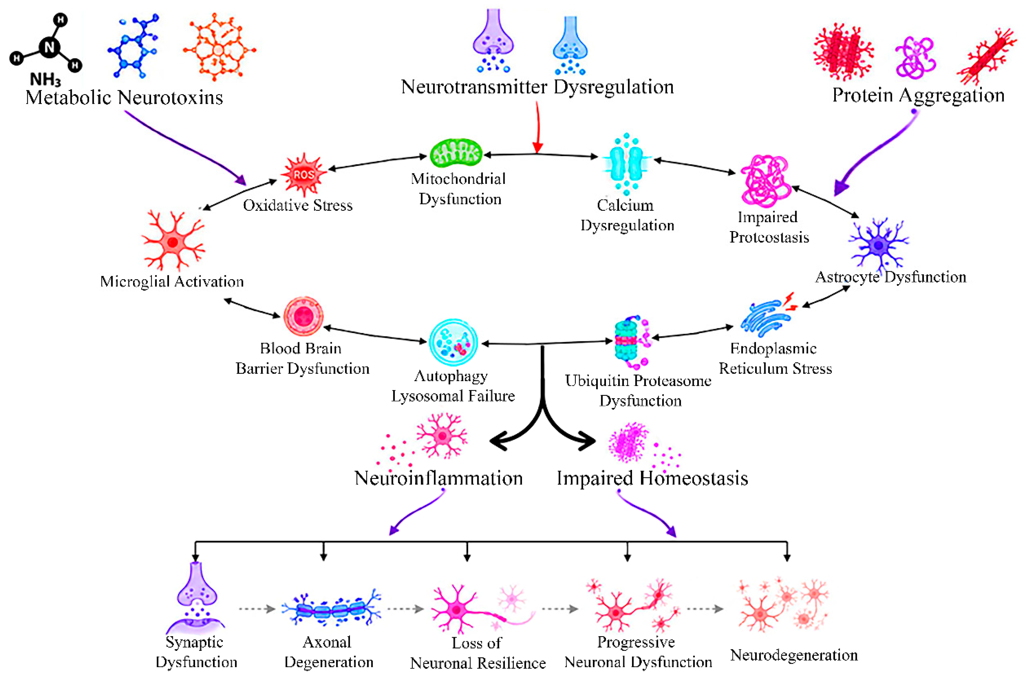
Figure 2.
Challenges in the clinical translation of endogenous neurotoxin-targeted therapies. Biological complexity, disease heterogeneity, biomarker limitations, blood–brain barrier permeability, preclinical model constraints, safety concerns, and clinical trial challenges collectively hinder therapeutic translation. Emerging approaches including precision patient care, biomarker-guided stratification, advanced drug delivery systems, multi-omics, artificial intelligence, human-relevant disease models, and combination therapies—offer promising strategies to bridge the translational gap and improve clinical outcomes in neurodegenerative diseases.
Figure 2.
Challenges in the clinical translation of endogenous neurotoxin-targeted therapies. Biological complexity, disease heterogeneity, biomarker limitations, blood–brain barrier permeability, preclinical model constraints, safety concerns, and clinical trial challenges collectively hinder therapeutic translation. Emerging approaches including precision patient care, biomarker-guided stratification, advanced drug delivery systems, multi-omics, artificial intelligence, human-relevant disease models, and combination therapies—offer promising strategies to bridge the translational gap and improve clinical outcomes in neurodegenerative diseases.
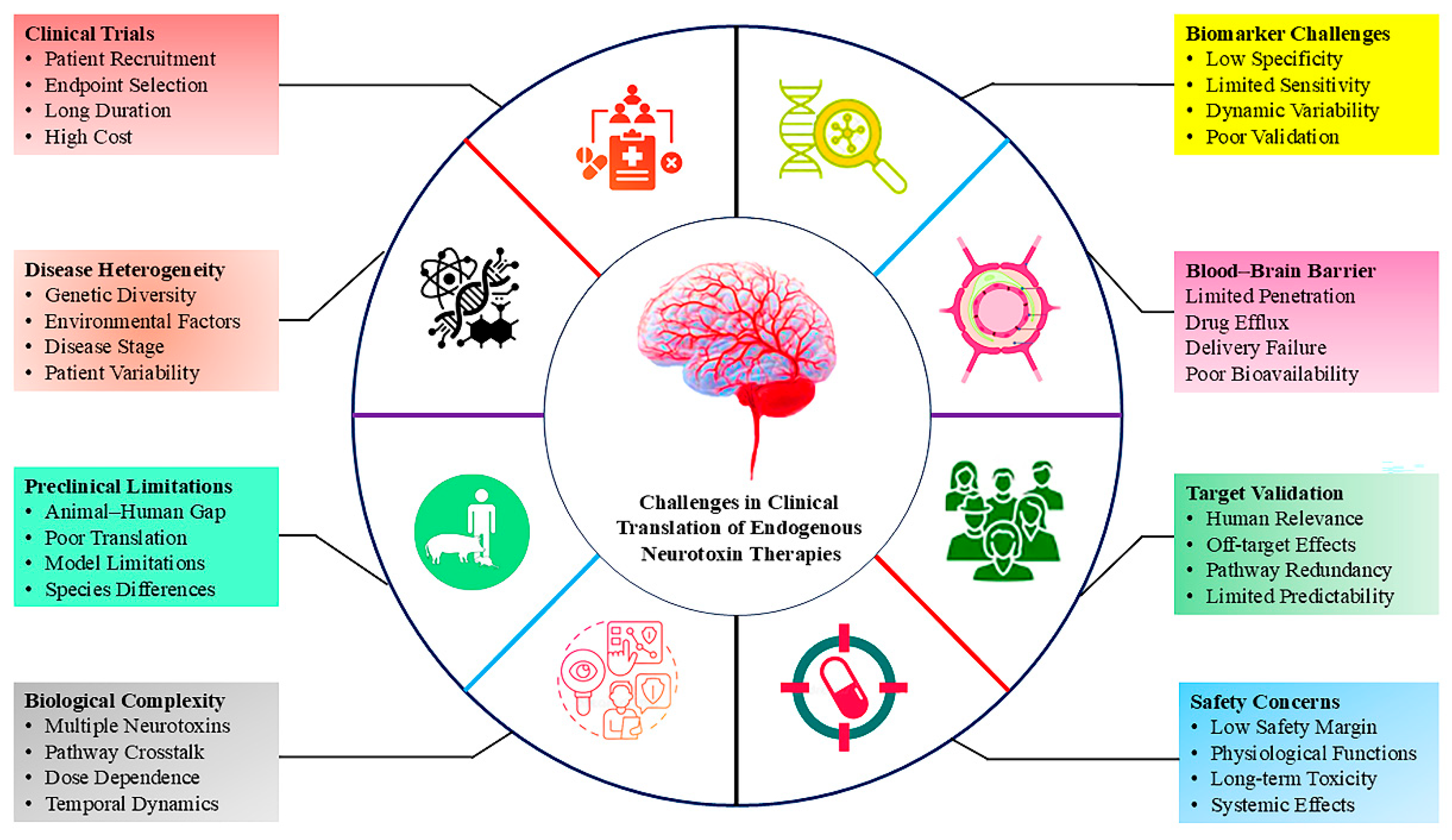

Table 1.
Major Classes of Endogenous Neurotoxins in the Central Nervous System and their principal neurotoxic mechanisms.
Table 1.
Major Classes of Endogenous Neurotoxins in the Central Nervous System and their principal neurotoxic mechanisms.
| Class | Representative endogenous neurotoxin | Physiological role | Principal neurotoxic mechanisms | Major neurological associations | Ref. |
|---|---|---|---|---|---|
| Metabolic | Ammonia (NH3/NH4+) | Nitrogen metabolism and maintenance of amino acid homeostasis through the urea cycle | Astrocyte swelling via glutamine accumulation, mitochondrial dysfunction, oxidative/nitrosative stress, glutamate imbalance, cerebral edema | Hepatic encephalopathy, hyperammonemic encephalopathy, cerebral edema | [12,13,19] |
| Metabolic | Homocysteine | Involved in methylation reactions and one-carbon metabolism | NMDA receptor-mediated excitotoxicity, oxidative stress, endothelial dysfunction, blood-brain barrier disruption, apoptosis | Cognitive decline, vascular dementia, stroke, Alzheimer's disease | [11,14,21,25] |
| Metabolic | Advanced Glycation End-products (AGEs) | Low-level physiological protein glycation | RAGE activation, ROS generation, chronic inflammation, mitochondrial dysfunction, impaired autophagy | Diabetic neuropathy, Alzheimer's disease, vascular cognitive impairment | [29,30,32] |
| Neurotransmitter | Glutamate | Principal excitatory neurotransmitter involved in learning, memory, and synaptic plasticity | NMDA/AMPA receptor overactivation, Ca2+ overload, mitochondrial dysfunction, oxidative stress, excitotoxicity | Stroke, ALS, epilepsy, Alzheimer's disease | [34,36,39] |
| Neurotransmitter | Dopamine | Motor control, cognition, motivation, and reward signaling | Auto-oxidation to dopamine quinones, ROS generation, mitochondrial impairment | Parkinson's disease | [43,47] |
| Protein | Amyloid-β (Aβ) | Synaptic regulation and neuronal signaling at physiological concentrations | Toxic oligomer formation, synaptic dysfunction, membrane disruption | Alzheimer's disease | [52,53,56] |
| Protein | α-Synuclein | Synaptic vesicle trafficking and neurotransmitter release | Misfolding, oligomerization, mitochondrial dysfunction, vesicle trafficking defects | Parkinson's disease | [50,63] |
| Protein | Tau | Microtubule stabilization and axonal transport | Microtubule destabilization, toxic oligomer formation, impaired axonal transport | Tauopathies, Alzheimer's disease | [49,73,79,80] |
Table 2.
Emerging biomarkers and therapeutic strategies targeting endogenous neurotoxicity.
| Homeostatic disturbance | Representative biomarker(s) | Pathophysiological significance | Potential therapeutic strategies | Ref. |
|---|---|---|---|---|
| Nitrogen dysregulation | Blood ammonia, glutamine, plasma amino acid profile | Excess ammonia disrupts astrocyte metabolism, promotes glutamine accumulation, cerebral edema, mitochondrial dysfunction, and excitotoxicity | Nitrogen-lowering therapies (lactulose, rifaximin), ammonia scavengers, metabolic correction, liver support strategies | [130] |
| One-carbon metabolism dysfunction | Homocysteine, folate, vitamin B12, methylmalonic acid | Hyperhomocysteinemia induces oxidative stress, endothelial dysfunction, DNA hypomethylation, blood-brain barrier disruption, and neuronal apoptosis | Folate and vitamin B supplementation, homocysteine-lowering therapy, dietary intervention, methyl donor replacement | [131,134] |
| Oxidative stress and redox imbalance | Reduced glutathione (GSH), malondialdehyde (MDA), 4-HNE, 8-OHdG | Excess ROS/RNS causes lipid peroxidation, protein oxidation, DNA damage, mitochondrial dysfunction, and neuronal degeneration | Antioxidants, Nrf2 pathway activators, mitochondrial antioxidants, ROS scavengers, redox-modulating therapies | [87,138] |
| Neuroaxonal injury | Neurofilament light chain (NfL), GFAP, UCH-L1 | Reflects axonal degeneration, neuronal injury, disease progression, and treatment response across neurodegenerative disorders | Neuroprotective therapies, disease monitoring, biomarker-guided therapeutic assessment | [135] |
| Protein aggregation and impaired proteostasis | Amyloid-β, phosphorylated tau (p-tau), total tau, α-synuclein | Misfolded protein accumulation disrupts synaptic function, mitochondrial integrity, axonal transport, and proteostasis networks | Anti-aggregation therapies, monoclonal antibodies, autophagy enhancers, proteasome activation, proteostasis modulation | [130,136] |
| Neuroinflammation | TREM2, IL-1β, IL-6, TNF-α, TSPO-PET imaging | Persistent microglial activation promotes cytokine release, oxidative stress, synaptic dysfunction, and progressive neuronal injury | Microglial modulators, anti-inflammatory agents, cytokine-targeted therapies, neuroglial communication modulation | [136,139] |
| Mitochondrial dysfunction | Mitochondrial metabolites, mtDNA, ATP/ADP ratio, multi-omics signatures | Impaired oxidative phosphorylation results in ATP depletion, ROS overproduction, calcium imbalance, and activation of apoptotic pathways | Mitochondrial-targeted antioxidants, mitophagy enhancers, metabolic modulators, mitochondrial quality-control therapies | [138,140] |
| Integrated homeostatic dysfunction | Multi-omics biomarker panels (proteomics, metabolomics, transcriptomics, lipidomics) | Simultaneous disruption of metabolic, inflammatory, oxidative, and proteostatic pathways drives disease progression and inter-individual heterogeneity | Precision medicine, systems biology-guided interventions, AI-assisted biomarker integration, personalized combination therapies | [130,131,137] |
Disclaimer/Publisher’s Note: The statements, opinions and data contained in all publications are solely those of the individual author(s) and contributor(s) and not of MDPI and/or the editor(s). MDPI and/or the editor(s) disclaim responsibility for any injury to people or property resulting from any ideas, methods, instructions or products referred to in the content. |
© 2026 by the authors. Licensee MDPI, Basel, Switzerland. This article is an open access article distributed under the terms and conditions of the Creative Commons Attribution (CC BY) license (http://creativecommons.org/licenses/by/4.0/).
Copyright: This open access article is published under a Creative Commons CC BY 4.0 license, which permit the free download, distribution, and reuse, provided that the author and preprint are cited in any reuse.


