

Submitted:
08 July 2026
Posted:
09 July 2026
You are already at the latest version
Abstract
Background: Maternal smoking during pregnancy remains a significant public health concern. The molecular mechanisms underlying smoking-induced maternal-fetal impact remain incompletely understood. The objective of this study is to identify adductomic signatures of oxidant stress in cord blood associated with tobacco exposure during pregnancy.
Methods: Through a prospective longitudinal cohort of mother-infant dyads enrolled at a single birth center (Chicago, IL) from 2008-2021, we linked prenatal maternal smoking status data to an existent database of 105 addition products (adducts) measured in cord blood plasma. Principal component analysis (PCA) and volcano plot analysis identified differential adduct profiles according to maternal smoking status using PERMANOVA (p≤0.05).
Results: Among 158 births included in the analysis, 6 women reported currently smoking, 26 were former smokers, and 126 identified as never smokers. PCA revealed significant separation between current smokers and both other groups (p≤0.002), with R² effect sizes of 0.026 for current vs never smokers and 0.115 for current vs former smokers. Among 56 known (annotated) adducts of oxidant stress, 33 were significantly upregu-lated in current versus never smokers, and 34 were upregulated in current versus former smokers. Maternal chronic hypertension, but not preeclampsia or other covariates, was associated with current smoking status (P=0.001).
Conclusions: Maternal smoking during pregnancy is associated with distinct cord blood adductomic signatures of oxidant stress. The persistence of these changes in current but not former smokers suggests potential reversibility upon smoking cessation.
Keywords:
maternal smoking
; hypertension
; perinatal oxidant stress
; preterm birth
; neonatal outcome
1. Introduction
Maternal smoking during pregnancy represents a preventable cause of adverse perinatal outcomes, including fetal growth restriction, preterm birth, stillbirth and placental com-plications.[1,2] Despite declining smoking rates in many developed countries, approximately 7-10% of pregnant women continue to smoke, with higher rates in certain demographic groups.[3,4] Cord blood, representing the offspring circulation at the time of birth, provides a unique window into fetal exposure to tobacco-related toxicants.[5]
While the epidemiological associations between smoking and adverse pregnancy out-comes are well established,[2,6] the molecular mechanisms underlying smoking-induced dysfunction remain incompletely characterized. Adductomic profiling is an emerging promising approach for identifying biochemical signatures of environmental exposures.[7,8,9,10] Specifically, adductomics measures the formation of multiple chemical addition products (adducts), which are covalent modifications of biomolecules by reactive com-pounds. These adductomics profiles represent a comprehensive molecular fingerprint of toxicant exposure.[11,12]
Previous studies have identified various smoking-related adducts in biological fluids and tissues,[8,13,14,15] but comprehensive characterization of cord blood (at birth) adductomic profiles across different smoking exposure groups remains limited. Understanding how adductomic changes persist after smoking cessation and how they vary with active smoking during pregnancy versus cessation during pregnancy could inform clinical counseling and public health intervention strategies.[16] The objective of this study was to characterize cord blood adductomic profiles across meaningful maternal smoking exposure categories.
2. Materials and Methods
2.1. Patient Population and Study Sample
The study population was drawn from a larger cohort of mother-infant dyads prospectively enrolled at Prentice Women’s Hospital in Chicago, IL. The database used at the time of this analysis included 2,287 mothers and infants enrolled from 2008 to 2021, for which both maternal and infant outcomes data were completely extracted from electronic medical records. The database consisted of consecutively born and enrolled preterm and full-term births for which cord blood was available at delivery. The inclusion/exclusion criteria, enrollment and data collection procedures for the cohort have been previously published.[17] Briefly, all study subjects provided informed consent prior to participation, and the study was approved by the Institution Review Board of Northwestern University (protocol number STU00201858). The study sample for the current analysis included the mother-infant dyads with confirmed maternal smoking status (below) and available cord blood adductomics data previously completed for 205 preterm and 51 full term births.[17]
2.2. Determination of Maternal Smoking Status
In the current study, we linked maternal smoking status data extracted from the social history section of the electronic medical records. Standardized data were obtained via self-report at the time of admission to the Labor and Delivery Unit from the patient intake form in EPIC, and included the following fields: 1) Never Smoker; 2) Former Smoker; 3) Current Every Day Smoker; 4) Current Some Day Smoker 5) Never Assessed; 6) Unknown if Ever Smoked. Maternal and infant clinical data were linked to the cord blood adductomics data via unique study codes without use of personal identifiers. Included in the present study were those births in which complete maternal smoking data were entered (i.e., complete self-reported entry in all relevant fields of the admission note). Maternal smoking status was categorized as “current smoker” (included current every day and some day smoking status), “former smoker” (quit prior to pregnancy or at least >30 days prior to admission), or “never smoker.” These data, along with all clinical data below, were extracted from electronic medical records using automated protocols in consultation with Northwestern’s Enterprise Data Warehouse (EDW). The study was approved by the institutional review board, and all participants provided informed consent.
2.3. Clinical Data Collection
Maternal and neonatal characteristics were abstracted from electronic medical records, including gestational age at delivery, birth weight, birth weight percentile, infant sex and neonatal outcomes. Maternal preeclampsia and chorioamnionitis were defined according to American College of Obstetrics and Gynecology (ACOG) criteria.[18,19]
2.4. Sample Collection and Processing
Cord blood samples were collected immediately following delivery, processed and archived per an established protocol with human serum albumin (HSA) isolated from plasma as previously published. The isolated HSA was subjected to trypsin digestion following established methodologies for adductomic analysis.[17]
2.5. Adductomic Analysis
Untargeted discovery of unknown adducts was performed using high-resolution mass spectrometry (HRMS) and targeted analyses of known adducts was performed using highly sensitive triple quadrupole mass spectrometry (QQQ-MS). A total of 56 known (annotated) adducts were quantified, including various cysteine modifications, oxidation products, and conjugates. Both adductomic approaches focused on HSA-Cys34 adducts, which serve as sentinel biomarkers of electrophilic exposure.[17,20] Unknown adducts were identified based on mass-to-charge ratio (m/z) and retention time using untargeted adductomic methods.[20]
2.6. Statistical Analysis
Principal component analysis (PCA) was performed to visualize overall adductomic patterns across smoking groups. PERMANOVA (Permutational Multivariate Analysis of Variance) was used to test for significant differences in adductomic profiles between groups,[21] with 95% confidence intervals calculated for each comparison. R² values were calculated to determine the proportion of variance explained by smoking status.
For individual adduct analysis, volcano plots were generated showing the relationship between fold change (Log2 scale) and statistical significance (-Log10 adjusted p-value).[22] Significance thresholds were set at adjusted p-value <0.05 (corresponding to -Log10 p = 1.3) and Log2 fold change > ±1.0. Multiple testing correction was performed using the Benjamini-Hochberg false discovery rate method.[23] Comparative analyses were performed for three pairwise comparisons: current vs never smokers, current vs former smokers, and former vs never smokers. Statistical significance was set at p ≤0.05 for all analyses.
3. Results
3.1. Characteristics of the Study Population and Patient Sample
Of the 2,287 mother-infant dyads enrolled at the time of this analysis, 36 (1.6%) were categorized as current smokers, 232 (10.1%) as former smokers and 1,679 (73.4%) as never smokers. The remaining births were unknown status. Linkage of smoking status data with the cord blood adductomics database resulted in identification of 158 births with complete data available. Thus, the current analysis included 6 (3.8%) current smokers, 26 (16.5%) former smokers, and 126 (79.7%) never smokers. Maternal and neonatal characteristics by smoking exposure status are presented in Table 1. Mean gestational age and birth weight were similar across groups, with a predominance of preterm (mean gestational age: 30.9 ± 5.9 weeks), low birth weight (mean birth weight: 1,745.5 ± 1,166.6 grams) deliveries reflecting the high-risk population. Overall, maternal and infant characteristics did not differ significantly by smoking status, with the exception of maternal chronic hypertension (50% among current smokers vs. 23% among former smokers and 5% among never smokers; P=0.001). This association was also significant in the larger cohort: 7/36 (19.4%) among current smokers vs. 39/232 (16.8%) among former smokers and 163/1679 (9.7%) among never smokers (P<0.001).
3.2. Overall Adductomic Patterns by Smoking Status
Table 2 lists the 56 known adducts that were quantified for this patient sample, including various cysteine modifications, oxidation products, and conjugates. Principle component analysis (PCA) revealed distinct clustering patterns based on maternal smoking status (Figure 1). The first two principal components explained substantial variance in the data: PC1 accounted for 35.3-40.8% and PC2 for 6.1-18.4% of variance depending on the comparison. PERMANOVA analysis demonstrated significant differences between current smokers and both other groups (Figure 1B,C; p≤0.002), but no significant difference between former and never smokers (Figure 1A; p=0.873, R²=0.004). The effect size was notably larger for the current vs former comparison (R²=0.115, 11% of variance) compared to current vs never comparison (R²=0.026, 26% of variance), indicating that current smokers have distinct adductomics pattern from former smokers.
3.3. Differential Annotated (Known) Adduct Formation
Volcano plot analysis identified numerous adducts significantly altered in current smokers compared to both control groups (Figure 2A-C). No significant adducts were identified in the former vs never smoker comparison, consistent with PCA findings. The current vs never smokers comparison revealed 33 significantly upregulated adducts with no downregulated adducts. Two adducts were unique to this comparison: A018 (S-methylthiolation.2) and A049 (S-addition of CysGly -H2O). The current vs former smokers comparison showed 34 significantly upregulated adducts. Three adducts were uniquely significant in this comparison: A019 (S–(O)–O–CH3), A022 (Cys34 sulfonic acid trioxidation), and A032 (S-addition of mercaptoacetic acid).
Figure 2D displays the comparative fold changes across both comparisons, with Log2FC values ranging from approximately 0.72 to 2.37 for current vs never and 0.98 to 2.30 for current vs former comparisons. The high degree of overlap between the two comparisons, with the majority of adducts showing similar directional changes, underscores the consistent adductomic signature of active smoking. Key adducts with the highest fold changes included A056 (S-addition of GSH) with Log2FC of 2.37 for current vs never and 2.30 for current vs former, A053 (Na adduct of S-CysGly) with Log2FC of 1.33 for current vs never and 2.02 for current vs former, and A048 (S-hCys, plus methylation not Cys34) with Log2FC of 1.80 for current vs never and 1.88 for current vs former. Other notable adducts with substantial increases included A045 (S-addition of Cys, methylation) with Log2FC of 1.33 for current vs never and 1.73 for current vs former, A022 (Cys34 sulfonic acid trioxidation) which was uniquely significant in current vs former with Log2FC of 1.69, and A028 (S-addition of crotonaldehyde) with Log2FC of 1.47 for current vs never and 1.39 for current vs former. Violin plots demonstrating the distribution of these individual adducts are shown in Figure 2E.
3.4. Unknown Adduct Discovery
Analysis of unknown adductomic features identified additional smoking-related adducts (Figure 3A). For current vs never smokers (Figure 3B), 9 unknown adducts showed significant upregulation, with one unique feature at 351.07 Da. For current vs former smokers (Figure 3C), 8 unknown adducts were significantly upregulated. The fold change patterns for unknown adducts revealed molecular weights ranging from 101.06 Da to 388.2 Da. The Unknown (351.07 Da) feature was uniquely significant in the current vs never comparison, while Unknown (388.2 Da) showed the highest fold change (Log2FC = 2.26 for current vs never, 2.76 for current vs former) (Figure 3D). Several unknown adducts showed consistency across both comparisons, suggesting they represent stable molecular markers of smoking exposure. The identification of unknown adducts highlights the potential for discovering novel biomarkers of tobacco toxicant exposure.
3.5. Figures, Tables and Schemes
4. Discussion
We employed novel adductomic approaches to identify protein adducts in cord blood associated with maternal smoking during pregnancy. The adducts included in this analysis are implicated in pathways of oxidative stress based upon their distinct biochemical structures, as previously published.[17] In total, 105 adducts were detected (56 known and 49 unknown), with certain adducts varying in concentration according to maternal smoking status. These adducts have been associated with environmental exposures and disease states including tobacco smoke, air pollution, and certain cancers.[17,24,25] The underlying chemistry and biology of these adducts as meaningful biomarkers of oxidant stress is also rapidly expanding.[26] While the literature on the role of adductomics in understanding the human exposome is rapidly expanding, this is the first study to investigate prenatal tobacco exposure through cord blood adductomic profiles.
Notable findings from this study include the following: cord blood adductomic profiles clearly distinguished current smokers from both former and never smokers; 33 and 34 known adducts were significantly upregulated in current smokers compared to never smokers and former smokers, respectively. Key adducts with the highest fold changes included A056 (S-addition of GSH), A053 (Na adduct of S-CysGly), and A048 (S-hCys, plus methylation not Cys34). Former smokers showed adductomic profiles indistinguishable from never smokers, suggesting normalization after smoking cessation. Finally, 8 to 9 unknown adducts were identified that correlated with current smoking status. Importantly, specific adducts were uniquely associated with current vs never smoker comparison (A018 and A049) or current vs former smoker comparison (A019, A022, A032), potentially representing biomarkers sensitive to recent smoking exposure or those that normalize at different rates after smoking cessation.
Elevated oxidative stress is an important mediator of tobacco-induced pathophysiology during pregnancy. Tobacco smoke contains thousands of chemicals, including reactive oxygen and nitrogen species (ROS/RNS) that damage DNA and proteins necessary for normal fetal development.[24,25] These electrophiles enter fetal circulation through the placenta, where they react with available proteins to form addition products, or “adducts”.[12] When bound to stable proteins such as human serum albumin (HSA), these adducts become significantly more stable (28 days in circulation) than the scavenged reactive electrophiles[17] and thus may serve as reliable biomarkers of exposure. The HSA-Cys34 residue is a sentinel nucleophilic hotspot for protein adducts that accounts for 80% of the antioxidant capacity of serum/plasma.[27] HSA-Cys34 adducts potentially play a pivotal role in understanding human disease arising from exposures to electrophilic species.[11,12] Our recently developed targeted assays can simultaneously quantify large panels of HSA-Cys34 adducts with higher detection rates than untargeted assays.[27,28] HSA-Cys34 protein adductomic profiles from cord blood could serve as a molecular fingerprint of prenatal tobacco exposures in the weeks leading up to delivery. These profiles captured at birth in cord blood provide the first report applying unbiased adductomics to assess maternal smoking exposure in a pregnancy cohort.
The patient sample represented a range of perinatal characteristics and included smoking exposure categories that allowed us to contrast the influence of current versus former smoking on adductomic profiles. An interesting finding was the increased incidence of maternal chronic hypertension with current and former smoking status (50% and 23%, respectively; P=0.001), which is consistent with well-known health effects of tobacco smoke exposure on chronic cardiovascular disease. The clear separation observed in PCA analysis between current smokers and both other groups (p≤0.002), but not between former and never smokers (p=0.873), suggests that smoking cessation leads to substantial normalization of the adductome. This finding provides molecular evidence supporting smoking cessation interventions and suggests that cardiometabolic recovery is possible when smoking is discontinued. As there is emerging and compelling evidence that maternal smoking is also linked to future hypertension in the offspring,[29,30,31] the findings of this study may also inform potential mechanisms and approaches by which smoking cessation programs and other early public health interventions may improve cardiovascular health in early childhood.
The adducts identified in our study have been associated with pathological disease states in other populations. The predominance of adducts in small thiols, direct oxidation products, and reactive aldehydes groups reflects the major categories of oxidative stress biomarkers. Small thiols, known for their antioxidant capacity, may represent a response to high oxidative stress exposure. Direct oxidation products result from reactions between Cys34 and ROS, providing evidence of oxidative burden. Reactive aldehydes are associated with lipid peroxidation, a process that triggers cellular dysfunction in the setting of elevated oxidative stress. Several specific adducts warrant particular attention: A056 (S-addition of GSH) showed the highest fold change (Log2FC=2.37) and represents glutathione conjugation, a major detoxification pathway. A053 (Na adduct of S-CysGly) and A048 (S-hCys, plus methylation) were also substantially elevated, suggesting activation of multiple antioxidant and detoxification systems in response to tobacco smoke exposure. The identification of unique adducts in specific comparisons (A018 and A049 for current vs never; A019, A022, and A032 for current vs former) provides insights into the temporal dynamics of adduct formation and clearance. These adducts may serve as particularly sensitive biomarkers for distinguishing active smoking from past exposure.
The unknown adducts identified in this study expand the potential repertoire of smoking-related biomarkers. The unknown adduct at 351.07 Da was uniquely significant in the current vs never comparison, while Unknown (388.2 Da) showed the highest fold changes across both comparisons. Validation studies employing synthetic chemistry work to characterize these unknown adducts associated with maternal smoking are important next steps. Further structural characterization through tandem mass spectrometry could reveal novel tobacco-specific metabolites and provide new insights into mechanisms of tobacco toxicity.
An important limitation of this study includes the relatively small number of current smokers identified (n=6), which limits statistical power and generalizability, though significant findings were still detected. Although a complete set of adducts for all 2,287 births of the parent cohort would have been more comprehensive, the relative proportions of current, former and never smokers were similarly represented in the smaller patient sample of 158 births. Another limitation is that self-reported smoking status may be subject to misclassification due to social desirability bias during pregnancy,[3] although adductomic validation supports the accuracy of classification. The cross-sectional design at delivery prevents assessment of temporal changes in adductomic profiles throughout pregnancy. Information on smoking intensity (cigarettes per day) was not available, precluding dose-response analysis. Future studies with detailed information on timing of smoking cessation in the former smoker group could address questions about the time course of adductomic normalization. We did not assess functional consequences of the observed adductomic changes on pregnancy outcomes or infant health. Additionally, the study utilized archived samples from a single center, which provided uniformity but requires multi-center studies to assess generalizability of findings.
Nevertheless, this study has several important strengths. The comprehensive adductomic approach covering both known and unknown adducts,[11,12,17,20,32] combined with rigorous statistical methods including PERMANOVA for multivariate analysis,[21] provides a robust characterization of smoking-related adduct formation. The inclusion of three smoking status groups enabled assessment of cessation effects, revealing that former smokers cluster metabolically with never smokers. The focus on HSA-Cys34 adducts provides a biologically relevant window into fetal exposure given the prolonged half-life of albumin.[17] The minimal blood volume needed (5 µL of sample for a single run) makes this approach practical for neonatal studies. Recent studies of adductomics performed on neonatal dried blood spots provide promise for the development of more practical approaches to tracking these exposure biomarkers over time.[8,33,34]
The investigation of adducts in validation studies and experimental models is warranted to better understand mechanistic links between prenatal tobacco exposure and adverse outcomes. Our understanding of specific maternal environmental exposures (toxins and pollutants from tobacco) and their effects on fetal development could be enhanced by integrating adductomics into larger epidemiologic studies and future clinical trials. Additionally, HSA-Cys34 biomarkers can be utilized in diagnostic laboratories to objectively assess smoking status and monitor cessation compliance during pregnancy.
Future research should focus on several key areas. Longitudinal studies tracking adductomic changes throughout pregnancy in women who quit smoking could define the precise time course of adductomic recovery and identify the optimal timing for interventions.[35,36,37] Investigation of dose-response relationships between smoking intensity and adduct levels would strengthen causal inference and inform risk assessment.[38,39] Studies linking specific adduct profiles to pregnancy outcomes and infant health would establish clinical relevance and identify which adducts best predict adverse outcomes.[5,40,41] Detailed structural characterization of unknown adducts through advanced mass spectrometry techniques could reveal novel tobacco-specific biomarkers and identify previously unrecognized pathways of tobacco toxicity.[20,32] Extension to other environmental exposures such as e-cigarettes, secondhand smoke, and air pollution would broaden understanding of environmental impacts on the prenatal exposome.[42,43,44] The adductomic approach developed here could be applied more comprehensively to characterize the full spectrum of prenatal environmental exposures and their biological consequences.
5. Conclusion
The application of adductomics to elucidate exposure pathways of prenatal tobacco exposure is a novel and promising approach in pregnancy research. These exposure pathways could inform the development of targeted interventions and monitoring strategies during pregnancy. Linked to other high-resolution and high-throughput technologies, investigating multiple omics could yield unprecedented findings and lead to innovative approaches in understanding and preventing tobacco-related pregnancy complications. This study highlights the central role of oxidative stress signaling in mediating the effects of maternal smoking on fetal health and provides a foundation for future investigations into the perinatal-neonatal exposome.
Author Contributions
J.N. drafted the initial manuscripts, J.N., E.B. and A.C. performed adductomics analyses and interpretation. J.N., E.B. and K.M. performed statistical analyses. E.B., E.L. and A.A. contributed to study design, data collection methods, cord blood assays and data annotation. K.M. and W.F. conceived and designed the study, supervised the research, and obtained funding. All authors contributed to manuscript preparation and approved the final version.
Funding
This study was supported by the National Institute of Child Health and Human Development (NICHD), Funding number: R21HD100831 (PI: Mestan), and the National Heart, Lung, and Blood Institute (NHLBI), Funding number: R01HL139798 (PI: Mestan), and in part by the National Center for Advancing Translational Sciences, Funding number: UL1TR001442 (UCSD Cooperative Agreement). Proteomics services were performed by the Northwestern Proteomics Core Facility, generously supported by NCI CCSG P30 CA060553 awarded to the Robert H Lurie Comprehensive Cancer Center, the instrumentation award (S10OD025194) from the NIH Office of Director, and the National Resource for Translational and Developmental Proteomics supported by P41 GM108569.
Institutional Review Board Statement
The study was conducted in accordance with the Declaration of Helsinki, and the protocol was approved by the Institutional Review Board of Northwestern University (protocol number STU00201858) on July 31, 2008.
Informed Consent Statement
Informed consent was obtained from all subjects involved in the study.
Data Availability Statement
The original contributions presented in this study are included in the article/supplementary material. Further inquiries can be directed to the corresponding author(s).
Acknowledgments
We thank the patients and families for their contributions and participation in this study. We thank additional members of the study team for their contributions and meticulous implementation of study protocols necessary for completing this project: Juanita Saqibuddin, RN, Kelly Stephens, RN, Rob Birkett, MSRC, Yeunook Bae, PhD.
Conflicts of Interest
The authors declare no conflicts of interest.
References
- Andres, R.L. and M.C. Day, Perinatal complications associated with maternal tobacco use. Semin Neonatol, 2000. 5(3): p. 231-41.
- Pineles, B.L., et al., Systematic Review and Meta-Analyses of Perinatal Death and Maternal Exposure to Tobacco Smoke During Pregnancy. Am J Epidemiol, 2016. 184(2): p. 87-97.
- Cnattingius, S., The epidemiology of smoking during pregnancy: smoking prevalence, maternal characteristics, and pregnancy outcomes. Nicotine Tob Res, 2004. 6 Suppl 2: p. S125-40.
- Creamer, M.R., et al., Tobacco Product Use and Cessation Indicators Among Adults - United States, 2018. MMWR Morb Mortal Wkly Rep, 2019. 68(45): p. 1013-1019.
- Jauniaux, E. and G.J. Burton, Morphological and biological effects of maternal exposure to tobacco smoke on the feto-placental unit. Early Hum Dev, 2007. 83(11): p. 699-706.
- Triche, E.W. and N. Hossain, Environmental factors implicated in the causation of adverse pregnancy outcome. Semin Perinatol, 2007. 31(4): p. 240-2.
- Fiehn, O., Metabolomics--the link between genotypes and phenotypes. Plant Mol Biol, 2002. 48(1-2): p. 155-71.
- Madera, D., et al., Adductomics of Newborn Dried Blood Spots Detects Constituents of Maternal Smoking During Pregnancy and Associated Oxidative Stress Exposure. Antioxidants (Basel), 2026. 15(4).
- Nicholson, J.K., J.C. Lindon, and E. Holmes, ‘Metabonomics’: understanding the metabolic responses of living systems to pathophysiological stimuli via multivariate statistical analysis of biological NMR spectroscopic data. Xenobiotica, 1999. 29(11): p. 1181-9.
- Patti, G.J., O. Yanes, and G. Siuzdak, Innovation: Metabolomics: the apogee of the omics trilogy. Nat Rev Mol Cell Biol, 2012. 13(4): p. 263-9.
- Grigoryan, H., et al., Cys34 Adductomics Links Colorectal Cancer with the Gut Microbiota and Redox Biology. Cancer Res, 2019. 79(23): p. 6024-6031.
- Rappaport, S.M., et al., Adductomics: characterizing exposures to reactive electrophiles. Toxicol Lett, 2012. 213(1): p. 83-90.
- Fu, K.T., D.C. Wu, and H.C. Chen, Elevated hemoglobin adducts derived from crotonaldehyde in healthy smokers and oral cancer patients by nanoflow liquid chromatography tandem mass spectrometry☆. Chem Biol Interact, 2025. 410: p. 111435.
- Imani, P., et al., HSA Adductomics in the Shanghai Women’s Health Study Links Lung Cancer in Never-Smokers with Air Pollution, Redox Biology, and One-Carbon Metabolism. Antioxidants (Basel), 2025. 14(3).
- Zhang, X., et al., Advances in the mechanistic understanding, biological consequences, and measurement of DNA adducts induced by tobacco smoke and e-cigarette aerosol: A review. Int J Biol Macromol, 2025. 306(Pt 2): p. 141574.
- McCowan, L.M., et al., Spontaneous preterm birth and small for gestational age infants in women who stop smoking early in pregnancy: prospective cohort study. BMJ, 2009. 338: p. b1081.
- Lin, E.T., et al., Cord Blood Adductomics Reveals Oxidative Stress Exposure Pathways of Bronchopulmonary Dysplasia. Antioxidants (Basel), 2024. 13(4).
- 18. Committee Opinion No. 712: Intrapartum Management of Intraamniotic Infection. Obstet Gynecol, 2017. 130(2): p. e95-e101.
- 19. Gestational Hypertension and Preeclampsia: ACOG Practice Bulletin, Number 222. Obstet Gynecol, 2020. 135(6): p. e237-e260.
- Carlsson, H. and M. Tornqvist, Strategy for identifying unknown hemoglobin adducts using adductome LC-MS/MS data: Identification of adducts corresponding to acrylic acid, glyoxal, methylglyoxal, and 1-octen-3-one. Food Chem Toxicol, 2016. 92: p. 94-103.
- Anderson, M., Permutational Multivariate Analysis of Variance (PERMANOVA). Wiley StatsRef: Statistics Reference Online 1-15. 2017.
- Xia, J. and D.S. Wishart, Using MetaboAnalyst 3.0 for Comprehensive Metabolomics Data Analysis. Curr Protoc Bioinformatics, 2016. 55: p. 14 10 1-14 10 91.
- Benjamini, Y, Hochberg, Y. Controlling the False Discovery Rate: A Practical and Powerful Approach to Multiple Testing. J.R. Stat. Soc. Series B Stat. Methodol. 57, 289-300. 1995.
- Chelchowska, M., et al., The effect of tobacco smoking during pregnancy on plasma oxidant and antioxidant status in mother and newborn. Eur J Obstet Gynecol Reprod Biol, 2011. 155(2): p. 132-6.
- Hecht, S.S., Biochemistry, biology, and carcinogenicity of tobacco-specific N-nitrosamines. Chem Res Toxicol, 1998. 11(6): p. 559-603.
- Jala, A., F. Tayyari, and W.E. Funk, Use of Human Serum Albumin Cys(34) (HSA-Cys(34)) Adductomics as a Multidimensional and Integrative Biomarker Approach to Assess Oxidative Stress. Antioxidants (Basel), 2026. 15(4).
- Grigoryan, H., et al., Cys34 adducts of reactive oxygen species in human serum albumin. Chem Res Toxicol, 2012. 25(8): p. 1633-42.
- Grigoryan, H., et al., Adductomics Pipeline for Untargeted Analysis of Modifications to Cys34 of Human Serum Albumin. Anal Chem, 2016. 88(21): p. 10504-10512.
- Niu, Z., et al., Involuntary tobacco smoke exposures from conception to 18 years increase midlife cardiometabolic disease risk: a 40-year longitudinal study. J Dev Orig Health Dis, 2023. 14(6): p. 689-698.
- Liu, M., et al., Maternal Smoking Intensity During Pregnancy and Early Adolescent Cardiovascular Health. J Am Heart Assoc, 2025. 14(5): p. e037806.
- Li, S., et al., Maternal smoking during pregnancy links to childhood blood pressure through birth weight and body mass index: NHANES 1999-2018. J Hum Hypertens, 2024. 38(2): p. 134-139.
- Balbo, S., R.J. Turesky, and P.W. Villalta, DNA adductomics. Chem Res Toxicol, 2014. 27(3): p. 356-66.
- Petrick, L.M., K. Uppal, and W.E. Funk, Metabolomics and adductomics of newborn bloodspots to retrospectively assess the early-life exposome. Curr Opin Pediatr, 2020. 32(2): p. 300-307.
- Funk, W.E., et al., Human Serum Albumin Cys34 Adducts in Newborn Dried Blood Spots: Associations With Air Pollution Exposure During Pregnancy. Front Public Health, 2021. 9: p. 730369.
- Fang, F., et al., Epigenetic biomarkers for smoking cessation. Addict Neurosci, 2023. 6.
- Lee, K.W. and Z. Pausova, Cigarette smoking and DNA methylation. Front Genet, 2013. 4: p. 132.
- Saygin Avsar, T., et al., Towards optimum smoking cessation interventions during pregnancy: a household model to explore cost-effectiveness. Addiction, 2022. 117(10): p. 2707-2719.
- Baba, S., et al., Changes in snuff and smoking habits in Swedish pregnant women and risk for small for gestational age births. BJOG, 2013. 120(4): p. 456-62.
- Hecht, S.S., et al., Similar exposure to a tobacco-specific carcinogen in smokeless tobacco users and cigarette smokers. Cancer Epidemiol Biomarkers Prev, 2007. 16(8): p. 1567-72.
- Bush, P.G., et al., Maternal cigarette smoking and oxygen diffusion across the placenta. Placenta, 2000. 21(8): p. 824-33.
- Zdravkovic, T., et al., The adverse effects of maternal smoking on the human placenta: a review. Placenta, 2005. 26 Suppl A: p. S81-6.
- Carlsten, C., et al., Genes, the environment and personalized medicine: We need to harness both environmental and genetic data to maximize personal and population health. EMBO Rep, 2014. 15(7): p. 736-9.
- Robinson, O. and M. Vrijheid, The Pregnancy Exposome. Curr Environ Health Rep, 2015. 2(2): p. 204-13.
- Tzoulaki, I., et al., Design and analysis of metabolomics studies in epidemiologic research: a primer on -omic technologies. Am J Epidemiol, 2014. 180(2): p. 129-39.
Figure 1.
Principal component analysis of cord blood adductomic profiles by maternal smoking status. PCA plots showing separation of samples based on adductomic signatures for (A) Former vs never smokers; (B) Current vs never smokers; and (C) Current vs former smokers. Each point represents an individual sample colored by smoking exposure group: current smokers (red), former smokers (blue), and never smokers (green). PC1 and PC2 capture the major sources of variance in the adductomic data. PERMANOVA results demonstrate significant differences between current smokers and both control groups (p≤0.002), with R² values indicating effect sizes. No significant differences were observed between former and never smokers (p=0.873, R²=0.004).
Figure 1.
Principal component analysis of cord blood adductomic profiles by maternal smoking status. PCA plots showing separation of samples based on adductomic signatures for (A) Former vs never smokers; (B) Current vs never smokers; and (C) Current vs former smokers. Each point represents an individual sample colored by smoking exposure group: current smokers (red), former smokers (blue), and never smokers (green). PC1 and PC2 capture the major sources of variance in the adductomic data. PERMANOVA results demonstrate significant differences between current smokers and both control groups (p≤0.002), with R² values indicating effect sizes. No significant differences were observed between former and never smokers (p=0.873, R²=0.004).
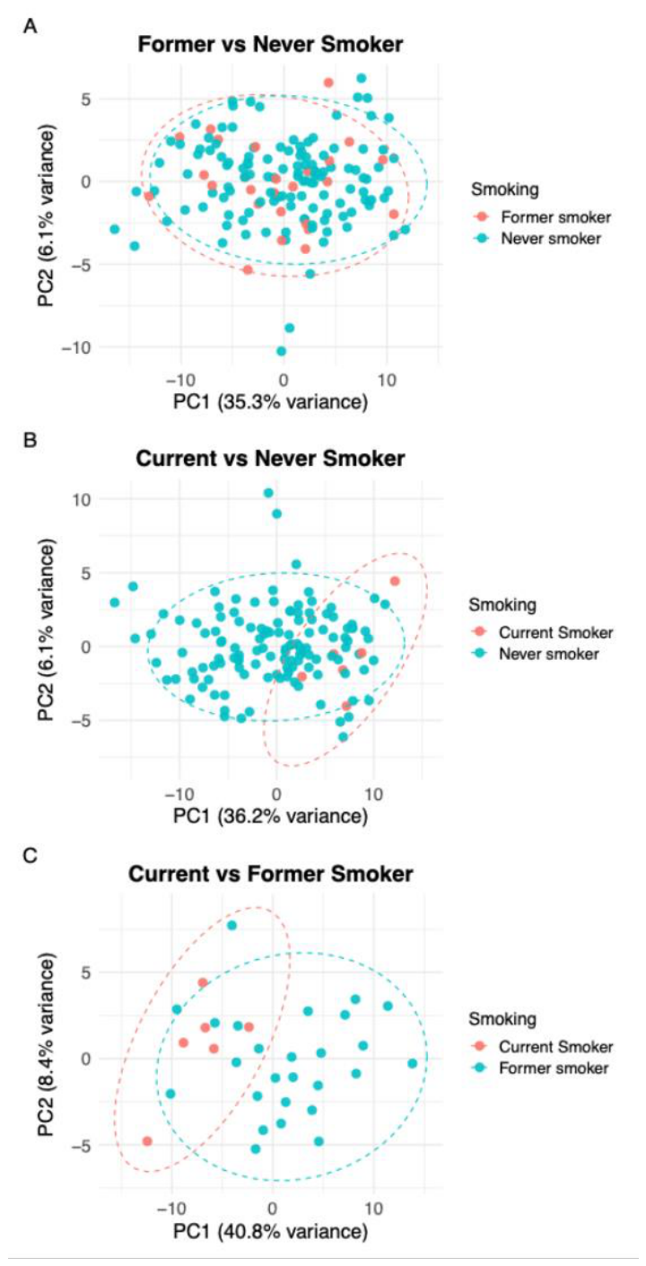
Figure 2.
Differential known adduct formation and comparative fold change analysis across maternal smoking groups. Volcano plots display the relationship between fold change (x-axis, Log2 scale) and statistical significance (y-axis, -Log10 adjusted P-value) for known adducts differentially formed between smoking groups. (A) Former vs never smokers comparison showing no significant adducts. (B) Current vs never smokers comparison revealing 33 significantly upregulated adducts (red points). (C) Current vs former smokers comparison showing 34 significantly upregulated adducts (red points). Horizontal dashed line indicates adjusted P-value threshold of 0.05 (-Log10 P = 1.3), and vertical dashed lines mark Log2 fold change thresholds of ±1.0. Points are colored by significance: gray (not significant) and red (significant). (D) Scatter plot comparing Log2 fold changes for individual adducts across both comparisons, with black circles showing current vs never and red circles showing current vs former fold changes, demonstrating consistent adductomic signatures of active smoking. (E) Violin plots illustrating concentration distributions of six key adducts representing major biochemical pathways: top row shows current vs never (A028 - S-addition of crotonaldehyde, A048 - S-hCys plus methylation, A056 - S-addition of GSH); bottom row shows current vs former (A022 - Cys34 sulfonic acid trioxidation, A053 - Na adduct of S-CysGly, A056 - S-addition of GSH). Never smokers shown in white, former smokers shown in orange and current smokers shown in red.
Figure 2.
Differential known adduct formation and comparative fold change analysis across maternal smoking groups. Volcano plots display the relationship between fold change (x-axis, Log2 scale) and statistical significance (y-axis, -Log10 adjusted P-value) for known adducts differentially formed between smoking groups. (A) Former vs never smokers comparison showing no significant adducts. (B) Current vs never smokers comparison revealing 33 significantly upregulated adducts (red points). (C) Current vs former smokers comparison showing 34 significantly upregulated adducts (red points). Horizontal dashed line indicates adjusted P-value threshold of 0.05 (-Log10 P = 1.3), and vertical dashed lines mark Log2 fold change thresholds of ±1.0. Points are colored by significance: gray (not significant) and red (significant). (D) Scatter plot comparing Log2 fold changes for individual adducts across both comparisons, with black circles showing current vs never and red circles showing current vs former fold changes, demonstrating consistent adductomic signatures of active smoking. (E) Violin plots illustrating concentration distributions of six key adducts representing major biochemical pathways: top row shows current vs never (A028 - S-addition of crotonaldehyde, A048 - S-hCys plus methylation, A056 - S-addition of GSH); bottom row shows current vs former (A022 - Cys34 sulfonic acid trioxidation, A053 - Na adduct of S-CysGly, A056 - S-addition of GSH). Never smokers shown in white, former smokers shown in orange and current smokers shown in red.
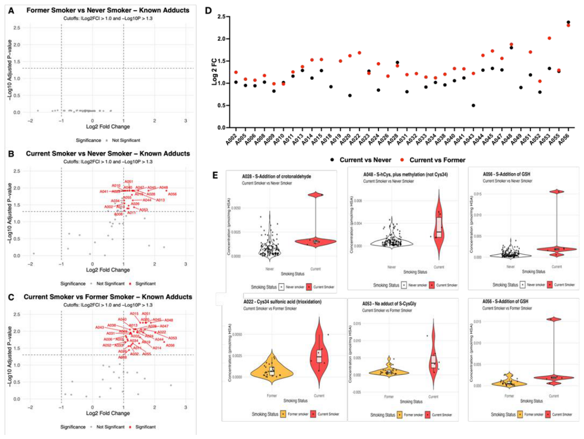
Figure 3.
Differential unknown adduct formation and comparative fold change analysis across maternal smoking groups. Volcano plots display the relationship between fold change (x-axis, Log2 scale) and statistical significance (y-axis, -Log10 adjusted P-value) for unknown adducts differentially formed between smoking groups. (A) Former vs never smokers comparison showing no significant unknown adducts. (B) Current vs never smokers comparison revealing 9 significantly upregulated unknown adducts (red points), including a unique feature at 351.07 Da. (C) Current vs former smokers comparison showing 8 significantly upregulated unknown adducts (red points). Horizontal dashed line indicates adjusted P-value threshold of 0.05 (-Log10 P = 1.3), and vertical dashed lines mark Log2 fold change thresholds of ±1.0. Points are colored by significance: gray (not significant) and red (significant). (D) Scatter plot comparing Log2 fold changes for individual unknown adducts across both comparisons, with black circles showing current vs never and red circles showing current vs former fold changes. Unknown adducts are labeled by their molecular weights (Da), with Unknown (388.2 Da) showing the highest fold changes in both comparisons. (E) Violin plots illustrating concentration distributions of four key unknown adducts: top row shows current vs never (Unknown 351.07 Da - unique to this comparison, Unknown 388.2 Da - highest fold change); bottom row shows current vs former (Unknown 101.06 Da - lower molecular weight range, Unknown 388.2 Da - highest overall fold change at Log2FC = 2.76). Never smokers shown in white, former smokers shown in orange and current smokers shown in red.
Figure 3.
Differential unknown adduct formation and comparative fold change analysis across maternal smoking groups. Volcano plots display the relationship between fold change (x-axis, Log2 scale) and statistical significance (y-axis, -Log10 adjusted P-value) for unknown adducts differentially formed between smoking groups. (A) Former vs never smokers comparison showing no significant unknown adducts. (B) Current vs never smokers comparison revealing 9 significantly upregulated unknown adducts (red points), including a unique feature at 351.07 Da. (C) Current vs former smokers comparison showing 8 significantly upregulated unknown adducts (red points). Horizontal dashed line indicates adjusted P-value threshold of 0.05 (-Log10 P = 1.3), and vertical dashed lines mark Log2 fold change thresholds of ±1.0. Points are colored by significance: gray (not significant) and red (significant). (D) Scatter plot comparing Log2 fold changes for individual unknown adducts across both comparisons, with black circles showing current vs never and red circles showing current vs former fold changes. Unknown adducts are labeled by their molecular weights (Da), with Unknown (388.2 Da) showing the highest fold changes in both comparisons. (E) Violin plots illustrating concentration distributions of four key unknown adducts: top row shows current vs never (Unknown 351.07 Da - unique to this comparison, Unknown 388.2 Da - highest fold change); bottom row shows current vs former (Unknown 101.06 Da - lower molecular weight range, Unknown 388.2 Da - highest overall fold change at Log2FC = 2.76). Never smokers shown in white, former smokers shown in orange and current smokers shown in red.
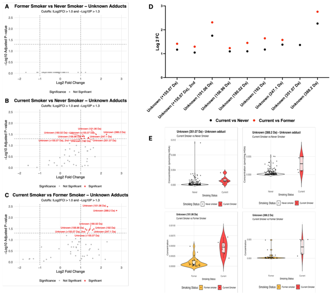

Table 1.
Maternal and Infant Characteristics by Smoking Exposure Status.
|
Current Smoker n=6 |
Former smoker n=26 |
Never smoker n=126 |
P | |
| Maternal Variables: | ||||
| Maternal age, mean yrs ± SD | 31.5 ± 6.4 | 33.0 ± 6.4 | 32.3 ± 5.2 | 0.79 |
|
Maternal Race Black/African American White Other/Unknown |
4 (67%) 1 (17%) 1 (17%) |
3 (11%) 19 (73%) 4 (15%) |
25 (20%) 65 (51%) 36 (29%) |
0.06 |
|
Maternal Ethnicity Hispanic or Latino Not Hispanic or Latino |
0 (0) 6 (100%) |
5 (19%) 21 (81%) |
27 (21%) 94 (75%) |
0.70 |
|
Maternal BMI, mean ± SD Pregravida Gravid |
29.3 ± 0.0 35.5 ± 5.7 |
27.8 ± 6.5 31.8 ± 7.1 |
27.5 ± 6.8 31.4 ± 6.2 |
0.93 0.29 |
| Preeclampsia, n (%) | 0 (0%) | 3 (11%) | 16 (13%) | 1.00 |
| Chorioamnionitis, n (%) | 1 (17%) | 4 (15%) | 12 (9%) | 0.39 |
|
Mode of Delivery Vaginal Cesarean |
2 (33%) 4 (67%) |
7 (27%) 19 (73%) |
69 (55%) 57 (45%) |
0.02 |
|
Rupture of Membranes Spontaneous Artificial |
2 (33%) 4 (67%) |
10 (38%) 16 (62%) |
55 (44%) 71 (56%) |
0.80 |
| Preterm Labor, n (%) | 4 (67%) | 15 (57%) | 62 (49%) | 0.58 |
| Chronic Hypertension, n (%) | 3 (50%) | 6 (23%) | 7 (5%) | 0.001 |
| Infant Outcomes: | ||||
| Gestational age, mean wks ± SD | 32.0 ± 6.0 | 29.2 ± 4.9 | 31.2 ± 6.1 | 0.28 |
| Birth weight, mean gms ± SD | 1,918 ± 1,136 | 1,457 ± 983 | 1,797 ± 1,201 | 0.38 |
| Birth weight percentile, mean ± SD | 56.2 ± 26.1 | 65.3 ± 23.5 | 58.2 ± 25.4 | 0.40 |
| Male sex, n (%) | 5 (83%) | 13 (50%) | 56 (44%) | 0.16 |
| Apgar (1-min), median [IQR] | 6.5 [17,2] | 6 [4,8] | 7 [4,8] | 0.59 |
| Apgar (5-min), median [IQR] | 8.5 [8,9] | 8 [7,9] | 8 [6,9] | 0.34 |
| NICU admission, n (%) | 5 (83%) | 22 (85%) | 85 (67%) | 0.17 |
Table 2.
Known adduct identification and nomenclature.
| Adduct ID | Adduct name | Adduct ID | Adduct name |
| A001 | -Lys from C-terminus | A029 | S-Phenylation |
| A002 | Cys34→Gly | A030 | S-Addition of tiglic aldehyde |
| A003 | Cys34→Dehydroalanine | A031 | S-Addition of pyruvate or malonate semialdehyde |
| A004 | Cys34→ Oxoalanine or formylglycine | A032 | S-Addition of mercaptoacetic acid |
| A005 | T3 dimer | A033 | S-Mercaptoacetamide |
| A006 | Unmodified T3 | A034 | S-Addition of Cys (-H2O) |
| A007 | CH2 crosslink | A035 | S-Cys (possibly NH2 → OH, -H2O) |
| A008 | Cys34-Gln cross-link (monooxidation), Cys34 Sulfinamide | A036 | S-addition of benzaldehyde or quinone methide |
| A009 | Methylation (not at Cys34) | A037 | S-Methylethyl-sulfonylation |
| A010 | S-Sodiation | A038 | S-Addition of S2O3H |
| A011 | S-Cyanylation | A039 | S-Addition of hCys (-H2O) |
| A012 | dehydrated form of Cys34 sulfinic acid plus methylation (not Cys34) | A040 | S-Cys |
| A013 | dehydrated form of Cys34 sulfonic acid (trioxidation) | A041 | S-Addition of Cys (NH2→OH) |
| A014 | Cys34 sulfinic acid (dioxidation) | A042 | S-Addition of BDE |
| A015 | K adduct of T3 | A043 | Oxindole |
| A016 | Ethylene oxide adduct | A044 | S-Addition of hCys |
| A017 | S-Methylthiolation.1 | A045 | S-Addition of Cys, methylation |
| A018 | S-Methylthiolation.2 | A046 | S-Addition of hCys (NH2→OH) |
| A019 | S–(O)–O–CH3 | A047 | Na adduct of S-Cys |
| A020 | Cys34 sulfinic acid plus methylation (not Cys34) | A048 | S-hCys plus methylation (not Cys34) |
| A021 | S-Methylthiolation | A049 | S-Addition of CysGly (-H2O) |
| A022 | Cys34 sulfonic acid (trioxidation) | A050 | S-(N-acetyl)Cys |
| A023 | Acrylonitrile adduct | A051 | S-Addition of CysGly |
| A024 | Na adduct of Cys34 sulfinic acid | A052 | S-CysGly plus methylation (not Cys34) |
| A025 | Putative S-addition of acrolein | A053 | Na adduct of S-CysGly |
| A026 | Methylisocyanate adduct | A054 | K Adduct of S-CysGly |
| A027 | S-Addition of SO2 | A055 | S-Addition of GluCys |
| A028 | S-Addition of crotonaldehyde | A056 | S-Addition of GSH |
| Complete list of 56 annotated (known) adducts analyzed in cord blood samples, including metabolite IDs (A001-A056) used in the text, and corresponding chemical names. Adducts include various cysteine modifications, oxidation products, cross-links, and conjugates with endogenous and exogenous compounds. | |||
Disclaimer/Publisher’s Note: The statements, opinions and data contained in all publications are solely those of the individual author(s) and contributor(s) and not of MDPI and/or the editor(s). MDPI and/or the editor(s) disclaim responsibility for any injury to people or property resulting from any ideas, methods, instructions or products referred to in the content. |
© 2026 by the authors. Licensee MDPI, Basel, Switzerland. This article is an open access article distributed under the terms and conditions of the Creative Commons Attribution (CC BY) license (http://creativecommons.org/licenses/by/4.0/).
Copyright: This open access article is published under a Creative Commons CC BY 4.0 license, which permit the free download, distribution, and reuse, provided that the author and preprint are cited in any reuse.


