
Submitted:
08 July 2026
Posted:
09 July 2026
You are already at the latest version
Abstract
Cancer remains a major global health challenge, and despite advances in chemotherapy, radiotherapy, immunotherapy, and targeted therapy, treatment outcomes are still limited by systemic toxicity, poor tumor selectivity, multidrug resistance, and inadequate efficacy against advanced disease. Nanotechnology has emerged as a promising strategy to address these limitations by improving drug delivery, imaging, radiosensitization, and immunomodulation. Among nanomaterials, phytochemical-mediated metal nanoparticles have attracted increasing interest as sustainable and potentially biocompatible alternatives to conventionally synthesized systems. However, their development remains constrained by poor reproducibility, lack of mechanistic understanding, and limited clinical translational progression, mainly because of the chemical complexity of plant extracts. In this review, we critically examine the role of metal nanoparticles in oncology, with emphasis on gold, silver, and zinc oxide, and we compare their anticancer mechanisms. We further evaluate phytochemical-mediated nanoparticle synthesis by discussing how plant-derived metabolites influence reduction, nucleation, capping, and the resulting physicochemical properties that influence biological activity. We argue that metabolomics offers a critical framework for overcoming one of the major limitations of green nanotechnology by enabling the comprehensive chemical characterization of plant extracts, identification of metabolites associated with nanoparticle formation, and correlation of metabolite profiles with nanoparticle physicochemical properties and anticancer activity. The integration of metabolomics with nanoparticle characterization could contribute to the advancement of green synthesis from empirical extract-based fabrication, toward a more predictive, standardized, and mechanistically informed nanoparticle design. Finally, we discuss the major barriers to clinical translation, including batch-to-batch variability, bio-corona effects, biodistribution, long-term toxicity, and regulatory compliance. Overall, metabolomics-guided phytochemical holds considerable promise for advancing reproducible and clinically relevant metal nanoparticles.
Keywords:
cancer
; nanoparticles
; green nanotechnology
; metabolomics
; phytochemicals
; metal nanoparticles
; oncology
; nanomedicine
; plant extract
1. Introduction
Cancer continues to impose a substantial and growing burden on global health, accounting for an estimated 9.6 million deaths worldwide in 2018 [1]. According to the World Health Organization (WHO), cancer ranks as the first leading cause of death in 112 of 183 countries [2]. Cancer is a disease known to develop through multiple steps of carcinogenesis and is characterized by uncontrolled cell proliferation that spreads from one part of the body to another, leading to death. Due to these reasons, earlier detection and treatment of cancer is key to reducing disease spread and mortality [3,4]. However, even with advancements in diagnostics and therapeutics, cancer cases and mortality remain unacceptably high. The development of some cancers is heavily associated with specific environmental factors such as radiation and pollution [5]. In South Africa, age is said to be one of the strongest risk factors for cancer incidence. The longer people live, the longer they are exposed to carcinogenic risk factors associated with lifestyle. Approximately 30% - 50% of cancer cases may be linked to modifiable risk factors like a poorly balanced diet, smoking, tobacco consumption, and a sedentary lifestyle. Some cancers have also been linked to HIV and HPV. While behavioral changes may be effective in the prevention of cancer, they are not sufficient for all types of cancer [5,6,7]. From 2011 to 2023, there was a significant jump in reported cancer cases to the National Cancer Registry (NCR), from 67 434 to 87 874 [7]. Data from the Global Cancer Observatory (GCO) reported a total of 111 321 new cases in 2022, with the top five most frequent cancers being breast (13.2%), prostate (11.6%), cervix uteri (9.5%), lung (8.5%), and colorectal cancer (6.6%). The reported mortality was 64 547 cancer deaths, with 11.8% dying before the age of 75 years, led by lung (13.4%), cervix uteri (9.3%), breast (8.1%), and prostate (8.1%). Prostate cancer has the leading incidence and mortality rates, 62.0% and 30.7%, respectively. Breast cancer follows with 47.8% and 17.0, cervix uteri with 33.2% and 19.0% [8].
Several conventional cancer treatments, including chemotherapy, radiotherapy, targeted therapy, immunotherapy, hormone therapy, and surgery, exist. However, these are associated with significant limitations, including non-specificity, non-selectivity, carcinogenicity, and lack of bioavailability [9]. Nanotechnology has emerged as one promising approach capable of overcoming the limitations associated with the existing conventional therapeutic strategies [6]. Nanotechnology is a science that deals with the manipulation of matter into reduced materials (<100 nm) called nanoparticles. Nanoparticles have the potential to improve drug delivery systems by demonstrating good pharmacokinetics, precise targeting, reduced side effects, and drug resistance [4]. Reduction in particle size results in a significant increase in surface area-to-volume ratio, improving the particles’ reactivity and their capability of interacting with biological systems [10]. Nanoparticles may vary in size, shape, and chemical composition, which determine the effectiveness and precision of both therapeutic and diagnostic applications [11,12]. Various nanoparticles, including liposomes, quantum dots, polymeric NPs, magnetic NPs, and metal NPs, have been developed for cancer therapy, each with unique physicochemical properties, mechanisms of action, and translational potential [13,14]. Metal nanoparticles (MNPs), in particular, have aroused great interest in cancer therapy. There have been several studies reporting on the application of MNPs in cancer cell elimination, yet the mechanism of action remains unclear.
Understanding how nanoparticles carry out cell elimination and their possible effects of toxicity and accumulation in normal cells is vital for their application in oncology, and further studies need to be conducted. Metal nanoparticles are conventionally synthesized through chemical and physical methods, which often require expensive equipment and lead to the generation of toxic by-products [15]. An alternative to these methods is green synthesis, which uses environmentally friendly solvents and non-toxic reducing agents extracted from plants and microorganisms [16]. Among the different green synthesis agents, the use of plant extracts provides the most economically and eco-friendly method for the synthesis of well-structured nanoparticles [17].
Plants are rich in metabolites called phytochemicals. These metabolites include alkaloids, terpenoids, quercetin, flavonoids, and catechins. In nanoparticle synthesis, phytochemicals act as reducing, capping, or stabilizing agents and may have antioxidant potential [18]. This review aims to critically examine the role that phytochemical-based metal nanoparticles may play in the advancement of cancer therapy. The review further examines the role of metabolomics in the biosynthesis of nanoparticles, with emphasis on how metabolite composition influences nanoparticle physicochemical properties and biological activity. Additionally, this review discusses current challenges and gaps hindering the clinical applications of nanoparticles in cancer. This review integrates metabolomics analysis with phytochemical-mediated nanoparticle synthesis, linking plant bioactive compounds to nanoparticle properties and biological activity. This review will start by summarizing and reviewing the current available cancer treatment modalities and some of the limitations associated with these methods. Next, we will discuss how nanotechnology, particularly phytochemical-based nanoparticles, can be implemented to improve the quality of cancer therapeutics. Given the vast potential of nanotechnology in cancer therapeutics, the goal of the current review is to provide insight into the possible mechanism of action of phytochemical-mediated metal nanoparticles to induce cytotoxicity in cancer cells. Furthermore, this review will explore the involvement of phytochemicals in metal nanoparticle synthesis and how the integration of metabolomics and bioinformatics can aid in providing a more comprehensive understanding of this mechanism.
2. Application of Metal Nanoparticles in Conventional Cancer Therapy
Global cancer mortality continues to rise annually, posing a consistent healthcare challenge. Over the years, substantial progress has been made in cancer therapy through the development and implementation of chemotherapy, hormonal therapy, targeted therapy, and immunotherapy, all of which have contributed to reduced cancer-related mortality in developed countries [19]. Treatment by removal of the cancerous part (surgery), burning the cancerous cells by radiation of a specific frequency band and intensity (radiotherapy), or by killing cancerous cells with the administration of cytotoxic drugs (chemotherapy) has been successful in some, but not all, cancer patients [20,21]. Moreover, current treatment modalities remain associated with limitations and side effects. For instance, surgery and radiotherapy are effective for localized tumors, but their curative potential declines once the disease becomes metastatic [22]. Consequently, scientists are striving to improve conventional therapeutic strategies by employing nanotechnology [23]. Metal nanoparticles have distinctive physicochemical properties that allow them to overcome the challenges faced by conventional cancer therapy modalities, making them potential candidates to improve the efficiency of cancer therapy [24]. This section explores how the application of metal nanoparticles improves the efficiency of current cancer therapeutic strategies, including chemotherapy, radiotherapy, and immunotherapy.
2.1. Radiotherapy
Radiotherapy, even with more accurate modern techniques, still damages surrounding tissue and is undermined by hypoxia-driven resistance [25,26]. Radiotherapy uses high-dose radiation to inhibit cell proliferation or induce death of cancerous cells [27]. One of the most challenging aspects of radiotherapy is the administration of high doses of radiation to specific target cells, while minimizing the risk of harming the surrounding healthy cells. Although technological advances such as intensity-modulated radiotherapy and proton therapy have improved targeting, radiotherapy still lacks the selectivity required to completely spare normal tissue [28]. Metal nanoparticles have been extensively utilized to enhance radiation toward the intended site, thereby reducing radiation dosage and mitigating the potential harm and toxicity to healthy tissues [29]. Metal nanoparticles have been found to enhance ROS-mediated oxidative stress, facilitating apoptosis and DNA damage of cancerous cells, and mitigating damage to the surrounding healthy cells [30].
Research has found that nanoparticles containing high-Z elements such as gold and gadolinium oxide are attributed to promoting radiation energy around tumors, enhancing the cytotoxic effects of radiotherapy [31,32]. High-Z nanoparticles enhance irradiation through altering the photoelectric effect. The interaction of high-Z nanoparticles with kV and MV energy photons in a tumor injures the tumor cells, leading to higher treatment efficacy than radiation alone [33]. Several metal nanoparticles have progressed toward clinical evaluation and have demonstrated improvement in radiotherapy efficacy, marking a significant step toward nano-oncology [34]. For example, in a single-arm clinical study, iron oxide nanoparticles were administered to 66 patients who were diagnosed with glioblastoma, 59 with recurring glioblastoma in the form of an aqueous suspension through the intratumoral route [35]. A dosage of 30 Gy stereotactic external radiation was administered in fractions of 5 x 2 Gy per week. The radiation dose was given immediately before or after the activation of nanoparticles through intratumoral thermotherapy. The median time interval from the first recurrence diagnosis in patients with recurrent glioblastoma was 13.4 months. The median time interval from first diagnosis to recurrence was 8 months [35]. No prolonged effects were observed, except for hemiparesis due to the primary disease. This study has several drawbacks. The metal materials within 40 cm of the treatment area need to be removed, and the exclusion of MRI for subsequent diagnosis of tumor progression is another obstacle [35].
Experimental studies have explored hafnium oxide nanoparticles (NBTXR3) as radioenhancers. These nanoparticles enhance the efficacy of X-ray irradiation and reduce cell proliferation and distant metastasis of malignant tumor cells [30,36,37]. A randomized controlled phase I/II clinical trial (NCT02379845) for patients diagnosed with locally advanced soft-tissue sarcoma injected with NBTXR3 demonstrated significant pathological complete response (pCR) or objective response rate (ORR). NBTXR3 induces energy dose deposit through high electron density and fosters immune system priming dependent on CD8+ lymphocyte T cells [38]. A group of 176 patients who were diagnosed with locally advanced soft-tissue sarcoma was randomly divided into two groups. One (n=87) received NBTXR3 activated by external-beam radiotherapy, and the other (n=89) received radiotherapy only. Of the 87 patients in the NBTXR3 group, 14 (16%) achieved the primary endpoint (pCR, <5% residual viable tumor cells), whereas in the radiotherapy group, only 7 (8%) of 89 patients achieved this [38]. The secondary endpoint relative to the resection margin after neoadjuvant treatment suggested that more patients in the NBTXR3 group achieved more R0 margins than the radiotherapy group. Any untoward events related to NBTXR3 did not last for a long duration and were immediately resolved in most patient cases [38]. A research group published a series of papers presenting studies using gold-based nanoparticles for synergistic chemo-radiotherapy. From this series, they found that AuNPs could significantly improve the hydroxyl radical production and the cell-killing effects of X-rays and fast carbons [39]. They also synthesized the reductive thiocyl tirapazamine (TPZs)-modified with AuNPs (TPZ-AuNPs) and demonstrated their ability to enhance radiation [40]. Additionally, Koonce et al. also reported that the novel nanomedicine CYT-6091 pegylated AuNPs incorporating tumor necrosis factor-α (TNF) combined with X-rays, could inhibit in vivo tumor growth. This work shows great promise for clinical translation because CYT-6091 has passed phase 1 trials (NCT00356980 and NCT00436410) [41].
2.2. Chemotherapy
Chemotherapy remains central to systemic treatment for metastatic cancer due to its capacity to access virtually all body tissues via the circulatory system, yet its non-selective toxicity affects normal proliferative tissues, resulting in severe adverse effects and dose limitations [19,22]. Chemotherapeutic drugs are non-specifically distributed and are susceptible to multidrug resistance (MDR) during treatment, thereby reducing their efficacy in inhibiting tumor growth, metastasis, and recurrence [42]. Their effectiveness is frequently accompanied by a broad spectrum of adverse effects, including myelosuppression, nausea and vomiting, alopecia, renal toxicity, cardiotoxicity, mucositis, diarrhoea, and neurotoxicity [43]. Chemotherapy does not have curative effects on some types of cancer, including carcinoma, but may offer prolonged overall survival. In a randomized literature search of clinical trials that included 154 971 patients with cancer from the USA and 72 903 patients with cancer from Australia, it was noted that chemotherapy contributed to the survival of 2.3% in Australia and 2.1% in the USA [44]. Several cancers are treated with combination chemotherapy, including Platinol (cisplatin) and Navelbine (vinorelbine) for non-small cell lung cancer [45,46]. However, due to different pharmacokinetics and biodistribution of drugs, using multiple drugs may not be effective. The advent of nanotechnology offers an opportunity to solve the problem.
Combination chemotherapy, with nanotechnology, can maintain the physicochemical properties of chemotherapeutic agents, which brings about their efficacy more efficiently [47]. Metal nanoparticles provide simultaneous stability and solubility of encapsulated drugs and regulate the delivery of therapeutic agents directly to tumor cells, which enhances therapeutic efficacy and minimizes drug toxicity and leakage into normal cells [48]. Metal nanoparticles exhibit improved pharmacokinetics and more efficient accumulation in tumor tissues. Moreover, metal nanoparticles exhibit a remarkable degree of biological selectivity, which allows them to selectively concentrate at tumor locations primarily because of the EPR effect [49]. Finally, metal nanoparticles can serve as carriers for the precise and targeted transportation of therapeutic drugs. Meng et al. reported the use of silica nanoparticles to improve the drug delivery of anticancer medications, including paclitaxel and gemcitabine, which were administered to mice with pancreatic cancer. Mesoporous silica entraps the medication, preventing its premature release before it reaches its target. Due to an existing concern about the toxicity of silica nanoparticles, the acute toxicity was investigated. The mice did not experience any harm [50]. Zinc oxide nanoparticles (ZnO NPs) are known to insert themselves into cells and induce cell damage, like chemotherapeutic medicine. Cell viability, apoptosis rate, and mRNA level of apoptosis genes of HepG2 (liver cancer) and MCF-7 (breast cancer) cancer cells were studied in relation to dosage variation. At a very low concentration of 25 µl/ml of ZnO NPs in HepG2 cells, the cell viability, as assessed by the MTT assay test, was less than 10%, and there was a dose-dependent decrease in response. At higher concentrations, apoptosis was accelerated. In another study, Liu et al. designed polymeric microspheres (PMs) based on the synthesis of oleic acid-conjugated chitosan (OA-CTS) to co-deliver quercetin, a natural nontoxic flavonoid and P-gp transporter inhibitor, and conventional chemotherapeutic drug paclitaxel (PTX) to the body. This method enhances the anticancer effects of PTX by prolonging its retention and release time, leading to more effective elimination of cancer cells by reversing MDR [51].
2.3. Immunotherapy
Immunotherapies have transformed treatment into selected cancers, achieving durable responses in some patients. Tumor vaccines, cytokine therapies, and adoptive T-cell transfer stimulate the immune system; immune checkpoint inhibitors (ICBs) inhibit or eliminate immunosuppressive such as cytotoxic T lymphocyte associate protein-4 (CTLA-4) inhibitors and programmed death receptor/ligand 1 (PD-(L)1) inhibitors. However, immunotherapy still suffers from many shortcomings, including its specificity to certain tumor types, low response rates, and immune-related toxicities [52,53]. Furthermore, immune-related adverse events, ranging from mild inflammation to life-threatening autoimmune reactions, highlight ongoing challenges in broad clinical translation [54]. Metal immunotherapy opens a new form of cancer immunotherapy by using metal ions to enhance anticancer immune responses. Metal ions interact with immunosensors and ion transporters and influence enzymes and downstream effector proteins through regulatory effects. Additionally, metal ions stimulate the cyclic GMP-AMP synthase (cGAS)-STING pathway, which stimulates the pro-inflammatory responses and production of type I IFNs, and induces immunogenic cell death [55,56].
Metal-based nanoparticles have been developed and applied in metal immunotherapy for the extrinsic or intrinsic immunomodulatory properties of metal ions. Sun et al. showed that Mn2+ can trigger the activity of the cyclic dinucleotide (CDN)-based STING agonist by an order of magnitude in multiple human STING haplotypes. The authors designed a self-assembled CDN-Mn2+ (CMP) nanoparticle that improves Mn2+ and CDN metabolic stability, cell permeability, intracellular activity, and in vivo performance. CMP triggered anticancer immune responses through NF-kB and IRF3, the downstream signaling of the cGAS-STING pathway. It also induced DC maturation, cytokine production, and M1 macrophage polarization, leading to CD8+ T cells and inhibition of immunosuppressive MDSCs for remarkable antitumor efficacy in multiple difficult-to-treat murine tumor models [57]. Zinc oxide nanoparticles activate anticancer immune responses with the production of ROS, which promotes apoptosis [58]. ZnO NPs serve as powerful drug carriers that improve the delivery of doxorubicin (DOX) by increasing intracellular drug accumulation and penetrating deep into solid tumor structures [59]. It has also been demonstrated that iron oxide nanoparticles can elicit anticancer immune responses. Ferumoxytol, an FDA-approved iron oxide nanoparticle for anemia treatment, has been shown to promote the polarization of macrophages into the M1 phenotype [60]. Furthermore, ferumoxytol catalyzes ROS generation through the Fenton reaction with hydrogen peroxide secreted by M1 macrophages, leading to apoptosis of cancer cells accompanied by the expression of caspase 3 [61]. Mice models have also demonstrated that ferumoxytol suppresses the tumor growth of early mammary cancers and inhibits the liver and lung metastasis of small-cell lung cancer [61,62].
Across the several cancer treatment modalities that are employed to date, there remain limitations, including a lack of selectivity, systemic toxicity, recurrence, and acquired resistance. Altogether, these challenges highlight the urgent need for innovative approaches such as nanotechnology, which offers metal nanoparticles as potential agents for targeted delivery and reduced systemic toxicity [63]. Despite promising pre-clinical and clinical outcomes, several challenges, such as toxicity, unpredictable accumulation, batch variability, and scaling limitations, remain. Addressing these challenges is critical and could unleash the full potential of metal nanoparticles in cancer therapy and drug delivery [64].
3. Metal Nanoparticles in Cancer Treatment (Oncology)
Nanoparticles (NPs) can be engineered to selectively accumulate in tumors via the enhanced permeability and retention (EPR) effect, carry therapeutic payloads, and release them in response to specific triggers (pH, enzymes, heat, radiation), evade immune clearance through surface modifications like PEGylation. NPs interact with cell components to alter intracellular signaling, particularly apoptosis and survival pathways. They can modulate the NF-κB and PI3K/AKT pathways, leading to increased inflammation and apoptosis in cancer cells. They can also activate caspases, specifically caspase-3, leading to programmed cell death. Furthermore, NPs catalyze the production of reactive oxygen species (ROS), leading to oxidative stress and DNA, lipid, and protein damage. ROS production can also activate pro-inflammatory signaling pathways [65,66]. Additionally, when NPs enter biological fluids, they are coated with proteins, forming a protein corona. The protein corona masks the synthetic identity of the NP, reducing their toxicity, allowing them to bind to biomolecules in the cell. NPs can cause conformational changes to certain proteins within the cell, leading to loss of function [67]. Different types of nanoparticles have applications in cancer diagnosis and treatment, offering advantages such as improved targeting, specificity, low toxicity, early cancer detection, and enhanced therapeutic drug activity (Table 1). Herein, we discuss the application of some metal nanoparticles in cancer.
Among metal nanoparticles investigated for cancer therapy, gold (Au), silver (Ag), and zinc oxide (ZnO) nanoparticles have attracted the greatest attention, owing to their distinct physicochemical properties and therapeutic mechanisms. All three exhibit anticancer activity, however, their mechanisms of action, safety profiles, and clinical translatability differ substantially [43,83,84].
Gold nanoparticles (Au NPs) are primarily valued for their optical imaging capabilities rather than their intrinsic cytotoxicity. Their unique localized surface plasmon resonance (LSPR) enables strong light absorption and scattering, which can be tuned into the near-infrared (NIR) region through control of particle size and morphology. This property allows Au NPs to function as photothermal agents, converting absorbed light into localized heat capable of selectively destroying tumor tissue [85]. Additionally, their high atomic number enhances X-ray absorption, making them attractive radiosensitizers and contrast agents for multimodal imaging and image-guided therapy [86]. Compared with other metal nanoparticles, Au NPs generally exhibit superior biocompatibility and lower intrinsic toxicity. However, their therapeutic efficacy often depends on external energy sources such as laser irradiation or radiotherapy, and concerns remain regarding long-term accumulation and clearance, particularly for larger particles and non-biodegradable formulations [87]. Consequently, despite being one of the most advanced nanoparticle systems, challenges associated with biodistribution retention and manufacturing costs limit clinical adoption.
On the other hand, silver nanoparticles (Ag NPs) derive their anticancer activity largely from their intrinsic cytotoxicity. Ag NPs induce reactive oxygen species (ROS) generation, oxidative stress, DNA damage, mitochondrial dysfunction, and disruption of multiple signaling pathways, ultimately leading to apoptosis or necrosis [88,89]. Unlike Au NPs, Ag NPs do not require external activation to exert therapeutic effects. Their ability to release Ag+ ions further contributes to cytotoxicity through interactions with thiol-containing enzymes and interference with essential metabolic processes [90]. However, the same mechanism responsible for their potent anticancer activity also raises significant safety concerns. Numerous studies have reported accumulation of Ag NPs in the liver, spleen, kidneys, and lungs following systemic administration, with evidence of oxidative stress and tissue in animal models [91,92]. As a result, Ag NPs present a narrower therapeutic window than Au NPs, and their long-term safety profile remains a major barrier to clinical translation [93].
Zinc oxide nanoparticles (ZnO NPs) occupy an intermediate position between Au and Ag NP systems by combining intrinsic anticancer activity with relatively favorable biocompatibility. Much like Ag NPs, ZnO NPs induce ROS generation, mitochondrial dysfunction, DNA damage, and activation of apoptotic pathways [94]. However, their anticancer activity is further enhanced by their pH-responsive dissolution behavior. In the acidic tumor microenvironment, ZnO NPs have demonstrated the ability to overcome multidrug resistance by bypassing efflux transporters such as P-glycoprotein and enhancing the efficacy of chemotherapeutic agents, including doxorubicin and paclitaxel [94,95]. Nevertheless, despite being widely regarded as less toxic than Ag NPs, concerns remain regarding dose-dependent oxidative damage, biodistribution, and potential toxicity arising from excessive zinc ion release [96].
Overall, these three nanoparticle systems illustrate the trade-offs that characterize metal-based nanomedicine. Au NPs offer excellent imaging capabilities, photothermal performance, and biocompatibility but possess limited anticancer activity [43]. Ag NPs exhibit potent tumoricidal effects but face significant toxicity and safety challenges [97]. ZnO NPs combine selective cytotoxicity, pH-responsive behavior, and drug-sensitizing properties, positioning them as promising candidates for cancer therapy, although their mechanisms of toxicity require further investigation [98]. Importantly, directly comparing these systems is difficult because studies employ different synthesis methods, particle sizes, surface modifications, cancer cell lines, and dosages. This lack of standardization complicated the interpretation of literature and highlights the need for more comparative studies conducted under harmonized experimental conditions.
Arguably, chemically synthesized nanoparticles are superior to green-synthesized nanoparticles. Chemical synthesis often offers better reproducibility, tighter size distribution, greater batch-to-batch consistency, more predictable pharmacokinetics, better reproducibility, and easier regulatory approval. These factors enhance anticancer performance in controlled studies because the nanoparticle properties are engineered more precisely. Chemical synthesis allows control over size, shape, crystallinity, surface charge, and drug loading. These characteristics have a strong influence on tumor penetration, long-term functionality, and efficiency. For instance, smaller-sized particles with irregular morphologies are more prone to aggregation and exhibit reduced stability, whereas spherical nanoparticles with an average size maintain greater stability. Chemically synthesized nanoparticles have been used in clinically relevant nanomedicines because surface functionalization and drug conjugation can be tightly controlled. In such systems, anticancer efficacy may exceed that of plant-mediated nanoparticles. There have been studies involving chemically engineered iron oxide, gold, and polymerase-coated nanoparticles, and they demonstrate highly controlled tumor targeting and reproducible therapeutic effects [99]. Plant extracts may vary according to season, geographic origin, plant age, and extraction. These differences introduce batch-to-batch variation and affect reproducibility. Plant-synthesized nanoparticles from different batches will therefore have different biological activities, which is one of the major concerns with utilizing the green route. Several studies report that plant-based nanoparticles show superior anticancer activity over chemically synthesized nanoparticles. This is because phytochemicals from the plant remain adsorbed on the nanoparticle surface, creating a synergistic effect between the metal core and bioactive metabolites, which enhances cellular uptake, ROS generation, apoptosis reduction, and tumor-cell selectivity [100]. Furthermore, plant-derived nanoparticles frequently demonstrate lower toxicity toward normal cells while maintaining cytotoxicity against cancer cells [101].
4. Phytochemical-Mediated Synthesis of Nanoparticles
Nanoparticles are synthesized through chemical, physical, and biological methods. However, due to the harmful side effects resulting from chemical methods, such as the release of environmentally unfriendly by-products, and the extravagant amount of energy required by physical methods, a strong interest has grown towards the eco-friendlier biological methods [102,103]. Green synthesis has gained prominence as a sustainable alternative to conventional chemical and physical methods. Green synthesis leverages bioresources that are readily available and easy to collect such as algae, fungi, bacteria, or plants, saving the cost of production due to energy-saving routes without complicated machines and equipment for the synthesis of nanoparticles [102,103,104]. Among all the bioresources, the utilization of plants has been considered the most reliable and eco-friendly method for the biosynthesis of nanoparticles because they offer several advantages. These advantages include being easily available, more environmentally friendly, better control over the size and shape of nanoparticles, suitable for large-scale synthesis, cost-effective, and safe to handle [105,106]. Plant extracts from medicinal plants are rich in secondary metabolites such as flavonoids, terpenoids, alkaloids, phenolic acids, and tannins, which provide electrons to reduce metal ions and subsequently stabilize the resulting nanoparticles [103,107]. Unlike chemical reductants, phytochemicals confer biocompatibility, scalability, and structural diversity, making them particularly attractive for biomedical applications [108].
Figure 1.
Biosynthesis of metal nanoparticles from plant extracts. Different plant tissues (leaves, stems, roots) can be used to prepare a plant extract. A metal salt solution is combined with the plant extract. The plant extract contains phytochemicals (terpenoids, alkaloids, polyphenols, flavonoids etc.) which stabilize and reduce metal ions to their nanoparticle form. The formed nanoparticles can be used for various applications.
Figure 1.
Biosynthesis of metal nanoparticles from plant extracts. Different plant tissues (leaves, stems, roots) can be used to prepare a plant extract. A metal salt solution is combined with the plant extract. The plant extract contains phytochemicals (terpenoids, alkaloids, polyphenols, flavonoids etc.) which stabilize and reduce metal ions to their nanoparticle form. The formed nanoparticles can be used for various applications.
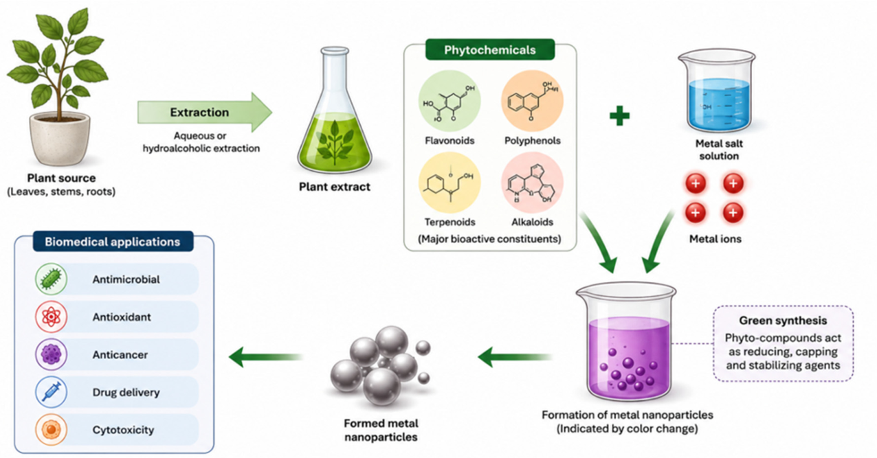
Phytochemical-mediated synthesis has been successful in producing various metal nanoparticles such as gold (Au), zinc oxide (ZnO), silver (Ag), copper (Cu), and more [109,110]. However, the molecular mechanisms behind the reduction and morphology control remain poorly explored. Most studies on phytochemical-mediated synthesis demonstrate nanoparticle formation and broadly link plant metabolites to reduction and stabilization without identifying the specific compounds or mechanisms that are responsible [106,111]. Hence, to bridge this gap in knowledge, metabolomics can be integrated with nanoparticle synthesis to unveil the bioactive compounds associated with the process. Metabolomics analysis of plant extracts can provide the plant’s profile, which could be linked to properties of nanoparticles and their biological functions.
Emerging evidence suggests that distinct metabolite classes influence nanoparticle morphology and function, as seen in Table 2. For example, flavonoids such as quercetin, myricetin, proanthocyanidin often yield small, spherical gold nanoparticles (AuNPs) with enhanced biological activities [127]. Current evidence exists, linking phytochemicals to nanoparticles across different studies, often using different plants, extraction methods, and characterization methods, and based on qualitative rather than quantitative data. This creates a major barrier to reproducibility and scalability [128,129]. Furthermore, very few studies have linked phytochemical composition directly to biological performance, leaving a gap in understanding how properties of plant-extract nanoparticles affect tumor uptake, cytotoxicity, or systemic toxicity [130]. In this regard, metabolomics offers a powerful, underexplored approach for identifying the specific metabolites that drive nanoparticle synthesis, stabilization, and biological activity. By coupling phytochemical diversity with advanced analytical platforms, metabolomics can advance green synthesis into a reproducible and clinically relevant strategy for cancer nanomedicine [131,132].
5. Integration of Metabolomics in Plant-Based Nanoparticle Synthesis
Plant extracts contain a diverse array of primary and secondary metabolites that regulate growth, development, and environmental response. This chemical diversity underpins their ability to act as reducing and stabilizing agents during nanoparticle synthesis, and functionalizing agents that influence biological activity after synthesis. Phytochemicals, including flavonoids, phenolic acids, tannins, terpenoids, alkaloids, proteins, sugars, and saponins, have been frequently implicated in nanoparticle formation. Despite their recognized importance, the phytochemical composition of plant extracts is usually treated as a ‘black box’, limiting mechanistic understanding and reproducibility. Most studies rely on indirect evidence from phytochemical screening and spectroscopic analyses that provide broad chemical classifications rather than molecular-level identification. Consequently, the field remains with a lack of comprehensive understanding of which phytochemicals participate in nanoparticle formation, become incorporated into the nanoparticle corona, or which of these metabolites influence physicochemical properties and biological activities. Metabolomics offers a powerful strategy to overcome the empirical nature of phytochemical-mediated nanoparticle synthesis by enabling comprehensive, untargeted profiling of plant metabolomes [133,134]. Defined as a systematic identification and quantification of low-molecular-weight metabolites within a biological system, metabolomics may be performed using targeted approaches to quantify predefined metabolites or untargeted approaches to profile metabolites without prior selection [131]. Within the context of nanoparticle synthesis, untargeted metabolomics is particularly valuable because it captures the extensive chemical diversity of plant extracts and generates global metabolic fingerprints that can be compared before and after nanoparticle formation [131,135]. By comparing metabolite profiles before and after synthesis, researchers can identify metabolites depleted during nanoparticle formation, determine compounds involved in reduction and capping, and establish a relationship between metabolite abundance and nanoparticle characteristics. By profiling plant metabolomes, metabolomics analysis can reveal the key reducing agents that reduce metal ions to elemental nanoparticles, stabilizing compounds that provide steric hindrance and electrostatic repulsion to prevent aggregation, and bioactive surface ligands that cap nanoparticles and provide them with biocompatibility and bioactivity [136,137]. Although this approach has been increasingly proposed in recent literature, its application remains uncommon in experimental studies. Additionally, metabolomics remains significantly underutilized in green nanotechnology, creating a major opportunity for studies that integrate untargeted metabolomics with nanoparticle characterization and biological activity assessment.
Untargeted metabolomics is commonly performed using liquid chromatography-mass spectrometry (LC-MS), gas chromatography-mass spectrometry (GC-MS), capillary electrophoresis-mass spectrometry (CE-MS), or nuclear magnetic resonance (NMR) spectroscopy. Among these platforms, LC-MS coupled with electrospray ionization (ESI) is the most widely used owing to its high sensitivity and broad metabolite coverage. However, no single analytical platform can capture the entire metabolome, and multi-platform strategies are increasingly adopted to improve metabolite detection [133]. The resulting datasets are large and complex and require preprocessing (peak detection, alignment, normalization) using software such as XCMS, MZmine, or MetAlign, followed by metabolite annotation via spectral libraries and databases including METLIN, NIST, and GOLM [138,139]. Multivariate analysis models such as principal component analysis (PCA), partial least squares regression (PLS), or orthogonal projection to latent structure (OPLS-DA) can be employed to identify associations between metabolite profiles and nanoparticle properties such as size, morphology, surface charge, and colloidal stability [140,141,142]. Metabolomics provides a high-throughput approach for investigating nanoparticle interactions with biological systems by elucidating synthesis mechanisms. Metabolic signatures can reveal cellular responses associated with oxidative stress, nanoparticle uptake, toxicity, and therapeutic activity. Since nanoparticle properties, including size, morphology, and stability, strongly influence cellular internalization and intracellular fate, metabolomic profiling can help explain the differences in biological responses elicited by nanoparticles with distinct physicochemical characteristics [143]. Consistent research results show that smaller NPs show higher cellular uptake and greater toxicity due to their high surface-to-volume ratio, causing greater metabolic disruption in cancer cells [144]. Whereas unstable nanoparticles often aggregate in biological media, lowering their toxicity and reducing their surface area [145]. Metabolomics can help to detect these biological responses by identifying metabolic signatures associated with nanoparticle exposure and ion-induced toxicity [86].
An additional challenge in plant-mediated synthesis is the considerable variability observed among plant species. Nanoparticles synthesized from different plants often exhibit distinct sizes, morphologies, and biological activities, despite being produced under similar synthesis conditions [146]. The idea that nanoparticle properties are not only determined by synthesis conditions but also by the metabolomic composition of the plant is widely acknowledged but rarely quantitatively tested. Plant species possess unique metabolomes, which can be decoded using metabolomics, to unveil how composition affects nanoparticle formation. Specific interactions and concentration determine the structural outcome of nanoparticles. The diversity in morphology, size, and stability of nanoparticles synthesized using different plant extracts may be partly explained by the difference in concentration and composition of phytochemicals. Phytochemicals have different binding affinities to nanoparticles, which dictate the final morphology, prevent agglomeration, and enhance colloidal stability [147]. As an example, plant extracts of Pelargonium graveolens have been used to form decahedral icosahedral-shaped nanoparticles measuring 20-40 nm in size. Whereas Cymbopogon flexuosus extracts form 0.05 -18 µm nanosphere and nanotriangles.
Nanoparticles adsorb phytochemicals on their surfaces, forming a layer called the bio-corona that influences their behavior in biological environments. The formation of a bio-corona modifies physicochemical properties, biodistribution, cellular interactions, and biological activity of nanoparticles [148]. Consequently, nanoparticles of similar size may exhibit markedly different biological activities owing to differences in surface chemistry. Given that the plant species is rich in bioactive compounds with inherent antimicrobial or antioxidant properties, these properties are then transferred and often amplified in the nanoparticle surface [149]. The newest direction in correlating metabolites with nanoparticle bioactivity involves linking the plant metabolome to nanoparticles’ physicochemical properties and then biological activity, rather than studying each component individually. Such approaches aim to establish relationships linking plant metabolomes to nanoparticle physicochemical properties and subsequent biological response (Figure 2). However, current evidence remains largely circumstantial, and relatively few studies have directly demonstrated these causal relationships through metabolomic modelling.
Conclusively, metabolomics offers considerable potential to improve mechanistic understanding, reproducibility, and standardization in phytochemical-mediated nanoparticle synthesis. By identifying metabolites involved in nanoparticle formation, tracking their depletion and transformation during synthesis, and linking metabolic composition to nanoparticle performance, metabolomics provides a foundation for developing more predictable and reproducible green nanotechnology platforms. The integration of metabolomics with nanoparticle characterization and biological activity assessment represents a significant opportunity for advancing the rational design of plant-derived nanoparticles for biomedical applications, including oncology.
6. Clinical Translation, Regulatory Consideration, and Safety Assessment
Despite the significant advances in phytochemical-mediated nanoparticle synthesis, clinical translation remains limited because of standardization, scalability and safety barriers [150]. While promising in pre-clinical settings, crude extract-based synthesis introduces batch-to-batch variability, while complex bio interactions raise toxicological concerns that may complicate regulatory approval [151]. Most studies focus on nanoparticle synthesis, physicochemical characterization, and in vitro biological activity, while relatively few evaluate pharmacokinetics, biodistribution, long-term toxicity, or regulatory compliance. Consequently, there remain substantial knowledge gaps regarding the safety and clinical applicability of plant-derived nanomaterials. The major challenge with clinical applicability is the comprehensive evaluation of nanoparticle toxicity. Although plant-derived nanomaterials are generally regarded as biocompatible, assessing their toxicity is uniquely challenging because it is influenced by multiple factors, including size, morphology, surface charge, composition, dissolution rate, and bio-corona formation [152,153].
Nanoparticle size determines cellular uptake and therapeutic activity, while metal ion release from metal oxide nanoparticles may contribute to their cytotoxicity through ion-mediated mechanisms that are independent of nanoparticle uptake [154]. Nanoparticles with smaller sizes often exhibit enhanced cellular uptake and therapeutic efficacy, but may also induce oxidative stress, mitochondrial dysfunction, DNA damage, and inflammatory responses due to their high surface area-to-volume ratio [155]. Regardless of their synthesis origin, a detailed toxicological evaluation remains essential for all phytochemical-mediated nanoparticles. Additionally, biodistribution and biological fate represent another barrier to clinical translation. The formation of a protein corona modifies their physicochemical identity, influencing their circulation time, cellular uptake, immune recognition, tissue accumulation, and clearance pathways [156]. While phytochemicals remain adsorbed onto nanoparticle surfaces during synthesis, forming an initial bio-corona, this layer may be disrupted or replaced by other biomolecules upon exposure to different physiological environments. This may result in an alteration in the biological behavior predicted by their initial physicochemical properties. Understanding the relationship between the phytochemical bio-corona and the acquired protein corona remains a critical yet underexplored area of research.
Regardless of whether a nanoparticle is synthesized through conventional or green routes, regulatory bodies such as the U.S. Food and Drug Administration (FDA) and the European Medicines Agency (EMA) require nanoparticles to adhere to a certain nanomedicine standard. They are evaluated not only based on their physicochemical properties but also on their intended function [157,158]. Both conventional and green nanomaterials face scrutiny regarding size, shape, surface charge, and particle agglomeration [159,160]. Because green synthesis relies on biological extracts, their inputs must be traced and standardized to avoid batch-to-batch variability and to ensure reproducible manufacture under Good Manufacturing Practice (GMP) conditions [161]. Regulators require rigorous preclinical and ADMET (absorption, distribution, metabolism, excretion, toxicity) profiling to prove that the drug delivery system does not cause unintended toxicity in vivo [162]. The current regulatory pathways were generally designed for conventional synthesis; the intrinsic variability of plant extracts complicates clinical translation. Integrating metabolomics into manufacturing workflows may provide a valuable quality assurance tool by generating comprehensive chemical fingerprints capable of monitoring extract consistency and identifying deviations that could affect nanoparticle performance [163]. Several nanoparticle-based therapeutics have successfully been clinically translated e.g., liposomal formulations, polymeric nanoparticles, and inorganic materials, yet clinical translation of plant-derived nanomaterials remains underexplored [164]. Most studies do not move beyond the proof-of-concept stage, with relatively few moving to pre-clinical evaluation and even fewer reaching clinical trials. Major challenges include limited mechanistic understanding, insufficient long-term safety data, manufacturing variability, and regulatory uncertainty [165,166]. In this context, metabolomics has the potential to facilitate the transition from empirically developed nanoparticles towards standardized, reproducible, and regulatory-compliant nanomaterials suitable for precision oncology and other biomedical applications.
7. Conclusion and Future Perspectives
Nanotechnology offers a transformative strategy for cancer therapy. The integration of nanoparticles and natural products has gained attention as an eco-friendlier, bio-compatible, and cost-effective approach to enhance the efficacy of current treatment modalities. Plants can be used as a bioresource for natural products because they are rich in bioactive compounds with pharmacological activities. Furthermore, these compounds have reducing and stabilizing effects, making them useful in nanoparticle synthesis. Among the various types of nanoparticles, metal nanoparticles have gained significant attention in nanomedicine due to their physicochemical properties, and they exhibit anticancer effects through various mechanisms. It is important to understand the interaction between plant extracts and metal during synthesis to aid in the large-scale production of nanoparticles and their subsequent biological functions for a more effective yet sustainable application in oncology. Metabolomics offers a powerful framework that decodes the phytochemistry of plants, enabling predictive and reproducible synthesis of nanoparticles, addressing the variability in green nanotechnology. The integration of nanotechnology with metabolomics offers a pathway toward precision oncology and holds promise for overcoming current limitations and advancing toward more effective and personalized treatments.
Author Contributions
N.V.M.: Conceptualization, writing and editing. T.T.: reviewing and editing. M.O.A.: supervision, reviewing and editing. F.T.: conceptualization, supervision, reviewing, editing, and project administration. All authors have read and agreed to the published version of the manuscript.
Funding
Not applicable.
Institutional Review Board Statement
Not applicable.
Informed Consent Statement
Not applicable.
Data Availability Statement
Not applicable.
Acknowledgments
During the preparation of this review article, AI tools were used to generate figures and to refine and correct the grammar of some content. The authors have reviewed and edited the output and take full responsibility for the content of this publication.
Conflicts of Interest
The authors declare no conflict of interest.
Abbreviations
The following abbreviations are used in this manuscript:
| WHO | World Health Organization |
| HIV | Human Immunodeficiency Virus |
| HPV | Human Papilloma Virus |
| NCR | National Cancer Registry |
| GCO | Global Cancer Observatory |
| pCR | Pathological complete response |
| ORR | Objective response rate |
| Au NP | Gold nanoparticle |
| MDR | Multidrug resistance |
| ZnO NP | Zinc oxide nanoparticle |
| ROS | Reactive oxygen species |
| EPR | Enhanced permeability and retention |
| Au | Gold |
| ZnO | Zinc oxide |
| Ag | Silver |
| LSPR | Localized surface plasmon resonance |
| Ag NP | Silver nanoparticle |
| Cu NP | Copper nanoparticle |
| LC-MS | Liquid chromatography-mass spectrometry |
| GC-MS | Gas chromatography-mass spectrometry |
| CE-MS | Capillary electrophoresis-mass spectrometry |
| NMR | Nuclear magnetic resonance |
| ESI | Electrospray ionization |
| PCA | Partial component analysis |
| PLS | Partial least squares |
| OPLS-DA | Orthogonal projection to latent structure discriminant analysis |
| FDA | Food and Drug Administration |
| EMA | European Medicines Agency |
| GMP | Good Manufacturing Practice |
| ADMET | Absorption, distribution, metabolism, excretion, toxicity |
References
- Gouws, C.; Smit, T.; Willers, C.; Svitina, H.; Calitz, C.; Wrzesinski, K. Anticancer potential of Sutherlandia frutescens and Xysmalobium undulatum in LS180 colorectal cancer mini-tumors. Molecules 2021, 26, 605. [Google Scholar] [CrossRef] [PubMed]
- Sung, H.; Ferlay, J.; Siegel, R.L.; Laversanne, M.; Soerjomataram, I.; Jemal, A.; Bray, F. Global cancer statistics 2020: GLOBOCAN estimates of incidence and mortality worldwide for 36 cancers in 185 countries. CA Cancer J. Clin. 2021, 71, 209–249. [Google Scholar] [CrossRef] [PubMed]
- Jin, C.; Wang, K.; Oppong-Gyebi, A.; Hu, J. Application of nanotechnology in cancer diagnosis and therapy: A mini review. Int. J. Med. Sci. 2020, 17, 2964–2973. [Google Scholar] [CrossRef] [PubMed]
- Misra, B.B.; van der Hooft, J.J.J. Updates in metabolomics tools and resources: 2014–2015. Electrophoresis 2016, 37, 86–110. [Google Scholar] [CrossRef] [PubMed]
- Gavas, S.; Quazi, S.; Karpinski, T.M. Nanoparticles for cancer therapy: Progress and challenges. Nanoscale Res. Lett. 2021, 16, 173. [Google Scholar] [CrossRef] [PubMed]
- Gmeiner, W.H.; Ghosh, S. Nanotechnology for cancer treatment. Nanotechnol. Revolut. 2014, 3, 111–122. [Google Scholar] [CrossRef]
- World Health Organization. Cancer; World Health Organization: Geneva, Switzerland, 2026; Available online: https://www.who.int/health-topics/cancer (accessed on 2 July 2026).
- Ferlay, J.; Colombet, M.; Soerjomataram, I.; Parkin, D.M.; Piñeros, M.; Znaor, A.; Bray, F. Cancer statistics for the year 2020: An overview. Int. J. Cancer 2021. [Google Scholar] [CrossRef] [PubMed]
- Peer, D.; Karp, J.M.; Hong, S.; Farokhzad, O.C.; Margalit, R.; Langer, R. Nanocarriers as an emerging platform for cancer therapy. Nat. Nanotechnol. 2007, 2, 751–760. [Google Scholar] [CrossRef] [PubMed]
- Duman, H.; Eker, F.; Akdaşçi, E.; Witkowska, A.M.; Bechelany, M.; Karav, S. Silver nanoparticles: A comprehensive review of synthesis methods and chemical and physical properties. Nanomaterials 2024, 14, 1527. [Google Scholar] [CrossRef] [PubMed]
- Cryer, A.M.; Thorley, A.J. Nanotechnology in the diagnosis and treatment of lung cancer. Pharmacol. Ther. 2019, 198, 189–205. [Google Scholar] [CrossRef] [PubMed]
- Pathakoti, K.; Manubolu, M.; Hwang, H. Nanotechnology applications for environmental industry. In Handbook of Nanomaterials for Industrial Applications; Elsevier: Amsterdam, The Netherlands, 2018; pp. 894–907. [Google Scholar]
- Fallatah, M.; Alradwan, I.; Al Fayez, N.; Aodah, A.H.; Alkhrayef, M.; Majrahi, M. Nanoparticles for cancer immunotherapy: Innovations and challenges. Pharmaceuticals 2025, 18, 1086. [Google Scholar] [CrossRef] [PubMed]
- Singh, P.; Pandit, S.; Bakusamy, S.R.; Madhusudanan, M.; Asmath Haseef, H.M.; Singh, H.; Mijakovic, I. Advanced nanomaterials for cancer therapy: Gold, silver, and iron oxide nanoparticles in oncological applications. Adv. Healthc. Mater. 2024, 14, 2403059. [Google Scholar] [CrossRef] [PubMed]
- Hanan, N.A.; Chiu, H.I.; Ramachandran, M.R.; Tung, W.H.; Mohamad Zain, N.N.; Yahaya, N.; Lim, V. Cytotoxicity of plant-mediated synthesis of metallic nanoparticles: A systematic review. Int. J. Mol. Sci. 2018, 19, 1725. [Google Scholar] [CrossRef] [PubMed]
- Soto, K.M.; Mendoza, S.; Lopez-Romero, J.M.; Gasca-Tirado, J.R.; Manzano-Ramirez, A. Gold nanoparticles: Synthesis, application in colon cancer therapy and new approaches. Green Chem. Lett. Rev. 2021, 14, 665–678. [Google Scholar] [CrossRef]
- Zuhrotun, A.; Oktaviani, D.J.; Hasanah, A.N. Biosynthesis of gold and silver nanoparticles using phytochemical compounds. Molecules 2023, 28, 3240. [Google Scholar] [CrossRef] [PubMed]
- Sawar, S.; Ali, S.; Khan, S.R.; Jamil, S.; Farooq, S.; Jafar, T.; Latif, M.J.; Shehroz, H. Marriage of green technology and chemistry: Bismuth-based nanoparticles as a remarkable combination. Environ. Technol. Rev. 2024, 13, 544–571. [Google Scholar] [CrossRef]
- Palumbo, M.O.; Kavan, P.; Miller, W.H.; Panasci, L.; Assouline, S.; Johnson, N.; Cohen, V.; Patenaude, F.; Pollak, M.; Jagoe, R.T.; Batist, G. Systemic cancer therapy: Achievements and challenges that lie ahead. Front. Pharmacol. 2013, 4, 57. [Google Scholar] [CrossRef] [PubMed]
- Zafar, A.; Khatoon, S.; Khan, M.J.; Abu, J.; Naeem, A. Advancements and limitations in traditional anti-cancer therapies: A comprehensive review of surgery, chemotherapy, radiation therapy, and hormonal therapy. Discov. Oncol. 2025, 16, 607. [Google Scholar] [CrossRef] [PubMed]
- Bray, F.; Laversanne, M.; Sung, H.; Ferlay, J.; Siegel, R.L.; Soerjomataram, I.; Jemal, A. Global cancer statistics 2022: GLOBOCAN estimates of incidence and mortality worldwide for 36 cancers in 185 countries. CA Cancer J. Clin. 2024, 74, 229–263. [Google Scholar] [CrossRef] [PubMed]
- Perez-Herrero, E.; Fernandez-Merino, A. Advanced targeted therapies in cancer: Drug nanocarriers and the future of chemotherapy. Eur. J. Pharm. Biopharm. 2015, 93, 52–79. [Google Scholar] [CrossRef] [PubMed]
- Sell, M.; Lopes, A.R.; Escudeiro, M.; Esteves, B.; Monteiro, A.R.; Trindade, T.; Cruz-Lopes, L. Application of nanoparticles in cancer treatment: A concise review. Nanomaterials 2023, 13, 2887. [Google Scholar] [CrossRef] [PubMed]
- Păduraru, D.N.; Ion, D.; Niculescu, A.G.; Mușat, F.; Andronic, O.; Grumezescu, A.M.; Bolocan, A. Recent developments in metallic nanomaterials for cancer therapy, diagnosis and imaging applications. Pharmaceutics 2022, 14, 435. [Google Scholar] [CrossRef] [PubMed]
- Delaney, G.; Jacob, S.; Featherstone, C.; et al. The role of radiotherapy in cancer treatment: Estimating optimal utilization from a review of evidence-based clinical guidelines. Cancer 2005, 104, 1129–1137. [Google Scholar] [CrossRef] [PubMed]
- Baumann, R.; Depping, R.; Delaperriere, M.; Dunst, J. Targeting hypoxia to overcome radiation resistance in head and neck cancers: Real challenge or clinical fairytale? Expert Rev. Anticancer Ther. 2016, 16, 751–758. [Google Scholar] [CrossRef] [PubMed]
- Carozza, J.A.; Böhnert, V.; Nguyen, K.C.; et al. Extracellular cGAMP is a cancer-cell-produced immunotransmitter involved in radiation-induced anticancer immunity. Nat. Cancer 2020, 1, 184–196. [Google Scholar] [CrossRef] [PubMed]
- Baskar, R.; Lee, K.A.; Yeo, R.; Yeoh, K.W. Cancer and radiation therapy: Current advances and future directions. Int. J. Med. Sci. 2012, 9, 193–199. [Google Scholar] [CrossRef] [PubMed]
- Schuemann, J.; Bagley, A.F.; Berbeco, R.; et al. Roadmap for metal nanoparticles in radiation therapy: Current status, translational challenges, and future directions. Phys. Med. Biol. 2020, 65, 21RM02. [Google Scholar] [CrossRef] [PubMed]
- Zhou, R.; Zhao, D.; Beeraka, N.M.; et al. Novel implications of nanoparticle-enhanced radiotherapy and brachytherapy: Z-effect and tumor hypoxia. Metabolites 2022, 12, 943. [Google Scholar] [CrossRef] [PubMed]
- Fan, W.; Shen, B.; Bu, W.; et al. Intranuclear biophotonics by smart design of nuclear-targeting photo-/radio-sensitizers co-loaded upconversion nanoparticles. Biomaterials 2015, 69, 89–98. [Google Scholar] [CrossRef] [PubMed]
- Zhang, P.; Qiao, Y.; Wang, C.; Ma, L.; Su, M. Enhanced radiation therapy with internalized polyelectrolyte-modified nanoparticles. Nanoscale 2014, 6, 10095–10099. [Google Scholar] [CrossRef] [PubMed]
- Jackson, N.; Cecchi, D.; Beckham, W.; Chithrani, D.B. Application of high-Z nanoparticles to enhance current radiotherapy treatment. Molecules 2024, 29, 2438. [Google Scholar] [CrossRef] [PubMed]
- Heidari, A.; Mohammadi, J.; Ghodousi, M.; et al. Metal-based nanoparticles in cancer treatment: Lessons learned and challenges. Front. Bioeng. Biotechnol. 2024, 12, 1436297. [Google Scholar] [CrossRef] [PubMed]
- Maier-Hauff, K.; Ulrich, F.; Nestler, D.; et al. Efficacy and safety of intratumoral thermotherapy using magnetic iron oxide nanoparticles combined with external beam radiotherapy in patients with recurrent glioblastoma multiforme. J. Neurooncol. 2011, 103, 317–324. [Google Scholar] [CrossRef] [PubMed]
- Maggiorella, L.; Barouch, G.; Devaux, C.; et al. Nanoscale radiotherapy with hafnium oxide nanoparticles. Future Oncol. 2012, 8, 1167–1181. [Google Scholar] [CrossRef] [PubMed]
- Zhang, P.; Darmon, A.; Marill, J.; Mohamed Anesary, N.; Paris, S. Radiotherapy-activated hafnium oxide nanoparticles produce an abscopal effect in a mouse colorectal cancer model. Int. J. Nanomed. 2020, 15, 3843–3850. [Google Scholar] [CrossRef] [PubMed]
- Bonvalot, S.; Rutkowski, P.L.; Thariat, J.; et al. NBTXR3, a first-in-class radioenhancer hafnium oxide nanoparticle, plus radiotherapy versus radiotherapy alone in patients with locally advanced soft-tissue sarcoma (Act.In.Sarc): A multicentre, phase II–III, randomized controlled trial. Lancet Oncol. 2019, 20, 1148–1159. [Google Scholar] [CrossRef] [PubMed]
- Liu, Y.; Liu, X.; Jin, X.; et al. The dependence of radiation enhancement effect on the concentration of gold nanoparticles exposed to low- and high-LET radiations. Phys. Med. 2015, 31, 210–218. [Google Scholar] [CrossRef] [PubMed]
- Liu, X.; Liu, Y.; Zhang, P.; et al. The synergistic radiosensitizing effect of tirapazamine-conjugated gold nanoparticles on human hepatoma HepG2 cells under X-ray irradiation. Int. J. Nanomed. 2016, 11, 3517–3531. [Google Scholar] [CrossRef] [PubMed]
- Lowenthal, R.M.; Eaton, K. Toxicity of chemotherapy. Hematol. Oncol. Clin. North Am. 1996, 10, 967–990. [Google Scholar] [CrossRef] [PubMed]
- Morgan, G.; Ward, R.; Barton, M. The contribution of cytotoxic chemotherapy to 5-year survival in adult malignancies. Clin. Oncol. 2004, 16, 549–560. [Google Scholar] [CrossRef] [PubMed]
- Faller, B.A.; Pandit, T.N. Safety and efficacy of vinorelbine in the treatment of non-small cell lung cancer. Clin. Med. Insights Oncol. 2011, 5, 131–144. [Google Scholar] [CrossRef] [PubMed]
- Funaguchi, N.; Iihara, H.; Kaito, D.; Gomyo, T.; Sasaki, Y.; Yanase, K.; Endo, J.; Ito, F.; Hirose, C.; Ohno, Y.; Okura, H. Efficacy of cisplatin plus vinorelbine adjuvant chemotherapy with split-dose administration of cisplatin after complete resection of stage II–IIIA non-small cell lung cancer. Mol. Clin. Oncol. 2022, 16, 76. [Google Scholar] [CrossRef] [PubMed]
- Gurunathan, S.; Kang, M.H.; Qasim, M.; Kim, J.H. Nanoparticle-mediated combination therapy: Two-in-one approach for cancer. Int. J. Mol. Sci. 2018, 19, 3264. [Google Scholar] [CrossRef] [PubMed]
- Roshani, M.; Rezaian-Isfahni, A.; Lotfalizadeh, M.H.; Khassafi, N.; Abadi, M.H.J.N.; Nejati, M. Metal nanoparticles as a potential technique for the diagnosis and treatment of gastrointestinal cancer: A comprehensive review. Cancer Cell Int. 2023, 23, 280. [Google Scholar] [CrossRef] [PubMed]
- Ammar, M.M.; Ali, R.; Abd Elaziz, N.A.; Habib, H.; Abbas, F.M.; Yassin, M.T.; Maniah, K.; Abdelaziz, R. Nanotechnology in oncology: Advances in biosynthesis, drug delivery, and theranostics. Discov. Oncol. 2025, 16, 1172. [Google Scholar] [CrossRef] [PubMed]
- Meng, H.; Wang, M.; Liu, H.; Liu, X.; Situ, A.; Wu, B.; Ji, Z.; Chang, C.H.; Nel, A.E. Use of a lipid-coated mesoporous silica nanoparticle platform for synergistic gemcitabine and paclitaxel delivery to human pancreatic cancer in mice. ACS Nano 2015, 9, 3540–3557. [Google Scholar] [CrossRef] [PubMed]
- Liu, K.; Chen, W.; Yang, T.; Wen, B.; Ding, D.; Keidar, M.; Tang, J.; Zhang, W. Paclitaxel and quercetin nanoparticles co-loaded in microspheres to prolong retention time for pulmonary drug delivery. Int. J. Nanomed. 2017, 12, 8239–8255. [Google Scholar] [CrossRef] [PubMed]
- Farkona, S.; Diamandis, E.P.; Blasutig, I.M. Cancer immunotherapy: The beginning of the end of cancer? BMC Med. 2016, 14, 73. [Google Scholar] [CrossRef] [PubMed]
- Tan, S.; Li, D.; Zhu, X. Cancer immunotherapy: Pros, cons and beyond. Biomed. Pharmacother. 2020, 124, 109821. [Google Scholar] [CrossRef] [PubMed]
- Siddiqui, M.A.; Usmani, A.; Ansari, M.N.; Almoselhy, R.I.M. Immune-related adverse events in immunotherapy: Challenges in diagnosis, monitoring, and management. Toxicol. Rep. 2025, 14, 102036. [Google Scholar] [CrossRef] [PubMed]
- Haase, H. Innate immune cells speak manganese. Immunity 2018, 48, 616–618. [Google Scholar] [CrossRef] [PubMed]
- Wang, L.; Lin, D.; Li, M.; Jiang, Y.; Yang, Y.; Wang, H.; Chu, H.; Ye, J.; Liu, Y. Bioactive metallic nanoparticles for synergistic cancer immunotherapy. Acta Pharm. Sin. B 2025, 15, 1869–1911. [Google Scholar] [CrossRef] [PubMed]
- Sun, X.; Zhang, Y.; Li, J.; Park, K.S.; Han, K.; Zhou, X.; Xu, Y.; Nam, J.; Xu, J.; Shi, X.; Wei, L. Amplifying STING activation by cyclic dinucleotide–manganese particles for local and systemic cancer metalloimmunotherapy. Nat. Nanotechnol. 2021, 16, 1260–1270. [Google Scholar] [CrossRef] [PubMed]
- Wang, A.Z.; Langer, R.; Farokhzad, O.C. Nanoparticle delivery of cancer drugs. Annu. Rev. Med. 2012, 63, 185–198. [Google Scholar] [CrossRef] [PubMed]
- Tseriotis, V.S.; Ampazis, D.; Karachrysafi, S.; Papamitsou, T.; Petrakis, G.; Kouvelas, D.; Mavropoulos, P.; Lallas, K.; Sič, A.; Fouskas, V.; Stergiou, K.; Pavlidis, P.; Arnaoutoglou, M. ZnO-based nanoparticles for targeted cancer chemotherapy and the role of the tumor microenvironment: A systematic review. Int. J. Mol. Sci. 2025, 26, 8417. [Google Scholar] [CrossRef] [PubMed]
- Zanganeh, S.; Hutter, G.; Spitler, R.; Lenkov, O.; Mahmoudi, M.; Shaw, A.; Pajarinen, J.S.; Nejadnik, H.; Goodman, S.; Moseley, M.; Coussens, L.M.; Daldrup-Link, H.E. Iron oxide nanoparticles inhibit tumour growth by inducing pro-inflammatory macrophage polarization in tumour tissues. Nat. Nanotechnol. 2016, 11, 986–994. [Google Scholar] [CrossRef] [PubMed]
- Zhang, Y.; Nayak, T.R.; Hong, H.; Cai, W. Biomedical applications of zinc oxide nanomaterials. Curr. Mol. Med. 2013, 13, 1633–1645. [Google Scholar] [CrossRef] [PubMed]
- Zhao, J.; Zhang, Z.; Xue, Y.; Wang, G.; Cheng, Y.; Pan, Y.; Zhao, S.; Hou, Y. Anti-tumor macrophages activated by ferumoxytol combined or surface-functionalized with the TLR3 agonist poly(I:C) promote melanoma regression. Theranostics 2018, 8, 6307–6321. [Google Scholar] [CrossRef] [PubMed]
- Chung, I.M.; Abdul Rahuman, A.; Marimuthu, S.; Kirthi, A.V.; Anbarasan, K.; Padmini, P.; Rajakumar, G. Green synthesis of copper nanoparticles using Eclipta prostrata leaf extract and their antioxidant and cytotoxic activities. Exp. Ther. Med. 2017, 14, 18–24. [Google Scholar] [CrossRef] [PubMed]
- Al-Samydai, A.; Abu Hajleh, M.N.; Al-Sahlawi, F.; Nsairat, H.; Khatib, A.A.; Alqaraleh, M.; Ibrahim, A.K. Advancements of metallic nanoparticles: A promising frontier in cancer treatment. Sci. Prog. 2024, 107, 368504241274967. [Google Scholar] [CrossRef] [PubMed]
- Abdal Dayem, A.; Hossain, M.K.; Lee, S.B.; Kim, K.; Saha, S.K.; Yang, G.M.; Choi, H.Y.; Cho, S.G. The role of reactive oxygen species (ROS) in the biological activities of metallic nanoparticles. Int. J. Mol. Sci. 2017, 18, 120. [Google Scholar] [CrossRef] [PubMed]
- Čapek, J.; Roušar, T. Detection of oxidative stress induced by nanomaterials in cells: The roles of reactive oxygen species and glutathione. Molecules 2021, 26, 4710. [Google Scholar] [CrossRef] [PubMed]
- Mayordomo, N.M.; Zatarain-Beraza, A.; Valerio, F.; Álvarez-Méndez, V.; Turegano, P.; Herranz-García, L.; López de Aguileta, A.; Cattani, N.; Álvarez-Alonso, A.; Fanarraga, M.L. The protein corona paradox: Challenges in achieving true biomimetics in nanomedicines. Biomimetics 2025, 10, 276. [Google Scholar] [CrossRef] [PubMed]
- Cui, G.; Wu, J.; Lin, J.; et al. Graphene-based nanomaterials for breast cancer treatment: Promising therapeutic strategies. J. Nanobiotechnol. 2021, 19, 211. [Google Scholar] [CrossRef] [PubMed]
- Alrushaid, N.; Khan, F.A.; Al-Suhaimi, E.A.; Elaissari, A. Nanotechnology in cancer diagnosis and treatment. Pharmaceutics 2023, 15, 1025. [Google Scholar] [CrossRef] [PubMed]
- Yadav, R.; Das, P.P.; Sharma, S.; Sengupta, S.; Kumar, D.; Sagar, R. Recent advancement of nanomedicine-based targeted delivery for cervical cancer treatment. Med. Oncol. 2023, 40, 347. [Google Scholar] [CrossRef] [PubMed]
- Zhang, Y.; Li, M.; Gao, X.; Chen, Y.; Liu, T. Nanotechnology in cancer diagnosis: Progress, challenges and opportunities. J. Hematol. Oncol. 2019, 12, 137. [Google Scholar] [CrossRef] [PubMed]
- Hasan, M.; Evett, C.G.; Burton, J. Advances in nanoparticle-based targeted drug delivery systems for colorectal cancer: A review. arXiv 2024, arXiv:2409.05222. [Google Scholar] [CrossRef]
- Liu, J.; Wu, T.; Lu, X.; Wu, X.; Liu, S.; Zhao, S.; Xu, X.; Ding, B. A self-assembled platform based on branched DNA for sgRNA/Cas9/antisense delivery. J. Am. Chem. Soc. 2019, 141, 19032–19037. [Google Scholar] [CrossRef] [PubMed]
- Lok, C.N.; Zou, T.; Zhang, J.J.; Lin, I.W.S.; Che, C.M. Controlled-release systems for metal-based nanomedicine: Encapsulated/self-assembled nanoparticles of anticancer gold(III)/platinum(II) complexes and antimicrobial silver nanoparticles. Adv. Mater. 2014, 26, 5550–5557. [Google Scholar] [CrossRef] [PubMed]
- Pissuwan, D.; Valenzuela, S.M.; Cortie, M.B. Therapeutic possibilities of plasmonically heated gold nanoparticles. Trends Biotechnol. 2006, 24, 62–67. [Google Scholar] [CrossRef] [PubMed]
- Singh, D.; Kumar, A. Next-generation nanocarriers for precision antitumor therapy: From passive targeting to intelligent response. Explor. Target Antitumor Ther. 2025, 6, 1002355. [Google Scholar] [CrossRef] [PubMed]
- Farzin, A.; Etesami, S.A.; Quint, J.; Memic, A.; Tamayol, A. Magnetic nanoparticles in cancer therapy and diagnosis. Adv. Healthc. Mater. 2020, 9, 1901058. [Google Scholar] [CrossRef] [PubMed]
- Xu, J.J.; Zhang, W.C.; Guo, Y.W.; Chen, X.Y.; Zhang, Y.N. Metal nanoparticles as a promising technology in targeted cancer treatment. Drug Deliv. 2022, 29, 664–678. [Google Scholar] [CrossRef] [PubMed]
- Hervault, A.; Thanh, N.T.K. Magnetic nanoparticle-based therapeutic agents for thermo-chemotherapy treatment of cancer. Nanoscale 2014, 6, 11553–11573. [Google Scholar] [CrossRef] [PubMed]
- Jose, J.; Kumar, R.; Harilal, S.; Mathew, G.E.; Parambi, D.G.T.; Prabhu, A.; et al. Magnetic nanoparticles for hyperthermia in cancer treatment: An emerging tool. Environ. Sci. Pollut. Res. 2020, 27, 19214–19225. [Google Scholar] [CrossRef] [PubMed]
- Suto, M.; Hirota, Y.; Mamiya, H.; Fujita, A.; Kasuya, R.; Tohji, K.; Jeyadevan, B. Heat dissipation mechanism of magnetite nanoparticles in magnetic fluid hyperthermia. J. Magn. Magn. Mater. 2009, 321, 1493–1496. [Google Scholar] [CrossRef]
- Al-Musywel, H.A.; Laref, A. Effect of gold nanoparticles on radiation doses in tumor treatment: A Monte Carlo study. Lasers Med. Sci. 2017, 32, 2073–2080. [Google Scholar] [CrossRef] [PubMed]
- Jain, S.; Hirst, D.G.; O’Sullivan, J.M. Gold nanoparticles as novel agents for cancer therapy. Br. J. Radiol. 2012, 85, 101–113. [Google Scholar] [CrossRef] [PubMed]
- Mu, S.; Yang, H.; Wang, S.; Tong, A.; Ding, R.; Wang, J.; Wang, D.; Li, J. Zinc-based nanomaterials in cancer therapy: Mechanisms, applications, and future directions. Theranostics 2025, 15, 7841–7871. [Google Scholar] [CrossRef] [PubMed]
- Badir, A.; Refki, S.; Sekkat, Z. Utilizing gold nanoparticles in plasmonic photothermal therapy for cancer treatment. Heliyon 2025, 11, e42738. [Google Scholar] [CrossRef] [PubMed]
- Chen, Y.; Yang, J.; Fu, S.; Wu, J. Gold nanoparticles as radiosensitizers in cancer radiotherapy. Int. J. Nanomed. 2020, 15, 9407–9430. [Google Scholar] [CrossRef] [PubMed]
- Bai, X.; Wang, Y.; Song, Z.; Feng, Y.; Chen, Y.; Zhang, D.; Feng, L. The basic properties of gold nanoparticles and their applications in tumor diagnosis and treatment. Int. J. Mol. Sci. 2020, 21, 2480. [Google Scholar] [CrossRef] [PubMed]
- Mikhailova, E.O. Silver nanoparticles: Mechanism of action and probable bio-application. J. Funct. Biomater. 2020, 11, 84. [Google Scholar] [CrossRef] [PubMed]
- Mostafavi, E.; Zarepour, A.; Barabadi, H.; Zarrabi, A.; Truong, L.B.; Medina-Cruz, D. Antineoplastic activity of biogenic silver and gold nanoparticles to combat leukemia: Beginning a new era in cancer theragnostic. Biotechnol. Rep. 2022, 34, e00714. [Google Scholar] [CrossRef] [PubMed]
- Liao, C.; Li, Y.; Tjong, S.C. Bactericidal and cytotoxic properties of silver nanoparticles. Int. J. Mol. Sci. 2019, 20, 449. [Google Scholar] [CrossRef] [PubMed]
- Ferdous, Z.; Nemmar, A. Health impact of silver nanoparticles: A review of the biodistribution and toxicity following various routes of exposure. Int. J. Mol. Sci. 2020, 21, 2375. [Google Scholar] [CrossRef] [PubMed]
- Wong, T.Y.; Wang, Y.; Kwan, K.K.L.; Pan, Y.; Lai, A.K.L.; Chen, S.; Xiao, Y.; Zhou, K.; Wu, L.; Huo, S.; Yan, N.; Lam, H. Exploring the potentials of silver nanoparticles in overcoming cisplatin resistance in lung adenocarcinoma: Insights from proteomic and xenograft mouse studies. ACS Nano 2025, 19, 34708–34723. [Google Scholar] [CrossRef] [PubMed]
- He, C.; Hu, Y.; Yin, L.; Tang, C.; Yin, C. Effects of particle size and surface charge on cellular uptake and biodistribution of polymeric nanoparticles. Biomaterials 2010, 31, 3657–3666. [Google Scholar] [CrossRef] [PubMed]
- Anjum, S.; Hashim, M.; Malik, S.A.; Khan, M.; Lorenzo, J.M.; Abbasi, B.H.; Hano, C. Recent advances in zinc oxide nanoparticles (ZnO NPs) for cancer diagnosis, targeted drug delivery, and treatment. Cancers 2021, 13, 4570. [Google Scholar] [CrossRef] [PubMed]
- Hu, C.; Du, W. Zinc oxide nanoparticles (ZnO NPs) combined with cisplatin and gemcitabine inhibit tumor activity of NSCLC cells. Aging 2020, 12, 25767–25777. [Google Scholar] [CrossRef] [PubMed]
- Zhang, P.; Qiao, Y.; Wang, C.; Ma, L.; Su, M. Enhanced radiation therapy with internalized polyelectrolyte-modified nanoparticles. Nanoscale 2014, 6, 10095–10099. [Google Scholar] [CrossRef] [PubMed]
- Tseriotis, V.-S.; Ampazis, D.; Karachrysafi, S.; Papamitsou, T.; Petrakis, G.; Kouvelas, D.; Mavropoulos, P.; Lallas, K.; Sič, A.; Fouskas, V.; Stergiou, K.; Pavlidis, P.; Arnaoutoglou, M. ZnO-based nanoparticles for targeted cancer chemotherapy and the role of tumor microenvironment: A systematic review. Int. J. Mol. Sci. 2025, 26, 8417. [Google Scholar] [CrossRef] [PubMed]
- Zarrinnahad, H.; Dehdast, S.A.; Fard, G.C.; Nourbakhsh, M.; Koohi, M.K.; Panahi, G.; Karimpour, A.; Rezayat, S.M.; Shabani, M. The effect of biosynthesized zinc oxide nanoparticles on gene expression and apoptosis in triple-negative breast cancer cells. DARU J. Pharm. Sci. 2024, 33, 10. [Google Scholar] [CrossRef] [PubMed]
- Catalano, E.; Di Benedetto, A. Characterization of physicochemical and colloidal properties of hydrogel chitosan-coated iron oxide nanoparticles for cancer therapy. J. Phys. Conf. Ser. 2017, 841, 012010. [Google Scholar] [CrossRef]
- Chaturvedi, V.K.; Sharma, B.; Tripathi, A.D.; Yadav, D.P.; Singh, K.R.; Singh, J.; Singh, R.P. Biosynthesized nanoparticles: A novel approach for cancer therapeutics. Front. Med. Technol. 2023, 5, 1236107. [Google Scholar] [CrossRef] [PubMed]
- Ahmed, S.; Ahmad, M.; Swami, B.L.; Ikram, S. A review on plants extract mediated synthesis of silver nanoparticles for antimicrobial applications: A green expertise. J. Adv. Res. 2016, 7, 17–28. [Google Scholar] [CrossRef] [PubMed]
- Balogun, F.O.; Ashafa, A.O.T. Potentials of synthesized Lessertia montana zinc oxide nanoparticles on free radical-mediated oxidative stress and carbohydrate-hydrolysing enzymes. Acta Biol. Szeged. 2020, 64, 239–249. [Google Scholar]
- Ovais, M.; Khalil, A.T.; Islam, N.U.; Ahmad, I.; Ayaz, M.; Saravanan, M.; Shinwari, Z.K.; Mukherjee, S. Role of plant phytochemicals and microbial enzymes in biosynthesis of metallic nanoparticles. Appl. Microbiol. Biotechnol. 2018, 102, 6799–6814. [Google Scholar] [CrossRef] [PubMed]
- Khalil, A.T.; Ovais, M.; Ullah, I.; Ali, M.; Shinwari, Z.K.; Hassan, D.; Maaza, M. Sageretia thea (Osbeck.)-mediated biosynthesis of NiO nanoparticles and their in vitro pharmacognostic, antioxidant, and cytotoxic potential. Artif. Cells Nanomed. Biotechnol. 2018, 46, 838–852. [Google Scholar] [PubMed]
- Naseer, M.; Aslam, U.; Khalid, B.; Chen, B. Green route to synthesize zinc oxide nanoparticles using leaf extracts of Cassia fistula and Melia azedarach and their antibacterial potential. Sci. Rep. 2020, 10, 9055. [Google Scholar] [PubMed]
- Vijayaraghavan, K.; Ashokkumar, T. Plant-mediated biosynthesis of metallic nanoparticles: A review of literature, factors affecting synthesis, characterization techniques, and applications. J. Environ. Chem. Eng. 2017, 5, 4866–4883. [Google Scholar] [CrossRef]
- Ogunyemi, S.O.; Abdallah, Y.; Zhang, M.; Fouad, H.; Hong, X.; Ibrahim, E.; Masum, M.M.I.; Hossain, A.; Mo, J.; Li, B. Green synthesis of zinc oxide nanoparticles using different plant extracts and their antibacterial activity against Xanthomonas oryzae pv. oryzae. Artif. Cells Nanomed. Biotechnol. 2019, 47, 341–352. [Google Scholar] [CrossRef] [PubMed]
- Ayub, A.; Wani, A.K.; Malik, S.M.; Ayub, M.; Singh, R.; Chopra, C.; Malik, T. Green nanoscience for healthcare: Advancing biomedical innovation through eco-synthesized nanoparticles. Biotechnol. Rep. 2025, 47, e00913. [Google Scholar] [CrossRef] [PubMed]
- Bhatt, S.; Saraswat, S. A review on phytochemical-mediated synthesis of nanoparticles through fruit and vegetable extracts and their potential applications. Nanotechnol. Environ. Eng. 2024, 9, 359–374. [Google Scholar] [CrossRef]
- Thomas, S.; Gonsalves, R.A.; Jose, J.; Zyoud, S.H.; Prasad, A.R.; Garvasis, J. Plant-based synthesis, characterization approaches, applications, and toxicity of silver nanoparticles: A comprehensive review. J. Biotechnol. 2024, 394, 135–149. [Google Scholar] [CrossRef] [PubMed]
- Mittal, A.K.; Chisti, Y.; Banerjee, U.C. Synthesis of metallic nanoparticles using plant extracts. Biotechnol. Adv. 2013, 31, 346–356. [Google Scholar] [CrossRef] [PubMed]
- Alhadrami, H.A.; Orfali, R.; Hamed, A.A.; Ghoneim, M.M.; Hassan, H.M.; Hassane, A.S.I.; Rateb, M.E.; Sayed, A.M.; Gamaleldin, N.M. Flavonoid-coated gold nanoparticles as efficient antibiotics against Gram-negative bacteria: Evidence from in silico-supported in vitro studies. Antibiotics 2021, 10, 968. [Google Scholar] [CrossRef] [PubMed]
- Alkhulaifi, M.M.; Alshehri, J.H.; Alwehaibi, M.A.; Awad, M.A.; Al-Enazi, N.M.; Aldosari, N.S.; Hatamleh, A.A.; Abdel-Raouf, N. Green synthesis of silver nanoparticles using Citrus limon peels and evaluation of their antibacterial and cytotoxic properties. Saudi J. Biol. Sci. 2020, 27, 3434–3441. [Google Scholar] [CrossRef] [PubMed]
- Babu, P.J.; Sharma, P.; Borthakur, B.B.; Das, R.K.; Nahar, P.; Bora, U. Synthesis of gold nanoparticles using Mentha arvensis leaf extract. Int. J. Green Nanotechnol. 2010, 2, 62–68. [Google Scholar] [CrossRef]
- Baishya, D.; Sharma, N.; Bora, R. Green synthesis of silver nanoparticles using Bryophyllum pinnatum (Lam.) and evaluation of their antibacterial activity. Arch. Appl. Sci. Res. 2012, 4, 2098–2104. [Google Scholar] [CrossRef]
- Jiménez-Rosado, M.; Gomez-Zavaglia, A.; Guerrero, A.; Romero, A. Green synthesis of ZnO nanoparticles using polyphenol extracts from pepper waste (Capsicum annuum). J. Clean. Prod. 2022, 350, 131541. [Google Scholar] [CrossRef]
- Liu, Y.S.; Chang, Y.C.; Chen, H.H. Silver nanoparticle biosynthesis using phenolic acids in rice husk extract as reducing agents and dispersants. J. Food Drug Anal. 2018, 26, 649–656. [Google Scholar] [CrossRef] [PubMed]
- Solís-Sandí, I.; Cordero-Fuentes, S.; Pereira-Reyes, R.; Vega-Baudrit, J.R.; Batista-Menezes, D.; Montes de Oca-Vásquez, G. Optimization of the biosynthesis of silver nanoparticles using bacterial extracts and their antimicrobial potential. Biotechnol. Rep. 2023, 40, e00816. [Google Scholar] [CrossRef] [PubMed]
- Melnikova, N.; Balakireva, A.; Orekhov, D.; Kamorin, D.; Didenko, N.; Malygina, D.; Knyazev, A.; Novopoltsev, D.; Solovyeva, A. Zinc oxide nanoparticles protected with terpenoids as a substance in redox imbalance normalization in burns. Pharmaceuticals 2021, 14, 492. [Google Scholar] [CrossRef] [PubMed]
- Nouren, S.; Bibi, I.; Kausar, A.; Sultan, M.; Bhatti, H.N.; Safa, Y.; Sadaf, S.; Alwadai, N.; Iqbal, M. Green synthesis of CuO nanoparticles using Jasminum sambac extract: Condition optimization and photocatalytic degradation of methylene blue dye. J. King Saud. Univ. Sci. 2024, 36, 103089. [Google Scholar] [CrossRef]
- Iravani, S. Green synthesis of metal nanoparticles using plants. Green Chem. 2011, 13, 2638–2650. [Google Scholar] [CrossRef]
- Pisitsak, P.; Chamchoy, K.; Chinprateep, V.; Khobthong, W.; Chitichotpanya, P.; Ummartyotin, S. Synthesis of gold nanoparticles using tannin-rich extract and coating onto cotton textiles for catalytic degradation of Congo red. J. Nanotechnol. 2021, 2021, 6380283. [Google Scholar] [CrossRef]
- Abdel-Aty, A.M.; Barakat, A.Z.; Bassuiny, R.I.; Mohamed, S.A. Statistical optimization, characterization, antioxidant, and antibacterial properties of silver nanoparticles biosynthesized using saw palmetto seed phenolic extract. Sci. Rep. 2023, 13, 15605. [Google Scholar] [CrossRef] [PubMed]
- Nagesh, M.R.; Kumar, N.; Khan, J.M.; Ahmed, M.Z.; Kavitha, R.; Kim, S.-J.; Vijayakumar, N. Green synthesis and pharmacological applications of silver nanoparticles using ethanolic extract of Salacia chinensis L. J. King Saud. Univ. Sci. 2022, 34, 102284. [Google Scholar] [CrossRef]
- Faisal, S.; Al-Radaddi, N.; Jan, H.; Shah, S.A.; Shah, S.; Rizwan, M.; Afsheen, Z.; Hussain, Z.; Uddin, M.N.; Idrees, M.; Bibi, N. Curcuma longa-mediated synthesis of copper oxide, nickel oxide and Cu–Ni bimetallic hybrid nanoparticles: Characterization and evaluation for antimicrobial, antiparasitic and cytotoxic potentials. Coatings 2021, 11, 849. [Google Scholar] [CrossRef]
- Hendi, A.A.; Awad, M.A.; Alanazi, M.M.; Virk, P.; Alrowaily, A.W.; Bahlool, T.; Merghan, N.M.; Aouaini, F.; Hagmusa, B. Phytomediated synthesis of bimetallic Ag/Au nanoparticles using orange peel extract and assessment of their antibacterial and anticancer potential. J. King Saud. Univ. Sci. 2023, 35, 102510. [Google Scholar] [CrossRef]
- Das, D.K.; Chakraborty, A.; Bhattacharjee, S.; Dey, S. Biosynthesis of stabilized gold nanoparticles using the aglycone flavonoid quercetin. J. Exp. Nanosci. 2013, 8, 649–655. [Google Scholar]
- Khan, F.; Shariq, M.; Asif, M.; Siddiqui, M.A.; Malan, P.; Ahmad, F. Green nanotechnology: Plant-mediated nanoparticle synthesis and application. Nanomaterials 2022, 12, 673. [Google Scholar] [CrossRef] [PubMed]
- Singh, J.; Dutta, T.; Kim, K.H.; Rawat, M.; Samddar, P.; Kumar, P. Green synthesis of metals and their oxide nanoparticles: Applications for environmental remediation. J. Nanobiotechnol. 2018, 16, 84. [Google Scholar] [CrossRef] [PubMed]
- Singh, H.; Desimone, M.F.; Pandya, S.; Jasani, S.; George, N.; Adnan, M.; Aldarhami, A.S.; Bazaid, A.S.; Alderhami, S.A. Revisiting the green synthesis of nanoparticles: Uncovering influences of plant extracts as reducing agents for enhanced synthesis efficiency and biomedical applications. Int. J. Nanomed. 2023, 18, 4727–4750. [Google Scholar] [CrossRef] [PubMed]
- Silva, S.; Dias, M.C.; Pinto, D.C.G.A.; Silva, A.M.S. Metabolomics as a tool to understand nano-plant interactions: The case study of metal-based nanoparticles. Plants 2023, 12, 491. [Google Scholar] [CrossRef] [PubMed]
- Dabbousy, R.; Rima, M.; Roufayel, R.; Rahal, M.; Legros, C.; Sabatier, J.-M.; Fajloun, Z. Plant metabolomics: The future of anticancer drug discovery. Pharmaceuticals 2024, 17, 1307. [Google Scholar] [CrossRef] [PubMed]
- Hong, J.; Yang, L.; Zhang, D.; Shi, J. Plant metabolomics: An indispensable systems biology tool for plant science. Int. J. Mol. Sci. 2016, 17, 767. [Google Scholar] [CrossRef] [PubMed]
- Wolfender, J.-L.; Rudaz, S.; Choi, Y.H.; Kim, H.K. Plant metabolomics: From holistic data to relevant biomarkers. Curr. Med. Chem. 2013, 20, 1056–1090. [Google Scholar] [CrossRef]
- Manickam, S.; Rajagopalan, V.R.; Kambale, R.; Rajasekaran, R.; Kanagarajan, S.; Muthurajan, R. Plant metabolomics: Current initiatives and future prospects. Curr. Issues Mol. Biol. 2023, 45, 8894–8906. [Google Scholar] [CrossRef] [PubMed]
- Hanna, D.H.; Nady, D.S.; Wasef, M.W.; Wasef, M.H.; Fakhry, M.H.; Mohamed, F.S.; Isaac, D.M.; Kirolos, M.M.; Azmy, M.S.; Hakeem, G.E.; Fathy, C.A. Plant-derived nanoparticles: Green synthesis, factors, and bioactivities. Next Mater. 2025, 9, 101275. [Google Scholar] [CrossRef]
- Singaravelu, S.; Motsoene, F.; Abrahamse, H.; Dhilip Kumar, S.S. Green-synthesized metal nanoparticles: A promising approach for accelerated wound healing. Front. Bioeng. Biotechnol. 2025, 13, 1637589. [Google Scholar] [CrossRef] [PubMed]
- Johnson, S.R.; Lange, B.M. Open-access metabolomics databases for natural product research: Present capabilities and future potential. Front. Bioeng. Biotechnol. 2015, 3, 22. [Google Scholar] [CrossRef] [PubMed]
- Fiehn, O. Metabolomics: The link between genotypes and phenotypes. Plant Mol. Biol. 2002, 48, 155–171. [Google Scholar] [CrossRef]
- Hall, R.D.; Beale, M.H.; Fiehn, O.; Hardy, N.; Sumner, L.W.; Bino, R.J. Plant metabolomics: The missing link in functional genomics strategies. Plant Cell 2002, 14, 1437–1440. [Google Scholar] [CrossRef] [PubMed]
- Troisi, J.; Troisi, G.; Scala, G.; Richards, S.M. Data analysis in metabolomics: From information to knowledge. Metabolomics Perspect. From Theory To Appl. 2022, Chapter 9, 287–309. [Google Scholar] [CrossRef]
- Worley, B.; Halouska, S.; Powers, R. Utilities for quantifying separation in PCA/PLS-DA scores plots. Anal. Biochem. 2013, 433, 102–104. [Google Scholar] [CrossRef] [PubMed]
- Awashra, M.; Młynarz, P. The toxicity of nanoparticles and their interaction with cells: An in vitro metabolomic perspective. Nanoscale Adv. 2023, 5, 2674–2723. [Google Scholar] [CrossRef] [PubMed]
- Zhang, J.; Tang, H.; Liu, Z.; Chen, B. Effects of major nanoparticle parameters on physicochemical properties and recent applications in targeted chemotherapy. Int. J. Nanomed. 2017, 12, 8483–8493. [Google Scholar] [CrossRef] [PubMed]
- Bhatti, R.; Shakeel, H.; Malik, K.; Qasim, M.; Khan, M.A.; Ahmed, N.; Jabeen, S. Inorganic nanoparticles: Toxic effects, mechanisms of cytotoxicity and phytochemical interactions. Adv. Pharm. Bull. 2022, 12, 757–762. [Google Scholar] [CrossRef] [PubMed]
- MacPhail, R.C.; Grulke, E.A.; Yokel, R.A. Assessing nanoparticle risk poses prodigious challenges. WIREs Nanomed. Nanobiotechnol. 2013, 5, 374–387. [Google Scholar] [CrossRef] [PubMed]
- Shahzadi, S.; Fatima, S.; Ul Ain, Q.; Shafiq, Z.; Janjua, M.R.S.A. A review on green synthesis of silver nanoparticles using plant extracts: A multifaceted approach in photocatalysis, environmental remediation, and biomedicine. RSC Adv. 2025, 15, 3858–3903. [Google Scholar] [CrossRef] [PubMed]
- Dube, E. From plant metabolites to functional nanomaterials: Advances in phytochemical-mediated silver nanoparticle synthesis and applications. Micro 2026, 6, 40. [Google Scholar] [CrossRef]
- Khan, F.; Shariq, M.; Asif, M.; et al. Green nanotechnology: Plant-mediated nanoparticle synthesis and application. Nanomaterials 2022, 12, 673. [Google Scholar] [CrossRef] [PubMed]
- Atanasov, A.G.; Zotchev, S.B.; Dirsch, V.M.; et al. Natural products in drug discovery: advances and opportunities. Nat. Rev. Drug Discov. 2021, 20, 200–216. [Google Scholar] [CrossRef] [PubMed]
- Singh, D.; Dilnawaz, F.; Sahoo, S.K. Challenges of moving theranostic nanomedicine into the clinic. Nanomedicine 2020, 15, 111–114. [Google Scholar] [CrossRef] [PubMed]
- Mahamuni-Badiger, P.; Dhanavade, M.J. Challenges and toxicity assessment of inorganic nanomaterials in biomedical applications: Current status and future roadmaps. J. Drug Deliv. Sci. Technol. 2023, 87, 104806. [Google Scholar] [CrossRef]
- Sharma, A.; Madhunapantula, S.V.; Robertson, G.P. Toxicological considerations when creating nanoparticle-based drugs and drug delivery systems. Expert Opin. Drug Metab. Toxicol. 2012, 8, 47–69. [Google Scholar] [CrossRef] [PubMed]
- Acar, C.A.; Sari, B.R.; Kostak, F. Cytotoxic effects of Ag–ZnO bimetallic nanoparticles synthesized with Alchemilla vulgaris extract on HT-29 and Caco-2 colon cancer cell lines. Appl. Vitr. Toxicol. 2024, 10. [Google Scholar] [CrossRef]
- Reda, A.T.; Park, J.Y.; Park, Y.T. Zinc oxide-based nanomaterials for microbiostatic activities: A review. J. Funct. Biomater. 2024, 15, 103. [Google Scholar] [CrossRef] [PubMed]
- Ziemy, A.; Kojic, M.; Milosevic, M.; Schrefler, B.; Ferrari, M. Multiscale models for transport and biodistribution of therapeutics in cancer. Comput. Aided Chem. Eng. 2018, 42, 209–237. [Google Scholar] [CrossRef]
- Rodríguez-Gómez, F.D.; Monferrer, D.; Penon, O.; Rivera-Gil, P. Regulatory pathways and guidelines for nanotechnology-enabled health products: A comparative review of EU and US frameworks. Front. Med. 2025, 12, 1544393. [Google Scholar] [CrossRef] [PubMed]
- Limaye, V.; Fortwengel, G.; Limaye, D. Regulatory roadmap for nanotechnology-based medicines. Int. J. Drug Regul. Aff. 2018, 2, 33–41. [Google Scholar] [CrossRef]
- Ali, F.; Neha, K.; Parveen, S. Current regulatory landscape of nanomaterials and nanomedicines: A global perspective. J. Drug Deliv. Sci. Technol. 2023, 80, 104118. [Google Scholar] [CrossRef]
- Bhoi, S.; Ramraje, G.; Ahire, N.; Patil, V. Bridging the gap in nanomedicine: Translational challenges and innovations in stimuli-responsive nanocarriers. Nano-Struct. Nano-Objects 2026, 46, 101652. [Google Scholar] [CrossRef]
- Tegtmeier, M.; Knierim, L.; Schmidt, A.; Strube, J. Green manufacturing for herbal remedies with advanced pharmaceutical technology. Pharmaceutics 2023, 15, 188. [Google Scholar] [CrossRef] [PubMed]
- Alum, E.U.; Manjula, V.S.; Uti, D.E.; Echegu, D.A.; Ugwu, O.P.; Rgba, S.I.; Agu, P.C. Metabolomics-driven standardization of herbal medicine: Advances, applications, and sustainability considerations. Nat. Prod. Commun. 2025, 20. [Google Scholar] [CrossRef]
- Herdiana, Y. Bridging the gap: The role of advanced formulation strategies in the clinical translation of nanoparticle-based drug delivery systems. Int. J. Nanomed. 2025, 20, 13039–13053. [Google Scholar] [CrossRef] [PubMed]
- Zhang, Q.; Duan, H.; Yang, Y.; Yu, H.; Wang, W.; Li, B. Recent advances in plant-derived extracellular vesicles as nanoparticles for cancer drug delivery. Int. J. Nanomed. 2025, 20, 14977–15016. [Google Scholar] [CrossRef] [PubMed]
- Avila, Y.I.; Rebolledo, L.P.; Skelly, E.; de Freitas Saito, R.; Wei, H.; Lilley, D.; Stanley, R.E.; Hou, Y.-M.; Yang, H.; Sztuba-Solinska, J.; Chen, S.-J.; Dokholyan, N.V.; Tan, C.; Li, S.K.; He, X.; Zhang, X.; Miles, W.; Franco, E.; Binzel, D.W.; Guo, P.; Afonin, K.A. Cracking the code: Enhancing molecular tools for progress in nanobiotechnology. ACS Appl. Bio Mater. 2024, 7, 3587–3604. [Google Scholar] [CrossRef] [PubMed]
- Zhang, K.; Fang, J. Extracellular vesicles in drug delivery: From quality assurance to therapeutic application. Int. J. Nanomed. 2026, 21, 579695. [Google Scholar] [CrossRef] [PubMed]
- Sun, D.; Gao, W.; Hu, H.; Zhou, S. Why 90% of clinical drug development fails and how to improve it? Acta Pharm. Sin. B 2022, 12, 3049–3062. [Google Scholar] [CrossRef] [PubMed]
- de la Torre Hernández, J.M.; Edelman, E.R. From nonclinical research to clinical trials and patient registries: Challenges and opportunities in biomedical research. Rev. Esp. Cardiol. (Engl. Ed.) 2017, 70, 1121–1133. [Google Scholar] [CrossRef] [PubMed]
Figure 2.
Metabolomics-guided framework for nanoparticle synthesis linking phytochemical composition of plants to nanoparticle physicochemical properties and downstream biological activity. (1) Plants have a diverse array of metabolites that can be identified and quantified by LC-MS/MS-based metabolomics. (2) Plant metabolites contribute to the reduction, nucleation, growth, and capping of metal ions, forming metal nanoparticles. (3) Specific metabolites encode for different sizes, morphologies, stability, and surface chemistry, which (4) determine the cellular interaction and biological function of nanoparticles. The integration of metabolomics in synthesis enables the predictive design of functional nanoparticles (image generated by AI).
Figure 2.
Metabolomics-guided framework for nanoparticle synthesis linking phytochemical composition of plants to nanoparticle physicochemical properties and downstream biological activity. (1) Plants have a diverse array of metabolites that can be identified and quantified by LC-MS/MS-based metabolomics. (2) Plant metabolites contribute to the reduction, nucleation, growth, and capping of metal ions, forming metal nanoparticles. (3) Specific metabolites encode for different sizes, morphologies, stability, and surface chemistry, which (4) determine the cellular interaction and biological function of nanoparticles. The integration of metabolomics in synthesis enables the predictive design of functional nanoparticles (image generated by AI).
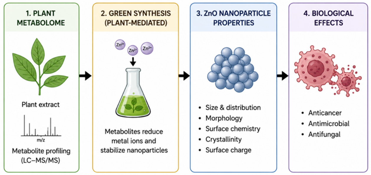

Table 1.
Applications of nanoparticles in cancer diagnosis and therapy.
| APPLICATION | MECHANISM | NANOPARTICLE TYPES | ADVANTAGES | REFERENCES |
|---|---|---|---|---|
| Cancer diagnosis | Nanoparticles interact with DNA/molecular biomarkers to detect genetic defects; improve imaging sensitivity and resolution. | Metallic NPs, magnetic NPs, polymeric NPs, quantum dots, graphene, carbon nanotubes, dendrimers, liposomes. | Early detection, high sensitivity, non-invasive, enhanced targeting | [68,69,70,71,72] |
| Cancer targeting |
Passive targeting: Exploits EPR effect due to leaky vasculature. Active targeting: Surface functionalization with ligands (antibodies, folic acid, peptides, siRNA). Gene silencing with antisense DNA/siRNA. |
Polymeric NPs, gold NPs, liposomes, dendrimers, siRNA/antisense DNA-loaded NPs. | Specificity for tumor cells, reduced off-target toxicity, improved tumor accumulation | [73,74,75,76] |
| Drug delivery | Nanocarriers enhance bioavailability, enable controlled release, and prolong blood circulation. | Liposomes, polymeric micelles, solid lipid NPs, dendrimers, gold/silver NPs. | Increased therapeutic index, reduced systemic toxicity, improved pharmacokinetics, steady-state drug release. | [69,77,78] |
|
Hyperthermia |
Magnetic NPs generate localized heat under alternating magnetic field via Néel and Brownian relaxation. | Iron oxide NPs, cobalt ferrite NPs, superparamagnetic NPs | Non-invasive tumor ablation, synergistic with radiotherapy/chemotherapy, localized heating | [79,80,81] |
|
Radiotherapy |
Metal NPs absorb radiation, induce oxidative stress, apoptosis, and improve radiation targeting. | Gold NPs, silver NPs, hafnium oxide NPs. | Dose reduction minimized collateral damage and radio sensitization. | [27,29,82] |
Table 2.
Nanoparticle outcomes of different phytochemical classes and their biological relevance.
| Phytochemical class | Typical NPs reported | Reported effects on synthesis (morphology/ size/ stability) | Biological relevance | References |
|---|---|---|---|---|
| Flavonoids (quercetin, chrysin, tricetin) | AuNPs, AgNPs | Act as reducing and stabilizing agent. Often yield spherical, monodisperse NPs; can increase stability and reduce aggregation | Improved cellular uptake and enhances ROS-mediated cytotoxicity in cancer cell lines; antibacterial activity. | [112,113,114] |
| Phenolic acids & polyphenols (tannic acid, gallic acid, general polyphenols) | AgNPs, AuNPs, ZnONPs | Strong reducing capacity. Enable size-tunable AgNPs; tannic acid produces monodisperse small AgNPs with negative zeta potential | Reported anticancer and antimicrobial activity. Often show enhanced oxidative stress induction in tumor cells and altered biodistribution due to surface chemistry | [115,116,117,118] |
| Terpenoids/ triterpenes/ essential oil components | ZnONPs, CuONPs, AgNPs | Reported as shape-directing and stabilizing agents for ZnO NPs, with rod/ flower morphologies and can alter crystallinity | ZnO and CuO NPs produced with terpenoids show photocatalytic and cytotoxic effects; surface terpenoids may modulate membrane interactions and uptake | [119,120] |
| Alkaloids | AgNPs, AuNPs | Often implicated in reduction but less common as sole reductants | Cytotoxicity enhancement; increase batch variability and necessitate careful toxicology | [121] |
| Tannins/ condensed polyphenols | AgNPs, AuNPs | Bio-reducing, stabilizing, and capping capability. Produce small, stable NPs. Often yield high colloidal stability and narrow size distributions. Au NPs are predominantly spherical | Reported antioxidant, antimicrobial, and anticancer properties; may alter protein corona formation in vivo | [122,123] |
| Crude extracts (mixtures) | Ag, Au, ZnO, bimetallic (Au-Ag) | Mixtures often yield heterogeneous, irregular size and shape distributions due to multiple competing metabolites | Reported antibacterial, antifungal, and antiproliferative activity. | [124] |
| Bimetallic/ hybrid systems | Au-Ag, Cu-Ni bimetallics | Plant extracts may mediate core-shell formation (hybrids) with spherical shapes | Enhanced catalytic activity and often improved photothermal/photocatalytic properties. Reports of antibacterial, antiparasitic and cytotoxic activity | [125,126] |
Disclaimer/Publisher’s Note: The statements, opinions and data contained in all publications are solely those of the individual author(s) and contributor(s) and not of MDPI and/or the editor(s). MDPI and/or the editor(s) disclaim responsibility for any injury to people or property resulting from any ideas, methods, instructions or products referred to in the content. |
© 2026 by the authors. Licensee MDPI, Basel, Switzerland. This article is an open access article distributed under the terms and conditions of the Creative Commons Attribution (CC BY) license.
Copyright: This open access article is published under a Creative Commons CC BY 4.0 license, which permit the free download, distribution, and reuse, provided that the author and preprint are cited in any reuse.


