Preprint
Article

This version is not peer-reviewed.

Occurrence, Species Distribution, and Antimicrobial Resistance of Listeria spp. in Foods from Retail and Farm Outlets in North Carolina, USA

Submitted:

07 July 2026

Posted:

09 July 2026

You are already at the latest version

Abstract

Retail food contamination by Listeria species and rising antimicrobial resistance (AMR) among Listeria populations further underscores need for continued surveillance. This study assessed prevalence, species distribution, and AMR profiles of Listeria spp. recovered from retail foods in North Carolina, USA. Food samples (866) collected between 2018 and 2020 were screened and presumptive isolates identified by multiplex PCR, and antimicrobial susceptibility was evaluated against sixteen antimicrobial agents. Listeria spp. were detected in 8.55% (74/866) of samples. Species-level identification was achieved for 68 isolates. Non-pathogenic species predominated, led by Listeria welshimeri (24.32%) and L. innocua (21.62%). Pathogenic species (L. monocytogenes, Listeria ivanovii) accounted for 17.57% of isolates and occurred exclusively in raw commodities. Prevalence was highest in raw milk (14.58%), mushrooms (13.64%), vegetables (11.49%), and raw meats (10.74%); processed foods showed minimal contamination (0.42%). Unprocessed foods were 28-times more likely to harbor Listeria than processed products (RR=28.00; 95% CI: 3.91-200.32; P<0.0001). Resistance was highest to Nitrofurantoin (51.47%) and Penicillin G (39.71%). Multidrug resistance (MDR) was detected in 64.71% of isolates including 84.62% of pathogenic and 60.00% of non-pathogenic species. Raw agricultural commodities are major Listeria reservoirs. Widespread MDR across species underscores the necessity for integrated surveillance and One Health monitoring frameworks.

Keywords: 
;  ;  ;  ;  ;  

1. Introduction

Foodborne diseases remain a major public health challenge globally, with bacterial pathogens accounting for a substantial proportion of foodborne illnesses and associated economic losses [1]. Among these pathogens, species of the genus Listeria have received considerable attention because of their widespread distribution in agricultural environments, food-processing facilities, and retail food products [2]. Their ability to persist throughout the food supply chain poses significant challenges for food safety and public health management.
A distinguishing characteristic of Listeria spp. is their exceptional environmental resilience. Unlike other foodborne bacteria, Listeria species are psychrotrophic and can survive and multiply at refrigeration temperatures, enabling them to persist under conditions commonly used to inhibit microbial growth in foods [3]. In addition, these organisms tolerate a wide range of environmental stresses, including high salt concentrations, acidic conditions, and desiccation. Such adaptive traits facilitate their survival throughout food production, processing, storage, distribution, and retail environments [3]. Furthermore, Listeria spp. readily forms biofilms on food-contact surfaces, including stainless steel equipment, conveyor belts, drains, and packaging systems. Biofilm formation enhances persistence within the food-processing environments and contributes to recurring contamination events despite routine sanitation procedures [4].
The taxonomy of the genus Listeria has expanded over the past two decades with tens of novel species from environmental and agricultural habitats. However, six species- Listeria monocytogenes, Listeria ivanovii, Listeria innocua, Listeria seeligeri, Listeria grayi and Listeria welshimeri, remain of relevance in food microbiology because of their frequent association with food products and food processing environments. Among these, L. monocytogenes is the primary etiological agent in human listeriosis, whereas L. ivanovii is principally associated with veterinary infections (ruminants), although sporadic human infections have also been reported [5,6]. The remaining species are regarded as non-pathogenic but are commonly recovered from food and environmental sources and may serve as indicators of environmental contamination and hygiene status [6].
Listeria monocytogenes is one of the most important foodborne pathogens because of its ability to cause invasive listeriosis, a disease associated with high hospitalization, and mortality rates. One peculiarity of this bacterium is that it is non-motile at elevated temperatures (37°C) and highly motile at low temperatures (22-28°C), which complicates its control in foods and the environment [7]. Clinical manifestations range from mild gastroenteritis to severe systemic infections, including septicemia, meningitis, encephalitis, maternal-fetal infections, spontaneous abortion, stillbirth, and neonatal disease [8,9]. Individuals at greatest risk include pregnant women, neonates, older adults, and immunocompromised persons, among which mortality rates may exceed 20-30% [7]. Although listeriosis occurs less frequently than other foodborne diseases, its severity makes it a major public health concern. In the United States, L. monocytogenes continues to be recognized as a priority foodborne pathogen because of its substantial disease burden and its frequent involvement in food recalls and outbreak investigations [10,11]. Currently, the Interagency Food Safety Analytics Collaboration listed Campylobacter spp., Listeria monocytogenes, nontyphoidal Salmonella and Shiga toxin-producing Escherichia coli (STEC) as priorities for source attribution and actions in the US [12]. Contamination by Listeria spp. can occur at multiple stages of the food supply chain. Fresh produce, edible fungi, and other raw agricultural commodities may become contaminated through exposure to soil, irrigation water, manure, wildlife, and other environmental sources [11]. Similarly, contamination of meat and dairy products may occur during animal production, slaughter, milking, processing, transportation, or retail handling [13]. Although interventions such as pasteurization, thermal processing, sanitation programs, and Hazard Analysis and Critical Control Point (HACCP) systems have reduced microbial hazards in food systems, post-processing contamination remains a persistent challenge [12]. Consequently, evaluating the occurrence of Listeria in both raw and processed food is essential for identifying contamination sources and improving food safety management strategies.
While L. monocytogenes remains the principal pathogen of concern, non-pathogenic species are frequently recovered from the same food matrices and environmental niches [14]. These species often coexist with pathogenic Listeria populations and share similar ecological adaptations that enable persistence in agricultural and food processing environments. Their presence may provide valuable information regarding environmental conditions that favor Listeria survival and dissemination. Accurate species level identification is thus important not only for risk assessment but also for understanding the ecology and transmission dynamics of Listeria within food systems [9,14].
The detection and identification of Listeria in food remain technically challenging because contamination levels are often low, and target organisms may be stressed or injured during food processing. Consequently, standard detection protocols developed by regulatory agencies rely on selective enrichment followed by selective plating and biochemical confirmation [11]. The development of chromogenic media, such as Agar Listeria according to Ottaviani and Agosti (ALOA), has improved the recovery and differentiation of Listeria species. More recently, molecular techniques, particularly multiplex polymerase chain reaction (PCR), have enhanced the speed and accuracy of species level identification and have become valuable tools for epidemiological investigations and surveillance programs [15,16]. In addition to contamination concerns, the global emergence of antimicrobial resistance (AMR) among Listeria species has become an increasing public health challenge [17]. Although listeriosis is traditionally treatedβ-lactam antibiotics particularly ampicillin or penicillin, often in combination with aminoglycosides, growing reports of resistance to clinically important antimicrobial agents have raised concerns regarding long-term effectiveness of available treatment options [18,19].
Furthermore, non-pathogenic Listeria species may serve as environmental reservoirs of antimicrobial resistance determinants within food production systems, underscoring the importance of monitoring susceptibility patterns across entire genus rather than focusing exclusively on Listeria monocytogenes [2]. Control of Listeria contamination relies on regulatory oversight and industry-based intervention strategies. Regulatory agencies, including the Centers for Disease Control and Prevention (CDC) and other food safety authorities, implement surveillance programs, microbiological standards, and environmental monitoring requirements to reduce contamination risks. In parallel, food producers employ a range of antimicrobial interventions, including thermal and non-thermal processing technologies, biological control agents such as nisin, and chemical preservatives. However, the effectiveness of these interventions varies according to the food matrix, application method, and processing conditions, necessitating continuous evaluation of their performance under commercial settings.
Despite extensive investigations of Listeria contamination in individual food commodities, comparative studies assessing species distribution, pathogenicity profiles, and antimicrobial resistance patterns across multiple food categories within the same retail environment remain limited. In addition, most surveillance programs focus primarily on L. monocytogenes, providing comparatively little information on the ecology, diversity, and epidemiological significance of other Listeria species.
Therefore, the present study conducted a cross-sectional surveillance of 866 food samples collected from retail markets and farm outlets in North Carolina, USA, to: (i) determine the prevalence and distribution of Listeria species across diverse food categories; (ii) evaluate the effect of food processing on contamination risk; (iii) characterize species diversity and pathogenicity profiles using multiplex PCR-based identification; and (iv) assess the antimicrobial susceptibility patterns of recovered isolates against 16 clinically relevant antimicrobial agents. By integrating prevalence, molecular characterization, and antimicrobial resistance data, this study provides valuable baseline information to support food safety surveillance, risk assessment, and the development of evidence-based control strategies for Listeria within contemporary food systems.

2. Materials and Methods

2.1. Sample Collection and Study Design

A cross-sectional surveillance study was conducted to determine the prevalence, species distribution, and antimicrobial profiles of Listeria species in retail foods. A total of 866 samples were collected from commercial grocery stores, supermarkets, farmers’ markets and open-air retail outlets in Kannapolis, Concord, Greensboro and Lexington, North Carolina, USA, between May 2018 and September 2020. Sampling was performed throughout the study period to capture potential seasonal variations in contamination patterns.
Samples were categorized into two groups based on processing status: unprocessed foods (n=626), comprising fresh vegetables, edible fungi (mushrooms), raw meats, and raw milk, and processed foods (n=240), comprising pasteurized milk, commercial cheeses, yoghurt, and processed meat products. Following purchase, samples were transported to the laboratory in insulated containers maintained below 4°C and processed within 2 H of collection.

2.2. Isolation and Phenotypic Identification of Listeria spp.

Isolation of Listeria spp. was performed using selective enrichment procedures following plating on Brilliance TM Listeria Selective Agar (Oxoid, UK). Presumptive colonies were sub-cultured to obtain pure culture isolates and incubated at 37°C for 24 h. Colony morphology, Gram staining and catalase testing were used as preliminary identification. Presumptive Listeria isolates were subsequently cultured in Brain Heart Infusion (BHI) broth at 37°C for 24 h prior to molecular characterization.

2.2.1. Genomic DNA Extraction and Multiplex PCR Identification of Listeria Species

Species identification was performed using a multiplex PCR assay adapted from Mazza et al. [20]. The assay targeted the genus specific prs gene (370 bp) for confirmation of Listeria spp. and species-specific markers/primer sets for differentiation of the six classical Listeria species (Table 1).
Polymerase chain reactions (PCR) were conducted in a final volume of 25 µL containing 12.5 µL of 2 × GoTaq ® PCR Master Mix (Promega, USA), 1.0 µL each of forward and reverse primers (10 µM), 1.5 µL of genomic DNA template and nuclease free water to final volume (Table 2).
Thermal cycling conditions consisted of an initial denaturation at 95°C for 5 min, followed by 35 cycles of denaturation at 94°C for 30 s, annealing at 60°C for 30 s, and extension at 72°C for 30 s with a final extension at 72°C for 5 min. PCR products were separated by electrophoresis on 1.5% (w/v) agarose gel in 1x TAE buffer and stained with ethidium bromide. Electrophoresis was performed at 100 V for 60 min, and amplicons visualized under UV illumination. Isolates positive for prs gene but negative for all species-specific targets were classified as unidentified Listeria species. Primers sets used for the studies are listed in Table 1 [20]

2.3. Antibiotic Susceptibility Testing

In vitro antibiotic susceptibility testing was performed on all identified classical Listeria isolates (n=68) using the Kirby-Bauer disk diffusion method on Mueller-Hinton agar according to Clinical and Laboratory Standards Institute (CLSI) guidelines. The six unidentified Listeria isolates were excluded from the antimicrobial susceptibility testing because species identity could not be confirmed. Bacterial suspensions were adjusted to 0.5 McFarland standard before inoculation. Sixteen antimicrobial agents were evaluated Amikacin (30 μg), Azithromycin (15 μg), Chloramphenicol (30 μg), Ciprofloxacin (5 μg), Clindamycin (2 μg), Erythromycin (15 μg), Kanamycin (30 μg), Levofloxacin (5 μg), Moxifloxacin (5 μg), Nitrofurantoin (300 μg), Penicillin G (10 U), Rifampin (5 μg), Tetracycline (30 μg), Tobramycin (10 μg), Trimethoprim (5 μg), and Trimethoprim–Sulfamethoxazole (1.25/23.75 μg). Following incubation at 35°C for 18-24 h, diameters of zones of inhibition were measured and interpreted as susceptible, (S), intermediate (I), or resistant (R), according to CLSI M100 Performance Standards for Antimicrobial Susceptibility Testing (26th edition) [21]. Multidrug resistance (MDR) is defined as non-susceptibility to at least one antimicrobial agent in three or more antimicrobial categories [22].

2.4. Statistical Analysis

Statistical analyses were performed using GraphPad Prism version 11.0.12 (GraphPad Software, San Diego, CA, USA). Prevalence differences among food categories were evaluated using the Fisher-Halton exact test. Comparisons between unprocessed and processed food groups were conducted using two-tailed Fisher’s exact tests. The Relative Risk (RR) was calculated as:
R R = U n p r o c e s s e d P r e v a l e n c e ( % ) P r o c e s s e d P r e v a l e n c e ( % )
The Odds Ratio (OR) was calculated as:
OR   =   U n p r o c e s s e d P o s i t i v e s x P r o c e s s e d N e g a t i v e s U n p r o c e s s e d N e g a t i v e s x P r o c e s s e d P o s i t i v e s
Corresponding 95% confidence interval (CI) was calculated for all effect size estimates. For antimicrobial susceptibility analyses, resistant and intermediate isolates were combined as non-susceptible. Differences in non-susceptibility frequencies between pathogenic (L. monocytogenes and L. ivanovii) and non-pathogenic (L. innocua, L. seeligeri, L. grayi and L. welshimeri) groups were evaluated using two-tailed Fisher’s exact tests. Proportions and 95% confidence intervals were calculated using the Wilson score algorithm in GraphPad Prism. Statistical significance was established at P<0.05.

3. Results

3.1. Prevalence and Distribution Listeria Species

A total of 866 retail food samples, including fresh produce, dairy products and meat products were collected from grocery stores and markets in Kannapolis, Concord, Greensboro and Lexington, North Carolina, between 2018-2020. Multiplex PCR analysis targeting the genus specific prs gene confirmed the presence of Listeria spp. in 74 samples, corresponding to 8.55% (74/866); 95% CI: 6.85-10.61) (Figure 1).
Species-specific multiplex PCR successfully identified 68 of the 74 isolates (91.89% resolution of taxonomic classification), while six isolates (8.11%) were positive for the genus-specific marker but negative for all species-specific targets and were therefore classified as unidentified Listeria spp. Non-pathogenic environmental species predominated, accounting for 82.43% (61/74) of all isolates and a prevalence of 7.04% (61/866) among sampled foods (Table A1). Pathogenic species (L. monocytogenes and L. ivanovii) represented 17.57% (13/74) of isolates and were recovered from 1.50% (13/866) of all samples examined (Table A1).
Among identified species, Listeria welshimeri was most frequently recovered species accounting for 24.32% (18/74) of all isolates, followed by Listeria innocua (21.62%, 16/74), Listeria seeligeri (14.86%, 11/74), and Listeria grayi (13.51, 10/74) (Figure 1). Pathogenic species were less common, with Listeria monocytogenes representing 9.46% (7/74) and Listeria ivanovii 8.11% (6/74) of the total isolate collection. Notably, all pathogenic species were recovered exclusively from raw food commodities, and no pathogenic species were detected in processed foods.

3.2. Product-Level Distribution of Listeria spp.

Analysis of individual food commodities revealed considerable variation in contamination rates (Table 3). Among fresh vegetable produce, the highest prevalence was observed in kale (22.22%, 8/36), followed by romaine lettuce (16.67%, 6/36) and spinach (13.33%, 8/60). Within the raw meat products, raw pork exhibited the highest prevalence (20.00%, 2/10), followed by ground beef (13.89%, 5/36) and raw chicken (9.52%, 4/42) (Table 3).
*L. mono-L. monocytogenes.
In contrast, recovery of Listeria spp. from processed foods was rare. No Listeria isolates were detected in processed meat products including pork ham, pork sausage, beef hotdogs, and smoked pork (0/39). Similarly, pasteurized milk products and ten of the eleven cheese varieties assessed were free of contamination. The only positive processed food sample was cream cheese, which exhibited a prevalence of 7.14% (1/14) (Table 3).

3.2.1. Effect of Food Processing on Listeria Contamination

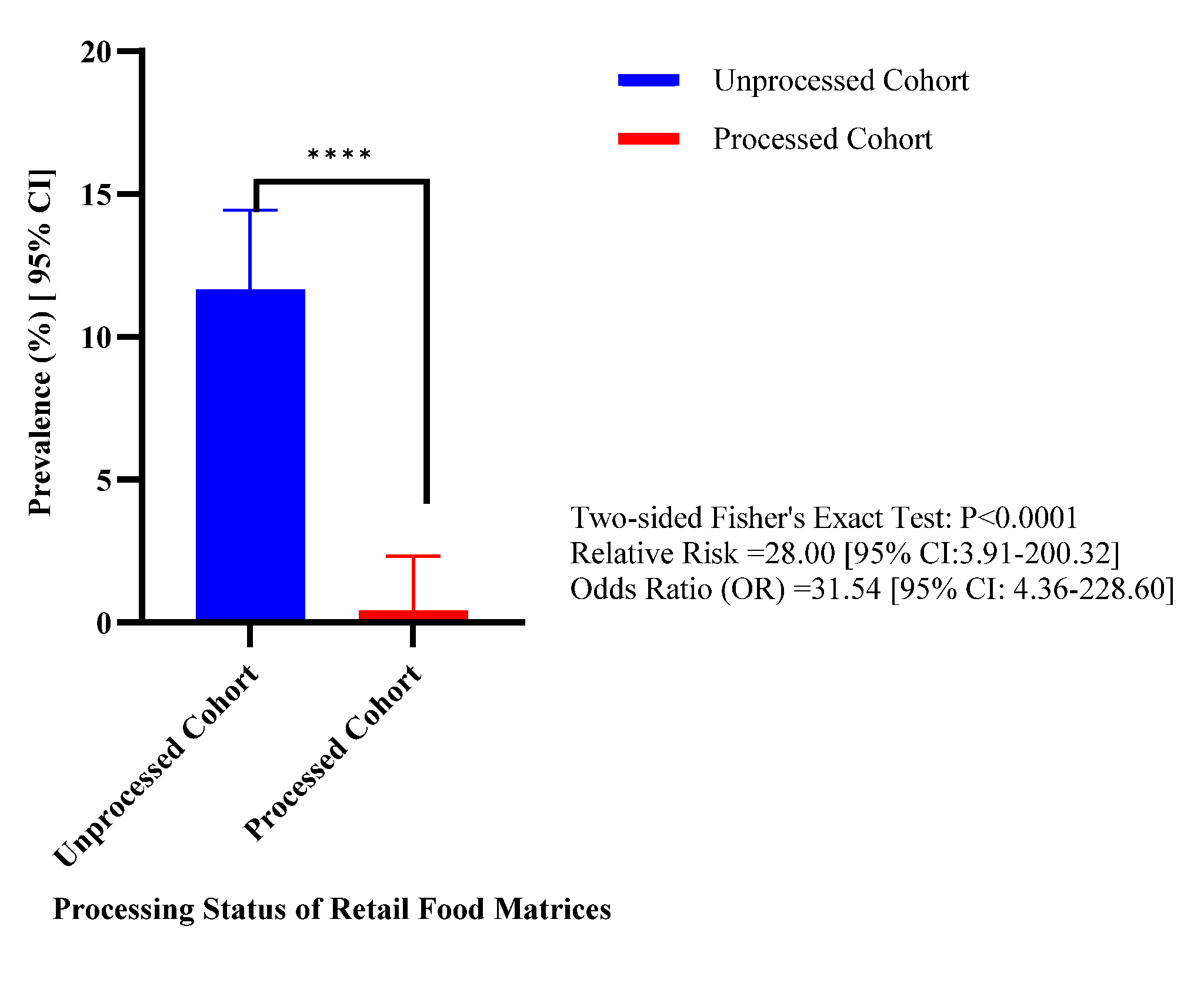
Food samples were further categorized according to processing status to evaluate the effect of commercial processing on Listeria occurrence (Figure 2).
The prevalence of Listeria spp. in unprocessed foods was 11.66% (73/626; 95% CI: 9.36-14.44), compared with only 0.42% (1/240; 95% CI: 0.07-2.33) in processed food. This difference was statistically significant (Fisher’s exact test, P<0.0001) (Figure 2). The calculated relative risk (RR) was 28.00 (95% CI: 3.91-200.32), indicating that unprocessed foods were approximately 28 times more likely to harbor Listeria spp. than processed foods. Similarly, the odds ratio (OR) was 31.57 (95% CI: 4.36-228.60), confirming a strong association between processing status and contamination risk (Figure 2).

3.2.2. Prevalence of Listeria spp. Across Major Food Categories

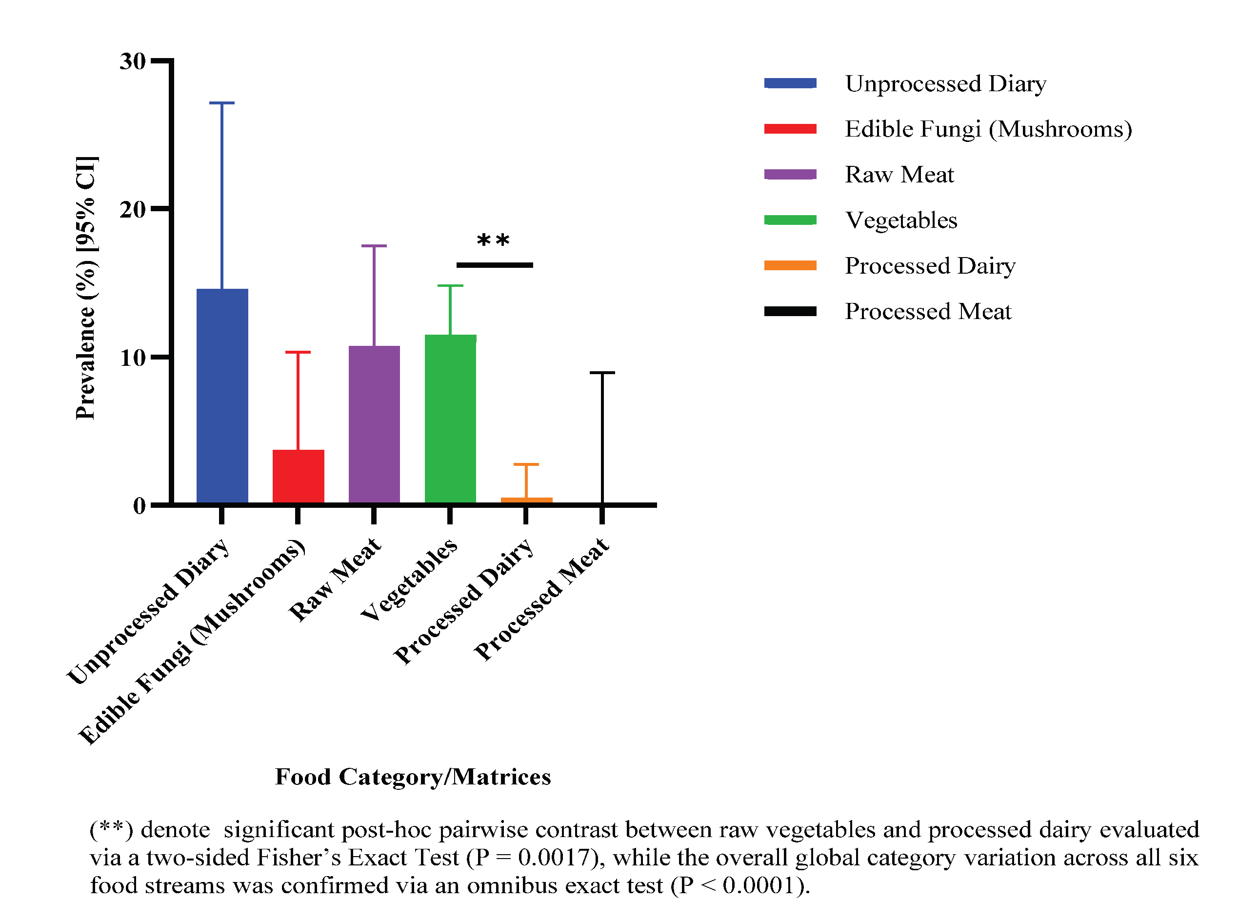
Prevalence varied among the six food categories evaluated (Figure 3).
The highest prevalence was observed in raw milk (14.58%, 7/48; 95% CI: 7.25-27.17), followed by mushrooms (13.64%, 3/22; 95% CI: 4.75-33.34), vegetables (11.49%, 50/435; 95% CI:8.83-14.83) and raw meats (10.74%, 13/121; 95% CI:6.39-17.52) (Figure 3). Processed dairy products exhibited a prevalence of only 0.50% (1/201), whereas no contamination was detected in processed meat products (0/39). Pairwise comparisons demonstrated a significant difference between vegetables and processed dairy products (P = 0.0017) (Figure 3).

3.2.3. Distribution of Pathogenic and Non-Pathogenic Species

When isolates were grouped according to pathogenic potential, pathogenic Listeria species were detected in 1.50% (13/866) of all samples whereas non-pathogenic species were detected in 6.35% (55/866) (Table A2). Vegetables contained both pathogenic and non-pathogenic species with prevalences of 1.38% (6/435) and 10.11% (44/435), respectively (Table A2). In raw milk, pathogenic species were recovered from 8.33% (4/48) of samples, compared with 6.25% (3/48) for non-pathogenic species (Table A2). Raw meat was dominated by non-pathogenic species, which occurred in 8.26% (10/121) of samples compared with 2.48% (3/121) for pathogenic species. Mushroom samples yielded only non-pathogenic Listeria species, which were detected in 13.64% (3/22) samples (Table A2).

3.3. Antimicrobial Susceptibility and Resistance Listeria Isolates

Antimicrobial susceptibility testing was conducted on 68 identified Listeria isolates

3.3.1. Overall Antimicrobial Resistance Profile

The highest resistance frequencies were observed for Nitrofurantoin (51.47%, 35/68), Penicillin G (39.71%, 27/68), Clindamycin (23.53%, 16/68), Levofloxacin (22.06%, 15/68), and Ciprofloxacin (14.71%, 10/68) (Table A3). Intermediate susceptibility was most common for Levofloxacin (32.35%, 22/68), followed by Chloramphenicol (16.18%, 11/68), and Nitrofurantoin (10.29%, 7/68). In contrast, Tobramycin, Tetracycline, and Trimethoprim-Sulfamethoxazole remained highly effective, with 98.53% of isolates been susceptible to them (67/68) of isolates (Table A3).

3.3.2. Species-Specific Antibiotic Resistance Patterns

Species-specific resistance patterns varied considerably (Table 4). Species-specific resistance patterns varied considerably. (Table 4). All L. monocytogenes isolates remained fully susceptible to Amikacin, Azithromycin, Tetracycline, Tobramycin and Trimethoprim-Sulfamethoxazole (Table 4).
However, resistance to Penicillin G and Levofloxacin was observed in 57.14% (4/7) and 42.86% of the same isolate, respectively. L. ivanovii exhibited high resistance to Nitrofurantoin (83.33%), Penicillin G (66.67%) and Levofloxacin (66.67%) (Table 4). Among non-pathogenic species, L. innocua showed resistance to Kanamycin and Penicillin G (25.00%), L. welshimeri exhibited intermediate susceptibility to Levofloxacin, Moxifloxacin, and Azithromycin (22.22%), while L. grayi displayed the highest resistance levels, particularly to Penicillin G (50.00%), Clindamycin (40.00%), and Levofloxacin (40.00%) (Table 4).

3.3.3. Non-Susceptibility Patterns Across Pathogenic and Non-Pathogenic Listeria spp.

When resistant and intermediate isolates were combined as non-susceptible, most antimicrobials exhibited comparable non-susceptibility between pathogenic and non-pathogenic Listeria isolates (Table A4). Although numerical differences were observed for resistance rates between the two cohorts for Penicillin G, Nitrofurantoin, and Levofloxacin, these did not reach statistical significance (P > 0.05) (Table A4). In contrast, Clindamycin non-susceptibility was significantly higher among pathogenic isolates (53.85% vs. 23.64%; P = 0.0175), whereas Azithromycin non-susceptibility was detected exclusively among non-pathogenic isolates (25.45% vs. 0%; P = 0.0461) (Table A4).

3.3.4. Multidrug Resistance Profiles of Listeria Phenotypes

Multidrug resistance (MDR) was defined according to established international criteria as non-susceptibility to at least one antimicrobial agent in three or more antimicrobial classes (Table A5). Overall, 64.71% (44/68) of isolates were classified as MDR. MDR prevalence was higher among pathogenic isolates (84.62%, 11/13) than non-pathogenic isolates (60.00%, 33/55). At the species level, MDR was detected in 71.43% (5/7) of L. monocytogenes and in all L. ivanovii isolates (100.00%, 6/6) (Table A5). Among non-pathogenic species, MDR was most frequent in L. grayi (100.00%, 10/10), followed by L. innocua (75.00%, 12/16), L. seeligeri (63.64%, 7/11), and L. welshimeri (22.22%, 4/18) (Table A5). Overall, MDR was widely distributed across both pathogenic and non-pathogenic Listeria, with the highest burden observed among pathogenic species.

4. Discussion

Listeria species remain important foodborne microorganisms because of their ability to persist in diverse environmental niches and contaminate foods throughout the production chain. Among them, Listeria monocytogenes is the principal human pathogen and a major concern in ready-to-eat (RTE) foods, which are particularly high-risk due to absence of terminal listericidal treatment step and the organisms’s capacity to survive and proliferate under conditions that inhibit many other pathogens [11]. This study investigated the occurrence, species diversity, distribution, and antimicrobial resistance profiles of Listeria spp. recovered from retail and farm-gate foods in North Carolina, USA. Overall, contamination was widespread in raw commodities, particularly vegetables, raw dairy products, and meats, while processed foods showed markedly lower prevalence, highlighting the effectiveness of post-harvest interventions. Although non-pathogenic species predominated, the detection of L. monocytogenes and L. ivanovii in raw food matrices underscores their continued relevance as potential sources of human exposure. Furthermore, the high prevalence of antimicrobial resistance (AMR) and multidrug resistance (MDR) across both pathogenic and environmental Listeria isolates suggests that food production environments may act as important reservoirs of resistance determinants. Collectively, these findings support the need for integrated food safety and AMR surveillance within a One Health framework.

4.1. Diversity and Distribution of Listeria Species in Retail Foods

The multiplex PCR assay successfully identified 91.89% of the presumptive isolates demonstrating the effectiveness of molecular methods for rapid species-level characterization. However, six isolates could not be assigned to any of the six classical Listeria species targeted suggesting the possible presence of atypical, novel, or less well characterized taxa. This limitation reflects the restricted coverage of species-specific PCR assays and highlights the need for complementary high-resolution approaches such as whole genome sequencing or multi-locus sequence analysis for comprehensive Listeria characterization. This finding highlights a recognized limitation of species-specific PCR assays, which are designed primarily to detect established classical Listeria species and may not fully capture the expanding taxonomic diversity of the genus [23,24]. Previous studies have reported higher identification rates using multiplex PCR, including a 98.7% identification rate of Listeria isolates recovered from 129 Flammulina velutipes (enoki mushroom) samples [25]. Differences in identification efficiency may be attributable to variations in sample type, species diversity, primer specificity, and the presence of atypical strains.
The overall prevalence of Listeria spp. was 8.55%, while L. monocytogenes was detected in 0.81% of samples (866). This prevalence is lower than reports from multi-state (3) U.S. surveys [26], likely reflecting differences in commodities, geography, and sampling design. Non-pathogenic species, particularly L. welshimeri and L. innocua, dominated the isolates, consistent with their ecological adaptability and persistence in food and agricultural environments [27]. Notably, L. welshimeri was the most prevalent species in this study (Table 3), differing from earlier reports where L. innocua typically predominates [26,27]. Such variation likely reflects differences in environmental conditions, food matrices, and regional agricultural practices. Although less frequent, the detection of L. monocytogenes confirms its continued presence in retail and farm-gate food systems. Previous studies have reported prevalence of approximately 1.6% of L. monocytogenes on food contact surfaces [28], suggesting that contamination may originate not only from primary production and processing environment but also from retail and market settings. The occurrence of L. monocytogenes in foods intended for human consumption highlights the need for sustained surveillance and strict hygienic control measures across the food supply chain.

4.2. Impact of Food Processing on Listeria Contamination

Listeria prevalence varied markedly by food type, with highest levels observed in raw milk, mushrooms, vegetables, and raw meats (Table 3). These findings reinforce raw commodities as primary reservoirs of contamination. In contrast, processed foods showed minimal contamination, with only one positive processed dairy sample and no detections in processed meat, confirming the effectiveness of thermal processing, pasteurization, and HACCP-based interventions [29,30].
Raw milk contamination fell within globally reported ranges for unpasteurized dairy products (5–20%) [31,32,33,34], although lower than some regional reports [35]. Sources of contamination likely include infected animals, environmental exposure, and milking equipment [32,33,34]. The absence of Listeria in pasteurized milk further supports the effectiveness of heat treatment and dairy safety programs.
Vegetables showed the highest number of positive samples, with kale exhibiting the greatest commodity-level prevalence. These findings align with global reports linking fresh produce contamination to soil, irrigation water, manure, and post-harvest handling [36,37,38,39]. Because these products are often consumed raw, they present a direct route for exposure.
Raw meat contamination (10.74%) was consistent with global retail surveys [40], with higher prevalence in pork suggesting contamination during slaughter and processing stages [41]. Mushrooms also showed moderate prevalence, although only non-pathogenic species were detected, reflecting environmental contamination during cultivation and handling unlike previous studies that recovered Listeria monocytogenes from mushroom production systems [42,43].
Overall, processing resulted in a 28-fold reduction in contamination risk, with significantly higher odds of contamination in raw foods (OR = 31.54) (Figure 1 and Figure 2). These results reinforce the critical role of post-harvest interventions in reducing Listeria burden in the food chain.

4.3. Distribution of Pathogenic and Non-Pathogenic Species

Pathogenic species were detected in 1.50% of samples, while non-pathogenic species occurred more frequently (6.35%). This pattern is consistent with previous studies showing dominance of environmental Listeria species in agricultural systems [6,44]. Although non-pathogenic, these species may indicate environmental conditions conducive to Listeria persistence [45].
Vegetables contained both pathogenic and non-pathogenic species, with the latter predominating, likely due to environmental exposure pathways such as soil, irrigation water, and fertilizers [36,39]. However, the presence of pathogenic species in raw produce remains a public health concern due to its common consumption without cooking.
Raw milk showed the highest prevalence of pathogenic Listeria, highlighting its importance as a transmission vehicle and supporting evidence of farm-level contamination and animal shedding [34]. In contrast, raw meat samples were dominated by non-pathogenic species, suggesting environmental contamination plays a greater role than direct pathogen carriage [41]. Mushrooms yielded only non-pathogenic species, consistent with environmental origin from substrates and cultivation systems [43].

4.4. Antimicrobial Resistance Profiles

The antimicrobial susceptibility profiles revealed widespread resistance among food-associated Listeria isolates. Resistance was most frequent to nitrofurantoin, penicillin G, clindamycin, levofloxacin, and ciprofloxacin. These patterns differ from some international reports, likely reflecting differences in antimicrobial usage practices, geography, and food systems [46,47]. For example, a study from Poland reported higher resistance rates to clindamycin (96.2%) and trimethoprim-sulfamethoxazole (91.3%) [46], whereas all isolates in the present study remained susceptible to trimethoprim-sulfamethoxazole. Similarly, Nikolaou et al. [47] reported 13.95% resistance to trimethoprim-sulfamethoxazole among Romanian Listeria isolates.
Of particular concern is resistance to penicillin G, a first-line treatment for invasive listeriosis. Although resistance remains relatively uncommon globally, increasing reports of reduced susceptibility among food and environmental isolates suggest emerging resistance reservoirs outside clinical settings. Fluoroquinolone resistance may similarly reflect selection pressure in agricultural environments. In contrast, tobramycin, tetracycline, and trimethoprim–sulfamethoxazole retained high efficacy.
Species-level analysis revealed marked heterogeneity. L. monocytogenes remained fully susceptible to several agents (amikacin, azithromycin, tetracycline, tobramycin, and trimethoprim-sulfamethoxazole) but showed resistance to penicillin G and levofloxacin, consistent with previous reports [48,49,50]. L. ivanovii exhibited even higher resistance levels, suggesting its potential role in maintaining resistance determinants in agricultural ecosystems [51,52]. Among non-pathogenic species, L. grayi showed the highest resistance burden, while L. innocua and L. welshimeri exhibited moderate resistance or intermediate susceptibility patterns. These findings support the role of environmental Listeria as reservoirs of antimicrobial resistance genes [53,54].
Multidrug resistance (MDR) was widespread, affecting 64.71% of isolates, and was more common in pathogenic (84.62%) than non-pathogenic (60.00%) groups. The detection of MDR in all L. ivanovii and most L. monocytogenes isolates is particularly concerning, as it may limit therapeutic options for invasive infections. High MDR levels among environmental species further suggest that food production environments may facilitate maintenance and dissemination of resistance determinants [55,56].

5. Conclusions

This study provides baseline data on the prevalence, species diversity, and antimicrobial resistance of Listeria spp. in retail and farm-gate foods in some regions in North Carolina, USA. Overall prevalence was 8.55%, with contamination concentrated on raw commodities, particularly milk, vegetables, mushrooms, and meats. Processed foods showed minimal contamination, confirming the effectiveness of post-harvest interventions. Molecular identification revealed a predominance of non-pathogenic species, while pathogenic L. monocytogenes and L. ivanovii were confined to raw foods, underscoring ongoing exposure risks. In addition, widespread antimicrobial resistance and high MDR prevalence across both pathogenic and environmental isolates indicate that food systems may serve as important reservoirs of resistance determinants. These findings highlight the need for continued surveillance of Listeria spp. beyond L. monocytogenes, strengthened farm-to-fork control strategies, and integrated monitoring of antimicrobial resistance within a One Health framework to mitigate foodborne and resistance-related risks.

Author Contributions

Conceptualization, A.K. and L.L.W.; methodology, L.L.W. and A.B.; validation, L.L.W., E.M.A. and S.D.; formal analysis, L.L.W. and E.M.A.; investigation, A.K., J.R.K. and S.D.; data curation, R.R.B.; writing—original draft preparation, L.L.W., A.K. and S.D.; writing—review and editing, L.L.W., A.K. and E.M.A.; supervision, L.L.W.; project administration, L.L.W.; funding acquisition, L.L.W. All authors have read and agreed to the published version of the manuscript.

Funding

This research was funded by the 1890 Capacity Building Grant Program from the United States Department of Agriculture and National Institute of Food and Agriculture (Project Award No.2023-38821-39979).

Institutional Review Board Statement

Not Applicable.

Data Availability Statement

The original contributions presented in this study are included in the article. Further enquiries can be directed at the corresponding author.

Conflicts of Interest

The authors declare no conflicts of interest.

Abbreviations

The following abbreviations are used in this manuscript:
AMR Antimicrobial Resistance
MDR Multidrug Resistance
TMP-SMX Trimethoprim-sulfamethoxazole.

Appendix A

Table A1. Taxonomic Distribution, and Public Health Risk Recovered Listeria Isolates (N=74).
Clinical Cohort and Species Isolation Count (n) Proportion of Total Genus
Incidence (%) (n=74)
Incidence/Prevalence in TotalRetail Samples (N=866)
Pathogenic Cohort 13 17.57 1.50
L. monocytogenes 7 9.46 0.81
L. ivanovii 6 8.11 0.69
Non-Pathogenic Cohort 61 82.43 7.04
L. welshimeri 18 24.32 2.08
L. innocua 16 21.62 1.85
L. seeligeri 11 14.86 1.27
L. grayi 10 13.51 1.15
Unidentified Listeria spp. 6 8.11 0.69
Total Yield 74 100.00 8.55
Table A2. Pathogenicity Distribution and Prevalence of Listeria Isolates Across Sampled Retail Food Matrices (N=866).
Food Cohort
and Category
Total Sample Size (N) Pathogenic Cohort a
% (n)
Non-Pathogenic Cohort b
% (n)
Overall Prevalence
% (n)
Vegetables 435 1.38% (6) 10.11% (44) 11.49% (50)
Edible Fungi (Mushrooms) 22 0.00% (0) 13.64% (3) 13.64% (3)
Raw Meat 121 2.48% (3) 8.26% (10) 10.74% (13)
Unprocessed Dairy 48 8.33% (4) 6.25% (3) 14.58% (7)
Processed Meat 39 0.00% (0) 0.00% (0) 0.00% (0)
Processed Dairy 201 0.00% (0) 0.50% (1) 0.50% (1)
TOTAL STUDIED 866 1.50% (13) 6.35% (55) 8.55% (74)
a Pathogenic Cohort reflects the combined isolation parameters of Listeria monocytogenes (n = 7) and Listeria ivanovii (n = 6). b Non-Pathogenic Cohort reflects the pooled parameters of Listeria innocua (n=16), Listeria seeligeri (n=11), Listeria grayi (n=10), Listeria welshimeri (n=18), and unidentified Listeria strains (n=6).
Table A3. Overall Phenotypic Distribution and Antibiotic Resistance Profiles of Recovered Listeria isolates (n = 68).
Antibiotics Resistant (R) % (n) Intermediate (I) % (n) Susceptible (S) % (n)
Amikacin 0.00 (0) 4.41 (3) 95.59 (65)
Azithromycin 7.35 (5) 13.24 (9) 79.41 (54)
Chloramphenicol 10.29 (7) 16.18 (11) 73.53 (50)
Ciprofloxacin 14.71 (10) 14.71 (10) 70.59 (48)
Clindamycin 23.53 (16) 13.24 (9) 63.24 (43)
Erythromycin 7.35 (5) 7.35 (5) 85.29 (58)
Kanamycin 26.47 (18) 23.53 (16) 50.00 (34)
Levofloxacin 22.06 (15) 32.35 (22) 45.59 (31)
Moxifloxacin 17.65 (12) 11.76 (8) 70.59 (48)
Nitrofurantoin 51.47 (35) 10.29 (7) 38.24 (26)
Penicillin G 39.71 (27) 0.00 (0) 60.29 (41)
Rifampin 14.71 (10) 5.88 (4) 79.41 (54)
Tetracycline 1.47 (1) 0.00 (0) 98.53 (67)
Tobramycin 1.47 (1) 0.00 (0) 98.53 (67)
Trimethoprim 19.12 (13) 5.88 (4) 75.00 (51)
TMP-SMX a 1.47 (1) 0.00 (0) 98.53 (67)
a TMP-SMX, trimethoprim-sulfamethoxazole.
Table A4. Comparative non-susceptibility/resistance of pathogenic and non-pathogenic Listeria isolates.
Antibiotic
Screening Panel
Pathogenic Cohort (N=3) a
Non-Susceptible Frequency
% (n)
Non-Pathogenic Cohort (N=55) b
Non-Susceptible Frequency
% (n)
Two-Sided Fisher’s Exact P-Value Significance Summary
Amikacin 7.69 (1) [1.37-33.31] 3.64 (2) [1.00-12.32] 0.4497 ns
Azithromycin 0.00 (0) [0.00-22.81] 25.45 (14) [15.81-38.30] 0.0461 *
Chloramphenicol 30.77 (4) [12.63-58.29] 14.55 (8) [7.56-26.16] 0.1332 ns
Ciprofloxacin 23.08 (3) [8.18-50.25] 32.73 (18) [21.81-45.90] 0.7423 ns
Clindamycin 58.85 (7) [29.13-76.79] 23.64 (13) [11.55-32.37] 0.0175 *
Erythromycin 7.69 (1) [1.37-33.31] 16.36 (9) [8.86-28.26] 0.6726 ns
Kanamycin 30.77 (4) [12.63-58.29] 50.91 (28) [38.08-63.62] 0.2227 ns
Levofloxacin 46.15 (6) [23.21-70.87] 43.64 (24) [31.37-56.73] 0.9999 ns
Moxifloxacin 15.38 (2) [4.33-42.23] 30.91 (17) [20.28-44.03] 0.3341 ns
Nitrofurantoin 69.23 (9) [42.37-87.37 56.36 (31) [43.27-68.63] 0.5401 ns
Penicillin G 61.54 (8) [35.53-82.28] 34.55 (19) [23.36-47.75] 0.0768 ns
Rifampin 23.08 (3) [8.18-50.25] 16.36 (9) [8.86-28.26] 0.6702 ns
Tetracycline 0.00 (0) [0.00-22.81] 1.82 (1) [0.32-9.61] >0.9999 ns
Tobramycin 0.00 (0) [0.00-22.81] 1.82 (1) [0.32-9.61] >0.9999 ns
Trimethoprim 15.38 (2) [4.33-42.23] 23.64 (13) [14.47-36.35] 0.7180 ns
TMP-SMX d 0.00 (0) [0.00-22.81] 1.82 (1) [0.32-9.61] >0.9999 ns
a Pathogenic isolates included Listeria monocytogenes (n = 7) and Listeria ivanovii (n = 6). b non-pathogenic isolates included Listeria innocua (n = 16), Listeria seeligeri (n = 11), Listeria grayi (n = 10), and Listeria welshimeri (n = 18). c Statistical significance was assessed using two-sided Fisher’s exact tests for each antibiotic. *P < 0.05; ns, not significant. d TMP-SMX, trimethoprim-sulfamethoxazole.
Table A5. Distribution of multidrug-resistant phenotypes among classical Listeria isolates (n = 68).
Taxonomy and Species Line Total Isolates Tested (N) Multidrug Resistant Isolates a Count (n) Species-Specific MDR Incidence (%) Primary Phenotypic Co-Resistance Signature Tracks b
Pathogenic Cohort 13 11 84.62% Penicillins + Fluoroquinolones + Lincosamides
L. monocytogenes 7 5 71.43% Penicillin G + Levofloxacin + Clindamycin
L. ivanovii 6 6 100.00% Penicillin G + Levofloxacin + Nitrofurantoin + Chloramphenicol
Non-Pathogenic Cohort 55 33 60.00% Penicillins + Fluoroquinolones + Aminoglycosides
L. welshimeri 18 4 22.22% Kanamycin + Levofloxacin + Azithromycin [Intermediate]
L. innocua 16 12 75.00% Kanamycin + Penicillin G + Levofloxacin + Azithromycin
L. seeligeri 11 7 63.64% Penicillin G + Kanamycin + Levofloxacin
L. grayi 10 10 100.00% Penicillin G + Clindamycin + Levofloxacin + Nitrofurantoin
Combined Genus Total 68 44 64.71% Overarching Retail Resistome Saturation Footprint
a MDR defined as absolute phenotypic non-susceptibility (Resistant or intermediate) to at least one antimicrobial agent across three or more distinct structural classes. b Indicate the most frequent overlapping therapeutic drug classes driving the multidrug resistance profile within specific species line.

References

  1. Yang, Y.; Xu, F.-R.; Zhou, Y.-B.; Hu, L.-Q.; Lu; Zhang, W.; Hu, S.-H.; Huang, H.; X.-R. Epidemiological Characteristics of Foodborne Disease Outbreaks in a Hospital: A 5-Year Retrospective Study. Int. J. Gen. Med. 2025. 18, 1529–1542. [Google Scholar] [CrossRef]
  2. Mohapi; D. A. Nkhebenyane, S. J. Listeria monocytogenes and Listeria ivanovii Virulence and Adaptations Associated with Leafy Vegetables from Small-Scale Farm and a Shift of Microbiota to a New Niche at Markets: A Systematic Review. Microorganisms 2025, 14(1), 76. [Google Scholar] [CrossRef] [PubMed]
  3. Osek, J.; Wieczorek, K. Why does Listeria monocytogenes survive in food and food-production environments? J. Vet. Res. 2023, 67(4), 537–544. [Google Scholar] [CrossRef] [PubMed]
  4. Mazaheri, T.; Cervantes-Huaman, B. R. H.; Bermudez-Caodevila, M.; Ripolles-Avila, C.; Rodriguez-Jerez, J. J. Listeria monocytogenes Biofilms in the Food Industry: Is the Current Hygiene Program Sufficient to Combat the Persistence of the Pathogen? Microorganisms 2021, 9(1), 182. [Google Scholar] [CrossRef]
  5. Manyi-Loh, C. E.; Lues, R. Listeria monocytogenes and Listeriosis: The Global Enigma. Foods 2025, 14(7), 1266. [Google Scholar] [CrossRef] [PubMed]
  6. Orsi, R. H.; Liao, J.; Carlin, C. R.; Weidmann, M. Taxonomy, ecology, and relevance to food safety of the genus Listeria with a particular consideration of new Listeria species described between 2010 and 2022. MBio. 2023, 15, e00938-23. [Google Scholar] [PubMed]
  7. Kathariou, S.; Kanenaka, R.; Allen, R. D.; Fok, A. K.; Mizumoto, C. Repression of motility and flagellin production at 37 degrees C is stronger in Listeria monocytogenes than in the nonpathogenic species Listeria innocua. Can. J. Microbiol. 1995, 41(7), 572–577. [Google Scholar] [CrossRef] [PubMed]
  8. Reis, J. O.; Teixeira, L. A. C.; Cunha-Neto, A.; Castro, V. S.; Figueiredo, E. E. S. Listeria monocytogenes in beef: a hidden risk. Res. Microbiol. 2024, 175(7), 104215. [Google Scholar] [CrossRef] [PubMed]
  9. Quereda, J. J.; Moron-Garcia, A.; Palacios-Gorba, C.; Dessaux, C.; Garcia-del Portillo, F.; Pucciarelli, M. G.; Ortega, A. D. Pathogenicity and virulence of Listeria monocytogenes: A trip from environmental to medical microbiology. Virulence 2021, 12(1), 2509–2545. [Google Scholar] [CrossRef] [PubMed]
  10. Su, Y.; Liu; A. Zhu, M. J. Mapping the landscape of listeriosis outbreaks (1998-2023): Trends, challenges, and regulatory responses in the United States. Trends Food Sci. Technol. 2024, 154, 104750. [Google Scholar] [CrossRef]
  11. D’Ambrosio, G.; Schirone, M.; Paparella, A. Listeria monocytogenes in Ready-to-Eat Foods: Risk Perspectives Across Different Regulatory Systems. Foods. 2026, 15(3), 470. [Google Scholar] [CrossRef] [PubMed]
  12. Scallan, W. E. J.; Cui, Z.; Tierney, R.; Griffin, P. M.; Hoekstra, R. M.; Payne, D. C.; et al. Foodborne Illness Acquired in the United States—Major Pathogens, 2019. Emerg. Infect. Dis. 2025, 31(4), 669–677. [Google Scholar] [CrossRef]
  13. Zhu, Q.; Gooneratne, R.; Hussain, M. A. Listeria monocytogenes in Fresh Produce: Outbreaks, Prevalence and Contamination Levels. Foods 2017, 6(3), 21. [Google Scholar] [CrossRef] [PubMed]
  14. Orsi, R. H.; Wiedmann, M. Characteristics and distribution of Listeria spp., including Listeria species newly described since 2009. Appl. Microbiol. Biotechnol. 2016, 100, 5273–5287. [Google Scholar] [CrossRef] [PubMed]
  15. Law, J. W.-F.; Mutalib, N.-S. A.; Chan, K.-G.; Lee, L.-H. An insight into the isolation, enumeration, and molecular detection of Listeria monocytogenes in food. Front Microbiol. 2015, 6, 1227. [Google Scholar] [CrossRef] [PubMed]
  16. Li, F.; Ye, Q.; Chen, M.; Zhang, J.; Xue, L.; Wang, J.; Wu, S.; et al. Multiplex PCR for the Identification of Pathogenic Listeria in Flammulina velutipes Plant Based on Novel Specific Targets Revealed by Pan-Genome Analysis. Front Microbiol. 2021, 11, 634255. [Google Scholar] [CrossRef] [PubMed]
  17. Soltysiuk, M.; Przyborowska, P.; Wiszniewska-Laszczch, A.; Tobolski, D. Prevalence and antimicrobial resistance profile of Listeria spp. isolated from raw fish. BMC Vet. Res. 2025, 21, 333. [Google Scholar] [CrossRef] [PubMed]
  18. Byathut, A.; Das, M.; Chowdhury, G.; Mukhopadhyay, A. K.; Khati, R.; Das, S.; Ramamorthy, T.; Dolma, K. G. Exploring the prevalence and antibiotic resistance of Listeria of Listeria monocytogenes in diverse food commodities across Sikkim, India. Braz. J. Microbiol.. 2026, 57, 23. [Google Scholar]
  19. Zawiasa, A.; Olejnik-Schmidt, A. Antibiotic Resistance Profiles and Genetic Determinants of Listeria innocua Isolated from Food Sources in Poland. Genes . 2025, 16(12), 1455. [Google Scholar] [CrossRef] [PubMed]
  20. Mazza, R.; Piras, F.; Ladu, D.; Putzolu, M.; Consolati, S. G.; Mazzette, R. Identification of Listeria spp. strains isolated from meat products and meat production plants by multiplex polymerase chain reaction. Ital. J. Food Saf.. 2016, 5, 5498. [Google Scholar]
  21. Clinical and Laboratory Standards Instititute (CLSI). Performance Standards for Antimicrobial Susceptibility Testing, 26th ed.; CLSI Supplement M100-S26: Wayne, PA, USA, 2016; pp. 1–204. [Google Scholar]
  22. Vatopoulos, A.; Weber, J. T.; Monnet, D. L. Multidrug-resistant, extensively drug-resistant and pandrug-resistant bacteria: an international expert proposal for interim standard definitions for acquired resistance. Clin. Microbiol. Infect. 2012, 18(3), 268–81. [Google Scholar] [CrossRef]
  23. Jagadeesan, B.; Baert, L.; Wiedmann, M.; Orsi, R. H. Comparative Analysis of Tools and Approaches for Source Tracking Listeria monocytogenes in a Food Facility Using Whole-Genome Sequence Data. Front. Moicrobiol. 2019, 10, 947. [Google Scholar] [CrossRef]
  24. Sullivan, G.; Orsi, R. H.; Estrada, E.; Strawn, L.; Wiedmann, M. Whole-Genome Sequencing-Based Characterization of Listeria Isolates from Produce Packinghouses and Fresh-Cut Facilities Suggests Both Persistence and Reintroduction of Fully Virulent L. monocytogenes. Appl. Env. Microbiol. 2022, 88(22), e01177-22. [Google Scholar] [CrossRef]
  25. Li, F.; Ye, Q.; Chen, M.; Zhang, J.; Xue, L.; Wang, J.; Wu, S.; Zeng, H.; Gu, Q.; et al. Multiplex PCR for the Identification of Pathogenic Listeria in Flammulina velutipes Plant Based on Novel Specific Targets Revealed by Pan-Genome Analysis. Front Microbiol. 2021, 11, 634255. [Google Scholar] [CrossRef] [PubMed]
  26. Simmons, C.; Stasiewicz, M. J.; Wright; Warchocki, E.; Roof, S.; Kause, S.; Bauer, J. R.; et al. Listeria monocytogenes and Listeria spp. Contamination Patterns in Retail Delicatessen Establishments in Three U.S. States. J. Food Prot. 2014, 77(11), 1929–1939. [Google Scholar] [CrossRef] [PubMed]
  27. Korsak, D.; Szuplewska, M. Characterization of nonpathogenic Listeria species isolated from food and food processing environment. Int. J. Food Microbiol. 2016, 238, 274–280. [Google Scholar] [CrossRef] [PubMed]
  28. Burnett, J.; Wu, S. T.; Bakker, H. D.; Cook, P. W.; Veenhuizen, D. R.; Hammons, S. R.; Singh, M.; Oliver, H. F. Listeria monocytogenes is prevalent in retail produce environments but Salmonella enterica is rare. Food Control. 2020, 113(2), 107173. [Google Scholar] [CrossRef]
  29. Ryzhova, E.; Janine, W.; Chantelle, H. D.; Holy, O. Listeria monocytogenes in organic and conventional farming: Epidemiology, risks, and solutions within a One Health framework. One Health 2025, 101173. [Google Scholar] [CrossRef] [PubMed]
  30. Schoder, D.; Pelz, A.; Paulsen, P. Transmission Scenarios of Listeria monocytogenes on Small Ruminant On-Farm Dairies. Foods 2023, 12, 265. [Google Scholar] [CrossRef] [PubMed]
  31. Li, X.; Zheng, J.; Zhao, W.; Wu, Y. Prevalence of Listeria monocytogenes in Milk and Dairy Product Supply Chains: A Global Systematic Review and Meta-analysis. Foodborne Patho Dis. 2024, 21(9), 526–535. [Google Scholar] [CrossRef]
  32. Borena, B. M.; Dilgasa, L.; Gebremedhin, E. Z.; Sarba, E. J.; Marami, L. M.; Kelbesa, K. A.; Tadese, N. D. Listeria Species Occurrence and Associated Risk Factors and Antibiogram of Listeria Monocytogenes in Milk and Milk Products in Ambo, Holeta, and Bako Towns, Oromia Regional State, Ethiopia. Vet. Med. Int. 2022, 5643478. [Google Scholar] [PubMed]
  33. Williams, E. N.; Van Doren, J. M.; Leonard, C. L.; Datta, A. R. Prevalence of Listeria monocytogenes, Salmonella spp., Shiga toxin-producing Escherichia coli, and Campylobacter spp. in raw milk in the United States between 2000 and 2019: A systematic review and meta-analysis. J. Food Prot. 2023, 86(2), 100014. [Google Scholar] [CrossRef] [PubMed]
  34. Zhao, Q.; Hu, P.; Li, Q.; Zhang, S.; Li, H.; Chang, J.; Jiang, Q.; Zheng, Y.; Li, Y.; et al. Prevalence and transmission characteristics of Listeria species from ruminants in farm and slaughtering environments in China. Emerg. Microbes Infect. 2021, 10(1), 356–364. [Google Scholar] [CrossRef] [PubMed]
  35. Shaaban, S. I.; Awwad, N.; Nossair, M.; Ayoub, M.; El-Majeed Eid, A. Gene profiling and serotyping of multidrug-resistant Listeria monocytogenes isolated from humans, animals and dairy products. BMC Vet. Res.. 2025, 21, 702. [Google Scholar] [CrossRef] [PubMed]
  36. Zhu, Q.; Gooneratne, R.; Hussain, M. A. Listeria monocytogenes in Fresh Produce: Outbreaks, Prevalence and Contamination Levels. Foods 2017, 6(3), 21. [Google Scholar] [CrossRef] [PubMed]
  37. Troung, H. N.; Garmyn, D.; Gal, L.; Fournier, C.; Sevellec, Y.; Jeandroz, S.; Piveteau, P. Plants as a realized niche for Listeria monocytogenes. MicrobiologyOPen 2021, 10(6), e1255. [Google Scholar] [CrossRef]
  38. Sullivan, G.; Wiedmann, M. Detection and Prevalence of Listeria in U.S. Produce Packinghouses and Fresh-Cut Facilities. J. Food Prot. 2020, 83(10), 1656–1666. [Google Scholar] [CrossRef] [PubMed]
  39. Weller, D.; Weidmann, M.; Strawn, L. K. Irrigation Is Significantly Associated with an Increased Prevalence of Listeria monocytogenes in Produce Production Environments in New York State. J. Food Prot. 2015, 78(6), 1132–1141. [Google Scholar] [CrossRef] [PubMed]
  40. Zhang, H.; Luo, X.; Aspridou, Z.; Misiou, O.; Dong, P.; Zhang, Y. The Prevalence and Antibiotic-Resistant of Listeria monocytogenes in Livestock and Poultry Meat in China and the EU from 2001 to 2022: A Systematic Review and Meta-Analysis. Foods 2023, 12(4), 769. [Google Scholar] [CrossRef] [PubMed]
  41. Matle, I.; Mbatha, K. R.; Madoroba, E. A review of Listeria monocytogenes from meat and meat products: Epidemiology, virulence factors, antimicrobial resistance and diagnosis. Onderstepoort J. Vet. Res. 2020, 87(1), 1869. [Google Scholar] [CrossRef] [PubMed]
  42. Tu, T.; Liu, Z.; Li, X.; Guo, C.; Chen, Z.; Wang, H.; Wang, L. Survival of Listeria monocytogenes on growing and harvested Trumpet Royale (Pleurotus eryngii), Alba Clamshell (Hypsizygus tessellatus), and Brown Clamshell (Hypsizygus tessellatus) mushrooms. Food Microbiol. 2025, 130, 104778. [Google Scholar] [CrossRef] [PubMed]
  43. Viswanath, P.; Murugesan, L.; Knabel, S. J.; Verghese, B.; Chikthimmah, N.; Laborde, L. F. Incidence of Listeria monocytogenes and Listeria spp. in a Small-Scale Mushroom Production Facility. J. Food Prot. 2013, 76(4), 608–615. [Google Scholar] [CrossRef] [PubMed]
  44. Townsend, A.; Strawn, L. K.; Chapman, B. J.; Dunn, L. L. A Systematic Review of Listeria Species and Listeria monocytogenes Prevalence, Persistence, and Diversity throughout the Fresh Produce Supply Chain. Foods 2021, 10(6), 1427. [Google Scholar] [CrossRef] [PubMed]
  45. Estrada, E. M.; Hamilton, A. M.; Sullivan, G. B.; Wiedmann, M.; Critzer, F. J.; Strawn, L. K. Prevalence, Persistence, and Diversity of Listeria monocytogenes and Listeria Species in Produce Packinghouses in Three U.S. States. J. Food Prot. 2020, 83(2), 277–286. [Google Scholar] [CrossRef] [PubMed]
  46. Soltysiuk, M.; Przyborowska, P.; Wiszniewska-Laszcych, A.; Tobolski, D. Prevalence and antimicrobial resistance profile of Listeria spp. isolated from raw fish. BMC Vet. Res. 2025, 21, 333. [Google Scholar] [CrossRef] [PubMed]
  47. Nikolaou, F. G.; Colobatiu, L. M.; Ciupescu, L. M.; Tabaran, A.; Hategan, A. R.; Mihaiu, R.; Tanassuica, R.; Poenaru, M. M.; Mihaiu, M. Prevalence and Antimicrobial Resistance of Listeria monocytogenes Isolated from Dairy Products in Romania. Antibiotics 2025, 14(5), 482. [Google Scholar] [CrossRef] [PubMed]
  48. Moabelo, K.; Gana, J.; Ngoshe, Y.; Moerane, R.; Adesiyun, A. Antimicrobial-Resistant Listeria Species Isolated From Beef and Beef Products Sold in Mpumalanga Province, South Africa. J. Food Prot. 2026, 89(5), 100767. [Google Scholar] [CrossRef] [PubMed]
  49. Kiss, R.; Marosi, B.; Petrik, B.; Lakatos, B.; Szabo, B. G. Clinical and microbiological characteristics and follow-up of invasive Listeria monocytogenes infection among hospitalized patients: real-world experience of 16 years from Hungary. BMC Microbiol. 2024, 24, 324. [Google Scholar] [CrossRef] [PubMed]
  50. Zawiasa, A.; Olejnik-Schmidt, A. Antibiotic Resistance Profiles and Genetic Determinants of Listeria innocua Isolated from Food Sources in Poland. Genes. 2025, 16(12), 1455. [Google Scholar] [CrossRef] [PubMed]
  51. Kozytska, T.; Neubauer, H.; Wareth, G. Listeria ivanovii-an underestimated pathogen in veterinary medicine. Front. Vet. Sci. 2026, 13, 1844936. [Google Scholar] [CrossRef] [PubMed]
  52. Nyenje, M. E.; Tanih, N. F.; Green, E.; Ndip, R. N. Current Status of Antibiograms of Listeria ivanovii and Enterobacter cloacae Isolated from Ready-to-Eat Foods in Alice, South Africa. Int. J. Env. Res. Public Heal. 2012, 9(9), 3101–3114. [Google Scholar] [CrossRef]
  53. Bertsch, D.; Muelli, M.; Weller, M.; Uruty, A.; Lacroix, C.; Meile, L. Antimicrobial susceptibility and antibiotic resistance gene transfer analysis of foodborne, clinical, and environmental Listeria spp. isolates including Listeria monocytogenes. MicrobiologyOpen 2014, 3(1), 118–127. [Google Scholar] [CrossRef] [PubMed]
  54. Anupama, A.; Pattapulavar, V.; Christopher, J. G. The past, present, future of Listeria monocytogenes: Understanding the molecular pathways, antibiotic resistance and public health implications. Med. Microecol. 2025, 100127. [Google Scholar] [CrossRef]
  55. Kayode, A. J.; Okoh, A. I. Assessment of multidrug-resistant Listeria monocytogenes in milk and milk product and One Health perspective. PLoS ONE 2022, 17(7), e270993. [Google Scholar] [CrossRef] [PubMed]
  56. Wartha, S.; Bretschneider, N.; Dangel, A.; Hobmaier, B.; Hormansdorfer, S.; Huber, I.; Murr, L.; Pavlovic, M.; et al. Genetic Characterization of Listeria from Food of Non-Animal Origin Products and from Producing and Processing Companies in Bavaria, Germany. Foods 2023, 12(6), 1120. [Google Scholar] [CrossRef] [PubMed]
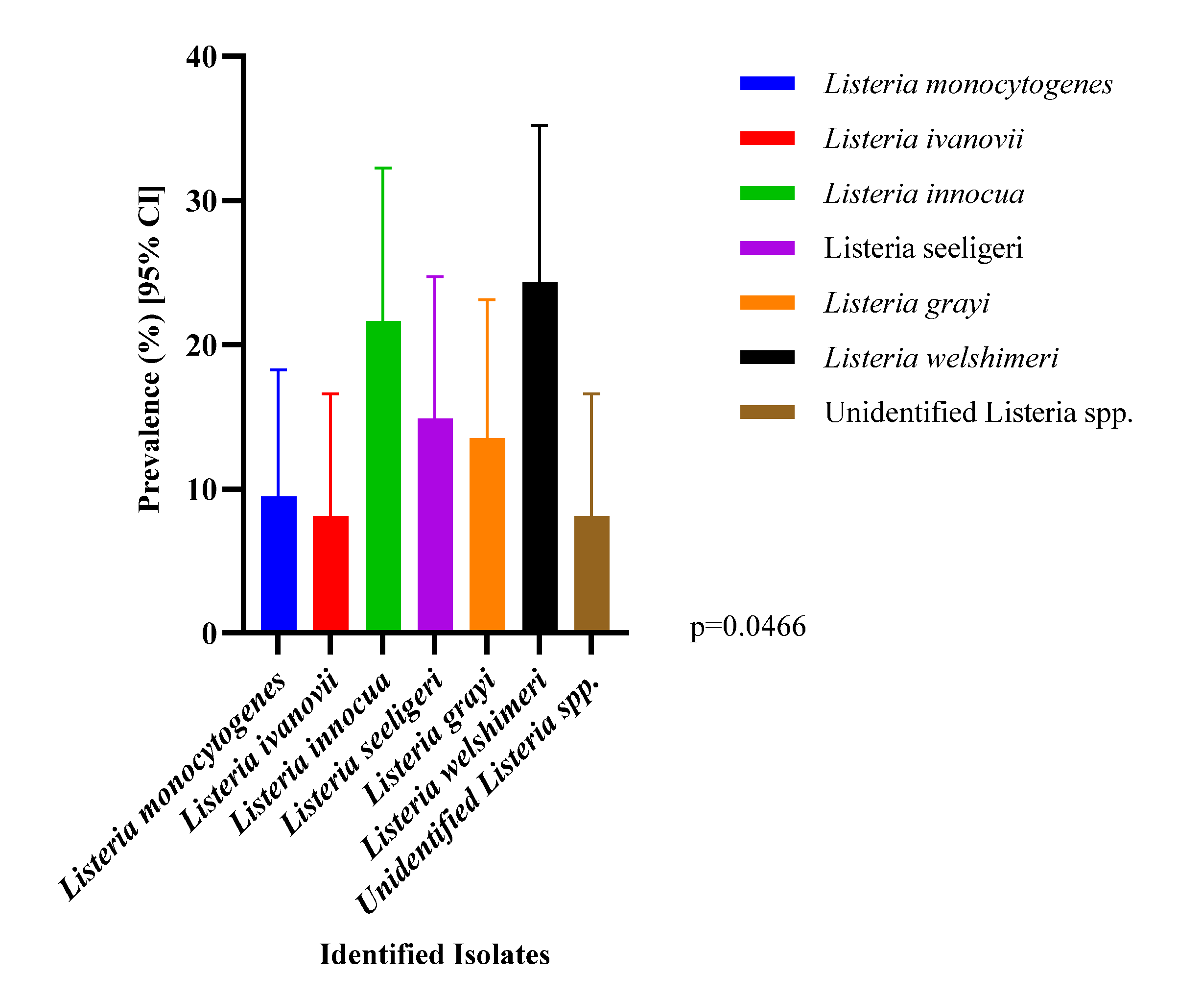
Figure 1. Molecular Speciation, Genus Diversity and Abundance of Isolates (n=74; N=866).
Preprints 222114 g001
Figure 2. Comparison of Listeria spp. Prevalence and calculated relative risk metrics across macro-environmental retail food (N=866).
Preprints 222114 g002
Figure 3. Distribution and Baseline Prevalence of Listeria spp. Contamination Across Individual Retail Food Matrices.
Preprints 222114 g003
Table 1. Oligonucleotide primer sequences and diagnostic amplicon configurations utilized for multiplex PCR speciation.
Target Group/
Species
Target
Gene
Primer
Name
Sequence (5′ → 3′) Product
Size (bp)
Diagnostic Role/Interpretation
Listeria Genus prs prs-F
prs-R
GCTGAAGAGATTGCGAAAGAAG
CAAAGAAACCTTGGATTTGCGG
370 Positive control/Genus
validation
L. monocytogenes hlyA hly-F
hly-R
GCAGTTGCAAGCGCTTGGAGTGAA
GCAACGTATCCTCCAGAGTGATCG
456 Clinical human
pathogen tracking
L. ivanovii namA liv22228F
liv22228R
CGAATTCCTTATTCACTTGAGC
GGTGCTGCGAACTTAACTCA
463 Veterinary ruminant pathogen tracking
L. innocua Lin0464 Lin0464F
Lin0464R
CGCATTTATCGCCAAAACTC
TGCTGACATAGACGCGATTG
749 Saprophytic environmental
indicator
L. seeligeri Lmo0333 lseelinF
lseelinR
GTACCTGCTGGAGTACATA
CTGTCTCCATATCCGTACAG
290 Saprophytic environmental
indicator
L. grayi Oxidoreductase JOgrayiF
JOgrayiR
GCGGATAAAGGTGTTCGGTCAA
ATTTGCTATCGTCCGAGGCTAGG
201 Saprophytic environmental
indicator
L. welshimeri scrA Lwe1801F
Lwe1801R
CGTGGCACAATAGCAATCTG
GACATGCCTGCTGAACTAGA
281 Saprophytic environmental
indicator
Table 2. The components of PCR master mix used for identification of presumptive Listeria spp.
Compound Volume (µL) Concentration
dH2O 11.8 -
5× Green GoTaq buffer 5
PCR Nucleotide mix 0.5 0.2mM
MgCl2, 25mM solution 4 4mM
Forward prs primer, 15 µM 1 0.6 µM
Reverse prs primer, 15 µM 1 0.6 µM
GoTaq DNA, polymerase, 5 U/µL 0.2 1 u/25 µL
Sum (master mix) 23.5 -
DNA Template 1.5 1.5 ng/25 µL
Total 25.0 -
Table 3. Distribution and Prevalence of Listeria species in fresh produce, meat and dairy samples from farms and retail outlets (n=866).
Food Category and
Individual Item Subtype
Total Listeria
isolates (N)
L. mono
% (n)
L. ivanovii
% (n)
L. innocua
% (n)
L. seeligeri
% (n)
L. grayi
% (n)
L. welshimeri
% (n)
Unknown Listeria spp.
% (n)
Overall Prevalence
% (n)
VEGETABLES 435 0.46 (2) 1.15 (5) 2.76 (12) 1.61 (7) 1.84 (8) 2.99 (13) 0.46 (2) 11.49 (50)
Cucumber 58 0.00 (0) 1.72 (1) 0.00 (0) 0.00 (0) 0.00 (0) 1.72 (1) 3.45 (2) 6.90 (4)
Lettuce 22 0.00 (0) 0.00 (0) 4.55 (1) 0.00 (0) 0.00 (0) 0.00 (0) 0.00 (0) 9.09 (2)
Alpha Sprouts + Alfalfa 23 4.35 (1) 0.00 (0) 4.35 (1) 0.00 (0) 4.35 (1) 4.35 (1) 0.00 (0) 17.39 (4)
Kale 36 0.00 (0) 2.78 (1) 5.56 (2) 2.78 (1) 8.34 (3) 2.78 (1) 0.00 (0) 22.22 (8)
Green onion 45 0.00 (0) 2.22 (1) 2.22 (1) 2.22 (1) 0.00 (0) 4.45 (2) 0.00 (0) 11.11 (5)
Spinach 60 0.00 (0) 0.00 (0) 3.33 (2) 5.00 (3) 1.66 (1) 3.33 (2) 0.00 (0) 13.33 (8)
Collard green 19 5.26 (1) 5.26 (1) 0.00 (0) 0.00 (0) 0.00 (0) 5.26 (1) 0.00 (0) 15.79 (3)
Romaine lettuce + Heart 36 0.00 (0) 0.00 (0) 2.78 (1) 2.78 (1) 5.56 (2) 5.56 (2) 0.00 (0) 16.67 (6)
Cilantro 24 0.00 (0) 0.00 (0) 8.33 (2) 0.00 (0) 0.00 (0) 4.17 (1) 0.00 (0) 12.50 (3)
Mixed lettuce 9 0.00 (0) 0.00 (0) 0.00 (0) 0.00 (0) 0.00 (0) 11.11 (1) 0.00 (0) 11.11 (1)
Carrot 9 0.00 (0) 0.00 (0) 0.00 (0) 0.00 (0) 0.00 (0) 0.00 (0) 0.00 (0) 0.00 (0)
Mixed vegetables 40 0.00 (0) 2.50 (1) 0.00 (0) 0.00 (0) 0.00 (0) 2.50 (1) 0.00 (0) 5.00 (2)
Tomato 54 0.00 (0) 0.00 (0) 3.70 (2) 1.85 (1) 1.85 (1) 0.00 (0) 0.00 (0) 7.41 (4)
EDIBLE FUNGI 22 0.00 (0) 0.00 (0) 9.09 (2) 4.55 (1) 0.00 (0) 0.00 (0) 0.00 (0) 13.64 (3)
Mushrooms 22 0.00 (0) 0.00 (0) 9.09 (2) 4.55 (1) 0.00 (0) 0.00 (0) 0.00 (0) 13.64 (3)
RAW MEAT 121 2.48 (3) 0.00 (0) 1.65 (2) 1.65 (2) 1.65 (2) 2.48 (3) 0.83 (1) 10.74 (13)
Raw Ground Beef 36 2.78 (1) 0.00 (0) 0.00 (0) 3.33 (1) 0.00 (0) 3.33 (1) 2.78 (1) 13.89 (5)
Raw Beef 30 0.00 (0) 0.00 (0) 3.33 (1) 0.00 (0) 0.00 (0) 3.33 (1) 0.00 (0) 6.67 (2)
Raw Chicken 42 2.38 (1) 0.00 (0) 2.38 (1) 2.38 (1) 2.38 (1) 0.00 (0) 0.00 (0) 9.52 (4)
Ground Pork 3 0.00 (0) 0.00 (0) 0.00 (0) 0.00 (0) 0.00 (0) 0.00 (0) 0.00 (0) 0.00 (0)
Raw Pork 10 10.00 (1) 0.00 (0) 0.00 (0) 0.00 (0) 10.00 (1) 0.00 (0) 0.00 (0) 20.00 (2)
UNPROCESSED DAIRY 48 4.17 (2) 2.08 (1) 0.00 (0) 2.08 (1) 0.00 (0) 4.17 (2) 2.08 (1) 12.50 (7)
Raw Milk 48 4.17 (2) 2.08 (1) 0.00 (0) 2.08 (1) 0.00 (0) 4.17 (2) 2.08 (1) 12.50 (7)
PROCESSED MEAT 39 0.00 (0) 0.00 (0) 0.00 (0) 0.00 (0) 0.00 (0) 0.00 (0) 0.00 (0) 0.00 (0)
Pork Ham 11 0.00 (0) 0.00 (0) 0.00 (0) 0.00 (0) 0.00 (0) 0.00 (0) 0.00 (0) 0.00 (0)
Pork Sausage 9 0.00 (0) 0.00 (0) 0.00 (0) 0.00 (0) 0.00 (0) 0.00 (0) 0.00 (0) 0.00 (0)
Beef Hotdog 16 0.00 (0) 0.00 (0) 0.00 (0) 0.00 (0) 0.00 (0) 0.00 (0) 0.00 (0) 0.00 (0)
Smoked Pork 3 0.00 (0) 0.00 (0) 0.00 (0) 0.00 (0) 0.00 (0) 0.00 (0) 0.00 (0) 0.00 (0)
PROCESSED DAIRY 201 0.00 (0) 0.00 (0) 0.00 (0) 0.00 (0) 0.00 (0) 0.00 (0) 0.50 (1) 0.50 (1)
Whole Milk 25 0.00 (0) 0.00 (0) 0.00 (0) 0.00 (0) 0.00 (0) 0.00 (0) 0.00 (0) 0.00 (0)
Milk (2%) 25 0.00 (0) 0.00 (0) 0.00 (0) 0.00 (0) 0.00 (0) 0.00 (0) 0.00 (0) 0.00 (0)
Mozzarella Cheese 17 0.00 (0) 0.00 (0) 0.00 (0) 0.00 (0) 0.00 (0) 0.00 (0) 0.00 (0) 0.00 (0)
Cream Cheese 14 0.00 (0) 0.00 (0) 0.00 (0) 0.00 (0) 0.00 (0) 0.00 (0) 7.14 (1) 7.14 (1)
Sandwich Cheese 9 0.00 (0) 0.00 (0) 0.00 (0) 0.00 (0) 0.00 (0) 0.00 (0) 0.00 (0) 0.00 (0)
Pepper Jack Cheese 12 0.00 (0) 0.00 (0) 0.00 (0) 0.00 (0) 0.00 (0) 0.00 (0) 0.00 (0) 0.00 (0)
Snack Cheese 3 0.00 (0) 0.00 (0) 0.00 (0) 0.00 (0) 0.00 (0) 0.00 (0) 0.00 (0) 0.00 (0)
Parmesan Cheese 9 0.00 (0) 0.00 (0) 0.00 (0) 0.00 (0) 0.00 (0) 0.00 (0) 0.00 (0) 0.00 (0)
Skimmed Milk Cheese 6 0.00 (0) 0.00 (0) 0.00 (0) 0.00 (0) 0.00 (0) 0.00 (0) 0.00 (0) 0.00 (0)
Cheddar Cheese 12 0.00 (0) 0.00 (0) 0.00 (0) 0.00 (0) 0.00 (0) 0.00 (0) 0.00 (0) 0.00 (0)
Feta Cheese 30 0.00 (0) 0.00 (0) 0.00 (0) 0.00 (0) 0.00 (0) 0.00 (0) 0.00 (0) 0.00 (0)
Yogurt 34 0.00 (0) 0.00 (0) 0.00 (0) 0.00 (0) 0.00 (0) 0.00 (0) 0.00 (0) 0.00 (0)
TOTAL STUDIED POOL 866 0.81 (7) 0.69 (6) 1.85 (16) 1.27 (11) 1.15 (10) 2.08 (18) 0.69 (6) 8.55 (74)
Table 4. Itemized phenotypic antimicrobial resistance, intermediate and susceptibility percentage distributions across individual classical Listeria isolates (n = 68).
L. MONOCYTOGENES
(N = 7)
R/I/S % (N)
L. IVANOVII
(N = 6)
R/I/S % (N)
L. INNOCUA
(N = 16)
R/I/S % (N)
L. SEELIGERI
(N = 11)
R/I/S % (N)
L. GRAYI
(N = 10)
R/I/S % (N)
L. WELSHIMERI
(N = 18)
R/I/S % (N)
AMIKACIN 0.0 (0)/0.0 (0)/100 (7) 0.0 (0)/16.7 (1)/83.3 (5) 0.0 (0)/0.0 (0)/100 (16) 0.0 (0)/0.0 (0)/100 (11) 0.0 (0)/20.0 (2)/80.0 (8) 0.0 (0)/0.0 (0)/100 (18)
AZITHROMYCIN 0.0 (0)/0.0 (0)/100 (7) 0.0 (0)/0.0 (0)/100 (6) 12.5 (2)/6.3 (1)/81.3 (13) 9.1 (1)/18.2 (2)/72.7 (8) 10.0 (1)/20.0 (2)/70.0 (7) 5.6 (1)/22.2 (4)/72.2 (13)
CHLORAMPHENICOL 0.0 (0)/14.3 (1)/85.7 (6) 16.7 (1)/50.0 (3)/33.3 (2) 18.8 (3)/18.8 (3)/62.5 (10) 0.0 (0)/0.0 (0)/100 (11) 0.0 (0)/0.0 (0)/100 (10) 5.6 (1)/5.6 (1)/88.9 (16)
CIPROFLOXACIN 14.3 (1)/0.0 (0)/85.7 (6) 33.3 (2)/16.7 (1)/50.0 (3) 12.5 (2)/25.0 (4)/62.5 (10) 0.0 (0)/0.0 (0)/100 (11) 30.0 (3)/30.0 (3)/40.0 (4) 22.2 (4)/11.1 (2)/66.7 (12)
CLINDAMYCIN 28.6 (2)/14.3 (1)/57.1 (4) 16.7 (1)/66.7 (4)/16.7 (1) 18.8 (3)/12.5 (2)/68.8 (11) 0.0 (0)/18.2 (2)/81.8 (9) 40.0 (4)/20.0 (2)/40.0 (4) 0.0 (0)/0.0 (0)/100 (18)
ERYTHROMYCIN 14.3 (1)/0.0 (0)/85.7 (6) 0.0 (0)/0.0 (0)/100 (6) 0.0 (0)/0.0 (0)/100 (16) 18.2 (2)/27.3 (3)/54.5 (6) 20.0 (2)/20.0 (2)/60.0 (6) 0.0 (0)/0.0 (0)/100 (18)
KANAMYCIN 14.3 (1)/14.3 (1)/71.4 (5) 50.0 (3)/0.0 (0)/50.0 (3) 25.0 (4)/25.0 (4)/50.0 (8) 27.3 (3)/36.4 (4)/36.4 (4) 30.0 (3)/20.0 (2)/50.0 (5) 16.7 (3)/27.8 (5)/55.6 (10)
LEVOFLOXACIN 42.9 (3)/0.0 (0)/57.1 (4) 66.7 (4)/0.0 (0)/33.3 (2) 18.8 (3)/31.3 (5)/50.0 (8) 18.2 (2)/36.4 (4)/45.5 (5) 40.0 (4)/20.0 (2)/40.0 (4) 0.0 (0)/22.2 (4)/77.8 (14)
MOXIFLOXACIN 14.3 (1)/0.0 (0)/85.7 (6) 16.7 (1)/16.7 (1)/66.7 (4) 18.8 (3)/12.5 (2)/68.8 (11) 18.2 (2)/9.1 (1)/72.7 (8) 20.0 (2)/0.0 (0)/80.0 (8) 16.7 (3)/22.2 (4)/61.1 (11)
NITROFURANTOIN 42.9 (3)/14.3 (1)/42.9 (3) 83.3 (5)/0.0 (0)/16.7 (1) 37.5 (6)/12.5 (2)/50.0 (8) 36.4 (4)/18.2 (2)/45.5 (5) 60.0 (6)/20.0 (2)/20.0 (2) 44.4 (8)/5.6 (1)/50.0 (9)
PENICILLIN G 57.1 (4)/0.0 (0)/42.9 (3) 66.7 (4)/0.0 (0)/33.3 (2) 25.0 (4)/0.0 (0)/75.0 (12) 36.4 (4)/18.2 (2)/45.5 (5) 50.0 (5)/10.0 (1)/40.0 (4) 11.1 (2)/5.6 (1)/83.3 (15)
RIFAMPIN 42.9 (3)/0.0 (0)/57.1 (4) 16.7 (1)/0.0 (0)/83.3 (5) 12.5 (2)/0.0 (0)/87.5 (14) 0.0 (0)/0.0 (0)/100 (11) 10.0 (1)/10.0 (1)/80.0 (8) 11.1 (2)/16.7 (3)/72.2 (13)
TETRACYCLINE 0.0 (0)/0.0 (0)/100 (7) 0.0 (0)/0.0 (0)/100 (6) 6.3 (1)/0.0 (0)/93.8 (15) 0.0 (0)/0.0 (0)/100 (11) 0.0 (0)/0.0 (0)/100 (10) 0.0 (0)/0.0 (0)/100 (18)
TOBRAMYCIN 0.0 (0)/0.0 (0)/100 (7) 0.0 (0)/0.0 (0)/100 (6) 0.0 (0)/0.0 (0)/100 (16) 0.0 (0)/0.0 (0)/100 (11) 0.0 (0)/0.0 (0)/100 (10) 5.6 (1)/0.0 (0)/94.4 (17)
TRIMETHOPRIM 14.3 (1)/0.0 (0)/85.7 (6) 16.7 (1)/0.0 (0)/83.3 (5) 12.5 (2)/6.3 (1)/81.3 (13) 18.2 (2)/0.0 (0)/81.8 (9) 30.0 (3)/30.0 (3)/40.0 (4) 11.1 (2)/0.0 (0)/88.9 (16)
TMP-SMX B 0.0 (0)/0.0 (0)/100 (7) 0.0 (0)/0.0 (0)/100 (6) 0.0 (0)/0.0 (0)/100 (16) 0.0 (0)/0.0 (0)/100 (11) 0.0 (0)/0.0 (0)/100 (10) 5.6 (1)/0.0 (0)/94.4 (17)
B TMP-SMX: Trimethoprim-Sulfamethoxazole. R-Resistant; I-Intermediate; S-Susceptible.
Disclaimer/Publisher’s Note: The statements, opinions and data contained in all publications are solely those of the individual author(s) and contributor(s) and not of MDPI and/or the editor(s). MDPI and/or the editor(s) disclaim responsibility for any injury to people or property resulting from any ideas, methods, instructions or products referred to in the content.
Copyright: This open access article is published under a Creative Commons CC BY 4.0 license, which permit the free download, distribution, and reuse, provided that the author and preprint are cited in any reuse.
Prerpints.org logo

Preprints.org is a free preprint server supported by MDPI in Basel, Switzerland.

Subscribe

© 2026 MDPI (Basel, Switzerland) unless otherwise stated

Accessibility

Disclaimer

Terms of Use

Privacy Policy

Privacy Settings