

Submitted:
07 July 2026
Posted:
08 July 2026
You are already at the latest version
Abstract
Maintenance of protein homeostasis (proteostasis) is essential for retinal integrity and visual function. The retina is among the most metabolically active tissues in the body, requiring precise control of protein synthesis, folding, trafficking, and degradation. Failure of these processes leads to accumulation of misfolded proteins, formation of toxic oligomers and aggregates, activation of cellular stress responses, and ultimately neuronal degeneration. Such mechanisms are increasingly recognized as central contributors to a broad spectrum of retinal disorders, including inherited retinal dystrophies, age-related macular degeneration, diabetic retinopathy, vitreoretinal amyloidoses, and retinal manifestations of systemic neurodegenerative diseases. Recent evidence indicates that protein aggregation, endoplasmic reticulum stress, impaired autophagy, ubiquitin–proteasome dysfunction, mitochondrial injury, and chronic neuroinflammation are shared pathogenic pathways across these disorders. Inherited forms of retinitis pigmentosa caused by rhodopsin mutations represent prototypical retinal conformational diseases, whereas age-related macular degeneration exhibits feature of a localized amyloidopathy characterized by extracellular deposits containing amyloid-β and other aggregation-prone proteins. Retinal abnormalities associated with Alzheimer's disease, Parkinson's disease, and prion disorders further support the concept that the retina mirrors molecular events occurring in the brain. This review examines current understanding of retinal proteinopathies, emphasizing mechanisms of proteostasis failure, disease-specific protein aggregates, and emerging therapeutic strategies aimed at restoring protein homeostasis. Understanding retinal conformational disorders may facilitate development of novel treatments and establish the retina as an accessible biomarker for neurodegenerative disease.
Keywords:
retina
; proteinopathy
; proteostasis
; protein misfolding
; amyloid
; retinitis pigmentosa
; age-related macular degeneration
; neurodegeneration
; α-synuclein
; transthyretin
Introduction
The correct folding of proteins into their native three-dimensional structures is fundamental for cellular function [1]. Protein folding is governed by intrinsic physicochemical properties and supported by a complex network of molecular chaperones and quality-control mechanisms collectively referred to as the proteostasis network [2,3]. Disturbances in these processes result in protein misfolding, aggregation, and accumulation of toxic intermediates that contribute to numerous human diseases [4].
The concept of conformational disease emerged from studies of neurodegenerative disorders characterized by abnormal protein aggregation, including Alzheimer’s disease, Parkinson’s disease, Huntington’s disease, and transmissible spongiform encephalopathies [5,6]. In recent years, similar mechanisms have been recognized in ophthalmic disorders, particularly those affecting the retina [7,8]. Because the retina originates from the neural tube and retains many structural and molecular characteristics of the central nervous system, it is especially susceptible to defects in proteostasis [9].
Photoreceptors continuously synthesize large quantities of proteins and renew their outer segments throughout life. Likewise, retinal pigment epithelial (RPE) cells process substantial amounts of photoreceptor-derived material and are exposed to chronic oxidative stress. These unique physiological demands make retinal cells highly vulnerable to disturbances in protein quality control [10,11,12].
Accumulating evidence suggests that retinal degeneration frequently results from the failure of proteastatic mechanisms. Misfolded proteins may accumulate intracellularly, form extracellular deposits, activate stress signaling pathways, impair mitochondrial function, and trigger inflammatory responses. Together, these processes contribute to progressive neuronal loss and visual dysfunction [9]
This review discusses retinal diseases through the framework of proteinopathy, highlighting common molecular mechanisms and disease-specific manifestations of proteostasis failure.
2. Proteostasis in the Retina
2.1. Molecular Chaperones
Molecular chaperones facilitate protein folding, prevent inappropriate intermolecular interactions, and promote refolding or degradation of damaged proteins. Heat-shock proteins (HSPs), including HSP70, HSP90, and small heat-shock proteins, are abundantly expressed in retinal cells and provide protection against oxidative stress and protein aggregation [13]. Chaperone dysfunction contributes to accumulation of aggregation-prone proteins and enhances susceptibility to retinal degeneration [14].
In the retina, these chaperone systems are tightly integrated with endoplasmic reticulum stress responses and the ubiquitin–proteasome and autophagy pathways, forming a coordinated proteostasis network essential for photoreceptor and retinal pigment epithelium (RPE) survival. When this balance is disrupted, sustained proteotoxic stress accelerates cellular dysfunction and neurodegenerative changes in retinal tissue [15].
2.2. The Ubiquitin–Proteasome System
The ubiquitin–proteasome system (UPS) selectively degrades damaged or misfolded proteins. Proteins destined for degradation are tagged with ubiquitin chains and subsequently processed by the proteasome. Defects in UPS function have been implicated in inherited retinal dystrophies, AMD, and diabetic retinopathy. Reduced proteasomal activity promotes accumulation of toxic protein species and amplifies cellular stress responses. In post-mitotic retinal neurons, where protein turnover is highly dependent on proteasomal efficiency, even modest UPS impairment can lead to progressive proteotoxic stress and synaptic dysfunction. Oxidative stress further exacerbates UPS inhibition, creating a feed-forward loop that accelerates retinal degeneration [16].
2.3. Autophagy and Lysosomal Pathways
Autophagy represents a major mechanism for degradation of large protein aggregates and damaged organelles. Efficient autophagic flux is essential for photoreceptor survival and RPE function. Impaired autophagy leads to accumulation of lipofuscin, dysfunctional mitochondria, and aggregated proteins, all of which contribute to retinal degeneration. Age-related decline in lysosomal acidification and autophagy efficiency further compromises the ability of RPE cells to process photoreceptor outer segment debris, thereby accelerating degenerative changes [17]. Because photoreceptors continually renew their outer segments and exhibit exceptionally high metabolic activity, they rely on tightly coordinated autophagic and lysosomal pathways to preserve cellular homeostasis under conditions of persistent oxidative and proteotoxic stress. Increasing evidence further suggests that crosstalk between autophagy, mitochondrial quality control (mitophagy), and endoplasmic reticulum stress responses forms an integrated proteostasis network whose dysfunction amplifies inflammation, cell death, and progressive retinal neurodegeneration.
2.4. Integration of the Retinal Proteostasis Network
The proteostasis network consists of interconnected pathways responsible for maintaining protein quality throughout the life of retinal cells. Molecular chaperones, the ubiquitin–proteasome system, autophagy, lysosomal degradation, and stress-response pathways function cooperatively to preserve cellular homeostasis. In photoreceptors, this network must accommodate exceptionally high rates of protein synthesis associated with continual renewal of outer segment discs [18,19,20].
Each photoreceptor replaces approximately 10% of its outer segment daily, requiring synthesis, transport, and degradation of large quantities of rhodopsin and other visual cycle proteins. Retinal pigment epithelial cells represent another major site of proteostatic demand. These cells phagocytose shed photoreceptor outer segments every day and must process large amounts of lipid-rich and protein-rich material. Failure of lysosomal degradation leads to accumulation of lipofuscin and other intracellular inclusions that compromise cellular function. Age-related decline in proteostasis efficiency further increases susceptibility to oxidative stress and aggregate formation [21,22,23].
Recent studies indicate that proteostasis pathways are highly interconnected. Impairment of proteasomal degradation can trigger compensatory activation of autophagy, whereas chronic ER stress can overwhelm both systems simultaneously. Understanding these interactions is important because therapeutic strategies targeting a single pathway may influence multiple components of the proteostasis network [24,25,26,27].
3. Molecular Mechanisms of Retinal Proteinopathy
3.1. Protein Misfolding and Aggregate Formation
Protein misfolding may arise from genetic mutations, post-translational modifications, oxidative damage, or age-related decline in cellular quality-control systems. Misfolded proteins can form various high molecular weight structures, e.g., soluble oligomers, intracellular inclusions, extracellular amyloid deposits, and insoluble fibrillar aggregates [28,29]. Increasing evidence indicates that soluble oligomeric species may be more toxic than mature aggregates [30,31,32,33].
3.2. Endoplasmic Reticulum Stress
Accumulating evidence indicates that prolonged ER stress in retinal cells—particularly in photoreceptors and RPE—activates PERK–eIF2α–ATF4 signaling and amplifies CHOP-dependent apoptosis, thereby linking unresolved proteostatic imbalance to progressive cell loss. In addition, ER stress can intersect with mitochondrial pathways and inflammatory signaling, further exacerbating retinal degeneration [34,35,36].
3.3. Oxidative Stress and Mitochondrial Dysfunction
The retina experiences continuous exposure to light, high oxygen consumption, and aDysfunctionbundant polyunsaturated lipids, creating conditions favorable for oxidative injury. In retinal cells, mitochondrial DNA damage and impaired respiratory chain activity further amplify reactive oxygen species (ROS) production, creating a self-propagating cycle of oxidative injury and bioenergetic failure. This oxidative–mitochondrial axis is particularly relevant in age-related retinal disorders, where chronic stress exceeds antioxidant defenses and accelerates photoreceptor and RPE degeneration [37,38,39].
3.4. Neuroinflammation
Sustained microglial activation in the retina promotes a shift toward a pro-inflammatory phenotype characterized by increased release of TNF-α, IL-1β, and complement components, which further amplifies neuronal stress. In parallel, complement cascade activation and recruitment of infiltrating immune cells exacerbate synaptic dysfunction and photoreceptor loss during progressive retinal degeneration [40,41,42,43].
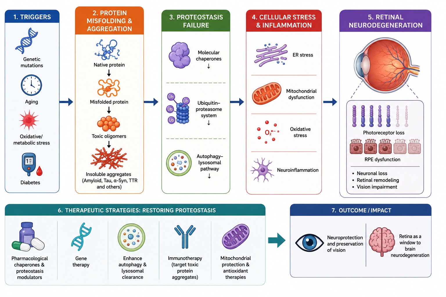
Figure 1.
Retinal conformational diseases: from protein misfolding to neurodegeneration. Conformational stress and proteostasis failure drive retinal neurodegeneration. Early detection and multi-targeted interactions are key to preserve vision.
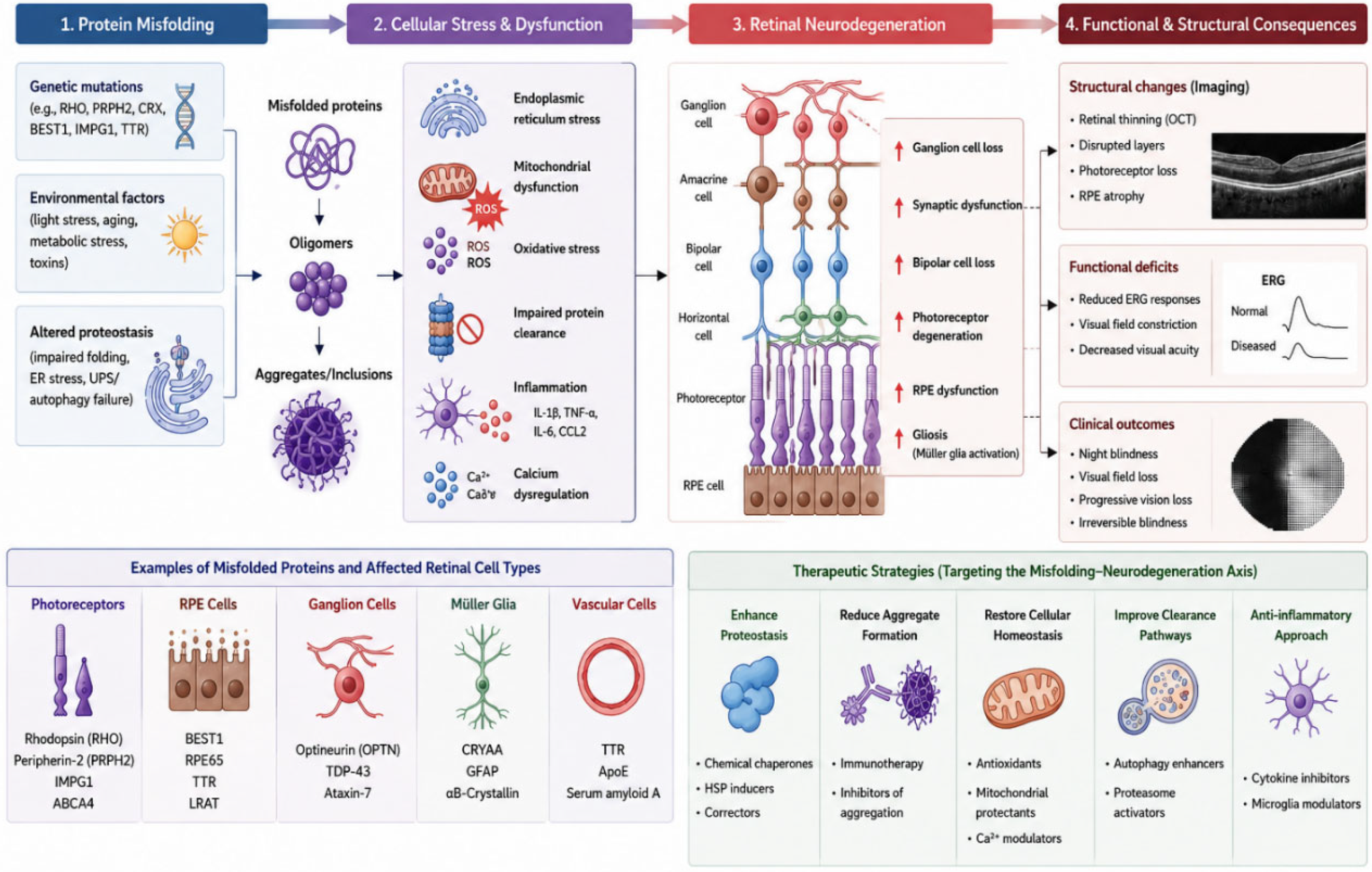
Figure 1.
Retinal conformational diseases: from protein misfolding to neurodegeneration. Conformational stress and proteostasis failure drive retinal neurodegeneration. Early detection and multi-targeted interactions are key to preserve vision.
4. Inherited Retinal Proteinopathies
4. Rhodopsin-Associated Retinitis Pigmentosa
Autosomal dominant retinitis pigmentosa caused by mutations in the rhodopsin gene represents one of the best-characterized retinal conformational diseases. Mutant rhodopsin proteins frequently fail to fold correctly, resulting in ER retention, aggregate formation, activation of the UPR and photoreceptor apoptosis. The P23H mutation has become a classic model for studying retinal protein misfolding. Rod opsin misfolding also disrupts its post-Golgi trafficking and impairs outer segment disc morphogenesis, leading to progressive structural instability of rod photoreceptors. Importantly, chronic ER stress in P23H models triggers both proteasomal overload and secondary activation of inflammatory and mitochondrial dysfunction pathways, thereby linking protein misfolding to multi-organellar collapse [44,45].
4.1. Other Inherited Retinal Dystrophies
Protein misfolding contributes to numerous inherited retinal disorders involving mutations in genes encoding: peripherin-2, RPGR, PRPF31, IMPDH1, ABCA4 and others. Although genetically heterogeneous, these disorders converge on common pathways involving proteostasis failure and cellular stress. Defective protein folding in these conditions often leads to impaired photoreceptor disc renewal, disrupted RNA splicing, or toxic accumulation of misprocessed visual cycle intermediates, depending on the affected gene product. Despite this diversity, sustained proteostatic imbalance consistently activates ER stress, mitochondrial dysfunction, and apoptotic signaling pathways that drive retinal cell loss [46]
4.2. Cell-Type Specific Vulnerability in Retinal Proteinopathies
Although proteostasis failure affects many retinal cell types, photoreceptors, retinal pigment epithelial cells, and retinal ganglion cells exhibit distinct patterns of vulnerability. Photoreceptors are particularly sensitive because of their high metabolic activity and continuous protein turnover. Rod photoreceptors produce enormous amounts of rhodopsin, making them susceptible to mutations that alter protein folding [47,48,49,50,51,52]
RPE cells are exposed to chronic oxidative stress generated by light exposure, mitochondrial respiration, and phagocytosis of photoreceptor outer segments. Consequently, impairment of autophagy or lysosomal degradation can rapidly lead to accumulation of damaged proteins and organelles. Retinal ganglion cells are especially vulnerable to mitochondrial dysfunction and axonal transport defects. Similar mechanisms contribute to neurodegeneration in glaucoma, Alzheimer’s disease, and Parkinson’s disease, suggesting convergence of pathogenic pathways throughout the nervous system [53,54,55,56].
5. Age-Related Macular Degeneration: A Retinal Amyloidopathy?
Age-related macular degeneration is the leading cause of irreversible vision loss among older adults in developed countries. A hallmark of AMD is the formation of drusen, extracellular deposits located between the RPE and Bruch’s membrane. Proteomic analyses have revealed that drusen contain numerous aggregation-prone proteins, including amyloid-β, apolipoproteins, complement factors, vitronectin, and clusterin [57].
5.1. Amyloid-β Accumulation
The identification of amyloid-β within drusen has led to the proposal that AMD shares important pathogenic features with cerebral amyloid disorders, including abnormal protein aggregation, chronic inflammation, and impaired clearance of extracellular deposits. Experimental studies further suggest that amyloid-associated activation of complement pathways and microglial cells contributes to RPE dysfunction and photoreceptor degeneration, supporting the view of AMD as a retinal amyloidopathy [58,59,60,61].
5.2. Tau Pathology
Abnormal phosphorylation and aggregation of tau proteins have also been reported in aging retinas and AMD tissue [62,63]. Tau-mediated disruption of axonal transport and cytoskeletal organization may contribute to retinal neuronal dysfunction. Accumulation of hyperphosphorylated tau in retinal ganglion cells and other retinal neurons has been associated with synaptic dysfunction, impaired intracellular trafficking, and increased susceptibility to oxidative stress. Furthermore, tau pathology may act synergistically with amyloid-β deposition and neuroinflammatory processes, thereby amplifying neurodegenerative changes in the aging retina and in AMD [64,65].
6. Transthyretin Amyloidosis and Vitreoretinal Degeneration
Transthyretin (TTR) is a transport protein synthesized predominantly in the liver and RPE. Mutations destabilizing the TTR tetramer promote amyloid formation and tissue deposition [66]. Ocular manifestations include: vitreous amyloid deposits, retinal vascular abnormalities, macular edema, and progressive visual loss. Because retinal TTR synthesis persists independently of hepatic production, ocular disease may continue despite successful liver transplantation [67]. Consequently, long-term ophthalmic surveillance remains essential in affected patients, even after systemic disease has been successfully treated, to enable early detection and management of progressive retinal and vitreous complications [68].
7. Proteostasis Dysfunction in Diabetic Retinopathy
Diabetic retinopathy has traditionally been considered a microvascular disease [69]. However, growing evidence supports an important role for neuronal injury and proteostasis impairment. Hyperglycemia induces protein glycation, ER stress, oxidative damage, impaired autophagy and mitochondrial dysfunction. These alterations precede overt vascular pathology and contribute to retinal neurodegeneration [70,71] Proteostasis dysfunction has therefore emerged as a central mechanism linking chronic metabolic stress to retinal neuronal injury, inflammation, and the progressive microvascular changes that characterize diabetic retinopathy [72].
8. Retinal Manifestations of Systemic Neurodegenerative Proteinopathies
8.1. Alzheimer’s Disease
In Alzheimer’s disease, retinal accumulation of amyloid-β and hyperphosphorylated tau has been reported in patients with Alzheimer’s disease. Structural changes include retinal thinning, synaptic alterations, and neuronal loss [73,74,75,76,77]. Retinal imaging and histopathological studies further suggest that amyloid-β deposition in the inner retinal layers correlates with disease severity and may parallel cerebral plaque burden. These findings support the concept that the retina mirrors central nervous system proteostatic failure, highlighting its potential as a readily accessible biomarker for the early diagnosis and monitoring of Alzheimer’s disease progression [77,78].
8.2. Parkinson’s Disease
In Parkinson’s disease, α-synuclein aggregates have been identified in retinal neurons and are associated with impaired visual processing. Retinal imaging studies demonstrate thinning of inner retinal layers and alterations in dopaminergic signaling. Retinal dopamine deficiency associated with degeneration of amacrine and ganglion cells may contribute to contrast sensitivity deficits and impaired color discrimination in Parkinson’s disease [79].These observations indicate that retinal α-synuclein pathology and proteostasis impairment closely reflect central neurodegenerative processes, supporting the retina as a promising biomarker for early diagnosis and disease monitoring in Parkinson’s disease [80].
8.3. Prion Diseases
Retinal prion accumulation has been detected in experimental models prior to overt CNS pathology, supporting the concept that the retina may serve as a peripheral site for early prion replication and detection [81,82]. This early retinal involvement is consistent with the neuroinvasive spread of prion proteins along interconnected neuronal pathways, including the optic nerve. As a result, the retina has been proposed as a potential accessible tissue for preclinical diagnosis and monitoring of transmissible spongiform encephalopathies. Collectively, these findings suggest that retinal proteostasis disruption parallels cerebral prion propagation and may provide a valuable platform for investigating disease pathogenesis, identifying early biomarkers, and evaluating therapeutic interventions targeting prion protein misfolding [81,82].
8.4. Prion-Like Propagation of Protein Aggregates
A growing body of evidence suggests that several aggregation-prone proteins may spread through neural tissues using mechanisms reminiscent of prion diseases. Although classical infectivity has not been demonstrated for most retinal proteinopathies, proteins such as amyloid-β, tau, α-synuclein, and transthyretin can act as self-templating [83,84,85,86,87,88,89]. A growing body of evidence suggests that several aggregation-prone proteins may spread through neural tissues using mechanisms reminiscent of prion diseases [90,91].
Although classical infectivity has not been demonstrated for most retinal proteinopathies, proteins such as amyloid-β, tau, α-synuclein, and transthyretin exhibit self-templating properties that enable misfolded aggregates to recruit native proteins into pathogenic conformations. This seeded aggregation can promote progressive accumulation and propagation of protein pathology within interconnected neural networks, thereby contributing to disease progression in both the retina and the brain [92].
Experimental studies indicate that misfolded proteins may be transferred between retinal cells through extracellular vesicles, tunneling nanotubes, synaptic connections, or release and uptake of aggregate-containing particles. Such propagation could explain the progressive spatial expansion of retinal pathology observed in several degenerative disorders [93,94]
The retina provides a unique model for investigating aggregate propagation because disease progression can be monitored noninvasively using advanced imaging techniques. Understanding the mechanisms governing aggregate spread may reveal new therapeutic opportunities aimed at interrupting disease transmission before widespread neuronal loss occurs [95,96,97].
9. The Retina as a Window to Neurodegeneration
The retina offers a unique opportunity for direct, non-invasive visualization of neural tissue [75,98,99]. Advances in optical coherence tomography, adaptive optics, hyperspectral imaging, and molecular imaging techniques have enhanced the ability to detect retinal changes associated with protein aggregation [100]]. Retinal biomarkers may ultimately facilitate: early diagnosis of neurodegenerative disease, monitoring of disease progression, assessment of therapeutic efficacy [78,98]. Longitudinal studies using OCT-based metrics have demonstrated that retinal nerve fiber layer and ganglion cell complex thinning can precede or parallel structural brain changes in neurodegenerative diseases. In addition, emerging retinal amyloid and tau imaging approaches suggest that molecular signatures of protein aggregation may be detectable in vivo before overt clinical symptoms appear. These advances support the integration of retinal imaging biomarkers into multimodal diagnostic frameworks for early neurodegeneration [101].
10. Therapeutic Strategies Targeting Proteostasis
10.1. Molecular and Pharmacological Chaperones
Pharmacological chaperones are small molecules that selectively bind unstable protein conformations and stabilize folding intermediates. By promoting correct folding, they can improve protein trafficking through the secretory pathway and reduce endoplasmic reticulum (ER)-associated degradation of mutant proteins. In many conformational diseases, this mechanism helps rescue partial loss-of-function mutants that would otherwise be targeted for degradation [102,103].
In retinal degenerative diseases, protein misfolding and chronic ER stress are key contributors to photoreceptor dysfunction and cell death. Pharmacological and molecular chaperones may therefore play a dual role: (i) directly stabilizing mutant photoreceptor proteins such as rhodopsin, improving their folding and localization, and (ii) enhancing broader cellular resilience to oxidative stress and ER stress responses. For example, 11-cis-retinal has been shown to act as a natural chaperone for opsin folding and trafficking, while chemical chaperones such as tauroursodeoxycholic acid (TUDCA) and 4-phenylbutyrate (4-PBA) reduce ER stress and improve photoreceptor survival in experimental models of retinal degeneration [104]. Together, these approaches highlight the therapeutic potential of modulating proteostasis networks in inherited retinal diseases.
10.2. Gene Therapy Approaches for Restoring Proteostasis in Retinal Degenerative Diseases
Gene therapy approaches can restore normal protein expression or silence toxic gain-of-function alleles, thereby addressing the primary cause of proteostasis imbalance [105,106]. Recent advances in AAV-mediated delivery have demonstrated sustained expression and functional rescue in several retinal degeneration models [107,108,109,110]. Gene therapy represents one of the most promising strategies for correcting the underlying molecular defects responsible for retinal degeneration and associated proteostasis imbalance. Unlike conventional pharmacological treatments that primarily address downstream pathological consequences, gene-based interventions have the potential to target the root cause of disease by restoring normal protein expression, suppressing toxic mutant proteins, or introducing protective genetic elements that enhance cellular resilience [111,112]. Because many inherited retinal disorders arise from mutations affecting proteins essential for photoreceptor function, retinal pigment epithelium (RPE) maintenance, or intracellular protein quality control pathways, gene therapy offers a rational and potentially long-lasting therapeutic approach [113].
The eye is particularly well suited for gene therapy applications due to its small size, relative immune privilege, compartmentalized anatomy, and accessibility for local delivery. Subretinal and intravitreal injection techniques permit direct administration of therapeutic vectors to target tissues while minimizing systemic exposure [106]. In addition, the fellow eye can often serve as an internal control during clinical studies, facilitating evaluation of treatment efficacy and safety. A major advance in the field has been the development of recombinant adeno-associated virus (AAV) vectors, which have emerged as the preferred delivery platform for retinal gene transfer. AAV vectors exhibit favorable safety profiles, low immunogenicity, and the capacity to mediate long-term transgene expression in both photoreceptors and RPE cells [106,114]. Numerous preclinical studies have demonstrated efficient transduction of retinal cells and sustained therapeutic benefits following AAV-mediated gene delivery. The clinical success of gene replacement therapy for RPE65-associated retinal dystrophy, culminating in regulatory approval of the AAV-based therapy Luxturna, provided proof of principle that genetic correction can restore visual function and alter disease progression in inherited retinal disorders [114,115].
Gene replacement strategies are particularly effective for recessive loss-of-function mutations in which disease results from the absence of a functional protein. Delivery of a normal copy of the affected gene can restore physiological protein synthesis, improve cellular homeostasis, and prevent progressive degeneration. In experimental models of retinal dystrophies caused by mutations in genes such as RPE65, CNGA3, CNGB3, and PDE6B, AAV-mediated gene supplementation has restored protein expression, preserved retinal structure, and improved visual function. These findings demonstrate that replenishing deficient proteins can re-establish proteostatic balance and reduce cellular stress responses associated with protein insufficiency [106,114].
In contrast, dominant retinal disorders often result from toxic gain-of-function mutations in which mutant proteins misfold, aggregate, or interfere with normal cellular processes. For these conditions, gene silencing approaches have attracted considerable attention [115,116,117] RNA interference (RNAi), antisense oligonucleotides (ASOs), and microRNA-based strategies can selectively reduce expression of pathogenic alleles while preserving or restoring normal protein function. Silencing mutant transcripts may reduce aggregate formation, alleviate endoplasmic reticulum stress, and decrease activation of apoptotic pathways. Such approaches are being investigated for disorders involving mutant rhodopsin, transthyretin, and other aggregation-prone proteins associated with retinal degeneration [118].
Recent advances in genome editing technologies have further expanded the therapeutic landscape. The CRISPR/Cas system enables precise modification of disease-causing mutations directly within the genome. Depending on the specific application, genome editing may correct pathogenic variants, disrupt toxic alleles, or insert protective genetic sequences [119,120]. Newer approaches including base editing and prime editing allow targeted nucleotide changes without generating double-stranded DNA breaks, potentially reducing the risk of off-target effects. In retinal disease models, CRISPR-based interventions have demonstrated successful mutation correction, preservation of photoreceptor survival, and improvement of retinal function [121,122].
Beyond correcting individual disease-causing genes, gene therapy can also be used to enhance the cellular proteostasis network itself. Delivery of genes encoding molecular chaperones, autophagy regulators, antioxidant enzymes, or neuroprotective factors may strengthen endogenous mechanisms responsible for protein folding, aggregate clearance, and stress adaptation. [123,124]. For example, overexpression of heat shock proteins such as HSP70 and HSP90 has been shown to improve protein quality control and reduce aggregation-induced toxicity in experimental models [125]. Similarly, gene transfer of autophagy-related proteins can enhance clearance of damaged proteins and dysfunctional organelles, thereby promoting cellular survival under conditions of chronic proteotoxic stress [126].
An emerging area of investigation involves the delivery of genes encoding trophic factors such as CNTF, BDNF, and GDNF. These molecules support neuronal survival, reduce inflammatory responses, and may indirectly improve proteostasis by enhancing cellular stress resistance. Long-term expression of neuroprotective factors through viral vectors could provide sustained protection against progressive retinal degeneration, regardless of the underlying genetic defect [127].
Gene therapy may also play an important role in treating retinal manifestations of systemic neurodegenerative diseases characterized by protein aggregation, including Alzheimer’s disease, Parkinson’s disease, and Huntington’s disease. Strategies designed to reduce production of amyloid-β, tau, or α-synuclein, enhance their clearance, or modulate cellular stress pathways may help prevent retinal neurodegeneration while simultaneously providing insights into disease mechanisms occurring within the brain [128]. Because retinal tissue can be visualized non-invasively, the retina may also serve as a valuable platform for monitoring therapeutic responses to gene-based interventions [129].
Despite remarkable progress, several challenges remain. AAV vectors have limited cargo capacity, restricting delivery of large genes such as ABCA4 and USH2A. Immune responses, variable transduction efficiency, and the need for precise targeting of specific retinal cell populations continue to present obstacles [107,130]. Moreover, long-term safety, durability of therapeutic effects, and potential off-target consequences of genome editing require continued investigation. Development of next-generation vectors with improved tropism, larger carrying capacity, and regulated transgene expression is expected to address many of these limitations [131].
Collectively, gene therapy approaches offer powerful opportunities to restore proteostasis and preserve retinal function by correcting genetic defects, suppressing toxic protein expression, enhancing cellular quality-control systems, and promoting neuronal survival. Continued advances in vector engineering, genome editing, and molecular understanding of proteostasis pathways are likely to expand the range of treatable retinal disorders and move the field closer to durable, disease-modifying therapies capable of preventing irreversible vision loss [132].
10.3. Autophagy Enhancement
Activation of autophagy pathways represents a promising therapeutic strategy for enhancing intracellular protein quality control in retinal degenerative diseases. By stimulating lysosomal degradation, autophagy promotes the clearance of misfolded and aggregated proteins as well as dysfunctional mitochondria and other damaged organelles, thereby restoring proteostasis and maintaining cellular homeostasis in stressed retinal neurons and retinal pigment epithelial cells [133]. Efficient autophagic flux also alleviates endoplasmic reticulum stress, reduces oxidative damage, and limits activation of pro-apoptotic signaling pathways that contribute to progressive photoreceptor and ganglion cell degeneration. [9,134]. Pharmacological modulation of autophagy has therefore emerged as an attractive approach to preserving retinal structure and function. Agents that inhibit the mammalian target of rapamycin (mTOR) pathway, such as rapamycin and its analogs, enhance autophagic activity by relieving suppression of the autophagy initiation complex, whereas activators of AMP-activated protein kinase (AMPK), including metformin and AICAR, stimulate autophagy through complementary signaling mechanisms that promote cellular energy homeostasis. In experimental models of inherited retinal degeneration, diabetic retinopathy, glaucoma, and age-related retinal injury, these pharmacological interventions have demonstrated neuroprotective effects by reducing protein aggregation, suppressing inflammation, improving mitochondrial integrity, and enhancing survival of retinal neurons [135]. Although additional studies are needed to optimize treatment timing, dosage, and long-term safety, modulation of autophagy remains one of the most promising strategies for slowing retinal degeneration and preserving vision by directly targeting the fundamental mechanisms underlying proteostasis dysfunction.
10.4. Anti-Amyloid Approaches
Anti-amyloid strategies seek to prevent the formation of toxic protein aggregates, inhibit the nucleation and propagation of amyloid fibrils, or facilitate the removal of existing deposits from neural tissues. These approaches have gained considerable attention in the treatment of neurodegenerative disorders, particularly Alzheimer’s disease, and are increasingly being explored for retinal diseases in which abnormal protein aggregation contributes to cellular dysfunction and degeneration. Accumulation of amyloidogenic proteins, including amyloid-β (Aβ), α-synuclein, tau, and other aggregation-prone proteins, disrupts proteostasis, impairs mitochondrial function, induces endoplasmic reticulum stress, and promotes oxidative injury within retinal neurons and retinal pigment epithelial (RPE) cells [9,136]. Consequently, therapies that reduce the burden of toxic protein aggregates may preserve retinal integrity and slow disease progression.
Passive immunotherapy using monoclonal antibodies directed against amyloid-β has demonstrated the ability to reduce cerebral amyloid burden and modestly slow cognitive decline in patients with early Alzheimer’s disease, providing proof of principle that immunological clearance of pathogenic protein aggregates is clinically feasible [137]. Although these therapies have been developed primarily for central nervous system disorders, similar approaches may be applicable to retinal neurodegeneration because the retina shares many pathological features with the brain, including extracellular amyloid deposition, neuroinflammation, and progressive neuronal loss [138].
Experimental studies have demonstrated that immunization against Aβ reduces retinal amyloid accumulation in transgenic mouse models [139]. Passive antibody-based therapies targeting pathogenic protein aggregates have also been shown to preserve retinal structure and function, reduce retinal degeneration, and improve visual function in experimental models. Although some active Aβ vaccination studies reported increased retinal vascular amyloid deposition and neuroinflammation despite reduced plaque burden, these findings collectively support the potential of immunotherapy to protect the retina from protein aggregation–associated neurodegeneration [139].
In addition to immunotherapy, numerous small-molecule compounds have been investigated for their ability to inhibit protein misfolding, stabilize native protein conformations, prevent oligomer formation, or promote disaggregation of existing fibrils. Several agents also enhance intracellular clearance mechanisms by stimulating autophagy and lysosomal degradation pathways, thereby reducing the intracellular accumulation of toxic protein species. Because soluble oligomeric intermediates are increasingly recognized as the most neurotoxic forms of many amyloidogenic proteins, therapeutic strategies targeting early stages of protein aggregation may prove more effective than those directed solely at mature fibrillar deposits [140].
Beyond directly reducing protein aggregation, anti-amyloid therapies may also mitigate secondary pathological mechanisms that contribute to retinal degeneration. Amyloid deposits can activate the complement cascade, stimulate microglial activation, and induce chronic production of pro-inflammatory cytokines, creating a self-perpetuating cycle of neuroinflammation and neuronal injury. Clearance of amyloid aggregates therefore has the potential to suppress complement-mediated tissue damage, reduce oxidative stress, preserve synaptic function, and improve survival of retinal neurons and retinal pigment epithelial cells. These anti-inflammatory effects may be particularly important in diseases such as age-related macular degeneration, diabetic retinopathy, glaucoma, and inherited retinal degenerations, where chronic inflammation amplifies the deleterious consequences of proteostasis failure [141].
Future therapeutic strategies are likely to combine anti-amyloid agents with complementary approaches that enhance protein quality-control systems, including pharmacological chaperones, autophagy activators, proteasome enhancers, gene-editing technologies, and neuroprotective compounds. Such multimodal interventions may simultaneously reduce aggregate formation, accelerate clearance of existing deposits, and restore cellular proteostasis, thereby providing more durable protection against progressive retinal degeneration. Continued improvements in molecular imaging and retinal biomarkers capable of detecting early protein aggregation will also facilitate patient stratification and enable monitoring of therapeutic responses in future clinical trials [9,142,143].
Future Perspectives
The emerging concept of retinal proteinopathies provides a unifying framework for understanding diverse retinal diseases, including inherited retinal degenerations, age-related macular degeneration, glaucoma, and retinal manifestations of systemic neurodegenerative disorders. Although the specific misfolded proteins differ among these conditions (e.g., rhodopsin, ABCA4, Aβ, tau, α-synuclein), many converge on shared pathogenic mechanisms involving protein misfolding, impaired proteostasis, intracellular and extracellular aggregate formation, mitochondrial dysfunction, ER stress, and chronic neuroinflammation [144]
A growing body of evidence indicates that proteostasis failure is an early and potentially reversible event preceding overt neuronal loss, suggesting a critical window for therapeutic intervention before irreversible degeneration occurs [145].
Future research should therefore prioritize the identification of early biomarkers of proteostasis imbalance, including molecular imaging of protein aggregates, retinal transcriptomic and metabolomic signatures, and functional readouts of cellular stress pathways [146]. Advances in adaptive optics, hyperspectral imaging, and targeted molecular probes now enable increasingly sensitive detection of early protein aggregation in vivo, supporting their potential use in early diagnosis and therapeutic monitoring [9,147].
In parallel, mechanistic studies are needed to elucidate how protein aggregates propagate within retinal circuits and whether prion-like mechanisms contribute to disease spread across retinal layers and potentially along the visual pathway. Understanding these processes may reveal new targets to halt disease progression at its earliest stages.
Finally, future therapeutic strategies are likely to emphasize restoration of cellular proteostasis, combining approaches such as pharmacological chaperones, autophagy enhancement, proteasome modulation, gene therapy, and anti-aggregation compounds. Such interventions may allow simultaneous reduction of toxic aggregate burden, restoration of protein quality-control systems, and preservation of retinal neuronal networks, ultimately preventing irreversible vision loss [148].
Conclusions
Retinal diseases associated with protein misfolding and aggregation represent an expanding class of neurodegenerative disorders. Inherited retinal dystrophies, age-related macular degeneration, transthyretin amyloidosis, diabetic retinopathy, and retinal manifestations of systemic proteinopathies all demonstrate the central importance of proteostasis in maintaining retinal integrity. Disruption of protein quality-control networks—including molecular chaperones, the ubiquitin–proteasome system, and autophagy–lysosomal pathways—emerges as a unifying pathogenic principle across these conditions [149].
Recognition of these shared mechanisms has transformed our understanding of retinal degeneration and highlights the concept of a convergent “proteostasis network failure” underlying both ocular and broader neurodegenerative diseases. This framework supports a shift from disease-specific models toward systems-level approaches that integrate protein homeostasis, metabolic stress, inflammation, and mitochondrial dysfunction as interconnected drivers of pathology [1,2,29].
From a translational perspective, these insights open new opportunities for early diagnosis, prevention, and treatment. The retina provides a uniquely accessible site for detecting early proteostatic imbalance through advanced imaging, molecular biomarkers, and functional assays, enabling a potential preclinical window for intervention. Therapeutic strategies targeting protein folding capacity, enhancing adaptive stress responses, modulating autophagy, or restoring proteostasis equilibrium represent promising avenues for disease modification. In parallel, gene-based and RNA-based approaches may allow precise correction of upstream folding defects in inherited disorders [150].
The retina serves as a valuable model system for investigating neurodegenerative processes within the central nervous system, providing a translational bridge between ophthalmology and neuroscience. Emerging evidence suggests that disruptions in proteostasis are a shared feature across retinal and br150oader neurodegenerative diseases, contributing to cellular dysfunction and progressive neuronal loss. Future research integrating systems biology, single-cell omics, and computational modeling will be essential to delineate disease-specific proteostasis signatures and identify patient-tailored therapeutic targets. Ultimately, defining mechanisms of proteostasis failure across retinal disorders may enable precision medicine approaches that transcend traditional diagnostic boundaries and support a unified, mechanism-based classification of neurodegenerative disease.
Importantly, the retina also serves as a model system for studying neurodegenerative processes throughout the central nervous system, offering a bridge between ophthalmology and neuroscience. Future research integrating systems biology, single-cell omics, and computational modeling will be essential to define disease-specific proteostasis signatures and identify patient-tailored therapeutic targets. Ultimately, a deeper understanding of proteostasis failure across retinal diseases may enable precision medicine approaches that transcend traditional disease boundaries and redefine neurodegenerative disease classification [97,151].
References
- Hartl, F.U.; Hayer-Hartl, M. Converging concepts of protein folding in vitro and in vivo. Nat. Struct. Mol. Biol. 2009, 16, 574–581. [Google Scholar] [CrossRef] [PubMed]
- Hartl, F.U.; Bracher, A.; Hayer-Hartl, M. Molecular chaperones in protein folding and proteostasis. Nature 2011, 475, 324–332. [Google Scholar] [CrossRef] [PubMed]
- Balchin, D.; Hayer-Hartl, M.; Hartl, F.U. In vivo aspects of protein folding and quality control. Science 2016, 353, aac4354. [Google Scholar] [CrossRef] [PubMed]
- Gandhi, J.; et al. Protein misfolding and aggregation in neurodegenerative diseases. Rev. Neurosci. 2019, 30, 339–358. [Google Scholar] [CrossRef] [PubMed]
- Soto, C. Unfolding the role of protein misfolding in neurodegenerative diseases. Nat. Rev. Neurosci. 2003, 4, 49–60. [Google Scholar] [CrossRef] [PubMed]
- Surguchov, A. Controversial properties of amyloidogenic proteins. Biomedicines 2023, 11, 1215. [Google Scholar] [CrossRef] [PubMed]
- Surguchev, A.; Surguchov, A. Conformational diseases: looking into the eyes. Brain Res. Bull. 2009, 81, 12–24. [Google Scholar]
- Masuzzo, A.; et al. Amyloidosis in retinal neurodegenerative diseases. Front. Neurol. 2016, 7, 127. [Google Scholar] [CrossRef] [PubMed]
- McLaughlin, T.; et al. Cellular stress signaling and unfolded protein response in retinal degeneration. Mol. Neurodegener. 2022, 17, 25. [Google Scholar] [CrossRef] [PubMed]
- Kevany, B.M.; Palczewski, K. Phagocytosis of photoreceptors. Physiology 2010, 25, 8–15. [Google Scholar] [CrossRef] [PubMed]
- Ambati, J.; et al. Immunology of age-related macular degeneration. Nat. Rev. Immunol. 2013, 13, 438–451. [Google Scholar] [CrossRef] [PubMed]
- Wimmers, S.; et al. Photoreceptor degeneration and stress responses. Front. Cell Dev. Biol. 2021, 9, 635123. [Google Scholar] [CrossRef]
- Piri, N.; et al. Heat shock proteins in the retina. Prog. Retin. Eye Res. 2016, 52, 22–46. [Google Scholar] [CrossRef] [PubMed]
- Kosmaoglou, M.; et al. Molecular chaperones and photoreceptor function. Prog. Retin. Eye Res. 2008, 27, 434–449. [Google Scholar] [CrossRef] [PubMed]
- Markitantova, Y.; Simirskii, V. RPE oxidative stress and autophagy. Int. J. Mol. Sci. 2025, 26, 1193. [Google Scholar] [CrossRef] [PubMed]
- Wang, A.L.; et al. Autophagy in the aged retina. Adv. Exp. Med. Biol. 2014, 801, 109–115. [Google Scholar]
- Kaarniranta, K.; et al. Autophagy dysfunction in AMD. Autophagy 2013, 9, 1031–1032. [Google Scholar] [CrossRef]
- Faber, S.; Roepman, R. Proteostasis network therapeutics in inherited retinal disease. Genes 2019, 10, 557. [Google Scholar] [CrossRef] [PubMed]
- Xu, J.; Zhao, C.; Kang, Y. Photoreceptor outer segment renewal. Cells 2024, 13, 1357. [Google Scholar] [CrossRef] [PubMed]
- Hsu, Y.C.; et al. Light regulates photoreceptor disc renewal. Dev. Cell 2015, 32, 731–742. [Google Scholar] [CrossRef] [PubMed]
- Ferrington, D.A.; Sinha, D.; Kaarniranta, K. RPE proteolysis defects in AMD. Prog. Retin. Eye Res. 2016, 51, 69–89. [Google Scholar] [CrossRef] [PubMed]
- Terman, A.; Brunk, U.T. Lipofuscin accumulation. Free Radic. Biol. Med. 2002, 33, 611–619. [Google Scholar] [CrossRef]
- Grune, T.; et al. Proteostasis, oxidative stress and aging. Redox Biol. 2017, 13, 550–567. [Google Scholar] [CrossRef]
- Hetz, C.; et al. UPR and proteostasis control. Nat. Cell Biol. 2015, 17, 829–838. [Google Scholar] [CrossRef] [PubMed]
- Chen, Y.; et al. ER homeostasis in retinal disease. Prog. Retin. Eye Res. 2024, 98, 101231. [Google Scholar]
- Dikic, I. Proteasomal and autophagic degradation. Annu. Rev. Biochem. 2017, 86, 193–224. [Google Scholar] [CrossRef] [PubMed]
- Kroeger, H.; et al. ER stress in retinal degeneration. Exp. Eye Res. 2014, 125, 30–40. [Google Scholar]
- Chiti, F.; Dobson, C.M. Protein misfolding and disease. Annu. Rev. Biochem. 2017, 86, 27–68. [Google Scholar] [CrossRef] [PubMed]
- Hartl, F.U. Protein misfolding diseases. Annu. Rev. Biochem. 2017, 86, 21–26. [Google Scholar] [CrossRef] [PubMed]
- Eisele, Y.S.; et al. Targeting aggregation in degenerative disease. Nat. Rev. Drug Discov. 2015, 14, 759–780. [Google Scholar] [CrossRef] [PubMed]
- Ross, C.A.; Poirier, M.A. Protein aggregation in neurodegeneration. Nat. Med. 2004, 10, S10–S17. [Google Scholar] [CrossRef] [PubMed]
- Haass, C.; Selkoe, D.J. Soluble oligomers in neurodegeneration. Nat. Rev. Mol. Cell Biol. 2007, 8, 101–112. [Google Scholar] [CrossRef] [PubMed]
- Phukan, H.; et al. Protein aggregation mechanisms. Process Biochem. 2016, 51, 1183–1192. [Google Scholar]
- Jing, G.; et al. ER stress and retinal apoptosis. Exp. Diabetes Res. 2012, 2012, 589589. [Google Scholar] [CrossRef] [PubMed]
- Liu, H.; et al. UPR in retinal degeneration. Mol. Neurodegener. 2022, 17, 15. [Google Scholar]
- Gorbatyuk, M.S.; et al. ER stress in retinal disease. Prog. Retin. Eye Res. 2020, 79, 100860. [Google Scholar] [CrossRef] [PubMed]
- Jarrett, S.G.; Boulton, M.E. Oxidative stress in AMD. Mol. Asp. Med. 2012, 33, 399–417. [Google Scholar] [CrossRef]
- Ozawa, Y. Retinal oxidative stress. Redox Biol. 2020, 37, 101779. [Google Scholar] [CrossRef] [PubMed]
- Fivenson, E.M.; et al. Mitochondrial dysfunction in aging retina. Antioxidants 2019, 8, 227. [Google Scholar] [CrossRef]
- Karlstetter, M.; et al. Retinal microglia in disease. Prog. Retin. Eye Res. 2015, 45, 30–57. [Google Scholar] [CrossRef] [PubMed]
- Rashid, K.; Akhtar-Schaefer, I.; Langmann, T. Microglia in retinal degeneration. Front. Immunol. 2019, 10, 1975. [Google Scholar] [CrossRef] [PubMed]
- Fan, W.; et al. Retinal microglia functions. Immunology 2022, 166, 268–286. [Google Scholar] [CrossRef] [PubMed]
- Akhtar-Schaefer, I.; et al. Innate immunity in retinal disease. EMBO Mol. Med. 2018, 10, e8259. [Google Scholar] [CrossRef]
- Bhattacharya, S.; et al. ERAD and rhodopsin P23H. Biochim. Biophys. Acta 2010, 1803, 424–434. [Google Scholar]
- Qiu, Y.; et al. Autophagy–proteasome balance in photoreceptors. Cell Death Dis. 2019, 10, 547. [Google Scholar] [CrossRef] [PubMed]
- Sizova, O.S.; et al. P23H rhodopsin signaling. Cell Signal. 2014, 26, 665–672. [Google Scholar] [CrossRef] [PubMed]
- Léveillard, T.; Sahel, J.A. Retinal metabolic signaling. Cell Mol. Life Sci. 2017, 74, 3649–3665. [Google Scholar] [PubMed]
- Lin, J.H.; LaVail, M.M. Misfolded proteins in retinal dystrophies. Adv. Exp. Med. Biol. 2010, 664, 115–121. [Google Scholar] [PubMed]
- Park, P.S. Rhodopsin aggregation. J. Membr. Biol. 2019, 252, 413–423. [Google Scholar] [CrossRef] [PubMed]
- Punzo, C.; et al. Metabolic dysregulation in retinal degeneration. J. Biol. Chem. 2012, 287, 1642–1648. [Google Scholar] [CrossRef] [PubMed]
- Fuhrmann, S.; et al. Aging retina degeneration. Prog. Mol. Biol. Transl. Sci. 2013, 118, 207–240. [Google Scholar]
- Ziaka, K.; van der Spuy, J. Hsp90 in retinal proteostasis. Biomolecules 2022, 12, 978. [Google Scholar] [CrossRef] [PubMed]
- Gauthier, A.C.; Liu, J. Glaucoma and neurodegeneration. Mol. Cell Neurosci. 2020, 786, 108323. [Google Scholar] [CrossRef]
- Duarte, J.; et al. Mitochondrial dysfunction in glaucoma. J. Ophthalmol. 2021, 4581909. [Google Scholar] [PubMed]
- Okan, I.C.T.; et al. Axonal transport defects. Adv. Exp. Med. Biol. 2023, 1415, 223–227. [Google Scholar] [CrossRef] [PubMed]
- Liang, Y.; et al. Axonal mitophagy in RGCs. Cell Commun. Signal. 2024, 22, 382. [Google Scholar] [CrossRef] [PubMed]
- Crabb, J.W.; et al. Drusen proteome. Proc. Natl. Acad. Sci. USA 2002, 99, 14682–14687. [Google Scholar] [CrossRef] [PubMed]
- Crabb, J.W. Drusen proteomics. Cold Spring Harb. Perspect. Med. 2014, 4, a017194. [Google Scholar] [CrossRef]
- Wang, L.; et al. Drusen composition. PLoS ONE 2010, 5, e10329. [Google Scholar] [CrossRef] [PubMed]
- Wang, J.; et al. Amyloid-β and complement activation. J. Cell Physiol. 2009, 220, 119–128. [Google Scholar] [CrossRef] [PubMed]
- Beclin, C.; et al. CCR2/CCL2 inflammation in retina. Neurobiol. Dis. 2011, 42, 55–72. [Google Scholar]
- Costagliola, C.; et al. Shared pathways in tauopathy and AMD. Front. Aging Neurosci. 2024, 16, 1371745. [Google Scholar] [CrossRef]
- Christinaki, E.; et al. Retinal imaging biomarkers. Clin. Exp. Optom. 2022, 105, 194–204. [Google Scholar] [PubMed]
- New, A.M.; et al. Retinal amyloid beta and tau pathology in age-related macular degeneration and Alzheimer’s disease. Prog. Retin. Eye Res. 2020, 78, 100843. [Google Scholar]
- Kaarniranta, K.; Pawlowska, E.; Szczepanski, M.J.; et al. Age-related macular degeneration (AMD): amyloid-β, complement, and inflammation in retinal degeneration. Prog. Retin. Eye Res. 2016, 50, 1–18. [Google Scholar]
- Ueda, M. Transthyretin: Its function and amyloid formation. Neurochem. Int. 2022, 155, 105313. [Google Scholar] [CrossRef] [PubMed]
- Beirão, N.M.; Matos, E.; Beirão, I.; Costa, P.P.; Torres, P. Ocular involvement in hereditary amyloidosis. Ophthalmol. Ther. 2021, 10, 845–864. [Google Scholar]
- Buxbaum, J.N.; Brannagan, T., 3rd; Buades-Reinés, J.; Cisneros, E.; Conceicao, I.; Kyriakides, T.; Merlini, G.; Obici, L.; Plante-Bordeneuve, V.; Rousseau, A.; Sekijima, Y.; Imai, A.; Waddington Cruz, M.; Yamada, M. Transthyretin deposition in the eye in the era of effective therapy for hereditary ATTRV30M amyloidosis. Amyloid 2019, 26(1), 10–14. [Google Scholar] [CrossRef] [PubMed]
- Wong, T.Y.; Cheung, C.M.G.; Larsen, M.; Sharma, S.; Simó, R. Diabetic retinopathy. Nat. Rev. Dis. Prim. 2016, 2, 16012. [Google Scholar] [CrossRef] [PubMed]
- Roy, S.; Trudeau, K.; Roy, S.; Tien, T.; Barrette, K.F. Mitochondrial dysfunction and endoplasmic reticulum stress in diabetic retinopathy: mechanistic insights into high glucose-induced retinal cell death. Curr. Clin. Pharmacol. 2013, 8(4), 278–84. [Google Scholar] [CrossRef] [PubMed]
- Volpe, C.M.O.; Villar-Delfino, P.H.; Dos Anjos, P.M.F.; Nogueira-Machado, J.A. Cellular death, reactive oxygen species (ROS) and diabetic complications. Cell Death Dis. 2018, 25;9(2), 119. [Google Scholar] [CrossRef] [PubMed]
- Toma, C.; Ferdeghini, D.; Ola Pour, M.M.; Venkatesan, S.; De Cillà, S.; Grossini, E. Oxidative Stress in Diabetic Retinopathy: Pathogenic Mechanisms, Biomarkers and Clinical Implications. Antioxidants 2026, 15(4), 425. [Google Scholar] [CrossRef] [PubMed]
- Hart, N.J.; Koronyo, Y.; Black, K.L.; Koronyo-Hamaoui, M. Ocular indicators of Alzheimer’s: exploring disease in the retina. Acta Neuropathol. 2016, 132, 767–787. [Google Scholar] [CrossRef] [PubMed]
- Liao, C.; Xu, J.; Chen, Y.; Ip, N.Y. Retinal Dysfunction in Alzheimer’s Disease and Implications for Biomarkers. Biomolecules 2021, 11, 1215. [Google Scholar] [CrossRef] [PubMed]
- Snyder, P.J.; Alber, J.; Alt, C.; Bain, L.J.; Bouma, B.E.; Bouwman, F.H.; DeBuc, D.C.; Campbell, M.C.W.; Carrillo, M.C.; Chew, E.Y.; Cordeiro, M.F.; Dueñas, M.R.; Fernández, B.M.; Koronyo-Hamaoui, M.; La Morgia, C.; Carare, R.O.; Sadda, S.R.; van Wijngaarden, P.; Snyder, H.M. Retinal imaging in Alzheimer’s and neurodegenerative diseases. Alzheimers Dement. 2021, 17(1), 103–111. [Google Scholar] [CrossRef] [PubMed]
- Masuzzo, A.; Dinet, V.; Cavanagh, C.; Mascarelli, F.; Krantic, S. Amyloidosis in Retinal Neurodegenerative Diseases. Front Neurol. 2016, 7, 127. [Google Scholar] [CrossRef] [PubMed]
- Ngolab, J.; Honma, P.; Rissman, R.A. Reflections on the Utility of the Retina as a Biomarker for Alzheimer’s Disease: A Literature Review. Neurol. Ther. 2019, 8 (Suppl 2), 57–72. [Google Scholar] [CrossRef] [PubMed]
- Czakó, C.; Kovács, T.; Ungvari, Z.; et al. Retinal biomarkers for Alzheimer’s disease and vascular cognitive impairment and dementia: implication for early diagnosis and prognosis. GeroScience. 2020, 42, 1499–1525. [Google Scholar] [CrossRef] [PubMed]
- Koronyo-Hamaoui, M.; Koronyo, Y.; Ljubimov, A.V.; Miller, C.A.; Ko, M.K.; Black, K.L.; Schwartz, M.; Farkas, D.L. Identification of amyloid plaques in retinas from Alzheimer’s patients and noninvasive in vivo optical imaging of retinal plaques in a mouse model. NeuroImage 2011, 54 (Suppl 1), S204–S217. [Google Scholar] [CrossRef] [PubMed]
- Ortuño-Lizarán, I.; Beach, T.G.; Serrano, G.E.; Walker, D.G.; Adler, C.H.; Cuenca, N. Phosphorylated α-synuclein in the retina is a biomarker of Parkinson’s disease pathology severity. Mov. Disord. 2018, 33(8), 1315–1324. [Google Scholar] [CrossRef] [PubMed]
- Striebel, J.F.; Race, B.; Leung, J.M.; Schwartz, C.; Chesebro, B. Prion-induced photoreceptor degeneration begins with misfolded prion protein accumulation in cones at two distinct sites: cilia and ribbon synapses. Acta Neuropathol. Commun. 2021, 9(1), 17. [Google Scholar] [CrossRef] [PubMed]
- Jucker, M.; Walker, L.C. Propagation and spread of pathogenic protein assemblies in neurodegenerative diseases. Nat. Neurosci. 2018, 21, 1341–1349. [Google Scholar] [CrossRef] [PubMed]
- Peng, C.; Trojanowski, J.Q.; Lee, V.M.-Y. Protein transmission in neurodegenerative disease. Nat. Rev. Neurol. 2020, 16, 199–212. [Google Scholar] [CrossRef] [PubMed]
- Soto, C.; Pritzkow, S. Protein misfolding, aggregation, and conformational strains in neurodegenerative diseases. Nat. Neurosci. 2018, 21, 1332–1340. [Google Scholar] [CrossRef] [PubMed]
- Kaufman, S.K.; Diamond, M.I. Prion-like propagation of protein aggregation and related therapeutic strategies. Neurotherapeutics 2013, 10, 371–382. [Google Scholar] [CrossRef] [PubMed]
- Mudher, A.; et al. What is the evidence that tau pathology spreads through prion-like propagation? Acta Neuropathol. Commun. 2017, 5, 99. [Google Scholar] [CrossRef] [PubMed]
- West Greenlee, M.H.; Lind, M.; Kokemuller, R.; et al. Temporal Resolution of Misfolded Prion Protein Transport, Accumulation, Glial Activation, and Neuronal Death in the Retinas of Mice Inoculated with Scrapie. Am. J. Pathol. 2016, 186(9), 2302–2309. [Google Scholar] [CrossRef] [PubMed]
- Weickenmeier, J.; Kuhl, E.; Goriely, A. Multiphysics of Prionlike Diseases: Progression and Atrophy. Phys. Rev. Lett. 2018, 121(15), 158101. [Google Scholar] [CrossRef] [PubMed]
- Walker, L. C.; Jucker, M. Neurodegenerative diseases: expanding the prion concept. Annu. Rev. Neurosci. 2015, 38, 87–103. [Google Scholar] [CrossRef] [PubMed]
- Brundin, P.; Ma, J.; Nathan, B. Prion-like transmission of protein aggregates in neurodegenerative diseases. Nat. Rev. Mol. Cell Biol. 2010, 11, 477–483. [Google Scholar] [CrossRef]
- Veys, L.; Van Houcke, J.; Aerts, J.; Van Pottelberge, S.; Mahieu, M.; Coens, A.; Melki, R.; Moechars, D.; De Muynck, L.; De Groef, L. Absence of Uptake and Prion-Like Spreading of Alpha-Synuclein and Tau After Intravitreal Injection of Preformed Fibrils. Front Aging Neurosci. 2021, 12, 614587. [Google Scholar] [CrossRef] [PubMed]
- Brunello, C.A.; Merezhko, M.; Uronen, R.L.; Huttunen, H.J. Mechanisms of secretion and spreading of pathological tau protein. Cell Mol. Life Sci. 2020, 77, 1721–1744. [Google Scholar] [PubMed]
- Thompson, A.G.; Gray, E.; Heman-Ackah, S.M.; Mäger, I.; Talbot, K.; Andaloussi, S.E.; Wood, M.J.; Turner, M.R. Extracellular vesicles in neurodegenerative disease—pathogenesis to biomarkers. Nat. Rev. Neurol. 2016, 12(6), 346–57. [Google Scholar] [CrossRef] [PubMed]
- Müller, P.L.; Wolf, S.; Dolz-Marco, R.; et al. Ophthalmic Diagnostic Imaging: Retina. In High Resolution Imaging in Microscopy and Ophthalmology; Springer, 2019. [Google Scholar]
- Bajwa, A.; Aman, R.; Reddy, A.K. A comprehensive review of diagnostic imaging technologies to evaluate the retina and the optic disk. Int. Ophthalmol. 2015, 35, 733–755. [Google Scholar] [CrossRef] [PubMed]
- London, A.; Benhar, I.; Schwartz, M. The retina as a window to the brain—from eye research to CNS disorders. Nat. Rev. Neurol. 2013, 9, 44–53. [Google Scholar] [PubMed]
- Nguyen, C.T.O.; Hui, F.; Charng, J.; et al. Retinal biomarkers provide “insight” into cortical pharmacology and disease. Pharmacol. Ther. 2017, 175, 151–177. [Google Scholar] [CrossRef] [PubMed]
- Gupta, S.; Zivadinov, R.; Ramanathan, M.; Weinstock-Guttman, B. Optical coherence tomography and neurodegeneration: are eyes the windows to the brain? Expert Rev. Neurother. 2016, 16, 765–775. [Google Scholar] [CrossRef] [PubMed]
- Jonnal, R.S.; Kocaoglu, O.P.; Zawadzki, R.J.; et al. Investigative Ophthalmology & Visual Science. 2016, 57, OCT51–OCT68. [Google Scholar] [PubMed]
- Asanad, S.; Fantini, M.; Sultan, W.; et al. Retinal nerve fiber layer thickness predicts CSF amyloid/tau before cognitive decline. PLoS ONE. 2020, 15, e0232785. [Google Scholar] [CrossRef] [PubMed]
- Loo, T. W.; Clarke, D. M. Chemical and pharmacological chaperones as new therapeutic agents. Expert Rev. Mol. Med. 2007, 9(16), 1–18. [Google Scholar] [CrossRef] [PubMed]
- Leidenheimer, N. J.; Ryder, K. G.; Noorwez, Syed M.; Malhotra, Ritu; McDowell, J. Hugh; Smith, Karen A.; Krebs, Mark P.; Kaushal, Shalesh. Retinoids Assist the Cellular Folding of the Autosomal Dominant Retinitis Pigmentosa Opsin Mutant P23H. J. Biol. Chem. 2014, 279(16, 2004), 16278–16284. [Google Scholar] [CrossRef] [PubMed]
- Noorwez, S.M.; Malhotra, R.; McDowell, J.H.; et al. Pharmacological Chaperone-mediated in Vivo Folding and Stabilization of the P23H-opsin Mutant Associated with Autosomal Dominant Retinitis Pigmentosa. J. Biol. Chem. 2003, 278(16), 14442–14450. [Google Scholar] [CrossRef] [PubMed]
- Binley, K.; Widdowson, P. S.; Loader, J.; Kelleher, M.; Iqball, S.; Ferryman, S.; Millington-Ward, S. Gene therapy for retinal degenerative diseases: Progress, challenges, and future directions. Int. J. Mol. Sci. 2023, 24(5), 4182. [Google Scholar] [PubMed Central]
- Trapani, I.; Auricchio, A. Gene therapy in inherited retinal degenerative diseases: A review. Curr. Gene Ther. 2019, 19(2), 77–89. [Google Scholar] [CrossRef] [PubMed]
- Ail, D.; Malki, H.; Zin, E.A.; Dalkara, D. Adeno-Associated Virus (AAV)-Based Gene Therapies for Retinal Diseases: Where are We? Appl. Clin. Genet. 2023, 16, 111–130. [Google Scholar] [CrossRef] [PubMed]
- Xia, J.; Gu, L.; Pan, Q. The landscape of basic gene therapy approaches in inherited retinal dystrophies. Front. Ophthalmol. 2023, 3, 1193595. [Google Scholar] [CrossRef] [PubMed]
- Petersen-Jones, S.M.; Beckwith-Cohen, B. Gene therapy advances using canine and feline animal models of inherited retinal degeneration. Eye 2025, 39, 2143–2150. [Google Scholar] [CrossRef] [PubMed]
- Buch, P.K.; Bainbridge, J.W.; Ali, R.R. AAV-mediated gene therapy for retinal disorders: from mouse to man. Gene Ther. 2008, 15(11), 849–857. [Google Scholar] [CrossRef] [PubMed]
- Drag, S.; Dotiwala, F.; Upadhyay, A.K. Gene Therapy for Retinal Degenerative Diseases: Progress, Challenges, and Future Directions. Investig. Ophthalmol. Vis. Sci. 2023, 64(7), 39. [Google Scholar] [CrossRef]
- Arbabi, A.; Liu, A.; Ameri, H. Gene Therapy for Inherited Retinal Degeneration. J. Ocul. Pharmacol. Ther. 2019, 35(2), 79–97. [Google Scholar] [CrossRef] [PubMed]
- Brar, A.S.; Parameswarappa, D.C.; Takkar, B.; et al. Gene Therapy for Inherited Retinal Diseases: From Laboratory Bench to Patient Bedside and Beyond. Ophthalmol. Ther. 2024, 13, 21–50. [Google Scholar] [CrossRef] [PubMed]
- Russell, S.; Bennett, J.; Wellman, J.A.; et al. Gene therapy and genome surgery in the retina. J. Clin. Investig. 2018, 128(6), 2177–2188. [Google Scholar] [CrossRef]
- Auricchio, Alberto; Trapani, Ivana. All roads lead to Rome: different gene therapy approaches for inherited retinal disorders. Hum. Gene Ther. 2021, 32(9–10), 529–540. [Google Scholar] [CrossRef]
- Narasimhan, Ishwarya; et al. Autosomal dominant retinitis pigmentosa with toxic gain of function: Mechanisms and therapeutics. Eur. J. Ophthalmol. 2021, 31(2), 304–320. [Google Scholar] [CrossRef] [PubMed]
- Farrar, G. Jane; Palfi, Arpad; O’Reilly, Mary. Gene therapeutic approaches for dominant retinopathies. Curr. Gene Ther. 2010, 10(5), 381–388. [Google Scholar] [CrossRef] [PubMed]
- Gemayel, Michael C.; Bhatwadekar, Ashay D.; Ciulla, Thomas. RNA therapeutics for retinal diseases. Expert Opin. Biol. Ther. 2021, 21(5), 603–613. [Google Scholar] [PubMed]
- Wu, Zhijian; Yu, Wenhan. In Vivo Applications of CRISPR-Based Genome Editing in the Retina. Front. Cell Dev. Biol. 2018, 6, 53. [Google Scholar] [CrossRef] [PubMed]
- MacLaren, Robert E.; Peddle, Caroline F. The Application of CRISPR/Cas9 for the Treatment of Retinal Diseases. Yale J. Biol. Med. 2017, 90(4), 533–541. [Google Scholar] [PubMed Central]
- Yee, Tiffany; Wert, Katherine J. Base and Prime Editing in the Retina—From Preclinical Research toward Human Clinical Trials. Int. J. Mol. Sci. 2022, 23(20), 12375. [Google Scholar] [CrossRef] [PubMed]
- Altay, H. Y. Y. Gene regulatory and gene editing tools and their applications for retinal diseases and neuroprotection. Front. Neurosci. 2022, 16, 924917. [Google Scholar] [CrossRef] [PubMed]
- Rowe, L.W.; Becerra, S.P.; MacLaren, R.E.; Avery, R.L.; Wykoff, C.C.; Ho, A.C.; Regillo, C.D.; Eliott, D.; Osborne, A.; Binley, K.M.; Ciulla, T.A. Gene-Agnostic Therapeutic Strategies for Inherited Retinal Diseases: Neuroprotection and Immunomodulation. Genes 2026, 17(4), 392. [Google Scholar] [CrossRef] [PubMed]
- Rhee, J.; Shih, K.C. Use of Gene Therapy in Retinal Ganglion Cell Neuroprotection: Current Concepts and Future Directions. Biomolecules 2021, 11(4), 581. [Google Scholar] [CrossRef] [PubMed]
- Nillegoda, N. B.; Bukau, B. Metazoan Hsp70-based protein disaggregases: emergence and mechanisms. Front. Mol. Biosci. 2015, 2, 57. [Google Scholar] [CrossRef] [PubMed]
- Sodhi, A.; et al. Intravitreal gene therapy restores the autophagy-lysosomal pathway and attenuates retinal degeneration in cathepsin D-deficient mice. Neurobiol. Dis. 2022, 164, 105628. [Google Scholar] [CrossRef] [PubMed]
- Ciulla, Thomas A.; Rowe, Lucas W.; et al. Gene-Agnostic Therapeutic Strategies for Inherited Retinal Diseases: Neuroprotection and Immunomodulation. Genes (Basel) 2026. Available online: https://www.mdpi.com/2073-4425/17/4/392. [CrossRef]
- Ngolab, J.; Honma, P.; Rissman, R.A. Reflections on the Utility of the Retina as a Biomarker for Alzheimer’s Disease: A Literature Review. Neurol. Ther. 2019, 8 (Suppl 2), 57–72. [Google Scholar] [CrossRef] [PubMed]
- Vujosevic, S.; Parra, M.M.; Hartnett, M.E.; et al. Optical coherence tomography as retinal imaging biomarker of neuroinflammation/neurodegeneration in systemic disorders in adults and children. Eye 2023, 37, 203–219. [Google Scholar] [CrossRef] [PubMed]
- Zin, E.A.; Ozturk, B.E.; Dalkara, D.; Byrne, L.C. Developing New Vectors for Retinal Gene Therapy. Cold Spring Harb. Perspect. Med. 2023, 13(12), a041291. [Google Scholar] [CrossRef] [PubMed]
- Naso, M.F.; Tomkowicz, B.; Perry, W.L.; et al. Adeno-Associated Virus (AAV) as a Vector for Gene Therapy. BioDrugs 2017, 31, 317–334. [Google Scholar] [CrossRef] [PubMed]
- McClements, M.E.; Elsayed, M.E.A.A.; Major, L.; et al. Gene Therapies in Clinical Development to Treat Retinal Disorders. Mol. Diagn. Ther. 2024, 28, 575–591. [Google Scholar] [CrossRef] [PubMed]
- Kaarniranta, K.; Tokarz, P.; Koskela, A.; et al. Autophagy regulates death of retinal pigment epithelium cells in age-related macular degeneration. Cell Biol. Toxicol. 2017, 33, 113–128. [Google Scholar] [CrossRef] [PubMed]
- Lin, W.J.; Kuang, H.Y. Oxidative stress induces autophagy in response to multiple noxious stimuli in retinal ganglion cells. Autophagy 2014, 10(10), 1692–701. [Google Scholar] [CrossRef] [PubMed]
- Ou, K.; Li, Y.; Liu, L.; Li, H.; Cox, K.; Wu, J.; Liu, J.; Dick, A.D. Recent developments of neuroprotective agents for degenerative retinal disorders. Neural Regen. Res. 2022, 17(9), 1919–1928. [Google Scholar] [CrossRef] [PubMed]
- Zhang, S.X.; Wang, J.J.; Starr, C.R.; Lee, E.J.; Park, K.S.; Zhylkibayev, A.; Medina, A.; Lin, J.H.; Gorbatyuk, M. The endoplasmic reticulum: Homeostasis and crosstalk in retinal health and disease. Prog. Retin Eye Res. 2024, 98, 101231. [Google Scholar] [CrossRef] [PubMed]
- Swanson, C.J.; Zhang, Y.; Dhadda, S.; et al. A randomized, double-blind, phase 2b proof-of-concept clinical trial in early Alzheimer’s disease with lecanemab, an anti-Aβ protofibril antibody. Alz Res. Ther. 2021, 13, 80. [Google Scholar] [CrossRef] [PubMed]
- van Dyck, C.H.; Swanson, C.J.; Aisen, P.; Bateman, R.J.; Chen, C.; Gee, M.; Kanekiyo, M.; Li, D.; Reyderman, L.; Cohen, S.; Froelich, L.; Katayama, S.; Sabbagh, M.; Vellas, B.; Watson, D.; Dhadda, S.; Irizarry, M.; Kramer, L.D.; Iwatsubo, T. Lecanemab in Early Alzheimer’s Disease. N Engl. J. Med. 2023, 388(1), 9–21. [Google Scholar] [CrossRef] [PubMed]
- Liu, B.; Rasool, S.; Yang, Z.; Glabe, C.G.; Schreiber, S.S.; Ge, J.; Tan, Z. Amyloid-peptide vaccinations reduce {beta}-amyloid plaques but exacerbate vascular deposition and inflammation in the retina of Alzheimer’s transgenic mice. Am. J. Pathol. 2009, 175(5), 2099–110. [Google Scholar] [CrossRef] [PubMed]
- Verma, M.; Vats, A.; Taneja, V. Toxic species in amyloid disorders: Oligomers or mature fibrils. Ann. Indian Acad. Neurol. 2015, 18(2), 138–45. [Google Scholar] [CrossRef] [PubMed]
- Xu, H.; Chen, M. Targeting the complement system for the management of retinal inflammatory and degenerative diseases. Eur. J. Pharmacol. 2016, 787, 94–104. [Google Scholar] [CrossRef] [PubMed]
- Tzekov, R.; Stein, L.; Kaushal, S. Protein misfolding and retinal degeneration. Cold Spring Harb. Perspect. Biol. 2011, 3(11), a007492. [Google Scholar] [CrossRef] [PubMed]
- Balch, W.E.; Morimoto, R.I.; Dillin, A.; Kelly, J.W. Adapting proteostasis for disease intervention. Science. 2008, 319(5865), 916–919. [Google Scholar] [CrossRef] [PubMed]
- Parfitt, D.A.; Cheetham, M.E. Targeting the proteostasis network in rhodopsin retinitis pigmentosa. Adv. Exp. Med. Biol. 2016, 854, 479–484. [Google Scholar] [CrossRef] [PubMed]
- Labbadia, J.; Morimoto, R.I. The biology of proteostasis in aging and disease. Annu Rev. Biochem. 2015, 84, 435–464. [Google Scholar] [CrossRef] [PubMed]
- Höhn, A.; Tramutola, A.; Cascella, R. Proteostasis failure in neurodegenerative diseases: focus on oxidative stress. Oxid. Med. Cell Longev. 2020, 2020, 5497046. [Google Scholar] [CrossRef] [PubMed]
- Kulenkampff, K.; Wolf Perez, A.M.; Sormanni, P.; et al. Quantifying misfolded protein oligomers as drug targets and biomarkers in Alzheimer and Parkinson diseases. Nat. Rev. Chem. 2021, 5, 277–294. [Google Scholar] [CrossRef] [PubMed]
- Kuzu, O.F.; Granerud, L.J.T.; Saatcioglu, F. Navigating the landscape of protein folding and proteostasis: from molecular chaperones to therapeutic innovations. Sig Transduct. Target Ther. 2025, 10, 358. [Google Scholar] [CrossRef] [PubMed]
- Livesey, F. J.; Cepko, C. L. Vertebrate neural cell-fate determination: lessons from the retina. Nat. Rev. Neurosci. 2001, 2, 109–118. [Google Scholar] [CrossRef] [PubMed]
- Chinchore, Y.; Gouras, P.; Michaels, K. Proteostasis in the retina: regulation and implications for retinal degeneration. Translational Neurodegeneration 2017. [Google Scholar]
- Ratnayaka, J.; Serpell, L.; Lotery, A. Dementia of the eye: the role of amyloid beta in retinal degeneration. Eye 2015, 29, 1013–1026. [Google Scholar] [CrossRef] [PubMed]
- Sorrentino, F. S.; Pizzimenti, L.; Bonifazzi, C. Role of the retina in neurodegenerative diseases. Prog. Retin. Eye Res. 2016, 50, 1–19. [Google Scholar] [CrossRef]
Disclaimer/Publisher’s Note: The statements, opinions and data contained in all publications are solely those of the individual author(s) and contributor(s) and not of MDPI and/or the editor(s). MDPI and/or the editor(s) disclaim responsibility for any injury to people or property resulting from any ideas, methods, instructions or products referred to in the content. |
© 2026 by the authors. Licensee MDPI, Basel, Switzerland. This article is an open access article distributed under the terms and conditions of the Creative Commons Attribution (CC BY) license (http://creativecommons.org/licenses/by/4.0/).
Copyright: This open access article is published under a Creative Commons CC BY 4.0 license, which permit the free download, distribution, and reuse, provided that the author and preprint are cited in any reuse.


