
Submitted:
07 July 2026
Posted:
08 July 2026
You are already at the latest version
Abstract
Music therapy is established in oncology as a supportive intervention to reduce anxiety, pain, distress and treatment-related symptom burden. Beyond these clinical effects, emerging experimental evidence suggests that audible sound and music may also exert direct biological effects on non-auditory cells, including cancer cells. This review summarizes current evidence on the effects of audible sound, music and acoustic stimulation on cancer cell viability and related cellular processes. Sound represents a mechanical pressure wave that may interact with cells through mechanotransductive processes involving the plasma membrane, cytoskeleton, integrins, focal adhesions and mechanosensitive ion channels. Since cancer cells frequently display altered stiffness, cytoskeletal organization, membrane dynamics and mechano-sensitivity, they may respond differently to acoustic or vibrational stimulation than non-malignant cells. Available in vitro studies indicate that audible sound, music and acoustic stimulation can modulate cancer cell viability, proliferation, migration, apoptosis-related signalling, gene expression and extracellular vesicle secretion. These effects have been reported in breast, gastric, prostate, glioblastoma, colon, ovarian and neuroblastoma cell models, but appear highly cell-type specific and dependent on exposure parameters. Proposed mechanisms include intracellular calcium signalling, cytoskeletal remodelling, mitochondrial dysfunction, reactive oxygen species generation and pathways involving PIEZO1, integrins, focal adhesion kinase and YAP/TAZ. However, much of the mechanistic framework derives from ultrasound-based research and cannot be directly extrapolated to audible sound or music. Conclusively, current findings suggest that audible sound and music may act as biologically active stimuli and modulators on cancer cels. However, the evidence is still in a preliminary stage and heterogeneous. Standardized studies are required before these observations can be translated into mechanistically defined or therapeutically relevant concepts in oncology.
Keywords:
sound
; music
; cancer cells
; viability
1. Introduction
Cancer remains one of the leading causes of mortality worldwide despite major advances in diagnostics and therapies. Complementary therapeutic approaches that have the potential to improve cancer treatment outcome have gained increased attention in recent years. Among these emerging areas, the biological effects of music and acoustic stimulation on early developmental stages of cancer have attracted growing scientific interest. While music therapy has long been employed in oncology to reduce anxiety, pain, and psychological distress in patients [1], emerging research suggests that music and acoustic stimuli may also exert direct effects on cellular physiology, including cancer cell viability and proliferation [2,3].
Traditionally, the effects of music on humans have been understood primarily through neuropsychological and emotional pathways mediated by the auditory system. However, there are observations from the biologist Helene Grimal and Fabien Maman from the 1980s who studied the relationship of sound waves to living cells. With a camera mounted on a microscope, they observed uterine cancer cells exposed to different acoustic instruments such as guitar, gong, xylophone as well as the human voice and found that cancer cells lost structural integrity until they exploded [4]. The first experimental, peer reviewed studies published stem from Lestard and colleagues who demonstrated that non-auditory cells in vitro can also respond to music and sound exposure [2,3]: in their studies, exposure to different musical compositions altered the viability, migration, and metabolic activity of cultured human cancer cell lines. These observations point towards evidence that acoustic energy itself may modulate cancer cells through direct biomechanical interactions.
From a biophysical perspective, music are vibrations transmitted in the form of pressure waves. Cells are sensitive to mechanical stimulation and continuously interact with their physical microenvironment through mechano-transduction [5,6]. Vibrations can influence cytoskeletal organization, membrane permeability, ion channel activation, intracellular signalling pathways, and gene expression [5]. Importantly, cancer cells often exhibit altered mechanical properties compared with healthy cells, such as structural changes, cytoskeletal disorganization, and abnormal membrane dynamics [6,7]. These differences raise the hypothesis that malignant cells may respond differently to acoustic or vibrational stimulation than non-malignant cells.
Beyond music exposure itself, related research fields have explored the effects of mechanic pressure waves on tumor cells, for example ultrasound, resonance-based therapies, and electromagnetic frequency modulation (8-10) [7,8,9]. Experimental approaches such as focused ultrasound [10] and the theoretical concept of “oncotripsy” suggest that cancer cells may possess unique mechanical resonance frequencies that could potentially be exploited for selective therapeutic targeting [8]. Furthermore, studies using tumor-specific amplitude-modulated radiofrequency electromagnetic fields have reported inhibitory effects on cancer cell proliferation in vitro and in clinical pilot investigations [7]. Although many of these approaches remain experimental and controversial, they collectively support the broader concept that physical forces and acoustic energy may influence cancer cell behavior.
Despite the heterogeneity of parameters from existing studies such as experimental conditions, cancer models, and outcome measurements, the observed interactions between sound-induced mechanical changes and tumor cell biology represent a scientifically relevant area worthwhile of systematic examination.
The aim of this review is therefore to summarize and critically evaluate the current experimental evidence regarding the effects of music, sound frequencies, and acoustic stimulation on cancer cell viability. In addition, we discuss potential mechanobiological mechanisms underlying these effects, highlights major methodological limitations within the field, and outlines future directions for research at the intersection of acoustics, mechano-transduction, and oncology.
2. Biological Mechanisms of Sound-Cell Interactions
Sound is a mechanical form of energy that propagates through a medium as oscillating pressure waves. Depending on their frequency, amplitude, and duration, sound waves can induce physical forces capable of interacting with biological tissues and individual cells.
Sound is not only subject to auditory perception but may directly influence cellular behaviour through biomechanical and biochemical mechanisms independent of the nervous system [6,7]. At a cellular level, mechanical stimuli are converted into biochemical signals through a process known as mechano-transduction. Cells continuously sense and respond to the physical properties of their microenvironment, including pressure, stiffness, tension, and vibration (6) [4]. This process is mediated by various cellular and membrane structures such as integrins, adhesion complexes, stretch-activated ion channels, the extracellular matrix, and the cytoskeleton (6, 7, 11) [4,5]. Mechanical forces transmitted through these structures can therefore induces cell membrane deformation, alter cytoskeletal organization, intracellular signalling pathways, gene expression, membrane permeability, and remodels extracellular matrix (ECM). Particularly the cytoskeleton plays a central role in cellular mechano-sensing. The cytoskeleton consists primarily of actin filaments, microtubules, and intermediate filaments that maintain structural integrity and facilitate intracellular force transmission. Acoustic vibrations may generate oscillatory mechanical stress within these structures, potentially influencing cell morphology, motility, and survival. Baumgartner and colleagues recently showed in an in vitro wound healing model that mechanic vibration alters the actin cytoskeleton, leading to a more directed and accelerated cell migration with a higher filament alignment along the wave´s propagation axis in surface acoustic wave wound healing [12].
Mechanical deformation of the plasma membrane can additionally activate mechanosensitive ion channels by the application of force to integrins, resulting in calcium influx into the cytoplasm which may modulate contractility through calmodulin–caldesmon interactions and thereby actively influence cytoskeletal organization [13]. An important mechanosensitive ion channel is Piezo1. Activation of Piezo1 results in the influx of ions such as Ca2+ thus triggering multiple signalling pathways such as PI3K/Akt and membrane type 1-matrix metalloproteinase (MT1-MMP) [14,15]. It is implicated in processes like vascular remodelling and angiogenesis and downstream signalling cascades associated with proliferation, stress responses, and apoptosis. Interestingly, there is an interaction between Piezo1 and the cytoskeleton: the loss of the actin cytoskeleton makes Piezo1 more sensitive and easier to activate. Thus, the cytoskeleton acts as a membrane tension regulator, playing a mechano-protective role at the same time affecting Piezo1 activity [16].
3. Mechanisms of Sound - Cancer Cell Interactions
Structural changes of the cytoskeletal are highly relevant in cancer because cytoskeletal plasticity controls migration, invasion, epithelial–mesenchymal transition and resistance to mechanical stress. Cancer cells are known to possess altered biomechanical properties and mechano-sensitivity compared with healthy cells. Malignant cells often exhibit reduced stiffness, cytoskeletal disorganization, altered adhesion dynamics, and increased deformability [7,17,18]. These characteristics contribute to tumor progression and metastatic potential but may also increase susceptibility to external mechanical stimuli. Several studies have therefore proposed that cancer cells may respond differently to acoustic or vibrational energy than non-malignant cells [19,20,21,22,23]. Mechanosensitive signalling pathways involving FAK, YAP/TAZ, integrins, and PIEZO channels play central roles in cancer cell adaptation to mechanical stress. Acoustic stimulation exploits these pathways by introducing oscillatory forces that disrupt mechanical homeostasis and activate downstream signalling cascade [24]. One proposed mechanism involves resonance. Resonance occurs when an external vibration matches the natural oscillatory frequency of a structure, thereby amplifying mechanical motion. The theoretical concept of “oncotripsy,” introduced by Heyden and Ortiz, suggests that cancer cells may possess unique resonance frequencies due to differences in cellular geometry, elasticity, and cytoskeletal architecture [25]. Dynamical models demonstrated that harmonic excitation at specific ultrasonic frequencies could selectively disrupt malignant cells while sparing healthy tissue [19]. Acoustic stimulation has been shown to activate intracellular calcium oscillations, cytoskeletal remodelling, mitochondrial signalling, reactive oxygen species (ROS) production, apoptotic pathways, and immune-related signalling cascades [26].
Though audible acoustic waves demonstrated measurable effects on mammalian cell, the strongest evidence exists for ultrasound-based interventions. The central early response to acoustic stimulation is the induction of intracellular calcium oscillations. Ultrasound-generated mechanical forces can deform the plasma membrane and activate mechanosensitive channels such as PIEZO1, thereby promoting calcium influx. In tumor cells, this calcium entry has been linked to downstream activation of calpain, mitochondrial dysfunction and apoptosis. Tijore et al. showed that ultrasound-mediated mechanical forces selectively induced apoptosis in tumor cells through a calcium-dependent, PIEZO1-associated and calpain-mediated mitochondrial pathway, while normal cells were less susceptible [27]. Calcium oscillations also serve as signalling hubs that connect acoustic stimulation with cytoskeletal dynamics, mitochondrial activity and transcriptional regulation, making Ca²⁺ signalling one of the most important interfaces between acoustic physics and cancer cell behaviour [28].
Another major response to acoustic stimuli involves mitochondrial signalling. Calcium influx induced by acoustic stimulation can be taken up by mitochondria, where it alters mitochondrial membrane potential, oxidative phosphorylation and apoptotic sensitivity. In sonodynamic therapy, mitochondria are frequently described as primary subcellular targets because many sonosensitizers accumulate in or near mitochondria. Upon ultrasound exposure, activated sonosensitizers generate reactive intermediates that damage mitochondrial membranes, reduce mitochondrial membrane potential, promote mitochondrial swelling and activate caspase-dependent apoptosis. A recent comprehensive review on nanosensitizer-assisted sonodynamic therapy in breast cancer summarizes that ultrasound-induced ROS can damage the mitochondrial membrane, decrease mitochondrial membrane potential, increase caspase-3 and caspase-9 activation, and induce apoptosis in breast cancer cells [29].
The generation of reactive oxygen species (ROS) represents a key biochemical bridge between acoustic energy and cancer-cell death. ROS may arise from cavitation, sonoluminescence, sonosensitizer activation, mitochondrial electron transport disruption and inflammatory cell responses. At moderate levels, ROS can act as secondary messengers; at excessive levels, they contribute to mitochondrial outer membrane permeabilization (MOMP), a critical event in the intrinsic apoptotic pathway, leading to activation of BAX/BAK proteins, cytochrome c release, and caspase-mediated cell death. This is particularly important in cancer cells, which often operate near a redox threshold because of oncogene-driven metabolism and chronic oxidative stress. Sonodynamic therapy deliberately exploits this vulnerability: low-intensity ultrasound activates sonosensitizers to produce ROS, which then induce mitochondrial dysfunction, apoptosis, ferroptosis, autophagy modulation and immunogenic tumor-cell death [30].
These calcium-, cytoskeleton-, mitochondrial- and ROS-dependent events converge on apoptotic pathways. Focused ultrasound can influence apoptotic signalling through membrane permeabilization, calcium overload, mitochondrial outer membrane permeabilization, caspase activation and DNA damage [31]. Particular the work by Tijore and colleagues emphasizes that ultrasound can trigger selective cancer-cell apoptosis through PIEZO1-dependent calcium influx and calpain-mediated mitochondrial signalling [27] or targeting cancer-associated fibroblasts [32].
Finally, acoustic stimulation can activate immune-related signalling cascades, particularly when tumor-cell death becomes immunogenic. Ultrasound, histotripsy and sonodynamic therapy can promote membrane disruption, antigen release, DAMP exposure, cytokine signalling and recruitment or activation of dendritic cells, macrophages and T cells [33]. Recent work on acoustic immune reprogramming describes ultrasound-responsive platforms as tools for spatially controlled modulation of the immune microenvironment, including induction of immunogenic cell death and remodelling of macrophage, dendritic-cell and T-cell functions [34]. Sonodynamic therapy can also induce immunogenic cell death through ROS generation and DAMP release, thereby linking direct tumor-cell killing with systemic anti-tumor immune activation [35]. Histotripsy provides an additional non-thermal acoustic strategy: by mechanically fractionating tumor tissue, it can release tumor antigens and DAMPs while preserving immunogenic material, thereby supporting local and abscopal immune responses.
Taken together, acoustic stimulation of cancer cells should be understood as a multi-layered mechanobiological process. Acoustic waves first perturb the membrane, cytoskeleton and focal adhesions; these physical signals then activate calcium oscillations, mitochondrial stress and ROS production; and these intracellular events converge on apoptosis, ferroptosis, autophagy and immune-related signalling.
4. Audible Sound- and Music-induced Anticancer Effects
In contrast to the well-established field of ultrasound-mediated cancer therapy, the biological effects of audible sound and music (20 Hz–20 kHz) on cancer cells is currently supported only by in vitro studies. Nevertheless, accumulating evidence suggests that audible acoustic stimulation can influence cellular physiology through mechano-transduction - dependent pathways. Cells exposed to audible sound waves experience minute mechanical oscillations that can be transmitted through the extracellular matrix, cell membrane, cytoskeleton, and nucleus, thereby modulating intracellular signalling networks. Kumeta et al. demonstrated that cultured cells exposed to different acoustic frequencies and musical stimuli between 55 Hz and 4 kHz sine-wave exhibited significant alterations in gene expression, including changes in genes associated with cytoskeletal organization, cell adhesion, and signal transduction, suggesting that mammalian cells can respond directly to audible acoustic stimulation independently of auditory organs [36].
At a cellular level, like ultrasound, audible sound exposure has also been associated with changes in cytoskeletal architecture, particularly involving actin filament organization and focal adhesion dynamics. As the cytoskeleton functions as a primary mechanosensory system, even low-amplitude acoustic vibrations may influence mechano-transduction pathways that regulate proliferation, migration, and cellular adaptation to mechanical stress [11]. Acoustic stimulation may also affect intracellular calcium signalling. Mechanical perturbations generated by sound waves can potentially activate mechanosensitive channels and induce transient calcium influx, which subsequently regulates cytoskeletal remodelling, mitochondrial activity, and transcriptional responses [26]. As calcium signalling is closely linked to cancer-cell proliferation and apoptosis, acoustic modulation of calcium homeostasis has been proposed as a possible mechanism underlying some of the reported biological effects of sound exposure. Mitochondrial activity and function have also been shown to be altered after audible sound exposure, potentially leading to changes in reactive oxygen species (ROS) production, both well recognized as regulators of tumor-cell behaviour [11,37]. Particularly intriguing are recent reports suggesting that audible sound may influence extracellular vesicle (EV) secretion and intercellular communication. Since extracellular vesicles play a critical role in tumor progression, metastasis, and immune modulation, sound-induced alterations in vesicle production could have downstream consequences for tumor–microenvironment interactions. Lei and colleagues showed that EV production and secretion from cancer cells can be enhanced by audible acoustic waves [38]. Del Rosario-Gilabert et al. nicely summarized the current evidence about stimulation of extracellular vesicle and cellular communication networks [11].
Taken together, audible sound can presently be regarded as a potential modulator of mechanobiological signalling given highly controlled laboratory study conditions. Cancer cell elimination in vivo, however, was not shown so far.
5. Experimental Studies About the Effect of Music and Audible Sound on Cancer Cell Viability and Cancer Cell Death
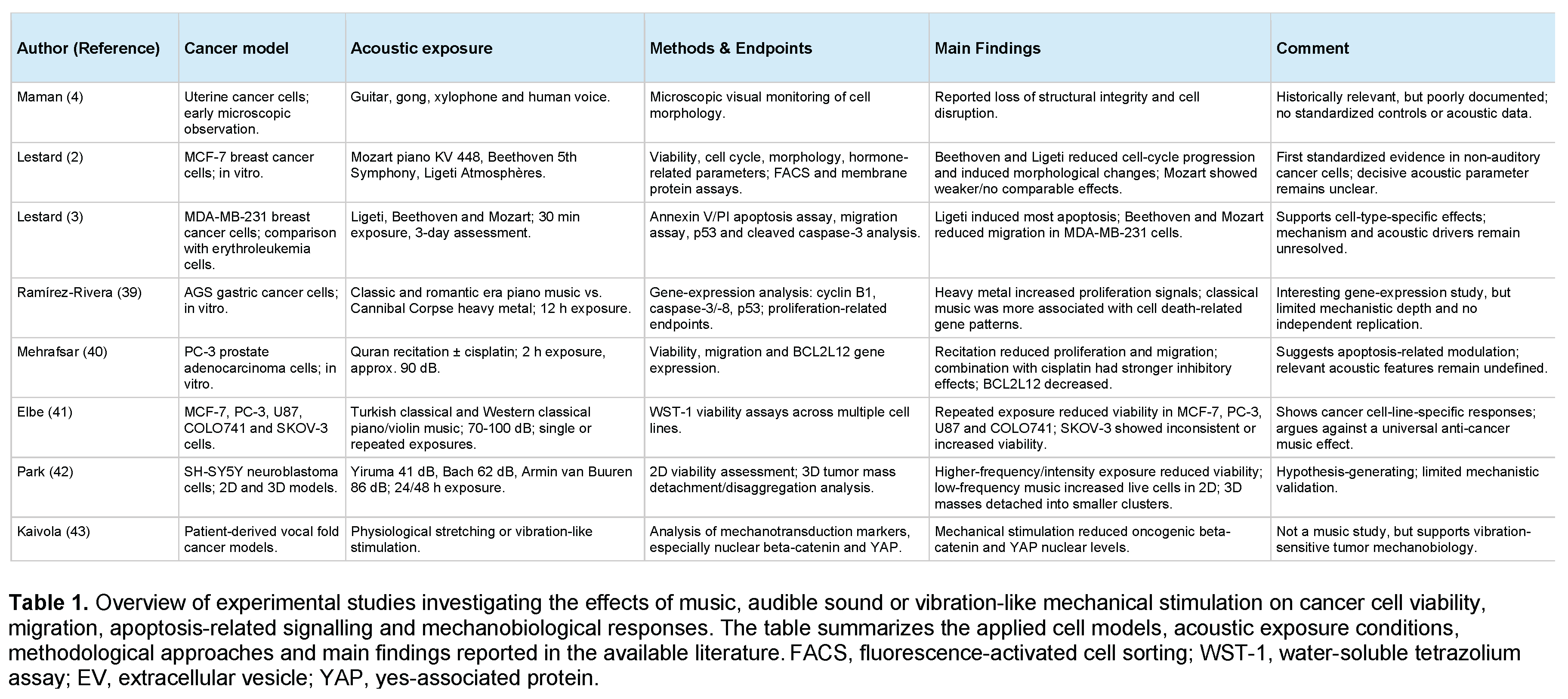
Compared to ultrasound application, experimental research on music or audible sound as a direct modulator of cancer cell viability remains limited and heterogeneous. The available evidence primarily relies on in vitro studies using loudspeaker-based exposure systems, different musical compositions, religious recitation, or audible vibration paradigms. These studies suggest that cancer cells may respond to audible acoustic stimulation through changes in viability, proliferation, motility, gene expression, apoptosis-related signalling, and mechano-transductive pathways. However, the evidence remains preliminary, and the field is limited by variability in sound intensity, frequency spectrum, exposure duration, distance from the sound source, culture conditions, and endpoint assays.
One of the earliest observations come from the biologist Helene Grimal and Fabien Maman from the 1980s. They studied the effect of sound waves on living cells and monitored under a camera uterine cancer cells that were exposed to the instruments guitar, gong, xylophone as well as the human voice. They found that cancer cells lost structural integrity until they exploded [4). No published data are known about these observations so the methodological details remain unknown rendering these results to be cautiously judged.
It was until 2013 that the first peer-reviewed study came out investigating music exposure to cancer cells under standardized experimental conditions [2]. Lestard, Valente, Lopes and Capella exposed MCF-7 human breast cancer cells to musical compositions and analyzed for cell viability, cell cycle distribution, morphology and hormone-related cellular parameters. They proofed that different biological effects on cells were not only dependent on sound but on different music styles. MCF-7 breast cancer cells were exposed to the first movement of Mozart’s Sonata for two Pianos (D major, KV. 448), to the first movement of Beethoven’s 5th Symphony, and to Ligeti’s Atmosphere. Authors found that Beethoven and Ligeti decreased the cell cycle up to cell death while Mozart did not. Instead, morphologic alterations, such as cell volume and granularity changed in Beethoven and Ligeti music exposure but not in Mozart´s music. Particularly Beethoven music induced a significant reduction in cancer cell volume. Authors here hypothesized that the observed effects depend on the frequency of music type and possibly the rhythm and mass of music exposed. While Mozart had a low frequency, Beethoven and Ligeti´s music had higher frequencies and were pieces with massive volumes. As authors used a standardized methodology employing FACS analysis and membrane protein binding assays, the observed effects seem plausible. However, beyond frequency and rhythm, it must be assumed that there are more sound parameters that potentially influence cancer cell behaviour. The three music pieces used did not only differ in style and volume, they differed also in harmony. While Mozart´s music is particularly tonal and harmonic, Beethoven´s music can occasionally become disharmonic while Ligeti is completely disharmonic, dissonant and atonal which might impact the results observed. Also, there is a structure in Mozart´s and Beethoven´s music, at least by the human ear, while Ligeti´s “Atmosphere” misses a recognizable audible structure. It thus remains unclear if harmony or disharmony impacts cancer cell behaviour, and if so, in which way. Nevertheless, this study is important as it is the first study that showed that non-auditory cancer cells may directly respond to music-generated acoustic vibrations. Lestard and Capella then provided a follow-up study in 2016 using the same music pieces in which they extended their analyses on another cancer cell line, MDA-MB-231 with apoptotic assay using annexin V/propidium iodide flow cytometry and a cell migration assay [3]. After 30 minutes of music exposure and three days of whole study period, they found that Ligeti´s “Atmosphere” induced the highest percentage of apoptotic cells and identified the p53 and Cleaved Caspase 3 pathway as responsible pathways. Instead, Beethoven’s 5th Symphony and Mozart’s sonata significantly diminished the migration of MDA-MB-231 cells. Authors could proof this only in the breast cancer cell lines but not in human erythroleukemia cell lines. Again, they owed the observed effects to mechanical forces induced by the music, but not to other music parameters such as harmony, volume or mass effect. However, the study at least proofed that the effect on cells by music was dependent on the cancer cell type. Overall, although authors employed a good method panel and study design, the experiments remain heterogenous and artificial.
A more pronounced difference in music style exposure was performed in a study by Ramírez-Rivera and Bernal in 2019 [39]. These authors exposed the human gastric cancer cell line AGS to classical versus heavy metal music, specifically as classical the piano sonata No.15, Op.28, the Bagatelle No.25 in A minor (for Elise), and the piano Sonata No.14 in C minor, Op.27 No.2 (Moonlight Sonata) and as heavy metal songs from the “Cannibal Corpse”. The core question in their study was if these music styles can alter gene expression. They exposed ASG cells 12 hours using an iPod Shuffle MP3 player. They found that, unlike classical music, metal induced cell proliferation. When testing genes responsible for apoptosis and cell cycle control, they found that both music styles augmented the expression of the genes cyclin B1 and caspases-3 and 8, while p53 was depressed by classical music. Heavy metal music, as the name implies, has much harder mechano-transduction forces than almost every other music style, though the frequency does not exceed 16kHz. Death metal amplitude and length of the music piece exposure could here most likely contribute to cancer cell proliferation. Summarizing their results from different pro- and anti-apoptotic genes, classical music was rather associated with gene-expression patterns related to cell death, whereas metal music was associated with increased signals related to cell proliferation. Nevertheless, though this is a relevant work, the study did not provide enough mechanistic insight to draw further conclusions as the results obtained require independent replication.
Another kind of audible sound that was exposed to cancer cells was proposed by Mehrafsar and Mokhtari. These authors examined the effect of Quran recitation, in one experimental arm alone and in another arm along with cisplatin. They analysed the cell viability, motility, and BCL2L12 (BCL2-like 12) gene expression on PC-3 human prostate adenocarcinoma cells [40]. Cells were exposed for 2 hours and with a sound pressure level at 90 dB. Because BCL2L12 is involved in apoptosis-related regulation and has been associated with several malignancies, authors suggested that audible recitation may influence prostate cancer cell proliferation and migration through apoptosis-related gene expression. Authors found a considerable inhibitory effect of Quran recitation on the proliferation and migration of PC-3 cells. The gene expression of BCL2L12 was significantly downregulated in PC-3 cells exposed to both cisplatin and Quran recitation. Taken together, authors concluded that Quran recitation can induce apoptosis in tumor cells through the downregulation of BCL2L12 expression. However, this study lacks other mechanistic insights so it remains unclear, what the cause of the changes observed are as this kind of music is monotonous, without a harmonic melody and rhythm, and without a clear structure.
Elbe and colleagues later investigated turkish (19th and early 20th century) and western violin and piano classical music (mainly from the classic and romantic era) in several cancer cell lines, including MCF-7 breast cancer, PC-3 prostate cancer, U87 glioblastoma, COLO741 colon cancer and SKOV-3 ovarian cancer cells [41). Using WST-1 viability assays, they reported that repeated exposure to turkish classical music, western classical piano music and western classical violin music reduced viability in several cell lines, particularly after two 40-minute exposure periods separated by a 1-hour interval in the frequency range of 70-100 dB. Authors found reductions in MCF-7, PC-3, U87 and COLO741 cells, whereas SKOV-3 ovarian cancer cells showed increased viability after single exposures, and less consistent suppression after repeated exposures. The significance of this study is that authors analysed a cancer cell-line-specific response. This is remarkable as it argues against a universal anti-cancer effect of music but rather suggests that cellular phenotype, tissue origin and mechano-sensitivity of different cancer cells react differently.
Recently, Park and colleagues examined SH-SY5Y neuroblastoma cells exposed to musical pieces differing in frequency and decibel characteristics at varying frequencies and decibel levels for 24 and 48 hours. They exposed cells to the music pieces "River Flows in You" by Yiruma (41 dB), "Brandenburg Concerto No.3" by Bach (62 dB), and "This is What it Feels Like" by Armin Van Buuren (86 dB) [42]. Authors reported that high-frequency music exposure was associated with reduced cancer cell viability and increased brain cell death, whereas low-frequency music increased the number of live cancer cells in a two-dimensional culture model, independent from the music genre. In the 3D tumor model, all music pieces effectively detached the cancer cells into smaller masses, indicating that physical vibration itself created by sound waves may effectively destroyed the cancer cells. Although this study is hypothesis-generating, it lacks the methodological depth that is expected from higher impact mechanobiology or oncology studies.
A remarkable study comes from Kaivola and colleagues that does not directly deal with the effect of audible sound on cancer cells but with the effect of vocal fold movement through stretching and vibration on vocal fold cancer. Authos showed that mimicking physiological vocal fold movement through stretching or vibration reduced oncogenic β-catenin and YAP nuclear levels in patient-derived vocal fold cancer models [43]. Although this study was not designed primarily to assess music-induced cancer cell death, it is relevant because it demonstrates that cancer cells from mechanically active tissues are mechanically sensitive and can respond to vibration-like stimulation through mechano-transduction pathways linked to malignancy. The study supports the broader concept that sound-related mechanical stimulation can alter cancer-cell behaviour, especially through YAP/TAZ-related mechanobiology.
Taken together, the available experimental studies suggest that music can change cancer cell viability and phenotype and can induce apoptosis and proliferation activity in vitro. However, the evidence available does not allow for the conclusion that audible sound can influence cancer cells in vivo. The strongest direct evidence for reduced viability and apoptosis comes from the Lestard studies in breast cancer cells, while Elbe et al. provide broader but still preliminary evidence across multiple cancer cell lines. Ramírez-Rivera and Bernal extend the scope of studies in this field towards a gene-expression perspective in gastric cancer cells, and Mehrafsar and Mokhtari suggest effects of Quran recitation on prostate cancer cell viability, migration and apoptosis-related gene expression. However, all these studies differ substantially in exposure systems, acoustic characterization, cell lines, endpoints and statistical robustness. Also, the assumption that certain frequencies within the range of audible sound, or the genre of music have a clear positive or negative effect on cancer cells is not supported by these studies. Therefore, audible sound and music should currently be discussed as experimental mechanobiological modulators rather than established anti-cancer interventions.
6. Limitation of Current Research
The current evidence on the effects of audible sound and music on cancer cell viability remains preliminary and should be interpreted with caution. A series of in vitro studies suggest that cancer cells may respond to audible acoustic stimulation through changes in viability, apoptosis, proliferation, migration, gene expression, extracellular vesicle secretion and mechano-transductive signalling. However, the available data are still insufficient to support a general anti-cancer effect of audible sound or music. The first limitation is the small number of peer-reviewed experimental studies directly addressing this question. Most available work is based on selected cancer cell lines but these studies differ substantially in sound source, acoustic exposure system, exposure duration, sound pressure level, culture conditions, endpoint assays and statistical robustness. As a result, the findings are difficult to compare, and no reproducible acoustic dose–response relationship could be established.
A central methodological problem is the incomplete biophysical characterization of the acoustic stimulus. In those studies available, exposure is described mainly by musical genre, composer, musical piece, religious recitation or decibel level, whereas essential parameters such as exact frequency spectrum, waveform characteristics, local sound pressure at the cell layer, vibration transmission through the culture vessel, resonance behaviour of the exposure chamber, distance from the sound source, thermal effects and possible electromagnetic interference are rarely sufficiently documented. This is particularly important because cancer cells do not perceive music in an emotional, neurological or aesthetic sense, as far as we can assume. Any direct cellular effect must therefore be mediated by measurable physical properties of acoustic waves, including pressure oscillations, vibration, shear stress, membrane deformation or resonance-related mechanical forces. Therefore, attributing specific biological effects to “Mozart”, “Beethoven”, “Ligeti”, “classical music”, “heavy metal” or Quran recitation remains scientifically problematic unless the underlying acoustic parameters are precisely quantified and reproducibly controlled.
Another important limitation is the insufficient use of standardized sham-exposure controls. In several studies, it remains unclear whether the observed effects are truly caused by acoustic stimulation itself or by secondary experimental factors such as handling stress, incubator opening, temperature or humidity changes, culture plate vibration, altered gas exchange or electromagnetic effects from playback devices. Furthermore, many studies rely mainly on short-term viability assays, selected apoptosis markers or limited gene-expression analyses. More robust mechanistic endpoints, including clonogenic survival, long-term proliferation, apoptosis versus necrosis discrimination, mitochondrial membrane potential, ROS generation, calcium imaging, cytoskeletal organization, mechanosensitive ion-channel activation, transcriptomic profiling and rescue experiments, are rarely integrated within the same study. Thus, the proposed mechanisms remain largely correlative. Although calcium signalling, cytoskeletal remodelling, mitochondrial dysfunction, ROS production, apoptosis-related pathways and extracellular vesicle release are plausible mechanisms linking acoustic stimulation to cancer-cell behaviour, direct causal evidence in the specific context of audible sound remains limited.
In contrast, the mechanistic evidence is considerably stronger for ultrasound-based approaches, where PIEZO1-dependent calcium influx, mitochondrial apoptosis, ROS generation, sonodynamic therapy, histotripsy and immune-related signalling have been investigated in greater detail. However, findings from ultrasound cannot simply be transferred to audible sound, because ultrasound differs fundamentally in frequency range, acoustic energy, cavitation potential, tissue penetration and clinical applicability. A further major limitation is the in vitro system employed. Conventional two-dimensional cell culture systems do not reproduce the complexity of tumors in vivo, including extracellular matrix composition, tissue stiffness, vascularization, hypoxia, immune-cell infiltration, stromal interactions, pharmacokinetics and systemic host responses. Therefore, the current data do not allow the conclusion that audible sound or music can reduce tumor growth, induce cancer-cell death or improve oncological outcomes in patients.
Finally, the available studies suggest that cellular responses are highly cell-type specific. Some acoustic exposures appear to reduce viability or increase apoptosis in certain cancer cell lines, whereas others show increased proliferation or inconsistent effects depending on tumor type, exposure condition and sound characteristics. This argues against a universal anti-cancer effect of music or audible sound. Future studies should therefore employ standardized acoustic exposure systems, precise physical characterization of sound and vibration at the cellular level, rigorous sham controls, temperature and vibration monitoring, multiple malignant and non-malignant control cell lines, three-dimensional tumor models, independent replication and integrated mechanistic analyses. Only such approaches will clarify whether audible sound represents a reproducible mechanobiological stimulus with therapeutic potential or whether the currently reported findings mainly reflect context-dependent cellular stress responses under artificial laboratory conditions.
7. Future Perspectives
Future research should not only consider the genre of audible sound, but also define which physical properties of audible sound are biologically relevant. The central aim should be to identify reproducible acoustic parameters, such as frequency composition, amplitude, rhythmical structure, vibration transfer and resonance, that can influence cancer-cell behaviour under controlled conditions. Comparative studies using malignant and non-malignant cells, three-dimensional spheroids and patient-derived organoids are needed to determine whether certain tumor types are selectively sensitive to acoustic stimulation.
Mechanistically, future studies should focus on causal pathways rather than endpoint observations alone. Acoustic exposure should be combined with calcium imaging, mitochondrial and ROS assays, cytoskeletal analysis, apoptosis profiling and targeted inhibition of mechanotransductive pathways such as PIEZO1, integrins, FAK and YAP/TAZ. Transcriptomic and proteomic approaches may further help define molecular signatures of acoustic responsiveness. If reproducible effects can be confirmed, audible sound may become a useful experimental tool to study cancer-cell mechanobiology. In the long term, such approaches could be explored in parallel with established music therapy, which already has a supportive role in oncology by reducing anxiety, distress and pain and improving quality of life. However, direct acoustic modulation of cancer-cell biology and conventional music therapy should remain clearly separated conceptually until biological effects are validated in standardized in vitro systems, advanced tumor models and eventually in vivo studies.
8. Conclusion
Audible sound and music represent an intriguing but still early field at the interface of acoustics, mechanobiology and oncology. Current evidence indicates that cancer cells can respond to acoustic stimulation under experimental conditions, particularly through changes in viability, migration, gene expression and mechanotransductive signalling. However, these findings should not yet be interpreted as evidence for a clinically applicable anti-cancer effect. Rather, audible sound should currently be understood as a potential experimental modulator of cellular mechanosensitivity. Its main scientific value lies in opening a new perspective on how physical forces may influence tumor-cell behaviour. Whether this concept can be translated into therapeutic strategies will depend on the demonstration of reproducible, cell-type-specific and mechanistically validated effects in advanced tumor models and, ultimately, in vivo systems.
References
- Bradt, J.; Dileo, C.; Magill, L.; Teague, A. Music interventions for improving psychological and physical outcomes in cancer patients. Cochrane Database Syst. Rev. 2016, CD006911. [Google Scholar] [CrossRef] [PubMed]
- Lestard Ndos, R.; Valente, R.C.; Lopes, A.G.; Capella, M.A. Direct effects of music in non-auditory cells in culture. Noise Health 2013, 15, 307–14. [Google Scholar] [CrossRef] [PubMed]
- Lestard, N.R.; Capella, M.A. Exposure to Music Alters Cell Viability and Cell Motility of Human Nonauditory Cells in Culture. Evid. Based Complement Altern. Med. 2016, 2016, 6849473. [Google Scholar] [CrossRef]
- Maman, F. The Role of music in the twenty-first century; Tama-Dõ Press: Redondo Beach, CA., 1997. [Google Scholar]
- Ingber, D.E. Mechanobiology and diseases of mechanotransduction. Ann. Med. 2003, 35, 564–77. [Google Scholar] [CrossRef] [PubMed]
- Jaalouk, D.E.; Lammerding, J. Mechanotransduction gone awry. Nat. Rev. Mol. Cell Biol. 2009, 10, 63–73. [Google Scholar] [CrossRef] [PubMed]
- Luo, T.; Mohan, K.; Iglesias, P.A.; Robinson, D.N. Molecular mechanisms of cellular mechanosensing. Nat. Mater. 2013, 12, 1064–71. [Google Scholar] [CrossRef] [PubMed]
- Zimmerman, J.W.; Jimenez, H.; Pennison, M.J.; Brezovich, I.; Morgan, D.; et al. Targeted treatment of cancer with radiofrequency electromagnetic fields amplitude-modulated at tumor-specific frequencies. Chin. J. Cancer 2013, 32, 573–81. [Google Scholar] [CrossRef] [PubMed]
- Wood, A.K.; Sehgal, C.M. A review of low-intensity ultrasound for cancer therapy. Ultrasound Med. Biol. 2015, 41, 905–28. [Google Scholar] [CrossRef] [PubMed]
- Liaquat, H.; Al-Jumaily, A.M. Resonance-Induced Therapeutic Technique for Skin Cancer Cells. Ultrasound Med. Biol. 2025, 51, 661–74. [Google Scholar] [CrossRef] [PubMed]
- Del Rosario-Gilabert, D.; Valenzuela-Miralles, A.; Esquiva, G. Advances in mechanotransduction and sonobiology: effects of audible acoustic waves and low-vibration stimulations on mammalian cells. Biophys. Rev. 2024, 16, 783–812. [Google Scholar] [CrossRef] [PubMed]
- Baumgartner, K.; Schleicher, M.T.; de Campos, A.M.; Taufer, P.; Engelke, H.; Westerhausen, C. Actin Filament and Cell Orientation Align with Surface Acoustic Wave Propagation and Cell Migration in Vibration-Enhanced Wound Healing. ACS Appl. Mater. Interfaces 2025, 17, 37586–600. [Google Scholar] [CrossRef] [PubMed]
- Sokabe, M.; Naruse, K.; Sai, S.; Yamada, T.; Kawakami, K.; et al. Mechanotransduction and intracellular signaling mechanisms of stretch-induced remodeling in endothelial cells. Heart Vessel.> Suppl. 1997, 12, 191–3. [Google Scholar]
- Kang, H.; Hong, Z.; Zhong, M.; Klomp, J.; Bayless, K.J.; et al. Piezo1 mediates angiogenesis through activation of MT1-MMP signaling. Am. J. Physiol. Cell Physiol. 2019, 316, C92–C103. [Google Scholar] [CrossRef] [PubMed]
- Lai, A.; Chen, Y.C.; Cox, C.D.; Jaworowski, A.; Peter, K.; Baratchi, S. Analyzing the shear-induced sensitization of mechanosensitive ion channel Piezo-1 in human aortic endothelial cells. J. Cell Physiol. 2021, 236, 2976–87. [Google Scholar] [PubMed]
- Cox, C.D.; Bae, C.; Ziegler, L.; Hartley, S.; Nikolova-Krstevski, V.; et al. Removal of the mechanoprotective influence of the cytoskeleton reveals PIEZO1 is gated by bilayer tension. Nat. Commun. 2016, 7, 10366. [Google Scholar] [CrossRef] [PubMed]
- Belousova, P.; Kwon, S.; Jung, M.H.; Kim, K.S. Impact of mechanical property alterations on cancer cell motility and metastasis. Crit. Rev. Oncol. Hematol. 2026, 220, 105165. [Google Scholar] [CrossRef] [PubMed]
- Massey, A.; Stewart, J.; Smith, C.; Parvini, C.; McCormick, M.; et al. Mechanical properties of human tumour tissues and their implications for cancer development. Nat. Rev. Phys. 2024, 6, 269–82. [Google Scholar] [CrossRef] [PubMed]
- Schibber, E.F.; Mittelstein, D.R.; Gharib, M.; Shapiro, M.G.; Lee, P.P.; Ortiz, M. A dynamical model of oncotripsy by mechanical cell fatigue: selective cancer cell ablation by low-intensity pulsed ultrasound. Proc. Math. Phys. Eng. Sci. 2020, 476, 20190692. [Google Scholar] [CrossRef] [PubMed]
- Heydarian, A.; Milani, D.; Beni, H.M.; Babaee, M.; Goudarzi, H.R. How do vibration stimulation frequencies affect the nonlinear dynamics and mechanical characterization of breast cancer cells? Biochem Biophys. Rep. 2026, 45, 102414. [Google Scholar] [CrossRef] [PubMed]
- Ambattu, L.A.; Ramesan, S.; Dekiwadia, C.; Hanssen, E.; Li, H.; Yeo, L.Y. High frequency acoustic cell stimulation promotes exosome generation regulated by a calcium-dependent mechanism. Commun. Biol. 2020, 3, 553. [Google Scholar] [CrossRef] [PubMed]
- Federico, G.; Carotenuto, A.R.; Cutolo, A.; Palumbo, S.; Moccia, M.; et al. Ultrasound-induced mechanical damage of cancer cell cytoskeleton causes disruption of nuclear envelope and activation of cGAS-STING. Sci. Rep. 2025, 15, 18037. [Google Scholar] [CrossRef] [PubMed]
- Anggayasti, W.L.; Imashiro, C.; Kuribara, T.; Totani, K.; Takemura, K. Low-frequency mechanical vibration induces apoptosis of A431 epidermoid carcinoma cells. Eng. Life Sci. 2020, 20, 232–8. [Google Scholar] [CrossRef] [PubMed]
- Kumari, A.; Veena, S.M.; Luha, R.; Tijore, A. Mechanobiological Strategies to Augment Cancer Treatment. ACS Omega 2023, 8, 42072–85. [Google Scholar] [CrossRef] [PubMed]
- Heyden, S. O.M. Oncotripsy: Targeting cancer cells selectively via resonant harmonic excitation. J. Mech. Phys. Solids 2016. [Google Scholar] [CrossRef]
- He, Y.; Xia, J.; Mai, J.D.H.; Upreti, N.; Lee, L.P.; Huang, T.J. Acoustic technologies for the orchestration of cellular functions for therapeutic applications. Sci. Adv. 2025, 11, eadu4759. [Google Scholar] [CrossRef] [PubMed]
- Tijore, A.; Margadant, F.; Dwivedi, N.; Morgan, L.; Yao, M.; et al. Ultrasound-mediated mechanical forces activate selective tumor cell apoptosis. Bioeng. Transl. Med. 2025, 10, e10737. [Google Scholar] [PubMed]
- Luo, A.C.; Qian, Z.; King, M.R. Focused ultrasound for cellular mechanoactivation and nanomedicine delivery in cancer. BioMed Microdevices 2026, 28. [Google Scholar] [CrossRef]
- Yu, J.; Hu, J.R.; Tian, Y.; Lei, Y.M.; Hu, H.M.; et al. Nanosensitizer-assisted sonodynamic therapy for breast cancer. J. Nanobiotechnology 2025, 23, 281. [Google Scholar] [CrossRef] [PubMed]
- Datta, P.; Moolayadukkam, S.; Chowdhury, D.; Rayes, A.; Lee, N.S.; et al. Recent Advances and Future Directions in Sonodynamic Therapy for Cancer Treatment. BME Front 2024, 2024, 0080. [Google Scholar] [CrossRef] [PubMed]
- Wang, N.; Luo, L.; Xu, X.; Zhou, H.; Li, F. Focused ultrasound-induced cell apoptosis for the treatment of tumours. PeerJ 2024, 12, e17886. [Google Scholar] [CrossRef] [PubMed]
- Sankar, G.; Roy Choudhury, A.; Luha, R.; Kumar, A.; Kulkarni, K.; et al. Selective killing of cancer-associated fibroblasts by ultrasound-mediated mechanical forces. Biomaterials 2026, 328, 123844. [Google Scholar] [CrossRef] [PubMed]
- Rix, A.; Heinrichs, H.; Porte, C.; Leenaars, C.; Bleich, A.; Kiessling, F. Ultrasound-induced immune responses in tumors: A systematic review and meta-analysis. J. Control Release 2024, 371, 146–57. [Google Scholar] [CrossRef] [PubMed]
- Chen, T.; Chen, J.; Chen, M.; Song, R.; Wang, M.; Yu, X. Acoustic immune reprogramming: a novel paradigm for spatiotemporally controlled immune regulation using ultrasound-responsive nanoplatforms. Front Immunol. 2025, 16, 1715455. [Google Scholar] [CrossRef]
- Du, Y.; Yang, J.; Xu, A.; Chen, S.; Fu, D. Research progress on the induction of immunogenic cell death in tumor immunotherapy using a sonodynamic therapy nanoparticle delivery system. Front Immunol. 2025, 16, 1681773. [Google Scholar] [CrossRef] [PubMed]
- Kumeta, M.; Takahashi, D.; Takeyasu, K.; Yoshimura, S.H. Cell type-specific suppression of mechanosensitive genes by audible sound stimulation. PLoS ONE 2018, 13, e0188764. [Google Scholar] [CrossRef] [PubMed]
- Valenti, D.; Atlante, A. Sound Matrix Shaping of Living Matter: From Macrosystems to Cell Microenvironment, Where Mitochondria Act as Energy Portals in Detecting and Processing Sound Vibrations. Int. J. Mol. Sci. 2024, 25. [Google Scholar] [CrossRef] [PubMed]
- Lei, Z.; Jiang, H.; Liu, J.; Liu, Y.; Wu, D.; et al. Audible Acoustic Wave Promotes EV Formation and Secretion from Adherent Cancer Cells via Mechanical Stimulation. ACS Appl. Mater. Interfaces 2023, 15, 53859–70. [Google Scholar] [CrossRef]
- Ramirez-Rivera, S.; Bernal, G. Music Is Capable of Inducing Changes in Gene Expression in Gastric Cancer Cells. J. Gastrointest. Cancer 2019, 50, 175–80. [Google Scholar] [CrossRef] [PubMed]
- Mehrafsar, A. M.M. Effect of Exposure to Quran Recitation on Cell Viability, Cell Migration, and BCL2L12 Gene Expression of Human Prostate Adenocarcinoma Cell Line in Culture. Health Spirit. Med. Ethics 2018, 5, 46–52. [Google Scholar] [CrossRef]
- Ceren Elbe, M.O.O.; Yigitturk, Gurkan; Turan, Seda Gelen; Elbe², H. Cellular Response of Cancer to Music: Mirror, Mirror, on the Wall, Which is the Most Effective of Them All? Med. Rec. 2023, 5, 237–43. [Google Scholar] [CrossRef]
- Park, J. Exploring the Impact of Music Frequency and Decibels on the Viability of Neuroblastoma Cancer Cells. Int. J. High Sch. Res. 2024, 16–20. [Google Scholar] [CrossRef]
- Kaivola, J.; Punovuori, K.; Chastney, M.R.; Abdo, H.; Follain, G.; et al. Restoring the tumour mechanophenotype of vocal fold cancer reverts its malignant properties. Nat. Mater. 2026, 25, 868–82. [Google Scholar] [CrossRef] [PubMed]
Disclaimer/Publisher’s Note: The statements, opinions and data contained in all publications are solely those of the individual author(s) and contributor(s) and not of MDPI and/or the editor(s). MDPI and/or the editor(s) disclaim responsibility for any injury to people or property resulting from any ideas, methods, instructions or products referred to in the content. |
© 2026 by the authors. Licensee MDPI, Basel, Switzerland. This article is an open access article distributed under the terms and conditions of the Creative Commons Attribution (CC BY) license (http://creativecommons.org/licenses/by/4.0/).
Copyright: This open access article is published under a Creative Commons CC BY 4.0 license, which permit the free download, distribution, and reuse, provided that the author and preprint are cited in any reuse.


