

Submitted:
07 July 2026
Posted:
08 July 2026
You are already at the latest version
Abstract
Background: Population aging is driving a convergent rise in three disorders historically managed in isolation: cardiovascular disease, neurocognitive decline, and osteoporotic bone loss. Accumulating mechanistic data indicate that these systems are coupled through shared regulators of calcium trafficking, inflammation resolution, vascular integrity, and inflammaging, supporting the concept of an integrated "Heart-Brain-Bone" axis. Three lipid-soluble nutrients-long-chain omega-3 polyunsaturated fatty acids (EPA/DHA), vitamin D3 (cholecalciferol), and vitamin K2 (menaquinone-7 [MK-7])-act on overlapping nodes of this network. Methods: We conducted a structured narrative review of mechanistic, observational, and randomized controlled trial (RCT) evidence, grading the strength of support for each claim. Results: Vitamin K2-dependent gamma-carboxylation of matrix Gla protein (MGP) and osteocalcin is proposed to reciprocally direct calcium away from the arterial wall and toward the bone matrix, offering a candidate mechanistic resolution of the "calcium paradox" associated with isolated vitamin D3 supplementation; EPA/DHA-derived specialized pro-resolving mediators may support resolution of endothelial and neuronal inflammation; and bone-, vascular-, and brain-derived signals (osteocalcin, FGF23, the neurovascular unit) interconnect the three organs. Human clinical evidence, however, is heterogeneous and formulation- and population-dependent: cardiovascular omega-3 RCTs are discordant (REDUCE-IT, which used icosapent ethyl [an EPA ethyl ester], positive; VITAL/STRENGTH/ASCEND null, predominantly in lower-risk or replete cohorts), cognitive trials are largely null or subgroup-dependent (MAPT, DO-HEALTH), and MK-7 improves surrogate bone and calcification biomarkers, with only a single recent hard-endpoint coronary calcification RCT (VitaK-CAC). Conclusions: The combined use of long-chain omega-3, vitamin D3, and MK-7 is mechanistically rational and hypothesis-generating rather than clinically established; benefit appears most plausible in individuals with elevated risk or demonstrable nutritional insufficiency, and least in replete, low-risk populations. Findings should be interpreted within a broader healthy-aging context that includes lifestyle and psychosocial factors. Adequately powered factorial RCTs stratified by baseline Omega-3 Index, 25(OH)D, and vitamin K status, with prespecified mechanistic biomarkers and hard endpoints, are required.
Keywords:
omega-3 fatty acids
; vitamin K2 (menaquinone-7)
; vitamin D3
; matrix Gla protein
; vascular calcification
; specialized pro-resolving mediators
; bone mineralization
; neurovascular unit
; inflammaging
; Heart-Brain-Bone axis
1. Introduction
Population aging is among the most consequential epidemiological transitions of the twenty-first century, and it is accompanied by a parallel rise in chronic, degenerative, non-communicable diseases. Cardiovascular disease (CVD) remains the leading global cause of mortality; neurocognitive disorders, chief among them Alzheimer’s disease and related dementias, are projected to affect an increasing share of the population as life expectancy lengthens; and osteoporosis with its attendant fragility fractures imposes enormous morbidity, loss of independence, and healthcare expenditure, particularly among postmenopausal women and older men. Historically, these three disease clusters have been investigated and managed within siloed clinical disciplines—cardiology, neurology, and orthopedics/endocrinology, respectively.
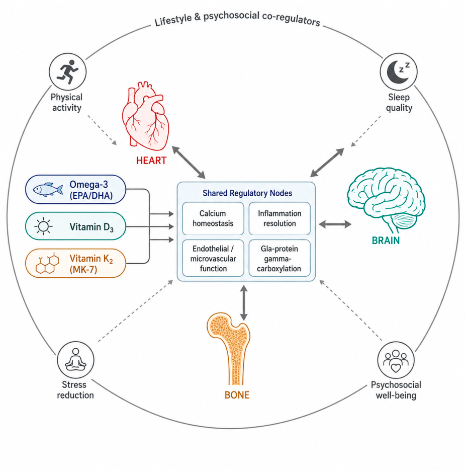
This compartmentalized framework increasingly conflicts with mechanistic data demonstrating that the cardiovascular, central nervous, and skeletal systems are bound by shared regulatory pathways [1,2]. The concept of a “Heart-Brain-Bone” axis posits that these systems do not degenerate in isolation but rather decline in a coordinated, often self-reinforcing manner, governed by common upstream drivers including chronic low-grade inflammation (“inflammaging”), dysregulated calcium trafficking, endothelial dysfunction, and impaired microcirculatory perfusion [3,4]. The same vascular calcification that stiffens large arteries and elevates cardiovascular risk also compromises the cerebral microvasculature that sustains neuronal metabolism, and reflects a systemic mishandling of calcium that simultaneously depletes the skeletal reservoir [1,5]. Bone is now recognized not as an inert scaffold but as an endocrine organ secreting osteocalcin and other osteokines that exert distal effects on the vasculature and the brain [6,7].
Within this integrative paradigm, lipid-soluble micronutrients occupy a privileged regulatory position because their receptors and molecular targets are expressed across all three organ systems. Three nutrients in particular—long-chain n-3 PUFAs (eicosapentaenoic acid, EPA; docosahexaenoic acid, DHA), vitamin D3, and vitamin K2—share a thematic mechanistic logic: each modulates inflammation, calcium handling, or membrane biophysics, and their biological actions are demonstrably interdependent [8,9]. Vitamin D3 enhances intestinal calcium absorption but, in the absence of adequate vitamin K2 status, may promote the deposition of that calcium in vascular and soft-tissue compartments rather than in bone—a phenomenon central to the “calcium paradox” [8,10]. Vitamin K2, by activating the calcium-binding Gla proteins MGP and osteocalcin, functions as the molecular regulator that redirects calcium toward the skeleton and away from the arterial wall [10,11]. EPA and DHA, in turn, are the substrates for specialized pro-resolving mediators that actively terminate inflammation in endothelial and neural tissues [12,13].
One determinant of the plasma and tissue concentrations achievable from a given intake is the molecular form in which these nutrients are delivered. Pharmacokinetic studies indicate that re-esterified triglyceride (rTG) omega-3 concentrates may show greater bioavailability than ethyl-ester (EE) forms [14,15], and that the long-chain menaquinone MK-7 has a longer circulating half-life and greater extrahepatic bioavailability than the shorter MK-4 homolog [16,17]. Such distinctions concern absorption and tissue delivery, not proven clinical superiority, and are considered in detail—together with their limits—in Section 2.
The objectives of this review are threefold. First, we examine the biochemical properties and bioavailability of long-chain omega-3 (EPA/DHA), vitamin D3, and vitamin K2 (MK-7). Second, we articulate the molecular architecture of the Heart-Brain-Bone axis—including vascular-bone coupling, neurovascular coupling, bone-brain endocrine crosstalk, and inflammaging as a common driver—and evaluate the rationale for combined use. Third, we critically appraise the clinical trial evidence, explicitly distinguishing mechanistic, observational, and randomized data; delineate the controversies and unresolved questions that temper any therapeutic claim; identify the populations in which supplementation is most plausibly warranted; and situate nutrient intake within the broader lifestyle and psychosocial context that co-regulates the axis. Throughout, claims are framed according to their evidentiary basis, and we deliberately avoid causal language where only mechanistic or associational data exist.
Figure 1.
Conceptual overview of the Heart-Brain-Bone axis. Three lipid-soluble nutrients—long-chain omega-3 (EPA/DHA), vitamin D3, and vitamin K2 (MK-7)—act on shared regulatory nodes (calcium homeostasis, inflammation resolution, endothelial/microvascular function, and Gla-protein gamma-carboxylation) that may coordinately influence cardiovascular, central nervous, and skeletal health. An outer ring depicts lifestyle and psychosocial co-regulators (physical activity, sleep quality, stress reduction, and psychosocial well-being) that modulate the same nodes, underscoring that nutrient intake is one component of a broader healthy-aging context (Section 7).
Figure 1.
Conceptual overview of the Heart-Brain-Bone axis. Three lipid-soluble nutrients—long-chain omega-3 (EPA/DHA), vitamin D3, and vitamin K2 (MK-7)—act on shared regulatory nodes (calcium homeostasis, inflammation resolution, endothelial/microvascular function, and Gla-protein gamma-carboxylation) that may coordinately influence cardiovascular, central nervous, and skeletal health. An outer ring depicts lifestyle and psychosocial co-regulators (physical activity, sleep quality, stress reduction, and psychosocial well-being) that modulate the same nodes, underscoring that nutrient intake is one component of a broader healthy-aging context (Section 7).
2. Biochemical Properties and Bioavailability
2.1. Formulation and Bioavailability: rTG Versus EE and Natural TG Forms of Omega-3
Marine omega-3 fatty acids are available in several molecular configurations whose digestion and absorption differ substantially. In native fish, EPA and DHA exist predominantly as natural triglycerides (TG). During industrial concentration, the triglyceride is transesterified to the ethyl-ester (EE) form, yielding a high-purity concentrate in which each fatty acid is bound to an ethanol moiety. Enzymatic re-esterification of the EE concentrate back onto a glycerol backbone produces the re-esterified triglyceride (rTG) form, combining high purity with a near-native structure [15].
The pharmacological relevance derives from lipid-digestion mechanics. Pancreatic lipase exhibits higher catalytic efficiency toward the glycerol-esterified sn-1 and sn-3 positions of triglycerides than toward the ethyl-ester bond; EE forms are therefore hydrolyzed more slowly and depend more heavily on co-ingested dietary fat [15]. A controlled bioavailability study reported that the rTG form achieved markedly higher EPA+DHA bioavailability (approximately 124% relative to natural fish oil) than the EE form (approximately 73%) [14]. Because steady-state erythrocyte membrane EPA/DHA content (the Omega-3 Index) governs eicosanoid and pro-resolving mediator synthesis, differences in absorption may translate into differences in the substrate available for downstream signaling [12,14], an effect likely to be most relevant in older or low-fat-diet populations with attenuated digestive lipase activity. Two caveats are essential. First, this evidence derives from pharmacokinetic (human bioavailability) studies and short-term biomarker endpoints, not clinical outcomes. Although rTG formulations may show superior bioavailability compared with ethyl-ester forms in pharmacokinetic studies, no adequately powered randomized trial has demonstrated superior cardiovascular, cognitive, or skeletal clinical outcomes attributable solely to the rTG formulation. Second, bioavailability is not equivalent to clinical efficacy: the trials that have shaped the omega-3 outcome literature (Section 4) have almost exclusively used ethyl-ester or carboxylic-acid preparations rather than rTG, so formulation and clinical outcome cannot presently be equated. For these reasons, the remainder of this review treats long-chain omega-3 (EPA/DHA) as the entity of interest, and confines formulation-specific claims to bioavailability.
2.2. Vitamin D3 (Cholecalciferol): Activation and Receptor Biology
Vitamin D3 is a secosteroid pro-hormone, biologically inert until sequentially hydroxylated: hepatic 25-hydroxylase (CYP2R1) generates 25-hydroxyvitamin D [25(OH)D], the principal circulating storage metabolite and status biomarker, which renal 1-alpha-hydroxylase (CYP27B1) converts to the active 1,25-dihydroxyvitamin D (calcitriol) [18,19]. Calcitriol acts through the nuclear vitamin D receptor (VDR), a ligand-activated transcription factor that heterodimerizes with the retinoid X receptor (RXR) and binds vitamin D response elements in target gene promoters [19]. VDR is expressed not only in intestine, kidney, and bone but also in cardiomyocytes, vascular smooth muscle, neurons, and glia, providing the molecular basis for the pleiotropic actions of vitamin D3 across the axis [19,20]. As a lipophilic secosteroid, vitamin D3 absorption is enhanced by co-administration with dietary lipids, mechanistically complementing a triglyceride-based omega-3 matrix. Importantly, because vitamin D3 increases the systemic calcium load, its supplementation heightens the requirement for the calcium-routing machinery discussed in Section 3.
2.3. Vitamin K2 Pharmacokinetics: MK-7 Versus MK-4
Vitamin K2 comprises menaquinones (MK-n) distinguished by the number (n) of isoprenoid side-chain units. MK-4 is rapidly absorbed but has a very short plasma half-life (1-2 h), so physiologically relevant systemic concentrations require high, divided doses [16]. MK-7, by contrast, has a longer side chain conferring greater lipoprotein binding, a circulating half-life measured in days, and greater accumulation in extrahepatic tissues such as the arterial wall and bone [16,17]. This extended residence time is functionally decisive: sustained plasma MK-7 ensures continuous delivery of the cofactor to gamma-glutamyl carboxylase in extrahepatic tissues, supporting ongoing carboxylation—and hence activation—of MGP and osteocalcin between doses [10,17]. A once-daily MK-7 regimen of approximately 90-180 micrograms maintains Gla-protein carboxylation more effectively than equivalent or higher doses of short-acting MK-4 [16,21].

Table 1.
Comparative biochemical and pharmacokinetic properties of the three lipid-soluble nutrients.
Table 1.
Comparative biochemical and pharmacokinetic properties of the three lipid-soluble nutrients.
| Property | Omega-3 (EPA/DHA) | Vitamin D3 | Vitamin K2 (MK-7) |
| Molecular class | Long-chain n-3 PUFA (TG backbone) | Secosteroid pro-hormone | Long-chain menaquinone |
| Active/effector species | EPA, DHA -> SPMs (resolvins, protectins) | 1,25(OH)2D (calcitriol) | Reduced MK-7 (GGCX cofactor) |
| Primary receptor/target | GPR120/FFAR4; membrane incorporation | Nuclear VDR-RXR heterodimer | gamma-Glutamyl carboxylase (GGCX) |
| Key downstream proteins | Pro-resolving mediator cascade | Calbindin, TRPV6; osteocalcin (expr.) | Carboxylated MGP and osteocalcin |
| Bioavailability note | rTG > natural TG > EE absorption | Enhanced by dietary-lipid co-ingestion | MK-7 half-life (days) >> MK-4 (hours) |
| Axis relevance | Anti-inflammatory; Heart + Brain | Calcium uptake; all three organs | Calcium routing; Heart + Bone |
3. The “Heart-Brain-Bone” Axis: Molecular Mechanisms and Potential Complementarity
The central thesis of this review is that the three nutrients act not as independent agents but as a functionally integrated module. The following subsections first dissect organ-specific mechanisms (Section 3.1, Section 3.2 and Section 3.3) and then develop the inter-organ coupling that constitutes the axis proper (Section 3.4, Section 3.5, Section 3.6 and Section 3.7), before synthesizing the integrated crosstalk (Section 3.8); the mechanistic roles are consolidated in Table 2.
3.1. Cardiovascular Protection and the Calcium Paradox
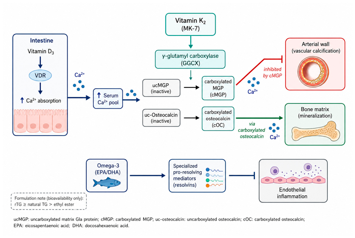
The “calcium paradox” describes the observation that calcium essential for skeletal integrity becomes pathogenic when deposited ectopically in the arterial wall. Vitamin D3, by enhancing intestinal calcium absorption, increases the bioavailable calcium pool; in subclinical vitamin K2 deficiency—widespread in modern diets—a fraction of this calcium may deposit in the vascular intima and media rather than in bone [8,10]. The molecular gatekeeper against this maladaptive deposition is matrix Gla protein (MGP), a potent endogenous calcification inhibitor synthesized by vascular smooth muscle cells [10]. MGP is functional only after vitamin K-dependent carboxylation: gamma-glutamyl carboxylase, using reduced MK-7 as cofactor, converts specific glutamate residues to gamma-carboxyglutamate (Gla) residues [10,11]. Carboxylated MGP binds calcium and hydroxyapatite and sequesters bone morphogenetic protein-2, preventing osteogenic transdifferentiation of vascular smooth muscle cells [5,10]. In vitamin K2 insufficiency, MGP remains undercarboxylated (ucMGP) and loses protective capacity; circulating dephospho-uncarboxylated MGP is an established biomarker of vascular calcification risk [22,23]. Adequate MK-7 status is therefore the mechanistic prerequisite allowing vitamin D3 to be given for skeletal and pleiotropic benefit without aggravating vascular calcium deposition [8,9].
Acting in parallel, long-chain n-3 PUFAs confer complementary protection through active resolution of inflammation. Once incorporated into endothelial and leukocyte membranes, EPA and DHA serve as substrates for specialized pro-resolving mediators (SPMs)—resolvins, protectins, and maresins—generated via lipoxygenase and cyclooxygenase-2 pathways [12,13]. Unlike passive dilution of arachidonic acid, SPMs are agonists that actively terminate inflammation: they limit neutrophil infiltration, promote macrophage efferocytosis, and downregulate NF-kappaB-driven adhesion molecules (VCAM-1, ICAM-1) and cytokines (TNF-alpha, IL-6) [12,13]. By attenuating endothelial dysfunction and lowering triglycerides, omega-3 addresses an inflammatory dimension of cardiovascular risk that calcium routing alone does not [13,24]. At the mechanistic level these actions may be complementary: K2-dependent carboxylation is proposed to influence where calcium is deposited, whereas omega-3-derived mediators are proposed to modulate the inflammatory milieu associated with vascular injury (clinical-outcome evidence is appraised in Section 4).
Figure 2.
Molecular resolution of the calcium paradox. Vitamin D3 increases intestinal calcium absorption; vitamin K2 (MK-7) supplies gamma-glutamyl carboxylase the cofactor required to carboxylate matrix Gla protein (MGP) and osteocalcin, directing calcium toward bone (via carboxylated osteocalcin) and away from the arterial wall (via carboxylated MGP). EPA/DHA-derived specialized pro-resolving mediators concurrently attenuate endothelial inflammation.
Figure 2.
Molecular resolution of the calcium paradox. Vitamin D3 increases intestinal calcium absorption; vitamin K2 (MK-7) supplies gamma-glutamyl carboxylase the cofactor required to carboxylate matrix Gla protein (MGP) and osteocalcin, directing calcium toward bone (via carboxylated osteocalcin) and away from the arterial wall (via carboxylated MGP). EPA/DHA-derived specialized pro-resolving mediators concurrently attenuate endothelial inflammation.
3.2. Neuroprotection and Cognitive Mechanisms
The brain is exceptionally lipid-rich, and DHA is its most abundant long-chain PUFA, a major structural component of neuronal and synaptic membrane phospholipids. DHA enrichment modulates membrane fluidity, the function of integral membrane proteins and ion channels, and synaptic vesicle trafficking, supporting synaptic plasticity and neurotransmission [25]. DHA is also the precursor of neuroprotectin D1 (NPD1), an SPM that suppresses neuronal apoptosis, attenuates microglial neuroinflammation, and promotes non-amyloidogenic processing of amyloid precursor protein in preclinical models [25,26]. Vitamin D3 exerts independent neuroprotective effects via CNS-expressed VDR: calcitriol regulates neurotrophic factor transcription, limits excitotoxic calcium influx by downregulating L-type voltage-gated calcium channels, and supports amyloid-beta clearance in experimental systems [20]. Vitamin K contributes through sphingolipid metabolism—serving as a cofactor in the synthesis of brain-enriched sphingolipids—and through carboxylation of the Gla protein Gas6, a ligand for TAM-family receptor tyrosine kinases that promotes neuronal survival and oligodendrocyte myelination [27,28]. Causal animal evidence links warfarin-induced brain vitamin K deficiency to altered sphingolipid profiles and cognitive-behavioral deficits [29]. It must be emphasized that the human cognitive data (Section 4.2) are far less consistent than these mechanistic and animal findings.
3.3. Bone Remodeling and Mineralization
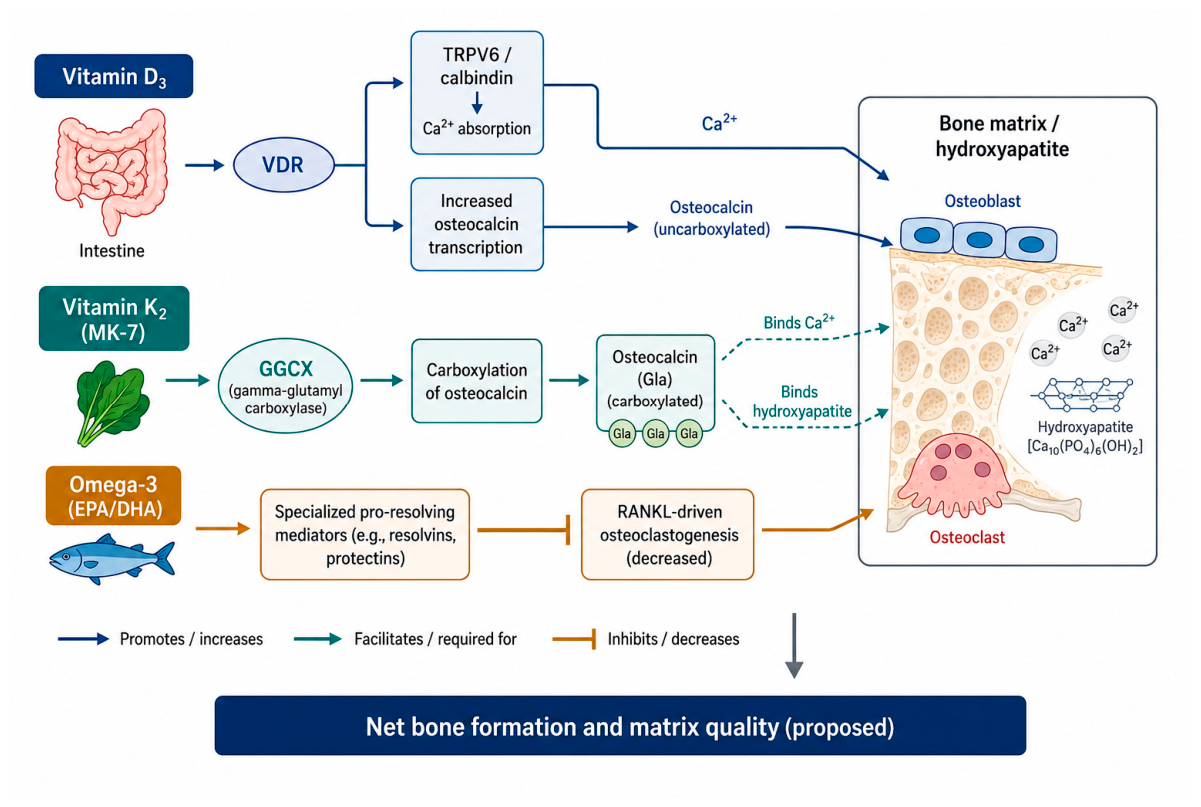
Skeletal homeostasis depends on coupling osteoclast-mediated resorption with osteoblast-mediated formation. Vitamin D3, via calcitriol-VDR signaling in the intestinal epithelium, upregulates the apical calcium channel TRPV6 and cytosolic calbindin-D9k, driving active transcellular calcium absorption and maintaining the calcium-phosphate supersaturation required for mineralization [18,19]. Vitamin K2 governs the qualitative incorporation of that calcium by activating osteocalcin (bone Gla protein), the most abundant non-collagenous bone protein [11,21]. Newly synthesized osteocalcin is undercarboxylated with low hydroxyapatite affinity; gamma-glutamyl carboxylase, using MK-7, carboxylates three glutamate residues to generate carboxylated osteocalcin, which anchors mineral to the collagenous matrix [11,30]. The proportion of undercarboxylated osteocalcin is an inverse marker of vitamin K status associated in cohorts with lower bone mineral density [21,30]. The D3-K2 relationship in bone mirrors that in the vasculature: D3 controls calcium availability and upregulates osteocalcin transcription, while K2 is required for the functional activity of the protein [8]. n-3 PUFAs further support bone indirectly by attenuating the pro-inflammatory cytokines that drive RANKL-mediated osteoclastogenesis [13].
Figure 3.
Integrated mechanisms of omega-3, vitamin D3, and MK-7 in bone mineralization. Vitamin D3 secures calcium supply (TRPV6/calbindin) and upregulates osteocalcin transcription; vitamin K2 (MK-7) carboxylates osteocalcin to its high-affinity form; omega-3 PUFAs dampen RANKL-driven osteoclastogenesis—together favoring net bone formation and matrix quality.
Figure 3.
Integrated mechanisms of omega-3, vitamin D3, and MK-7 in bone mineralization. Vitamin D3 secures calcium supply (TRPV6/calbindin) and upregulates osteocalcin transcription; vitamin K2 (MK-7) carboxylates osteocalcin to its high-affinity form; omega-3 PUFAs dampen RANKL-driven osteoclastogenesis—together favoring net bone formation and matrix quality.
3.4. Vascular-Bone Coupling
The most robust pillar of the axis is the bone-vascular coupling captured by the calcification paradox: epidemiologically, low bone mineral density and arterial calcification co-occur more often than chance, and progress in parallel with age, independent of conventional risk factors [1,2]. This inverse bone-vessel relationship is not merely coincident; it reflects a shared set of molecular regulators. Vascular calcification is an active, cell-mediated process in which contractile vascular smooth muscle cells transdifferentiate into an osteoblast-like phenotype, upregulating the master osteogenic transcription factor RUNX2 and depositing a mineralized matrix recapitulating bone formation [5]. The same signaling families that govern skeletal remodeling—the RANK/RANKL/osteoprotegerin (OPG) triad, bone morphogenetic proteins, and Wnt/beta-catenin—operate in the vessel wall, such that dysregulation simultaneously promotes bone resorption and arterial mineralization [1,31]. MGP sits at the center of this coupling: as a vitamin K-dependent inhibitor expressed in both compartments, its carboxylation status determines whether calcium and BMP-2 signaling drive physiological bone mineralization or pathological vascular calcification [10,31]. The bone-vascular axis is best characterized in chronic kidney disease, where mineral-bone disorder and accelerated vascular calcification are tightly linked, but the principle generalizes to vascular aging [31]. This subsection rests primarily on observational and mechanistic evidence; intervention data confirming that modulating the shared pathways alters both endpoints concurrently remain limited.
3.5. Neurovascular Coupling and Blood-Brain Barrier Integrity
The brain’s dependence on its microcirculation provides the second pillar. The neurovascular unit—the functional ensemble of endothelial cells, pericytes, astrocytic end-feet, and neurons—matches regional cerebral blood flow to metabolic demand and maintains the blood-brain barrier (BBB) [4]. Endothelial dysfunction, arterial stiffness, and microvascular calcification impair neurovascular coupling and BBB integrity, producing chronic cerebral hypoperfusion and the small-vessel pathology that underlies vascular cognitive impairment [4,32]. With aging, BBB breakdown permits extravasation of neurotoxic plasma components, perivascular inflammation, and impaired clearance of amyloid-beta—mechanisms increasingly implicated in neurodegeneration [32,33]. Because the nutrient triad acts directly on this vascular substrate—omega-3-derived SPMs preserving endothelial function and resolving perivascular inflammation, vitamin K2-activated MGP limiting cerebral microvascular calcification, and vitamin D3 supporting endothelial and BBB-stabilizing signaling [13,20,34]—cardiovascular protection mechanistically extends to the maintenance of cerebral perfusion. A recent review explicitly links vitamin K2, improved vascular health, and cognitive function along precisely this vascular-to-brain route [34].
3.6. Bone-Brain and Bone-Heart Endocrine Crosstalk
The third pillar reframes bone as an endocrine organ whose secreted factors (osteokines) act on distant targets [6,35]. The vitamin K-dependent Gla protein osteocalcin is the prototypical example: beyond anchoring bone mineral, its undercarboxylated fraction enters the circulation as a hormone. In rodent models, osteocalcin crosses the blood-brain barrier, binds neurons, promotes monoamine neurotransmitter synthesis, and acts in the hippocampus via the receptor GPR158 to enhance learning and memory and counter age-related cognitive decline [7,36]. The same Gla-protein carboxylation machinery that activates osteocalcin for bone is therefore positioned to influence its endocrine signaling to the brain, offering a vitamin K2-centered mechanistic thread linking the skeletal and neural poles of the axis [7,37]. Bone also signals to the heart: the osteocyte-derived phosphaturic hormone FGF23, elevated in mineral-bone disorders, directly induces left-ventricular hypertrophy via calcineurin-NFAT signaling, mechanistically connecting skeletal mineral metabolism to cardiac remodeling [38]. Osteokines including osteocalcin, FGF23, and osteoprotegerin have additionally been implicated in atherosclerosis [35,39]. These bone-brain and bone-heart links derive largely from preclinical and mechanistic studies, and the human relevance of osteocalcin’s neuroendocrine actions in particular remains to be established.
3.7. Inflammaging as a Common Upstream Driver
Underlying all three pillars is inflammaging—the chronic, sterile, low-grade inflammation that accompanies aging and is now recognized as a hallmark of the aging process [3,40]. Inflammaging is sustained by immunosenescence, accumulation of senescent cells secreting a pro-inflammatory secretome, oxidative stress, and mitochondrial dysfunction, and it operates in a self-amplifying loop with the other hallmarks of aging [41,42,43]. This shared inflammatory substrate provides a unifying explanation for the co-segregation of cardiovascular, cognitive, and skeletal decline: persistent NF-kappaB activation simultaneously promotes endothelial dysfunction, neuroinflammation, and RANKL-driven bone resorption [41,44]. The nutrient triad intersects this driver at multiple points—EPA/DHA-derived SPMs actively resolve inflammation rather than merely suppressing it [12], vitamin D3 exerts anti-inflammatory transcriptional effects via VDR [19], and vitamin K has been reported to attenuate pro-inflammatory signaling [11]. By dampening a common upstream amplifier, the triad’s components could in principle exert correlated benefit across the axis, although this remains a mechanistic hypothesis requiring direct clinical testing.
3.8. Integrated Crosstalk: A Unifying Synthesis
Synthesizing Section 3.4, Section 3.5, Section 3.6 and Section 3.7, the Heart-Brain-Bone axis can be understood as a network with two shared currencies—calcium and inflammation—propagated along a common vascular conduit and modulated by endocrine osteokine signaling. Misrouted calcium links bone loss to vascular and cerebral microvascular calcification (vascular-bone and neurovascular coupling); unresolved inflammaging concurrently degrades endothelium, neurons, and bone; and bone-derived hormones feed back onto heart and brain. The three nutrients map onto this network with complementary, non-redundant roles, summarized in Table 2: vitamin K2 is implicated in calcium routing and Gla-protein-mediated osteokine activation; vitamin D3 in calcium supply and VDR-mediated pleiotropy across all three organs; and long-chain omega-3 (EPA/DHA) in the inflammatory milieu and membrane lipid composition of vasculature and brain. The mechanistic case for complementarity is coherent; the remainder of this review tests it against clinical evidence, which is considerably more equivocal.
Table 2.
Mechanistic integration of the Heart-Brain-Bone axis across the three nutrients. Entries summarize the principal proposed mechanism in each organ; strength of supporting evidence is graded in Table 3.
Table 2.
Mechanistic integration of the Heart-Brain-Bone axis across the three nutrients. Entries summarize the principal proposed mechanism in each organ; strength of supporting evidence is graded in Table 3.
| Pathway/shared node | Heart/vasculature | Brain | Bone | Omega-3 (EPA/DHA) | Vitamin D3 | Vitamin K2 (MK-7) |
| Calcium handling | MGP-gated arterial calcification | Cerebral microvascular calcification; Ca2+ excitotoxicity | Calcium uptake & matrix mineralization | Indirect | Increases Ca2+ supply (TRPV6/calbindin) | Carboxylates MGP & osteocalcin to route Ca2+ |
| Inflammation resolution | Resolves endothelial inflammation | Resolves microglial neuroinflammation | Lowers RANKL-driven resorption | SPMs (resolvins, NPD1) | VDR anti-inflammatory transcription | Reported anti-inflammatory effects |
| Endothelial/microvascular function | Preserves endothelium; lowers TG | Maintains NVU & BBB integrity | Supports marrow microcirculation | Improves endothelial function | VDR endothelial signaling | Prevents microvascular calcification |
| Gla-protein carboxylation | Active MGP | Gas6-TAM neuronal survival | Active osteocalcin | None | Upregulates osteocalcin expression | Essential GGCX cofactor |
| Membrane/endocrine signaling | FGF23-cardiac remodeling axis | DHA membranes; osteocalcin-GPR158 | Osteokine secretion | DHA membrane incorporation | Calcitriol genomic signaling | Activates osteocalcin as osteokine |
4. Clinical Evidence: Trials of the Individual Nutrients
Mechanistic plausibility must be distinguished from demonstrated clinical benefit. This section appraises the human evidence by organ system and study design. The overall strength of evidence by topic is graded in Table 3, and the major clinical trials are summarized in Table 4.
4.1. Cardiovascular Outcomes
The omega-3 cardiovascular literature is the most extensive and the most discordant. The GISSI-Prevenzione trial (n = 11,324 post-myocardial-infarction patients, ~1 g/day n-3 PUFA) reported reduced death and cardiovascular events, providing early secondary-prevention support [45]. Decades later, REDUCE-IT (n = 8,179 statin-treated patients with elevated triglycerides) found that high-dose (4 g/day) icosapent ethyl—a purified EPA ethyl ester, and specifically not an rTG preparation—reduced major adverse cardiovascular events by approximately 25% [46], and the EVAPORATE imaging substudy demonstrated regression of low-attenuation coronary plaque on serial CT [47]. It is therefore notable that the single most influential positive cardiovascular omega-3 trial used an ethyl-ester formulation, so this signal provides no support for any claim of rTG clinical superiority. In stark contrast, three large RCTs were null: VITAL (n = 25,871, 1 g/day EPA+DHA, primary prevention), ASCEND (n = 15,480, diabetes), and STRENGTH (n = 13,078, 4 g/day mixed EPA+DHA carboxylic acid versus corn oil) all failed to reduce major cardiovascular events [48,49,50]. The divergence between REDUCE-IT and STRENGTH—both high-dose—has fueled debate over EPA-only versus EPA+DHA formulations and the choice of mineral-oil versus corn-oil comparators (Section 5). Recent meta-analyses partially reconcile these data: pooled analyses report modest reductions in cardiovascular events that are larger for EPA-only trials and for higher doses, and an inverse association of EPA+DHA with sudden cardiac death, alongside a dose-dependent atrial-fibrillation safety signal [51,52,53,54]. For vitamin K2, evidence had until recently been confined to observational data (the Rotterdam Study associated higher dietary menaquinone intake with lower coronary heart disease and aortic calcification) and surrogate-biomarker RCTs [55]. The VitaK-CAC randomized trial now provides the first hard-imaging-endpoint evidence that MK-7 supplementation slows coronary artery calcification progression [56,57], while the AVADEC substudy of combined K2+D3 modified a vitamin K-status marker without significantly changing epicardial adipose tissue or systemic inflammation [58].
4.2. Cognitive Outcomes
Cognitive trial evidence is weaker and predominantly null at the level of confirmatory RCTs. The MIDAS trial (n = 485, 900 mg/day DHA, 24 weeks) reported improved episodic and visuospatial memory in healthy older adults with age-related cognitive decline [59]. However, the larger and longer MAPT trial (n = 1,680, omega-3 with or without multidomain intervention, 3 years) found no significant overall cognitive benefit, with a signal only in pre-specified high-risk subgroups [60], and the factorial DO-HEALTH trial (n = 2,157; vitamin D3, omega-3, and exercise in generally healthy seniors) found no effect on its prespecified clinical outcomes [61]. Observational evidence is more favorable: a large pooled analysis (>100,000 participants) associated higher omega-3 intake, supplementation, and blood levels with lower dementia and cognitive-decline risk, with the strongest signal in APOE epsilon-4 carriers [62], and higher omega-3 status has been linked to better memory and greater regional brain volume [63]. For vitamin D, meta-analyses associate low 25(OH)D with higher odds of Alzheimer’s disease and a dose-dependent increase in dementia risk [64,65], and a recent review links vitamin K2 to cognition through the vascular route [34]. The recurring pattern—positive observational associations not confirmed by RCTs—is the central interpretive challenge (Section 5).
4.3. Bone Outcomes
Bone evidence is most consistent for the surrogate-biomarker and bone-density endpoints relevant to vitamin K2 and vitamin D. The pivotal Knapen trial (n = 244 postmenopausal women, MK-7 180 micrograms/day, 3 years) demonstrated improved osteocalcin carboxylation and significantly reduced age-related bone mineral density loss at the lumbar spine and femoral neck [21]. Recent meta-analyses corroborate that K2 supplementation raises carboxylated osteocalcin and lowers undercarboxylated osteocalcin in postmenopausal osteoporosis, and that habitual dietary MK-7 (natto) intake is associated with osteocalcin carboxylation and bone density [30,66]. A 2025 prospective study reported that combined K2+D3 improved spinal fusion outcomes in osteoporotic patients [67]. Vitamin D3’s role in calcium absorption and musculoskeletal maintenance is well established mechanistically, though supplementation trials for fracture prevention in unselected, replete populations have been mixed [19,61]. Overall, bone-density and biomarker endpoints constitute the strongest clinical pillar for the triad, but fracture-incidence RCTs specifically testing the combination are lacking.
5. Limitations, Controversies, and Unresolved Questions
A balanced appraisal must foreground the substantial uncertainties that qualify the preceding mechanistic narrative.
Omega-3. The discordance among high-quality cardiovascular RCTs is unresolved. The opposing results of REDUCE-IT (positive) and STRENGTH (null) have been attributed variously to the EPA-only versus EPA+DHA composition, dose and achieved plasma EPA levels, and—controversially—to possible harm from the mineral-oil placebo used in REDUCE-IT raising the comparator’s event rate [46,50]. Null primary-prevention trials (VITAL, ASCEND) used lower (1 g/day) doses, complicating cross-trial comparison [48,49]. A dose-dependent increase in atrial fibrillation is a consistent safety signal across high-dose trials and meta-analyses [53,54]. Formulation heterogeneity (rTG vs EE vs carboxylic acid) further confounds the literature [15]. Two interpretive points follow. First, greater bioavailability is not equivalent to greater clinical efficacy: a formulation may raise the Omega-3 Index more efficiently without any demonstrated advantage in cardiovascular, cognitive, or skeletal endpoints, and no trial has isolated formulation as the determinant of outcome. Second, the totality of evidence argues against describing omega-3 as uniformly effective in the general population; it is more accurately characterized as a population- and baseline-status-dependent intervention whose benefit concentrates in higher-risk or nutritionally insufficient individuals (Section 6) [46,48].
Vitamin D. Cognitive and many extraskeletal outcomes are inconsistent. A recurring theme is baseline-status dependency: benefit, where seen, concentrates in deficient individuals, whereas supplementation of replete populations yields little (the basis for several null trials such as DO-HEALTH) [19,61]. The shape of the dose-response relationship and optimal target 25(OH)D concentration remain contested, and most positive cognitive data are observational and vulnerable to reverse causation and confounding [64,65].
Vitamin K2. The principal limitation is reliance on surrogate biomarkers (undercarboxylated osteocalcin, dephospho-uncarboxylated MGP, bone mineral density, coronary calcium score) rather than hard clinical endpoints [21,22]. Large fracture- and cardiovascular-event RCTs are scarce; the VitaK-CAC trial is an important advance but reports an imaging endpoint rather than clinical events, and requires replication [56]. Optimal dose, the MK-7 versus MK-4 question in hard-endpoint terms, and interactions with anticoagulants all remain open.
The Heart-Brain-Bone axis. Most fundamentally, the mechanistic evidence for the axis substantially exceeds the clinical evidence for coordinated, modifiable benefit. The bone-vascular coupling is well supported observationally and mechanistically, but bone-brain endocrine signaling (e.g., osteocalcin-GPR158) rests largely on rodent models of uncertain human relevance [7,36]. Critically, no trial has tested the full triad against its individual components for multi-organ endpoints; the complementarity hypothesis is therefore mechanistically motivated but clinically unproven. Causality—as opposed to association—remains to be established for most of the axis-level claims.
6. Who May Benefit Most from Omega-3 Supplementation?
The heterogeneity of omega-3 trial results (Section 4.1 and Section 5) is increasingly interpreted not as evidence of no effect, but as evidence that any effect is conditional on the population and its baseline status. Framing omega-3 as universally beneficial is not supported by the randomized evidence; a more defensible position is that omega-3 is a population- and baseline-status-dependent intervention, most plausibly warranted in the following, overlapping groups.
Hypertriglyceridemia and elevated cardiometabolic risk. The most consistent randomized signal arises in statin-treated patients with elevated triglycerides, in whom high-dose EPA (as icosapent ethyl) reduced cardiovascular events [46]. High-dose omega-3 also lowers triglycerides and may modify other cardiometabolic risk factors [24].
Secondary prevention and established high cardiovascular risk. Early secondary-prevention data in post-myocardial-infarction patients suggested benefit [45], and recent meta-analyses report that event reductions are larger in higher-risk populations and with higher-dose, EPA-predominant regimens [52,54]. By contrast, primary-prevention trials in generally healthy or lower-risk cohorts (VITAL, ASCEND) were null [48,49].
Low baseline omega-3 status or low habitual fish intake. Benefit is mechanistically more plausible where the baseline Omega-3 Index is low and there is headroom for membrane enrichment; older adults with low habitual marine intake are a pertinent example. Several null trials enrolled cohorts that were relatively replete or at low risk, which may have limited their capacity to detect benefit [48,54,61].
Cognitive-risk subgroups (interpreted with caution). Observational data associate higher omega-3 intake and status with lower dementia risk, with a stronger signal reported in APOE epsilon-4 carriers [62]. This remains a hypothesis: confirmatory randomized trials have not established cognitive benefit, and subgroup findings require prospective validation [60].
Baseline nutritional status as a determinant of triad response. The same logic extends to the full triad. The response to vitamin D concentrates in individuals who are deficient at baseline, whereas supplementation of replete populations yields little [19,61]; and the scope for vitamin K2 to increase MGP and osteocalcin carboxylation depends on pre-existing undercarboxylation, i.e., on vitamin K insufficiency [21,22]. Accordingly, the plausibility of benefit from the combined regimen is greatest in individuals with demonstrable insufficiency and/or elevated risk, and least in replete, low-risk populations—an argument for risk- and biomarker-stratified use rather than indiscriminate supplementation.
8. Potential Clinical Implications and Dosage Considerations
Building on the population (Section 6) and lifestyle (Section 7) considerations above, this section summarizes evidence-informed dosage ranges and research priorities. All recommendations are framed as potential implications contingent on the evidentiary limits already discussed.
8.1. Dosage Considerations: Evidence-Informed Ranges
Dosing should reflect both individual pharmacology and interdependence. For vitamin D3, maintenance intakes of approximately 800-2000 IU/day are commonly used to achieve and sustain serum 25(OH)D in the generally accepted sufficiency range (~30-50 ng/mL), with the lower end (~800 IU) frequently cited for musculoskeletal maintenance in older adults [18,19]. For vitamin K2 as MK-7, approximately 90-180 micrograms/day improves carboxylation of MGP and osteocalcin, with ~90 micrograms a commonly referenced baseline [16,21]. For omega-3, the emphasis is on delivering an adequate combined EPA+DHA dose (commonly 1-2 g/day, with higher doses used in hypertriglyceridemia) from a high-purity concentrate (e.g., >80% total omega-3); where digestion or adherence is limiting, a better-absorbed formulation such as rTG may help achieve target intake, although, as noted in Section 2.1, this is a bioavailability rather than a proven clinical-outcome consideration [14,46]. Table 5 summarizes these ranges. They are reference ranges drawn from the literature and adequate-intake frameworks, not individualized medical advice; supplementation—especially in patients on anticoagulants, for whom vitamin K intake requires medical supervision—should be clinician-guided.
8.2. Future Perspectives
Several priorities emerge. First, adequately powered factorial-design RCTs are needed that compare the triad against its individual components and placebo, enrolling appropriately selected (insufficient or higher-risk) populations and stratifying by baseline Omega-3 Index, 25(OH)D, vitamin K status, and cardiometabolic risk, with prespecified mechanistic biomarkers (Omega-3 Index, 25(OH)D, dephospho-uncarboxylated MGP, undercarboxylated osteocalcin) and hard endpoints (cardiovascular events, validated cognitive trajectories, fracture incidence) [54,56]. Second, optimal inter-nutrient dosing ratios and the influence of baseline status, genetic polymorphisms (VDR, GGCX, APOE), and the gut microbiome on response must be defined to enable precision-nutrition stratification [40,62]. Third, formulation should be standardized and reported (rTG vs EE; MK-7 vs MK-4) so that bioavailability does not confound outcome interpretation [15,16]. Fourth, lifestyle and psychosocial co-variables (Section 7) should be measured and, where feasible, used for stratification. Fifth, safety—particularly the omega-3 atrial-fibrillation signal and the vitamin K-anticoagulant interaction—must be rigorously characterized [53]. Figure 4 maps the current evidence landscape and the translational pathway from mechanism to confirmatory trials.
9. Conclusions
What is known. The cardiovascular, central nervous, and skeletal systems are mechanistically coupled through shared calcium-handling and inflammatory pathways, a common vascular conduit, and endocrine osteokine signaling. Within this Heart-Brain-Bone framework, vitamin D3 secures calcium supply and exerts VDR-mediated pleiotropy; vitamin K2 (MK-7) activates MGP and osteocalcin in a manner proposed to route calcium toward bone and away from the arterial wall, offering a candidate resolution of the calcium paradox; and long-chain omega-3 (EPA/DHA) supplies the substrate for specialized pro-resolving mediator biosynthesis and membrane integrity. The comparatively firmer clinical observations are MK-7’s effects on bone and calcification biomarkers (recently extended to a coronary-calcification imaging endpoint) and the benefit of high-dose EPA (as an ethyl ester) in selected high-risk cardiovascular patients.
What remains uncertain. The mechanistic case consistently exceeds the clinical evidence. Omega-3 cardiovascular RCTs are discordant and appear population- and dose-dependent; cognitive RCTs are largely null or subgroup-dependent; vitamin K2 evidence still rests substantially on surrogate biomarkers; and bone-brain endocrine signaling is principally preclinical. No trial has tested the full triad for multi-organ endpoints, so axis-level complementarity remains a hypothesis rather than a demonstrated effect. With respect to formulation, rTG omega-3 has pharmacokinetic (bioavailability) advantages but has not been shown to produce superior clinical outcomes.
Where benefit is most plausible. On current evidence, omega-3 benefit is most plausible in selected higher-risk or nutritionally insufficient populations—such as those with hypertriglyceridemia, established cardiovascular risk, low baseline Omega-3 Index, or low habitual fish intake—and is limited or absent in replete, low-risk individuals. The same baseline-status dependence applies to vitamin D and, through carboxylation headroom, to vitamin K2, favoring risk- and biomarker-stratified use over indiscriminate supplementation.
Why the axis matters, in context. The Heart-Brain-Bone framework is valuable because it reframes three leading burdens of aging as manifestations of partly shared, potentially modifiable biology. Crucially, however, lifestyle and psychosocial factors—physical activity, sleep, stress, and social connection—are co-regulators of the same axis, and nutrient supplementation is best understood as one component of a broader healthy-aging strategy rather than a stand-alone intervention.
What future trials are required. Definitive evaluation will require adequately powered, factorial-design randomized controlled trials that compare the combination with its components and placebo; enroll appropriately selected (insufficient or higher-risk) populations; stratify by baseline Omega-3 Index, 25(OH)D, vitamin K status, cardiometabolic risk, and lifestyle factors; standardize and report formulation; embed prespecified mechanistic biomarkers; and measure hard clinical endpoints over sufficient duration, while monitoring the known safety signals. Until such data exist, the combined regimen is best characterized as a biologically rational, hypothesis-generating strategy for preserving multi-organ homeostasis across the lifespan, not an established therapy.
Author Contributions
Conceptualization, H.-T.S., B.-X.B.Z. and C.-C.W.; methodology (literature search strategy and evidence grading), S.-C.F. and M.-K.H.; investigation and data curation (evidence appraisal), S.-C.F., M.-K.H., T.-H.C.C. and B.-X.B.Z.; writing-original draft preparation, B.-X.B.Z. and S.-C.F.; writing-review and editing, M.-K.H., H.-T.S., T.-H.C.C. and C.-C.W.; visualization, B.-X.B.Z. and T.-H.C.C.; supervision, C.-C.W. and H.-T.S.; project administration, C.-C.W. All authors have read and agreed to the published version of the manuscript.
Funding
This research received no external funding.
Institutional Review Board Statement
Not applicable.
Informed Consent Statement
Not applicable.
Data Availability Statement
No new data were created or analyzed in this study. Data sharing is not applicable to this article.
Conflicts of Interest
Authors H.-T.S. and B.-X.B.Z. are affiliated with YD Bio Limited. The remaining authors declare no conflict of interest. The affiliations had no role in the design of the review, in the collection, analysis, or interpretation of the literature, in the writing of the manuscript, or in the decision to publish.
Abbreviations
The following abbreviations are used in this manuscript:
| 1,25(OH)2D | 1,25-dihydroxyvitamin D (calcitriol) |
| 25(OH)D | 25-hydroxyvitamin D |
| APOE | apolipoprotein E |
| BBB | blood–brain barrier |
| BMD | bone mineral density |
| BMP-2 | bone morphogenetic protein-2 |
| CAC | coronary artery calcification |
| cMGP | carboxylated matrix Gla protein |
| CVD | cardiovascular disease |
| CYP2R1 | cytochrome P450 2R1 (vitamin D 25-hydroxylase) |
| CYP27B1 | cytochrome P450 27B1 (25-hydroxyvitamin D 1α-hydroxylase) |
| DHA | docosahexaenoic acid |
| EE | ethyl ester |
| EPA | eicosapentaenoic acid |
| FFAR4 | free fatty acid receptor 4 (GPR120) |
| FGF23 | fibroblast growth factor 23 |
| Gas6 | growth arrest-specific protein 6 |
| GGCX | γ-glutamyl carboxylase |
| GPR158 | G protein-coupled receptor 158 |
| HPA | hypothalamic–pituitary–adrenal (axis) |
| ICAM-1 | intercellular adhesion molecule-1 |
| IL-6 | interleukin-6 |
| MACE | major adverse cardiovascular events |
| MGP | matrix Gla protein |
| MK-4 | menaquinone-4 |
| MK-7 | menaquinone-7 |
| n-3 PUFA | omega-3 polyunsaturated fatty acid |
| NF-κB | nuclear factor kappa B |
| NPD1 | neuroprotectin D1 |
| OPG | osteoprotegerin |
| RANKL | receptor activator of NF-κB ligand |
| RCT | randomized controlled trial |
| rTG | re-esterified triglyceride |
| RUNX2 | runt-related transcription factor 2 |
| RXR | retinoid X receptor |
| SPM | specialized pro-resolving mediator |
| TAM | Tyro3/Axl/MerTK receptor tyrosine kinase family |
| TG | triglyceride |
| TNF-α | tumor necrosis factor-alpha |
| TRPV6 | transient receptor potential vanilloid 6 |
| ucMGP | undercarboxylated matrix Gla protein |
| ucOC | undercarboxylated osteocalcin |
| VCAM-1 | vascular cell adhesion molecule-1 |
| VDR | vitamin D receptor |
| VDRE | vitamin D response element |
References
- Thompson, B., & Towler, D. A. (2012). Arterial calcification and bone physiology: role of the bone-vascular axis. Nature reviews. Endocrinology, 8(9), 529–543. [CrossRef]
- Lampropoulos, C. E., Papaioannou, I., & DCruz, D. P. (2012). Osteoporosis--a risk factor for cardiovascular disease? Nature Reviews Rheumatology, 8(10), 587+. https://link.gale.com/apps/doc/A305249801/AONE?u=anon~3bb8078e&sid=googleScholar&xid=488a2747.
- Franceschi, C., & Campisi, J. (2014). Chronic inflammation (inflammaging) and its potential contribution to age-associated diseases. The journals of gerontology. Series A, Biological sciences and medical sciences, 69 Suppl 1, S4–S9. [CrossRef]
- Iadecola C. (2017). The Neurovascular Unit Coming of Age: A Journey through Neurovascular Coupling in Health and Disease. Neuron, 96(1), 17–42. [CrossRef]
- Hofmann Bowman, M. A., & McNally, E. M. (2012). Genetic pathways of vascular calcification. Trends in cardiovascular medicine, 22(4), 93–98. [CrossRef]
- Karsenty, G., & Olson, E. N. (2016). Bone and Muscle Endocrine Functions: Unexpected Paradigms of Inter-organ Communication. Cell, 164(6), 1248–1256. [CrossRef]
- Khrimian, L., Obri, A., Ramos-Brossier, M., Rousseaud, A., Moriceau, S., Nicot, A. S., Mera, P., Kosmidis, S., Karnavas, T., Saudou, F., Gao, X. B., Oury, F., Kandel, E., & Karsenty, G. (2017). Gpr158 mediates osteocalcin’s regulation of cognition. The Journal of experimental medicine, 214(10), 2859–2873. [CrossRef]
- van Ballegooijen, A. J., Pilz, S., Tomaschitz, A., Grübler, M. R., & Verheyen, N. (2017). The Synergistic Interplay between Vitamins D and K for Bone and Cardiovascular Health: A Narrative Review. International journal of endocrinology, 2017, 7454376. [CrossRef]
- D’Elia, S., Bottino, R., Carbone, A., Formisano, T., Orlandi, M., Sperlongano, S., Castaldo, P., Molinari, D., Palladino, A., Morello, M., Titolo, G., Loffredo, F. S., Natale, F., Cirillo, P., & Cimmino, G. (2025). Modulation of Cardiometabolic Risk by Vitamin D and K2: Simple Supplementation or Real Drug? Uncovering the Pharmacological Properties. International journal of molecular sciences, 27(1), 298. [CrossRef]
- Schurgers, L. J., Cranenburg, E. C., & Vermeer, C. (2008). Matrix Gla-protein: the calcification inhibitor in need of vitamin K. Thrombosis and haemostasis, 100(4), 593–603. [CrossRef]
- Booth S. L. (2009). Roles for vitamin K beyond coagulation. Annual review of nutrition, 29, 89–110. [CrossRef]
- Serhan C. N. (2014). Pro-resolving lipid mediators are leads for resolution physiology. Nature, 510(7503), 92–101. [CrossRef]
- Calder P. C. (2017). Omega-3 fatty acids and inflammatory processes: from molecules to man. Biochemical Society transactions, 45(5), 1105–1115. [CrossRef]
- Dyerberg, J., Madsen, P., Møller, J. M., Aardestrup, I., & Schmidt, E. B. (2010). Bioavailability of marine n-3 fatty acid formulations. Prostaglandins, leukotrienes, and essential fatty acids, 83(3), 137–141. [CrossRef]
- Schuchardt, J. P., & Hahn, A. (2013). Bioavailability of long-chain omega-3 fatty acids. Prostaglandins, leukotrienes, and essential fatty acids, 89(1), 1–8. [CrossRef]
- Sato, T., Schurgers, L. J., & Uenishi, K. (2012). Comparison of menaquinone-4 and menaquinone-7 bioavailability in healthy women. Nutrition journal, 11, 93. [CrossRef]
- Schurgers, L. J., Teunissen, K. J., Hamulyák, K., Knapen, M. H., Vik, H., & Vermeer, C. (2007). Vitamin K-containing dietary supplements: comparison of synthetic vitamin K1 and natto-derived menaquinone-7. Blood, 109(8), 3279–3283. [CrossRef]
- Holick M. F. (2007). Vitamin D deficiency. The New England journal of medicine, 357(3), 266–281. [CrossRef]
- Bouillon, R., Marcocci, C., Carmeliet, G., Bikle, D., White, J. H., Dawson-Hughes, B., Lips, P., Munns, C. F., Lazaretti-Castro, M., Giustina, A., & Bilezikian, J. (2019). Skeletal and Extraskeletal Actions of Vitamin D: Current Evidence and Outstanding Questions. Endocrine reviews, 40(4), 1109–1151. [CrossRef]
- Eyles, D. W., Burne, T. H., & McGrath, J. J. (2013). Vitamin D, effects on brain development, adult brain function and the links between low levels of vitamin D and neuropsychiatric disease. Frontiers in neuroendocrinology, 34(1), 47–64. [CrossRef]
- Knapen, M. H., Drummen, N. E., Smit, E., Vermeer, C., & Theuwissen, E. (2013). Three-year low-dose menaquinone-7 supplementation helps decrease bone loss in healthy postmenopausal women. Osteoporosis international : a journal established as result of cooperation between the European Foundation for Osteoporosis and the National Osteoporosis Foundation of the USA, 24(9), 2499–2507. [CrossRef]
- Cranenburg, E. C., Koos, R., Schurgers, L. J., Magdeleyns, E. J., Schoonbrood, T. H., Landewé, R. B., Brandenburg, V. M., Bekers, O., & Vermeer, C. (2010). Characterisation and potential diagnostic value of circulating matrix Gla protein (MGP) species. Thrombosis and haemostasis, 104(4), 811–822. [CrossRef]
- Schurgers, L. J., Barreto, D. V., Barreto, F. C., Liabeuf, S., Renard, C., Magdeleyns, E. J., Vermeer, C., Choukroun, G., & Massy, Z. A. (2010). The circulating inactive form of matrix gla protein is a surrogate marker for vascular calcification in chronic kidney disease: a preliminary report. Clinical journal of the American Society of Nephrology : CJASN, 5(4), 568–575. [CrossRef]
- Mozaffarian, D., & Wu, J. H. (2011). Omega-3 fatty acids and cardiovascular disease: effects on risk factors, molecular pathways, and clinical events. Journal of the American College of Cardiology, 58(20), 2047–2067. [CrossRef]
- Bazinet, R. P., & Layé, S. (2014). Polyunsaturated fatty acids and their metabolites in brain function and disease. Nature reviews. Neuroscience, 15(12), 771–785. [CrossRef]
- Lukiw, W. J., Cui, J. G., Marcheselli, V. L., Bodker, M., Botkjaer, A., Gotlinger, K., Serhan, C. N., & Bazan, N. G. (2005). A role for docosahexaenoic acid-derived neuroprotectin D1 in neural cell survival and Alzheimer disease. The Journal of clinical investigation, 115(10), 2774–2783. [CrossRef]
- Ferland G. (2012). Vitamin K and the nervous system: an overview of its actions. Advances in nutrition (Bethesda, Md.), 3(2), 204–212. [CrossRef]
- Gruber, R. C., Ray, A. K., Johndrow, C. T., Guzik, H., Burek, D., de Frutos, P. G., & Shafit-Zagardo, B. (2014). Targeted GAS6 delivery to the CNS protects axons from damage during experimental autoimmune encephalomyelitis. The Journal of neuroscience : the official journal of the Society for Neuroscience, 34(49), 16320–16335. [CrossRef]
- Tamadon-Nejad, S., Ouliass, B., Rochford, J., & Ferland, G. (2018). Vitamin K Deficiency Induced by Warfarin Is Associated With Cognitive and Behavioral Perturbations, and Alterations in Brain Sphingolipids in Rats. Frontiers in aging neuroscience, 10, 213. [CrossRef]
- Wen, Z., Zhen, M., Wang, J., Agudamu, A., & Zhang, Y. (2025). Habitual natto intake elevates serum MK-7 levels, enhances osteocalcin carboxylation, and supports bone density: a meta-analysis of Japanese evidence. Frontiers in nutrition, 12, 1713726. [CrossRef]
- Vachey, C., Candellier, A., Toutain, S., & Mac-Way, F. (2024). The Bone-Vascular Axis in Chronic Kidney Disease: From Pathophysiology to Treatment. Current osteoporosis reports, 22(1), 69–79. [CrossRef]
- Sweeney, M. D., Zhao, Z., Montagne, A., Nelson, A. R., & Zlokovic, B. V. (2019). Blood-Brain Barrier: From Physiology to Disease and Back. Physiological reviews, 99(1), 21–78. [CrossRef]
- Andjelkovic, A. V., Situ, M., Citalan-Madrid, A. F., Stamatovic, S. M., Xiang, J., & Keep, R. F. (2023). Blood-Brain Barrier Dysfunction in Normal Aging and Neurodegeneration: Mechanisms, Impact, and Treatments. Stroke, 54(3), 661–672. [CrossRef]
- Roumeliotis, S., Kontogiorgos, I., de Vries, F., Maresz, K., Jeanne, J. F., Leivaditis, K., & Schurgers, L. J. (2025). The role of vitamin K2 in cognitive impairment: linking vascular health to brain health. Frontiers in aging neuroscience, 16, 1527535. [CrossRef]
- He, T., Qin, L., Chen, S., Huo, S., Li, J., Zhang, F., Yi, W., Mei, Y., & Xiao, G. (2025). Bone-derived factors mediate crosstalk between skeletal and extra-skeletal organs. Bone research, 13(1), 49. [CrossRef]
- Oury, F., Khrimian, L., Denny, C. A., Gardin, A., Chamouni, A., Goeden, N., Huang, Y. Y., Lee, H., Srinivas, P., Gao, X. B., Suyama, S., Langer, T., Mann, J. J., Horvath, T. L., Bonnin, A., & Karsenty, G. (2013). Maternal and offspring pools of osteocalcin influence brain development and functions. Cell, 155(1), 228–241. [CrossRef]
- Booth, S. L., Centi, A., Smith, S. R., & Gundberg, C. (2013). The role of osteocalcin in human glucose metabolism: marker or mediator?. Nature reviews. Endocrinology, 9(1), 43–55. [CrossRef]
- Faul, C., Amaral, A. P., Oskouei, B., Hu, M. C., Sloan, A., Isakova, T., Gutiérrez, O. M., Aguillon-Prada, R., Lincoln, J., Hare, J. M., Mundel, P., Morales, A., Scialla, J., Fischer, M., Soliman, E. Z., Chen, J., Go, A. S., Rosas, S. E., Nessel, L., Townsend, R. R., … Wolf, M. (2011). FGF23 induces left ventricular hypertrophy. The Journal of clinical investigation, 121(11), 4393–4408. [CrossRef]
- Liu, Y. F., Tian, Y., Chen, X. F., Zhang, C., & Huang, L. (2024). Role of osteokines in atherosclerosis. Cell biochemistry and function, 42(6), e4107. [CrossRef]
- López-Otín, C., Blasco, M. A., Partridge, L., Serrano, M., & Kroemer, G. (2023). Hallmarks of aging: An expanding universe. Cell, 186(2), 243–278. [CrossRef]
- Franceschi, C., Garagnani, P., Parini, P., Giuliani, C., & Santoro, A. (2018). Inflammaging: a new immune-metabolic viewpoint for age-related diseases. Nature reviews. Endocrinology, 14(10), 576–590. [CrossRef]
- Baechle, J. J., Chen, N., Makhijani, P., Winer, S., Furman, D., & Winer, D. A. (2023). Chronic inflammation and the hallmarks of aging. Molecular metabolism, 74, 101755. [CrossRef]
- Wrona, M. V., Ghosh, R., Coll, K., Chun, C., & Yousefzadeh, M. J. (2024). The 3 I’s of immunity and aging: immunosenescence, inflammaging, and immune resilience. Frontiers in aging, 5, 1490302. [CrossRef]
- López-Otín, C., Blasco, M. A., Partridge, L., Serrano, M., & Kroemer, G. (2013). The hallmarks of aging. Cell, 153(6), 1194–1217. [CrossRef]
- Dietary supplementation with n-3 polyunsaturated fatty acids and vitamin E after myocardial infarction: results of the GISSI-Prevenzione trial. Gruppo Italiano per lo Studio della Sopravvivenza nell’Infarto miocardico. (1999). Lancet (London, England), 354(9177), 447–455. [CrossRef]
- Bhatt, D. L., Steg, P. G., Miller, M., Brinton, E. A., Jacobson, T. A., Ketchum, S. B., Doyle, R. T., Jr, Juliano, R. A., Jiao, L., Granowitz, C., Tardif, J. C., Ballantyne, C. M., & REDUCE-IT Investigators (2019). Cardiovascular Risk Reduction with Icosapent Ethyl for Hypertriglyceridemia. The New England journal of medicine, 380(1), 11–22. [CrossRef]
- Budoff, M. J., Muhlestein, J. B., Bhatt, D. L., Le Pa, V. T., May, H. T., Shaikh, K., Shekar, C., Kinninger, A., Lakshmanan, S., Roy, S. K., Tayek, J., & Nelson, J. R. (2021). Effect of icosapent ethyl on progression of coronary atherosclerosis in patients with elevated triglycerides on statin therapy: a prospective, placebo-controlled randomized trial (EVAPORATE): interim results. Cardiovascular research, 117(4), 1070–1077. [CrossRef]
- Manson, J. E., Cook, N. R., Lee, I. M., Christen, W., Bassuk, S. S., Mora, S., Gibson, H., Albert, C. M., Gordon, D., Copeland, T., D’Agostino, D., Friedenberg, G., Ridge, C., Bubes, V., Giovannucci, E. L., Willett, W. C., Buring, J. E., & VITAL Research Group (2019). Marine n-3 Fatty Acids and Prevention of Cardiovascular Disease and Cancer. The New England journal of medicine, 380(1), 23–32. [CrossRef]
- ASCEND Study Collaborative Group, Bowman, L., Mafham, M., Wallendszus, K., Stevens, W., Buck, G., Barton, J., Murphy, K., Aung, T., Haynes, R., Cox, J., Murawska, A., Young, A., Lay, M., Chen, F., Sammons, E., Waters, E., Adler, A., Bodansky, J., Farmer, A., … Armitage, J. (2018). Effects of n-3 Fatty Acid Supplements in Diabetes Mellitus. The New England journal of medicine, 379(16), 1540–1550. [CrossRef]
- Nicholls, S. J., Lincoff, A. M., Garcia, M., Bash, D., Ballantyne, C. M., Barter, P. J., Davidson, M. H., Kastelein, J. J. P., Koenig, W., McGuire, D. K., Mozaffarian, D., Ridker, P. M., Ray, K. K., Katona, B. G., Himmelmann, A., Loss, L. E., Rensfeldt, M., Lundström, T., Agrawal, R., Menon, V., … Nissen, S. E. (2020). Effect of High-Dose Omega-3 Fatty Acids vs Corn Oil on Major Adverse Cardiovascular Events in Patients at High Cardiovascular Risk: The STRENGTH Randomized Clinical Trial. JAMA, 324(22), 2268–2280. [CrossRef]
- Huang, L., Zhang, F., Xu, P., Zhou, Y., Liu, Y., Zhang, H., Tan, X., Ge, X., Xu, Y., Guo, M., & Long, Y. (2023). Effect of Omega-3 Polyunsaturated Fatty Acids on Cardiovascular Outcomes in Patients with Diabetes: A Meta-analysis of Randomized Controlled Trials. Advances in nutrition (Bethesda, Md.), 14(4), 629–636. [CrossRef]
- Dinu, M., Sofi, F., Lotti, S., Colombini, B., Mattioli, A. V., Catapano, A. L., Casula, M., Baragetti, A., Wong, N. D., Steg, P. G., & Ambrosio, G. (2024). Effects of omega-3 fatty acids on coronary revascularization and cardiovascular events: a meta-analysis. European journal of preventive cardiology, 31(15), 1863–1875. [CrossRef]
- Kim, J. Y., Kong, S. Y. J., Jung, E., & Cho, Y. S. (2024). Omega-3 Fatty Acids as Potential Predictors of Sudden Cardiac Death and Cardiovascular Mortality: A Systematic Review and Meta-Analysis. Journal of clinical medicine, 14(1), 26. [CrossRef]
- Djuricic, I., & Calder, P. C. (2025). N-3 Fatty Acids (EPA and DHA) and Cardiovascular Health - Updated Review of Mechanisms and Clinical Outcomes. Current atherosclerosis reports, 27(1), 116. [CrossRef]
- Geleijnse, J. M., Vermeer, C., Grobbee, D. E., Schurgers, L. J., Knapen, M. H., van der Meer, I. M., Hofman, A., & Witteman, J. C. (2004). Dietary intake of menaquinone is associated with a reduced risk of coronary heart disease: the Rotterdam Study. The Journal of nutrition, 134(11), 3100–3105. [CrossRef]
- Vossen LM, de Leeuw PW, Schurgers LJ, et al. Two Years of Menaquinone-7 Supplementation and Coronary Artery Calcification: A Randomized Clinical Trial. JAMA Cardiol. Published online June 10, 2026. [CrossRef]
- Vossen, L. M., Schurgers, L. J., van Varik, B. J., Kietselaer, B. L., Vermeer, C., Meeder, J. G., Rahel, B. M., van Cauteren, Y. J., Hoffland, G. A., Rennenberg, R. J., Reesink, K. D., de Leeuw, P. W., & Kroon, A. A. (2015). Menaquinone-7 Supplementation to Reduce Vascular Calcification in Patients with Coronary Artery Disease: Rationale and Study Protocol (VitaK-CAC Trial). Nutrients, 7(11), 8905–8915. [CrossRef]
- Hasific, S., Ravn, E. J., Rasmussen, L. M., Mejldal, A., Dey, D., Frandsen, N. E., Lindholt, J. S., Auscher, S., Lambrechtsen, J., Hosbond, S., Alan, D., Urbonaviciene, G., Becker, S., Øvrehus, K. A., & Diederichsen, A. (2025). Effects of vitamin K2 and D3 supplementation on epicardial adipose tissue and systemic inflammation: A substudy of the AVADEC trial. Atherosclerosis, 410, 120540. [CrossRef]
- Yurko-Mauro, K., McCarthy, D., Rom, D., Nelson, E. B., Ryan, A. S., Blackwell, A., Salem, N., Jr, Stedman, M., & MIDAS Investigators (2010). Beneficial effects of docosahexaenoic acid on cognition in age-related cognitive decline. Alzheimer’s & dementia : the journal of the Alzheimer’s Association, 6(6), 456–464. [CrossRef]
- Andrieu, S., Guyonnet, S., Coley, N., Cantet, C., Bonnefoy, M., Bordes, S., Bories, L., Cufi, M. N., Dantoine, T., Dartigues, J. F., Desclaux, F., Gabelle, A., Gasnier, Y., Pesce, A., Sudres, K., Touchon, J., Robert, P., Rouaud, O., Legrand, P., Payoux, P., … MAPT Study Group (2017). Effect of long-term omega 3 polyunsaturated fatty acid supplementation with or without multidomain intervention on cognitive function in elderly adults with memory complaints (MAPT): a randomised, placebo-controlled trial. The Lancet. Neurology, 16(5), 377–389. [CrossRef]
- Bischoff-Ferrari, H. A., Vellas, B., Rizzoli, R., Kressig, R. W., da Silva, J. A. P., Blauth, M., Felson, D. T., McCloskey, E. V., Watzl, B., Hofbauer, L. C., Felsenberg, D., Willett, W. C., Dawson-Hughes, B., Manson, J. E., Siebert, U., Theiler, R., Staehelin, H. B., de Godoi Rezende Costa Molino, C., Chocano-Bedoya, P. O., Abderhalden, L. A., … DO-HEALTH Research Group (2020). Effect of Vitamin D Supplementation, Omega-3 Fatty Acid Supplementation, or a Strength-Training Exercise Program on Clinical Outcomes in Older Adults: The DO-HEALTH Randomized Clinical Trial. JAMA, 324(18), 1855–1868. [CrossRef]
- Wei, B. Z., Li, L., Dong, C. W., Tan, C. C., Alzheimer’s Disease Neuroimaging Initiative, & Xu, W. (2023). The Relationship of Omega-3 Fatty Acids with Dementia and Cognitive Decline: Evidence from Prospective Cohort Studies of Supplementation, Dietary Intake, and Blood Markers. The American journal of clinical nutrition, 117(6), 1096–1109. [CrossRef]
- Loong, S., Barnes, S., Gatto, N. M., Chowdhury, S., & Lee, G. J. (2023). Omega-3 Fatty Acids, Cognition, and Brain Volume in Older Adults. Brain sciences, 13(9), 1278. [CrossRef]
- Annweiler, C., Llewellyn, D. J., & Beauchet, O. (2013). Low serum vitamin D concentrations in Alzheimer’s disease: a systematic review and meta-analysis. Journal of Alzheimer’s disease : JAD, 33(3), 659–674. [CrossRef]
- Huang, Y., Chen, Y., Wu, Y., Wu, Y., Dai, X., Feng, J., & Li, X. (2025). Association of vitamin D with risk of dementia: a dose-response meta-analysis of observational studies. Frontiers in neurology, 16, 1649841. [CrossRef]
- Zhang, Z., Li, Y., Li, J., Yuan, Y., Liu, K., & Shi, X. (2025). The effect of vitamin K2 supplementation on bone turnover biochemical markers in postmenopausal osteoporosis patients: a systematic review and meta-analysis. Frontiers in endocrinology, 16, 1703116. [CrossRef]
- Wang, Y., Wang, Y., Wang, F., Wang, Z., Sun, R., Huang, X., & Fu, S. (2025). Combined vitamin K2 and D3 therapy improves endoscopic fusion outcomes in osteoporotic lumbar degenerative disease: a prospective study. Scientific reports, 15(1), 15422. [CrossRef]
Figure 4.
Evidence map and clinical translation framework. For each organ axis (heart, brain, bone) and nutrient, the strength of current evidence is depicted across a hierarchy (mechanistic -> animal -> observational -> RCT -> meta-analysis), highlighting where evidence is strong (e.g., MK-7 and bone biomarkers; EPA and high-risk cardiovascular prevention) versus where it is preliminary (e.g., bone-brain endocrine crosstalk; the full triad for multi-organ endpoints), and outlining the factorial-RCT pathway required for confirmation. Lifestyle and psychosocial factors (physical activity, sleep, stress reduction, psychosocial well-being) are depicted as cross-cutting co-regulators of the axis (Section 7).
Figure 4.
Evidence map and clinical translation framework. For each organ axis (heart, brain, bone) and nutrient, the strength of current evidence is depicted across a hierarchy (mechanistic -> animal -> observational -> RCT -> meta-analysis), highlighting where evidence is strong (e.g., MK-7 and bone biomarkers; EPA and high-risk cardiovascular prevention) versus where it is preliminary (e.g., bone-brain endocrine crosstalk; the full triad for multi-organ endpoints), and outlining the factorial-RCT pathway required for confirmation. Lifestyle and psychosocial factors (physical activity, sleep, stress reduction, psychosocial well-being) are depicted as cross-cutting co-regulators of the axis (Section 7).
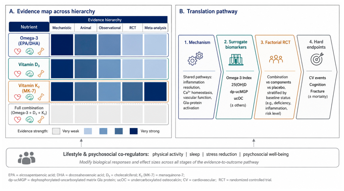
Table 3.
Evidence hierarchy for the principal nutrient-organ relationships. Strength reflects the highest consistent level of evidence available; “+” qualitative grading: (-) absent/insufficient, (+) limited, (++) moderate, (+++) strong/consistent.
Table 3.
Evidence hierarchy for the principal nutrient-organ relationships. Strength reflects the highest consistent level of evidence available; “+” qualitative grading: (-) absent/insufficient, (+) limited, (++) moderate, (+++) strong/consistent.
| Topic | Preclinical | Observational | RCT | Meta-analysis | Overall strength |
| Omega-3 & CV events | +++ | ++ | ++ (discordant) | ++ | Moderate (population/dose-dependent) |
| Omega-3 & cognition | +++ | ++ | + (mostly null) | + | Limited |
| Vitamin D & bone/calcium | +++ | ++ | ++ | ++ | Moderate |
| Vitamin D & cognition | ++ | ++ | + (null) | + (obs.) | Limited (baseline-dependent) |
| Vitamin K2 (MK-7) & bone | +++ | ++ | ++ | ++ | Moderate-Strong (surrogate/BMD) |
| Vitamin K2 & vascular calcification | +++ | ++ | ++ (imaging endpoint) | + | Moderate (emerging hard endpoint) |
| Heart-Brain-Bone integration | ++ | + | - (triad untested) | - | Hypothesis-generating |
Table 4.
Major clinical trials and analyses relevant to the triad. EE, ethyl ester; IPE, icosapent ethyl; CAC, coronary artery calcification; MACE, major adverse cardiovascular events.
Table 4.
Major clinical trials and analyses relevant to the triad. EE, ethyl ester; IPE, icosapent ethyl; CAC, coronary artery calcification; MACE, major adverse cardiovascular events.
| Study (design) | Population (n) | Intervention | Duration | Primary outcome | Key findings | Limitations |
| REDUCE-IT (RCT) | Statin-treated, high TG (8,179) | IPE 4 g/d (EPA-EE) | ~4.9 y | MACE composite | ~25% MACE reduction | Mineral-oil placebo debate; EPA-only |
| STRENGTH (RCT) | High CV risk (13,078) | EPA+DHA 4 g/d (CA) vs corn oil | ~3.5 y | MACE composite | No benefit | Contrasts REDUCE-IT; formulation differs |
| VITAL (RCT) | Primary prevention (25,871) | EPA+DHA 1 g/d (+/- vit D) | ~5.3 y | MACE & cancer | No significant CV benefit | Low dose; healthy cohort |
| GISSI-P (RCT) | Post-MI (11,324) | n-3 PUFA ~1 g/d | 3.5 y | Death/CV events | Reduced events/mortality | Open-label; pre-statin era |
| EVAPORATE (RCT) | High TG on statin (80) | IPE 4 g/d | 18 mo | Plaque volume (CT) | Low-attenuation plaque regression | Small; imaging surrogate |
| ASCEND (RCT) | Diabetes (15,480) | n-3 1 g/d | ~7.4 y | Vascular events | No benefit | Low dose |
| MIDAS (RCT) | Age-related decline (485) | DHA 900 mg/d | 24 wk | Memory tests | Improved episodic/visuospatial memory | Short; surrogate cognitive endpoints |
| MAPT (RCT) | Memory complaints, elderly (1,680) | Omega-3 +/- multidomain | 3 y | Cognitive composite | No overall benefit; subgroup signal | Low baseline omega-3 deficiency |
| DO-HEALTH (RCT) | Healthy seniors (2,157) | Vit D3 + omega-3 + exercise | 3 y | Composite clinical | No effect on primary outcomes | Replete, healthy cohort |
| Knapen (RCT) | Postmenopausal women (244) | MK-7 180 mcg/d | 3 y | BMD, osteocalcin carboxylation | Reduced BMD loss; better carboxylation | Surrogate/BMD, not fracture |
| VitaK-CAC (RCT) | Coronary artery disease | MK-7 360 mcg/d | 2 y | CAC progression | Slowed CAC progression | Imaging endpoint; needs replication |
| AVADEC substudy (RCT) | Aortic valve calcification | Vit K2 + D3 | 24 mo | Epicardial fat/inflammation | K-status marker changed; no EAT effect | Substudy; surrogate endpoints |
Table 5.
Evidence-informed reference dosage ranges for combined supplementation. Ranges reflect published clinical literature and adequate-intake frameworks; they are not a substitute for individualized medical advice.
Table 5.
Evidence-informed reference dosage ranges for combined supplementation. Ranges reflect published clinical literature and adequate-intake frameworks; they are not a substitute for individualized medical advice.
| Nutrient | Reference daily range | Primary biomarker | Mechanistic rationale |
| Vitamin D3 (cholecalciferol) | ~800-2000 IU | Serum 25(OH)D (~30-50 ng/mL) | Calcium absorption; VDR-mediated pleiotropy |
| Vitamin K2 (MK-7) | ~90-180 micrograms | Undercarboxylated MGP/osteocalcin | Carboxylates MGP & osteocalcin; calcium routing |
| Omega-3 (EPA/DHA, high-purity) | ~1-2 g EPA+DHA | Omega-3 Index (RBC %) | Membrane incorporation; SPM-mediated resolution (rTG affects absorption only) |
Disclaimer/Publisher’s Note: The statements, opinions and data contained in all publications are solely those of the individual author(s) and contributor(s) and not of MDPI and/or the editor(s). MDPI and/or the editor(s) disclaim responsibility for any injury to people or property resulting from any ideas, methods, instructions or products referred to in the content. |
© 2026 by the authors. Licensee MDPI, Basel, Switzerland. This article is an open access article distributed under the terms and conditions of the Creative Commons Attribution (CC BY) license (http://creativecommons.org/licenses/by/4.0/).
Copyright: This open access article is published under a Creative Commons CC BY 4.0 license, which permit the free download, distribution, and reuse, provided that the author and preprint are cited in any reuse.


