

Submitted:
07 July 2026
Posted:
08 July 2026
You are already at the latest version
Abstract
Brain metastases represent a major clinical challenge in the management of non-small cell lung cancer, particularly in patients with epidermal growth factor receptor mutations constituting a leading cause of morbidity and mortality. In recent years, significant advancements have been made in both local therapies and systemic treatments. Nevertheless, managing central nervous system disease remains complex, encompassing challenges such as optimal therapy sequencing, resistance mechanisms, oligoprogression, the role of the blood-brain barrier, diagnostic strategies, and the influence of the brain tumor microenvironment. Despite advances, brain metastases in non-small cell lung cancer remain an unmet need. Local treatments like radiotherapy are effective but carry risks of long-term neurological side effects, while the number and burden of brain metastases remain key prognostic factors. Surgical resection may be considered even in patients with multiple brain metastases to improve clinical status, enable further therapy, and enhance survival and quality of life. With the extension of survival afforded by systemic therapies, optimizing the balance between treatment efficacy and quality of life has become critical. Integrating patient perspectives is essential in navigating these complex clinical decisions.
Keywords:
brain
; metastasis
; non-small cell lung cancer
; NSCLC
; EGFR-mutant NSCLC
1. Introduction
Brain metastases (BM) represent a significant clinical challenge in the management of non-small cell lung cancer (NSCLC), particularly in patients harboring epidermal growth factor receptor mutations (EGFRm), where its incidence can be notably high (Figure 1) [1,2]. The development of BM and leptomeningeal disease (LMD) often leads to poor prognosis and significantly impacts patient quality of life, underscoring the critical need for optimized therapeutic strategies [2,3]. In recent years, there have been rapid advancements in both local treatments, such as stereotactic radiosurgery (SRS), and systemic therapies, including newer-generation tyrosine kinase inhibitors (TKIs) with improved central nervous system (CNS) penetration [1,4,5,6,7] and emerging antibody-based treatments [8,9]. However, navigating the complexities of CNS disease management, including optimal sequencing of therapies, managing resistance and oligoprogression [10], understanding the role of the blood-brain barrier (BBB) [11], leveraging diagnostic tools like liquid biopsy [12], and considering the unique brain tumor microenvironment (TME) [13], remains an area of active investigation and debate.
To address these evolving challenges, a panel of experts identified critical questions related to the contemporary management of BM in NSCLC. This review synthesizes current evidence from the literature to examine these critical areas, with the aim of providing insights into optimizing treatment strategies for this specific population of patients.
2. Methods
A multidisciplinary panel of eleven experts participated in structured discussions and reached consensus on six critical questions and unresolved challenges in the field:
- Should upfront systemic or local therapy be pursued for BM in patients with EGFRm NSCLC?
- Could antibody-based regimens or novel therapeutic regimens offer a potential alternative to radiotherapy for EGFRm NSCLC patients with untreated baseline BM?
- Can liquid biopsy inform the clinical management of BM and LMD in patients with EGFRm NSCLC? What are the limitations and potential applications of this approach?
- What strategies can be employed to improve CNS penetration of systemic therapy?
- In patients with NSCLC, does the TME in the brain play a role in promoting or inhibiting BM or LMD?
- What biological factors mediate organ-specific metastasis of cancer?
Following this consensus-building process, a targeted literature review was conducted in PubMed/MEDLINE using broad search terms, including “NSCLC” and “metastasis”, combined with question-specific keywords tailored to each of the six priority areas. To ensure a contemporary perspective, English-language publications published between 2020 and 2025 were considered, regardless of study design.
3. Results
Should upfront systemic or local therapy be pursued for BM in patients with EGFRm NSCLC if technically adequate?
EGFRm non-small cell lung cancer has a well-recognized predilection for CNS involvement, and the introduction of highly CNS-penetrant systemic therapies has substantially reshaped treatment paradigms for BM. Third generation EGFR TKIs, most notably osimertinib, demonstrate superior blood–brain barrier penetration and achieve higher intracranial response rates with prolonged intracranial progression-free survival (PFS) and improved overall survival (OS) compared with earlier generation agents [1,4,14,15,16,17]. For asymptomatic patients with small or multiple BM, frontline systemic therapy with a CNS active TKI is therefore widely endorsed because it concurrently addresses intracranial and extracranial disease and frequently permits safe deferral of cranial irradiation [14]. When intracranial progression occurs on osimertinib 80 mg, contemporary expert consensus advocates continuing osimertinib while adding focal SRS rather than immediate whole brain radiotherapy (WBRT). Dose escalation of osimertinib to 160 mg can be considered in selected situations and is often preferred for LMD when supported by clinical judgment and availability [18]. Combination systemic approaches have further expanded options for CNS disease control: the FLAURA2 trial demonstrated that adding chemotherapy to osimertinib significantly reduced CNS progression and markedly increased CNS complete remission rates among patients with measurable lesions ≥1 cm (48% vs 16%) [19], and MARIPOSA reported that first line amivantamab plus lazertinib prolonged intracranial durability and halved the 3 year intracranial progression risk relative to osimertinib, despite comparable intracranial objective response rate (ORR) [20]. These data highlight the emerging role of antibody-containing CNS active regimens and support the strategy of prioritizing CNS-penetrant systemic molecules when clinically appropriate.
Local therapies remain pivotal in scenarios where immediate decompression or high precision local control is required. SRS is the focal modality of choice for limited intracranial disease and for symptomatic or large lesions with mass effect because it affords high rates of local control while sparing uninvolved brain tissue [14,21]. Neurosurgical resection retains its indication for accessible lesions that cause mass effect or require histologic confirmation. By contrast, WBRT - while effective for widespread intracranial involvement - has an increasingly circumscribed role given its well documented and often irreversible neurocognitive sequelae; accordingly, WBRT is generally reserved for patients with diffuse CNS disease not amenable to focal techniques [15,21,22]. Real world data showed that upfront EGFR-TKI with deferred radiotherapy was associated with inferior overall survival, whereas stereotactic radiosurgery followed by EGFR-TKI achieved the longest OS while avoiding the risk of neurocognitive decline associated with whole-brain radiotherapy [23]. However, analyses that include modern CNS penetrant agents yield heterogeneous results: some reports indicate no clear OS advantage to adding upfront SRS to high penetrance TKIs [23], whereas others, including the TURBO trial, demonstrate improved time to CNS progression and superior local control with upfront SRS, particularly for lesions ≥1 cm [23,24,25]. These collective findings suggest that lesion size (notably ≥1 cm), symptom burden and expected survival are critical determinants of whether upfront focal therapy provides an incremental survival or quality of life benefit beyond systemic monotherapy.
CNS oligoprogression, defined as limited intracranial progression in the context of otherwise controlled systemic disease, has become a common clinical pattern as systemic therapies achieve durable extracranial control. While the optimal management is still evolving, a possible strategy—drawing parallels from established paradigms in other oncogene-driven diseases like HER2-positive breast cancer—is to pursue focal local interventions while continuing the current effective systemic agent. This approach aims to preserve extracranial disease control and delay wholesale changes in systemic therapy. [10,26]. In this context, focal treatments may include stereotactic radiosurgery (SRS) to the progressing sites, or, for selected lesions, neurosurgical resection followed by SRS to the resection cavity. When a change in systemic therapy is eventually required due to broader resistance, completing focal radiation before initiating the next agent is often prudent to minimize overlapping toxicities and optimize local control [26,27]. The biological rationale for such selective local interventions is supported by evidence that brain metastases (BM) frequently exhibit distinct genomic profiles compared to primary or extracranial sites, including higher tumor mutational burden, an increased fraction of the genome altered, more frequent whole genome duplication, and enrichment of cell cycle pathway alterations. [28,29]. These genomic and microenvironmental distinctions suggest that intracranial lesions may undergo independent clonal evolution, potentially explaining why site-specific focal interventions remain necessary for a subset of patients even when systemic disease remains otherwise well-controlled.
Radiation related neurotoxicity is a major trade off that must be weighed carefully, especially as effective systemic therapies lengthen survival. WBRT is associated with measurable declines in neurocognitive function and reductions in quality of life, effects that are of consequence for patients with longer life expectancy [14,15]. Although SRS is more sparing of global cognitive function, it is not without risk: focal radiation necrosis, symptomatic edema and cumulative toxicity from multiple or repeated treatments can produce substantial morbidity [30,31]. Given the enhanced CNS activity of contemporary systemic agents, there is increasing justification for deferring or avoiding cranial irradiation when clinically appropriate to minimize late neurotoxicity. When WBRT cannot be avoided, hippocampal sparing techniques and neuroprotective pharmacotherapy such as memantine should be employed to mitigate cognitive decline [15].
Despite the advances summarized above, several critical evidence gaps persist. Prospective, randomized trials are needed to define which patients derive an OS advantage from upfront focal therapy in the context of potent CNS penetrant systemic regimens, and to determine whether antibody plus TKI combinations can reliably obviate the need for radiation in selected cohorts without compromising long term outcomes. Biomarker driven studies that explicitly address the distinct genomics of BM are required to refine patient selection for local interventions and to elucidate intracranial resistance mechanisms. Mechanistic preclinical work should also clarify whether combined TKI radiation regimens produce additive or synergistic antitumor effects in EGFRm disease [32]. In clinical practice, treatment selection should be individualized within a multidisciplinary tumor board, integrating lesion size and number, symptom severity, performance status, anticipated survival, resistance biology and patient preferences.
In summary, for most patients with EGFRm NSCLC and asymptomatic or small multiple BM, initial management should prioritize systemic therapy with CNS penetrant molecules (third generation TKIs and emerging CNS active antibody combinations) because these agents target both intracranial and extracranial disease while minimizing immediate exposure to radiation and its attendant neurocognitive risks [1,4,14,15,16,17,18,19,20]. Upfront local therapy—consisting of surgical resection or, for unresectable lesions, fractionated stereotactic radiotherapy (fSRT)—remains mandatory for symptomatic, large, or life-threatening lesions. While SRS is effective for smaller targets, larger volumes often necessitate fractionation to mitigate the risk of radionecrosis and exacerbated perilesional edema. Local consolidation should also be strongly considered for selected patients with limited intracranial disease (particularly lesions ≥1 cm) where achieving durable local control is likely to yield clinical benefit [21,22,23,24,25]. WBRT should be reserved for diffuse, unresectable intracranial disease and, when used, combined with neuroprotective strategies [15]. Ultimately, multidisciplinary, biomarker driven decision making and prospective clinical trials are urgently needed to optimize selection for upfront SRS versus systemic first strategies in the era of highly CNS penetrant therapies [1,4,14,15,16,17,18,19,20,21,22,23,24,25,26,27,28,29,30,31,32,33].
Could antibody-based regimens or novel therapeutic regimens offer a potential alternative to radiotherapy for EGFRm NSCLC patients with untreated baseline BM?
Patients with EGFRm NSCLC exhibit a notably high incidence of BM, with rates reaching up to 70%, significantly exceeding those observed in non–EGFR-driven NSCLC. While current treatment options include EGFR TKIs, WBRT, SRS, and chemotherapy, the optimal management and sequencing remain undefined [5,34]. Newer generation EGFR TKIs like osimertinib have demonstrated improved BBB penetration and significant intracranial activity that may contribute to improved OS [5]. However, the management of BM remains a critical challenge due to high morbidity and mortality risks.
Recently, antibody-based regimens, including antibody-drug conjugates, bispecific T-cell engagers, and bispecific antibodies, have emerged as promising options for managing EGFRm NSCLC with BM [8].
HER3 is frequently overexpressed in BMs of NSCLC (73%) and breast cancer (75%), often exceeding expression levels found in extracranial sites [35,36,37]. This makes it a critical target, particularly as HER3 has been described to facilitate CNS colonization by cancer cells. Although data on CNS efficacy for antibody-drug conjugates remain limited (a critical consideration in oncogene-driven NSCLC37), recent clinical trials have shown promising results.
In the HERTHENA-Lung01 trial, patritumab deruxtecan demonstrated an intracranial ORR of 33% among 30 patients with untreated baseline BM [38]. The median OS observed in this trial for patients with advanced EGFR-mutated NSCLC without LMD was 11.9 months, while a separate cohort of patients with LMD showed a median OS of 10.5 months [39]. Further investigation is underway in the TUXEDO-3 trial (NCT05865990) and the PARAMETer trial (NCT05620914). However, this drug is not approved for NSCLC, which limits its use in the clinical setting.
Similarly, biomarker-agnostic strategies targeting TROP2 are being explored post-osimertinib failure [40,41,42]. In the TROPION-Lung05 study, datopotamab deruxtecan (Dato-DXd) showed comparable systemic efficacy regardless of baseline BM status. Among patients with measurable intracranial lesions, the ORR was 22% (18% in EGFRm) with a disease control rate of 72% [43]. Preclinical models suggest Dato-DXd can penetrate the BBB to mediate these effects [44], warranting further study in trials such as TUXEDO-2 (NCT05866432) and DATO-BASE (NCT06176261).
The bispecific EGFR/MET antibody amivantamab has demonstrated robust intracranial efficacy. In the MARIPOSA-2 trial, the combination of amivantamab and chemotherapy significantly delayed CNS recurrence. Among patients with BM and no prior radiotherapy, the median intracranial PFS was 11.1 months for amivantamab-lazertinib-chemotherapy compared to 6.3 months for chemotherapy alone (hazard ratio 0.36) [21].
Amivantamab plus lazertinib also shows anti-tumor activity in patients developing new or progressing brain or leptomeningeal metastases. Patients with BM had an ORR of 50%, median PFS of 5.8 months, and median OS of 17.4 months. Those with LMD had an ORR of 33%, median PFS of 7.8 months, and median OS of 14.4 months [45]. Despite being a large molecule, amivantamab demonstrates clinical efficacy, potentially mediated by direct antitumor effects and/or immune-mediated mechanisms, challenging the traditional view that large antibodies generally lack BBB penetration [21].
Emerging evidence suggests immune checkpoint inhibitors (ICIs) also hold promise, with several mechanisms explaining their efficacy in this context [9]. By blocking inhibitory pathways, ICIs like pembrolizumab and nivolumab enhance the immune response. In patients with NSCLC and BM, ICIs have demonstrated significant efficacy, with studies reporting disease control rates of 39% in patients with asymptomatic BM [46].
The efficacy of large molecules challenges the historical dogma that the BBB is impermeable to antibodies. Evidence suggests that in patients with BM, the BBB is inherently compromised, forming a "blood–tumor barrier" (BTB) (Figure 2). This heterogeneous vasculature is characterized by disrupted neurovascular units, altered transporter expression, and loss of astrocytic endfeet connections [47,48].
This structural compromise may facilitate the passage of therapeutic antibodies. Furthermore, combination therapies pairing ICIs with anti-angiogenic agents like bevacizumab may further enhance drug delivery by normalizing tumor vasculature [49]. Bevacizumab, by inhibiting vascular endothelial growth factor (VEGF), suppresses angiogenesis and has shown promise in preclinical studies by suppressing the growth of established BM [50].
From a clinical perspective, this combination is particularly valuable when strategies to reduce peritumoral vasogenic edema are required. Given that corticosteroids can inhibit the efficacy of ICIs, the bevacizumab plus ICI regimen serves as a crucial alternative for managing edema without compromising the immune response.
Before broadly considering these new molecules as alternatives to radiotherapy, caution is warranted. Robust data on CNS efficacy for antibody-drug conjugates remain limited [50], and the optimal integration of these therapies with TKIs, chemotherapy, or radiotherapy remains an open question [51,52]. Other molecules or combinations, such as zipalertinib, sunvozertinib, furmonertinib, or combinations with osimertinib plus savolitinib, also show encouraging brain activity [53].
However, the shift toward antibody-based regimens offers a potential avenue to reduce the cumulative neurological toxicity associated with repeated radiotherapy while increasing intracranial control [5,20,22,54]. As research evolves, clarifying the role of these agents will be essential for optimizing treatment strategies.
Can liquid biopsy inform the clinical management of BM and LMD in patients with EGFRm NSCLC? What are the limitations and potential applications of this approach?
Liquid biopsy refers to a set of minimally invasive techniques used to analyze biomarkers, such as cell-free DNA, extracellular vesicles, microRNAs, circulating tumor cells, and tumor-derived metabolites, detected in bodily fluids like blood or cerebrospinal fluid (CSF) [55]. While activation of the EGFR pathway drives oncogenesis in 15% to 40% of NSCLC cases [23], brain metastatic sites are highly divergent from primary tumors despite sharing a common ancestor [56].
In the context of systemic disease, circulating tumor DNA (ctDNA) levels detected in plasma are strongly prognostic for disease-free and distant metastasis-free survival, serving as indicators of micro metastases. However, no reliable inferences can be made regarding CNS disease due to the BBB. Data suggests that "ctDNA shedding" (detecting the driver EGFR mutation in plasma) is associated with a higher incidence of BM. Conversely, patients who are "non-shedders" before and after osimertinib treatment and have no BM show longer post-progression survival [57,58]. Nevertheless, for patients with stable or active BM, data on using plasma liquid biopsy to guide therapy remains limited (Figure 3).
A particularly challenging scenario arises in patients with metastases confined to the brain or LMD, where plasma liquid biopsy often fails due to the BBB. In these cases, CSF is emerging as a valuable alternative [59]. Its proximity to brain tumors and low baseline cellularity reduces background noise, enhancing sensitivity for detecting tumor-derived material compared to plasma [58].
Studies indicate that CSF ctDNA provides a more comprehensive driver gene profile during CNS progression. For instance, EGFR mutations were detected in 85.5% of LMD cases in CSF, including the T790M mutation in 16.1% [60]. Comparative studies show EGFR detection rates of 82% in CSF versus 45% in matched plasma, with similar superiority observed for ALK and ROS1 [61]. Furthermore, mutant-allelic frequency levels are significantly higher in CSF [61], and some studies report a 100% detection rate for driver mutations in CSF samples [62]. Consequently, NCCN guidelines contemplate liquid biopsy in CSF for cases of negative-cytology leptomeningeal dissemination, where available [63].
The role of CSF in LMD is multifaceted. CSF cytology remains the gold standard for diagnosis, and investigation is mandatory whenever LMD is suspected [64]. However, sensitivity can be low, often requiring multiple samples (up to three) to improve diagnostic yield [65,66,67]. In approximately 25% of cases, positive cytology does not align with clinical features or biochemical markers like glucose and protein [68].
In cases where cytology is negative, but suspicion remains high, CSF ctDNA analysis becomes critical. It can reveal actionable mutations missed by blood samples and provide evidence of malignancy when magnetic resonance imaging is inconclusive [58,69,70,71]. Conversely, plasma liquid biopsy approaches have not achieved sufficient diagnostic accuracy for LMD and are not currently recommended [67,72,73,74].
CSF analysis is crucial for tailoring targeted therapies. Genotyping can identify resistance mechanisms, such as T790M or specific EGFR 19 deletions, informing treatment strategies more accurately than plasma [71]. For example, the BLOSSOM trial demonstrated the intracranial efficacy of 80 mg osimertinib in LMD, with pharmacokinetic analysis showing a CSF-to-free-plasma ratio of 22% [75]. Similarly, the combination of amivantamab and lazertinib has shown promise, with 64% of LMD patients exhibiting a decrease in CSF circulating tumor cells [76].
While head-to-head comparisons are limited, single cell sequencing of circulating tumor cells may offer better characterization of mutational diversity than cell-free DNA [72,77,78,79]. However, interpreting CSF findings requires caution. Extracranial resistance mechanisms cannot be assumed to exist in the CNS without direct evidence [80]. Furthermore, CNS progression on TKIs may result from low drug exposure rather than true resistance, potentially supporting the continuation of TKIs with high BBB penetration alongside systemic treatments [81].
Despite its promise, CSF analysis faces challenges. Technical factors such as sampling site, volume, and processing affect accuracy [67]. Interpreting CSF cell-free DNA is complex; molecular variations may reflect clonal randomness rather than dominant drivers, and "lessons from plasma" warn that high variant allele frequencies could indicate non-tumor sources [80]. Additionally, longitudinal CSF assessment is invasive and often not feasible. Response assessment in LMD remains complicated due to the lack of validated imaging criteria and the difficulty in distinguishing cancer progression from neurotoxicity or comorbidities [72].
In conclusion, liquid biopsy, particularly CSF ctDNA analysis, offers a minimally invasive method to monitor disease progression and guide treatment in EGFRm NSCLC patients with CNS involvement [82]. While challenges in standardization and sensitivity remain, the integration of CSF analysis into clinical practice represents a significant step toward optimizing the management of this complex patient population.
What strategies can be employed to improve CNS penetration of systemic therapy?
The development of CNS therapeutics is challenging due to the BBB, a complex interface that maintains CNS homeostasis by regulating nutrient passage (e.g., amino acids, glucose) while blocking harmful molecules like neurotoxins [15,27]. However, the BBB also restricts the entry of certain drugs and large biopharmaceuticals into the brain [11]. For instance, after parenteral administration of therapeutic antibodies, their concentration in the brain is only 0.01–0.10% of plasma levels. While these low antibody levels in the brain may produce pharmacological effects in certain cases, depending on the molecular target and the antibody's interaction dynamics, it is evident that, in many instances, achieving a higher concentration of the antibody in the brain would be advantageous, reducing the required dose and enhancing the therapeutic index [11,83].
BM are a hallmark of EGFRm (Figure 1) and ALK fusion NSCLC, with a higher proclivity for brain involvement compared to EGFR wild-type cases [84]. Their presence negatively impacts clinical outcomes, as patients treated with first- or second-generation TKIs often experience shorter PFS [3]. Improving CNS penetration of systemic therapies is essential for effectively managing BM and LMD, especially in patients with EGFRm. To treat metastatic brain disease, BBB permeability is considered desirable for increasing clinical efficacy. The development of brain-penetrant molecules for oncogene-defined NSCLC is an active area of research [85].
The emergence of novel TKIs with enhanced CNS penetration has led to a paradigm shift in treating NSCLC patients with BM. These newer generation TKIs have demonstrated promising intracranial activity in preclinical and clinical studies [6]. For instance, ALK inhibitors like alectinib, lorlatinib, and brigatinib, as well as the third-generation EGFR inhibitor osimertinib, lazertinib or aumolertinib, have shown improved CNS penetrance and efficacy compared to earlier generations [7]. Some of these agents have achieved CNS ORR of up to 70-80% in patients with targetable molecular alterations [30].
Notably, high-dose pulsatile therapy has been explored as a potential strategy to improve CNS penetration. In one case study, high-dose pulsatile crizotinib (1000 mg/day) administered on an alternating-day schedule (one day on, one day off) resulted in a significant CNS response and prolonged time to neurological progression in a patient with NSCLC and multiple intracranial metastases. According to the ESMO expert consensus recommendations, the standard approach for patients with intracranial progression despite osimertinib 80 mg is to continue the current dose of Osimertinib. An increased dose of osimertinib (160 mg) may be considered if accessible [86]. This approach may help overcome pharmacokinetic and biodynamic resistance phenomena associated with CNS metastases.
The limited brain penetration of certain clinically used EGFR TKIs may be attributed to their physicochemical properties, which fall outside the optimal ranges for crossing BBB, including molecular weight, hydrogen bond donors and acceptors, polar surface area, and rotatable bonds. These properties also impact the interaction with P-glycoprotein (P-gp) and breast cancer resistance protein, critical efflux transporters in the BBB, further restricting drug exposure in the CNS [87].
Intrathecal administration is also an approach. In a retrospective cohort study of 23 patients with NSCLC and LMD treated with the novel EGFR TKI furmonertinib, administered intrathecally alongside either methotrexate or pemetrexed, the median PFS was 10.8 months, while the median OS was not reached [88]. Though not specifically targeting NSCLC patients, several strategies have emerged to enhance drug delivery to the CNS, including the use of targeted therapies, innovative drug delivery systems, and techniques to temporarily disrupt the BBB. One promising approach involves the use of targeted therapies that are specifically designed to penetrate the BBB. For instance, early research on next-generation EGFR TKIs such as zorifertinib, approved in China since 2024, has shown improved CNS penetration compared to first-generation TKIs like gefitinib and erlotinib [89,90]. These newer agents might be particularly beneficial for patients with EGFRm who develop BM, as they could effectively target and inhibit tumor growth within the CNS [91,92]. Zorifertinib exhibits superior BBB penetration compared to earlier-generation EGFR TKIs, a property that enhances its potential efficacy in treating BM in patients with EGFR-mutant non-small cell lung cancer (NSCLC). This enhanced CNS distribution is primarily attributed to its low affinity for efflux transporters such as P-gp and breast cancer resistance protein, which are highly expressed at the BBB and actively restrict the entry of many therapeutic agents into the central nervous system. Unlike first- and second-generation EGFR TKIs, which are efficiently excluded from the brain by these transporters, zorifertinib avoids this mechanism and achieves sustained intracranial exposure. Furthermore, its physicochemical properties - moderate molecular weight, balanced lipophilicity, and favorable ionization state - facilitate passive diffusion across the BBB. Preclinical studies have demonstrated a high unbound brain-to-plasma ratio (Kp,uu) indicating effective free drug availability within the CNS. These characteristics, combined with emerging clinical data showing intracranial responses, position zorifertinib as a promising candidate for the treatment of EGFR-mutant NSCLC with BM [90,93].
In addition to targeted therapies, innovative drug delivery systems such as nanoparticles and liposomes have been developed to improve drug delivery across the BBB. Nanoparticles can be engineered to enhance their ability to penetrate the BBB by modifying their surface properties or by using specific ligands that facilitate transport across the barrier [94]. For example, the incorporation of targeting ligands such as arginine-glycine-aspartic acid peptides into nanoparticles has been shown to enhance their penetration into brain tissues [95]. Similarly, liposomal formulations can protect encapsulated drugs from degradation and improve their bioavailability in the CNS [96]. Other examples include the pulmonary delivery of cisplatin-hyaluronan conjugates via endotracheal instillation for the treatment of lung cancer, and the use of quaratusugene ozeplasmid and osimertinib in patients with advanced lung cancer who progressed on osimertinib in clinical trial Acclaim-1 [97,98].
Another effective strategy could be the use of convection-enhanced delivery, which allows for direct infusion of therapeutic agents into the brain interstitial space, bypassing the BBB altogether. Convection-enhanced delivery utilizes a pressure gradient to distribute drugs uniformly throughout the target area, achieving higher local concentrations than conventional systemic administration [99,100]. This method has been explored in an experimental setting for the treatment of glioblastomas and could possibly be adapted for use in NSCLC with BM [101]. Recent advancements in catheter design, such as step-design catheters, have further improved the efficacy of convection-enhanced delivery by minimizing reflux and enhancing drug distribution [99].
Moreover, techniques to temporarily disrupt the BBB can also facilitate drug delivery. Methods such as focused ultrasound combined with microbubble technology have been explored to transiently open the BBB, allowing for increased penetration of therapeutic agents into the CNS [102,103]. This approach has shown promise in preclinical models and may be applicable in clinical settings for patients with BM. Another therapeutic approach is Tumor Treating Fields, a non-invasive cancer therapy that delivers low-intensity, intermediate-frequency alternating electric fields directly to the tumor. It has been demonstrated to disrupt mitotic spindle assembly by interfering with the significant dipole moment of microtubules, leading to metaphase arrest, extended mitosis, abnormal daughter cell formation, and ultimately cell death [104]. An electromagnetic field is generated to disrupt the mitotic spindles during anaphase, leading the tumor to apoptosis [105].
Improving CNS penetration of systemic therapies in NSCLC involves a multifaceted approach that includes optimizing drug properties, utilizing BBB modulation techniques, leveraging nanotechnology, and integrating combination therapies. Continued research into these strategies and emerging technologies holds promise for improving the management of CNS involvement in NSCLC and other cancers with a high propensity for CNS metastases.
In patients with NSCLC, does the TME in the brain play a role in promoting or inhibiting BM or LMD?
TME in the brain plays a significant role in both promoting and inhibiting BM and LMD in NSCLC patients. The brain TME is unique due to its composition and interactions among various cell types, which can either support or impede the growth and spread of metastatic cells. Several factors within the brain TME, including immune cells, astrocytes, microglia, the extracellular matrix (ECM), and the BBB, are involved in these processes [106].
As stated above, there are some promoting factors in the TME. The expression of specific chemokine receptors, such as C-X-C chemokine receptor type 4 (CXCR4), has been associated with brain-specific metastasis in NSCLC. Studies have shown that high levels of CXCR4 expression correlate with an increased likelihood of BM, suggesting that the CXCL12/CXCR4 axis facilitates tumor cell migration and invasion into the brain [107,108]. This interaction not only promotes the establishment of metastases but also influences the immune landscape within the brain, potentially leading to a more favorable environment for tumor growth. The presence and polarization of immune cells within the TME can also significantly impact tumor behavior. For instance, the infiltration of tumor-associated macrophages has been shown to promote BM by enhancing angiogenesis and providing growth factors that support tumor survival [109]. In contrast, a suppressed immune microenvironment characterized by a predominance of M2 macrophages can facilitate tumor progression and metastasis [110]. The balance between different immune cell types, including T cells and macrophages, can therefore dictate the metastatic potential of NSCLC cells in the brain. The TME is also influenced by the composition and remodeling of the ECM. Cancer-associated fibroblasts play a significant role in this process, contributing to ECM changes that can enhance tumor cell invasion and survival [111,112,113]. The remodeling of the ECM can create a permissive environment for metastasis by facilitating tumor cell migration and providing biochemical signals that promote growth.
As for inhibiting factors in the TME, the BBB serves as a protective barrier that can inhibit the entry of therapeutic agents and metastatic cells into the brain. While some tumor cells can exploit pathways to breach the BBB, the integrity of this barrier can act as a significant obstacle to metastasis. The ability of NSCLC cells to penetrate the BBB is influenced by their molecular characteristics and the presence of specific signaling pathways that facilitate this process [106]. The immune system plays a dual role in TME. While certain immune cells can promote tumor growth, others can exert anti-tumor effects. The presence of activated T cells and natural killer cells can inhibit the establishment and growth of metastases [110]. However, the effectiveness of immune surveillance can be compromised by the immunosuppressive environment often found in BM, where factors such as PD-L1 expression can inhibit T cell activity [114]. TME can also influence the epigenetic landscape of tumor cells, affecting their metastatic potential. For example, microRNAs such as miR-596-3p have been shown to suppress BM by modulating pathways involved in tumor growth and invasion [115]. This suggests that the interactions within the TME can lead to changes in gene expression that either promote or inhibit metastasis.
In summary, the TME in the brain plays a complex role in the development of BM and LMD in NSCLC patients. Factors such as chemokine signaling, immune cell dynamics, and ECM remodeling can promote metastasis, while the integrity of the BBB and immune surveillance can inhibit it. Understanding these mechanisms is crucial for developing targeted therapies that can effectively manage BM in NSCLC.
What biological factors mediate organ-specific metastasis of cancer?
Tumors that arise in different organs often show a preference for metastasizing to certain organs, a phenomenon known as 'organotropism.' This process is thought to be influenced by various factors, including blood circulation patterns, anatomical closeness, the metastatic microenvironment, and the inherent traits of the tumor cells [116]. This necessitates the establishment of self-sufficiency, interactions with the microenvironment, and the production of growth factors that drive tumor progression within this specific context. Epithelial-mesenchymal transition (EMT) is an evolutionarily conserved process essential for embryonic development, but recent studies have shown its involvement in cancer progression and metastasis. EMT plays a critical role in the development of BM, as metastatic brain lesions commonly exhibit a loss of epithelial characteristics [117]. E-Cadherin, which mediates cell adhesion, is notably diminished in patients with NSCLC who have BM, compared to those without, providing evidence for the link between EMT and BM development [118]. In patients with NSCLC and BM, markers such as N-cadherin and vimentin are often over-expressed, which are associated with migration and adhesion mechanisms, and they may indicate an increased risk of relapse [119]. The loss of EMT during metastatic development also leads to tumor stromal degradation due to the upregulation of matrix metalloproteases and plasminogen activators, promoting an invasive phenotype [120]. Notably, elevated levels of MMP-9 have been observed in lung cancer BM compared to other tumors, suggesting its key role in facilitating the migration of tumor cells across the BBB into the CNS [121].
TME plays a pivotal role in determining the metastatic potential of cancer cells. The TME consists of various cell types, including stromal cells, immune cells, and ECM components, which can either support or inhibit tumor progression. For instance, tumor-associated macrophages can adopt pro-tumorigenic roles by secreting growth factors and cytokines that promote metastasis [122]. Additionally, the presence of specific immune cell populations can create an immunosuppressive environment that facilitates tumor cell survival and dissemination [123].
Chemokines and their receptors are critical mediators of organ-specific metastasis. Tumor cells often express specific chemokine receptors that guide their migration to specific organs where corresponding chemokines are produced. For example, the interaction between vascular cell adhesion molecule-1 on endothelial cells and integrin α4β1 on cancer cells has been shown to promote the adhesion and migration of metastatic cells to the bone [124]. Studies have indicated that levels of CXCL12 and its receptor CXCR4 are elevated in patients with BM compared to those with other tumors that do not have metastases [108]. This suggests that the former exhibits a phenotype that is more prone to cell migration, as the interaction between these chemokines is linked to cell proliferation and migration. Additionally, it was found that carcinomas lacking CX3CR1 tend to metastasize preferentially to the brain, while those expressing CX3CR1 tend to spread to other sites [125].
In addition to cytokines and markers of EMT loss, several growth factor pathways with protein kinase activity and their receptors have been implicated in the development of BM. Transcriptional studies of BM across various tumors have demonstrated that EGFR/ERK expression is increased in tumors with brain involvement. Specifically, in lung cancer, it has been observed that the expression levels of the proteins in this pathway are even higher in metastases than in the primary tumor [126].
The c-MET-HIF pathway is over-expressed in BM, particularly in patients with EGFRm NSCLC, contributing to resistance to EGFR inhibitors [127]. Additionally, the angiogenesis pathway plays a critical role in tumor invasion and CNS infiltration, with VEGF and its receptors promoting neo-vascularization and vascular permeability. Increased VEGF expression has been observed in NSCLC patients with brain involvement, especially those with adenocarcinoma. Preclinical experiments indicate that silencing VEGF reduces the incidence of BM [128]. Moreover, studies have shown that monoclonal antibodies targeting VEGF receptor, such as bevacizumab, exhibit intracerebral activity due to the pathway's overexpression in the brain [129].
The establishment of a pre-metastatic niche is a crucial step in the metastatic process. Tumor cells can secrete factors that modify the microenvironment of distant organs, making them more conducive to tumor growth. For instance, tumor-secreted proteins can recruit immune cells and create an inflammatory environment that supports the survival of disseminated tumor cells [130]. This niche formation is often mediated by exosomes and other extracellular vesicles that carry signaling molecules to the target organs [131].
Different organs exhibit unique gene expression profiles that can influence the behavior of metastatic cells. Studies have identified organ-specific genes that are upregulated in metastases, suggesting that the local microenvironment can induce specific transcriptional programs in tumor cells [132]. For example, the expression of certain integrins and adhesion molecules can be modulated by the organ microenvironment, affecting the ability of cancer cells to adhere and invade [133].
Hypoxic conditions within the TME can significantly influence metastatic behavior. Tumors often adapt to low oxygen levels by activating hypoxia-inducible factors (HIFs), which promote angiogenesis and metabolic reprogramming [134]. These adaptations can enhance the survival and proliferation of metastatic cells in specific organs, such as the bone or liver, where hypoxic conditions may prevail [135].
The composition and remodeling of the ECM in target organs can also dictate metastatic behavior. The ECM provides structural support and biochemical signals that influence tumor cell migration and invasion. For instance, the presence of specific ECM components, such as fibronectin and collagen, can facilitate the adhesion and growth of metastatic cells in the liver or lung [136]. Additionally, the interaction between tumor cells and the ECM can trigger signaling pathways that promote EMT, enhancing the invasive potential of cancer cells [137].
Overall, the elevated incidence of CNS metastases and their associated dismal prognosis underscore the critical need to delineate molecular determinants of metastatic dissemination. Genomic divergence has emerged as a key mechanism, with distinct alterations such as mutations within the PI3K/AKT/mTOR signaling axis identified in BM but absent from primary or extracranial lesions [138]. CNS metastases in lung cancer exhibit increased tumor mutational burden, a greater fraction of genome altered, and evidence of whole-genome duplication [28]. Although recurrent alterations in CDKN2A/B, TP53, EGFR, KRAS, and MYC have been documented across diverse lung cancer histologies [28,139,140,141], their specific contributions to CNS tropism and progression remain unresolved. Notably, CNS metastases correlate with a higher frequency of CARD11 amplifications and a lower frequency of MDM2 amplifications, while patients harbor atypical EGFR mutations or developing CNS involvement under osimertinib therapy experience inferior survival outcomes. Furthermore, comparative analyses reveal that genomic landscapes between BM and leptomeningeal metastases, as well as paired intra- versus extracranial tumor samples, are broadly similar, suggesting that additional non-genomic factors may influence CNS colonization [142].
In conclusion, organ-specific metastasis is mediated by a complex interplay of biological factors within the TME, including immune cell dynamics, chemokine signaling, pre-metastatic niche formation, organ-specific gene expression, hypoxic conditions, and ECM composition. Understanding these mechanisms is essential for developing effective therapeutic strategies to target metastasis and improve patient outcomes in cancer treatment.
4. Conclusion
Brain metastases represent a substantial clinical challenge, constituting a leading cause of morbidity and mortality in lung cancer and exhibiting a rising incidence. Patients with BM endure considerable neurological and systemic symptom burden, compounded by poor prognosis and profound psychological distress, including anxiety and depression. Recent evidence indicates that the majority of individuals with BM experience significant psycho-oncological needs, with female sex, synchronous BM presentation, and reduced Karnofsky performance status independently predicting the requirement for specialized intervention [143]. Despite therapeutic advances, BM in non-small cell lung cancer remains an unmet medical need. While local modalities such as radiotherapy offer efficacy, they warrant strong consideration, their potential for long-term neurological sequelae necessitates judicious use. In this era of modern systemic treatments for cancer, the number of BM and total cerebral tumor burden remain significant prognostic factors of OS. It remains to be clarified if surgical resection should be considered as an option even in those patients with multiple BM to enhance patient clinical status, enable further local and systemic treatment delivery, and improve their survival and quality of life, as well as the need for radiation therapy or upfront SRS treatment.
As systemic therapies extend survival (either due to activity in the brain or because they offer strong systemic control and thus reducing the ability to metastasize for the brain), optimizing the balance between treatment efficacy, including local therapies, and quality of life becomes paramount. Having a drug that can be initiated quickly to achieve a systemic response is particularly valuable when patients are symptomatic or have life threatening disease and allows them to start a treatment without having to wait for radiation to be completed. Incorporating patient perspectives into clinical decision-making is essential to address these complex challenges.
Author Contributions
All authors were involved in the conception and methodology of this project, the writing of the manuscript and critical revision for intellectual content. All authors approved the final version of the manuscript.
Funding
This study was funded by Johnson & Johnson Innovative Medicine, Portugal.
Acknowledgments
The authors would like to acknowledge Catarina Silva (Instituto de Saúde Baseada na Evidência, Senior Researcher) for support in the submission of the manuscript.
Conflicts of Interest
JRC is employee of J&J Innovative Medicine. JR is an external consultant of Instituto de Saúde Baseada na Evidência which was contracted by J&J Innovative Medicine to provide support in medical writing activities. All remaining authors have no conflict of interests.
Ethical Statement
This project did not involve the collection of any data requiring submission to or approval by an Institutional Review Board/Ethics Committee, in accordance with Portuguese Law No. 58/2019 of 8 August (https://diariodarepublica.pt/dr/detalhe/lei/58-2019-123815982). Prior to expert meetings, all participants were informed about the objectives, methodology, and their responsibilities under this project. Ethical considerations were upheld throughout the entire process, ensuring that participants’ contributions were voluntary.
References
- Ballard, P.; Yates, J.W.T.; Yang, Z.; Kim, D.W.; Yang, J.C.H.; Cantarini, M.; et al. Preclinical Comparison of Osimertinib with Other EGFR-TKIs in EGFR-Mutant NSCLC Brain Metastases Models, and Early Evidence of Clinical Brain Metastases Activity. Clin. Cancer Res. Off. J. Am. Assoc. Cancer Res. 2016, 22(20), 5130–5140. [Google Scholar] [CrossRef]
- Imber, B.S.; Sehgal, R.; Saganty, R.; Reiner, A.S.; Ilica, A.T.; Miao, E.; Li, B.T.; Riely, G.J.; Yu, H.A.; Panageas, K.S.; et al. Intracranial Outcomes of De Novo Brain Metastases Treated With Osimertinib Alone in Patients With Newly Diagnosed EGFR-Mutant NSCLC. JTO Clin. Res. Rep. 2023, 4, 100607. [Google Scholar] [CrossRef] [PubMed]
- Schuler, M.; Wu, Y.-L.; Hirsh, V.; O’byrne, K.; Yamamoto, N.; Mok, T.; Popat, S.; Sequist, L.V.; Massey, D.; Zazulina, V.; et al. First-Line Afatinib versus Chemotherapy in Patients with Non–Small Cell Lung Cancer and Common Epidermal Growth Factor Receptor Gene Mutations and Brain Metastases. J. Thorac. Oncol. 2016, 11, 380–390. [Google Scholar] [CrossRef] [PubMed]
- Colclough, N.; Ballard, P.; Barton, P.; Chen, K.; Cross, D.; Finlay, M.; Han, L.; Janefeldt, A.; Johnström, P.; Ward, R.; et al. Preclinical comparison of the blood brain barrier (BBB) permeability of osimertinib (AZD9291) with other irreversible next generation EGFR TKIs. Eur. J. Cancer 2016, 69, S28. [Google Scholar] [CrossRef]
- Nishino, M.; Soejima, K.; Mitsudomi, T. Brain metastases in oncogene-driven non-small cell lung cancer. Transl Lung Cancer Res. 2019, 8, S298–S307. [Google Scholar] [CrossRef] [PubMed]
- Pan, K.; Concannon, K.; Li, J.; Zhang, J.; Heymach, J.V.; Le, X. Emerging therapeutics and evolving assessment criteria for intracranial metastases in patients with oncogene-driven non-small-cell lung cancer. Nat. Rev. Clin. Oncol. 2023, 20, 716–732. [Google Scholar] [CrossRef] [PubMed]
- O’kAne, G.M.; Leighl, N.B. Systemic Therapy of Lung Cancer CNS Metastases Using Molecularly Targeted Agents and Immune Checkpoint Inhibitors. CNS Drugs 2018, 32, 527–542. [Google Scholar] [CrossRef] [PubMed]
- Torchia, A.; Ciappina, G.; Giammaruco, M.; Monteferrante, I.; Landi, L.; Cappuzzo, F. Antibody-Based Therapeutics in Small Cell Lung Cancer: A Narrative Review. Biol. Targets Ther. 2025, 19, 189–199. [Google Scholar] [CrossRef] [PubMed]
- Ranjan, T.; Podder, V.; Margolin, K.; Velcheti, V.; Maharaj, A.; Ahluwalia, M.S. Immune Checkpoint Inhibitors in the Management of Brain Metastases from Non-Small Cell Lung Cancer: A Comprehensive Review of Current Trials, Guidelines and Future Directions. Cancers 2024, 16, 3388. [Google Scholar] [CrossRef] [PubMed]
- Rossi, A.; Galetta, D. Systemic Therapy for Oligoprogression in Patients with Metastatic NSCLC Harboring Activating EGFR Mutations. Cancers 2022, 14, 832. [Google Scholar] [CrossRef] [PubMed]
- Terstappen, G.C.; Meyer, A.H.; Bell, R.D.; Zhang, W. Strategies for delivering therapeutics across the blood–brain barrier. Nat. Rev. Drug Discov. 2021, 20, 362–383. [Google Scholar] [CrossRef]
- Wilhelm, I.; Molnár, J.; Fazakas, C.; Haskó, J.; Krizbai, I.A. Role of the Blood-Brain Barrier in the Formation of Brain Metastases. Int. J. Mol. Sci. 2013, 14, 1383–1411. [Google Scholar] [CrossRef] [PubMed]
- Suwinski, R. Combination of immunotherapy and radiotherapy in the treatment of brain metastases from non-small cell lung cancer. J. Thorac. Dis. 2021, 13, 3315–3322. [Google Scholar] [CrossRef] [PubMed]
- Ernani, V.; Stinchcombe, T.E. Management of Brain Metastases in Non–Small-Cell Lung Cancer. J. Oncol. Pr. 2019, 15, 563–570. [Google Scholar] [CrossRef] [PubMed]
- Myall, N.J.; Yu, H.; Soltys, S.G.; A Wakelee, H.; Pollom, E. Management of brain metastases in lung cancer: Evolving roles for radiation and systemic treatment in the era of targeted and immune therapies. Neuro-Oncol. Adv. 2021, 3, v52–v62. [Google Scholar] [CrossRef]
- Soria, J.C.; Ohe, Y.; Vansteenkiste, J.; Reungwetwattana, T.; Chewaskulyong, B.; Lee, K.H.; et al. Osimertinib in Untreated EGFR-Mutated Advanced Non–Small-Cell Lung Cancer. N Engl. J. Med. 2018, 378(2), 113–125. [Google Scholar] [CrossRef] [PubMed]
- Planchard, D.; Jänne, P.A.; Cheng, Y.; Yang, J.C.-H.; Yanagitani, N.; Kim, S.-W.; Sugawara, S.; Yu, Y.; Fan, Y.; Geater, S.L.; et al. Osimertinib with or without Chemotherapy in EGFR -Mutated Advanced NSCLC. New Engl. J. Med. 2023, 389, 1935–1948. [Google Scholar] [CrossRef] [PubMed]
- Passaro, A.; Leighl, N.; Blackhall, F.; Popat, S.; Kerr, K.; Ahn, M.; Arcila, M.; Arrieta, O.; Planchard, D.; de Marinis, F.; et al. ESMO expert consensus statements on the management of EGFR mutant non-small-cell lung cancer. Ann. Oncol. 2022, 33, 466–487. [Google Scholar] [CrossRef] [PubMed]
- Jänne, P.A.; Planchard, D.; Kobayashi, K.; Cheng, Y.; Lee, C.K.; Valdiviezo, N.; Laktionov, K.; Yang, T.-Y.; Yu, Y.; Kato, T.; et al. CNS Efficacy of Osimertinib With or Without Chemotherapy in Epidermal Growth Factor Receptor–Mutated Advanced Non–Small-Cell Lung Cancer. J. Clin. Oncol. 2024, 42, 808–820. [Google Scholar] [CrossRef] [PubMed]
- Cho, B.C.; Lu, S.; Felip, E.; Spira, A.I.; Girard, N.; Lee, J.S.; et al. Amivantamab plus Lazertinib in Previously Untreated EGFR-Mutated Advanced NSCLC. N Engl. J. Med. 2024, 391, 1486–1498. [Google Scholar] [CrossRef] [PubMed]
- Passaro, A.; Wang, J.; Wang, Y.; Lee, S.-H.; Melosky, B.; Shih, J.-Y.; Azuma, K.; Juan-Vidal, O.; Cobo, M.; Felip, E.; et al. Amivantamab plus chemotherapy with and without lazertinib in EGFR-mutant advanced NSCLC after disease progression on osimertinib: Primary results from the phase III MARIPOSA-2 study. Ann. Oncol. 2023, 35, 77–90. [Google Scholar] [CrossRef] [PubMed]
- Khasraw, M.; Yalamanchili, P.; Santhanagopal, A.; Wu, C.; Salas, M.; Meng, J.; Karnoub, M.; Esker, S.; Felip, E. Clinical Management of Patients with Non-Small Cell Lung Cancer, Brain Metastases, and Actionable Genomic Alterations: A Systematic Literature Review. Adv. Ther. 2024, 41, 1815–1842. [Google Scholar] [CrossRef] [PubMed]
- Magnuson, W.J.; Lester-Coll, N.H.; Wu, A.J.; Yang, T.J.; Lockney, N.A.; Gerber, N.K.; Beal, K.; Amini, A.; Patil, T.; Kavanagh, B.D.; et al. Management of Brain Metastases in Tyrosine Kinase Inhibitor–Naïve Epidermal Growth Factor Receptor–Mutant Non–Small-Cell Lung Cancer: A Retrospective Multi-Institutional Analysis. J. Clin. Oncol. 2017, 35, 1070–1077. [Google Scholar] [CrossRef] [PubMed]
- Pike, L.R.; Miao, E.; Boe, L.A.; Patil, T.; Imber, B.S.; Myall, N.J.; Pollom, E.L.; Hui, C.; Qu, V.; Langston, J.; et al. Tyrosine Kinase Inhibitors With and Without Up-Front Stereotactic Radiosurgery for Brain Metastases From EGFR and ALK Oncogene–Driven Non–Small Cell Lung Cancer (TURBO-NSCLC). J. Clin. Oncol. 2024, 42, 3606–3617. [Google Scholar] [CrossRef] [PubMed]
- Tozuka, T.; Noro, R.; Mizutani, H.; Kurimoto, F.; Hakozaki, T.; Hisakane, K.; Naito, T.; Takahashi, S.; Taniuchi, N.; Yajima, C.; et al. Osimertinib plus local treatment for brain metastases versus osimertinib alone in patients with EGFR-Mutant Non-Small Cell Lung Cancer. Lung Cancer 2024, 191, 107540. [Google Scholar] [CrossRef] [PubMed]
- Fong, C.H.; Meti, N.; Kruser, T.; Weiss, J.; Liu, Z.A.; Takami, H.; Narita, Y.; de Moraes, F.Y.; Dasgupta, A.; Ong, C.K.; et al. Recommended first-line management of asymptomatic brain metastases from EGFR mutant and ALK positive non-small cell lung cancer varies significantly according to specialty: An international survey of clinical practice. J. Thorac. Dis. 2023, 15, 4367–4378. [Google Scholar] [CrossRef] [PubMed]
- Pellerino, A.; Bruno, F.; Rudà, R.; Soffietti, R. Systemic Therapy for Lung Cancer Brain Metastases. Curr. Treat. Options Oncol. 2021, 22, 1–23. [Google Scholar] [CrossRef] [PubMed]
- Skakodub, A.; Walch, H.; Tringale, K.R.; Eichholz, J.; Imber, B.S.; Vasudevan, H.N.; Li, B.T.; Moss, N.S.; Yu, K.K.H.; Mueller, B.A.; et al. Genomic analysis and clinical correlations of non-small cell lung cancer brain metastasis. Nat. Commun. 2023, 14, 1–11. [Google Scholar] [CrossRef] [PubMed]
- Wang, X.; Bai, H.; Zhang, J.; Wang, Z.; Duan, J.; Cai, H.; Cao, Z.; Lin, Q.; Ding, X.; Sun, Y.; et al. Genetic Intratumor Heterogeneity Remodels the Immune Microenvironment and Induces Immune Evasion in Brain Metastasis of Lung Cancer. J. Thorac. Oncol. 2023, 19, 252–272. [Google Scholar] [CrossRef] [PubMed]
- D’aiello, A.; Miao, E.; Cheng, H. Advances in the Management of Central Nervous System Metastases in Non-Small Cell Lung Cancer. Cancers 2023, 15, 844. [Google Scholar] [CrossRef] [PubMed]
- Peña-Pino, I.; Chen, C.C. Stereotactic Radiosurgery as Treatment for Brain Metastases: An Update. Asian J. Neurosurg. 2023, 18, 246–257. [Google Scholar] [CrossRef] [PubMed]
- Bokobza, S.M.; Jiang, Y.; Weber, A.M.; Devery, A.M.; Ryan, A.J. Short-Course Treatment With Gefitinib Enhances Curative Potential of Radiation Therapy in a Mouse Model of Human Non-Small Cell Lung Cancer. Int. J. Radiat. Oncol. 2014, 88, 947–954. [Google Scholar] [CrossRef] [PubMed]
- Perng, P.-S.; Hsu, H.-J.; Lee, J.-S.; Wang, L.-C.; Huang, C.-Y.; Tien, C.-H.; Lai, Y.-H.; Su, P.-L.; Hsu, H.-H.; Chen, L.-Y.; et al. Outcomes of surgery and subsequent therapy for central nervous system oligoprogression in EGFR-mutated NSCLC patients. World J. Surg. Oncol. 2023, 21, 1–10. [Google Scholar] [CrossRef] [PubMed]
- Fan, Y.; Yu, X.; Wang, H. P3.01-069 Three Treatments for EGFR-Mutant Non-Small-Cell Lung Cancer with Brain Metastases. J. Thorac. Oncol. 2017, 12, S2228. [Google Scholar] [CrossRef]
- Da Silva, L.; Simpson, P.T.; Smart, C.E.; Cocciardi, S.; Waddell, N.; Lane, A.; Morrison, B.J.; Vargas, A.C.; Healey, S.; Beesley, J.; et al. HER3 and downstream pathways are involved in colonization of brain metastases from breast cancer. Breast Cancer Res. 2010, 12, R46. [Google Scholar] [CrossRef] [PubMed]
- Tomasich, E.; Steindl, A.; Paiato, C.; Hatziioannou, T.; Kleinberger, M.; Berchtold, L.; et al. Frequent Overexpression of HER3 in Brain Metastases from Breast and Lung Cancer. Clin. Cancer Res. Off. J. Am. Assoc. Cancer Res. 2023, 29, 3225–3236. [Google Scholar] [CrossRef]
- Sun, M.; Behrens, C.; Feng, L.; Ozburn, N.; Tang, X.; Yin, G.; et al. HER family receptor abnormalities in lung cancer brain metastases and corresponding primary tumors. Clin. Cancer Res. Off. J. Am. Assoc. Cancer Res. 2009, 15, 4829–4837. [Google Scholar] [CrossRef]
- Yu, H.A.; Goto, Y.; Hayashi, H.; Felip, E.; Yang, J.C.-H.; Reck, M.; Yoh, K.; Lee, S.-H.; Paz-Ares, L.; Besse, B.; et al. HERTHENA-Lung01, a Phase II Trial of Patritumab Deruxtecan (HER3-DXd) in Epidermal Growth Factor Receptor–Mutated Non–Small-Cell Lung Cancer After Epidermal Growth Factor Receptor Tyrosine Kinase Inhibitor Therapy and Platinum-Based Chemotherapy. J. Clin. Oncol. 2023, 41, 5363–5375. [Google Scholar] [CrossRef] [PubMed]
- Preusser, M.; Garde-Noguera, J.; García-Mosquera, J.J.; Gion, M.; Greil, R.; Arumi, M.; Ruiz-Borrego, M.; Llombart-Cussac, A.; Valero, M.; Cortés, J.; et al. Patritumab deruxtecan in leptomeningeal metastatic disease of solid tumors: The phase 2 TUXEDO-3 trial. Nat. Med. 2025, 31, 2797–2805. [Google Scholar] [CrossRef] [PubMed]
- Yang, Y.; Zhang, L.; Ma, Y.; Zhao, Y.; Fang, W.; Zhao, H.; Huang, Y.; Chen, L.; Hou, X.; Li, Y.; et al. Phase I study of iza-bren (BL-B01D1), an EGFR x HER3 bispecific antibody-drug conjugate (ADC), in patients with locally advanced or metastatic non-small cell lung cancer (NSCLC) with driver genomic alterations (GA) outside of classic EGFR mutations. J. Clin. Oncol. 2025, 43, 3001. [Google Scholar] [CrossRef]
- Zhang, L.; Fang, W.; Li, X.; Wang, Q.; Meng, X.; Zheng, W.; Sun, L.; Yao, W.; Zhuang, W.; Fan, Y.; et al. Sacituzumab tirumotecan (sac-TMT) in patients (pts) with previously treated advanced EGFR-mutated non-small cell lung cancer (NSCLC): Results from the randomized OptiTROP-Lung03 study. J. Clin. Oncol. 2025, 43, 8507. [Google Scholar] [CrossRef]
- Camidge, D.R.; Raimbourg, J.; Lee, Y.-G.; de Miguel, M.J.; Hollebecque, A.; Kim, T.M.; Fujiwara, Y.; Ghiringhelli, F.; Lee, D.H.; Goldman, J.W.; et al. Telisotuzumab adizutecan (ABBV-400; Temab-A), a c-Met protein–targeting antibody-drug conjugate (ADC), in patients (pts) with advanced EGFR -mutated (MT) non-squamous (NSQ) non-small cell lung cancer (NSCLC): Results from a phase 1 study. J. Clin. Oncol. 2025, 43, 8512. [Google Scholar] [CrossRef]
- Lisberg, A.; Ahn, M.-J.; Kitazono, S.; Cho, B.C.; Blumenschein, G.R.; Shum, E.; Pons-Tostivint, E.; Goto, Y.; Yoh, K.; Paz-Ares, L.G.; et al. Intracranial efficacy of datopotamab deruxtecan (Dato-DXd) in patients (pts) with previously treated advanced/metastatic non-small cell lung cancer (a/m NSCLC) with actionable genomic alterations (AGA): Results from TROPION-Lung05. J. Clin. Oncol. 2024, 42, 8593. [Google Scholar] [CrossRef]
- Jones, K.J.; Suksomboon, M.; rosenthal, S.; Gordon, J.; Reimer, C.; Sung, M.; Rane, C. Abstract 1911: Dato-DXd mediates anti-tumor activity in preclinical TROP2-expressing intracranial tumor model. Cancer Res. 2024, 84. [Google Scholar] [CrossRef]
- Chen, M.F.; Lee, J.J.-K.; Choudhury, N.J.; Hui, A.B.; Jeng, M.Y.; Zheng, J.; Aly, R.G.; Sielski, N.; Ahn, L.; Pupo, A.; et al. Phase 2 Study of Amivantamab Plus Lazertinib in Previously Treated Patients With EGFR-Mutant Lung Cancers With Brain and Leptomeningeal Metastases. J. Thorac. Oncol. 2025, 21, 103505. [Google Scholar] [CrossRef] [PubMed]
- Yang, K.; Li, J.; Bai, C.; Sun, Z.; Zhao, L. Efficacy of Immune Checkpoint Inhibitors in Non-small-cell Lung Cancer Patients With Different Metastatic Sites: A Systematic Review and Meta-Analysis. Front. Oncol. 2020, 10, 1098. [Google Scholar] [CrossRef] [PubMed]
- Kim, Y.H.; Nagai, H.; Ozasa, H.; Sakamori, Y.; Mishima, M. Therapeutic strategy for non-small-cell lung cancer patients with brain metastases (Review). Biomed. Rep. 2013, 1, 691–696. [Google Scholar] [CrossRef] [PubMed]
- Arvanitis, C.D.; Ferraro, G.B.; Jain, R.K. The blood–brain barrier and blood–tumour barrier in brain tumours and metastases. Nat. Rev. Cancer 2019, 20, 26–41. [Google Scholar] [CrossRef] [PubMed]
- Fang, L.; Zhao, W.; Ye, B.; Chen, D. Combination of Immune Checkpoint Inhibitors and Anti-Angiogenic Agents in Brain Metastases From Non-Small Cell Lung Cancer. Front. Oncol. 2021, 11. [Google Scholar] [CrossRef] [PubMed]
- Masuda, C.; Sugimoto, M.; Wakita, D.; Monnai, M.; Ishimaru, C.; Nakamura, R.; Kinoshita, M.; Yorozu, K.; Kurasawa, M.; Kondoh, O.; et al. Bevacizumab suppresses the growth of established non-small-cell lung cancer brain metastases in a hematogenous brain metastasis model. Clin. Exp. Metastasis 2019, 37, 199–207. [Google Scholar] [CrossRef] [PubMed]
- Ameku, K.; Higa, M. Complete Remission of Multiple Brain Metastases in a Patient with EGFR-Mutated Non-Small-Cell Lung Cancer Treated with First-Line Osimertinib without Radiotherapy. Case Rep. Oncol. Med. 2020, 2020, 1–6. [Google Scholar] [CrossRef] [PubMed]
- Yang, T.J.; Wu, A.J. Cranial irradiation in patients with. EGFR-Mutant Non-Small Cell Lung Cancer Brain Metastases 2016, 5, 134–137. [Google Scholar] [CrossRef] [PubMed]
- Yu, H.A.; Nguyen, D.; Ruiter, G.; Lee, V.H.-F.; Soo, R.A.; Kim, S.H.; Tan, D.S.-W.; Lee, S.-H.; Daga, H.; Velcheti, V.; et al. Efficacy of zipalertinib in NSCLC patients with EGFR exon 20 insertion mutations who received prior platinum-based chemotherapy with or without amivantamab. J. Clin. Oncol. 2025, 43, 8503. [Google Scholar] [CrossRef]
- Ma, J.; Han, C.; Huang, L.; Sun, L.; Zhang, S.; Pang. First-line treatment of EGFR-mutated non-small cell lung cancer with brain metastases: A systematic review and meta-analysis. Sci Rep. 2024, 14, 22901. [Google Scholar] [CrossRef] [PubMed]
- Rolfo, C.; Russo, A. Liquid biopsy for early stage lung cancer moves ever closer. Nat. Rev. Clin. Oncol. 2020, 17, 523–524. [Google Scholar] [CrossRef] [PubMed]
- Paik, P.K.; Shen, R.; Won, H.; Rekhtman, N.; Wang, L.; Sima, C.S.; Arora, A.; Seshan, V.; Ladanyi, M.; Berger, M.F.; et al. Next-Generation Sequencing of Stage IV Squamous Cell Lung Cancers Reveals an Association of PI3K Aberrations and Evidence of Clonal Heterogeneity in Patients with Brain Metastases. Cancer Discov. 2015, 5, 610–621. [Google Scholar] [CrossRef] [PubMed]
- Lin, C.-C.; Shih, J.-Y.; Yu, C.-J.; Ho, C.-C.; Liao, W.-Y.; Lee, J.-H.; Tsai, T.-H.; Su, K.-Y.; Hsieh, M.-S.; Chang, Y.-L.; et al. Outcomes in patients with non-small-cell lung cancer and acquired Thr790Met mutation treated with osimertinib: A genomic study. Lancet Respir. Med. 2018, 6, 107–116. [Google Scholar] [CrossRef] [PubMed]
- Seoane, J.; De Mattos-Arruda, L.; Le Rhun, E.; Bardelli, A.; Weller, M. Cerebrospinal fluid cell-free tumour DNA as a liquid biopsy for primary brain tumours and central nervous system metastases. Ann. Oncol. 2019, 30, 211–218. [Google Scholar] [CrossRef] [PubMed]
- Rolfo, C.; Mack, P.; Scagliotti, G.V.; Aggarwal, C.; Arcila, M.E.; Barlesi, F.; Bivona, T.; Diehn, M.; Dive, C.; Dziadziuszko, R.; et al. Liquid Biopsy for Advanced NSCLC: A Consensus Statement From the International Association for the Study of Lung Cancer. J. Thorac. Oncol. 2021, 16, 1647–1662. [Google Scholar] [CrossRef] [PubMed]
- Chiang, C.-L.; Ho, H.-L.; Yeh, Y.-C.; Lee, C.-C.; Huang, H.-C.; Shen, C.-I.; Luo, Y.-H.; Chen, Y.-M.; Chiu, C.-H.; Chou, T.-Y. Prognosticators of osimertinib treatment outcomes in patients with EGFR-mutant non-small cell lung cancer and leptomeningeal metastasis. J. Cancer Res. Clin. Oncol. 2022, 149, 5–14. [Google Scholar] [CrossRef] [PubMed]
- Yang, H.; Wen, L.; Zhao, C.; Chen, J.; Zhou, Z.; Zhou, C.; Cai, L.; Zhou, C. Cerebrospinal fluid-derived circulating tumor DNA is more comprehensive than plasma in NSCLC patients with leptomeningeal metastases regardless of extracranial evolution. Heliyon 2022, 8, e12374. [Google Scholar] [CrossRef] [PubMed]
- Wu, X.; Xing, P.; Shi, M.; Guo, W.; Zhao, F.; Zhu, H.; Xiao, J.; Wan, J.; Li, J. Cerebrospinal Fluid Cell-Free DNA-Based Detection of High Level of Genomic Instability Is Associated With Poor Prognosis in NSCLC Patients With Leptomeningeal Metastases. Front. Oncol. 2022, 12, 664420. [Google Scholar] [CrossRef] [PubMed]
- NCCN. NCCN Guidelines Version 2.2025 Central Nervous System Cancers. Available online: https://www.nccn.org/guidelines/guidelines-detail?category=1&id=1425 (accessed on 17 October 2025).
- Bönig, L.; Möhn, N.; Ahlbrecht, J.; Wurster, U.; Raab, P.; Puppe, W.; Sühs, K.-W.; Stangel, M.; Skripuletz, T.; Schwenkenbecher, P. Leptomeningeal Metastasis: The Role of Cerebrospinal Fluid Diagnostics. Front. Neurol. 2019, 10, 839. [Google Scholar] [CrossRef] [PubMed]
- Nakasu, Y.; Deguchi, S.; Nakasu, S.; Yamazaki, M.; Notsu, A.; Mitsuya, K.; Hayashi, N. Diagnostic accuracy of cerebrospinal fluid liquid biopsy and MRI for leptomeningeal metastases in solid cancers: A systematic review and meta-analysis. Neuro-Oncol. Adv. 2023, 5, vdad002. [Google Scholar] [CrossRef] [PubMed]
- Ozcan, G.; Singh, M.; Vredenburgh, J.J. Leptomeningeal Metastasis from Non–Small Cell Lung Cancer and Current Landscape of Treatments. Clin. Cancer Res. 2022, 29, 11–29. [Google Scholar] [CrossRef] [PubMed]
- Le Rhun, E.; Weller, M.; Bent, M.v.D.; Brandsma, D.; Furtner, J.; Rudà, R.; Schadendorf, D.; Seoane, J.; Tonn, J.-C.; Wesseling, P.; et al. Leptomeningeal metastasis from solid tumours: EANO–ESMO Clinical Practice Guideline for diagnosis, treatment and follow-up. ESMO Open 2023, 8, 101624. [Google Scholar] [CrossRef] [PubMed]
- Naidoo, J.; Schreck, K.C.; Fu, W.; Hu, C.; Carvajal-Gonzalez, A.; Connolly, R.M.; et al. Pembrolizumab for patients with leptomeningeal metastasis from solid tumors: Efficacy, safety, and cerebrospinal fluid biomarkers. J. Immunother. Cancer 2021, 9, e002473. [Google Scholar] [CrossRef] [PubMed]
- Ogasawara, T.M.; Batista, T.Y.K.; dos Santos, R.R.P.; Rampazzo, A.C.M.; Badaoui, C.A.P.; Caetano, I.R.d.A.; Martins, J.V.; Nogueira, M.L.; Guarnieri, J.Â.; Zicarelli, C.A.M. Literature Review: Role of Neurosurgery in Leptomeningeal Carcinomatosis. Arq. Bras. De Neurocir. Braz. Neurosurg. 2023, 42, e337–e342. [Google Scholar] [CrossRef]
- Ma, C.; Yang, X.; Xing, W.; Yu, H.; Si, T.; Guo, Z. Detection of circulating tumor DNA from non-small cell lung cancer brain metastasis in cerebrospinal fluid samples. Thorac. Cancer 2020, 11, 588–593. [Google Scholar] [CrossRef] [PubMed]
- Zheng, M.-M.; Li, Y.-S.; Tu, H.-Y.; Jiang, B.-Y.; Yang, J.-J.; Zhou, Q.; Xu, C.-R.; Yang, X.-R.; Wu, Y.-L. Genotyping of Cerebrospinal Fluid Associated With Osimertinib Response and Resistance for Leptomeningeal Metastases in EGFR-Mutated NSCLC. J. Thorac. Oncol. 2021, 16, 250–258. [Google Scholar] [CrossRef] [PubMed]
- Ozair, A.; Wilding, H.; Bhanja, D.; Mikolajewicz, N.; Glantz, M.; Grossman, S.A.; Sahgal, A.; Le Rhun, E.; Weller, M.; Weiss, T.; et al. Leptomeningeal metastatic disease: New frontiers and future directions. Nat. Rev. Clin. Oncol. 2024, 22, 134–154. [Google Scholar] [CrossRef] [PubMed]
- Li, Y.; Jiang, B.; Yang, J.; Zhang, X.; Zhang, Z.; Ye, J.; Zhong, W.; Tu, H.; Chen, H.; Wang, Z.; et al. Unique genetic profiles from cerebrospinal fluid cell-free DNA in leptomeningeal metastases of EGFR-mutant non-small-cell lung cancer: A new medium of liquid biopsy. Ann. Oncol. 2018, 29, 945–952. [Google Scholar] [CrossRef] [PubMed]
- Ying, S.; Ke, H.; Ding, Y.; Liu, Y.; Tang, X.; Yang, D.; Li, M.; Liu, J.; Yu, B.; Xiang, J.; et al. Unique genomic profiles obtained from cerebrospinal fluid cell-free DNA of non-small cell lung cancer patients with leptomeningeal metastases. Cancer Biol. Ther. 2018, 20, 562–570. [Google Scholar] [CrossRef] [PubMed]
- Park, S.; Baldry, R.; Jung, H.A.; Sun, J.-M.; Lee, S.-H.; Ahn, J.S.; Kim, Y.J.; Lee, Y.; Kim, D.-W.; Kim, S.-W.; et al. Phase II Efficacy and Safety of 80 mg Osimertinib in Patients With Leptomeningeal Metastases Associated With Epidermal Growth Factor Receptor Mutation–Positive Non–Small Cell Lung Cancer (BLOSSOM). J. Clin. Oncol. 2024, 42, 2747–2756. [Google Scholar] [CrossRef] [PubMed]
- Yu, H.A.; Chen, M.F.; Hui, A.B.; Choudhury, N.J.; Lee, J.J.-K.; Zheng, J.; Ahn, L.S.H.; Pupo, A.; Nesselbush, M.; Jabara, I.; et al. A phase 2 study of amivantamab plus lazertinib in patients with EGFR-mutant lung cancer and active central nervous system disease. J. Clin. Oncol. 2024, 42, 8517. [Google Scholar] [CrossRef]
- Gao, T.; Chen, F.; Li, M. Sequencing of cerebrospinal fluid in non-small-cell lung cancer patients with leptomeningeal metastasis: A systematic review. Cancer Med. 2022, 12, 2248–2261. [Google Scholar] [CrossRef] [PubMed]
- Morganti, S.; Parsons, H.A.; Lin, N.U.; Grinshpun, A. Liquid biopsy for brain metastases and leptomeningeal disease in patients with breast cancer. npj Breast Cancer 2023, 9, 1–9. [Google Scholar] [CrossRef] [PubMed]
- Boire, A.; Brandsma, D.; Brastianos, P.K.; Le Rhun, E.; Ahluwalia, M.; Junck, L.; Glantz, M.; Groves, M.D.; Lee, E.Q.; Lin, N.; et al. Liquid biopsy in central nervous system metastases: A RANO review and proposals for clinical applications. Neuro-Oncology 2019, 21, 571–584. [Google Scholar] [CrossRef] [PubMed]
- Tsui, D.C.C.; Camidge, D.R. Molecular Profiling of the Cerebrospinal Fluid in Leptomeningeal NSCLC: The Shape of Things to Come? J. Thorac. Oncol. 2021, 16, 194–196. [Google Scholar] [CrossRef] [PubMed]
- Deng, Y.; Feng, W.; Wu, J.; Chen, Z.; Tang, Y.; Zhang, H.; Liang, J.; Xian, H.; Zhang, S. The concentration of erlotinib in the cerebrospinal fluid of patients with brain metastasis from non-small-cell lung cancer. Mol. Clin. Oncol. 2013, 2, 116–120. [Google Scholar] [CrossRef] [PubMed]
- Desai, A.; Vázquez, T.A.; Arce, K.M.; Corassa, M.; Mack, P.C.; Gray, J.E.; Pellini, B. ctDNA for the Evaluation and Management of EGFR-Mutant Non-Small Cell Lung Cancer. Cancers 2024, 16, 940. [Google Scholar] [CrossRef] [PubMed]
- St-Amour, I.; Paré, I.; Alata, W.; Coulombe, K.; Ringuette-Goulet, C.; Drouin-Ouellet, J.; Vandal, M.; Soulet, D.; Bazin, R.; Calon, F. Brain Bioavailability of Human Intravenous Immunoglobulin and its Transport through the Murine Blood–Brain Barrier. J. Cereb. Blood Flow Metab. 2013, 33, 1983–1992. [Google Scholar] [CrossRef] [PubMed]
- Ge, M.; Zhuang, Y.; Zhou, X.; Huang, R.; Liang, X.; Zhan, Q. High probability and frequency of EGFR mutations in non-small cell lung cancer with brain metastases. J. Neuro-Oncol. 2017, 135, 413–418. [Google Scholar] [CrossRef] [PubMed]
- Bui, N.; Woodward, B.; Johnson, A.; Husain, H. Novel Treatment Strategies for Brain Metastases in Non-small-cell Lung Cancer. Curr. Treat. Options Oncol. 2016, 17, 1–17. [Google Scholar] [CrossRef] [PubMed]
- Wang, S.; Chen, J.; Xie, Z.; Xia, L.; Luo, W.; Li, J.; Li, Q.; Yang, Z. Pulsatile crizotinib treatment for brain metastasis in a patient with non-small-cell lung cancer. J. Clin. Pharm. Ther. 2017, 42, 627–630. [Google Scholar] [CrossRef] [PubMed]
- Tsang, J.E.; Urner, L.M.; Kim, G.; Chow, K.; Baufeld, L.; Faull, K.; Cloughesy, T.F.; Clark, P.M.; Jung, M.E.; Nathanson, D.A. Development of a Potent Brain-Penetrant EGFR Tyrosine Kinase Inhibitor against Malignant Brain Tumors. ACS Med. Chem. Lett. 2020, 11, 1799–1809. [Google Scholar] [CrossRef] [PubMed]
- Li, X.; Zheng, X.; An, J.; Hua, Y.; Ding, X.; Shi, H.; Xu, W.; Li, Y.; Zhou, L. 1346P Efficacy and safety of high dose furmonertinib combined with intrathecal injection in EGFR-mutated advanced NSCLC patients with LM progressed on osimertinib. Ann. Oncol. 2023, 34, S775. [Google Scholar] [CrossRef]
- Zhang, Y.; Zhang, Y.; Niu, W.; Ge, X.; Huang, F.; Pang, J.; Li, X.; Wang, Y.; Gao, W.; Fan, F.; et al. Experimental Study of Almonertinib Crossing the Blood-Brain Barrier in EGFR-Mutant NSCLC Brain Metastasis and Spinal Cord Metastasis Models. Front. Pharmacol. 2021, 12. [Google Scholar] [CrossRef] [PubMed]
- Zeng, Q.; Wang, J.; Cheng, Z.; Chen, K.; Johnström, P.; Varnäs, K.; Li, D.Y.; Yang, Z.F.; Zhang, X. Discovery and Evaluation of Clinical Candidate AZD3759, a Potent, Oral Active, Central Nervous System-Penetrant, Epidermal Growth Factor Receptor Tyrosine Kinase Inhibitor. J. Med. Chem. 2015, 58, 8200–8215. [Google Scholar] [CrossRef] [PubMed]
- Schoenmaekers, J.J.A.O.; Paats, M.S.; Dingemans, A.-M.C.; Hendriks, L.E.L. Central nervous system metastases and oligoprogression during treatment with tyrosine kinase inhibitors in oncogene-addicted non-small cell lung cancer: How to treat and when? Transl. Lung Cancer Res. 2020, 9, 2599–2617. [Google Scholar] [CrossRef] [PubMed]
- Lee, J.; Ahn, M.-J.; Park, S. Durable response to first-line treatment with AZD3759 (zorifertinib) in a patient with epithelial growth factor receptor mutated non-small cell lung cancer and untreated multiple brain metastasis. Precis. Futur. Med. 2024, 8, 23–27. [Google Scholar] [CrossRef]
- Zhou, Q.; Yu, Y.; Xing, L.; Cheng, Y.; Wang, Y.; Pan, Y.; Fan, Y.; Shi, J.; Zhang, G.; Cui, J.; et al. First-line zorifertinib for EGFR-mutant non-small cell lung cancer with central nervous system metastases: The phase 3 EVEREST trial. Med 2024, 6, 100513. [Google Scholar] [CrossRef] [PubMed]
- Schneider, C.S.; Perez, J.G.; Cheng, E.; Zhang, C.; Mastorakos, P.; Hanes, J.; Winkles, J.A.; Woodworth, G.F.; Kim, A.J. Minimizing the non-specific binding of nanoparticles to the brain enables active targeting of Fn14-positive glioblastoma cells. Biomaterials 2015, 42, 42–51. [Google Scholar] [CrossRef] [PubMed]
- Zeng, L.; Zou, L.; Yu, H.; He, X.; Cao, H.; Zhang, Z.; Yin, Q.; Zhang, P.; Gu, W.; Chen, L.; et al. Treatment of Malignant Brain Tumor by Tumor-Triggered Programmed Wormlike Micelles with Precise Targeting and Deep Penetration. Adv. Funct. Mater. 2016, 26, 4201–4212. [Google Scholar] [CrossRef]
- Ying, M.; Shen, Q.; Liu, Y.; Yan, Z.; Wei, X.; Zhan, C.; Gao, J.; Xie, C.; Yao, B.; Lu, W. Stabilized Heptapeptide A7R for Enhanced Multifunctional Liposome-Based Tumor-Targeted Drug Delivery. ACS Appl. Mater. Interfaces 2016, 8, 13232–13241. [Google Scholar] [CrossRef] [PubMed]
- Xie, Y.; Aillon, K.L.; Cai, S.; Christian, J.M.; Davies, N.M.; Berkland, C.J.; Forrest, M.L. Pulmonary delivery of cisplatin–hyaluronan conjugates via endotracheal instillation for the treatment of lung cancer. Int. J. Pharm. 2010, 392, 156–163. [Google Scholar] [CrossRef] [PubMed]
- Genprex Inc. A Phase 1&2 Open-Label, Dose-Escalation and Clinical Response Study of Quaratusugene Ozeplasmid in Combination With Osimertinib in Patients With Advanced, Metastatic EGFR-Mutant, Metastatic Non-Small Cell Lung Cancer. clinicaltrials.gov; Report No.: NCT04486833. Available online: https://clinicaltrials.gov/study/NCT04486833 (accessed on 8 October 2025).
- Halle, B.; Mongelard, K.; Poulsen, F.R. Convection-enhanced drug delivery for glioblastoma: A systematic review focused on methodological differences in the use of the convection-enhanced delivery method. Asian J. Neurosurg. 2019, 14, 5–14. [Google Scholar] [CrossRef] [PubMed]
- Mehta, A.M.; Sonabend, A.M.; Bruce, J.N. Convection-Enhanced Delivery. Neurother. J. Am. Soc. Exp. Neurother. 2017, 14, 358–371. [Google Scholar] [CrossRef]
- Shi, M.; Sanche, L. Convection-Enhanced Delivery in Malignant Gliomas: A Review of Toxicity and Efficacy. J. Oncol. 2019, 2019, 1–13. [Google Scholar] [CrossRef] [PubMed]
- Ye, D.; Zhang, X.; Yue, Y.; Raliya, R.; Biswas, P.; Taylor, S.; Tai, Y.-C.; Rubin, J.B.; Liu, Y.; Chen, H. Focused ultrasound combined with microbubble-mediated intranasal delivery of gold nanoclusters to the brain. J. Control. Release 2018, 286, 145–153. [Google Scholar] [CrossRef] [PubMed]
- He, C.; Wu, Z.; Zhuang, M.; Li, X.; Xue, S.; Xu, S.; Xu, J.; Wu, Z.; Lu, M. Focused ultrasound-mediated blood-brain barrier opening combined with magnetic targeting cytomembrane based biomimetic microbubbles for glioblastoma therapy. J. Nanobiotechnology 2023, 21, 1–12. [Google Scholar] [CrossRef] [PubMed]
- Shams, S.; Patel, C.B. Anti-cancer mechanisms of action of therapeutic alternating electric fields (tumor treating fields [TTFields]). J. Mol. Cell Biol. 2022, 14. [Google Scholar] [CrossRef] [PubMed]
- Iv, M.; Naya, L.; Sanan, S.; Van Buskirk, S.L.; Nagpal, S.; Thomas, R.P.; Recht, L.D.; Patel, C.B. Tumor treating fields increases blood-brain barrier permeability and relative cerebral blood volume in patients with glioblastoma. Neuroradiol. J. 2023, 37, 107–118. [Google Scholar] [CrossRef] [PubMed]
- Wen, J.; Yu, J.-Z.; Liu, C.; Ismail, A.A.O.O.; Ma, W. Exploring the Molecular Tumor Microenvironment and Translational Biomarkers in Brain Metastases of Non-Small-Cell Lung Cancer. Int. J. Mol. Sci. 2024, 25, 2044. [Google Scholar] [CrossRef] [PubMed]
- Wang, L.; Wang, Z.; Liu, X.; Liu, F. High-level C-X-C chemokine receptor type 4 expression correlates with brain-specific metastasis following complete resection of non-small cell lung cancer. Oncol. Lett. 2014, 7, 1871–1876. [Google Scholar] [CrossRef] [PubMed]
- Chen, G.; Wang, Z.; Liu, X.; yan; Liu, F.; ying. High-Level CXCR4 Expression Correlates with Brain-Specific Metastasis of Non-Small Cell Lung Cancer. World J. Surg. 2011, 35, 2014. [Google Scholar]
- Zhang, W.; Karschnia, P.; von Mücke-Heim, I.A.; Mulazzani, M.; Zhou, X.; Blobner, J.; et al. In vivo two-photon characterization of tumor-associated macrophages and microglia (TAM/M) and CX3CR1 during different steps of brain metastasis formation from lung cancer. Neoplasia 2021, 23, 1089–1100. [Google Scholar] [CrossRef] [PubMed]
- Kudo, Y.; Haymaker, C.; Zhang, J.; Reuben, A.; Duose, D.Y.; Fujimoto, J.; et al. Suppressed immune microenvironment and repertoire in brain metastases from patients with resected non-small-cell lung cancer. Ann Oncol Off J Eur Soc Med Oncol 2019, 30, 1521–1530. Available online: https://pubmed.ncbi.nlm.nih.gov/31282941/ (accessed on 6 November 2024).
- Navab, R.; Strumpf, D.; Bandarchi, B.; Zhu, C.Q.; Pintilie, M.; Ramnarine, V.R.; Ibrahimov, E.; Radulovich, N.; Leung, L.; Barczyk, M.; et al. Prognostic gene-expression signature of carcinoma-associated fibroblasts in non-small cell lung cancer. Proc. Natl. Acad. Sci. USA 2011, 108, 7160–7165. [Google Scholar] [CrossRef] [PubMed]
- Hao, J.; Zeltz, C.; Pintilie, M.; Li, Q.; Sakashita, S.; Wang, T.; Cabanero, M.; Martins-Filho, S.N.; Wang, D.Y.; Pasko, E.; et al. Characterization of Distinct Populations of Carcinoma-Associated Fibroblasts from Non–Small Cell Lung Carcinoma Reveals a Role for ST8SIA2 in Cancer Cell Invasion. Neoplasia 2019, 21, 482–493. [Google Scholar] [CrossRef] [PubMed]
- Levental, K.R.; Yu, H.; Kass, L.; Lakins, J.N.; Egeblad, M.; Erler, J.T.; Fong, S.F.T.; Csiszar, K.; Giaccia, A.; Weninger, W.; et al. Matrix Crosslinking Forces Tumor Progression by Enhancing Integrin Signaling. Cell 2009, 139, 891–906. [Google Scholar] [CrossRef] [PubMed]
- Lee, K.; Choi, Y.J.; Kim, J.S.; Kim, D.S.; Lee, S.Y.; Shin, B.K.; Kang, E.J. Association between PD-L1 expression and initial brain metastasis in patients with non-small cell lung cancer and its clinical implications. Thorac. Cancer 2021, 12, 2143–2150. [Google Scholar] [CrossRef] [PubMed]
- Li, C.; Zheng, H.; Xiong, J.; Huang, Y.; Li, H.; Jin, H.; Ai, S.; Wang, Y.; Su, T.; Sun, G.; et al. miR-596-3p suppresses brain metastasis of non-small cell lung cancer by modulating YAP1 and IL-8. Cell Death Dis. 2022, 13, 1–13. [Google Scholar] [CrossRef] [PubMed]
- Massagué, J.; Obenauf, A.C. Metastatic colonization by circulating tumour cells. Nature 2016, 529, 298–306. [Google Scholar] [CrossRef] [PubMed]
- Thiery, J.P.; Acloque, H.; Huang, R.Y.J.; Nieto, M.A. Epithelial-Mesenchymal Transitions in Development and Disease. Cell 2009, 139, 871–890. [Google Scholar] [CrossRef] [PubMed]
- Yoo, J.Y.; Yang, S.H.; Lee, J.E.; Cho, D.G.; Kim, H.K.; Kim, S.H.; et al. [논문]E-cadherin as a predictive marker of brain metastasis in non-small-cell lung cancer, and its regulation by pioglitazone in a preclinical model. J. Neurooncol 2012, 109, 219–227. [Google Scholar] [CrossRef] [PubMed]
- Dauphin, M.; Barbe, C.; Lemaire, S.; Nawrocki-Raby, B.; Lagonotte, E.; Delepine, G.; Birembaut, P.; Gilles, C.; Polette, M. Vimentin expression predicts the occurrence of metastases in non small cell lung carcinomas. Lung Cancer 2013, 81, 117–122. [Google Scholar] [CrossRef] [PubMed]
- Orlichenko, L.S.; Radisky, D.C. Matrix metalloproteinases stimulate epithelial-mesenchymal transition during tumor development. Clin. Exp. Metastasis 2008, 25, 593–600. [Google Scholar] [CrossRef] [PubMed]
- Hu, L.; Zhang, J.; Zhu, H.; Min, J.; Feng, Y.; Zhang, H. Biological characteristics of a specific brain metastatic cell line derived from human lung adenocarcinoma. Med. Oncol. 2009, 27, 708–714. [Google Scholar] [CrossRef] [PubMed]
- Pollard, J.W. Tumour-educated macrophages promote tumour progression and metastasis. Nat. Rev. Cancer 2004, 4, 71–78. [Google Scholar] [CrossRef] [PubMed]
- Park, E.S.; Kim, S.J.; Kim, S.W.; Yoon, S.L.; Leem, S.H.; Kim, S.B.; et al. Cross-species hybridization of microarrays for studying tumor transcriptome of brain metastasis. Proc. Natl. Acad. Sci. 2011, 108, 17456–17461. [Google Scholar] [CrossRef] [PubMed]
- Lu, X.; Mu, E.; Wei, Y.; Riethdorf, S.; Yang, Q.; Yuan, M.; Yan, J.; Hua, Y.; Tiede, B.J.; Lu, X.; et al. VCAM-1 Promotes Osteolytic Expansion of Indolent Bone Micrometastasis of Breast Cancer by Engaging α4β1-Positive Osteoclast Progenitors. Cancer Cell 2011, 20, 701–714. [Google Scholar] [CrossRef] [PubMed]
- Mauri, F.A.; Pinato, D.J.; Trivedi, P.; Sharma, R.; Shiner, R.J. Isogeneic comparison of primary and metastatic lung cancer identifies CX3CR1 as a molecular determinant of site-specific metastatic diffusion. Oncol. Rep. 2012, 28, 647–653. [Google Scholar] [CrossRef] [PubMed]
- Sun, M.; Behrens, C.; Feng, L.; Ozburn, N.; Tang, X.; Yin, G.; et al. HER family receptor abnormalities in lung cancer brain metastases and corresponding primary tumors. Clin. Cancer Res. Off. J. Am. Assoc. Cancer Res. 2009, 15, 4829–4837. [Google Scholar] [CrossRef]
- Tsuta, K.; Kozu, Y.; Mimae, T.; Yoshida, A.; Kohno, T.; Sekine, I.; Tamura, T.; Asamura, H.; Furuta, K.; Tsuda, H. c-MET/Phospho-MET Protein Expression and MET Gene Copy Number in Non-small Cell Lung Carcinomas. J. Thorac. Oncol. 2012, 7, 331–339. [Google Scholar] [CrossRef] [PubMed]
- Yano, S.; Shinohara, H.; Herbst, R.S.; Kuniyasu, H.; Bucana, C.D.; Ellis, L.M.; Davis, D.W.; McConkey, D.J.; Fidler, I.J. Expression of vascular endothelial growth factor is necessary but not sufficient for production and growth of brain metastasis. Cancer Res. 2000, 60, 4959–4967. [Google Scholar] [PubMed]
- Jubb, A.M.; Cesario, A.; Ferguson, M.; Congedo, M.T.; Gatter, K.C.; Lococo, F.; Mulè, A.; Pezzella, F. Vascular phenotypes in primary non-small cell lung carcinomas and matched brain metastases. Br. J. Cancer 2011, 104, 1877–1881. [Google Scholar] [CrossRef] [PubMed]
- Chen, L.; Zheng, H.; Yu, X.; Liu, L.; Li, H.; Zhu, H.; Zhang, Z.; Lei, P.; Shen, G. Tumor-Secreted GRP78 Promotes the Establishment of a Pre-metastatic Niche in the Liver Microenvironment. Front. Immunol. 2020, 11, 584458. [Google Scholar] [CrossRef] [PubMed]
- Urabe, F.; Patil, K.; Ramm, G.A.; Ochiya, T.; Soekmadji, C. Extracellular vesicles in the development of organ-specific metastasis. J. Extracell. Vesicles 2021, 10, e12125. [Google Scholar] [CrossRef] [PubMed]
- Zhang, L.; Fan, M.; Napolitano, F.; Gao, X.; Xu, Y.; Li, L. Transcriptomic analysis identifies organ-specific metastasis genes and pathways across different primary sites. J. Transl. Med. 2021, 19, 1–11. [Google Scholar] [CrossRef] [PubMed]
- Oo, H.Z.; Sentani, K.; Sakamoto, N.; Anami, K.; Naito, Y.; Uraoka, N.; Oshima, T.; Yanagihara, K.; Oue, N.; Yasui, W. Overexpression of ZDHHC14 promotes migration and invasion of scirrhous type gastric cancer. Oncol. Rep. 2014, 32, 403–410. [Google Scholar] [CrossRef] [PubMed]
- Lu, X.; Yan, C.H.; Yuan, M.; Wei, Y.; Hu, G.; Kang, Y. In vivo Dynamics and Distinct Functions of Hypoxia in Primary Tumor Growth and Organotropic Metastasis of Breast Cancer. Cancer Res. 2010, 70, 3905–3914. [Google Scholar] [CrossRef] [PubMed]
- Das, S.; Samant, R.S.; Shevde, L.A. The Hedgehog Pathway Conditions the Bone Microenvironment for Osteolytic Metastasis of Breast Cancer. Int. J. Breast Cancer 2012, 2012, 1–9. [Google Scholar] [CrossRef] [PubMed]
- Li, G.; Qin, L.; Ye, Q.; Dong, Q.; Ren, N.; Jia, H. Organ microenvironment affects growth and metastasis of hepatocellular carcinoma via the TGF-β/Smad pathway in mice. Exp. Ther. Med. 2012, 5, 133–137. [Google Scholar] [CrossRef] [PubMed]
- Gao, Y.; Bado, I.; Wang, H.; Zhang, W.; Rosen, J.M.; Zhang, X.H.-F. Metastasis Organotropism: Redefining the Congenial Soil. Dev. Cell 2019, 49, 375–391. [Google Scholar] [CrossRef] [PubMed]
- Brastianos, P.K.; Carter, S.L.; Santagata, S.; Cahill, D.P.; Taylor-Weiner, A.; Jones, R.T.; Van Allen, E.M.; Lawrence, M.S.; Horowitz, P.M.; Cibulskis, K.; et al. Genomic Characterization of Brain Metastases Reveals Branched Evolution and Potential Therapeutic Targets. Cancer Discov. 2015, 5, 1164–1177. [Google Scholar] [CrossRef] [PubMed]
- Shih, D.J.H.; Nayyar, N.; Bihun, I.; Dagogo-Jack, I.; Gill, C.M.; Aquilanti, E.; Bertalan, M.; Kaplan, A.; D’aNdrea, M.R.; Chukwueke, U.; et al. Genomic characterization of human brain metastases identifies drivers of metastatic lung adenocarcinoma. Nat. Genet. 2020, 52, 371–377. [Google Scholar] [CrossRef] [PubMed]
- Huang, R.S.P.; Harries, L.; Decker, B.; Hiemenz, M.C.; Murugesan, K.; Creeden, J.; Tolba, K.; Stabile, L.P.; Ramkissoon, S.H.; Burns, T.F.; et al. Clinicopathologic and Genomic Landscape of Non-Small Cell Lung Cancer Brain Metastases. Oncologist 2022, 27, 839–848. [Google Scholar] [CrossRef] [PubMed]
- Srinivasan, E.S.; Tan, A.C.; Anders, C.K.; Pendergast, A.M.; Sipkins, D.A.; Ashley, D.M.; Fecci, P.E.; Khasraw, M. Salting the Soil: Targeting the Microenvironment of Brain Metastases. Mol. Cancer Ther. 2021, 20, 455–466. [Google Scholar] [CrossRef] [PubMed]
- Wilcox, J.; Jeng, M.; Tischfield, S.; Sui, J.; Nemirovsky, D.; Heller, G.; Choudhury, N.; Ross, J.; Rudin, C.; Riely, G.; et al. Identifying the genomic landscape of EGFR-mutant lung cancers with central nervous system metastases. Ann. Oncol. 2025, 36, 1142–1153. [Google Scholar] [CrossRef] [PubMed]
- Araceli, T.; Fischl, A.; Haj, A.; Doenitz, C.; Stoerr, E.-M.; Hillberg, A.; Vogelhuber, M.; Rosengarth, K.; Riemenschneider, M.J.; Hau, P.; et al. Psycho-oncological burden in patients with brain metastases undergoing neurological surgery. Front. Oncol. 2024, 14, 1463467. [Google Scholar] [CrossRef] [PubMed]
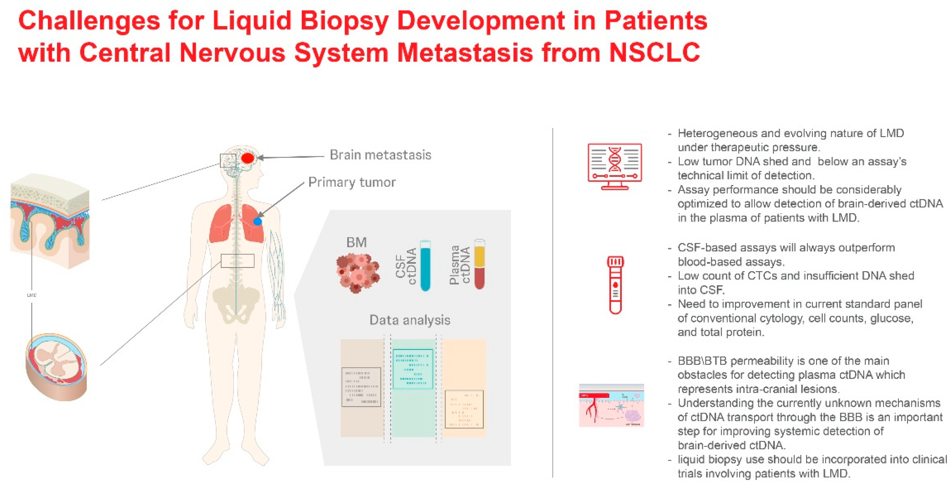
Figure 2.
Blood-brain barrier (BBB) versus blood-tumor barrier (BTB) and implications for drug delivery. (A) Schematic comparison of the BBB and BTB, (B) Key functional differences, (C) Therapeutic implications.
Figure 2.
Blood-brain barrier (BBB) versus blood-tumor barrier (BTB) and implications for drug delivery. (A) Schematic comparison of the BBB and BTB, (B) Key functional differences, (C) Therapeutic implications.
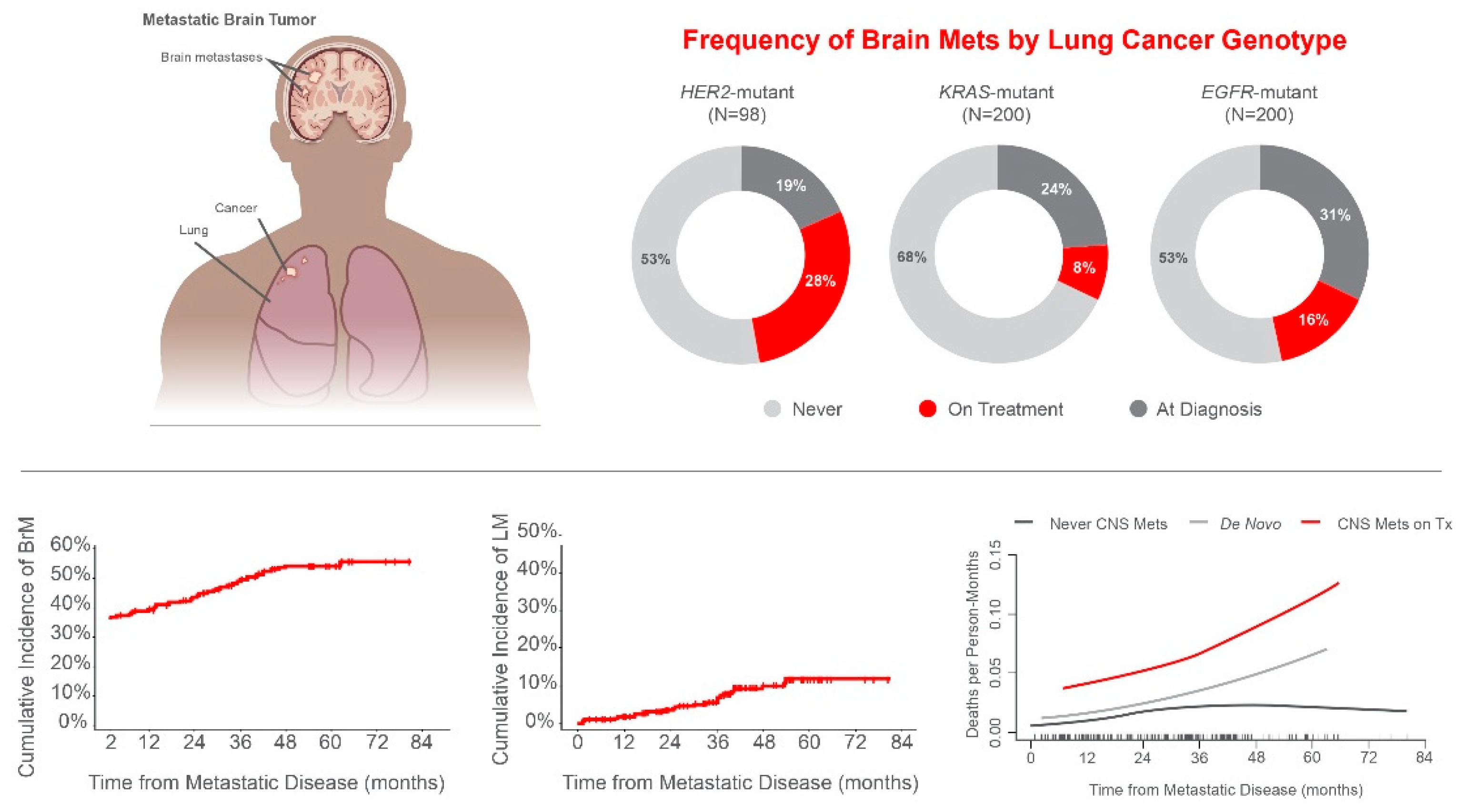
Figure 3.
Challenges for liquid biopsy development in patients with CNS metastases from NSCLC.
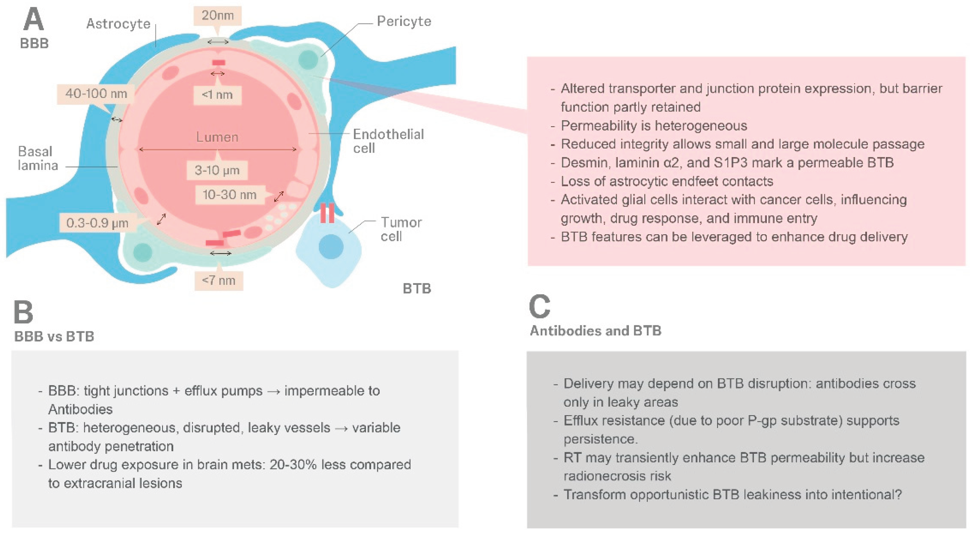
Disclaimer/Publisher’s Note: The statements, opinions and data contained in all publications are solely those of the individual author(s) and contributor(s) and not of MDPI and/or the editor(s). MDPI and/or the editor(s) disclaim responsibility for any injury to people or property resulting from any ideas, methods, instructions or products referred to in the content. |
© 2026 by the authors. Licensee MDPI, Basel, Switzerland. This article is an open access article distributed under the terms and conditions of the Creative Commons Attribution (CC BY) license.
Copyright: This open access article is published under a Creative Commons CC BY 4.0 license, which permit the free download, distribution, and reuse, provided that the author and preprint are cited in any reuse.


