
Submitted:
05 July 2026
Posted:
08 July 2026
You are already at the latest version
Abstract
Craniocerebral firearm injuries are associated with high mortality rates which increase in proportion to the damage to the brain and skull produced by the projectile, as a result of the kinetic energy dissipated during the projectile-tissue interaction. Ballistic factors that contribute to the brain injury are related to the ballistic behavior of the bullet (whether it yaws, tumbles, mushrooms or disintegrates following skull penetration) and its effects. While these injuries are complicated by the creation of bone fragments causing further damage to the brain tissue, the pressure waves generated intracranially as a result of the temporary cavitation phenomenon are the landmark of bullet penetration of the head. Because within the skull there is no mechanism of pressure relief as in other parts of the body during cavitation, the largely incompressible brain tissue sustains the pressure built up, transmitting the pressure wave and causing indirect bone fractures. Although cavitation occurs with low-velocity projectiles too, high-velocity projectiles are capable of high energy transfer secondary to bullet tumbling, mushrooming and often fragmentation, resulting in marked cavitation and more widespread tissue damage. The sudden increase in the intracranial pressure and the transient deformation of the brain tissue contributes to the development of diffuse brain edema and the cardiac and respiratory centers of the brainstem when not involved in the path of the bullet can still be affected indirectly by the pressure transmission with catastrophic results. Shotgun injuries to the head at close range cause extensive destruction of the brain involving a different mechanism, as the pellets enter the cranial cavity bunched together, thus acting as a single projectile of large diameter.
Keywords:
gunshot wounds of the head
; wound ballistics
; penetrating brain injury
; shotgun injuries
; military wounds
; traumatic brain injury
1. Introduction
Craniocerebral gunshot and shotgun injuries are associated with mortality reportedly greater than 90% when deaths occurring on the scene are included, while it still exceeds 50% for victims transported alive to the hospital [1,2,3,4]. Lethality is greatest with self-inflicted injuries [5], which typically involve contact shots directed towards the central core of the brain, but non-suicidal firearm-related traumatic brain injuries also have a poor prognosis compared to firearm injuries to other body areas [6]. This indicates the devastating nature of these injuries involving a complex mechanism of both direct and indirect brain injury [2,6,7], which in gunshot wounds is related to the performance of the penetrating projectile, namely, its ballistic properties (kinetic energy, velocity), design (jacketing) and behavior (yaw/tumbling, deformation). The resultant trauma from the direct action of the bullet is exacerbated by the formation of bone fragments secondary to skull fractures and any possible fragmentation of the bullet itself [8] while more extensive indirect injury occurs due to the cavitation effect [7,9,10]. Consequently, a knowledge of the wounding effects of projectiles penetrating the head is essential for understanding of the underlying mechanism of brain damage [11,12,13,14].
In order to review these issues regarding craniocerebral gunshot wounds, in view of the current epidemic of gun violence and mass shooting incidents [15], an online search was conducted in the PubMed database using the MeSH terms “firearms”, “forensic ballistics”, “penetrating head injuries” and “gunshot wounds”, revealing a limited number of relevant sources, spanning five decades [11,16,17,18,19,20]. A further stimulus was the increasing use of military-style rifles by both terrorists and lone actors against civilians [21,22], as these weapons have been associated with more deaths and a larger number of people injured [23,24], which was not a concerning issue in the past.
2. Ballistics of the Wounding Process (Wound Ballistics)
The bullet is designed to deliver a large amount of energy, due to its velocity imparted by the weapon, by concentrating all its force upon a very small impact area. The penetration process following impact upon the target is determined by the retarding force (drag) and the deceleration encountered by the bullet [25], which according to Newton’s second law of motion is proportional to the force acting upon it and inversely proportional to its mass. Since the projectile mass is very small and the change in its momentum occurs over a very short time, it follows that the retarding force is enormous and the same holds true for the opposite and equal force and the associated pressure exerted by the projectile to the tissue.
Bullet penetration of soft tissue involves two main mechanisms of tissue damage due to pressure effects [20]. Initially, extremely high pressures (reaching thousands of atmospheres or hundreds of thousands of kilopascals) are generated around the forepart of the bullet [26], resulting in tissue crushing and laceration, which is macroscopically conceived as penetration injury and formation of the primary wound channel. After several milliseconds (ms), a second zone of injury appears surrounding the primary wound channel, as its expansion leads to the formation of a temporary cavity under the influence of the energy released by the projectile.
The amount of tissue crushed, which is a consequence of the direct tissue contact with the projectile, increases markedly when the presented area of the projectile increases as a result of tumbling of a non-deforming (i.e., military) bullet or mushrooming of an expanding (e.g., hunting) bullet. Expanding bullets, such as semi-jacketed soft point and hollow-point bullets, are considered to cause “unnecessary suffering” thus violating a fundamental principle of the Law of War. They have been prohibited for military use by the Hague Convention of 18991 following the introduction of the notorious dum-dum bullets used by the British in India. Standard military bullets with a soft lead core are full-metal jacketed, with the jacket covering the nose to prevent deformation. Reports for the use of illegal deforming bullets in armed conflicts surfaced soon after World War I and recently during the War in Ukraine [27].
The second mechanism of injury which involves the formation of a temporary cavity, is a manifestation of high energy transfer as the projectile expends large amounts of energy mobilizing radially the tissue immediately adjacent to the wound channel [25,26,28]. The term cavitation applied to this process denotes a high-velocity impact phenomenon of hydrodynamic origin [25], although in soft tissue it is seen to a lesser extent with low-velocity projectiles too.
Soft tissue behaves like a fluid, which detaches from the projectile surface at some point of its periphery, in a similar manner as the flow around an immersed body separates from it creating a wake region [29]. With high-velocity projectiles, this phenomenon attains an explosive character as there is massive transfer of momentum to the tissue particles that come in contact with the projectile surface, forcing the tissue to move outwards thus creating the cavity [30]. Bullet tumbling in tissue increases the contact area leading to marked cavitation (Figure 1). Due to the tissue inertia, the cavity reaches its maximum diameter about 2-4 ms after the passage of the projectile, and subsequently, in sufficiently elastic tissue, it collapses violently under the influence of the dynamic energy stored in its walls. The phenomenon continues as the cavity undergoes some additional expansion and contraction in a declining manner [26]. The subatmospheric pressure within the expanding cavity creates a suction effect causing bacteria and debris to enter the wound.
The temporary cavity contributes to the morphology of the permanent wound channel (Figure 1). As its maximum size may exceed many times the diameter of the projectile, this results in tensile stress and tearing of tissue when stretched beyond its elastic limit [31]. Shearing of organs fixed to surrounding anatomical structures also occurs. Elastic tissue, such as the skeletal muscle, exhibits largely reversible damage from cavitation and the lung parenchyma due to its compliance is even more tolerant to the stretching effect. Inelastic organs, on the other hand, such as the liver, sustain extensive damage around the wound channel.
A third mechanism of Injury appears with fragmentation of the projectile, which commonly occurs with expanding bullets sustaining mushrooming of their nose under the pressure of the impact but may also occur with high-velocity non-deforming (full metal-jacketed) military bullets. In addition to the creation of secondary wound channels by the metallic fragments, this mechanism is considered to act synergistically with the temporary cavity formed within this widely traumatized area to cause extensive tissue disruption.
Gunshot injuries have traditionally been classified according to the projectile velocity into high- and low-velocity ones depending on the estimated impact velocity of the causative agent [3], which, however, is not always known. Rifle bullets (military and hunting), which are by definition high-velocity projectiles with an initial (muzzle) velocity exceeding 600–760 m/s, have the potential for far greater tissue destruction because of the cavitation [32,33]. Common handgun bullets, on the other hand, responsible for the vast majority of civilian gunshot injuries, are launched at speeds around that of the sound in air (1000 ft/s, approximately 340 m/s) [7], and they are considered low-velocity projectiles. A medium category exists in between, encompassing more powerful handgun projectiles, i.e., Magnum [34].
Velocity is relatively important as the predominant factor imparting kinetic energy to the projectile [7,17]; kinetic energy K as a physical quantity is proportional to mass m but it is proportional to the square of velocity v (K = 1/2 mv2), hence the latter becomes the major determinant of energy [17]. Kinetic energy is considered an expression of the wounding potential of the projectile (the maximum damage that the projectile is capable of) but it is the amount and rate of energy transferred to the tissue that mostly correlates with wound severity [26,35]. Emphasis on projectile velocity as a defining factor of the nature of the injury also reflects the epidemiology of civilian gunshot injuries, the vast majority of which result from low-velocity bullets [7]. However, the increasing availability and use of military-style rifles against civilians in recent decades has blurred the role of velocity as a dividing line between civilian and military injuries [3].
Velocity itself can only decrease along the course of the bullet due to its deceleration in tissue and thus it is not an accurate predictor of variations in the behavior of the projectile in soft tissue, which can actually increase the rate of energy transfer. Yaw (divergence of the bullet’s axis with respect to its line of trajectory) and tumbling (rapid yaw) increases the effective surface of the bullet crushing tissue, while this also leads to higher amounts of energy transferred to the wound and a larger temporary cavity [11,36,37]. When these variations are eliminated with the use of non-deforming non-tumbling spherical projectiles, experiments with soft tissue simulants have shown that a larger, heavier and slower projectile has greater penetration capacity (resulting in greater penetration depth) than a smaller, faster one of the same kinetic energy; while the former crushes more tissue due to its diameter, the latter because of its higher velocity produces a greater cavitation phenomenon [38].
When first introduced, full metal-jacketed (FMJ) rifle bullets, particularly of the pointed (spitzer) type, were thought of as more “humane” compared to solid lead projectiles used previously, which caused more extensive soft tissue and orthopedic wounds because of their deformation upon impact with the target [39]. This view proved to be fallacious, especially in the case of head injuries [40]. The capacity of FMJ rifle bullets for excessive wounding largely depends on their tendency to yaw and tumble following impact [28,41], a characteristic feature of these projectiles [42]. Yaw occurs as the drag force acting on the forepart of the bullet creates a strong overturning moment due to the center of mass of the bullet (its heavier part) lying towards the rear. The gyroscopic stabilization of the bullet from the rifling of the barrel is sufficient to counteract this moment during flight in air but is overcome by the tissue resistance which is about 800 times greater than air drag [37].
The wounding effect of an FMJ bullet becomes maximum at 90 degrees of yaw. At this point the bullet comes into contact with the surrounding medium with its whole length [37], crushing a volume of tissue that may be three times greater than from a bullet with a yaw angle close to 0 [43]. A 90 degrees yaw angle also increases the drag and the rate of energy transfer to the maximum; while the resultant increase in the drag is 3-5 times for a tumbling handgun bullet, the drag on a rifle bullet moving sideways increases by a factor of 10 or more [44] accounting for the difference in the respective tissue damage (which is not a direct result of higher velocity in the case of the rifle bullet but rather its greater efficiency in delivering energy).
Expanding soft-point or hollow-point bullets tend to cause larger wounds than undeformed bullets, with their greatest diameters near the point of entrance to the tissue [36,44]. Mushrooming causes the presented area of the bullet to increase in diameter and such a bullet with a diameter 2.5 times greater than its original can cause prompt damage by tissue crushing over a tissue surface area 6.25 times greater than that from the same bullet while undeformed [43]. There is a resultant increase in the rate of energy transfer early in the course of the bullet due to the increased drag experienced which also significantly reduces the distance (depth) of penetration [44]. The mushroom shape provides stability during tissue penetration, so tumbling is not a feature of these bullets [44,45].
2.1. Biomechanics of Bullet Penetration of the Head
A bullet can cause damage to the head in three ways: by inflicting a tangential wound without penetration, which may result in skull fracture and brain damage nevertheless; by penetrating the skull and becoming lodged within the brain (penetrating brain wound), sometimes following a complex ricochet from the inner surface of the bone; and by completely perforating and exiting the skull (perforating head wound) [17,46,47,48]. It is not uncommon for bullets to perforate the skull without having enough energy to disrupt the overlying scalp, thus becoming lodged beneath it.
Bone preforation by a low-velocity projectile demonstrates a pattern known as cone cracking creating a conoidal defect, with the exit hole larger than the entrance . This pattern is typically seen in the bones of the cranial vault [36]. These bones are composed of an outer and an inner table of compact bone with cancellous bone (diploe) intervening between them. A bullet penetrating the skull leaves a punched out hole on the outer surface and a beveled crater on the inner table. The mechanism is related to the biomechanics of the inner table and the transmission of tensile forces to it through the diploe from the point of impact [46]. However, a similar pattern occurs on the outer table when the bullet exits the cranial cavity. The resultant defect is of importance for forensic pathologists in cases of complete perforation of the skull, in order to differentiate the exit wound from the entry hole [36].
The damage produced by a bullet to the skull can be explained In terms of energy transfer to the tissue [44]. The projectile acts as a kinetic energy penetrator and the amount of energy lost during complete perforation of both sides of the skull is independent of its impact energy and is actually very low, around 70 J [49,50]. A threshold velocity for perforation of the bones of the cranial vault (4-6 mm thick) by a 9 mm Luger bullet with a nominal energy around 490 J, has been estimated at approximately 90 m/s [36]. Perforation of the flat bones of the calvarium is a complex process. Bone fragments are commonly carried into the brain as a secondary effect [49], even by low-velocity projectiles [7,17,51]. These secondary missiles are generally found within the main wound track accounting for its irregular configuration but they may also diverge creating secondary tracks. With contact wounds, these secondary wound channels are of such volume as to contribute substantially to the total brain injury [17].
A 7.62 mm NATO military rifle bullet, with an impact velocity of 750 m/s, completely perforating a head simulant (dried skull filled with 20% gelatin and covered with shamois leather) can deposit 351 J to it [50]. Similarly, a simulated 9 mm handgun bullet studied with impact velocities 300 and 370 m/s, during complete perforation of an anatomical model of cranial skin and cranium has been estimated to lose a total of 380 J [52]. The bone impact destabilizes the bullet inviting early yaw and tumbling, and may also cause bullet fragmentation resulting in increased energy transfer [44]. As a result, while decelerating, a rifle bullet actually transfers significantly greater amounts of momentum and energy to the brain within milliseconds, compared to a similar bullet travelling at stable flight with its velocity less affected. A 9 mm handgun bullet can dissipate more than 40% of its energy while penetrating skin and skull on the side of entrance, with another 40% dissipated during subsequent passage through the brain, suggesting that a substantial portion of its energy is transferred to the brain tissue [51]. Forensic studies suggest that a bullet with sufficient energy to enter the skull, usually perforates the brain completely [17].
2.2. Pressure Effects
Because of the role played by the unyielding confinement of the skull and the interaction with the bullet and the brain, the sudden pressure build-up due to temporary cavitation is the major secondary effect from high-velocity penetration [20,30] (Figure 2).
The brain acts as a medium for transmission of the pressure waves from cavity development. This places the bony surroundings under extreme stress resulting in shattering of the bone from inside, like an egg shell [54,55]. High-velocity projectiles penetrating skulls filled with gelatin simulating brain create pressures due to cavitation, from 2200 kPa in the case of 6-mm sphere to more than 2750 kPa from a 7.62 mm NATO bullet [50,56]. Pressure waves of such magnitude result in extensive fracturing of the skull [44,56,57,58] often disarticulating bone along suture lines [30]. This “explosive” type of skull damage is illustrated in the case of the President Kennedy assassination; a 6.5 mm Mannlicher-Carcano round-nosed FMJ bullet striking the back of the President’s head at approximately 580 m/s produced a wound of exit involving the temporo-parieto-occipital region, approximately 13 mm in diameter [59]. However, in the absence of such pressure effects, i.e., in an empty skull devoid of brain, the resultant damage consists only of minor fracturing around the entry and exit holes created in the bone by the projectile passage [30,57].
The mechanism of explosive injury of the skull by high-velocity projectiles is very similar to the hydrodynamic (hydraulic) ram effect (Figure 3). This is encountered when a high kinetic energy projectile penetrates a fluid-filled container causing structural damage to it that far exceeds the diameter of the entry hole. This event has four major components. Initially, a high-pressure shock wave appears at the point of impact (shock phase) travelling at approximately 1450 m/s, i.e., the speed of sound in water. Subsequently, as the projectile decelerates (drag phase), it transfers momentum and kinetic energy at high rates to the surrounding liquid, which is propelled outwards generating relatively slow pressure waves and leading to cavity formation (cavity phase). Finally, rupture of the container may ensue due to the sudden release of the pressure through the exit hole (exit phase). In the case of head injuries, the initial shock wave is considered of lesser importance because it does not cause gross tissue displacement due to its short duration. It can damage nerve tissue [48], nevertheless, and there have been anecdotal cases of bullets following an extracranial path that affected brain function, apparently through the shock wave.
The temporary cavity from a 9 mm handgun bullet can create peak pressures exceeding 600 kPa [60] and stress waves in the skull [51]. These bullets tend to cause bursting injuries of the calvarium and skull base whereas secondary fractures of the skull base by smaller bullets typically involve the paper-thin orbital plates resulting in the appearance of “panda eyes” [36,45]. That even such low-velocity projectiles penetrating the head can cause widespread damage in areas of the brain at distance from the bullet track has been demonstrated by Oechmichen et al. [61]. Pathologic examination of actual wounds produced by such projectiles has shown extensive injury of brain tissue extending up to 3.6 cm in diameter around the central defect, due to the pressure effects of the temporary cavity and probably the additional deformation and displacement of the brain. The damaged tissue consists of an inner zone of astrocytic destruction extending up to 4 mm, next, a zone of hemorrhage with an irregular appearance (due to local differences in blood supply), and finally an outer zone of diffuse axonal and neuronal injury. The extent of the latter (17-18 mm) can explain the occurrence of early respiratory arrest following wounding due to involvement of remote areas of the brainstem [61].
2.3. “Falling Bullets”
Shooting into the air, commonly as celebratory fire, can result in gunshot wounds which may be encountered at distance from the shooting incident, with victims or relatives completely unaware of the causative event. These bullets are referred to as falling bullets, as they start moving downhill under the influence of gravity once they lose their upward momentum, and the respective injuries are called celebratory firearm injuries [55]. The majority of these injuries are sustained in the head, often with a fatal outcome [62,63], and some of the victims are children. Doctors must keep a high index of suspicion in cases involving sudden loss of consciousness outdoors in a young victim without an obvious cause, especially from geographical areas where such celebratory fires have been reported.
Celebratory firearm injuries are commonly attributed to vertical firings, but they are most likely due to high-angled discharges of firearms [36,64]. Bullets fired vertically enter free falling at the highest point of their flight, eventually reaching a terminal velocity once the air resistance balances the weight. For handgun bullets descending base-first the terminal velocity has been calculated at 45-76 m/s, depending on the bullet’s weight. Experiments of the United States Army in 1919-20 concluded that the 7.62 mm service rifle bullet fired straight up to the sky returned with about 100 m/s terminal velocity, not possessing enough energy to be considered lethal threat for a soldier [65]. Indeed, since most of the falling bullets from a vertical discharge tend to tumble or descend base-first, they may not attain such energy as to cause a deep penetrating or fatal wound [36,66].
2.4. Shotgun Injuries
Shotguns are long-barreled firearms, typically smoothbore (non-rifled), which fire either multiple spherical projectiles (pellets) collectively known as shot or a single large projectile (slug). Shotgun pellets, which are manufactured in various sizes, are discharged from a single cartridge known as shotshell. It also contains the wad, a plastic insert used to isolate the shot from the propellant. The most popular and versatile shotgun design is the 12 gauge (the gauge number refers to the internal diameter of the bore, with smaller numbers indicating larger barrels) [16,67].
After exiting the muzzle, the pellets spread in a conical fashion and the resultant dispersion represents the shot pattern. The pellet density in the pattern is determined by the design of the gun, the number of pellets in the shell, and the distance from the gun barrel [44,54,68]. At closer distances, the shot enters the body en masse with the initial velocity of the pellets virtually unchanged, resulting in maximum energy deposition and creation of a large wound cavity corresponding to the diameter of the pattern, usually less than 25 mm. It is within this range, generally less than 3 m, that the shotgun demonstrates its greatest destructive capacity [44]. The wad also acts as a ballistic projectile and within the range at which the shot creates a single wound, the wad should be expected to be found within it as a radiolucent foreign body [44]. With increasing distance, the density of the pattern decreases dramatically as the pellets begin to separate, and because of their unfavorable ballistic shape their velocity drops quickly [36]. Beyond a distance of 6 m, they tend to cause wounds of limited depth with lesser tissue destruction although sensitive structures such as the eyes can certainly be damaged while larger (buckshot) pellets can still hit the target with sufficient energy to inflict severe injury [54]. Shotgun slugs produce extensive internal injuries within a much longer effective range of 100 m, as they rapidly deliver their kinetic energy to the wound because of their blunt shape, also inducing marked cavitation [44,55].
Contact or near contact shotgun blasts to the head are fatal injuries associated with extensive destruction of soft and hard tissues. With 12-gauge shotguns, a wound of exit is likely to be created as the shot at such range acts as a single bolus facilitated by the propellant gases that enter the wound under pressure creating an explosive effect. As a result, the intracranial contents are likely to become pulverized and the brain can be partly or as a whole expelled [53].
2.5. Craniofacial Gunshot Injuries
The length of the standard barrel of shotguns and other long-barreled weapons placed submentally or inserted into the mouth in a suicide attempt, does not seem to prevent reaching the trigger by an average person [69]. However, psychological factors and reflex movement may cause tilting of the barrel anteriorly or overextension of the neck at the moment of discharge resulting in massive craniofacial trauma which spares the vital structures of the brain. Craniofacial gunshot wounds result in wide communication of the intracranial cavity with the paranasal sinuses and cerebrospinal fluid rhinorrhea. Extensive contamination of the brain with oral and nasal secretions and bone and tooth fragments (which also act as secondary projectiles) can lead to development of infectious complications [70] (Figure 4).
3. Ballistic-Related Factors Affecting Outcome
A number of prognostic factors in gunshot wounds of the head, particularly those predicting mortality, have been identified by several authors, but mostly limited to civilian cases [4,70,71,72,73,74,75,76,77]. Suicide attempts have a higher probability of fatal outcome [1,48,71], apparently due to the direction of the weapon and the close range of most self-inflicted wounds [4,73], and often appear as an independent predictor of mortality [78,79]. The Glasgow Coma Scale (GCS) score is another factor often mentioned as the most important one in predicting survival or the feasibility of surgical intervention. However, as with gunshot wounds in other parts of the body, the placement of the shot and the associated trajectory is the most important factor affecting wound severity [41,80], which can explain the relatively high mortality rate associated with suicide attempts. The GCS score, on the other hand, is a clinical measurement which is the result of the wounding process.
These prognostic factors are usually collectively listed although, previously, the high velocity of the projectile had been characterized as the “most important element in mortality at the time of impact” whereas the state of consciousness was the most important prognostic factor at the time of surgery [81]. Therefore, it seems appropriate to organize in a hierarchical manner ballistic and relevant aspects of wounding pertinent to the causative agent and the tissue response to injury, followed by clinical factors that emerge as significant predictors after initiation of resuscitative procedures (Table 1).
High-velocity weapons have consistently been correlated with poorer outcome of gunshot wounds to the head [4,47,48,70]. While this is not a direct consequence of velocity per se, high-velocity military bullets have the capacity to inflict more widespread damage to the brain and skull due to their tumbling tendency resulting in higher energy transfer and the associated cavitation effect. In addition, bullet fragmentation frequently accompanies bone penetration.
Caliber of the weapon, on the other hand, was previously considered not to be an independent factor predicting mortality [78]. However, a multivariate analysis [82] of data on shooting cases over a 5-year period retrieved from Boston Police investigation files, showed that firearms caliber was associated with the likelihood of death from assaults involving gunshots. In this study, injuries located in the head and neck area were the most serious and, along with chest, back and abdominal wounds, predominated in fatal cases. Reasonably, a large caliber weapon can be expected to inflict more serious wounds to the head or neck because of the larger volume of the wound channel produced by the projectile but also because of its greater penetration capacity due to its larger mass.
As previously noted, the temporary cavity, which is an energy-related phenomenon, is the main aggravating factor in the case of high-velocity penetration of the head. The direct injury produced by the projectile appears along its track and is mainly focal with contusions, lacerations, intracranial hemorrhage and wound contamination. The formation of the temporary cavity, however, has indirect effects that can result in diffuse damage [48], including diffuse axonal injury, widespread cortical contusions (from the impact of the brain tissue to the raw interior surface of the skull), rapidly developing intracerebral edema in the absence of significant blood loss or intracranial hematoma formation, and petechial hemorrhages remote from the bullet track [20]. For these reasons, the consequences of the temporary cavity in the brain should be considered comparable to blunt rather than to penetrating injury.
A recent study [83] compared military and civilian patients with gunshot wounds to the head. Despite the former patients being theoretically in a more critical condition compared to civilian cases, as suggested by the nature of the wounding agent and the fact that they received more aggressive treatment with significantly more blood products, intracranial pressure (ICP) monitoring and cranial decompression, they did better, with significantly lower mortality rates than their civilian counterparts (27% vs 38%). The authors mentioned these measures as possible reasons for the improved results reported among the military population. This study [83] provides evidence supporting decompressive craniectomy as a means to reduce the ICP in penetrating brain injury. The procedure remains controversial and its use in patients with severe traumatic brain injury is supported mainly by observational studies [84]. Nevertheless, its rationale is fundamentally biomechanical since the rigid cranial vault converts cerebral edema into dangerous intracranial hypertension, and removing part of the skull alters the boundary conditions of the system. If decompression craniotomy or craniectomy was indeed lifesaving in some of these patients, this suggests that the temporary cavitation and its secondary pressure effects contributing to the development of intracerebral edema are important prognostic factors affecting outcome.
The trajectory of the bullet, demonstrated in head computed tomography (CT) scan, is the strongest prognostic factor of survival [85]. Deep bihemispheric and transventricular trajectories as well as those demonstrating multilobar or posterior fossa involvement are associated with high mortality rates [4,78,86].
An analysis of bullet trajectories in CT scans after civilian perforating wounds of the head by Kim et al. [87] revealed that, while GCS score and pupil irregularity were significantly associated with fatal outcome, penetration through the midline of a central anatomic area of the brain termed zona fatalis, approximately 4 cm above the dorsum sella, invariably led to brain death and proximity of the bullet trajectory to that area was also associated with a fatal outcome in 7 out of 10 patients. The authors also identified the so-called “tram-track sign” on CT scans correlating with fatal injury; it consists of dense or hyperdense tracks outlining a dark center representing the bullet track and it is considered an imaging finding suggestive of the effects of cavitation [87] (Figure 2). A trend of bullet trajectories in non-survivors crossing a wider “potential lethal zone” of the brain, involving the ventricular system, brainstem, dorsum sellae and the circle of Willis, has recently been reported by other authors [88].
4. Fragment Wounds of the Head
Blast injuries from improvised explosive devices (IEDs) have become in recent decades more common in battlefields compared to gunshot wounds but they have also been encountered in civilian practice due to terrorist acts using IEDs in several countries [4,89]. In addition, such injuries are caused by explosion of mines, grenades or shells. They are complex injuries involving three mechanisms: barotrauma to air-filled organs such as the lungs and ears, from exposure to the overpessure of the blast wave unique to high-order explosives (primary effects); penetrating injuries from flying debris, fragments and shrapnel (secondary effects); and blunt injuries as the blast wind accelerates the body through the air causing impact to solid surfaces (tertiary effects) [89].
The blast wave can cause severe cerebral edema [4]. Penetrating craniocerebral injuries from fragments are isolated as they are usually seen in polytrauma patients. The ballistic profile of individual fragment wounds is characterized by a wound channel that has its largest diameter at the point of entry becoming progressively narrower towards the depth of the wound [90,91]. This occurs because fragments, despite their initial high velocity, have an irregular or roughly spherical shape, which results in greatest amounts of energy transfer at the entry site [91]. Regardless of the shape of the fragment, the entry wound is more or less circular and the depth of the wound and the degree of tissue damage are determined by the mass and velocity of the fragment [92]. The wound consists of a cone of tissue destruction, with pulped brain tissue, blood, and also hair, skin and bone fragments. Since the kinetic energy of the projectile itself is greater than that imparted to bone fragments, its position within the wound is always deeper than any accompanying bone fragment [90]. Fragment wounds of the brain are typically heavily contaminated [4]. Metallic fragments with sharp edges tend to cause lacerations to the brain even near the end of the wound channel [90] and subarachnoid hemorrhage is a common feature in these wounds [4] (Figure 5).
5. Conclusions
Craniocerebral wounds from bullets, pellets and fragments are devastating injuries, which continue to carry a grave prognosis for a large percentage of patients. The unique characteristics of these injuries are related to the energy transferred by the projectile(s) to the skull and the brain, both directly and indirectly. The diffuse brain damage that occurs especially with high-velocity projectiles, although disproportionately large compared to their small size, can be explained and understood on the basis of high-energy hydrodynamic phenomena, such as the hydrodynamic ram. The factors that are implicated in the pathogenesis of these wounds also have a prognostic significance in the context of clinical and imaging assessment of the patient, which may have not been fully investigated. The improved outcome recently reported in large series with aggressive treatment of more severe wounds appears to have some link with the role of ballistic and biomechanical aspects of wounding. The role of ballistic factors as independent predictive parameters of the outcome should be examined in view of the increased incidence of civilian head injuries inflicted by military-type high-velocity weapons.
Funding
This article received no external funding.
Informed Consent Statement
Not applicable, since the visual material included in the article does not contain information about the identity of the respective patients.
Conflicts of Interest
The authors declare no conflicts of interest.
| 1 | The common belief that the Hague Convention actually mandated full-metal jacketed projectiles for military purposes, for example [11], is incorrect |
References
- Krieger, M.D.; Levy, M.L.; Apuzzo, M.L.J. Gunshot Wounds to Head in an Urban Setting. Neurosurg. Clin. N. Am. 1995, 6, 605–610. [Google Scholar] [CrossRef] [PubMed]
- Maas, A.I.R.; Stocchetti, N.; Bullock, R. Moderate and Severe Traumatic Brain Injury in Adults. Lancet Neurol. 2008, 7, 728–741. [Google Scholar] [CrossRef] [PubMed]
- Likun, Y.; Opalak, C.F.; Valadka, A.B. Brain. In Trauma; Feliciano, D.L., Mattox, K.L., Moore, E.E., Eds.; McGraw-Hill: New York, NY, 2021; pp. 457–478. ISBN 978-1-260-14335-5. [Google Scholar]
- Rosenfeld, J.V.; Bell, R.S.; Armonda, R. Current Concepts in Penetrating and Blast Injury to the Central Nervous System. World J. Surg. 2015, 39, 1352–1362. [Google Scholar] [PubMed]
- Kaufman, H.H.; Makela, M.E.; Lee, F.; Haid, R.W., Jr.; Gildenberg, P.L. Gunshot Wounds to the Head: A Perspective. Neurosurgery 1986, 18, 605–610. [Google Scholar] [CrossRef]
- Waltzman, D.; Sarmiento, K.; Daugherty, J.; Lumba-Brown, A.; Klevens, J.; Miller, G.F. Firearm-Related Traumatic Brain Injury Homicides in the United States, 2000-2019. Neurosurgery 2023, 93, 43–49. [Google Scholar] [CrossRef] [PubMed]
- Kaufman, H.H. Gunshot Wounds to the Head. In The Textbook of Penetrating Trauma; Ivatury, R.R., Cayten, C.G., Eds.; Williams & Wilkins: Baltimore, MD, 1996; pp. 431–442. ISBN 0-683-04338-2. [Google Scholar]
- Greenberg, M.S. Gunshot Wounds and Non-Missile Penetrating Brain Injuries. In Greenberg’s Handbook of Neurosurgery; Thieme: New York, NY, 2023; pp. 1093–1097. ISBN 978-1-68420-504-2. [Google Scholar]
- Carey, M.E. Bullet Wounds to the Brain Among Civilians. In Youmans Neurological Surgery; Marshall, L.F., Grady, M.S., Eds.; Saunders: Philadelphia, PA, 2004; Vol. 4, pp. 5223–5242. ISBN 0-7216-8291-X. [Google Scholar]
- Gean, A.D. Concussion, Contusion, Hematoma. In Imaging of Head Trauma; Raven Press: New York, NY, 1994; pp. 147–206. ISBN 0-7817-0025-6. [Google Scholar]
- Aarabi, B.; Alden, T.D.; Chesnut, R.M.; Downs, H.I.; Ecklund, J.M.; Eisenberg, H.M.; Farace, E.; Florin, R.E.; Jane, J.A.J.; Krieger, M.D.; et al. Management and Prognosis of Penetrating Brain Injury. Part 1: Guidelines for the Management of Penetrating Brain Injury. Introd. Methodol. J. Trauma 2001, 51, S3–S6. [Google Scholar]
- Bhatoe, H.S. Missile Injuries of the Brain, Spine, and Peripheral Nerves, 2nd ed.; Thieme: Delhi, 2019; ISBN 978-93-88257-10-7. [Google Scholar]
- Carey, M.E.; Sarna, G.S.; Farrell, B.; Happel, L.T. Experimental Missile Wound to the Brain. J. Neurosurg. 1989, 71, 754–764. [Google Scholar] [CrossRef] [PubMed]
- Liker, M.A.; Aarabi, B.; Levy, M.L. Missile Wounds to the Head: Ballistics and Forensics. In Missile Wounds of the Head and Neck; Kaufman, H.H., Aarabi, B., Eds.; The American Association of Neurological Surgeons: Park Ridge, IL, 1999; Vol. 1, pp. 35–55. ISBN 0-9624246-6-8. [Google Scholar]
- Spence, C.A.; Eden, S.V.; Pennicooke, B.; Adogwa, O.; Holly, L.T.; Welch, B.G.; Mbabuike, N.; Nduom, E.; Ashley, W.W., Jr. Mass Shootings in the United States: An Alarming Trend of Violence and Public Health Threat. J. Neurosurg. 2022, 138, 882–883. [Google Scholar] [PubMed]
- Jandial, R.; Reichwage, B.; Levy, M.; Duenas, V.; Sturdivan, L. Ballistics for the Neurosurgeon. Neurosurgery 2008, 62, 472–480. [Google Scholar] [CrossRef] [PubMed]
- Kirkpatrick, J.B.; Di Maio, V. Civilian Gunshot Wounds of the Brain. J. Neurosurg. 1978, 49, 185–198. [Google Scholar] [CrossRef] [PubMed]
- Selden, B.S. Craniocerebral Wound Ballistics. Ind. Med. 1987, 80, 150–152. [Google Scholar]
- Young, L.; Rule, G.T.; Bocchieri, R.T.; Waliko, T.J.; Burns, J.M.; Ling, G. When Physics Meets Biology: Low and High-Velocity Penetration, Blunt Impact, and Blast Injuries to the Brain. Front. Neurol. 2015, 6, 89. [Google Scholar] [CrossRef] [PubMed]
- Karger, B. Penetrating Gunshots to the Head and Lack of Immediate Incapacitation. I. Wound Ballistics and Mechanisms of Incapacitation. Int. J. Leg. Med. 1995, 108, 53–61. [Google Scholar]
- Harrington, T.; Apostolides, P. Penetrating Brain Injury. In Head Injury; Cooper, P.R., Golfinos, J.G., Eds.; McGraw-Hill: New York, NY, 2000; pp. 349–359. ISBN 0-8385-3687-5. [Google Scholar]
- Ashkenazi, I.; Bemelman, M. Editorial: “Focus on Disaster and Military Surgery.”. Eur. J. Trauma Emerg. Surg. 2017, 43, 575–577. [Google Scholar] [CrossRef] [PubMed]
- Barnard, L.M.; Wright-Kelly, E.; Brooks-Russell, A.; Betz, M.E. Firearm Type and Number of People Killed in Publicly Targeted Fatal Mass Shooting Events. JAMA Netw. Open 2025, 8, e2458085. [Google Scholar] [CrossRef] [PubMed]
- Goldman, S.; Lipsky, A.M.; Radimislensky, I.; Givon, A.; Almog, O.; Benov, A.; Katorza, E. October 7th Mass Casualty Attack in Israel: Injury Profiles of Hospitalized Casualties. Ann. Surg. Open 2024, 5, e481. [Google Scholar] [CrossRef] [PubMed]
- Rosenberg, Z.; Dekel, E. Terminal Ballistics, 3rd ed.; Springer: Cham, CH, 2020; ISBN 978-3-030-46611-4. [Google Scholar]
- Carey, M.E. Experimental Missile Wounding of the Brain. Neurosurg. Clin. North Am. 1995, 6, 629–642. [Google Scholar] [CrossRef]
- Gumeniuk, K.; Lurin, I.A.; Tsema, I.; Malynovska, L.; Gorobeiko, M.; Dinets, A. Gunshot Injury to the Colon by Expanding Bullets in Combat Patients Wounded in Hybrid Period of the Russian-Ukrainian War during 2014-2020. BMC Surg. 2023, 23, 23. [Google Scholar] [PubMed]
- Stefanopoulos, P.K.; Mikros, G.; Pinialidis, D.E.; Oikonomkais, I.E.; Tsiatis, N.E.; Janzon, B. Wound Ballistics of Military Rifle Bullets: An Update on Controversial Issues and Associated Misconceptions. J. Trauma Acute Care Surg. 2019, 87, 690–698. [Google Scholar] [CrossRef] [PubMed]
- Gerhart, P.M.; Gerhart, A.L.; Hochstein, J.I. Flow over Immersed Bodies. In Fundamentals of Fluid Mechanics; Munson, Young, Okiishi’s, Eds.; Wiley: Hoboken, NJ, 2016; p. 489. ISBN 978-1-119-08070-1. [Google Scholar]
- Butler, E.G.; Puckett, W.O.; Harvey, E.N.; McMillen, J.H. Experiments on Head Wounding by High Velocity Missiles. J. Neurosurg. 1945, 2, 358–363. [Google Scholar] [CrossRef]
- Janzon, B.; Hull, J.B.; Ryan, J.M. Projectile-Material Interactions: Soft Tissue and Bone. In Scientific Foundations of Trauma; Butterworth-Heinemann: Oxford, 1997; pp. 37–52. ISBN 0 7506 1585 0. [Google Scholar]
- Strong, B.L.; Lauerman, M.H.; Scalea, T.M. Sequelae and Care After Firearm Injury. JAMA 2025. [Google Scholar] [CrossRef] [PubMed]
- Ling, G.S.F.; Ecklund, J.M. Severe TBI in Military: Medical and Surgical Interventions. In Neurotrauma: A Comprehensive Book on Traumatic Brain Injury and Spinal Cord Injury; Wang, K.K.W., Ed.; Oxford: New York, NY, 2019; pp. 13–20. ISBN 978– 0– 19– 027943– 1. [Google Scholar]
- Stefanopoulos, P.K.; Breglia, G.A.; Bissias, C.; Nikita, A.S.; Papageorgiou, C.; Tsiatis, N.E.; Serafetinides, E.; Gyftokostas, D.A.; Aloizos, S.; Mikros, G. Firearm Injuries: A Review of Wound Ballistics and Related Emergency Management Considerations. Emerg. Care Med. 2 52. [CrossRef]
- Dagi, T.F. Emergency Management of Missile Injuries to the Brain: Resuscitation, Triage, and Preoperative Stabilization. Am. J. Emerg. Med. 1987, 5, 140–148. [Google Scholar] [CrossRef] [PubMed]
- DiMaio, V.J.M. Gunshot Wounds, 3rd ed.; CRC Press: Boca Raton, FL, 2016; ISBN 978-1-4987-2569-9. [Google Scholar]
- French, R.W.; Callender, G.R. Ballistic Characteristics of Wounding Agents. In Wound Ballistics; Beyer, J.C., Ed.; Office of the Surgeon General: Washington, D.C., 1962; pp. 91–141. [Google Scholar]
- Fackler, M.L. Wound Ballistics: A Review of Common Misconceptions. JAMA 1988, 259, 2730–2736. [Google Scholar] [CrossRef] [PubMed]
- La Garde, L.A. Gunshot Injuries: How They Are Inflicted Their Complications and Treatment, 2nd ed.; William Wood: New York, NY, 1916. [Google Scholar]
- Hesketh-Prichard, H. Huchinson. In Sniping in France; 1920. [Google Scholar]
- Hatcher, J.S. Textbook of Pistols and Revolvers; Small-Arms Technical Publishing: Marines, NC, 1935. [Google Scholar]
- Wen, Y.; Xu, C.; Jin, Y.; Batra, R.C. Rifle Bullet Penetration into Ballistic Gelatin. J. Mech. Behav. BioMed Mater. 2017, 67. [Google Scholar] [CrossRef] [PubMed]
- Hollerman, J.J.; Fackler, M.L. Wound Ballistics. In Tintinalli’s Emergency Medicine: A Comprehensive Study Guide; Tintinalli, J.E., Ed.; McGraw-Hill: New York, NY, 2011; pp. e38–e43. ISBN 978-0-07-148480-0. [Google Scholar]
- Sturdivan, L.M.; Sacco, W.J.; Edgerton, M.T.; Edlich, R.F. Firearm Injuries: Medicolegal Considerations of Ballistics. In Emergency Medicine Annual; Wolcott, B.W., Rund, D.A., Eds.; Appleton-Century-Crofts: Norwalk, CT, 1984; Vol. 3, pp. 125–167. ISBN 0-8385-2197-5. [Google Scholar]
- Kneubuehl, B.P. General Wound Ballistics. In Wound Ballistics: Basics and Applications; Kneubuehl, B.P., Ed.; Springer: Berlin, 2022; pp. 87–163. ISBN 978-3-662-64854-4. [Google Scholar]
- Maynard, R.L.; Allen, I.V.; Hull, J.B. Penetrating Head Injury. In Scientific Foundations of Trauma; Cooper, G.J., Dudley, H.A.F., Gann, D.S., Little, R.A., Maynard, R.L., Eds.; Butterworth-Heinemann: Oxford, 1997; pp. 53–62. ISBN 0 7506 1585 0. [Google Scholar]
- Graham, D.I.; Gennarelli, T.A. Pathology of Brain Damage After Head Injury. In Head Injury; Cooper, P.R., Ed.; McGraw-Hill: New York, NY, 2000; pp. 133–153. ISBN 0-8385-3687-5. [Google Scholar]
- Roberts, S.A.G. Management of Ballistic Trauma to the Head. In Ballistic Trauma: A Practical Guide; Breeze, J., Penn-Barwell, J.G., Keene, D., O’Reilly, D., Jeyanathan, J., Mahoney, P.F., Eds.; Springer: Cham, CH, 2017; pp. 175–199. ISBN 978-3-319-61364-2. [Google Scholar]
- Rothschild, M.A. Wound Ballistics and Forensic Medicine. In Wound Ballistics: Basics and Applications; Kneubuehl, B.P., Ed.; Springer: Berlin, 2022; pp. 263–300. ISBN 978-3-662-64854-4. [Google Scholar]
- Watkins, F.P.; Pearce, B.P.; Stainer, M.C. Physical Effects of the Penetration of Head Simulants by Steel Spheres. J. Trauma 1988, 28, S40–S54. [Google Scholar] [CrossRef] [PubMed]
- Chattrairat, A.; Kandare, E.; Aimmanee, S.; Tran, P.; Das, R. Understanding Post-Impact Biomechanics of Ballistic Cranial Injury by Smoothed Particle Hydrodynamics Numerical Modelling. Comput. Part. Mech. 2025, 12, 133–152. [Google Scholar]
- Kwon, E.; Singh, M.R.; Vallabh, R.D.; Das, R.; Taylor, M.C.; Fernandez, J.W. Modelling Ballistic Cranial Injury and Backspatter Using Smoothed Particle Hydrodynamics. Comput. Methods Biomech. Biomed. Eng. Imaging Vis. 2020, 8, 478–491. [Google Scholar]
- Pollak, S.; Saukko, P.; Knight, B. Gunshot and Explosion Deaths. In Knight’s Forensic Pathology; CRC Press: Boca Raton, FL, 2026; ISBN 978-1-041-13001-7. [Google Scholar]
- Schmidt, A.; Wise, K.; Kelly, B. The Physics of Trauma. In PHTLS: Prehospital Trauma Life Support; Pollak, A.N., Ed.; Jones & Bartlett Learning: Burlington, MA, 2023; pp. 103–147. ISBN 978-1-284-27227-7. [Google Scholar]
- Stefanopoulos, P.K.; Tsiatis, N.E.; Herbstein, J.A. Gunshot Wounds. Encycl. Forensic Sci. 2023, Vol. 3, 75–98. [Google Scholar]
- Owen-Smith, M.S. High Velocity Missile Wounds; Edward Arnold: London, 1981; ISBN 0-7131-4371-1. [Google Scholar]
- Carr, D.; Lindstrom, A.-C.; Jareborg, A.; Champion, S.; Waddell, N.; Miller, D.; Teagle, M.; Horsfall, I.; Kieser, J. Development of a Skull/Brain Model for Military Wound Ballistics Studies. Int. J. Leg. Med. 2015, 129, 505–510. [Google Scholar]
- Taylor, S.C.; Ondruschka, B.; Kieser, D.C.; Hammer, N.; Lee, M.; Hooper, G.J.; Kranioti, E. Ballistic Trauma Caused by Military Rifles: Experimental Study Based on Synthetic Skull Proxies. Forensic Sci. Med. Pathol. 18 30–36. [CrossRef]
- Finck, P.A. Ballistic and Forensic Pathologic Aspects of Missile Wounds. Conversion between Anglo-American and Metric-System Units. Mil. Med. 1965, 130, 545–569. [Google Scholar] [PubMed]
- Zhang, J.; Yoganandan, N.; Pintar, F.A.; Guan, Y.; Gennarelli, T.A. Experimental Model for Civilian Ballistic Brain Injury Biomechanics Quantification. J. Biomech. 2007, 40, 2341–2346. [Google Scholar] [CrossRef] [PubMed]
- Oehmichen, M.; Meissner, C.; König, H.G.; Gehl, H.-B. Gunshot Injuries to the Head and Brain Caused by Low-Velocity Handguns and Rifles. A Review. Forensic Sci. Int. 2004, 146, 111–120. [Google Scholar] [CrossRef] [PubMed]
- Ordog, G.J.; Dronhoffer, P.; Ackroyd, G.; Wasserberger, J.; Bishop, M.; Shoemaker, W.; Balasubramanium, S. Spent Bullets and Their Injuries: The Result of Firing Weapons into the Sky. J. Trauma 1994, 37, 1003–1006. [Google Scholar] [PubMed]
- Çelik, E.; Koç, A. Analysis of Free-Fall Bullet Injury Potential in the Cranium via Finite Elements Method. J. Forensic Leg. Med. 2023, 97, 102552. [Google Scholar] [CrossRef] [PubMed]
- Shuker, S.T.; Sadda, R. Craniomaxillofacial Falling Bullet Injuries and Management. J. Oral Maxillofac. Surg. 2010, 68, 1593–1601. [Google Scholar] [CrossRef] [PubMed]
- Hatcher, J.S. Bullets from the Sky. In Hatcher’s Notebook; Telegraph Press, 1957; pp. 511–519. [Google Scholar]
- Rapkiewicz, A.V.; Shuman, M.J.; Hutchins, K.D. Fatal Wounds Sustained from “Falling Bullets”: Maintaining a High Index of Suspicion in a Forensic Setting. J. Forensic Sci. 2014, 59, 268–270. [Google Scholar] [PubMed]
- Levy, M.L.; Davis, S.E.; Russell, M. Ballistics and Forensics. In Traumatic Brain Injury; Marion, D.W., Ed.; Thieme: New York, NY, 1999; pp. 201–213. ISBN 0-86577-727-6. [Google Scholar]
- Sights, W.P., Jr. Ballistic Analysis of Shotgun Injuries to the Central Nervous System. J. Neurosurg. 1969, 31, 25–33. [Google Scholar] [CrossRef] [PubMed]
- Haug, R.H. Discussion: Gunshot Wounds of the Face in Attempted Suicide Patients. J. Oral Maxillofac. Surg. 1998, 56, 933–934. [Google Scholar]
- Cooper, P.R. Gunshot Wounds of the Brain. In Head Injury; Williams & Wilkins: Baltimore, MD, 1993; pp. 355–371. ISBN 0-683-02108-7. [Google Scholar]
- Aldrich, E.F.; Eisenberg, H.M.; Saydjari, C.; Foulkes, M.A.; Jane, J.A.; Marshall, L.F.; Young, H.; Marmarou, A. Predictors of Mortality in Severely Head-Injured Patients with Civilian Gunshot Wounds: A Report from the NIH Traumatic Coma Data Bank. Surg. Neurol. 1992, 38, 418–423. [Google Scholar] [CrossRef] [PubMed]
- Polin, R.S.; Shaffrey, M.E.; Phillips, C.D.; Germanson, T.; Jane, J.A. Multivariate Analysis and Prediction of Outcome Following Penetrating Head Injury. Neurosurg. Clin. North Am. 1995, 6, 689–699. [Google Scholar] [CrossRef]
- Aldrich, E.F.; Eisenberg, H.M. Prognostic Factors. In Traumatic Brain Injury; Marion, D.W., Ed.; Thieme: New York, NY, 1999; pp. 223–229. ISBN 0-86577-727-6. [Google Scholar]
- Levy, M.L.; Davis, S.E. Outcome Prediction Following Craniocerebral Missile Wounds: How Do We Identify the Salvageable? In Missile Wounds of the Head and Neck; Kaufman, H.H., Aarabi, B., Eds.; The American Association of Neurological Surgeons: Park Ridge, IL, 1999; Vol. 1, pp. 135–150. ISBN 1-879284-64-2. [Google Scholar]
- Levy, M.L. Outcome Prediction Following Penetrating Craniocerebral Injury in a Civilian Population: Aggressive Surgical Management in Patients with Admission Glasgow Coma Scale Scores of 6 to 15. Neurosurg. Focus. 2000, 8, 1–6. [Google Scholar] [CrossRef]
- Glapa, M.; Zorio, M.; Snyckers, F.D.; Bowley, D.M.; Yilmaz, T.H.; Doll, D.; Degiannis, E. Gunshot Wounds to the Head in Civilian Practice. Am. Surg. 2009, 74, 223–226. [Google Scholar] [CrossRef]
- Aarabi, B.; Tofighi, B.; Kufera, J.A.; Hadley, J.; Ahn, E.S.; Cooper, C.; Malik, J.M.; Naff, N.J.; Chang, L.; Radley, M.; et al. Predictors of Outcome in Civilian Gunshot Wounds to the Head. J. Neurosurg. 2014, 120, 1138–1146. [Google Scholar] [CrossRef] [PubMed]
- Aarabi, B.; Alden, T.D.; Chesnut, R.M.; Downs, H.I.; Ecklund, J.M.; Eisenberg, H.M.; Farace, E.; Florin, R.E.; Jane, J.A.J.; Krieger, M.D.; et al. Management and Prognosis of Penetrating Brain Injury. Part 2: Prognosis in Penetrating Brain Injury. 2001, 51, S44–S86. [Google Scholar]
- Skarupa, D.J.; Khan, M.; Hsu, A.; Madbak, F.G.; Ebler, D.J.; Yorkgitis, B.; Rahmathulla, G.; Alcindor, D.; Joseph, B. Trends in Civilian Penetrating Brain Injury: A Review of 26,871 Patients. Am. J. Surg. 2019, 218, 255–260. [Google Scholar] [CrossRef] [PubMed]
- MacPherson, D. Bullet Penetration: Modeling the Dynamics and the Incapacitation Resulting from Wound Trauma; Ballistic Publications: El Segundo, CA, 2005. [Google Scholar]
- Haddad, F.S. Wilder Penfield Lecture: Nature and Management of Penetrating Head Injuries during the Civil War in Lebanon. Can. J. Surg. 1978, 21, 233–237, 240. [Google Scholar] [PubMed]
- Braga, A.A.; Cook, P.J. The Association of Firearm Caliber With Likelihood of Death From Gunshot Injury in Criminal Assaults. JAMA Netw. Open. 2018, 1, e180833. [Google Scholar] [CrossRef] [PubMed]
- Benjamin, E.R.; Demetriades, D.; Owattanapanich, N.; Shackelford, S.A.; Roedel, E.; Polk, T.M.; Biswas, S.; Rasmussen, T. Therapeutic Interventions and Outcomes in Civilian and Military Isolated Gunshot Wounds to the Head: A Department of Defense Trauma Registry and ACS TQIP-Matched Study. Ann. Surg. 2023, 278, e131–e136. [Google Scholar] [PubMed]
- O’Reilly, G.; Cameron, P. Traumatic Brain Injury. In Textbook of Adult Emergency Medicine; Cameron, P., Mitra, B., Hansen, K., Little, M., Deasy, C., Eds.; Elsevier: Oxford, 2026; pp. 76–80. ISBN 978-0-443-24899-3. [Google Scholar]
- Aarabi, B.; Eisenberg, H. Surgical Management and Prognosis of Penetrating Brain Injury. In Youmans & Winn Neurological Surgery; Elsevier: Philadelphia, PA, 2023; Vol. 4, pp. 3092–3104. [Google Scholar]
- Zakrison, T.L.; Essig, R.; Polcari, A.; McKinley, W.; Arnold, D.; Beyene, R.; Wilson, K.; Rogers, S.J.; Matthews, J.B.; Millis, J.M.; et al. Review Paper on Penetrating Brain Injury: Ethical Quandaries in the Trauma Bay and Beyond. Ann. Surg. 2023, 277, 66–72. [Google Scholar] [PubMed]
- Kim, K.A.; Wang, M.Y.; McNatt, S.A.; Pinsky, G.; Liu, C.Y.; Giannota, S.L.; Apuzzo, M.L.J. Vector Analysis Correlating Bullet Trajectory to Outcome after Civilian Through-and-through Gunshot Wound to the Head: Using Imaging Cues to Predict Fatal Outcome. Neurosurgery. 2005, 57, 737–747. [Google Scholar] [CrossRef] [PubMed]
- Ordoñez, S.; Ledesma, M.A.; Villegas-Trujillo, L.M.; Velásquez, M.; Trujillo, M.; Rubiano, A.M. Analysis of Ballistic Trajectories and Its Association with Clinical Outcomes in Civilian Penetrating Brain Injury. Eur. J. Trauma Emerg. Surg. 2024, 50, 2527–2537. [Google Scholar] [CrossRef] [PubMed]
- Powers, D.B.; Rodriguez, E.D. Characteristics of Ballistic and Blast Injuries. In Facial Trauma Surgery: From Primary repair to reconstruction; Dorafshar, A.H., Rodriguez, E.D., Manson, P.N., Eds.; Elsevier: Oxford, 2020; pp. 261–272. ISBN 978-0-323-49755-8. [Google Scholar]
- Giannou, C.; Baldan, M.; Molde, Å. Cranio-Cerebral Injuries. In War Surgery: Working With Limited Resources in Armed Conflict and Other Situations of Violence; International Committee of the Red Cross (ICRC): Geneva, CH, 2021; Vol. 2, pp. 251–287. [Google Scholar]
- Kneubuehl, B.P. Wound Ballistics of Bullets and Fragments. In Wound Ballistics: Basics and Applications; Kneubuehl, B.P., Ed.; Springer: Berlin, 2022; pp. 165–262. ISBN 978-3-662-64854-4. [Google Scholar]
- Coupland, R.M. Wound Ballistics and Surgery. In Wound Ballistics: Basics and Applications; Kneubuehl, B.P., Ed.; Springer: Berlin, 2022; pp. 321–336. ISBN 978-3-662-64854-4. [Google Scholar]
Figure 1.
Schematic depiction of the various stages of the wounding process by penetrating projectiles. (a) Initial impact with shock wave propagation; (b) Initiation of tissue penetration; (c) Expanding temporary cavity formation in the wake of the tumbling bullet; (d) Final appearance of the wound (permanent wound channel) after collapse of the temporary cavity. Direction of the bullet motion indicated by solid arrow; vertical arrows indicating expansion of the temporary cavity.
Figure 1.
Schematic depiction of the various stages of the wounding process by penetrating projectiles. (a) Initial impact with shock wave propagation; (b) Initiation of tissue penetration; (c) Expanding temporary cavity formation in the wake of the tumbling bullet; (d) Final appearance of the wound (permanent wound channel) after collapse of the temporary cavity. Direction of the bullet motion indicated by solid arrow; vertical arrows indicating expansion of the temporary cavity.
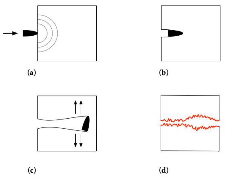
Figure 2.
Catastrophic transcranial gunshot wound by military rifle bullet penetrating the skull from left to right. The patient survived a few hours in the intensive care unit. (a) Axial non-contrast head CT scan demonstrating the entry wound, with bullet and bone fragments dispersed around it. There is ventricular hemorrhage, diffuse cerebral swelling, midline shifting and also pneumocephalus due to bullet entrance and the suction effect of cavitation. Linear hyperattenuation outlining the wound track (“tram track sign”) is visible adjacent to the entry trajectory, consistent with hemorrhagic coating of the track walls due to the temporary cavitation; (b) Three-dimensional CT reconstruction (oblique coronal view) provides visualization of both the entry and exit wounds, clearly demonstrating the explosive character of the soft tissue exit wound (on the right).
Figure 2.
Catastrophic transcranial gunshot wound by military rifle bullet penetrating the skull from left to right. The patient survived a few hours in the intensive care unit. (a) Axial non-contrast head CT scan demonstrating the entry wound, with bullet and bone fragments dispersed around it. There is ventricular hemorrhage, diffuse cerebral swelling, midline shifting and also pneumocephalus due to bullet entrance and the suction effect of cavitation. Linear hyperattenuation outlining the wound track (“tram track sign”) is visible adjacent to the entry trajectory, consistent with hemorrhagic coating of the track walls due to the temporary cavitation; (b) Three-dimensional CT reconstruction (oblique coronal view) provides visualization of both the entry and exit wounds, clearly demonstrating the explosive character of the soft tissue exit wound (on the right).
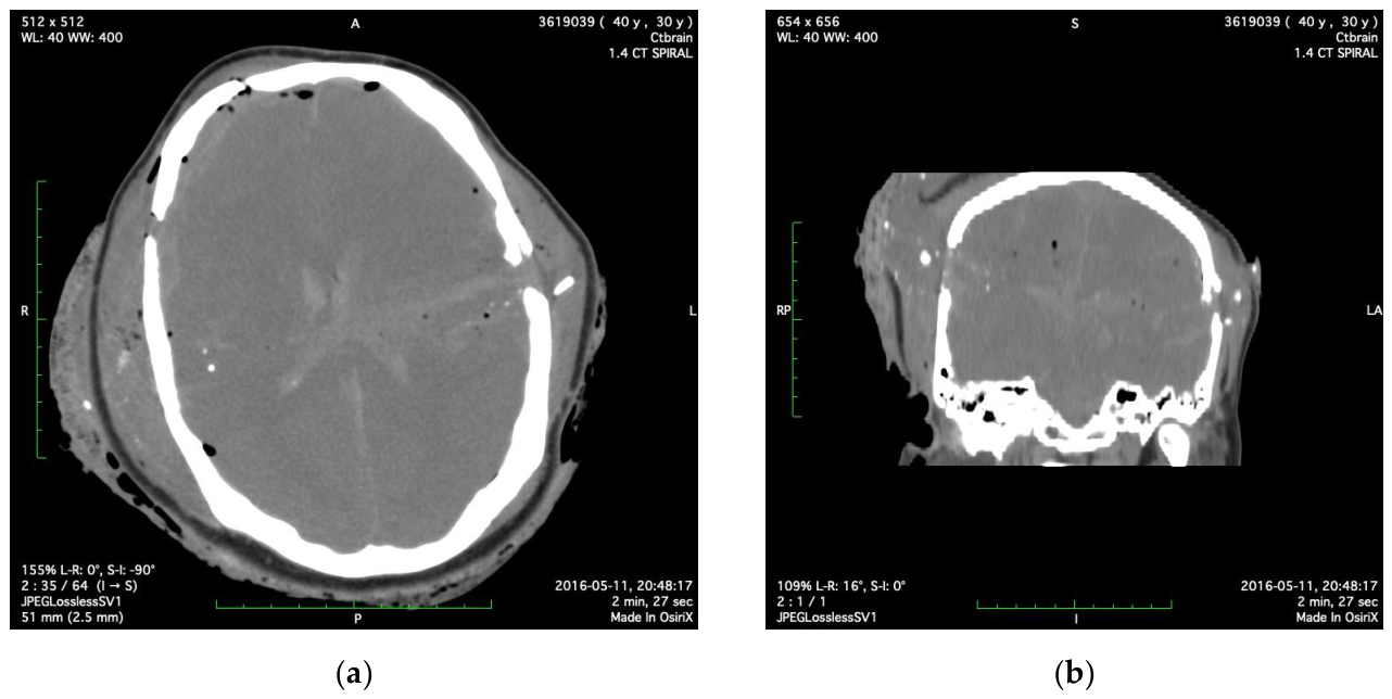
Figure 3.
Comparison of the corresponding phases of bullet penetration of the head and the hydrodynamic ram effect. (a) Skull and brain penetration by bullet creating an explosive fracture at the exit site due to temporary cavity formation and the intracranial pressure build-up (bullet size exaggerated for illustrative purposes); (b) Penetration of a fluid-filled tank (corresponding to skull-and-brain model) by bullet (hydrodynamic ram) resulting in deformation and explosive damage of the walls of the container. This occurs because of the pressure waves initiated as the momentum and kinetic energy of the bullet are transferred to the fluid across all directions. Bullet motion indicated by solid arrow.
Figure 3.
Comparison of the corresponding phases of bullet penetration of the head and the hydrodynamic ram effect. (a) Skull and brain penetration by bullet creating an explosive fracture at the exit site due to temporary cavity formation and the intracranial pressure build-up (bullet size exaggerated for illustrative purposes); (b) Penetration of a fluid-filled tank (corresponding to skull-and-brain model) by bullet (hydrodynamic ram) resulting in deformation and explosive damage of the walls of the container. This occurs because of the pressure waves initiated as the momentum and kinetic energy of the bullet are transferred to the fluid across all directions. Bullet motion indicated by solid arrow.
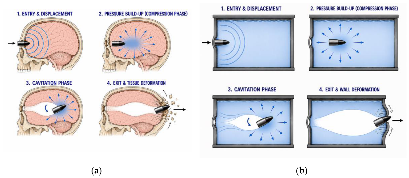
Figure 4.
Craniofacial gunshot wound due to self-inflicted submental discharge of high-velocity weapon. (a) Axial CT scan demonstrates complete destruction of the middle facial skeleton; (b) Image at a lower level shows comminuted fracture of the mandible with loss of its anterior part and presence of air collections in the soft tissues of the floor of the mouth.
Figure 4.
Craniofacial gunshot wound due to self-inflicted submental discharge of high-velocity weapon. (a) Axial CT scan demonstrates complete destruction of the middle facial skeleton; (b) Image at a lower level shows comminuted fracture of the mandible with loss of its anterior part and presence of air collections in the soft tissues of the floor of the mouth.
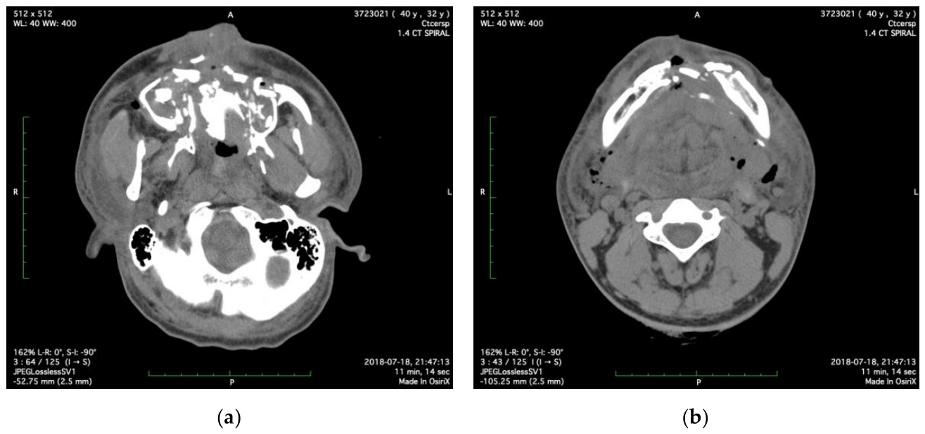
Figure 5.
Axial non-contrast CT image obtained after a grenade explosion in a military sericeman who sustained amputation of his right hand while wearing a protective helmet. The image demonstrates penetrating craniocerebral injury with numerous metallic (shrapnel) fragments lodged in the right temporal region and in the soft tissues at the site of projectile entry. The injury is associated with temporal bone and skull base fractures. Acute traumatic subarachnoid hemorrhage is present within the basal cisterns, with multiple intraparenchymal hemorrhagic contusions. Additional metallic fragments are evident in the soft tissues adjacent to a midfacial fracture.
Figure 5.
Axial non-contrast CT image obtained after a grenade explosion in a military sericeman who sustained amputation of his right hand while wearing a protective helmet. The image demonstrates penetrating craniocerebral injury with numerous metallic (shrapnel) fragments lodged in the right temporal region and in the soft tissues at the site of projectile entry. The injury is associated with temporal bone and skull base fractures. Acute traumatic subarachnoid hemorrhage is present within the basal cisterns, with multiple intraparenchymal hemorrhagic contusions. Additional metallic fragments are evident in the soft tissues adjacent to a midfacial fracture.
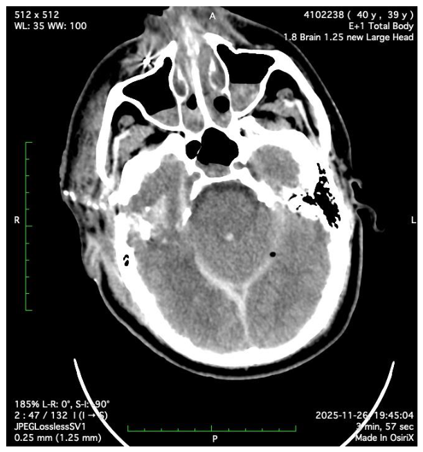

Table 1.
Hierarchical organization of prognostic factors for gunshot wounds of the head.
| Category | Primary | Secondary (during projectile-tissue interaction) | Tissue effect/response |
| Ballistic factors (projectile-related) * | velocity, kinetic energy | stability/yaw | bone fracture (entry/exit) |
| caliber | mushrooming | contusion | |
| shape/construction | fragmentation | laceration | |
| Biomechanical factors (energy-related) | energy transfer | shock/stress wave | stretching, shearing |
| pressure transmission | temporary cavity | indirect fractures | |
| Imaging (CT)/anatomical factors | transventricular | raised ICP | |
| projectile trajectory | bihemispheric/multilobar | brain swelling | |
| posterior | herniation (coning) | ||
| Clinical factors (measured at presentation) | level of consciousness | impaired consciousness | GCS <5 |
| pupillary response | dilated, unreactive pupil |
* Ballistic factors include “primary” (intrinsic) properties of the projectile and secondary ones appearing as a result of the projectile-tissue interaction. CT: computed tomography; GCS: Glasgow Coma Scale; ICP: intracranial pressure.
Disclaimer/Publisher’s Note: The statements, opinions and data contained in all publications are solely those of the individual author(s) and contributor(s) and not of MDPI and/or the editor(s). MDPI and/or the editor(s) disclaim responsibility for any injury to people or property resulting from any ideas, methods, instructions or products referred to in the content. |
© 2026 by the authors. Licensee MDPI, Basel, Switzerland. This article is an open access article distributed under the terms and conditions of the Creative Commons Attribution (CC BY) license (http://creativecommons.org/licenses/by/4.0/).
Copyright: This open access article is published under a Creative Commons CC BY 4.0 license, which permit the free download, distribution, and reuse, provided that the author and preprint are cited in any reuse.


