

Submitted:
21 June 2026
Posted:
07 July 2026
You are already at the latest version
Abstract
Cardiolipin (CL), a unique dimeric phospholipid with four acyl chains and a characteristically small polar head group, stands as one of the most compelling examples of evolutionary continuity in cell biology. Present in the plasma membrane of a-proteobacteria and conserved without fundamental modification in the inner mitochondrial membrane (IMM) of all eukaryotes examined, CL has been retained across approximately two billion years of evolution, a period over which the mitochondrion shed thousands of its original genes. This degree of conservation demands an explanation that transcends structural necessity alone. Here we propose, and document with biochemical and cell biological evidence, that CL functions as a programmable signaling hub: a lipid species whose physical chemistry and membrane address allow it to assemble distinct supramolecular platforms in response to discrete stress signals, each platform transducing a specific mitochondrial state into a defined cell fate outcome. Three core CL signaling platforms are described. Platform 1, the catalytic peroxidase platform, converts the constitutive CL–cytochrome c (cyt c) structural complex into an enzymatic reaction under oxidative stress, generating oxidized CL (oxCL) species that commit the cell to apoptosis by releasing cyt c from the IMM. Platform 2, the receptor-like mitophagy platform, exploits NME4-dependent CL scramblase activity to translocate CL from the IMM to the outer mitochondrial membrane (OMM) surface upon membrane potential dissipation, creating an externalized "eat-me" signal that LC3-II on autophagic membranes recognizes directly. Platform 3, the caspase-8/BID activation platform, assembles a CL microdomain scaffold at the OMM that recruits caspase-8 from death receptor complexes, accelerates Bid cleavage by three orders of magnitude, and couple extrinsic apoptotic signals to mitochondrial outer membrane permeabilization (MOMP). An emerging fourth axis links CL externalization to innate immune activation via NLRP3 inflammasome recruitment. We argue that the deep evolutionary conservation of CL reflects not its structural roles per se, but the irreplaceable nature of these signaling functions. Functions already present in ancestral bacteria and progressively elaborated as eukaryotic cell death, quality control, and immunity coevolved with the organelle itself.
Keywords:
cardiolipin
; mitochondria
; apoptosis
; mitophagy
; caspase-8
; Bid
; NLRP3
; signaling hub
; evolutionary conservation
; Barth syndrome
1. Introduction
Among the roughly 200 molecular species that constitute the mammalian lipidome, cardiolipin (CL; 1,3-bis(sn-3'-phosphatidyl)-sn-glycerol) occupies a singular position. It is the only phospholipid with a dimeric architecture — two phosphatidic acid moieties linked through a central glycerol — and four acyl chains that generate a distinctive cone-shaped molecular geometry, favouring negative membrane curvature [1,2]. It is the only lipid whose distribution is essentially confined to the inner mitochondrial membrane, where it constitutes 20–25% of the total phospholipid content, particularly at cristae rims and contact sites [3,4]. As it can be seen in Figure 1, CL are strictly confined to the mitochondria a subcellular compartment with a particularly distinguishable lipid composition (Figure 1).
The lipid composition of different membranes varies throughout the cell and mitochondria exhibit a special composition. The lipid compositional data are expressed as a percentage of the total phospholipid (PL) in mammals (blue) and yeast (light blue). As a measure of sterol content, the molar ratio of cholesterol (CHOL; in mammals) and ergosterol (ERG; in yeast) to phospholipid is also included. The main panel shows the site of synthesis of the major phospholipids (blue) and lipids that are involved in signaling and organelle recognition pathways (red). It should be appreciated that the levels of signaling and recognition lipids are significantly below 1% of the total phospholipid, except for ceramide (Cer). The major glycerophospholipids assembled in the endoplasmic reticulum (ER) are phosphatidylcholine (PtdCho; PC), phosphatidylethanolamine (PtdEtn; PE), phosphatidylinositol (PtdIns; PI), phosphatidylserine (PtdSer; PS) and phosphatidic acid (PA). In addition, the ER synthesizes Cer, galactosylceramide (GalCer), cholesterol and ergosterol. Both the ER and lipid droplets participate in steryl ester and triacylglycerol (TG) synthesis. The Golgi lumen is the site of synthesis of sphingomyelin (SM), complex glycosphingolipids (GSLs) and yeast inositol sphingolipid (ISL) synthesis. PtdCho is also synthesized in the Golgi, and may be coupled to protein secretion at the level of its diacylglycerol (DAG) precursor. Approximately 45% of the phospholipid in mitochondria (mostly PtdEtn, PA and cardiolipin (CL) is autonomously synthesized by the organelle. BMP (bis(monoacylglycero)phosphate) is a major phospholipid in the inner membranes of late endosomes26. PI(3,5)P2, phosphatidylinositol-(3,5)-bisphosphate; PI(4,5)P2, phosphatidylinositol-(4,5)-bisphosphate; PI(3,4,5)P3, phosphatidylinositol-(3,4,5)-trisphosphate; PI4P, phosphatidylinositol- 4-phosphate; R, remaining lipids; S1P, sphingosine-1-phosphate
And it is, with only minor structural modifications, the same molecule found in the plasma membrane of a-proteobacteria, the group from which mitochondria descended by endosymbiosis roughly 1.5 - 2 billion years ago [5,6] (Figure 2).
Cardiolipin (CL) is one of the very few lipid species whose biosynthetic pathway has been conserved without fundamental modification across approximately two billion years of evolution. This figure illustrates the endosymbiotic transition that transferred CL from the plasma membrane of an ancestral a-proteobacterium into the inner mitochondrial membrane (IMM) of the emerging eukaryotic cell.
The left panel depicts the ancestral prokaryote, in which CL is concentrated in high-curvature membrane domains associated with the respiratory chain and serves as a structural scaffold for ATP synthase and electron transfer complexes.
The central panel represents the endosymbiotic event, showing how gene transfer to the nuclear genome progressively relocated control of CL biosynthesis and remodeling while preserving the lipid's membrane address.
The right panel shows the modern mitochondrion, in which CL remains confined to the IMM, particularly at cristae rims, and retains its ancestral role in organizing respiratory supercomplexes (CI-CIII₂-CIV). The conservation of the entire CL biosynthetic pathway (PA → CDP-DAG → PGP → PG → CL) and of key enzymes such as cardiolipin synthase (CLS/CRD1) across bacteria, fungi, plants, and animals is represented by the color-coded homology bars at the bottom of each panel.
The strong purifying selection acting on these genes (low dN/dS ratios across eukaryotic phylogeny) underscores that CL's retention is driven by irreplaceable functional necessity rather than evolutionary inertia. The phylogenetic banner at the top of the figure contextualizes the diagram within the broader eukaryotic tree of life, emphasizing that every lineage examined, from Saccharomyces cerevisiae to Homo sapiens, and including deeply divergent lineages such as Dictyostelium discoideum and Leishmania spp. that has maintained CL as an obligate component of the IMM.
The textbook account of CL assigns it structural functions: stabilizing respiratory chain supercomplexes (the respirasome, CI–CIII₂–CIV), maintaining the proton impermeability of the IMM, and providing the negative surface charge that attracts matrix-targeted import sequences [7,8,9]. These roles are real and important. Yet they do not easily explain why CL is under strong purifying selection across the entirety of eukaryotic phylogeny, why its acyl chain composition is precisely controlled by a dedicated remodeling enzyme (tafazzin), and why mutations in CL biosynthesis and remodeling produce systemic disease, most dramatically in Barth syndrome, the X-linked cardiomyopathy caused by tafazzin loss of function [10,11]. Structural lipids are rarely subject to such enzymatic precision.
We propose here that the resolution of this apparent paradox lies in recognizing CL as a signaling lipid specifically, as a programmable platform molecule that assembles distinct supramolecular machines in response to different mitochondrial stress signals. This reframing is not merely semantic. It predicts that (i) specific protein–CL interactions should be stimulus-dependent and mechanistically necessary for downstream signaling; (ii) the precise molecular species of CL (i.e., its acyl chain composition) should influence the efficiency of each platform; and (iii) deep evolutionary conservation of CL should be paralleled by conservation of the enzymes that generate and remodel it. All three predictions are supported by experimental evidence, which we review here in the context of three well-characterized platforms and one emerging axis.
2. Mitochondrial Phylogeny and the Deep Conservation of Cardiolipin
2.1. The Endosymbiotic Origin of CL in Eukaryotes
Phylogenomic analyses of mitochondrial and nuclear-encoded mitochondrial genes consistently place the mitochondrial ancestor within the a-proteobacteria, though the precise sister group remains debated [12,13]. What is not debated is that the Last Eukaryotic Common Ancestor (LECA) already possessed functional mitochondria, and that every eukaryotic lineage examined - from opisthokonts and plants to excavates such as Giardia and Leishmania - retains CL or structurally equivalent anionic lipids in mitochondrial-derived compartments [14,15]. Even hydrogenosomes and mitosomes, the most highly derived mitochondria-related organelles that have lost the electron transport chain entirely, retain CL biosynthetic machinery in several lineages, suggesting that CL's roles extend beyond oxidative phosphorylation [16].
The biosynthetic route to CL: - phosphatidic acid → CDP-DAG → phosphatidylglycerophosphate (PGP) → phosphatidylglycerol (PG) → CL - is essentially identical in bacteria and eukaryotic mitochondria, with clear orthologous relationships at each enzymatic step [17]. Cardiolipin synthase (CLS1 in mammals, CRD1 in yeast) is a direct homolog of bacterial CLS, reflecting the organelle's prokaryotic ancestry at the molecular level [18]. Crucially, the CL remodeling system, absent in bacteria but present in all eukaryotes examined, adds a layer of enzymatic control that points to additional, eukaryote-specific functions for the mature lipid species.
2.2. Acyl Chain Remodeling: Precision as Evidence of Function
In most mammalian tissues, CL carries four linoleoyl chains (18:2), yielding the species tetralinoleoyl-CL (TLCL) as the predominant molecular form [19]. This is not the product of de novo synthesis — CL is initially synthesized with a heterogeneous acyl chain composition — but of post-synthetic remodeling by tafazzin, a phospholipid transacylase encoded on the X chromosome [10]. The substrate specificity of tafazzin, and the tissue-specific TLCL profiles it generates, have been conserved from yeast to humans, implying strong selection on CL molecular species rather than CL quantity alone [20].
Barth syndrome, caused by hemizygous loss-of-function mutations in TAZ (the tafazzin gene), provides unambiguous genetic evidence that CL remodeling is clinically essential [10,11]. Affected individuals present with dilated cardiomyopathy, skeletal myopathy, neutropenia, and growth retardation — a phenotype that cannot be explained solely by impaired OXPHOS, since respiratory chain assembly is only modestly affected in Barth syndrome fibroblasts [21]. As discussed below, the acyl chain composition of CL directly determines the efficiency of at least two signaling platforms (Platforms 1 and 3), providing a mechanistic basis for the specificity of disease manifestation [22,23].
2.3. Purifying Selection and the Hub Hypothesis
Comparative genomics of CL pathway enzymes across eukaryotic phyla reveals consistently low dN/dS ratios, indicating that these genes evolve under strong negative (purifying) selection [24]. This is the evolutionary fingerprint of genes whose products perform non-redundant functions: mutations are systematically removed from populations because they impair fitness. For a structural lipid, modest impairment of OXPHOS efficiency would plausibly be sufficient to explain this; but the degree of conservation observed — including the conservation of acyl remodeling machinery across lineages that have otherwise dramatically diversified their membrane lipidomes — argues for additional, non-structural selection pressures. The signaling platform hypothesis provides a coherent explanation: each CL-dependent platform is indispensable, so neither the lipid itself nor the enzymes controlling its molecular species can be freely varied without fitness cost.
3. Platform 1: The CL-Cytochrome c Peroxidase Axis
3.1. The Constitutive CL–Cyt c Complex
Under homeostatic conditions, 15–20% of the total mitochondrial cytochrome c pool exists in tight association with CL at the IMM [25]. This interaction is electrostatic in origin — the lysine-rich face of cyt c (pI ~10) binds to the anionic head group of CL — and partially occludes the heme crevice, reducing cyt c's electron transfer activity in its membrane-bound state [26]. This constitutive complex represents a kinetic reservoir that also, critically, positions cyt c adjacent to its enzymatic substrate under pro-apoptotic conditions [27].
3.2. Activation of CL-Specific Peroxidase Activity
The seminal work of Kagan and colleagues demonstrated that, upon oxidative stress, cyt c acquires potent peroxidase activity specifically toward CL [28,29]. The mechanism involves H₂O₂-dependent oxidation of the heme iron to compound I/II intermediates that abstract hydrogen from the bis-allylic methylene groups of CL's polyunsaturated acyl chains (principally 18:2 in TLCL), generating a spectrum of hydroperoxy- and hydroxy-CL species designated collectively as oxidized CL (oxCL) [30]. The reaction is enzymatic rather than a random oxidative attack: the molecular profiles of oxCL in apoptotic cells are non-random and site-specific, consistent with active-site catalysis [31] (Figure 3).
The peroxidase platform has two downstream consequences. First, accumulation of oxCL progressively weakens the CL–cyt c electrostatic interaction, because the negatively charged phosphate groups of oxCL no longer present an optimal binding surface for cyt c [32]. This releases cyt c into the intermembrane space, where MOMP subsequently permits its cytosolic entry and apoptosome assembly (cyt c–APAF-1–pro-caspase-9). Second, oxCL species are themselves bioactive: they modulate the membrane curvature of the IMM, destabilize respiratory supercomplexes, and generate lipid peroxide propagation chains that amplify the initial oxidative insult [33,34].
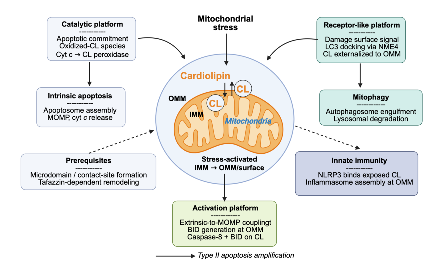
This figure presents the conceptual framework at the heart of the manuscript: the proposition that cardiolipin functions not as a passive membrane component but as a programmable signaling hub operating through three distinct, stimulus-coupled platform modes. Each platform is triggered by a specific upstream stress signal, engages a defined set of protein partners, and produces a discrete cellular output.
Platform 1 (the catalytic peroxidase platform, shown in teal) is activated by mitochondrial reactive oxygen species (ROS) and converts the constitutive CL-cytochrome c (cyt c) interaction into an enzymatic reaction: cyt c acquires CL-specific peroxidase activity, oxidizes CL at polyunsaturated acyl chains, and generates oxidized CL species (oxCL) that weaken CL-cyt c binding, ultimately releasing cyt c into the intermembrane space (IMS) as an apoptotic initiator.
Platform 2 (the receptor-like mitophagy platform, shown in blue) is activated by mitochondrial membrane potential collapse (Δψm dissipation) and drives NME4-dependent phospholipid scramblase activity that translocates CL from the IMM to the outer mitochondrial membrane (OMM) surface, where externalized CL is recognized by LC3-II on autophagic membranes, selectively targeting the damaged organelle for elimination.
Platform 3 (the caspase activation platform, shown in coral) is activated by extrinsic death receptor signaling and assembles a CL microdomain-based supramolecular complex at the OMM, in which caspase-8 cleaves Bid to generate tBid, which inserts into CL-rich microdomains and drives Bax/Bak oligomerization and mitochondrial outer membrane permeabilization (MOMP).
The figure emphasizes that these three platforms are not independent pathways but form a hierarchically organized signaling network, with oxCL production from Platform 1 feeding back to modulate the efficiency of Platforms 2 and 3. The dashed arrows between platforms indicate cross-talk nodes and potential therapeutic intervention points
3.3. Acyl Chain Composition as a Determinant of Platform Sensitivity
The preference of the cyt c peroxidase for polyunsaturated acyl chains explains why TLCL, the product of tafazzin-mediated remodeling, is particularly susceptible. Cells with saturated or monounsaturated CL species (as occurs in tafazzin-deficient Barth syndrome cells) show substantially blunted oxCL generation and delayed apoptotic commitment in response to equivalent oxidative insults [22,35]. This molecular mechanism links the Barth syndrome phenotype to a specific defect in Platform 1 signaling fidelity, rather than to a generic bioenergetic impairment.
4. Platform 2: NME4-Driven CL Externalization and Selective Mitophagy
4.1. The Asymmetric Distribution of CL
In the resting mitochondrion, CL is highly asymmetric: it is essentially confined to the inner leaflet of the IMM, where its anionic head group faces the mitochondrial matrix [36]. This asymmetry is maintained by the combined action of the membrane potential and ATP-dependent lipid translocases. Loss of membrane potential (Δψm dissipation) therefore represents a permissive condition for CL redistribution — a redistribution that constitutes the basis of Platform 2 (Figure 4).
Selective elimination of irreparably damaged mitochondria by autophagy (mitophagy) is essential for cellular homeostasis, yet the molecular signal that marks a dysfunctional mitochondrion for autophagic engulfment remained elusive until the identification of externalized CL as a damage-associated "eat-me" signal.
This figure maps the complete sequence of events from Δψm dissipation to LC3-II-mediated autophagosome docking.
The upper portion of the figure shows the resting state, in which CL is asymmetrically distributed across the two mitochondrial membranes, confined almost exclusively to the inner leaflet of the IMM by the combined action of ATP-dependent flippases and the energized membrane potential. The central portion shows the activation sequence triggered by membrane depolarization: loss of Δψm activates NME4 (nucleoside diphosphate kinase 4), a mitochondrial intermembrane space enzyme that moonlights as a phospholipid scramblase; NME4 translocates CL across the IMM from inner to outer leaflet, across the IMS, and finally to the cytoplasmic face of the OMM.
The right portion illustrates the recognition event: LC3-II, the lipidated autophagosomal membrane protein that drives cargo selection, binds directly to externalized CL through a specific interaction that does not require any adaptor protein, bridging the damaged mitochondrion to the growing phagophore membrane.
A lower panel shows the experimental evidence basis for each step, fluorescent CL reporter localization, NME4 knockdown rescue experiments, and LC3-II co-immunoprecipitation with CL liposomes and distinguishes Platform 2 mitophagy from the PINK1–Parkin ubiquitin-dependent pathway, emphasizing that these are parallel, mechanistically independent routes to mitophagy that may be engaged by different magnitudes or qualities of mitochondrial damage. The clinical relevance is highlighted in a side box: impaired CL externalization has been documented in neurodegeneration-associated contexts where mitophagic clearance is compromised.
4.2. NME4 as the CL Scramblase
The enzyme responsible for stress-induced CL externalization is NME4 (nucleoside diphosphate kinase 4), a member of the NME/NDPK family that moonlights as a phospholipid scramblase in the mitochondrial intermembrane space [37,38]. Upon Δψm dissipation, NME4 catalyzes the ATP-independent bidirectional transfer of CL across the IMM inner leaflet to the outer leaflet, across the IMS, and ultimately to the cytoplasmic face of the OMM [37]. The directionality toward the OMM surface is driven by the concentration gradient created by LC3-II binding at the outer face, which acts as a thermodynamic sink.
4.3. LC3-II Recognizes Externalized CL Directly
The autophagosome membrane protein LC3-II binds directly to externalized CL without the requirement for any adaptor protein - a mechanistically distinctive feature that sets this mitophagy pathway apart from the PINK1–Parkin ubiquitin-dependent route [39]. The CL–LC3-II interaction has been reconstituted in vitro with liposomes, demonstrating that CL surface exposure is both necessary and sufficient for LC3-II docking [40]. In cells, NME4 knockdown abolishes CL surface exposure and blocks CL-dependent mitophagy without affecting PINK1–Parkin-driven mitophagy, confirming the specificity of Platform 2 [37] (Figure 4).
Platform 2 mitophagy represents a damage-sensing mechanism of remarkable elegance: the very lipid that characterizes the mitochondrial inner membrane becomes, when translocated to the outer surface, the molecular flag for organelle elimination. In Barth syndrome, impaired CL maturation reduces the density and accessibility of externalized CL, contributing to the defective mitophagy documented in tafazzin-deficient cardiomyocytes [21,41] (Figure 4).
5. Platform3: CL Microdomains as a Caspase-8/BID Activation Scaffold
5.1. CL Microdomains at the OMM
CL, despite its predominant IMM localization, is also present at the OMM in discrete, cholesterol-excluding microdomains that are enriched in the pro-apoptotic proteins BAX, BAK, and VDAC [42,43]. These microdomains represent lateral organization of the OMM distinct from bulk-phase phospholipids and are stabilized by the cone-shaped geometry of CL, which promotes negative curvature and loose packing at microdomain boundaries [44]. Their existence has been demonstrated by detergent-free density gradient fractionation, fluorescence correlation spectroscopy, and super-resolution STED microscopy [43].
5.2. Caspase-8 Recruitment and Accelerated BID Cleavage
Upon ligation of death receptors (FAS, DR4/5, TNFR1), activated caspase-8 is released from the DISC complex and, rather than acting solely in the cytoplasm, translocates to the OMM surface via direct interaction with CL microdomains [45]. This membrane recruitment increases the local concentration of fully activated caspase-8 and co-concentrates its substrate BID in the same CL-rich environment. As a consequence, the rate of BID cleavage to truncated BID (tBID) is approximately 1,000-fold greater at CL-containing membranes than in solution [46]. tBID inserts also into the CL microdomain, where it triggers BAX conformational change and oligomerization, culminating in MOMP and the full commitment to intrinsic apoptosis [47] (Figure 5).
This Platform 3 mechanism was first proposed as the "mitochondrial activating platform" concept by Petit and colleagues [48], and has since been extended and confirmed in multiple model systems including reconstituted giant unilamellar vesicles (GUVs), isolated mitochondria, and intact cells subjected to extrinsic apoptotic stimulation [49,50]. The GUV reconstitution experiments are particularly compelling: a minimal system comprising CL-containing membranes, caspase-8, BID, and BAX reconstitutes the complete sequence from BID cleavage to membrane permeabilization, establishing the minimal molecular requirements of the platform [50].
The classical model of apoptosis posits a strict separation between the extrinsic pathway (death receptor → caspase-8 activation) and the intrinsic pathway (mitochondrial → MOMP → cyt c → apoptosome), connected only through caspase-8 cleavage of BID. This figure presents evidence that CL microdomains at the OMM constitute a physical platform that dramatically accelerates and amplifies this connection, effectively functioning as a signal integration surface at the mitochondrial outer membrane.
The left panel depicts the resting OMM with CL organized into discrete, cholesterol-excluding lipid microdomains (shown as darker patches) that are enriched in BAX, BAK, and VDAC. Upon death receptor ligation (FAS-L, TRAIL, TNF), activated caspase-8 is recruited to the OMM surface. A step that requires direct caspase-8 interaction with CL, where it cleaves BID with approximately 1,000-fold greater efficiency than in the cytosol, as shown in the kinetic inset.
The central panel shows tBID insertion into the CL microdomain, where it triggers BAX conformational change and oligomerization, leading to the formation of proteolipidic pores in the OMM and MOMP. A critical mechanistic detail illustrated in the lower inset is that CL acyl chain remodeling by tafazzin determines microdomain fluidity and packing, and therefore the efficiency of caspase-8 recruitment and tBID insertion: Barth syndrome cells with CL remodeling deficiency show blunted Platform 3 responses despite intact upstream death receptor signaling.
The right panel maps the therapeutic implications of this platform: small molecules that stabilize or destabilize CL microdomains, or that compete with caspase-8 for the CL binding interface, represent conceptually novel intervention strategies in diseases where the extrinsic-to-intrinsic apoptosis amplification loop is either over-activated (ischemia-reperfusion, septic cardiomyopathy) or pathologically suppressed (cancer drug resistance).
5.3. The 2025 Extension: Multiple Pathway Entanglements
Recent work from our laboratory has substantially extended the Platform 3 model in two directions [51,52]. First, we have documented that Platform 3 does not operate in isolation but intersects with several other cell death pathways — including necroptosis (via RIPK3 interaction with CL microdomains) and ferroptosis (via GPX4-mediated suppression of CL peroxidation) — creating a network of CL-dependent pathway entanglements rather than a linear hierarchy [51]. Second, detailed biochemical mapping of the caspase-8/BID/CL ternary interaction has revealed specific binding interfaces on both proteins that represent potential pharmacological targets for modulating Platform 3 activity selectively, without disrupting the constitutive CL–cyt c interaction of Platform 1 [52].
6. Emerging Platform: CL and Innate Immune Signaling
Beyond its roles in cell death and quality control, CL has recently emerged as a participant in innate immune signal transduction — a finding that carries particular evolutionary resonance given CL's bacterial ancestry. The NLRP3 inflammasome, a multimolecular danger-sensing complex that processes pro-IL-1β and pro-IL-18 into mature cytokines and initiates pyroptosis, is activated by mitochondrial signals including externalized CL [53,54].
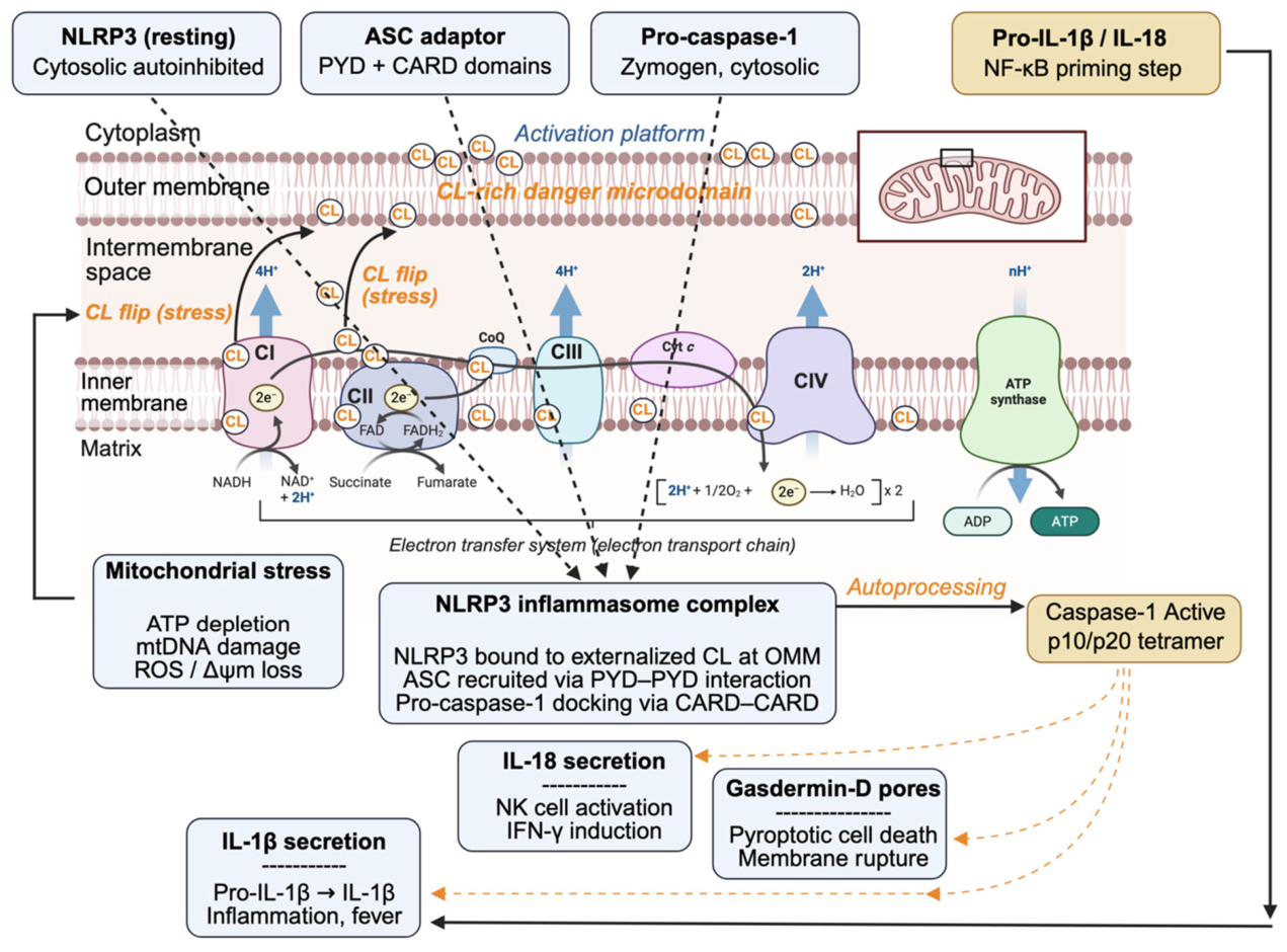
The mechanistic basis of CL-driven NLRP3 activation follows the two-signal model of inflammasome biology. A priming signal (typically TLR ligation activating NF-κB) upregulates NLRP3 and pro-IL-1β expression. CL externalization to the OMM surface then provides the second signal: NLRP3 docks directly onto exposed CL via its NACHT domain, recruits ASC through PYD–PYD interactions, and assembles pro-caspase-1 via CARD–CARD contacts, generating the active inflammasome complex at the mitochondrial surface [54,55]. Active caspase-1 processes both cytokine precursors and gasdermin D (GSDMD), whose N-terminal domain forms plasma membrane pores, driving the lytic inflammatory cell death designated pyroptosis [56].
An additional immune-relevant role of CL has been identified in the context of cGAS-STING signalling. Oxidized mitochondrial DNA released from damaged mitochondria activates cGAS; the subsequent downstream signalling is amplified by CL-dependent mitochondrial membrane disruption, creating a mechanistic link between Platform 1 (oxCL generation) and innate DNA sensing [57]. The evolutionary logic is notable: eukaryotic innate immunity appears to have co-opted the "bacterial signature" of CL — recognizing it as a danger-associated molecular pattern (DAMP) with structural similarities to the pathogen-associated molecular patterns (PAMPs) of its alphaproteobacterial ancestors.
7. Integration: CL as a Decision-Making Interface
The figure shows, layer by layer: At the Cytosol (top); the four cytosolic actors before activation: autoinhibited NLRP3, ASC, pro-caspase-1, and pro-IL-1β/IL-18 produced during the NF-κB priming step. At the OMM surface, the CL-rich danger microdomain where NLRP3 docks directly onto externalized CL, the critical mechanistic claim from [55]. At the IMS / IMM level, the CL flip arrow from IMM to OMM surface, connecting this figure to the same CL redistribution mechanism shown in Figure 4.
At the Inflammasome complex, NLRP3 on CL recruits ASC via PYD–PYD and pro-caspase-1 via CARD–CARD, producing active caspase-1. Indeed at whole, the three downstream consequences are: IL-1β maturation, IL-18 maturation,`and gasdermin-D pore formation / pyroptosis.
The three CL platforms and the emerging immune axis are not parallel, independent pathways but an interconnected decision network. Several integration points are worth highlighting. First, Platform 1 (oxCL generation) is upstream of Platform 2 (mitophagy) under mild oxidative stress: sub-lethal oxCL levels impair TLCL-dependent NME4 activity, attenuating mitophagy and thereby modulating the threshold between organelle repair and elimination [58]. Second, the magnitude of Platform 1 output determines whether Platform 3 is engaged: moderate cyt c release initiates the apoptosome without MOMP-dependent amplification, while maximal cyt c release combined with full MOMP requires Platform 3 activation [33,47]. Third, the inflammasome platform shares the NME4/CL externalization machinery with Platform 2, creating a molecular switch in which the identity of the CL-binding partner (LC3-II versus NLRP3) determines whether the response is autophagic elimination or inflammatory pyroptosis [55] (Figure 6).
This integration is governed, at the lipid level, by CL molecular species. TLCL is the preferred substrate of the cyt c peroxidase (Platform 1) and the optimal ligand for LC3-II docking (Platform 2). Barth syndrome's CL composition defect therefore impairs the fidelity of both platforms simultaneously, explaining why the disease manifests as a combined defect in apoptosis, mitophagy, and inflammatory regulation: a phenotype not easily reconciled with a purely structural or bioenergetic model of CL function [21,22,35,41].
From a signaling theory perspective, CL behaves as a bifunctional hub molecule: it is constitutively present at the relevant membrane address and constitutively pre-loaded with binding partners (cyt c, partially assembled NLRP3 components), but switches from dormant to active states only upon specific stress-triggered modifications (peroxidation, translocation, microdomain reorganization). This architecture, i.e., stimulus-dependent activation of a pre-formed scaffold, resembles the logic of second messenger systems and kinase cascades more closely than that of a passive structural lipid.
8. Therapeutic Implications
Reframing CL as a signaling hub rather than a structural component opens several pharmacological windows. In ischemia-reperfusion injury, where Platform 1 is catastrophically activated by burst ROS production, selective scavengers of oxCL (e.g., the mitochondria-targeted antioxidant XJB-5-131) have shown efficacy in pre-clinical models without disrupting physiological CL–cyt c electron shuttling [59]. In Barth syndrome, the recognition that tafazzin loss impairs signaling fidelity rather than just bioenergetics has motivated gene therapy approaches (AAV-mediated TAZ replacement) that have shown promise in mouse models and are entering clinical development [60].
Platform 3's pharmacological interface, i.e., the caspase-8/CL and BID/CL binding surfaces offers targets for modulating the extrinsic-to-intrinsic apoptosis amplification loop. This is directly relevant to cancer drug resistance: many tumours evade TRAIL-induced apoptosis by downregulating Platform 3 components, and small molecules that restore CL microdomain assembly could re-sensitize resistant cells to death receptor agonists [52]. Conversely, diseases characterized by pathological over-activation of Platform 3 - including septic cardiomyopathy and myocardial infarction - would benefit from inhibitors of this axis [47].
The inflammasome axis (Section 6) suggests that CL-targeted interventions could modulate pyroptotic inflammation in settings including NLRP3-driven sterile inflammation, sepsis, and neurodegeneration. The shared NME4 machinery between Platforms 2 and 4 implies that NME4 modulators might selectively redirect the response from inflammatory pyroptosis to autophagic clearance, a therapeutic goal of considerable interest in chronic inflammatory diseases [55].
9. Conclusions
The deep evolutionary conservation of cardiolipin across two billion years of eukaryotic evolution is not an accident of lipid biosynthetic pathway inertia. It reflects the progressive recruitment and elaboration of CL signaling functions that were already present in the ancestral alphaproteobacterial plasma membrane and that became irreplaceable components of eukaryotic cell biology as mitochondria were integrated into the cell's regulatory economy. The three platforms described here: - catalytic peroxidase, - receptor-like mitophagy, and - caspase-8/BID activation together with the emerging innate immune axis, constitute a mechanistically coherent framework for understanding why this lipid is both constitutively essential and acutely responsive to stress. The mitochondrial inner membrane, from this perspective, is not simply a bioenergetic structure: it is a programmable signaling surface, and cardiolipin is its master organizer.
Author Contributions
P.X.P.: conceptualization, writing — original draft, writing — review and editing, figure design (realized with “Biorender” and “Illustrator”, adobe). The author has read and agreed to the published version of the manuscript.
Funding
This research received no external funding. But benefit from the core funding of the CNRS and INSERM.
Conflicts of Interest
The author declares no conflict of interest.
References
- Schlame, M.; Rua, D.; Greenberg, M.L. The biosynthesis and functional role of cardiolipin. Prog. Lipid Res. 2000, 39, 257–288. [Google Scholar] [CrossRef] [PubMed]
- Jouhet, J. Importance of the hexagonal II phase in biological membrane organization. Front. Plant Sci. 2013, 4, 494. [Google Scholar] [CrossRef] [PubMed]
- Mileykovskaya, E.; Dowhan, W. Cardiolipin-dependent formation of mitochondrial respiratory supercomplexes. Chem. Phys. Lipids 2014, 179, 42–48. [Google Scholar] [CrossRef] [PubMed]
- Letts, J.A.; Sazanov, L.A. Clarifying the supercomplex: the higher-order organization of the mitochondrial electron transport chain. Nat. Struct. Mol. Biol. 2017, 24, 800–808. [Google Scholar] [CrossRef] [PubMed]
- Gray, M.W.; Burger, G.; Lang, B.F. Mitochondrial evolution. Science 1999, 283, 1476–1481. [Google Scholar] [CrossRef] [PubMed]
- Roger, A.J.; Muñoz-Gómez, S.A.; Kamikawa, R. The origin and diversification of mitochondria. Curr. Biol. 2017, 27, R1177–R1192. [Google Scholar] [CrossRef] [PubMed]
- Pfeiffer, K.; Gohil, V.; Stuart, R.A.; Hunte, C.; Brandt, U.; Greenberg, M.L.; Schägger, H. Cardiolipin stabilizes respiratory chain supercomplexes. J. Biol. Chem. 2003, 278, 52873–52880. [Google Scholar] [CrossRef] [PubMed]
- Böttinger, L.; Horvath, S.E.; Kleinschroth, T.; Hunte, C.; Daum, G.; Pfanner, N.; Becker, T. Phosphatidylethanolamine and cardiolipin differentially affect the stability of mitochondrial respiratory chain supercomplexes. J. Mol. Biol. 2012, 423, 677–686. [Google Scholar] [CrossRef] [PubMed]
- Ikon, N.; Ryan, R.O. Cardiolipin and mitochondrial cristae organization. Biochim. Biophys. Acta Biomembr. 2017, 1859, 1156–1163. [Google Scholar] [CrossRef] [PubMed]
- Bione, S.; D'Adamo, P.; Maestrini, E.; Gedeon, A.K.; Ballabio, A.; Toniolo, D. A novel X-linked gene, G4.5, is responsible for Barth syndrome. Nat. Genet. 1996, 12, 385–391. [Google Scholar] [CrossRef] [PubMed]
- Schlame, M.; Ren, M. The role of cardiolipin in the structural organization of mitochondrial membranes. Biochim. Biophys. Acta 2009, 1788, 2080–2083. [Google Scholar] [CrossRef] [PubMed]
- Muñoz-Gómez, S.A.; Wideman, J.G.; Roger, A.J.; Slamovits, C.H. The origin of mitochondrial cristae from alphaproteobacteria. Mol. Biol. Evol. 2017, 34, 943–956. [Google Scholar] [CrossRef] [PubMed]
- Martijn, J.; Vosseberg, J.; Guy, L.; Offre, P.; Ettema, T.J.G. Deep mitochondrial origin outside the sampled alphaproteobacteria. Nature 2018, 557, 101–105. [Google Scholar] [CrossRef] [PubMed]
- Daum, G. Lipids of mitochondria. Biochim. Biophys. Acta 1985, 822, 1–42. [Google Scholar] [CrossRef] [PubMed]
- Dolezal, P.; Likic, V.; Tachezy, J.; Lithgow, T. Evolution of the molecular machines for protein import into mitochondria. Science 2006, 313, 314–318. [Google Scholar] [CrossRef] [PubMed]
- Tielens, A.G.M.; Rotte, C.; van Hellemond, J.J.; Martin, W. Mitochondria as we don't know them. Trends Biochem. Sci. 2002, 27, 564–572. [Google Scholar] [CrossRef] [PubMed]
- Dowhan, W. Molecular basis for membrane phospholipid diversity: why are there so many lipids? Annu. Rev. Biochem. 1997, 66, 199–232. [Google Scholar] [CrossRef] [PubMed]
- Lu, Y.-W.; Claypool, S.M. Disorders of phospholipid metabolism: an emerging class of mitochondrial disease due to defects in remodeling. Front. Genet. 2015, 6, 3. [Google Scholar] [CrossRef] [PubMed]
- Schlame, M.; Hostetler, K.Y. Cardiolipin synthase from rat liver mitochondria, purification, and properties. J. Biol. Chem. 1991, 266, 22398–22403. [Google Scholar] [CrossRef] [PubMed]
- Claypool, S.M.; Koehler, C.M. The complexity of cardiolipin in health and disease. Trends Biochem. Sci. 2012, 37, 32–41. [Google Scholar] [CrossRef] [PubMed]
- Dudek, J.; Hartmann, M.; Rehling, P. The role of mitochondrial cardiolipin in heart function and its implication in cardiac disease. Biochim. Biophys. Acta Mol. Basis Dis. 2019, 1865, 810–821. [Google Scholar] [CrossRef]
- Petrosillo, G.; Matera, M.; Casanova, G.; Ruggiero, F.M.; Paradies, G. Mitochondrial dysfunction in rat brain with aging: involvement of complex I, reactive oxygen species and cardiolipin. Neurochem. Int. 2008, 53, 126–131. [Google Scholar] [CrossRef] [PubMed]
- Maguire, J.J.; Tyurina, Y.Y.; Mohammadyani, D.; Kapralov, A.A.; Anthonymuthu, T.S.; Bluteau, D.; Lahiri, S.; Vladimirov, Y.A.; Stark, H.J.; Kagan, V.E. Known unknowns of cardiolipin signaling: The best is yet to come. Biochim. Biophys. Acta Mol. Cell Biol. Lipids 2017, 1862, 8–24. [Google Scholar] [CrossRef] [PubMed]
- Zhang, M.; Mileykovskaya, E.; Dowhan, W. Cardiolipin is essential for organization of complexes III and IV into a supercomplex in intact yeast mitochondria. J. Biol. Chem. 2005, 280, 29403–29408. [Google Scholar] [CrossRef] [PubMed]
- Tuominen, E.K.J.; Wallace, C.J.A.; Kinnunen, P.K.J. Phospholipid-cytochrome c interaction: evidence for the extended lipid anchorage. J. Biol. Chem. 2002, 277, 8822–8826. [Google Scholar] [CrossRef] [PubMed]
- Sinibaldi, F.; Fiorucci, L.; Patriarca, A.; Lauceri, R.; Ferri, T.; Coletta, M.; Santucci, R. Insights into cytochrome c–cardiolipin interaction. Role played by ionic strength. Biochemistry 2008, 47, 6928–6935. [Google Scholar] [CrossRef] [PubMed]
- Planas-Iglesias, J.; Dwarakanath, H.; Mohammadyani, D.; Yanamala, N.; Kagan, V.E.; Klein-Seetharaman, J. Cardiolipin interactions with proteins. Biophys. J. 2015, 109, 1282–1294. [Google Scholar] [CrossRef]
- Kagan, V.E.; Tyurin, V.A.; Jiang, J.; Tyurina, Y.Y.; Ritov, V.B.; Amoscato, A.A.; Osipov, A.N.; Belikova, N.A.; Kapralov, A.A.; Kini, V.; et al. Cytochrome c acts as a cardiolipin oxygenase required for release of proapoptotic factors. Nat. Chem. Biol. 2005, 1, 223–232. [Google Scholar] [CrossRef] [PubMed]
- Jiang, J.; Kini, V.; Belikova, N.; Serinkan, B.F.; Borisenko, G.G.; Tyurina, Y.Y.; Tyurin, V.A.; Kagan, V.E. Cytochrome c release is required for phosphatidylserine peroxidation during Fas-triggered apoptosis in lung epithelial A549 cells. Lipids 2004, 39, 1133–1142. [Google Scholar] [CrossRef] [PubMed]
- Tyurin, V.A.; Tyurina, Y.Y.; Kochanek, P.M.; Hamilton, R.; DeKosky, S.T.; Greenberger, J.S.; Bhatt, D.L.; Bayir, H.; Kagan, V.E. Oxidative lipidomics of programmed cell death. Methods Enzymol. 2008, 442, 375–393. [Google Scholar] [CrossRef] [PubMed]
- Tyurina, Y.Y.; Polimova, A.M.; Maciel, E.; Tyurin, V.A.; Kapralova, V.I.; Winnica, D.E.; Vikulina, A.S.; Domingues, M.R.M.; McCoy, J.; Sanders, L.H.; et al. LC/MS analysis of cardiolipins in substantia nigra and plasma of rotenone-treated rats: implication for mitochondrial dysfunction in Parkinson's disease. Free Radic. Res. 2015, 49, 681–691. [Google Scholar] [CrossRef]
- Kapralov, A.A.; Kurnikov, I.V.; Vlasova, I.I.; Belikova, N.A.; Tyurin, V.A.; Basova, L.V.; Zhao, Q.; Tyurina, Y.Y.; Jiang, J.; Bayir, H.; et al. The hierarchy of structural transitions induced in cytochrome c by anionic lipids during peroxidase activation. Biochemistry 2007, 46, 14232–14244. [Google Scholar] [CrossRef] [PubMed]
- Kagan, V.E.; Jiang, J.; Huang, Z.; Tyurina, Y.Y.; Desbourdes, C.; Cottet-Rousselle, C.; Dar, H.H.; Verma, M.; Tyurin, V.A.; Kapralov, A.A.; et al. NDPK-D (NME4)-mediated externalization of cardiolipin enables elimination of depolarized mitochondria by mitophagy. Cell Death Differ. 2016, 23, 1140–1151. [Google Scholar] [CrossRef] [PubMed]
- Anthonymuthu, T.S.; Kenny, E.M.; Bayir, H. Emerging roles for oxidized phospholipids in cellular dysfunction and disease. Cell. Biochem. Biophys. 2018, 76, 19–37. [Google Scholar]
- Huang, W.; Choi, W.; Hu, W.; Mi, N.; Guo, Q.; Ma, M.; Liu, M.; Tian, Y.; Lu, P.; Wang, F.-L.; et al. Crystal structure and biochemical analyses reveal Beclin 1 as a novel membrane binding protein. Cell Res. 2012, 22, 473–489. [Google Scholar] [CrossRef] [PubMed]
- Schlame, M. Cardiolipin synthesis for the assembly of bacterial and mitochondrial membranes. J. Lipid Res. 2008, 49, 1607–1620. [Google Scholar] [CrossRef] [PubMed]
- Kagan, V.E.; Jiang, J.; Huang, Z.; Tyurina, Y.Y.; Desbourdes, C.; Cottet-Rousselle, C.; Dar, H.H.; Verma, M.; Tyurin, V.A.; Kapralov, A.A.; et al. NDPK-D (NME4)-mediated externalization of cardiolipin enables elimination of depolarized mitochondria by mitophagy. Cell Death Differ. 2016, 23, 1140–1151. [Google Scholar] [CrossRef] [PubMed]
- Lacombe, M.-L.; Tokarska-Schlattner, M.; Epand, R.F.; Boissan, M.; Epand, R.M.; Schlattner, U. Interaction of NDPK-D with cardiolipin-containing membranes: structural basis and implications for mitochondrial physiology. Biochimie 2009, 91, 779–783. [Google Scholar] [CrossRef] [PubMed]
- Chu, C.T.; Ji, J.; Dagda, R.K.; Jiang, J.F.; Tyurina, Y.Y.; Kapralov, A.A.; Tyurin, V.A.; Yanamala, N.; Shrivastava, I.H.; Mohammadyani, D.; et al. Cardiolipin externalization to the outer mitochondrial membrane acts as an elimination signal for mitophagy in neuronal cells. Nat. Cell Biol. 2013, 15, 1197–1205. [Google Scholar] [CrossRef] [PubMed]
- Sentelle, R.D.; Senkal, C.E.; Jiang, W.; Ponnusamy, S.; Gencer, S.; Selvam, S.P.; Bhatt, M.V.; Liu, J.; Loewen, M.C.; Bielawski, J.; et al. Ceramide targets autophagosomes to mitochondria and induces lethal mitophagy. Nat. Chem. Biol. 2012, 8, 831–838. [Google Scholar] [CrossRef]
- Hsu, P.; Liu, X.; Zhang, J.; Wang, H.-G.; Ye, J.-M.; Shi, Y. Cardiolipin remodeling by TAZ/tafazzin is selectively required for the initiation of mitophagy. Autophagy 2015, 11, 643–652. [Google Scholar] [CrossRef] [PubMed]
- Sorice, M.; Manganelli, V.; Matarrese, P.; Tinari, A.; Misasi, R.; Malorni, W.; Garofalo, T. Cardiolipin-enriched raft-like microdomains are essential activating platforms for apoptotic signals on mitochondria. FEBS Lett. 2009, 583, 2447–2450. [Google Scholar] [CrossRef] [PubMed]
- Garofalo, T.; Giammarioli, A.M.; Misasi, R.; Tinari, A.; Manganelli, V.; Gambardella, L.; Pavan, A.; Malorni, W.; Sorice, M. Lipid microdomains contribute to apoptosis-associated modifications of mitochondria in T cells. Cell Death Differ. 2005, 12, 1378–1389. [Google Scholar] [CrossRef] [PubMed]
- Khalifat, N.; Puff, N.; Bonneau, S.; Fournier, J.-B.; Angelova, M.I. Membrane deformation under local pH gradient: mimicking mitochondrial cristae dynamics. Biophys. J. 2008, 95, 4924–4933. [Google Scholar] [CrossRef] [PubMed]
- Lutter, M.; Fang, M.; Luo, X.; Nishijima, M.; Xie, X.; Wang, X. Cardiolipin provides specificity for targeting of tBid to mitochondria. Nat. Cell Biol. 2000, 2, 754–761. [Google Scholar] [CrossRef] [PubMed]
- Jalmar, O.; García-Sáez, A.J.; Berland, L.; Gonzalvez, F.; Petit, P.X. Giant unilamellar vesicles (GUVs) as a new tool for analysis of caspase-8/Bid-FL complex binding to cardiolipin and its functional activity. Cell Death Dis. 2010, 1, e103. [Google Scholar] [CrossRef] [PubMed]
- Gonzalvez, F.; Bessoule, J.-J.; Rocchiccioli, F.; Manon, S.; Petit, P.X. Role of cardiolipin on tBid and tBid/Bax synergistic effects on yeast mitochondria. Cell Death Differ. 2005, 12, 659–667. [Google Scholar] [CrossRef] [PubMed]
- Petit, P.X.; Lecoeur, H.; Zorn, E.; Dauguet, C.; Mignotte, B.; Gougeon, M.-L. Alterations in mitochondrial structure and function are early events of dexamethasone-induced thymocyte apoptosis. J. Cell Biol. 1995, 130, 157–167. [Google Scholar] [CrossRef] [PubMed]
- Jalmar, O.; Andreu-Fernandez, V.; Schlattner, U.; Garcia-Saez, A. Caspase-8 binding to cardiolipin in giant unilamellar vesicles provides a functional docking platform for bid. PLoS ONE 2013, 8, e55024. [Google Scholar] [CrossRef] [PubMed]
- Gonzalvez, F.; Pariselli, F.; Dupaigne, P.; Budihardjo, I.; Lutter, M.; Antonsson, B.; Diolez, P.; Manon, S.; Martinou, J.-C.; Goubern, M.; et al. tBid interaction with cardiolipin primarily orchestrates mitochondrial dysfunctions and subsequently activates Bax and Bak. Cell Death Differ. 2005, 12, 614–626. [Google Scholar] [CrossRef] [PubMed]
- Petit, P.X. Multiple entanglements of different cell death pathways, in which caspase-8 and BID interact with cardiolipin, have been identified. Front. Cell Dev. Biol. 2025, 13, 1667611. [Google Scholar] [CrossRef] [PubMed]
- Petit, P.X. Caspase-8 and BID caught in the act with cardiolipin: a new platform to provide mitochondria with microdomains of apoptotic signals. Cells 2025, 14, 1678. [Google Scholar] [CrossRef] [PubMed]
- Iyer, S.S.; He, Q.; Janczy, J.R.; Elliott, E.I.; Zhong, Z.; Olivier, A.K.; Sadler, J.J.; Knepper-Adrian, V.; Han, R.; Qiao, L.; et al. Mitochondrial cardiolipin is required for Nlrp3 inflammasome activation. Immunity 2013, 39, 311–323. [Google Scholar] [CrossRef] [PubMed]
- Shimada, K.; Crother, T.R.; Karlin, J.; Dagvadorj, J.; Chiba, N.; Chen, S.; Ramanujan, V.K.; Wolf, A.J.; Vergnes, L.; Ojcius, D.M.; et al. Oxidized mitochondrial DNA activates the NLRP3 inflammasome during apoptosis. Immunity 2012, 36, 401–414. [Google Scholar] [CrossRef] [PubMed]
- Tapia-Abellán, A.; Angosto-Bazarra, D.; Martínez-Banaclocha, H.; de Torre-Minguela, C.; Cerón-Carrasco, J.P.; Pérez-Sánchez, H.; Arostegui, J.I.; Pelegrin, P. MCC950 closes the NLRP3 inflammasome and inhibits NLRP3-driven mouse models of disease. Sci. Adv. 2019, 5, eaay closed. [Google Scholar]
- Kayagaki, N.; Stowe, I.B.; Lee, B.L.; O'Rourke, K.; Anderson, K.; Warming, S.; Cuellar, T.; Haley, B.; Roose-Girma, M.; Phung, Q.T.; et al. Caspase-11 cleaves gasdermin D for non-canonical inflammasome signalling. Nature 2015, 526, 666–671. [Google Scholar] [CrossRef] [PubMed]
- West, A.P.; Khoury-Hanold, W.; Staron, M.; Tal, M.C.; Pineda, C.M.; Lang, S.M.; Bestwick, M.; Duguay, B.A.; Raimundo, N.; MacDuff, D.A.; et al. Mitochondrial DNA stress primes the antiviral innate immune response. Nature 2015, 520, 553–557. [Google Scholar] [CrossRef] [PubMed]
- Frezza, C.; Cipolat, S.; Martins de Brito, O.; Micaroni, M.; Beznoussenko, G.V.; Rudka, T.; Bartoli, D.; Polishchuk, R.S.; Danial, N.N.; De Strooper, B.; et al. OPA1 controls apoptotic cristae remodeling independently from mitochondrial fusion. Cell 2006, 126, 177–189. [Google Scholar] [CrossRef] [PubMed]
- Bayir, H.; Fadeel, B.; Palladino, M.J.; Witasp, E.; Kurnikov, I.V.; Tyurina, Y.Y.; Tyurin, V.A.; Amoscato, A.A.; Jiang, J.; Kochanek, P.M.; et al. Apoptotic interactions of cytochrome c: redox flirting with anionic phospholipids within and outside of mitochondria. Biochim. Biophys. Acta 2006, 1757, 648–659. [Google Scholar] [CrossRef] [PubMed]
- Spencer, C.T.; Bryant, R.M.; Day, J.; Bhatt, M.H.; Towbin, J.A.; Karr, D.J.; Bernstein, D.; Child, J.S.; et al. Cardiac and clinical phenotype in Barth syndrome. Pediatrics 2006, 118, e337–e346. [Google Scholar] [CrossRef] [PubMed]
Figure 1.
Mitochondrial membranes lipid composition.
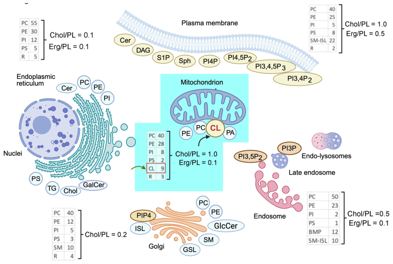
Figure 2.
Evolutionary continuity of cardiolipin from α-proteobacteria to mitochondria.
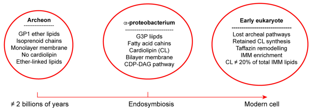
Figure 3.
The three CL platform modes: a unified overview.
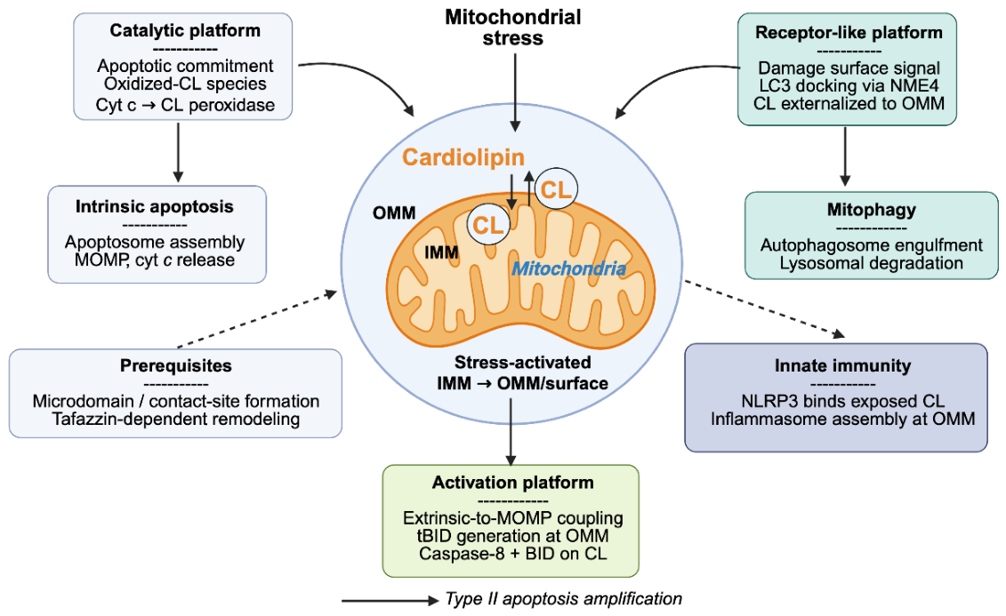
Figure 4.
NME4-driven CL externalization and selective mitophagy.
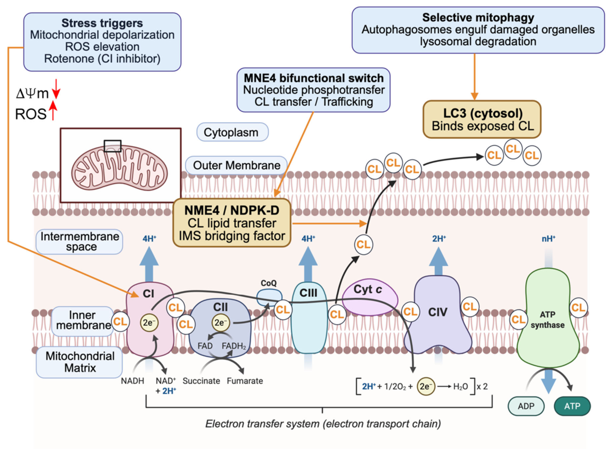
Figure 5.
CL microdomains as a caspase-8/BID activation scaffold coupling extrinsic death signals to MOMP.
Figure 5.
CL microdomains as a caspase-8/BID activation scaffold coupling extrinsic death signals to MOMP.
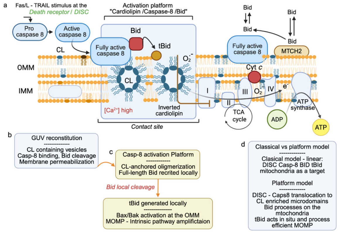
Figure 6.
NLRP3/inflammasome / innate immunity axis.
Disclaimer/Publisher’s Note: The statements, opinions and data contained in all publications are solely those of the individual author(s) and contributor(s) and not of MDPI and/or the editor(s). MDPI and/or the editor(s) disclaim responsibility for any injury to people or property resulting from any ideas, methods, instructions or products referred to in the content. |
© 2026 by the authors. Licensee MDPI, Basel, Switzerland. This article is an open access article distributed under the terms and conditions of the Creative Commons Attribution (CC BY) license (http://creativecommons.org/licenses/by/4.0/).
Copyright: This open access article is published under a Creative Commons CC BY 4.0 license, which permit the free download, distribution, and reuse, provided that the author and preprint are cited in any reuse.


