

Submitted:
03 July 2026
Posted:
07 July 2026
You are already at the latest version
Abstract
Fungi recovered from human fecal samples represent an understudied component of the gastrointestinal microbiome, yet fungi can play important roles in health and disease, particularly in hospitalized and immunocompromised patients. Here, we report the culture-based characterization of fungi isolated from fecal samples of 39 hospitalized patients with nosocomial diarrhea in Mexico City. Fungal growth was observed in approximately 40% of samples, yielding 26 isolates: 20 yeasts and 6 filamentous fungi. The predominant genera were Candida (mainly C. albicans) and Rhodotorula, with less common species including Nakaseomyces glabratus, Lodderomyces elongisporus, and yeasts previously explored for probiotic applications, Meyerozyma and Metschnikowia spp. Among filamentous fungi, Paecilomyces variotii and multiple Penicillium species were identified. Phenotypic assays demonstrated that several yeast isolates tolerated gut-associated stress conditions, including elevated temperature, bile salts, and oxidative stress, while thermotolerant filamentous fungi remained viable at temperatures up to 42°C. Cell-free supernatants and organic extracts of selected thermotolerant filamentous fungi showed moderate cytotoxic activity against non-differentiated Caco-2 cells.
Keywords:
mycobiota
; hospitalized patients
; nosocomial diarrhea
; culturable fungi
; metabolomics
; cytotoxicity
; Mexico
1. Introduction
Fungi play critical ecological roles in diverse environments, acting as decomposers, mutualists, and pathogens [1]. In the human gastrointestinal tract, fungi constitute approximately 0.1% of the total gut microbiota and form a distinct community known as the gut mycobiome [2]. Although less diverse than the bacterial microbiome, the mycobiome includes species that persist among individuals, suggesting the existence of recurrent fungal taxa in the gastrointestinal tract. Ten fungal genera are consistently found in the human gut, including Candida species (mainly Candida albicans), Saccharomyces, Penicillium, Aspergillus, Cryptococcus, Malassezia, Cladosporium, Galactomyces, Debaryomyces, and Trichosporon [3,4]. Distinguishing true gut symbionts from transient fungi remains a key challenge in the field [5,6].
The functional role of the gut mycobiome in intestinal homeostasis and disease is still being elucidated. Gut fungi are thought to contribute to immune system training and gastrointestinal homeostasis [7,8]. Conversely, the overgrowth or expansion of opportunistic fungal species can disrupt host immunity through toxin production and inflammatory signaling [9]. Alterations in gut fungal communities have been associated with a range of conditions, including metabolic disorders, inflammatory bowel diseases (IBDs), and colorectal adenomas [10]. Additionally, bacteria and fungi interact through multiple mechanisms within the gut, and changes in one kingdom may reciprocally affect the other [9].
Antibiotic treatment can dramatically alter the bacterial microbiome, creating a permissive environment for the expansion of pathogens such as Clostridioides difficile (CDI) [11] but also fungi. Notably, co-colonization with Candida albicans has been observed in patients with CDI, and experimental evidence suggests that fungal-bacterial interactions may modulate the course of infection [12,13]. These observations highlight the potential relevance of fungi in the context of gut bacterial infections, though the mechanisms underlying these interactions remain to be fully characterized.
Changes in fungal community composition have been documented in association with gut inflammation, immune suppression, and antibiotic use, and are thought to create conditions permissive to opportunistic fungal growth [9,10]. As the number of hospitalized and immunocompromised patients continues to grow, characterizing the fungi present in the gut during infection-associated diarrhea becomes increasingly relevant. Culture-dependent approaches remain essential for this purpose, as they allow phenotypic and metabolic characterization that sequencing-based methods alone cannot provide.
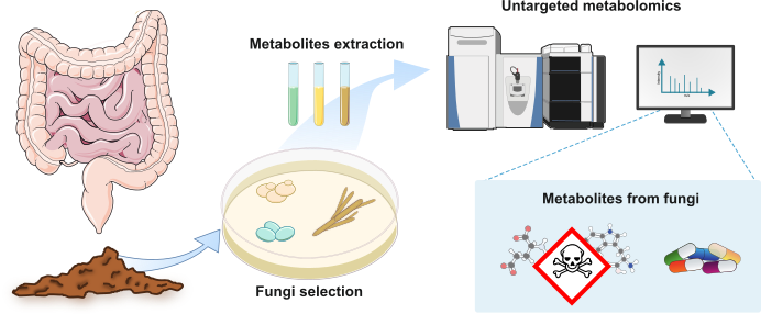
In this study, we used culture-dependent approaches to recover and characterize viable fungal isolates from fecal samples of hospitalized patients with nosocomial diarrhea in Mexico City. Rather than comparing fungal community composition across clinical groups, our objective was to explore the physiological traits, stress tolerance, cytotoxic potential, and secondary metabolite production of cultivable fungi recovered from this clinical setting. By combining fungal isolation, phenotypic characterization, and untargeted metabolomics, we aimed to generate an initial functional framework for future studies addressing the ecological and biological significance of gut-associated fungi.
2. Materials and Methods
2.1. Sample Collection
This study used fecal samples from patients hospitalized at a hospital in Mexico City. The samples were discarded biological material generated during routine hospital monitoring procedures and were transferred to our laboratory only after being designated for disposal. All samples were fully coded and contained no direct or indirect identifiers, and no information related to patient identity was accessible to the researchers. The study did not involve direct interaction with human subjects, nor the use of identifiable private information.
2.2. Isolation of Cultivable Fungal Species from Fecal Samples
Stool samples were diluted in PBS and plated on Brain Heart Infusion agar (BHI BD Bioxon™) supplemented with antibiotics (rifamycin, 32 µg/mL; gentamicin, 16 µg/mL; apramycin, 32 µg/mL; chloramphenicol, 32 µg/mL; nalidixic acid, 32 µg/mL; cycloserin, 10 µg/mL; cefoxitin, 32 µg/mL; and ampicillin, 32 µg/mL) and incubated aerobically at 27 °C for at least ten days with daily monitoring. Morphologically different yeast colonies and all molds were picked for further analysis. All selected isolates were further subcultivated to obtain pure colonies. Molds were subcultivated in Malt Extract Agar (agar, 15 g/L; malt extract, 40 g/L; peptone, 10 g/L) or Potato Dextrose Agar (agar, 15 g/L; dextrose, 20 g/L; potato extract, 4 g/L). Yeasts were subcultivated in YPD medium (10 g/L yeast extract, 20 g/L peptone, 20 g/L dextrose).
The isolates, grown on potato dextrose agar (PDA) or malt extract agar (MEA) for 48–72 h, were examined; colonial morphology and pigment production were recorded. The colony morphologies and pigmentation of the presumptive yeast isolates were also assessed on Candida chromogenic media (TM media™). The colors of each colony were recorded after 72 h of incubation.
2.3. DNA Extraction and Molecular Characterization of Fungal Isolates
Genomic DNA from five- to seven-day-old cultures of filamentous fungi was extracted from either liquid SMA broth or PDA culture plates. Molecular identification of isolates was performed by amplification and sequencing of the internal transcribed spacer (ITS) region and, when required for species-level assignment, the β-tubulin gene. Sequences were compared against the GenBank database, and phylogenetic trees were constructed in MEGA to confirm identity. The ITS sequences of all isolates have been deposited in NCBI GenBank under accession numbers PZ513273–PZ513288 (BioProject PRJEB107195).
2.4. Phenotypical Characterization of Fungal Isolates
Three independent experiments were performed with yeast and filamentous fungi to assess the effect of oxidative stress, pH, and bile salts on fungal growth. Cultures were exposed to H₂O₂ at 2 and 5 mM, pH values of 4, 6, 6.8, and 8, and taurocholate at 2 and 5%. The concentrations of H₂O₂ (2 and 5 mM) and taurocholate (2 and 5%) were selected based on previously reported physiologically relevant ranges in the gut lumen [15] and to allow comparison with prior studies on fungal stress tolerance. Growth was recorded from 72 to 120 h.
2.5. Antagonism Assays of Filamentous Fungi
For confrontation assays against Paecilomyces variotii YTC22, filamentous fungi were cultivated on SMA for 7 days. A 0.5 cm plug of each isolate was placed at each extreme of the plate, and plates were incubated at 29 °C for 7 days. This approach, adapted from classical fungal confrontation methods [16], was used as a qualitative screening tool to identify isolates exhibiting competitive interactions, which could then be prioritized for more quantitative analyses.
Fungal growth inhibition was evaluated by measuring mycelial area using ImageJ. Each test was performed in triplicate (n = 3).
2.6. Cell Culture and Cell Viability Assay
Caco-2 cells (ATCC HTB-37™) were cultured in DMEM containing 10% FBS, 5 µg/mL penicillin–streptomycin, and 5 mM sodium pyruvate, at 37 °C and 5% CO₂. Caco-2 cells (1 × 10⁴) were seeded in 96-well plates and cultured for 3 days post-confluence for the cell viability assay. It should be noted that Caco-2 cells were used in a non-differentiated state, as the goal of this initial screening was to assess general cytotoxic potential rather than epithelial barrier function. Cells were treated with cell-free supernatants or fungal organic extracts for 24 h. Prior to cell treatment, cell-free supernatants were adjusted to pH 7.0 using 1 M NaOH or 1 M HCl to minimize any cytotoxic effects attributable to pH differences rather than fungal metabolites. Cell viability was determined by the MTT assay. Experiments were repeated in triplicate across at least three independent experiments. Results were expressed as the mean percentage of viable cells ± standard deviation relative to untreated controls. Statistical significance was determined using one-way ANOVA followed by Tukey's post hoc test, with p < 0.05 considered significant.
2.7. Filamentous Fungi Culture and Extracts
Paecilomyces variotii YTC22, Penicillium chrysogenum YTC25, and Penicillium crustosum YTC26 were cultivated on 4 × PDA plates under aerobic conditions for 15 days at 37 °C. The mycelium was extracted with 1:1 CHCl₃:MeOH. Filtrates were dried under vacuum, and the resulting extracts were stored at −4 °C until further analysis.
2.8. LC–HRMS/MS, Dereplication, and Untargeted Metabolomics Analyses
Fungal organic extracts at 1.0 mg/mL in 1:1 dioxane–MeOH were subjected to LC–HRMS–MS/MS (ESI positive mode) on a Q Exactive mass spectrometer coupled with UPLC. Dereplication was performed against an in-house library of more than 750 authenticated standards. Untargeted metabolomics analyses were carried out using the GNPS platform. MZmine v3.8 parameters were set as follows: precursor ion mass tolerance, 0.02 Da; MS/MS fragment ion mass tolerance, 0.02; minimum matched peaks, 6; cosine score, 0.7. Annotations were reported at confidence levels 1 and 2 according to the Metabolomics Standards Initiative (<5 ppm).
2.9. Statistical Analysis
All data were analyzed using GraphPad Prism V7.0 (San Diego, CA, USA). For phenotypic characterization and interaction assays, results are presented as means ± standard deviation from three independent experiments. For cell viability assays, statistical significance was assessed by one-way ANOVA (p < 0.05).
3. Results
3.1. Cultivable Fungal Isolates From Fecal Samples of Patients With Diarrhea
Culturable fungi were recovered from 39 fecal samples obtained from hospitalized patients with nosocomial diarrhea. Fungal growth was observed in approximately 40% of samples, yielding 26 isolates that were subsequently purified and identified. These included twenty yeasts (74%) and six filamentous fungi (26%), indicating that yeasts represented the dominant cultivable fungal group recovered under the culture conditions employed (Figure 1A). The taxonomic identity of the fungal strains was determined by molecular sequencing of the ITS rDNA region, followed by BLAST searches and maximum likelihood phylogenetic analysis (Figure S1-S2). For certain filamentous fungi, β-tubulin gene sequencing was additionally performed to improve resolution (Figure S2). Yeast typing was conducted using ITS sequencing and chromogenic agar (Figure S3-S4).
Among the yeast isolates, Candida and Rhodotorula were the most prevalent genera (Figure 1A). Specifically, C. albicans and Rhodotorula mucilaginosa each accounted for 25% of all yeast isolates (Figure 1A). Less common Candida species, including Nakaseomyces glabratus (formerly Candida glabrata) was detected in approximately 10% of the samples, while Candida inopsicua, Candida parapsilosis, and Candida tropicalis, were each recovered from 5% of the samples (Figure 1A). In addition we detected Lodderomyces elongisporus, an infrequent pathogen whose true incidence may be underreported due to its close genetic similarity to C. parapsilosis [14]. Notably, yeasts previously explored for probiotic applications from the Meyerozyma and Metschnikowia genera were also isolated (Figure 1B).
For filamentous fungi, amplification and sequencing of the ITS and β-tubulin loci enabled the identification of the Penicillium and Paecilomyces genera (Figure 1C and Figure S1-S2)). Within Penicillium, five distinct taxa were detected: Penicillium mexicanum YTC23, P. chrysogenum YTC25, P. crustosum YTC26, and an unclassified Penicillium sp. YTC24, and P. brevicompactum YTC27. In the Paecilomyces, only P. variotii YTC22 was recovered.
3.2. Phenotyping of Intestinal Fungal Isolates
We conducted assays to evaluate fungal tolerance to temperature, oxidative stress, and bile salts as indicators of their capacity to withstand gut-associated environmental conditions.
As expected, a progressive reduction in growth ability was observed in correspondence with incubation temperature, which was particularly relevant for filamentous fungi (Figure 2). All yeasts could thrive at temperatures ranging from 29°C to 37°C. In contrast, the growth of R. mucilaginosa strains was impeded at 42°C (Figure 2A). At the same time, the majority of yeasts tolerated bile salts; R. mucilaginosa demonstrated only limited proliferation in media supplemented with 2% taurocholate (Figure 2A). In stress-tolerance assays, L. elongisporus proved more susceptible than C. albicans and C. tropicalis to bile acids, hydrogen peroxide, and pH fluctuations. These findings suggest differential tolerance to gut-associated stress conditions among the recovered isolates, with some species displaying physiological traits compatible with persistence in the intestinal environment.
While most environmental fungi cannot withstand mammalian body temperatures, recent years have seen the emergence of novel fungal pathogens, driven in part by the evolution of thermotolerance. Given that P. variotii is both thermotolerant and an opportunistic human pathogen, we assessed its competitive interactions with other filamentous isolates using a dual-culture confrontation assay on PDA. In pairwise cocultures with P. variotii YTC22, this strain generally outgrew its competitors, except P. crustosum YTC26, which markedly inhibited its expansion (Figure 3A).
3.3. Cytotoxic Activity Against Caco-2 Cells
To assess the cytotoxic potential of fungal metabolites from human feces, we performed MTT assays on Caco-2 cells using cell-free supernatants (CFS; 5, 10, 15, and 20 µL) and organic extracts (20 and 200 µg/mL). CFS were collected from ten yeast strains (YTC1, YTC3, YTC4, YTC8, YTC9, YTC10, YTC14, YTC15, YTC17, and YTC19) and three filamentous isolates (YTC22, YTC25, and YTC26). After 24 h of exposure to yeast CFS, no significant differences were found (Figure S5). In contrast, CFS from the filamentous fungi decreased viability by approximately 30% relative to controls (Figure 4). Consequently, we evaluated the cytotoxicity of organic extracts from YTC22, YTC25, and YTC26 at 20 and 200 µg/mL, observing moderate toxicity for the P. variotii extract at 200 µg/mL (Figure 5A). To further define its concentration-response profile, P. variotii extracts were tested over a concentration series ranging from 0.4 µg/mL to 200 µg/mL (Figure 5B).
3.4. Metabolomics Analysis
Emerging evidence suggests that fungi associated with the gastrointestinal tract produce diverse metabolites, some of which have reported antimicrobial, cytotoxic, neuromodulatory, or immunomodulatory activities. Whether these compounds are produced in sufficient concentrations in the gut to exert biological effects remains to be determined. Both endogenous fungal metabolites and those derived from dietary fungi may act as bioactive signals, yet their roles remain poorly characterized. Therefore, we performed a dereplication analysis and untargeted metabolomic profiling on three thermotolerant filamentous fungi isolated from human feces to elucidate their metabolic repertoires.
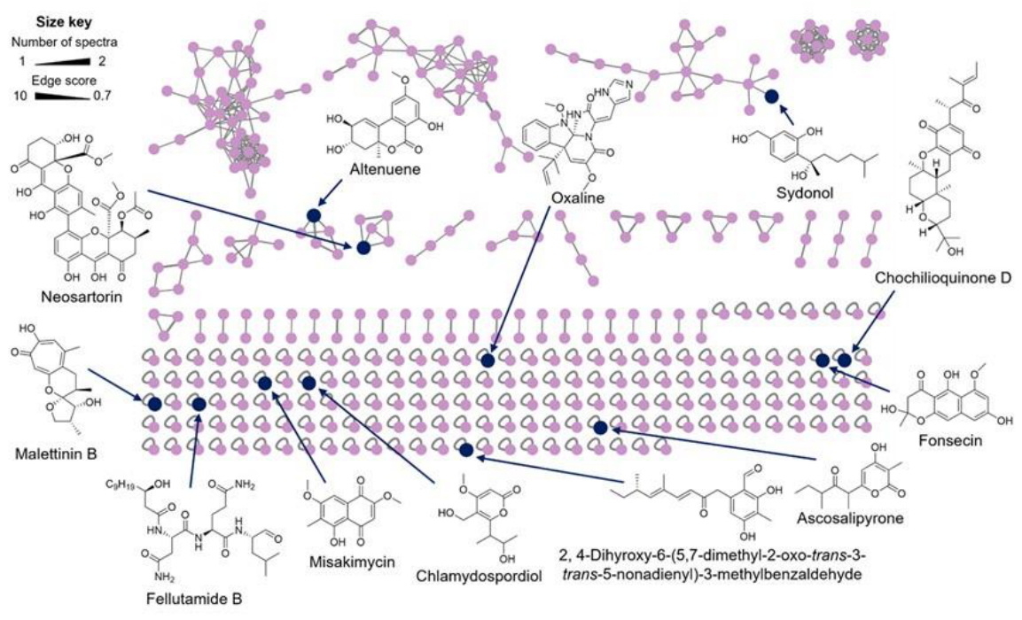
The metabolic profile of each fungus during growth showed distinct patterns of molecule production (Table 1). Paecilomyces variotii YTC22 metabolite features observed in the HRMS-MS/MS data were arranged in a molecular network of 369 nodes, grouped into 20 clusters (3 nodes per cluster, 23 with 2 nodes, and 164 singletons). Dereplication analysis using the in-house database of fungal mycotoxins revealed the presence of S-sydonol, altenuene, fellutamide B, neosartorin, ascosalipyrone, 6-[(3E,5E,7S)-5,7-dimethyl-2-oxonona-3,5-dienyl]-2,4-dihydroxy-3-methylbenzaldehyde, misakimycin, fonsecin, chochilioquinone D, malettinin B, and chlamydospordiol, while by GNPS analysis only oxaline was detected (Figure 6). The molecular networking of Penicillium chrysogenum YTC25 showed features arranged in 167 nodes grouped into seven clusters; the metabolites talarolutin B and phomopsidin MK8383 were detected. Finally, the molecular networking of Penicillium crustosum YTC26 consisted of 344 nodes grouped into 21 clusters; meleagrin, trichothecin, paxilline, hypothemycin, and roquefortine C were identified. The annotated secondary metabolites detected in these isolates and their previously reported biological activities are summarized in Table 2.
4. Discussion
The present study provides a culture-based characterization of fungi recovered from fecal samples of hospitalized patients with nosocomial diarrhea. Although culture-dependent methods capture only a subset of fungal diversity, they permit isolation of viable organisms for downstream physiological and metabolomic analyses that cannot be performed using sequencing approaches alone.
In healthy adults, the gut mycobiome is characterized by low species richness and is predominantly composed of yeast, especially members of the Saccharomyces, Malassezia, and Candida genera. In our cohort of patients with nosocomial diarrhea, yeasts remained dominant but with a distinctly altered composition: Candida and Rhodotorula species prevailed, and notably, no Saccharomyces spp. were detected. Although C. albicans is the archetypal core gut yeast in healthy individuals [10], environmental yeasts such as Rhodotorula spp., commonly transmitted via air and food, emerged as the second-most-abundant genus among our isolates [18]. Prior work has reported intestinal Rhodotorula colonization in up to 5% of healthy children and 12% of young adults [10]. Our data demonstrates its recovery from fecal samples of hospitalized patients with nosocomial diarrhea, although its status as a stable intestinal colonizer remains uncertain. Less prevalent opportunistic yeasts, including N. glabratus, C. inospicua, C. parapsilosis, and C. tropicalis, each accounted for approximately 5% of samples, aligning with their sporadic but clinically relevant presence in the gastrointestinal tract [10,20]. In particular, immunocompromised patients often experience fungemia caused by these same Candida species [18].
We also detected L. elongisporus, a rarely reported opportunist often misidentified as C. parapsilosis due to phenotypic similarity [14]. Its presence in fecal samples likely reflects antibiotic treatment and nosocomial exposures. Notably, we recovered yeasts belonging to Meyerozyma and Metschnikowia, genera that have attracted interest because of their reported probiotic-associated properties in experimental systems. Their ecological significance in the human gastrointestinal tract remains poorly understood and warrants further investigation.
Filamentous fungi are relatively uncommon in both healthy and diseased adult guts. In healthy individuals, environmental and food-associated genera, such as Cladosporium, Penicillium, and Aspergillus, are occasionally detected, though their roles in the gut remain speculative. In contrast, our cohort yielded only Penicillium species as filamentous isolates. Growth-temperature assays showed that two of five Penicillium isolates thrived at 37°C, while P. variotii isolates grew at 42°C, highlighting their thermotolerance. Although these molds are often dismissed as contaminants, they are increasingly implicated in invasive infections in both immunocompromised and immunocompetent patients. For instance, P. variotii has been associated with 59 reported human infections through 2020, exhibiting a 28.8% overall mortality and accounting for ten direct deaths [18].
Functional assays using yeast cell-free supernatants (CFS) revealed no cytotoxicity against Caco-2 cells. While supernatants from the thermotolerant filamentous strains P. variotii YTC22, P. chrysogenum YTC25, and P. crustosum YTC26 each decreased cell viability by approximately 30%. When tested as organic extracts, only the fraction derived from P. variotii YTC22 retained cytotoxic activity, suggesting that critical bioactive compounds were lost during extract processing.
It should also be noted that the culture conditions used to generate the CFS may not fully replicate the stressful environment of the gut, where nutrient limitation, pH shifts, and microbial competition could alter metabolite secretion profiles. Future studies under gut-simulating conditions may reveal a broader or different cytotoxic repertoire.
Untargeted metabolomic profiling of the three thermotolerant filamentous fungi revealed a diverse repertoire of secondary metabolites previously reported in the literature to possess antimicrobial, cytotoxic, neuroactive, or immunomodulatory activities. These findings highlight the metabolic versatility of the recovered isolates and suggest that fungi present in fecal samples may represent a previously underexplored source of bioactive molecules.
Several annotated compounds, including roquefortine C, paxilline, meleagrin, fellutamide B, hypothemycin, and neosartorin, have been associated with biological activities in experimental systems. However, it is important to emphasize that the present study does not establish whether these metabolites are produced in the human gastrointestinal tract, nor whether they reach concentrations sufficient to influence host physiology or microbial community structure. Consequently, any potential effects on intestinal homeostasis, epithelial function, immune responses, or microbial interactions remain speculative.
The moderate cytotoxicity observed for extracts and cell-free supernatants of selected filamentous fungi supports the notion that some recovered isolates produce biologically active compounds under laboratory conditions. Nevertheless, the specific metabolites responsible for these effects were not identified, and additional studies will be required to establish causal links between metabolite production and biological activity.
More broadly, these findings illustrate the value of combining culture-dependent approaches with metabolomic analyses to explore the functional potential of the fungi recovered from fecal samples. Future work integrating fungal isolation, culture-independent community profiling, and direct metabolomic analysis of fecal samples will be necessary to determine whether fungal secondary metabolites contribute to host physiology or microbial ecology within the gastrointestinal tract.
This study has several limitations. First, no healthy or hospitalized non-diarrheal control groups were included, preventing assessment of whether the recovered fungal taxa are specifically associated with nosocomial diarrhea or simply reflect background colonization. Second, detailed clinical metadata, including antibiotic exposure, immune status, were not available for analysis. Third, culture-dependent approaches recover only a fraction of the fungal diversity present in fecal samples and may favor fast-growing organisms. Finally, metabolomic analyses were performed under laboratory culture conditions and do not demonstrate production of the detected metabolites in vivo. Future studies integrating clinical metadata, culture-independent sequencing, and direct metabolomic analyses of fecal samples will be required to address these limitations.
5. Conclusion
This exploratory study provides a culture-based characterization of fungi recovered from fecal samples of hospitalized patients with nosocomial diarrhea in Mexico City. Opportunistic yeasts, particularly Candida and Rhodotorula species, represented the dominant cultivable fungi, while a subset of thermotolerant filamentous fungi exhibited resistance to gut-associated stresses, moderate cytotoxic activity, and diverse secondary metabolite repertoires. Although the ecological and clinical significance of these findings remains to be determined, the recovered isolates constitute a valuable resource for future investigations into fungal-host and fungal-bacterial interactions in the gastrointestinal tract.
Funding
This research was funded by UNAM-DGAPA PAPIIT grant IA206823 (A.R.R.) and grant IN203923 (M.F.), and by FQ-PAIP grant 5000-9145 (M.F.).
Institutional Review Board Statement
This study used fecal samples that were discarded biological material generated during routine hospital diagnostic procedures. All samples were fully anonymized prior to receipt by the research team; no patient identifiers, clinical records, or private information were accessible to the researchers at any point. The study did not involve direct interaction with human subjects. Under applicable Mexican bioethics regulations (Ley General de Salud, Título Quinto; NOM-012-SSA3-2012), research conducted exclusively on discarded, non-identifiable biological material without patient contact is classified as minimal-risk.
Informed Consent Statement
Not applicable. This study used fully anonymized, discarded biological material; no patient identifiers were accessible to the researchers.
Data Availability Statement
The ITS nucleotide sequences generated in this study have been deposited in NCBI GenBank under accession numbers PZ513273–PZ513288 (BioProject PRJEB107195).
Conflicts of Interest
The authors declare no conflict of interest. The funders had no role in the design of the study; in the collection, analyses, or interpretation of data; in the writing of the manuscript; or in the decision to publish the results.
References
- Zhang, D.; Wang, Y.; Shen, S.; Hou, Y.; Chen, Y.; Wang, T. The mycobiota of the human body: a spark can start a prairie fire. Gut Microbes 2020, 11, 655–679. [Google Scholar] [CrossRef] [PubMed]
- Nash, A.K.; Auchtung, T.A.; Wong, M.C.; et al. The gut mycobiome of the Human Microbiome Project healthy cohort. Microbiome 2017, 5, 153. [Google Scholar] [CrossRef] [PubMed]
- Wu, X.; Xia, Y.; He, F.; Zhu, C.; Ren, W. Intestinal mycobiota in health and diseases: from a disrupted equilibrium to clinical opportunities. Microbiome 2021, 9, 60. [Google Scholar] [CrossRef] [PubMed]
- Ferrocino, I.; Ponzo, V.; Pellegrini, M.; Goitre, I.; Papurello, M.; Franciosa, I.; D'Eusebio, C.; Ghigo, E.; Cocolin, L.; Bo, S. Mycobiota composition and changes across pregnancy in patients with gestational diabetes mellitus (GDM). Sci. Rep. 2022, 12, 9192. [Google Scholar] [CrossRef] [PubMed]
- Suhr, M.J.; Hallen-Adams, H.E. The human gut mycobiome: pitfalls and potentials — a mycologist's perspective. Mycologia 2015, 107, 1057–1073. [Google Scholar] [PubMed]
- Lavrinienko, A.; Scholier, T.; Bates, S.T.; Miller, A.N.; Watts, P.C. Defining gut mycobiota for wild animals: a need for caution in assigning authentic resident fungal taxa. Anim. Microbiome 2021, 3, 75. [Google Scholar] [CrossRef] [PubMed]
- Kumamoto, C.A. The fungal mycobiota: small numbers, large impacts. Cell Host Microbe 2016, 19, 750–751. [Google Scholar] [CrossRef] [PubMed]
- Santus, W.; Devlin, J.R.; Behnsen, J. Crossing kingdoms: how the mycobiota and fungal-bacterial interactions impact host health and disease. Infect. Immun. 2021, 89, e00648-20. [Google Scholar] [CrossRef] [PubMed]
- Richard, M.L.; Sokol, H. The gut mycobiota: insights into analysis, environmental interactions and role in gastrointestinal diseases. Nat. Rev. Gastroenterol. Hepatol. 2019, 16, 331–345. [Google Scholar] [CrossRef] [PubMed]
- Strati, F.; Di Paola, M.; Stefanini, I.; et al. Age and gender affect the composition of fungal population of the human gastrointestinal tract. Front. Microbiol. 2016, 7, 1227. [Google Scholar] [CrossRef] [PubMed]
- Mullish, B.H.; Williams, H.R. Clostridium difficile infection and antibiotic-associated diarrhoea. Clin. Med. (Lond.) 2018, 18, 237–241. [Google Scholar] [CrossRef] [PubMed]
- van Leeuwen, P.T.; van der Peet, J.M.; Bikker, F.J.; Hoogenkamp, M.A.; Oliveira Paiva, A.M.; Kostidis, S.; Mayboroda, O.A.; Smits, W.K.; Krom, B.P. Interspecies interactions between Clostridium difficile and Candida albicans. mSphere 2016, 1, e00187-16. [Google Scholar] [CrossRef] [PubMed]
- Lambooij, J.M.; Hoogenkamp, M.A.; Brandt, B.W.; Janus, M.M.; Krom, B.P. Fungal mitochondrial oxygen consumption induces the growth of strict anaerobic bacteria. Fungal Genet. Biol. 2017, 109, 1–6. [Google Scholar] [CrossRef] [PubMed]
- Wang, Y.; Xu, J. Lodderomyces elongisporus: an emerging human fungal pathogen. PLoS Pathog. 2023, 19, e1011613. [Google Scholar] [CrossRef] [PubMed]
- Garrido-Bazán, V.; Jaimes-Arroyo, R.; Sanchez, O.; Lara-Rojas, F.; Aguirre, J. SakA and MpkC stress MAPKs show opposite and common functions during stress responses and development in Aspergillus nidulans. Front. Microbiol. 2018, 9, 2518. [Google Scholar] [CrossRef] [PubMed]
- Steyaert, J.M.; Weld, R.J.; Mendoza-Mendoza, A.; Stewart, A. Reproduction without sex: conidiation in the filamentous fungus Trichoderma. Microbiology 2010, 156, 2887–2900. [Google Scholar] [CrossRef] [PubMed]
- Brunetti, G.; Giuliani, A.; Navazio, A.S.; Paradisi, C.; Raponi, F.; Conti, L.A.; Raponi, G. Candida gut colonization, yeast species distribution, and biofilm production in Clostridioides difficile infected patients. Braz. J. Microbiol. 2021, 52, 1845–1852. [Google Scholar] [CrossRef] [PubMed]
- Sprute, R.; Salmanton-Garcia, J.; Sal, E.; et al. Characterization and outcome of invasive infections due to Paecilomyces variotii: analysis from the FungiScope® registry. J. Antimicrob. Chemother. 2021, 76, 765–774. [Google Scholar] [PubMed]
- Vargas-Espindola, L.A.; Cuervo-Maldonado, S.I.; Enciso-Olivera, J.L.; et al. Fungemia in hospitalized adult patients with hematological malignancies: epidemiology and risk factors. J. Fungi 2023, 9, 191. [Google Scholar] [CrossRef]
- Hof, H. Rhodotorula spp. in the gut — foe or friend? GMS Infect. Dis. 2019, 7, Doc02. [Google Scholar] [PubMed]
- Aron, A.T.; Gentry, E.C.; McPhail, K.L.; et al. Reproducible molecular networking of untargeted mass spectrometry data using GNPS. Nat. Protoc. 2020, 15, 1954–1991. [Google Scholar] [CrossRef] [PubMed]
- Nothias, L.F.; Petras, D.; Schmid, R.; et al. Feature-based molecular networking in the GNPS analysis environment. Nat. Methods 2020, 17, 905–908. [Google Scholar] [CrossRef] [PubMed]
- Sumner, L.W.; Amberg, A.; Barrett, D.; et al. Proposed minimum reporting standards for chemical analysis — Chemical Analysis Working Group (CAWG) Metabolomics Standards Initiative (MSI). Metabolomics 2007, 3, 211–221. [Google Scholar] [PubMed]
- Wang, M.; Carver, J.J.; Phelan, V.V.; et al. Sharing and community curation of mass spectrometry data with Global Natural Products Social Molecular Networking. Nat. Biotechnol. 2016, 34, 828–837. [Google Scholar] [CrossRef] [PubMed]
- Shannon, P.; Markiel, A.; Ozier, O.; Baliga, N.S.; Wang, J.T.; Ramage, D.; Amin, N.; Schwikowski, B.; Ideker, T. Cytoscape: a software environment for integrated models of biomolecular interaction networks. Genome Res. 2003, 13, 2498–2504. [Google Scholar] [CrossRef] [PubMed]
- Robert, V.; Vu, D.; Amor, A.B.; et al. MycoBank gearing up for new horizons. IMA Fungus 2013, 4, 371–379. [Google Scholar] [CrossRef] [PubMed]
- Al-Samarrai, T.H.; Schmid, J. A simple method for extraction of fungal genomic DNA. Lett. Appl. Microbiol. 2000, 30, 53–56. [Google Scholar] [CrossRef] [PubMed]
- Harju, S.; Fedosyuk, H.; Peterson, K.R. Rapid isolation of yeast genomic DNA: Bust n' Grab. BMC Biotechnol. 2004, 4, 8. [Google Scholar] [CrossRef] [PubMed]
- McCullough, M.J.; Clemons, K.V.; McCusker, J.H.; Stevens, D.A. Intergenic transcribed spacer PCR ribotyping for differentiation of Saccharomyces species and interspecific hybrids. J. Clin. Microbiol. 1998, 36, 1035–1038. [Google Scholar] [CrossRef] [PubMed]
- Kumar, S.; Stecher, G.; Tamura, K. MEGA7: Molecular Evolutionary Genetics Analysis Version 7.0 for Bigger Datasets. Mol. Biol. Evol. 2016, 33, 1870–1874. [Google Scholar] [CrossRef] [PubMed]
Figure 1.
Culturable mycobiota isolated from fecal samples of hospitalized patients with nosocomial diarrhea. (A) Pie charts showing the percentage of fungal genera and species identified by ITS sequencing. (B) Representative photographs of fungal colony growth on agar medium and Candida chromogenic agar. (C) Colony morphology and microscopic characteristics of filamentous fungi isolates.
Figure 1.
Culturable mycobiota isolated from fecal samples of hospitalized patients with nosocomial diarrhea. (A) Pie charts showing the percentage of fungal genera and species identified by ITS sequencing. (B) Representative photographs of fungal colony growth on agar medium and Candida chromogenic agar. (C) Colony morphology and microscopic characteristics of filamentous fungi isolates.
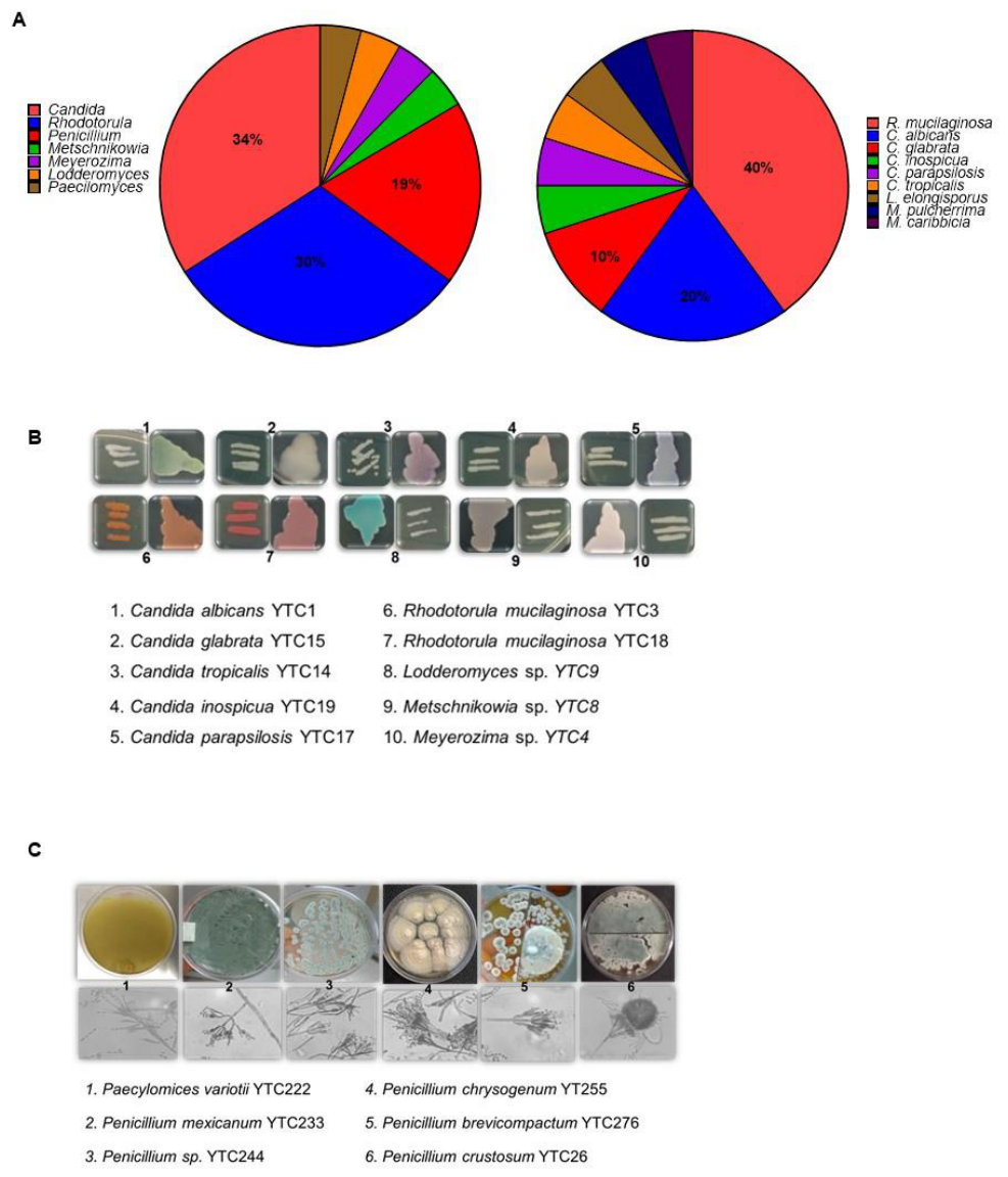
Figure 2.
In vitro fungal growth and thermotolerance assays. (A) Experimental plate design and representative photographs of filamentous fungi growth under tested conditions. (B) Growth of filamentous fungi under different stress conditions: P. variotii YTC22 was unaffected by all conditions tested; P. chrysogenum YTC25 showed reduced pigment production at 5% taurocholate; P. crustosum YTC26 exhibited severely impaired pigmentation at 37 °C. (C) Heatmap of yeast growth under different stress conditions; all yeasts were thermotolerant, although R. mucilaginosa showed reduced growth at 42 °C and pH 5. TCA, taurocholic acid.
Figure 2.
In vitro fungal growth and thermotolerance assays. (A) Experimental plate design and representative photographs of filamentous fungi growth under tested conditions. (B) Growth of filamentous fungi under different stress conditions: P. variotii YTC22 was unaffected by all conditions tested; P. chrysogenum YTC25 showed reduced pigment production at 5% taurocholate; P. crustosum YTC26 exhibited severely impaired pigmentation at 37 °C. (C) Heatmap of yeast growth under different stress conditions; all yeasts were thermotolerant, although R. mucilaginosa showed reduced growth at 42 °C and pH 5. TCA, taurocholic acid.
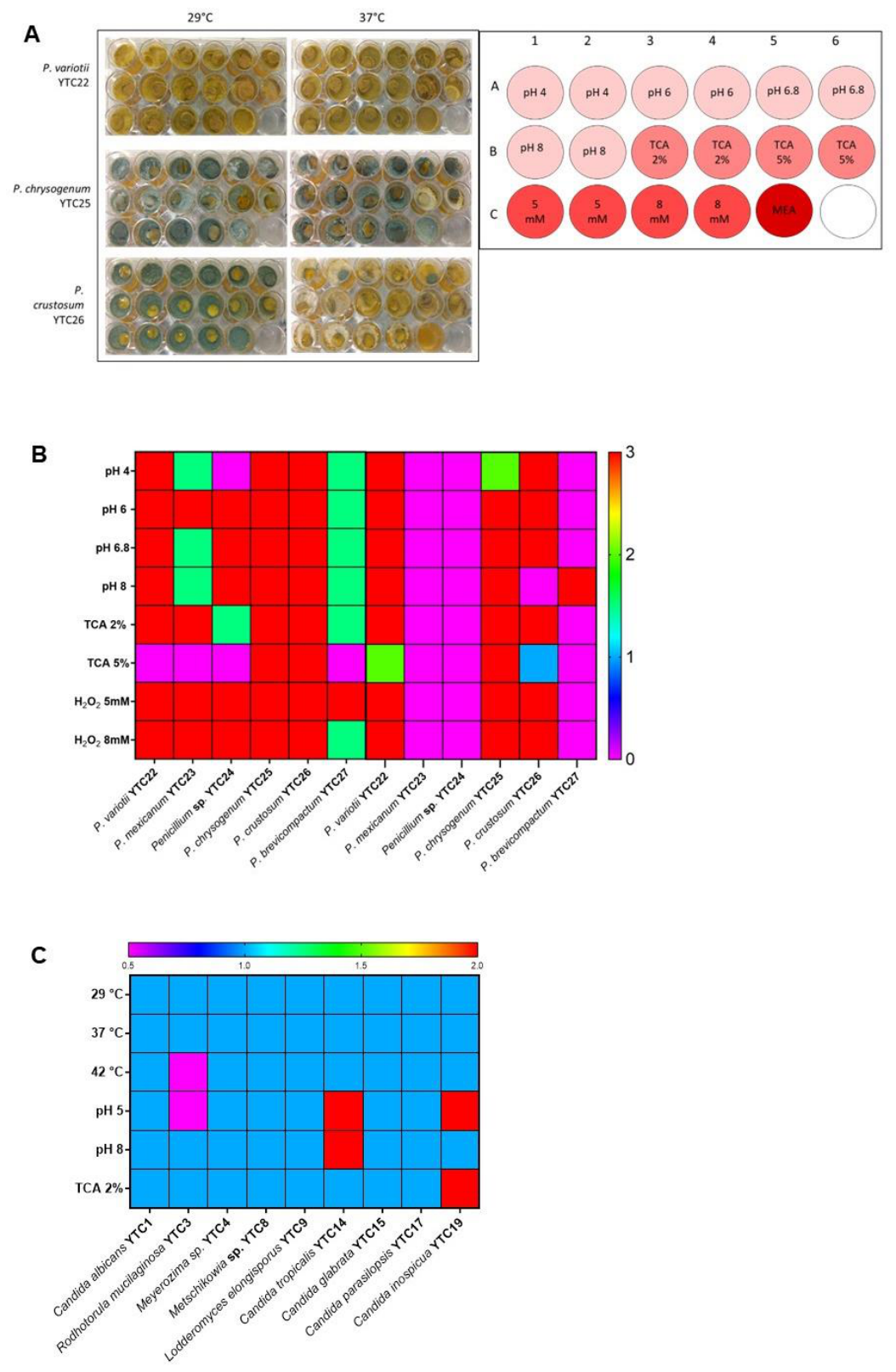
Figure 3.
Confrontation assays against P. variotii YTC22. Fungi were inoculated on PDA medium, each plug placed at opposite extremes of the Petri dish, and incubated at 24 °C for 72 h. (A) Representative photographs of the confrontation assay. P. variotii YTC22 inhibited the growth of most competitor strains; however, P. crustosum YTC26 inhibited the expansion of P. variotii YTC22 in confrontation. (B) Quantification of mycelial growth, expressed as mycelial diameter relative to P. variotii grown alone (control). Data represent the mean (±SD) of three independent experiments. Asterisk (*) indicates a statistically significant difference by one-way analysis of variance (ANOVA) (p ≤ 0.05) followed by the Tukey test.
Figure 3.
Confrontation assays against P. variotii YTC22. Fungi were inoculated on PDA medium, each plug placed at opposite extremes of the Petri dish, and incubated at 24 °C for 72 h. (A) Representative photographs of the confrontation assay. P. variotii YTC22 inhibited the growth of most competitor strains; however, P. crustosum YTC26 inhibited the expansion of P. variotii YTC22 in confrontation. (B) Quantification of mycelial growth, expressed as mycelial diameter relative to P. variotii grown alone (control). Data represent the mean (±SD) of three independent experiments. Asterisk (*) indicates a statistically significant difference by one-way analysis of variance (ANOVA) (p ≤ 0.05) followed by the Tukey test.
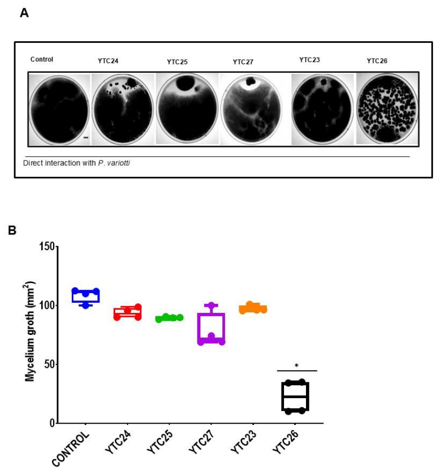
Figure 4.
Cytotoxicity of cell-free supernatants (CFS) of thermotolerant filamentous fungi against non-differentiated Caco-2 cells. Cytotoxic activity of CFS from (a) P. chrysogenum YTC25, (b) P. variotii YTC22, and (c) P. crustosum YTC26. Fungi were cultivated in MEA at 37 °C for seven days. CFS were adjusted to pH 7.0 prior to cell treatment. Cell viability was assessed by MTT assay after 24 h of exposure. Data represent the mean (±SD) of three independent experiments. Asterisk (*) indicates a statistically significant difference by one-way ANOVA (p ≤ 0.05).
Figure 4.
Cytotoxicity of cell-free supernatants (CFS) of thermotolerant filamentous fungi against non-differentiated Caco-2 cells. Cytotoxic activity of CFS from (a) P. chrysogenum YTC25, (b) P. variotii YTC22, and (c) P. crustosum YTC26. Fungi were cultivated in MEA at 37 °C for seven days. CFS were adjusted to pH 7.0 prior to cell treatment. Cell viability was assessed by MTT assay after 24 h of exposure. Data represent the mean (±SD) of three independent experiments. Asterisk (*) indicates a statistically significant difference by one-way ANOVA (p ≤ 0.05).
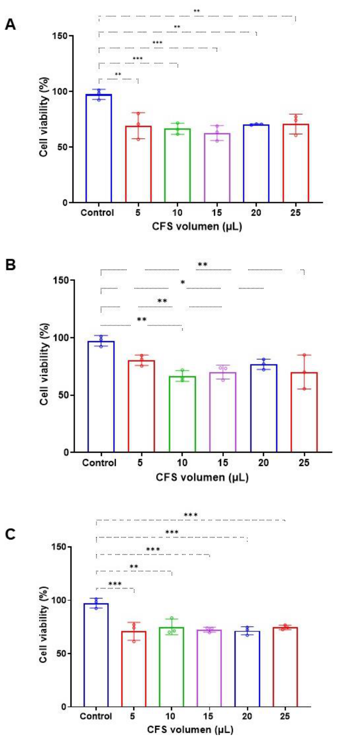
Figure 5.
Cytotoxicity of organic extracts of thermotolerant filamentous fungi against non-differentiated Caco-2 cells. (a) Cytotoxicity of organic extracts from P. variotii YTC22, P. chrysogenum YTC25, and P. crustosum YTC26 at 20 and 200 µg/mL, assessed by MTT assay. (b) Concentration-response curve of Caco-2 cell viability after treatment with P. variotii YTC22 organic extract at concentrations ranging from 0.4 to 200 µg/mL. Data represent the mean (±SD) of three independent experiments. Asterisk (*) indicates a statistically significant difference by one-way ANOVA (p ≤ 0.05).
Figure 5.
Cytotoxicity of organic extracts of thermotolerant filamentous fungi against non-differentiated Caco-2 cells. (a) Cytotoxicity of organic extracts from P. variotii YTC22, P. chrysogenum YTC25, and P. crustosum YTC26 at 20 and 200 µg/mL, assessed by MTT assay. (b) Concentration-response curve of Caco-2 cell viability after treatment with P. variotii YTC22 organic extract at concentrations ranging from 0.4 to 200 µg/mL. Data represent the mean (±SD) of three independent experiments. Asterisk (*) indicates a statistically significant difference by one-way ANOVA (p ≤ 0.05).
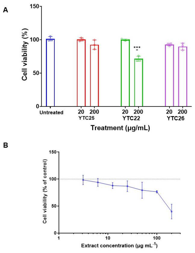
Figure 6.
Molecular network of the P. variotii YTC22 organic extract. Nodes represent MS/MS spectra; edges connect spectra with cosine similarity ≥ 0.7 and at least six matched fragment ions. Colored nodes indicate compounds annotated by dereplication against an in-house library (confidence levels 1–2, <5 ppm) or by GNPS spectral matching.
Figure 6.
Molecular network of the P. variotii YTC22 organic extract. Nodes represent MS/MS spectra; edges connect spectra with cosine similarity ≥ 0.7 and at least six matched fragment ions. Colored nodes indicate compounds annotated by dereplication against an in-house library (confidence levels 1–2, <5 ppm) or by GNPS spectral matching.
Disclaimer/Publisher’s Note: The statements, opinions and data contained in all publications are solely those of the individual author(s) and contributor(s) and not of MDPI and/or the editor(s). MDPI and/or the editor(s) disclaim responsibility for any injury to people or property resulting from any ideas, methods, instructions or products referred to in the content. |
© 2026 by the authors. Licensee MDPI, Basel, Switzerland. This article is an open access article distributed under the terms and conditions of the Creative Commons Attribution (CC BY) license.
Copyright: This open access article is published under a Creative Commons CC BY 4.0 license, which permit the free download, distribution, and reuse, provided that the author and preprint are cited in any reuse.


