

Submitted:
05 July 2026
Posted:
07 July 2026
You are already at the latest version
Abstract
Reducing sodium in meat products is nutritionally desirable, but salt reduction often weakens myofibrillar protein gelation and reduces texture quality and water retention. This study investigated whether konjac glucomannan (KGM) and L-lysine (Lys) could improve the quality of beef myofibrillar protein (MP) gels prepared under low-salt conditions and explored the related structural mechanisms. The results indicated that reducing NaCl from 0.6 to 0.2 M decreased water-holding capacity (WHC), increased cooking loss, and produced a loose gel network. Compared with the 0.2 M NaCl group, the combined Lys-KGM treatment increased WHC from 28.53% to 56.70% and reduced cooking loss to 22.19% (P < 0.05). Texture analysis showed that Lys-KGM increased hardness and springiness by 69.40% and 80.43%, respectively (P < 0.05). LF-NMR indicated a higher proportion of immobilized water and reduced water mobility in the combined treatment. Structural analyses suggested that Lys mainly promoted reactive sulfhydryl exposure and protein association, whereas KGM contributed through hydration and hydrogen-bond interactions. Their combined use altered secondary structure, intermolecular force distribution, and gel network organization. Molecular docking and molecular dynamics simulations provided supporting evidence for different interaction patterns between myosin and Lys/KGM. These results suggested that Lys and KGM might help maintain the quality of low-salt meat protein gels and provide a formulation basis for reduced-sodium meat products.
Keywords:
beef myofibrillar proteins
; reduced-sodium meat products
; konjac glucomannan
; L-lysine
; intermolecular interaction
; gel properties
1. Introduction
The gelling properties of myofibrillar proteins (MP) largely determine the texture and water-holding capacity (WHC) of meat products (Gao et al., 2023; Li et al., 2019; Xu & Xu, 2021). In gel-type meat processing, 2%–3% NaCl is commonly used to promote MP solubilization and heat-induced gel network formation, thereby helping maintain desirable sensory and textural properties (Cao, Zhu, et al., 2023; Zhang, Li, et al., 2022). Nevertheless, excessive sodium intake is closely related to hypertension and cardiovascular diseases (Xiang et al., 2025). The World Health Organization recommends reducing population salt intake by 30% and limiting daily salt intake to no more than 5 g for adults. As processed meat products contribute approximately 20%–30% of total dietary sodium intake, the development of reduced-sodium meat products has received increasing attention (Zhang, Guo, et al., 2022). However, NaCl reduction generally decreases MP solubilization and consequently weakens its gel-forming ability (Shi et al., 2024). Maintaining MP gel quality under low-salt conditions remains a challenge.
L-Lysine (Lys) has been widely used in meat processing to enhance flavor and improve the functional properties of meat proteins (Zhang, Guo, et al., 2022). Lys can enhance MP solubility and induce changes in the secondary and tertiary structures of MP (Cao et al., 2021; Li et al., 2022). These structural modifications may further regulate intermolecular interactions and thermal aggregation behavior of MP, thereby promoting gel formation under low-salt conditions. (Wang et al., 2021; Xu & Xu, 2021; Guo et al., 2022; Xie et al., 2024). Nevertheless, the improvement in gel quality by Lys alone remains limited under reduced-salt conditions, especially in maintaining gel integrity and water retention. Therefore, additional strategies are still required to improve low-salt MP gels.
Plant-derived polysaccharides have gained interest as functional ingredients for improving meat quality under low-salt conditions (Lee et al., 2023). Konjac glucomannan (KGM), a water-soluble polysaccharide mainly composed of D-glucose and D-mannose linked by β-1,4-glycosidic bonds, possesses excellent hydration, thickening, and gel-forming properties (Cao, Zhao, et al., 2023; Xiong et al., 2009). KGM has been reported to increase the gel properties of MP systems by enhancing water retention, modulating protein aggregation behavior, and contributing to a denser and more stable gel structure during heating (Zhuang et al., 2021; Gao et al., 2022; Li, Lin, et al., 2024). Therefore, the combination of Lys and KGM may provide complementary regulation for low-salt MP gelation through simultaneous modulation of protein molecular behavior and gel network structure. Such an ingredient-based strategy may help maintain texture and water retention in reduced-sodium meat products. However, the combined effects of Lys and KGM on intermolecular interactions, protein conformation, and gel network formation in low-salt MP systems remain unclear.
Therefore, this study aimed to evaluate the combined effects of Lys and KGM on the quality and structural characteristics of beef MP gels under low-salt conditions. WHC, cooking loss, water distribution, texture, rheological behavior, and microstructure were determined to assess gel quality, while sulfhydryl groups, surface hydrophobicity, FT-IR spectra, and intermolecular forces were used to characterize protein structural changes. Molecular docking and molecular dynamics simulations were further used as supporting tools to explore possible interactions between myosin and Lys/KGM. The results may support the combined use of Lys and KGM as functional ingredients to improve the quality of reduced-sodium meat products.
2. Materials and Methods
2.1. Materials and Chemicals
Fresh beef Longissimus dorsi muscle was obtained from Suguo Supermarket (Yangzhou, China). Lys and KGM were obtained from Aladdin Biochemical Technology Company (Shanghai, China). All chemicals were of analytical grade.
2.2. Extraction of Mp
MP extraction was performed according to Xu et al. (2024) with slight modifications. All operations were performed at 4 °C. Beef samples were homogenized with buffer at a ratio of 1:3 (w/v). Four cycles of homogenization (30 s) and centrifugation (2000 × g, 10 min) were performed. 0.01 M phosphate-buffered saline (PBS) was used for the first two cycles, and 0.1 M NaCl buffer was used for the last two cycles. The precipitate was collected for use.
2.3. Sample Preparation
The MP pellet was resuspended in five solutions prepared with 20 mM PBS (pH 7.0): 0.6 M NaCl, 0.2 M NaCl, 0.2 M NaCl with 0.4% Lys, 0.2 M NaCl with 0.5% KGM, and 0.2 M NaCl with 0.4% Lys and 0.5% KGM. The concentrations of Lys and KGM used in the treatment groups were selected based on preliminary trials. The suspensions were then kept at 4 °C until use. For gel preparation, 5 g of MP suspension (40 mg/mL) was heated from 25 °C to 75 °C at 1 °C/min and held at 75 °C for 30 min, followed by cooling in an ice-water bath for 20 min.
2.4. Cooking Loss
The combined weight of the centrifuge tube and sample was measured before heating (m1) and after
heating (m2). Cooking loss (%) was calculated as:
where m0 refers to the weight of empty centrifuge tube.
2.5. Whc of Gels
Gels were blotted to remove surface moisture and centrifuged at 5000 × g for 10 min at 4 °C. The weight of the empty tube (M0), tube with sample before centrifugation (M1), and tube with sample after centrifugation (M2) were recorded. WHC (%) was calculated as follows:
2.6. Low-Field Nuclear Magnetic Resonance (Lf-Nmr) of Mp Gels
Gel samples were cut into cylinders (diameter 1 cm) and analyzed using a LF-NMR analyzer (AccuFat-1050, Magmai, Nanjing, China) with a Carr-Purcell-Meiboom-Gill (CPMG) pulse sequence. Each sample was scanned 16 times with a 2 s interval and a receiver gain of 4. Transverse relaxation time (T2) distributions and response peak area of samples were obtained using the NMR inversion software.
2.7. Texture Analysis of Mp Gels
Sample texture was measured with a TA-XT2i texture analyzer (Stable Micro Systems, UK) equipped with a P/50 probe. Before testing, the gels were cut into 1 cm cubes. The samples were compressed twice to 50% deformation. The pre-test, test, and post-test speeds were 120, 60, and 60 mm/min, respectively, and the trigger force was 5 g.
2.8. Dynamic Rheological Measurements
The rheological properties of MP suspensions was measured using a rheometer (MARS III, Thermo Fisher Scientific, Germany). The concentration of MP was adjusted to 10 mg/mL. Measurements were conducted using a 35 mm parallel plate with a 1 mm gap. Silicone oil was spread around the sample to minimize moisture loss during testing. Frequency sweep, shear rate sweep, and temperature sweep tests were performed within the linear viscoelastic region. Frequency-dependent changes in G′ and G″ were monitored at 25 °C over 0.1–16 Hz with 1% strain. The apparent viscosity was measured over a shear rate range of 0.1–100 s⁻¹ at 25 °C. Changes in G′, G″, and tan δ were monitored during heating from 20 °C to 80 °C at a rate of 1 °C/min.
2.9. Scanning Electron Microscopy (Sem)
Samples (2 mm × 2 mm × 1 mm) were immersed in 2.5% glutaraldehyde at 4 °C for 12 h. Then, the samples were rinsed thrice with 0.1 M PBS (pH 7.2) and dehydrated stepwise in 50%, 70%, 80%, 90%, and 100% ethanol for 15 min at each concentration. After freeze-drying, the samples were gold-coated and observed using a GeminiSEM 300 (Carl Zeiss, UK) at 5 kV.
2.10. Total and Reactive Sulfhydryl Groups
Sulfhydryl content was determined following Guo et al. (2024) with modifications. Total sulfhydryl (T-SH) content was measured by mixing 1 mL of MP solution (5 mg/mL) with 2 mL of Tris-glycine buffer containing 10.4 mg/mL Tris, 6.9 mg/mL glycine, 1.2 mg/mL EDTA, and 480 mg/mL urea (pH 8.0). Then, 0.02 mL of Ellman’s reagent was added, and the mixture was incubated at 25 °C for 5 min in the dark. Absorbance was recorded at 412 nm.
Reactive sulfhydryl (R-SH) content was measured in the same way, except that urea was removed from the buffer. The sulfhydryl contents were expressed as μmol/g protein using a molar extinction coefficient of 13,600 M⁻¹ cm⁻¹.
2.11. Surface Hydrophobicity of MP
Surface hydrophobicity was measured using bromophenol blue (BPB) binding (Guo et al., 2023). 1 mL of MP suspension (5 mg/mL) was mixed with 0.2 mL BPB solution (1 mg/mL), vortexed for 10 min, and centrifuged at 4000 × g for 15 min at 4 °C. The supernatant was diluted 10-fold with PBS before measuring absorbance at 595 nm. Surface hydrophobicity was determined as the amount of BPB bound per mg protein using the following equation.
where A0 and A1 represent the absorbance values measured at 595 nm for the control and sample, respectively.
2.12. Fourier Transform Infrared Spectroscopy (FT-IR)
Lyophilized samples were ground with dried KBr (1:50, w/w), pressed into tablets (1 mm thick), and analyzed using an FT-IR spectrometer (Cary 610/670, Varian, USA). The spectra were acquired in the range of 4000-400 cm⁻¹ with 32 scans.
2.13. Determination of the intermolecular interactions
Intermolecular forces in MP gels were determined according to Xu et al. (2022) with minor modifications. 2 g of gel samples was homogenized with 18 mL of five extraction solutions: SA, 0.05 M NaCl; SB, 0.6 M NaCl; SC, SB with 1.5 M urea; SD, SB with 8 M urea; and SE, SB with 8 M urea and 0.05 M β-mercaptoethanol. The mixtures were heated at 80 °C for 30 min and then cooled to 25 °C. The supernatant was collected for protein quantification after centrifugation at 5000 × g for 15 min. Differences in solubility were used to estimate ionic bonds, hydrophobic interactions, hydrogen bonds, and disulfide bonds.
2.14. Molecular Docking
Interactions between KGM/Lys and myosin were analyzed by AutoDock Vina 1.1.2. The bovine myosin sequence was obtained from UniProtKB, with accession number E1BPK6. Lys (CAS: 56-87-1) and KGM (CAS: 37220-17-0) were used as ligands. The docking pose with the lowest binding energy was selected for analysis, and the interaction structures were visualized using Discovery Studio 2019.
2.15. Molecular Dynamics Simulation
Molecular dynamics simulations were conducted with GROMACS 2022 according to Guo et al. (2024) with minor changes. The stability and conformational behavior of the complexes were evaluated using SASA, RMSD, Rg, RMSF, and hydrogen bond analysis. Binding free energy was obtained using the MM/PBSA approach (Wang et al., 2022).
2.16. Statistical Analysis
All experiments were performed in triplicate, and results are shown as mean ± standard deviation (SD). Statistical differences were tested with one-way ANOVA and Duncan’s multiple range test (P < 0.05) using SPSS 26.0 (IBM Corp., Armonk, NY, USA). Figures were plotted with GraphPad Prism 10 (GraphPad Software Inc., Boston, MA, USA), Origin 9.0 software (OriginLab, Northampton, MA, USA).
3. Results and Discussion
3.1. Cooking Loss and Whc
Cooking loss and WHC are commonly used to evaluate water retention and overall quality of heat-induced MP gels (Huang et al., 2024). As presented in Figure 1, reducing NaCl from 0.6 M to 0.2 M increased cooking loss and decreased WHC, which might be attributed to insufficient MP solubilization and weakened intermolecular interactions under low-salt conditions, thereby impairing gel network formation during thermal gelation (Chin et al., 2009; Shi et al., 2024). Compared with the 0.2 M NaCl group, Lys and/or KGM treatments significantly decreased cooking loss from 41.71% to 31.05%, 24.10%, and 22.19% and increased WHC from 28.53% to 34.21%, 49.38%, and 56.70%, respectively (P < 0.05). The Lys-KGM treatment showed the lowest cooking loss and the highest WHC, indicating complementary contributions of Lys and KGM to water retention under low-salt conditions. Lys has been reported to improve MP solubility and promote protein unfolding and intermolecular interactions during thermal gelation, which contributed to greater water retention in the gel (Guo et al., 2015; Zhang et al., 2017). In contrast, KGM mainly improved hydration capacity and reduced water mobility during thermal gelation, thereby facilitating the development of a more uniform gel network (Gao et al., 2022; Zhuang et al., 2021).
3.2. Water Distribution
LF-NMR relaxation behavior reflects water status in meat protein gels (Yang et al., 2016). As presented in Figure 2A, three water populations were identified in all samples, including T2b (1.96-6.83 ms), T21 (155.22-880.48 ms), and T22 (1245.88-7067.18 ms), which corresponded to bound water, immobilized water, and free water, respectively. Immobilized water was the predominant water population in the samples, suggesting that most water was retained in the gel network (Xu et al., 2022).
Peak area proportion (P2) reflected the relative distribution of water in the gel samples. As presented in Figure 2 B-D, the 0.6 M NaCl group exhibited significantly higher relative contents of bound and immobilized water, accompanied by a lower relative content of free water, than the 0.2 M NaCl group (P < 0.05). This was consistent with Lin et al. (2026), who found that higher NaCl levels shifted water toward the immobilized fraction and decreased the free water fraction in MP gels. In addition, the presence of Lys and/or KGM increased the relative contents of bound water and immobilized water while decreasing the relative content of mobile water. Notably, the Lys-KGM group exhibited the highest immobilized water content and the lowest mobile water content, reaching 98.15% and 1.42%, respectively (P < 0.05). This water distribution pattern agreed with the improved cooking loss and WHC results, suggesting better water retention in the gel network. The improved water distribution in the Lys-KGM group was likely related to their complementary regulation of MP structure and water immobilization. Lys could improve MP dispersion and promote protein unfolding under low-salt conditions, thereby facilitating intermolecular interaction during gel formation (Guo et al., 2015; Li et al., 2022). Unlike Lys, KGM mainly restricted water migration through its strong hydration capacity and stabilization of the protein-polysaccharide gel matrix (Xu et al., 2022; Lin et al., 2026). Therefore, more water was retained as immobilized water in the Lys-KGM gel, which contributed to the improved WHC and reduced cooking loss.
3.3. Texture Analysis of Mp Gels
Textural properties of MP gels are strongly linked to gel network formation and are important indicators for evaluating the quality of gel-type meat products (Gao et al., 2023). As presented in Table 1, the 0.6 M NaCl group showed higher hardness, springiness, cohesiveness, gumminess, and chewiness than the 0.2 M NaCl group (P < 0.05), indicating that salt reduction weakened the mechanical strength of MP gels. This might result from insufficient MP solubilization and limited intermolecular association at low salt levels, leading to weak gel network development (Zhu et al., 2022). Lys and/or KGM treatments improved the texture properties compared with 0.2 M NaCl group. Hardness increased from 1.83 N to 2.20 N, 2.87 N, and 3.10 N in the Lys, KGM, and Lys-KGM groups, respectively (P < 0.05). A similar trend was observed for springiness, cohesiveness, gumminess, and chewiness. The Lys-treated group showed improved texture properties, which might be associated with the regulation of intermolecular interactions by Lys, thereby helping develop a more integrated protein network during heating (Guo et al., 2015; Li et al., 2022). KGM showed a stronger effect on hardness and chewiness, which was likely related to its contribution to structural stability and gel matrix continuity (Gao et al., 2022; Xu et al., 2022). Notably, the Lys-KGM group showed the highest hardness, even exceeding that of the 0.6 M NaCl group (P < 0.05), indicating a marked improvement in gel strength. The results were consistent with the higher immobilized water content, lower cooking loss, and higher WHC observed in the Lys-KGM group.
3.4. Rheological Properties
Dynamic rheology was used to evaluate the viscoelastic behavior and thermal gelation characteristics of MP suspensions. As presented in Figure 3A and B, both G′ and G″ values increased with increasing frequency in all groups, indicating frequency-dependent viscoelastic behavior. Across the tested frequency range, G′ values were consistently higher than G″ values, suggesting that all samples exhibited elastic-dominant viscoelastic behavior. The storage modulus reflected elastic energy storage during deformation, whereas the loss modulus reflected viscous energy dissipation (Su et al., 2023). The 0.6 M NaCl group showed higher G′ and G″ values than the 0.2 M NaCl group, which was mainly attributed to improved MP solubilization and enhanced protein association under high-salt conditions. Lys and/or KGM increased the viscoelastic response of low-salt MP suspensions. The Lys group showed higher G′ and G″ values, suggesting that Lys helped improve the elastic network of MP gels under low-salt conditions. This might be related to the ability of Lys to improve myosin dispersion and promote protein association during gelation (Guo et al., 2015; Zhang et al., 2017). KGM-containing groups showed more significant increases in G′ and G″, especially in the KGM group. This result indicated that KGM strongly contributed to the elastic and viscous bahavior of samples. The improvement was likely associated with the hydrated polysaccharide chains of KGM, which strengthened matrix continuity andincreased the viscoelastic response of the gel network (Hu et al., 2019; Ran & Yang, 2022). Nevertheless, the Lys-KGM group showed lower G′ and G″ values than KGM group, indicating that the combined treatment did not further enhance viscoelasticity through the thickening effect of KGM alone.
All samples showed shear-thinning behavior, and apparent viscosity decreased with increasing shear rate (Figure 3C). This trend might be due to the gradual disruption of weak physical interactions in the gel sols under shear (Wang et al., 2018). Apparent viscosity mainly reflected the flow resistance of MP dispersions before gel formation. Lys caused a slight increase in viscosity, indicating a limited thickening effect in the low-salt MP system. KGM and Lys-KGM markedly increased apparent viscosity, especially at low shear rates. This increase was mainly related to the strong hydration capacity and intermolecular entanglement of KGM chains, which increased flow resistance in the continuous phase (Fang et al., 2026; Hu et al., 2019). The Lys-KGM group maintained high viscosity, suggesting that KGM mainly determined the flow behavior of samples, whereas Lys contributed more to MP dispersion than to viscosity increase.
The temperature sweep results were shown in Figure 3D and E. During heating, G′ showed a typical three-stage pattern, including an initial increase, a temporary decrease, and a sharp increase at higher temperatures. This pattern was associated with protein unfolding, structural rearrangement, and heat-induced aggregation during MP gel formation (Egelandsdal et al., 1986; Tornberg, 2005; Zhang, Li, et al., 2025). Lys, KGM, and Lys-KGM treatments increased the final G′ value, indicating improved heat-induced gelation under low-salt conditions. The KGM group showed the highest final G′. This result suggested that KGM strongly contributed to elastic structure development during heating, probably due to its hydration effects and matrix-thickening effects. However, the Lys-KGM group showed better WHC and texture properties than the individual treatments, although its final G′ was not the highest. This indicated that the quality improvement in the Lys-KGM gel was not determined only by elastic modulus, but also by water immobilization and network organization.
As shown in Figure 3F, tan δ values remained below 1 in all groups, confirming the solid-like behavior of MP gels. The decrease in tan δ at the later heating stage suggested that elastic properties gradually became dominant during gel formation. KGM mainly increased viscosity and improved the viscoelastic properties of samples, whereas Lys showed a weaker effect on rheological behavior. These changes were in agreement with the improved water retention and texture observed in the Lys-KGM gels.
3.5. Microstructure of Mp Gels
The microstructure of MP gels was shown in Figure 4. The 0.6 M NaCl group formed a dense and continuous network with small pores, whereas the 0.2 M NaCl group showed an irregular structure with large voids and weak continuity. This weak structure might result from poor MP solubilization and limited protein association at low salt levels, leading to insufficient network formation during heating (Tornberg, 2005; Chin et al., 2009; Zhu et al., 2022). The microstructural changes were consistent with the high cooking loss, low WHC, reduced immobilized water content, and weak texture observed in the 0.2 M NaCl group. After Lys addition, the gel network became more continuous with smaller pores, although some irregular pores were still present. This suggested that Lys partially improved the microstructure of low-salt MP gels. Previous studies reported that Lys could improve myosin dispersion and regulate protein association under reduced-salt conditions, thereby facilitating the development of a more integrated protein network (Guo et al., 2015; Zhang et al., 2017). The KGM group showed a more homogeneous and continuous structure than the Lys group, with fewer large pores and improved matrix integrity. This result suggested that KGM contributed more strongly to network organization. The high hydration capacity and chain interaction of KGM might help stabilize the protein-polysaccharide matrix and reduce structural defects during gel formation (Hu et al., 2019; Zhuang et al., 2021; Ran & Yang, 2022; Gao et al., 2022). Notably, the Lys-KGM group exhibited the most uniform and continuous network among all low-salt groups, with fewer large cavities and better structural integrity. This morphology indicated that Lys and KGM had complementary effects on low-salt MP gel formation. The improved microstructure was consistent with the significantly higher WHC, higher immobilized water content, and improved hardness observed in the Lys-KGM group.
3.6. T-Sh and R-Sh Content
Sulfhydryl groups are sensitive indicators of MP conformational changes and thiol-related reactions during protein structural transition (Shi et al., 2024). No significant difference was observed in T-SH content among the treatment groups (P > 0.05; Table 2), indicating that NaCl reduction and Lys/KGM addition did not markedly change the total amount of sulfhydryl groups in MP. In contrast, R-SH content changed significantly among treatments (P < 0.05). Compared with the 0.6 M NaCl group, R-SH content decreased from 1.81 to 1.59 μmol/g in the 0.2 M NaCl group (P < 0.05). This decrease might be related to insufficient MP solubilization at low salt levels, which reduced the exposure of buried sulfhydryl groups (Lin & Park, 1998; Zhu et al., 2022). After Lys addition, R-SH content increased from 1.59 to 1.80 μmol/g, which was close to the value of the 0.6 M NaCl group (P < 0.05). Lys could promote MP dispersion and protein unfolding, leading to increased exposure of sulfhydryl groups. (Guo et al., 2015; Zhang et al., 2017). R-SH content also increased to 1.73 μmol/g in the presence of KGM (P < 0.05), which might be related to MP-KGM interactions that altered protein conformation and improved sulfhydryl exposure. (Gao et al., 2022; Li, Li, et al., 2024).
3.7. Surface Hydrophobicity of Mp
The effect of Lys and/or KGM on the surface hydrophobicity of MP were shown in Table 2. The BPB binding value of 0.2 M NaCl group was decreased from 109.48 mg/mL to 91.50 mg/mL compared to the 0.6 M NaCl group (P < 0.05), resulting from to insufficient MP solubilization and limited conformational unfolding of MP at low-salt concentrations (Guo et al., 2015; Dai et al., 2021). Lys increased BPB binding to 95.86 mg/mL compared with 0.2 M NaCl group (P < 0.05), which might be attributed to the increased MP solubility and protein unfolding induced by Lys under low-salt conditions. (Guo et al., 2015; Li et al., 2022). In contrast, KGM treatment markedly reduced BPB binding to 54.85 mg/mL (P < 0.05). This decrease might result from MP-KGM interactions that altered the protein surface environment and limited BPB binding to hydrophobic sites (Gao et al., 2022). Lys-KGM group showed higher BPB binding than the KGM group but lower values than the 0.2 M NaCl and Lys groups (P < 0.05), suggesting that Lys partly reduced the effect of KGM on hydrophobic exposure.
3.8. Ft-Ir of Mp Gels
FT-IR analysis was performed to characterize changes in the secondary structure and hydrogen-bonding environment of MP gels. As shown in Figure 5A, no new absorption peaks appeared after Lys, KGM, or Lys-KGM treatment, indicating that no new covalent functional groups were formed in the MP gels. The amide A band, mainly related to N-H stretching vibration, shifted slightly from 3281 cm⁻¹ to 3282 cm⁻¹ in the Lys-KGM group. This result suggested a change in the hydrogen-bonding environment of MP, which might be related to interactions among amino groups of Lys, hydroxyl groups of KGM, and polar groups of MP (Gao et al., 2022).
The amide I band at 1600–1700 cm⁻¹ was further analyzed because it is sensitive to changes in protein secondary structure. The bands assigned to α-helix, β-sheet, β-turn, and random coil were located at 1650–1658 cm⁻¹, 1600–1640 cm⁻¹ and 1670–1690 cm⁻¹, 1660–1700 cm⁻¹, and 1640–1650 cm⁻¹, respectively (Gao et al., 2022). As presented in Figure 5B-E, the 0.2 M NaCl group showed higher α-helix and β-turn contents, but lower β-sheet content than the 0.6 NaCl group (P < 0.05). This suggested that salt reduction changed the secondary structure distribution of MP gels and limited the formation of β-sheet structures, which might be related to insufficient MP unfolding and weakened intermolecular interaction at low salt concentration. Compared with the 0.2 M NaCl group, Lys treatment did not significantly alter α-helix, β-sheet, or random coil contents (P > 0.05), although β-turn content decreased significantly (P < 0.05). The presence of KGM decreased α-helix content (P < 0.05). In addition, the Lys-KGM group showed significantly lower α-helix and higher β-sheet contents than the 0.2 M NaCl group (P < 0.05), likely resulting from hydrogen bonding and protein-polysaccharide interactions between KGM and MP (Gao et al., 2022; Li, Li, et al., 2024). This structural transformation might facilitate the formation of a more stable gel matrix (Liu et al., 2008). These results were in line with the improved WHC, water immobilization, texture, and microstructure observed in the Lys-KGM group.
3.9. Molecular Forces in Mp Gels
Intermolecular forces are important role for the formation and stability of MP gel networks (Cao, Zhu, et al., 2023). Ionic bonds, hydrogen bonds, hydrophobic interactions, and disulfide bonds were involved in the formation of MP gels (Figure 5F-I), among which hydrophobic interactions and disulfide bonds were predominant. The 0.2 M NaCl group showed significantly lower hydrogen bonds, hydrophobic interactions, and disulfide bonds (P < 0.05) than the 0.6 M NaCl group, indicating that salt reduction weakened intermolecular association during gel formation. This result aligned with the lower WHC, weaker textural properties, and looser microstructure observed in the low-salt group (Tornberg, 2005; Zhu et al., 2022). In contrast, ionic bonds increased significantly after salt reduction (P < 0.05), probably due to reduced ionic shielding. Lys and/or KGM treatments significantly changed intermolecular force distribution compared with the 0.2 M NaCl group. Lys treatment increased hydrogen bonds, hydrophobic interactions, and disulfide bonds (P < 0.05), suggesting enhanced intermolecular interactions during gel formation. This agreed with the increased R-SH content and surface hydrophobicity. KGM treatment increased ionic bonds and hydrogen bonds (P < 0.05), which might result from MP-KGM interactions and the hydrogen bonding capacity of hydroxyl groups in KGM (Gao et al., 2022; Li, Li, et al., 2024). This agreed with Cao, Zhu, et al. (2023), who reported that polysaccharides changed intermolecular interactions and improved gel stability. Meanwhile, KGM markedly reduced hydrophobic interactions (P < 0.05), which agreed with the lower surface hydrophobicity observed previously. The Lys-KGM group showed the highest ionic bond content and maintained a high level of disulfide bonds among the low-salt groups (P < 0.05), indicating that the combined treatment altered the intermolecular force distribution during gel formation. These changes were consistent with the higher WHC, increased immobilized water content, improved texture, and more continuous microstructure observed in the Lys-KGM group.
3.10. Molecular Docking of KGM/Lys with MP
Molecular docking simulations were used to predict the preferred binding conformation and affinity between ligands and macromolecular receptors. When combined with molecular dynamics simulations, they can further provide information on the stability and dynamic behavior of the interaction system (Guo et al., 2024; Zhang, Bai, et al., 2025). As the main component of MP, myosin is selected as the receptor to examine its interactions with Lys and KGM (Jiang et al., 2025). The binding energies of KGM-myosin and Lys-myosin were -8.4 and -7.7 kcal/mol, respectively. The negative binding energies suggested that both KGM and Lys could spontaneously interact with myosin. The more negative value of KGM-myosin indicated a stronger predicted binding affinity than Lys-myosin (Guo et al., 2025; Jiang et al., 2025).
As presented in Figure 6A and B, KGM and Lys bound to different sites of myosin. KGM showed more binding residues than Lys, including PHE224, VAL225, SER226, HIS227, TYR228, ASN440, HIS434, ASN437, PHE621, SER623, SER624, ASN627, and ASN628. Additional residues, including HIS430, GLU606, THR631, SER639, LEU601, ALA600, THR426, ILE641, ASP599, LYS232, SER429, LYS646, ASP433, and GLN650, were also involved in KGM-myosin interactions. In contrast, Lys mainly interacted with residues including ILE1016, GLU464, SER1019, GLU1020, ILE1024, VAL198, HIS464, GLU241, LYS240, LEU1023, ARG199, and PHE463. As shown in Figure 6C and D, the surface interaction maps further confirmed that KGM and Lys occupied different local regions of myosin and exhibited different interaction patterns. KGM interacted with myosin mainly through conventional hydrogen bonds, carbon hydrogen bonds, and van der Waals forces, with limited π-alkyl/hydrophobic contacts. These interactions were probably related to the abundant hydroxyl groups and flexible chain structure of KGM, which favored contacts with polar residues of myosin. Lys interacted with myosin mainly through salt bridges, charge interactions, hydrogen bonds, carbon hydrogen bonds, and van der Waals forces. The presence of unfavorable charge interactions indicated that electrostatic effects in the Lys-myosin complex were not fully favorable. Thus, KGM and Lys appeared to interact with myosin through different binding patterns. This agreed with previous studies showing that KGM could interact with proteins through hydrogen bonds and other non-covalent forces, thereby affecting protein conformation and gel properties (Xiong et al., 2009; Li et al., 2023; Li, Li, et al., 2024). Lys had also been reported to improve functional properties of MP by changing protein conformation and molecular interactions (Guo et al., 2022). Taken together, the docking results showed that both KGM and Lys could bind to myosin, but with different binding sites and interaction types. KGM had stronger predicted affinity and involved more interaction residues, whereas Lys was more affected by charge-related interactions. These findings supported the different roles of KGM and Lys in regulating MP conformation and intermolecular interactions, which was in line with the FT-IR and molecular force results.
3.11. Molecular dynamics simulations
3.11.1. RMSD
RMSD was used to follow the backbone deviation of myosin during simulation and to assess the stability of the protein-ligand complex (Eslami-Farsani et al., 2022). As shown in Figure 7A, RMSD values increased quickly at the beginning and then became stable, indicating that the systems gradually reached equilibrium. After 10 ns, the Lys-KGM-myosin complex showed lower RMSD values than free myosin, suggesting that Lys-KGM binding helped stabilize myosin during simulation (Cao, Zhu, et al., 2023; Guo et al., 2024). This lower RMSD indicated reduced structural fluctuation after ligand binding.
3.11.2. RMSF
RMSF was used to analyze the flexibility of amino acid residues during simulation (Wu et al., 2023). RMSF values varied among different residue regions (Figure 7B), indicating differences in local flexibility along the myosin chain. Compared with free myosin, the Lys-KGM-myosin complex showed lower RMSF values in several regions, suggesting reduced residue mobility after ligand binding (Liu et al., 2023; Zhang et al., 2020). However, some residues still showed relatively high RMSF values, indicating that local flexibility was retained during simulation. These results suggested that Lys-KGM mainly affected local residue movement rather than restricting the whole protein structure.
3.11.3. Rg, SASA, and Hydrogen Bonds
Rg reflects the compactness of protein structure during simulation (Wu et al., 2023). As illustrated in Figure 7C, the Rg value decreased rapidly at the initial stage and then tended to stabilize, indicating that the Lys-KGM-myosin complex became more compact after structural adjustment. This result suggested that Lys-KGM binding helped maintain a compact conformation of myosin rather than inducing structural expansion.
SASA reflects the solvent-accessible surface area of proteins (Liu et al., 2017). As shown in Figure 7D, the SASA value gradually decreased and then remained relatively stable, indicating that the solvent-exposed surface area of myosin was reduced after complex formation. This change suggested that part of the myosin surface became less accessible to solvent, which was consistent with the reduced surface exposure observed in KGM-containing systems (Gao et al., 2022).
Hydrogen bonds are important non-covalent interactions involved in protein-ligand binding (Xue et al., 2023; Zhang, Bai, et al., 2025). As illustrated in Figure 7E, the number of hydrogen bonds fluctuated within a relatively stable range during simulation, suggesting that hydrogen bonding maintained the interaction between Lys-KGM and myosin. However, the hydrogen bond profile should be interpreted as evidence of dynamic binding stability rather than direct proof of gel network formation.
3.11.4. Binding Energy and Free Energy Landscape
Binding free energy was analyzed to evaluate the interaction strength between KGM/Lys and myosin. As illustrated in Figure 8A and B, the total binding free energies of both Lys-myosin and KGM-myosin systems were below zero, indicating thermodynamically favorable interactions (Guo et al., 2024). Among the energy components, ΔVDWAALS, ΔEEL, and ΔGGAS showed negative values, suggesting that van der Waals forces and electrostatic interactions contributed to complex stability, whereas positive ΔEGB and ΔGSOLV values indicated unfavorable polar solvation contributions (Farhadian et al., 2019; Xue et al., 2023). The total binding free energy of the KGM-myosin system was more negative than that of the Lys-myosin system, indicating stronger predicted binding stability of KGM with myosin.
The energy contribution maps further showed different residue contribution patterns between Lys and KGM. For the Lys-myosin system, ARG199A, LG2:1297C, GLU1020A, and GLU464A made the main favorable energy contributions (Figure 8C). For the KGM-myosin system, ASN440A, ASN627A, ASN437A, ASN628A, ASP433A, ASP599A, GLU606A, and GLN650A were the main favorable residues (Figure 8D). These results indicated that Lys and KGM interacted with myosin through different binding regions and energy contribution patterns.
The free energy landscape was further used to describe conformational stability during simulation. As shown in Figure 8E and F, both systems showed low-energy basins, whereas the Lys-KGM-myosin complex exhibited a more concentrated low-energy region at a lower Rg range, suggesting that the complex tended to adopt a relatively stable and compact conformational state during simulation. Overall, the molecular dynamics results provided supporting evidence for the docking analysis, suggesting that Lys and KGM might interact with myosin through different molecular interaction modes and contribute to stabilization of the complex.
3.12. Proposed Mechanism Underlying the Improvement of Low-Salt MP Gels Induced by Lys-KGM
Based on our results, a possible mechanism for the improvement of low-salt MP gels by Lys and KGM is proposed in Figure 9. Under low-salt conditions, insufficient ionic strength limited MP solubilization and unfolding, thereby weakening intermolecular association during heating. This led to a loose gel network with enlarged pores, allowing more water to migrate from immobilized water to mobile water and leading to higher cooking loss, lower WHC, weaker texture, and a less continuous microstructure. Lys and KGM improved low-salt MP gel formation through different but complementary pathways. Lys mainly promoted MP dispersion and reactive group exposure, as indicated by the increased reactive sulfhydryl content and surface hydrophobicity. This promoted hydrophobic interactions and disulfide bond formation during heating. KGM mainly acted through water binding and MP-KGM interactions. Its hydroxyl groups could form hydrogen bonds with MP, reduce excessive hydrophobic exposure, and help limit water migration. In the Lys-KGM group, Lys promoted protein association, while KGM improved hydration and network continuity. Their combined effect changed the secondary structure and intermolecular force distribution, resulting in better water retention. Docking and molecular dynamics results further showed that Lys and KGM interacted with myosin at different sites and through different forces. KGM had stronger predicted binding affinity and mainly interacted with myosin through hydrogen bonds, van der Waals forces, and carbon hydrogen bonds. Lys binding was more affected by charge-related interactions. Overall, the improved gel quality of the Lys-KGM group was related to changes in protein conformation, intermolecular interactions, and water distribution. These changes led to a more continuous gel matrix, lower cooking loss, higher water retention, and better texture under low-salt conditions.
4. Conclusions
This study demonstrated that Lys and KGM improved the quality of low-salt beef MP gels through complementary mechanisms. Compared with the 0.2 M NaCl group, the combined Lys-KGM treatment improved WHC, reduced cooking loss, increased immobilized water, enhanced texture, and produced a more continuous gel network. Lys mainly promoted reactive group exposure and protein association, whereas KGM contributed through hydration and hydrogen-bond interactions. These changes altered protein secondary structure, intermolecular force distribution, and water immobilization. Docking and molecular dynamics simulations provided supporting evidence that Lys and KGM interacted with myosin through different binding regions and interaction modes. Overall, the combined use of Lys and KGM might provide a practical strategy for maintaining the quality of reduced-sodium meat protein gels. However, this study was conducted using a beef MP model gel system, and further studies were needed to validate the effects of Lys and KGM in real reduced-sodium meat formulations, including sodium content, sensory acceptability, saltiness perception, digestion behavior, and consumer acceptance.
Author Contributions
Xiuyun Guo: Conceptualization; Data curation; Writing - review & editing; Jinsheng Yang: Formal analysis; Methodology; Software; Writing - original draft; Chao Fu: Software; Investigation; Jiangpeng Yao: Validation; Visualization; Zhikun Yang: Validation; Visualization; Xiangren Meng: Validation; Project administration; Supervision
Declaration of Competing Interest
The authors declare no conflicts of interest.
Acknowledgements
The present study was supported by the National Natural Science Foundation of China (No.32402122), the Natural Science Foundation of Yangzhou, Jiangsu Province, China (YZ2024162), and the Program of Cuisine Science Key Laboratory of Sichuan Province (PRKX2023Z01).
References
- Cao, C. A., Zhu, Z. C., Liang, X., Kong, B. H., Xu, Z. H., Shi, P. R., Li, Y. G., Ji, Y. L., Ren, Z. X., & Liu, Q. (2023). Elucidation of interactions between myofibrillar proteins and κ-carrageenan as mediated by NaCl level: Perspectives on multiple spectroscopy and molecular docking. International Journal of Biological Macromolecules, 248, 125903. [CrossRef]
- Cao, Y., Li, B., Fan, X., Wang, J., Zhu, Z., Huang, J., & Xiong, Y. L. (2021). Synergistic recovery and enhancement of gelling properties of oxidatively damaged myofibrillar protein by L-lysine and transglutaminase. Food Chemistry, 358, 129860. [CrossRef]
- Cao, Y. Y., Zhao, L. L., & Li, H. Y. (2023). Effects of Combining High Pressure Processing Treatments and Konjac Glucomannan and Sodium Caseinate on Gel Properties of Myosin Protein. Foods, 12(4), 691. [CrossRef]
- Chin, K. B., Go, M. Y., & Xiong, Y. L. (2009). Konjac flour improved textural and water retention properties of transglutaminase-mediated, heat-induced porcine myofibrillar protein gel: Effect of salt level and transglutaminase incubation. Meat Science, 81(3), 565-572. [CrossRef]
- Dai, H. J., Sun, Y., Xia, W. Y., Ma, L., Li, L., Wang, Q., & Zhang, Y. H. (2021). Effect of phospholipids on the physicochemical properties of myofibrillar proteins solution mediated by NaCl concentration. LWT - Food Science & Technology, 141, 110895. [CrossRef]
- Egelandsdal, B., Fretheim, K., & Samejima, K. (1986). Dynamic rheological measurements on heat-induced myosin gels: Effect of ionic strength, protein concentration and addition of adenosine triphosphate or pyrophosphate. Journal of the Science of Food and Agriculture, 37(9), 915-926. [CrossRef]
- Eslami-Farsani, R., Farhadian, S., Shareghi, B., & Asgharzadeh, S. (2022). Structural change of myoglobin structure after binding with spermidine. Journal of Molecular Liquids, 352, 118691. [CrossRef]
- Fang, S., Huang, X., Nsor-Atindana, J., Chen, L., Xu, F., Liu, F., Chen, M., & Zhong, F. (2026). Investigating critical determinants governing hydration characteristics of konjac glucomannan. Food Hydrocolloids, 172, 111986. [CrossRef]
- Farhadian, S., Shareghi, B., Asgharzadeh, S., Rajabi, M., & Asadi, H. (2019). Structural characterization of α-chymotrypsin after binding to curcumin: Spectroscopic and computational analysis of their binding mechanism. Journal of Molecular Liquids, 289, 111111. [CrossRef]
- Gao, Y. F., Hu, Y. P., Wang, J. K., Ahmad, H. N., & Zhu, J. (2023). Modification of low-salt myofibrillar protein using combined ultrasound pre-treatment and konjac glucomannan for improving gelling properties: Intermolecular interaction and filling effect. International Journal of Biological Macromolecules, 250, 126195. [CrossRef]
- Gao, Y. F., Luo, C., Zhang, J. Y., Wei, H. J., Zan, L. S., & Zhu, J. (2022). Konjac glucomannan improves the gel properties of low salt myofibrillar protein through modifying protein conformation. Food Chemistry, 393, 133400. [CrossRef]
- Guo, X. Y., Fu, C., Xu, S. Y., Yao, J. P., Zhu, K. X., & Meng, X. R. (2025). Dual enhancement of pork color stability: Antioxidant activity and myoglobin structural compactness mediated by L-histidine/L-arginine. Food Chemistry: X, 31, 103035. [CrossRef]
- Guo, X. Y., Peng, Z. Q., Zhang, Y. W., Liu, B., & Cui, Y. Q. (2015). The solubility and conformational characteristics of porcine myosin as affected by the presence of L-lysine and L-histidine. Food Chemistry, 170, 212-217. [CrossRef]
- Guo, X. Y., Wu, J. J., Meng, X. R., Zhang, Y. W., & Peng, Z. Q. (2022). Oxidative characteristics and gel properties of porcine myofibrillar proteins affected by L-lysine and L-histidine in a dose-dependent manner at a low and high salt concentration. International Journal of Food Science & Technology, 57(4), 2556-2567. [CrossRef]
- Guo, X. Y., Xu, S. Y., Jiang, P. Q., Fu, C., Wang, J. P., & Meng, X. R. (2024). L-lysine enhances pork color through antioxidant activity and myoglobin conformational changes. Food Research International, 197, 115148. [CrossRef]
- Guo, X. Y., Xu, S. Y., Meng, X. R., & Peng, Z. Q. (2023). Dose-Dependent Effect of Hyperoside on the Physicochemical and Gel Properties of Porcine Myofibrillar Proteins at Different NaCl Concentrations under Oxidative Stress. Foods, 12(8), 1684. [CrossRef]
- Hu, Y., Tian, J., Zou, J., Yuan, X. Q., Li, J., Liang, H. S., Zhan, F. C., & Li, B. (2019). Partial removal of acetyl groups in konjac glucomannan significantly improved the rheological properties and texture of konjac glucomannan and κ-carrageenan blends. International Journal of Biological Macromolecules, 123, 1165-1171. [CrossRef]
- Huang, C. Y., Blecker, C., Wei, X. R., Xie, X. R., Li, S. B., Chen, L., & Zhang, D. Q. (2024). Effects of different plant polysaccharides as fat substitutes on the gel properties, microstructure and digestion characteristics of myofibrillar protein. Food Hydrocolloids, 150, 109717. [CrossRef]
- Jiang, S. S., Huang, Y. T., Li, Q., Luo, T. T., & Guan, T. Z. (2025). Effects of three polyphenols on the gel properties and structures of goose myofibrillar protein. Food Hydrocolloids, 167, 111407. [CrossRef]
- Lee, S., Jo, K., Jeong, S.-K.-C., Jeon, H., Choi, Y.-S., & Jung, S. (2023). Recent strategies for improving the quality of meat products. Journal of Animal Science and Technology, 65(5), 895-911. [CrossRef]
- Li, D. Y., Li, N., Wang, Y. F., Zhang, K. X., Tan, Z. F., Liu, H. L., Liu, X. Y., Wu, Q., & Zhou, D. Y. (2024). Effect of konjac glucomannan on gelling and digestive properties of myofibrillar protein in Litopenaeus vannamei based on molecular docking. Food Hydrocolloids, 149, 109595. [CrossRef]
- Li, S., Lin, S. Y., Jiang, P. F., Bao, Z. J., Qian, X. X., Wang, S. Q., & Sun, N. (2024). The interaction mechanism of different ionic polysaccharides with myofibrillar protein and its contribution to the heat-induced gels. Food Frontiers, 5(4), 1613-1628. [CrossRef]
- Li, X. P., Wang, W. H., Wang, S. Y., Shen, Y. Q., Pan, J. F., Dong, X. P., & Li, S. J. (2022). The Solubility and Structures of Porcine Myofibrillar Proteins under Low-Salt Processing Conditions as Affected by the Presence of L-Lysine. Foods, 11(6), 855. [CrossRef]
- Li, Y., Cai, M. S., Ma, S. Q., Lu, H. J., & Liu, X. (2023). Heat-induced gel formation by whey protein isolate-deacetylated konjac glucomannan at varying pH conditions. Food Hydrocolloids, 145, 109076. [CrossRef]
- Li, Z. Y., Wang, J. Y., Zheng, B. D., & Guo, Z. B. (2019). Effects of high pressure processing on gelation properties and molecular forces of myosin containing deacetylated konjac glucomannan. Food Chemistry, 291, 117-125. [CrossRef]
- Lin, S., Liu, Y., Li, C., Kong, B., Li, M., Liu, Q., & Cao, C. (2026). Underlying the regulatory mechanism of κ-carrageenan on improving the water holding capacity of myofibrillar protein gels at different NaCl levels: Emphasis on aggregation behavior and molecular docking during heating process. Food Hydrocolloids, 174, 112339. [CrossRef]
- Lin, T. M., & Park, J. W. (1998). Solubility of salmon myosin as affected by conformational changes at various ionic strengths and pH. Journal of Food Science, 63(2), 215-218. [CrossRef]
- Liu, G. X., Li, Z. X., Li, Z. K., Hao, C. C., & Liu, Y. F. (2023). Molecular dynamics simulation and in vitro digestion to examine the impact of theaflavin on the digestibility and structural properties of myosin. International Journal of Biological Macromolecules, 247, 125836. [CrossRef]
- Liu, R., Zhao, S. M., Xiong, S. B., Xie, B. J., & Qin, L. H. (2008). Role of secondary structures in the gelation of porcine myosin at different pH values. Meat Science, 80(3), 632-639. [CrossRef]
- Liu, S. C., Liu, Y., Luo, S., Dong, A. D., Liu, M. N., Ji, H. W., Gao, J., & Hao, J. M. (2017). Molecular dynamics simulation of the interaction between dense-phase carbon dioxide and the myosin heavy chain. Journal of CO2 Utilization, 21, 270-279. [CrossRef]
- Ran, X. L., & Yang, H. S. (2022). Promoted strain-hardening and crystallinity of a soy protein-konjac glucomannan complex gel by konjac glucomannan. Food Hydrocolloids, 133, 107959. [CrossRef]
- Shi, H. B., Li, Y. J., Zheng, J. B., Yao, X. Q., Wang, W., Tomasevic, I., & Sun, W. Z. (2024). Effect of NaCl replacement by other salt mixtures on myofibrillar proteins: Underlining protein structure, gel formation, and chewing properties. Journal of Food Science, 89(12), 9060-9072. [CrossRef]
- Su, X. L., Cui, W., Zhang, Z., Zhang, J., Zhou, H., Zhou, K., Xu, Y. J., Wang, Z. M., & Xu, B. C. (2023). Effects of L-lysine and L-arginine on the structure and gel properties of konjac glucomannan. Food Hydrocolloids, 137, 108404. [CrossRef]
- Tornberg, E. (2005). Effects of heat on meat proteins – Implications on structure and quality of meat products. Meat Science, 70(3), 493-508. [CrossRef]
- Wang, H. T., Zhu, J. M., Zhang, H. W., Chen, Q., & Kong, B. H. (2022). Understanding interactions among aldehyde compounds and porcine myofibrillar proteins by spectroscopy and molecular dynamics simulations. Journal of Molecular Liquids, 349, 118190. [CrossRef]
- Wang, L., Zhang, B., Xiao, J., Huang, Q., Li, C., & Fu, X. (2018). Physicochemical, functional, and biological properties of water-soluble polysaccharides from Rosa roxburghii Tratt fruit. Food Chemistry, 249, 127-135. [CrossRef]
- Wang, X. J., Feng, T. T., Wang, X. W., Zhang, X. M., & Xia, S. Q. (2021). Gelation and microstructural properties of fish myofibrillar protein gels with the incorporation of L-lysine and L-arginine at low ionic strength. Journal of the Science of Food and Agriculture, 101(13), 5469-5477. [CrossRef]
- Wu, W. M., Jiang, Q. X., Gao, P., Yu, D. W., Yu, P. P., & Xia, W. S. (2023). L-histidine-assisted ultrasound improved physicochemical properties of myofibrillar proteins under reduced-salt condition - Investigation of underlying mechanisms. International Journal of Biological Macromolecules, 253, 126820. [CrossRef]
- Xiang, Y. M., Lin, X. Y., Zheng, M. X., Deng, K. B., Miao, S., Zheng, B. D., & Zhang, L. T. (2025). Salt reduction in processed meat products: Integrated strategies and mechanisms to maintain saltiness perception. Trends in Food Science & Technology, 166, 105420. [CrossRef]
- Xie, Y. K., Chen, D., Cao, J. X., Wang, X. J., & Yin, X. Y. (2024). Synergistic Effects of High-Intensity Ultrasound Combined with L-Lysine for the Treatment of Porcine Myofibrillar Protein Regarding Solubility and Flavour Adsorption Capacity. Foods, 13(4), 629. [CrossRef]
- Xiong, G. Q., Cheng, W., Ye, L. X., Du, X., Zhou, M., Lin, R. T., Geng, S. R., Chen, M. L., Corke, H., & Cai, Y. Z. (2009). Effects of konjac glucomannan on physicochemical properties of myofibrillar protein and surimi gels from grass carp (Ctenopharyngodon idella). Food Chemistry, 116(2), 413-418. [CrossRef]
- Xu, L. L., Wang, J., Lv, Y. Q., Su, Y. J., Chang, C. H., Gu, L. P., Yang, Y. J., & Li, J. H. (2022). Influence of konjac glucomannan on the emulsion-filled/non-filled chicken gel: Study on intermolecular forces, microstructure and gelling properties. Food Hydrocolloids, 124, Article 107269. [CrossRef]
- Xu, S. Y., Guo, X. Y., Fu, C., Wang, J. P., Meng, X. R., Teng, H., & Peng, Z. Q. (2024). Effect of ultrasound-assisted L-lysine treatment on pork meat quality and myofibrillar protein properties during postmortem aging. Journal of Food Science, 89(7), 4162-4177. [CrossRef]
- Xu, Y. J., & Xu, X. L. (2021). Modification of myofibrillar protein functional properties prepared by various strategies: A comprehensive review. Comprehensive Reviews in Food Science and Food Safety, 20(1), 458-500. [CrossRef]
- Xue, C., You, J., Xiong, S. B., Yin, T., Du, W. Y., & Huang, Q. L. (2023). Myosin head as the main off-odors binding region: Key binding residues and conformational changes in the binding process. Food Chemistry, 403, 134437. [CrossRef]
- Yang, H. J., Zhang, W. G., Li, T., Zheng, H. B., Khan, M. A., Xu, X. L., Sun, J. X., & Zhou, G. H. (2016). Effect of protein structure on water and fat distribution during meat gelling. Food Chemistry, 204, 239-245. [CrossRef]
- Zhang, K. X., Li, J. Z., Tan, Z. F., Yu, X. L., Wang, S. Q., Zhou, D. Y., & Li, D. Y. (2025). Investigation of Soy Protein Isolate-Konjac Glucomannan Sodium Salt Hydrogel: Molecular Docking, Microstructure, Rheological Properties, and 3D Printing Characteristics. Food and Bioprocess Technology, 18(4), 1-16. [CrossRef]
- Zhang, L., Wang, P., Yang, Z. Y., Du, F. F., Li, Z., Wu, C. L., Fang, A. H., Xu, X. L., & Zhou, G. H. (2020). Molecular dynamics simulation exploration of the interaction between curcumin and myosin combined with the results of spectroscopy techniques. Food Hydrocolloids, 101, 105455. [CrossRef]
- Zhang, M., Li, C. L., Zhang, Y. P., Pan, J. J., Huang, S. J., He, L. C., & Jin, G. F. (2022). Impact of salt content and hydrogen peroxide-induced oxidative stress on protein oxidation, conformational/morphological changes, and micro-rheological properties of porcine myofibrillar proteins. Food Chemistry, 370, 131074. [CrossRef]
- Zhang, Y. M., Bai, G. P., Wang, J. P., Teng, W. D., Ni, L. X., Wang, W., Cao, J. X., & Wang, Y. (2025). Insights into interaction mechanism between fibrinogen hydrolyzed peptides and myosin during gelation by molecular docking and molecular dynamic simulation. Food Chemistry, 472, 142900. [CrossRef]
- Zhang, Y. W., Guo, X. Y., Peng, Z. Q., & Jamali, M. A. (2022). A review of recent progress in reducing NaCl content in meat and fish products using basic amino acids. Trends in Food Science & Technology, 119, 215-226. [CrossRef]
- Zhang, Y. W., Wu, J. J., Jamali, M. A., Guo, X. Y., & Peng, Z. Q. (2017). Heat-induced gel properties of porcine myosin in a sodium chloride solution containing L-lysine and L-histidine. LWT - Food Science and Technology, 85, 16-21. [CrossRef]
- Zhu, N., Zang, M. W., Wang, S. W., Zhang, S. L., Zhao, B., Liu, M., Li, S., Wu, Q. R., Liu, B. W., Zhao, Y., & Qiao, X. L. (2022). Modulating the structure of lamb myofibrillar protein gel influenced by psyllium husk powder at different NaCl concentrations: Effect of intermolecular interactions. Food Chemistry, 397, 133852. [CrossRef]
- Zhuang, X. B., Wang, L. J., Jiang, X. P., Chen, Y. J., & Zhou, G. H. (2021). Insight into the mechanism of myofibrillar protein gel influenced by konjac glucomannan: Moisture stability and phase separation behavior. Food Chemistry, 339, 127941. [CrossRef]
Figure 1.
Water-holding capacity and cooking loss of MP gels under different treatments. The values are the mean ± standard deviation. Different capital letters indicate significant differences in water-holding capacity among different treatment groups. Different lowercase letters indicate that there are significant differences in cooking loss among different treatment groups (P < 0.05) (n = 3).
Figure 1.
Water-holding capacity and cooking loss of MP gels under different treatments. The values are the mean ± standard deviation. Different capital letters indicate significant differences in water-holding capacity among different treatment groups. Different lowercase letters indicate that there are significant differences in cooking loss among different treatment groups (P < 0.05) (n = 3).
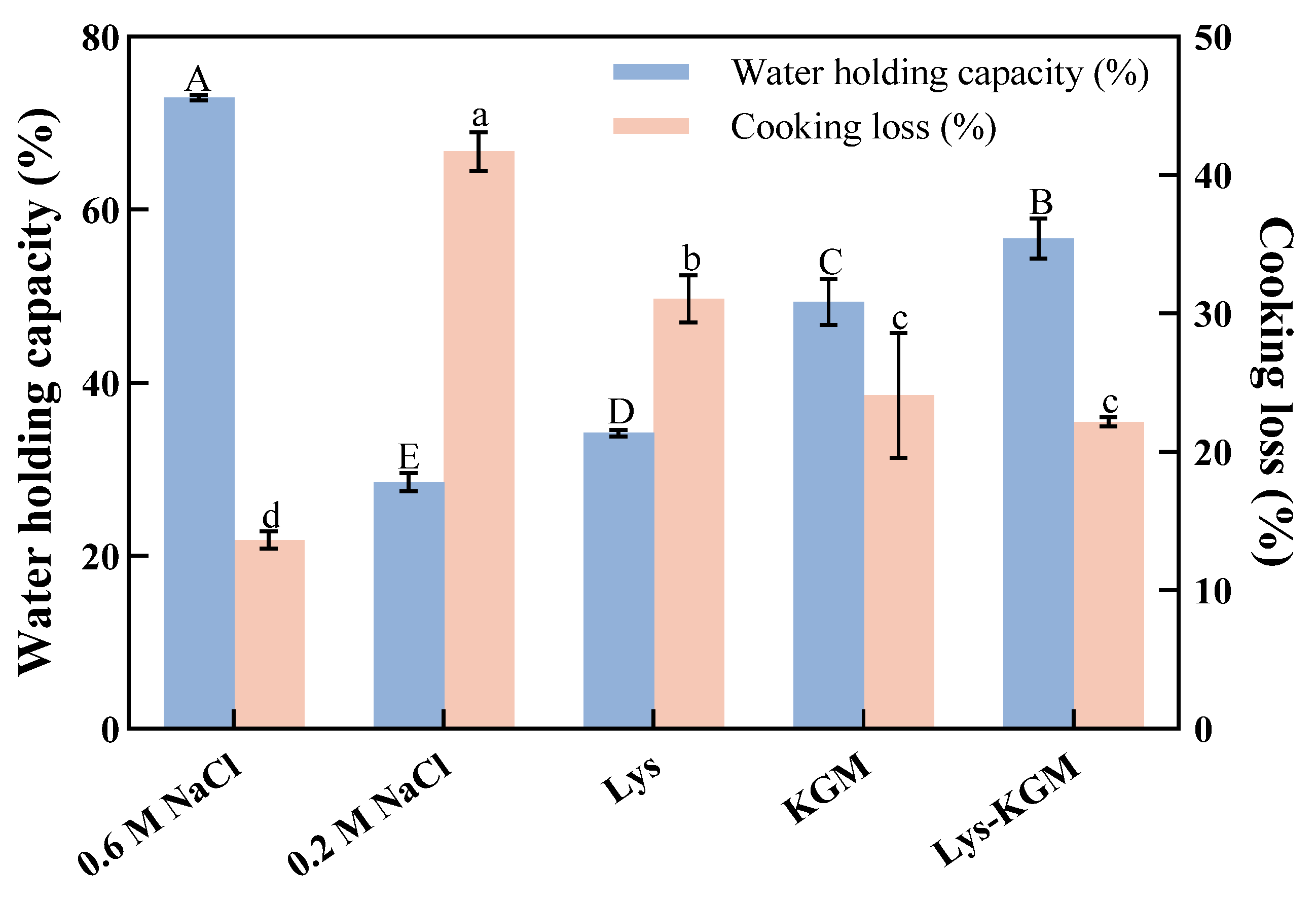
Figure 2.
T2 relaxation time distribution (A) and water population proportions (B) of MP gels under different treatments. The values are the mean ± standard deviation. Different lowercase letters indicate significant differences among different treatment groups (P < 0.05) (n=3).
Figure 2.
T2 relaxation time distribution (A) and water population proportions (B) of MP gels under different treatments. The values are the mean ± standard deviation. Different lowercase letters indicate significant differences among different treatment groups (P < 0.05) (n=3).
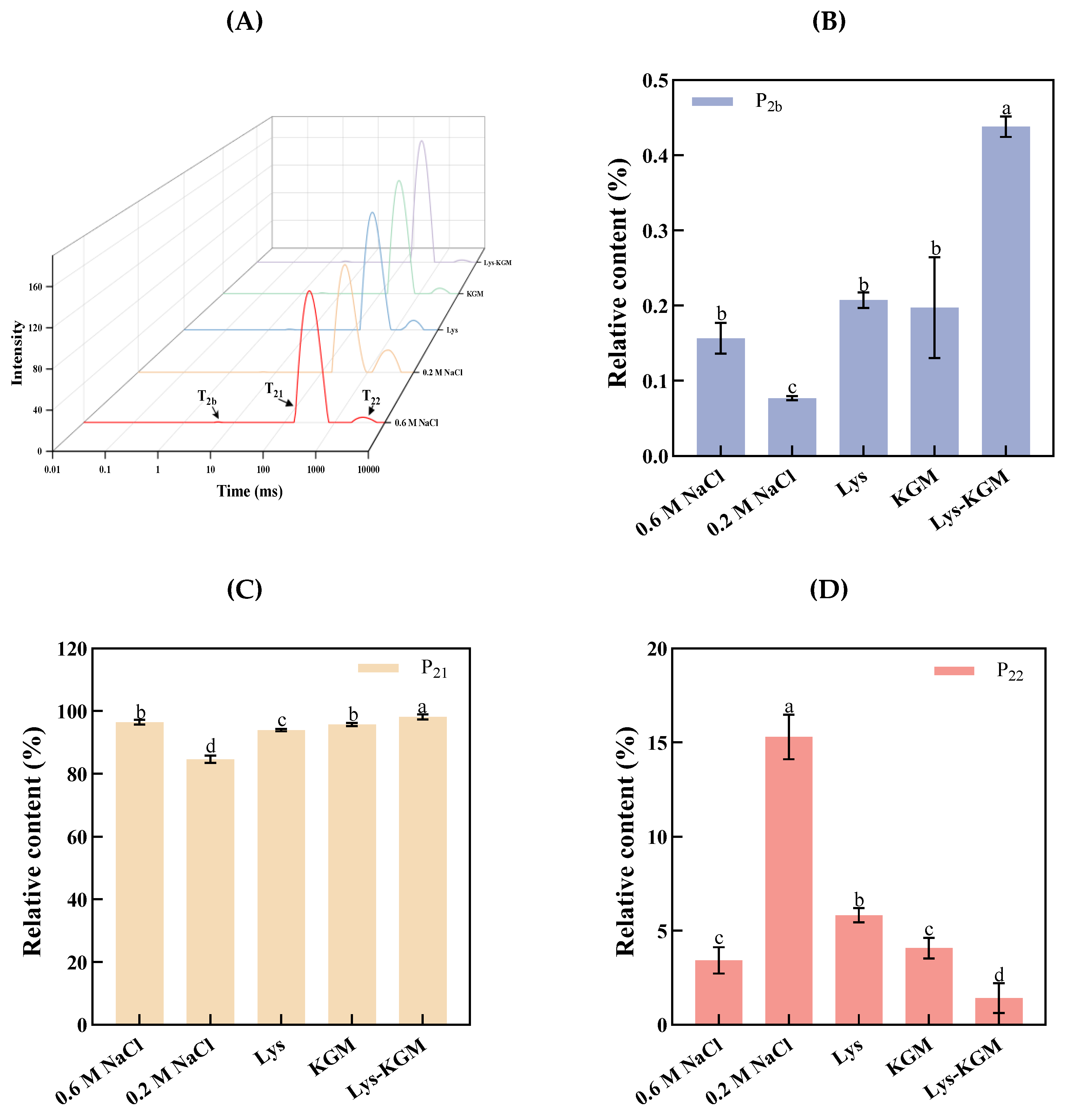
Figure 3.
Frequency sweep (A, B), apparent viscosity (C), and temperature sweep (D-F) of MP systems under different treatments.
Figure 3.
Frequency sweep (A, B), apparent viscosity (C), and temperature sweep (D-F) of MP systems under different treatments.
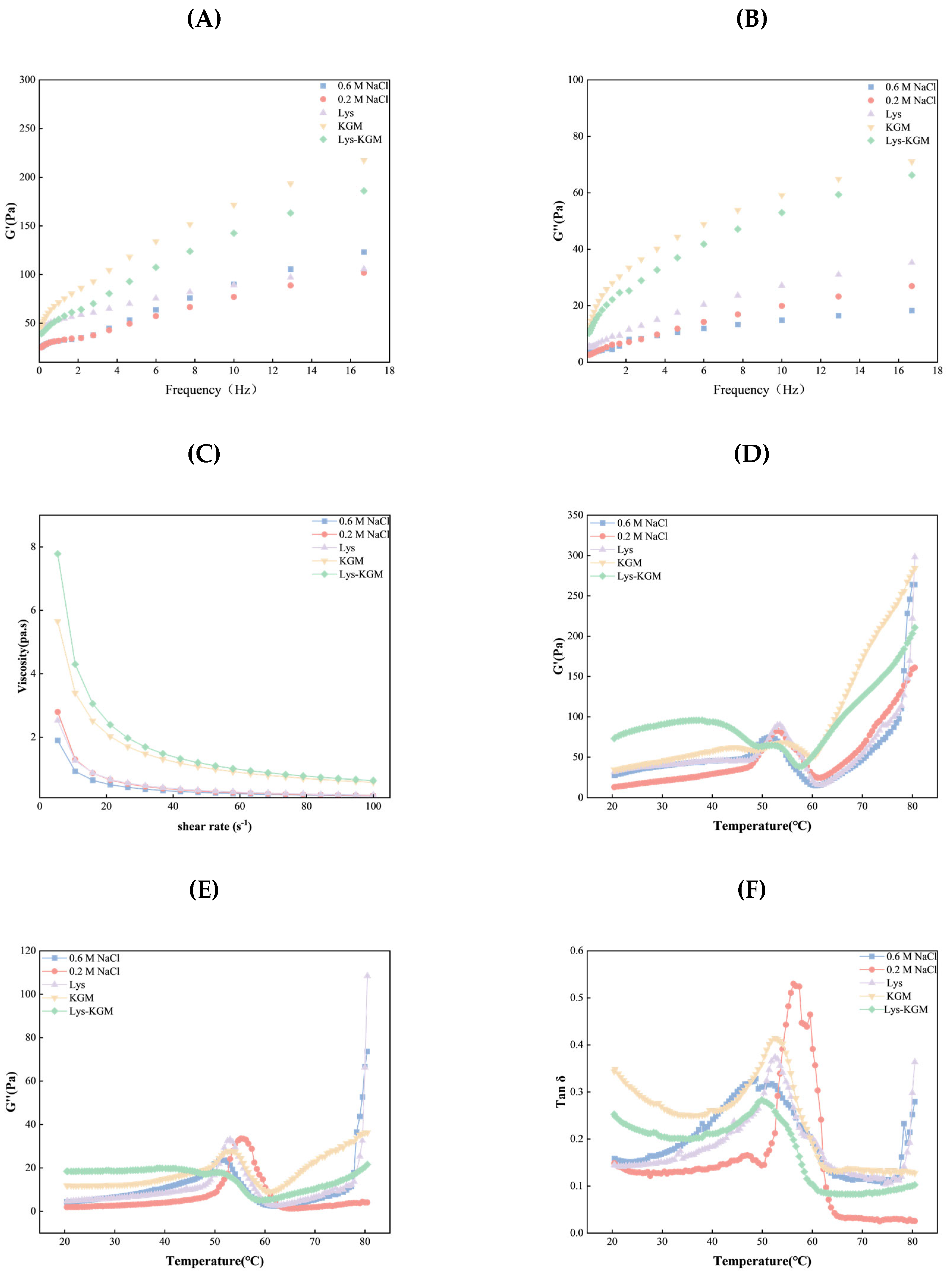
Figure 4.
SEM micrographs of MP gels under different treatments: 0.6 M NaCl (A), 0.2 M NaCl (B), Lys (C), KGM (D), and Lys-KGM (E).
Figure 4.
SEM micrographs of MP gels under different treatments: 0.6 M NaCl (A), 0.2 M NaCl (B), Lys (C), KGM (D), and Lys-KGM (E).
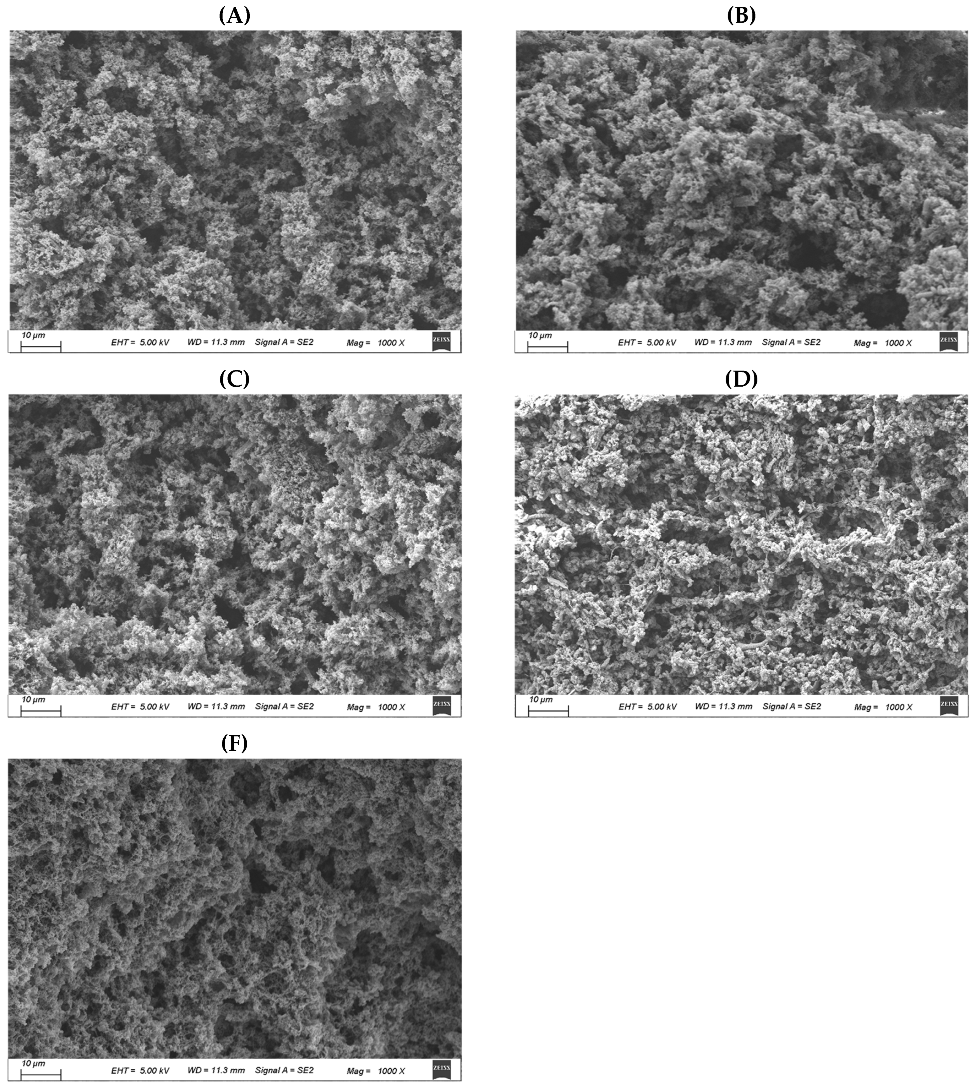
Figure 5.
FT-IR spectra (A), secondary structure composition (B-E), and intermolecular forces (F-I) of MP gels under different treatments. The values are the mean ± standard deviation. Different lowercase letters indicate significant differences among different treatment groups (P < 0.05) (n = 3).
Figure 5.
FT-IR spectra (A), secondary structure composition (B-E), and intermolecular forces (F-I) of MP gels under different treatments. The values are the mean ± standard deviation. Different lowercase letters indicate significant differences among different treatment groups (P < 0.05) (n = 3).
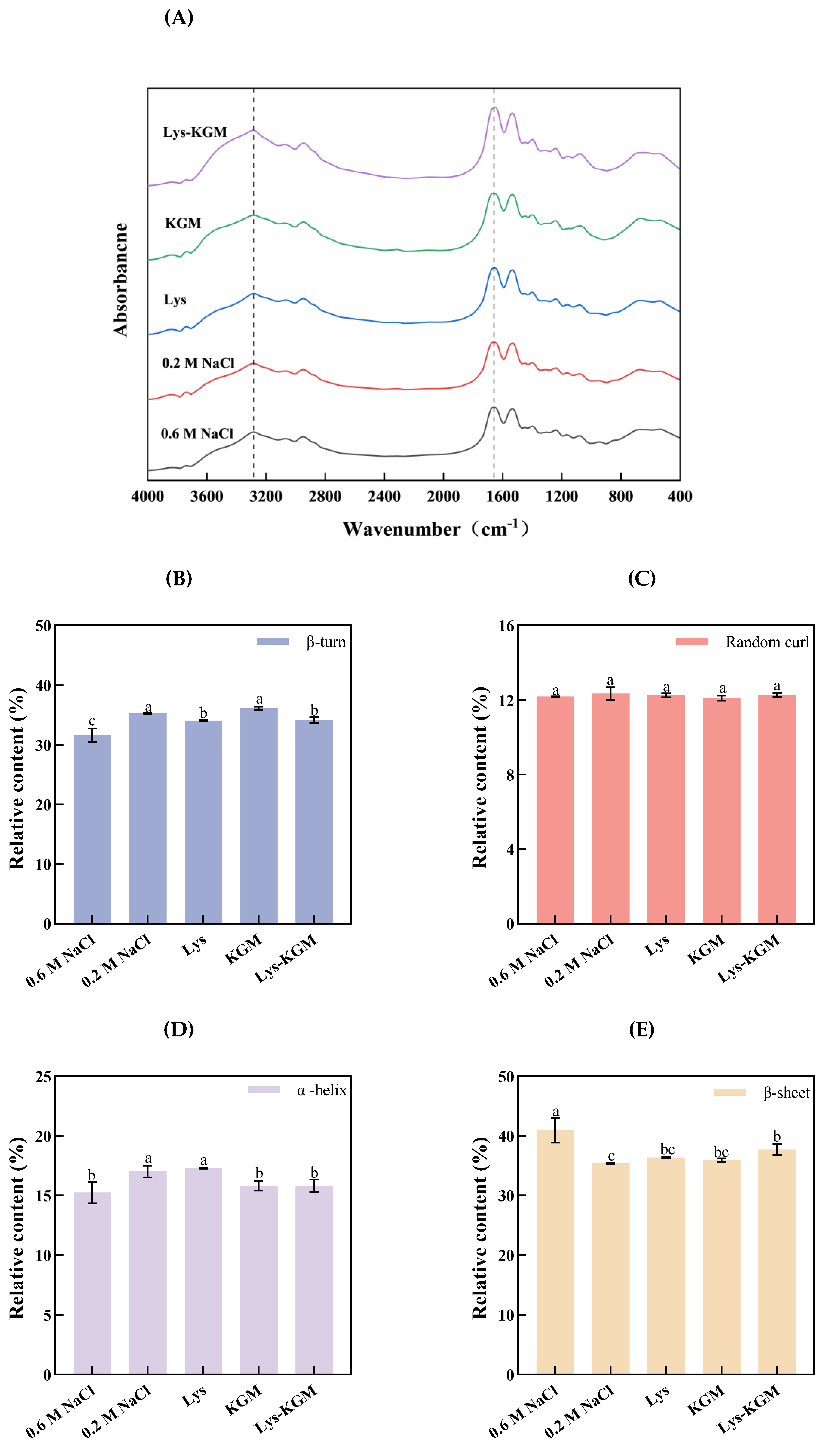
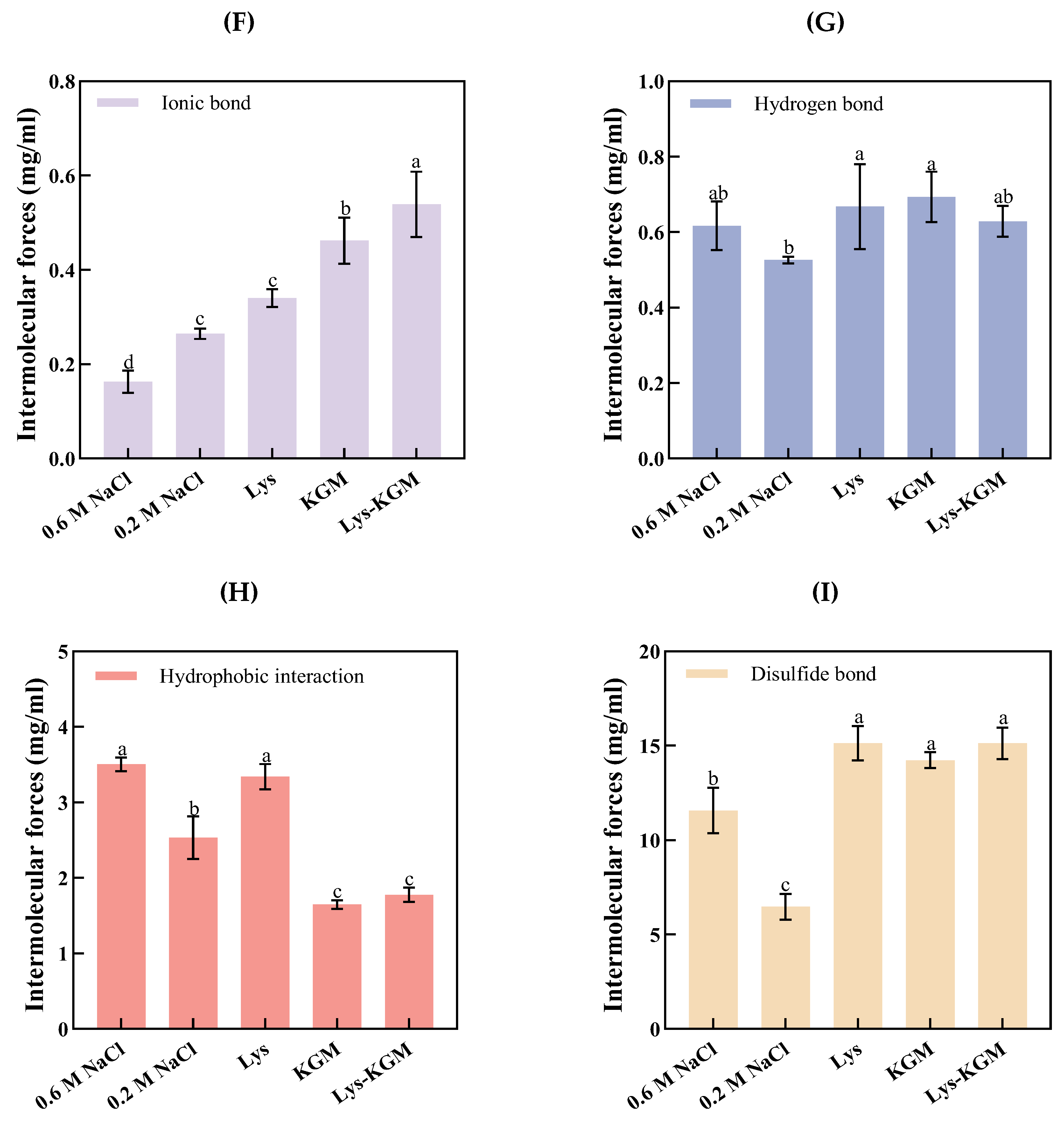
Figure 6.
Molecular docking analysis of the interaction between Myosin and KGM/Lys. The 2D structures of Myosin docking with KGM and Lys are (A, B), and the 3D structures are (C, D). In the 3D structure, a-f represent the hydrophobicity, ionizability, interpolated charge, SAS, aromatic, and H-Bond of KGM/Lys and Myosin, respectively.
Figure 6.
Molecular docking analysis of the interaction between Myosin and KGM/Lys. The 2D structures of Myosin docking with KGM and Lys are (A, B), and the 3D structures are (C, D). In the 3D structure, a-f represent the hydrophobicity, ionizability, interpolated charge, SAS, aromatic, and H-Bond of KGM/Lys and Myosin, respectively.
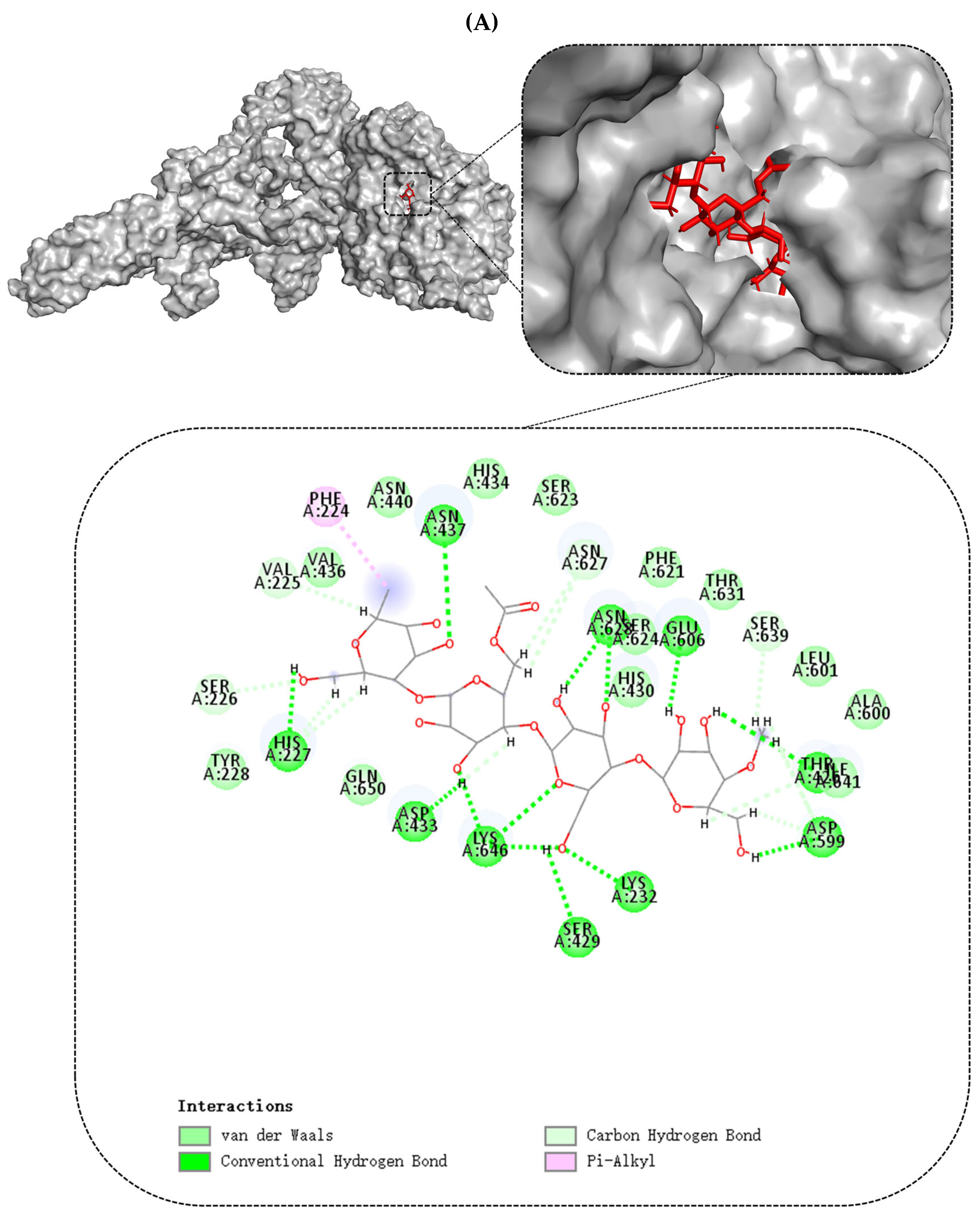
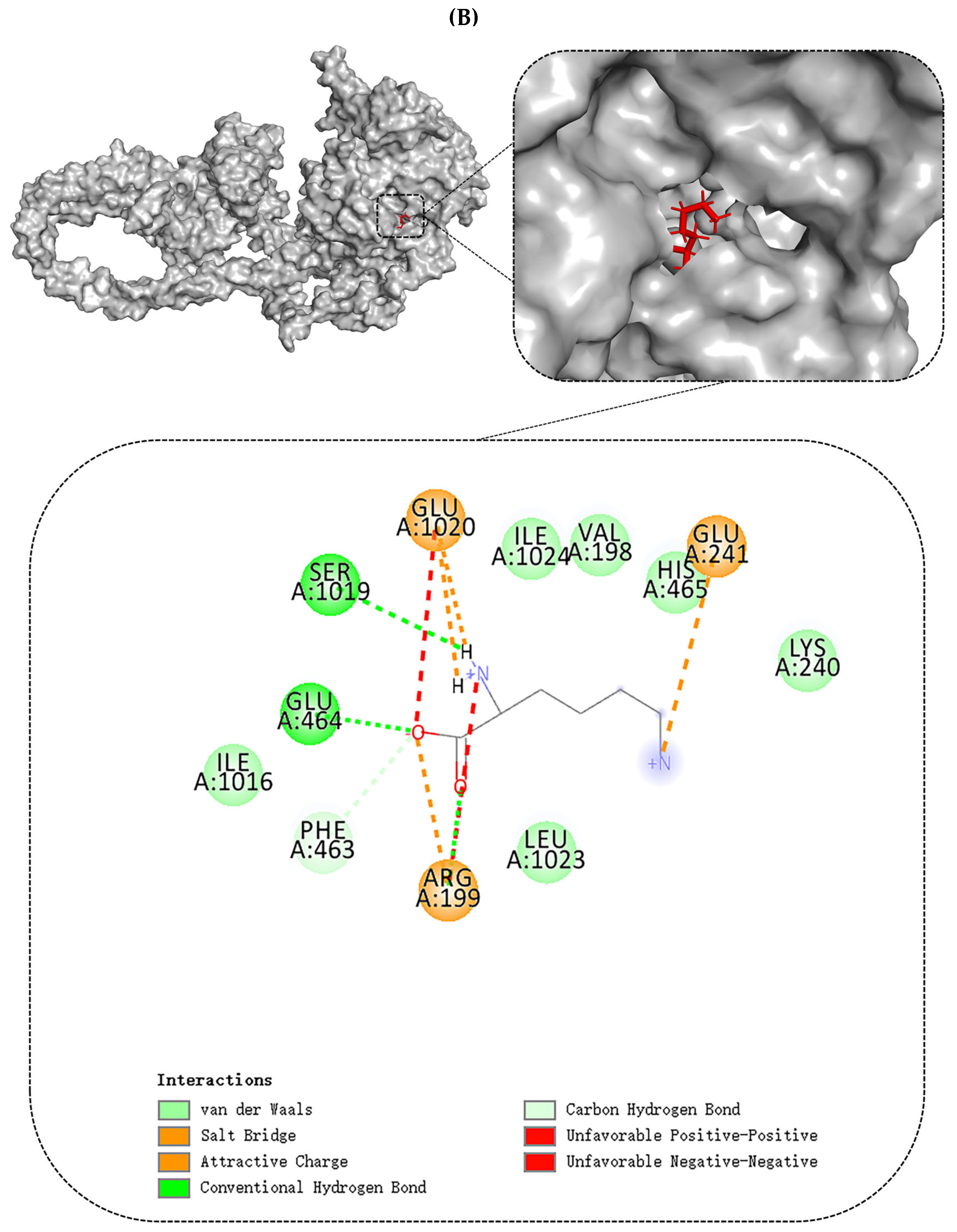
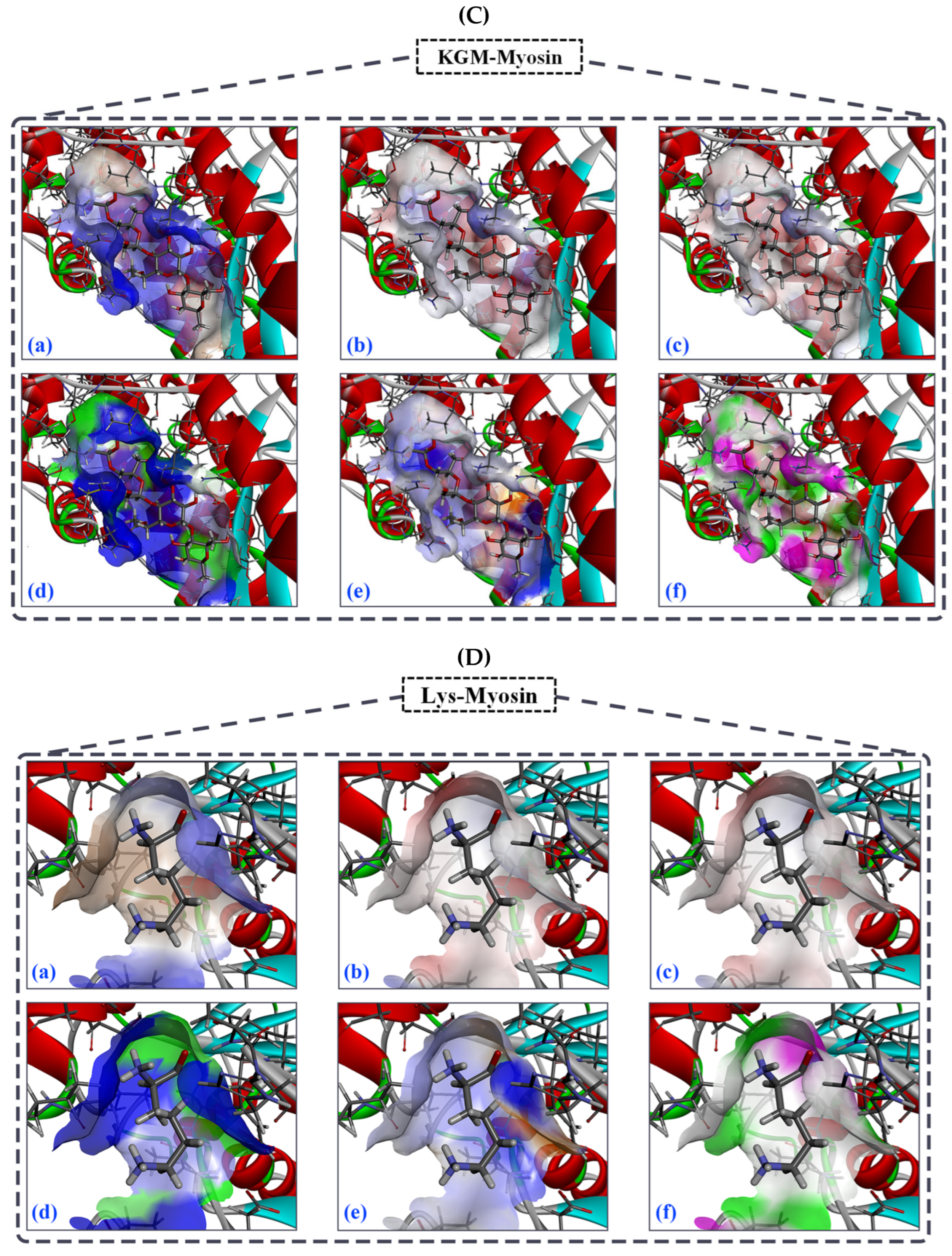
Figure 7.
The molecular dynamics simulation of myosin and Lys-KGM-Myosin complex (a-b). The RMSD (A), RMSD (B), Rg (C), and solvent accessible surface area (SASA, D) of the myosin and Lys-KGM-Myosin complex. Number of hydrogen bonds in Myosin and Lys-KGM-Myosin complexe (E).
Figure 7.
The molecular dynamics simulation of myosin and Lys-KGM-Myosin complex (a-b). The RMSD (A), RMSD (B), Rg (C), and solvent accessible surface area (SASA, D) of the myosin and Lys-KGM-Myosin complex. Number of hydrogen bonds in Myosin and Lys-KGM-Myosin complexe (E).
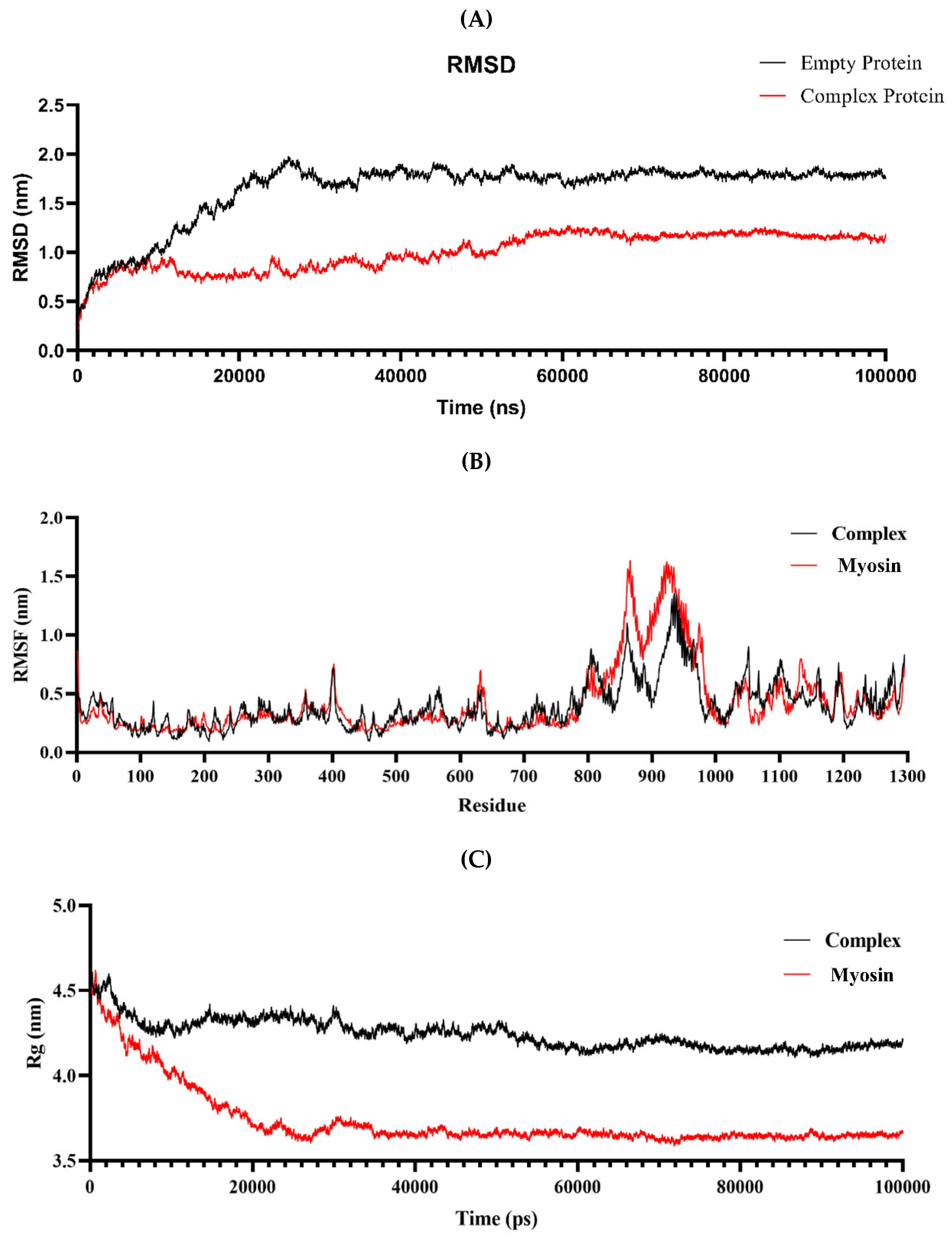
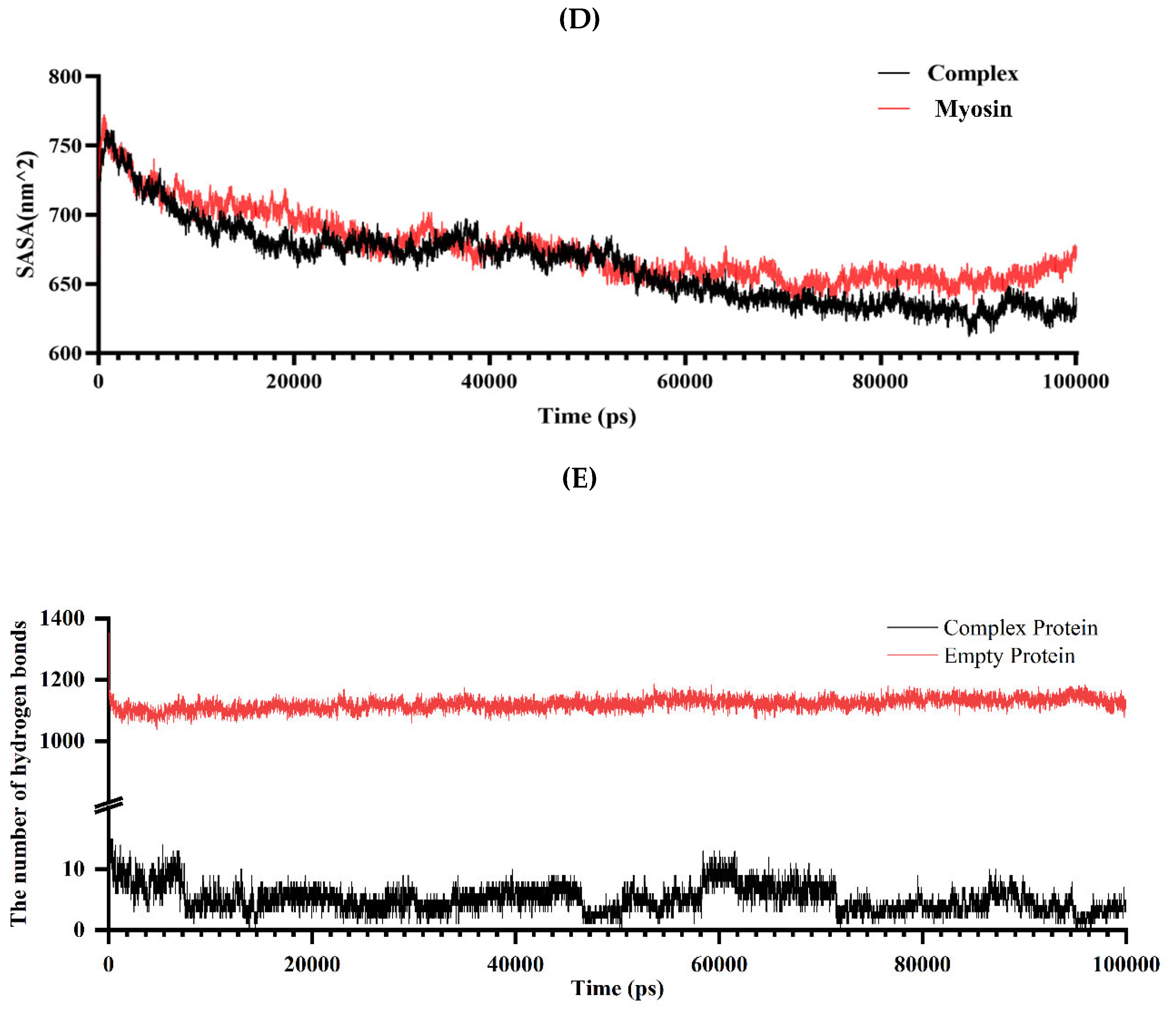
Figure 8.
Binding free energy maps of Lys-Myosin complex (A) and KGM-Myosin complex (B). energy contribution maps of amino acid residues of Lys-Myosin complex (C) and KGM-Myosin complex (D). The free energy landscape map of Myosin (E) and Lys-KGM-Myosin complex (F). Note: VDWAALS: van der Waals energy, EEl: Electrostatic energy, EGB: Polar solvation energy, ESURF: Non-polar solvation energy, GGAS: Total gas phase free energy, GSOLV: Total solvation free energy, TOTAL: GSOLV + GGAS.
Figure 8.
Binding free energy maps of Lys-Myosin complex (A) and KGM-Myosin complex (B). energy contribution maps of amino acid residues of Lys-Myosin complex (C) and KGM-Myosin complex (D). The free energy landscape map of Myosin (E) and Lys-KGM-Myosin complex (F). Note: VDWAALS: van der Waals energy, EEl: Electrostatic energy, EGB: Polar solvation energy, ESURF: Non-polar solvation energy, GGAS: Total gas phase free energy, GSOLV: Total solvation free energy, TOTAL: GSOLV + GGAS.
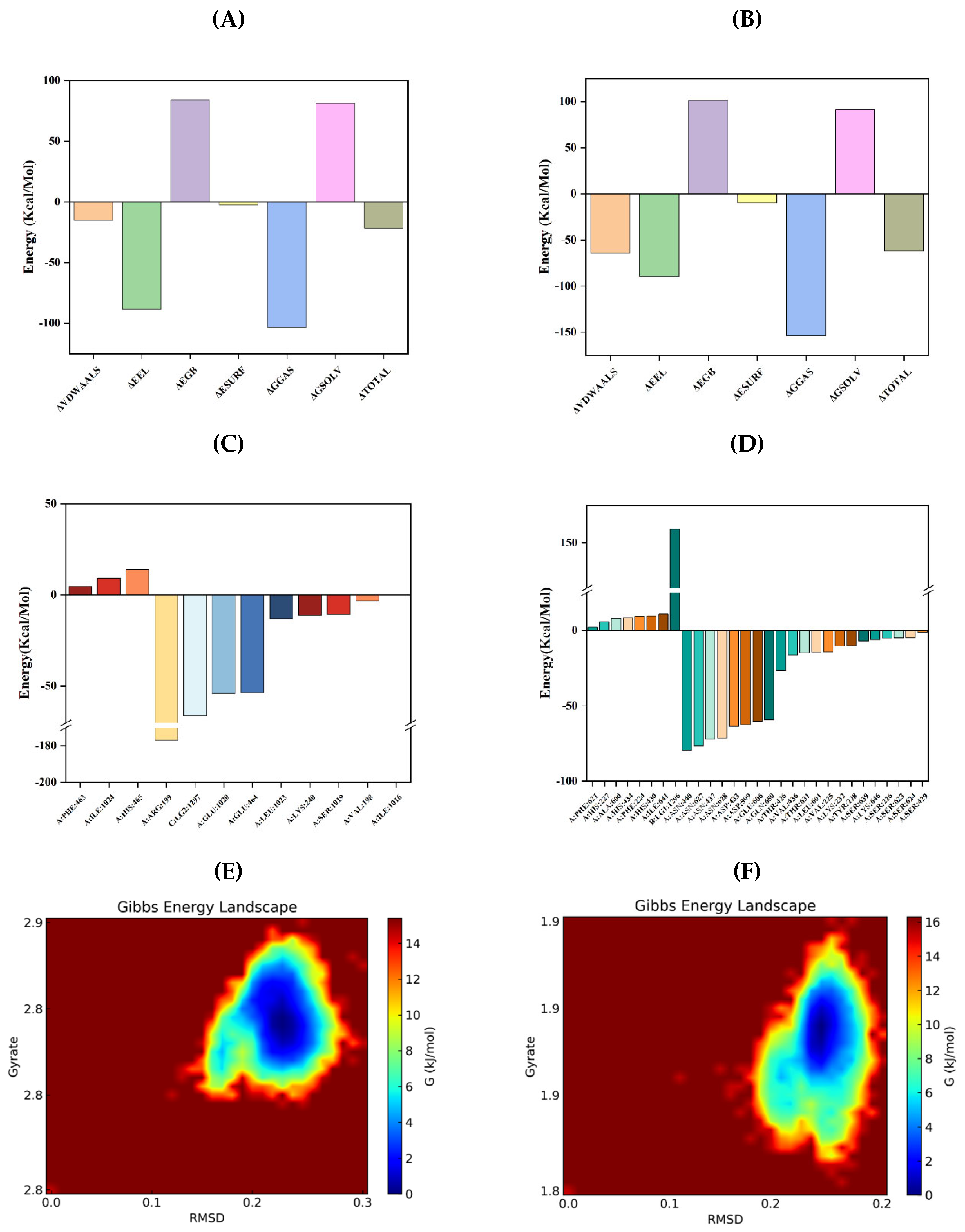
Figure 9.
Schematic of the formation mechanism of MP gels with Lys and/or KGM treatments.
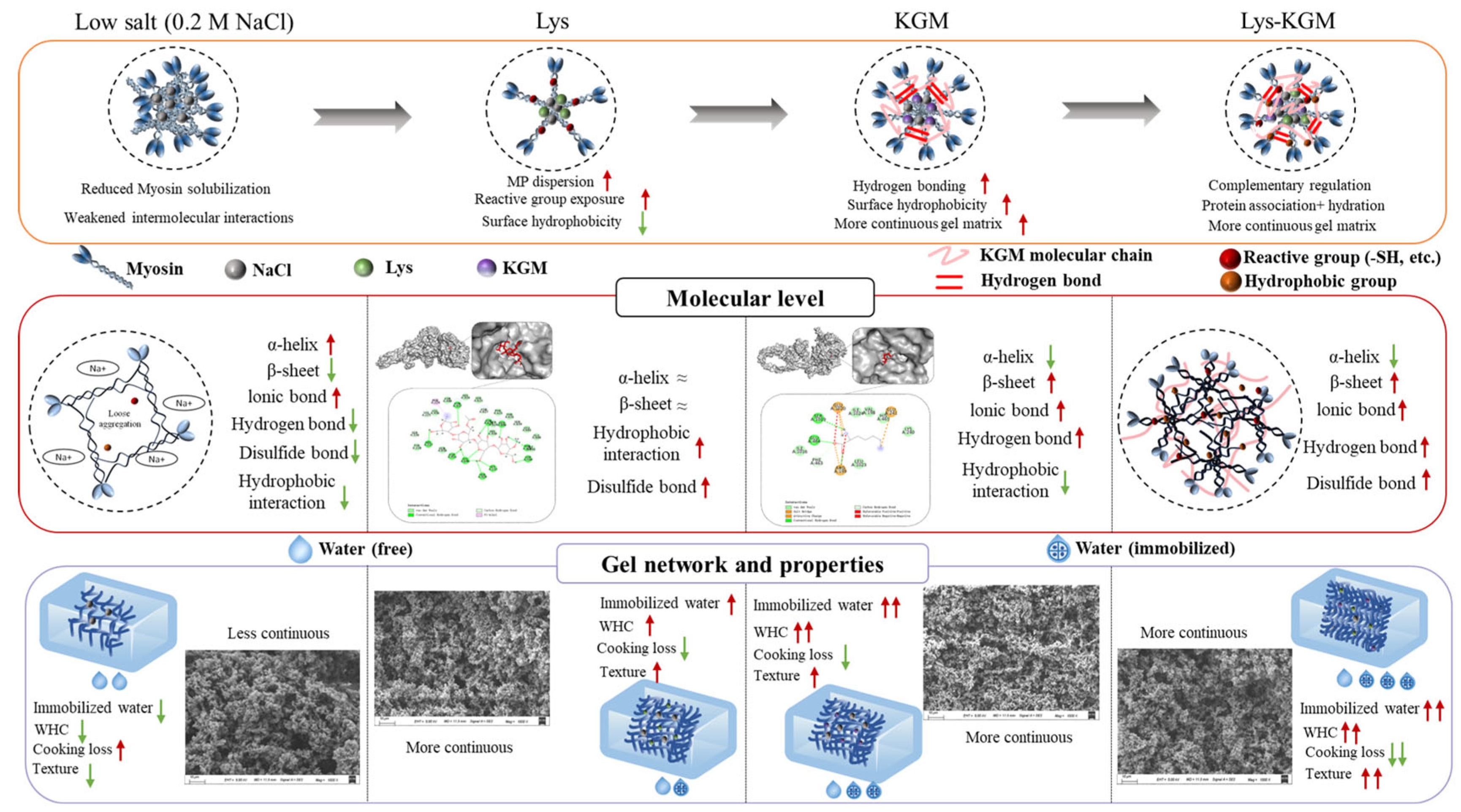

Table 1.
Texture properties of MP gels under different treatments.
| Group | Hardness (N) |
Springiness (mm) | Cohesiveness (Ratio) | Adhesion (mJ) | Gumminess (N) | Chewiness (mJ) |
|---|---|---|---|---|---|---|
| 0.6 M NaCl | 2.80 ± 0.17b | 1.20 ± 0.15a | 0.24 ± 0.03ab | 0.26 ± 0.09c | 0.71 ± 0.11a | 0.84 ± 0.04a |
| 0.2 M NaCl | 1.83 ± 0.06d | 0.46 ± 0.02d | 0.18 ± 0.01b | 0.89 ± 0.11a | 0.56 ± 0.05b | 0.31 ± 0.04d |
| Lys | 2.20 ± 0.10c | 0.58 ± 0.01cd | 0.26 ± 0.02a | 0.53 ± 0.08b | 0.54 ± 0.07b | 0.31 ± 0.05e |
| KGM | 2.87 ± 0.12b | 0.70 ± 0.08bc | 0.28 ± 0.04a | 0.66 ± 0.19ab | 0.80 ± 0.11a | 0.56 ± 0.08c |
| Lys-KGM | 3.10 ± 0.10a | 0.83 ± 0.04b | 0.28 ± 0.01a | 0.78 ± 0.17a | 0.86 ± 0.04a | 0.71 ± 0.05b |
Note: The values are the mean ± standard deviation. Different lowercase letters in the same column indicate statistically significant differences (P < 0.05) (n = 3).
Table 2.
Sulfhydryl content and surface hydrophobicity of MP under different treatments.
| Group | R-SH (μmol/g) | T-SH (μmol/g) | BPB (mg/mL) |
|---|---|---|---|
| 0.6 M NaCl | 1.81 ± 0.04a | 2.36 ± 0.03a | 109.48 ± 1.1a |
| 0.2 M NaCl | 1.59 ± 0.02c | 2.34 ± 0.04a | 91.50 ± 1.99c |
| Lys | 1.80 ± 0.01a | 2.32 ± 0.02a | 95.86 ± 0.92b |
| KGM | 1.73 ± 0.04b | 2.38 ± 0.11a | 54.85 ± 0.51e |
| Lys-KGM | 1.58 ± 0.01c | 2.35 ± 0.13a | 71.84 ± 2.01d |
Note: The values are the mean ± standard deviation. Different lowercase letters in the same column indicate statistically significant differences (P < 0.05) (n = 3).
Disclaimer/Publisher’s Note: The statements, opinions and data contained in all publications are solely those of the individual author(s) and contributor(s) and not of MDPI and/or the editor(s). MDPI and/or the editor(s) disclaim responsibility for any injury to people or property resulting from any ideas, methods, instructions or products referred to in the content. |
© 2026 by the authors. Licensee MDPI, Basel, Switzerland. This article is an open access article distributed under the terms and conditions of the Creative Commons Attribution (CC BY) license (http://creativecommons.org/licenses/by/4.0/).
Copyright: This open access article is published under a Creative Commons CC BY 4.0 license, which permit the free download, distribution, and reuse, provided that the author and preprint are cited in any reuse.


